C9ORF72, implicated in amytrophic lateral sclerosis and frontotemporal dementia, regulates endosomal...
17
C9ORF72, implicated in amytrophic lateral sclerosis and frontotemporal dementia, regulates endosomal trafficking Manal A. Farg 1 , Vinod Sundaramoorthy 1 , Jessica M. Sultana 1 , Shu Yang 3 , Rachel A.K. Atkinson 5 , Vita Levina 1 , Mark A. Halloran 2 , Paul A. Gleeson 4 , Ian P. Blair 3 , Kai Y. Soo 1 , Anna E. King 5 and Julie D. Atkin 1, ∗ 1 Department of Biochemistry, 2 Department of Neuroscience, School of Psychological Science, La Trobe University, Victoria, Australia, 3 Australian School of Advanced Medicine, Macquarie University, Sydney, NSW, Australia, 4 Department of Biochemistry and Molecular Biology, Bio21 Molecular Science and Biotechnology Institute, The University of Melbourne, Victoria, Australia, and 5 Wicking Dementia Research and Education Centre, University of Tasmania, Hobart, Tasmania, Australia Received October 14, 2013; Revised January 13, 2014; Accepted February 10, 2014 Intronic expansion of a hexanucleotide GGGGCC repeat in the chromosome 9 open reading frame 72 (C9ORF72) gene is the major cause of familial amyotrophic lateral sclerosis (ALS) and frontotemporal dementia. However, the cellular function of the C9ORF72 protein remains unknown. Here, we demonstrate that C9ORF72 regulates endo- somal trafficking. C9ORF72 colocalized with Rab proteins implicated in autophagy and endocytic transport: Rab1, Rab5, Rab7 and Rab11 in neuronal cell lines, primary cortical neurons and human spinal cord motor neu- rons, consistent with previous predictions that C9ORF72 bears Rab guanine exchange factor activity. Consistent with this notion, C9ORF72 was present in the extracellular space and as cytoplasmic vesicles. Depletion of C9ORF72 using siRNA inhibited transport of Shiga toxin from the plasma membrane to Golgi apparatus, intern- alization of TrkBreceptor andaltered the ratio of autophagosome marker light chain 3 (LC3) II:LC3I, indicating that C9ORF72 regulates endocytosis and autophagy. C9ORF72 also colocalized with ubiquilin-2 and LC3-positive vesicles, and co-migrated with lysosome-stained vesicles in neuronal cell lines, providing further evidence that C9ORF72 regulates autophagy. Investigation of proteins interacting with C9ORF72 using massspectrometry identified other proteins implicated in ALS; ubiquilin-2 and heterogeneous nuclear ribonucleoproteins, hnRNPA2/B1 and hnRNPA1, and actin. Treatment of cells overexpressing C9ORF72 with proteasome inhibitors induced the formation of stress granules positive for hnRNPA1 and hnRNPA2/B1. Immunohistochemistry of C9ORF72 ALS patient motor neurons revealed increased colocalization between C9ORF72 and Rab7 and Rab11 compared with controls, suggesting possible dysregulation of trafficking in patients bearing the C9ORF72 repeat expansion. Hence, this study identifies a role for C9ORF72 in Rab-mediated cellular trafficking. INTRODUCTION Amyotrophic lateral sclerosis (ALS) is characterized by degen- eration of upper and lower motor neurons in the brain, brainstem and spinal cord, leading to progressive paralysis. Frontotem- poral dementia (FTD) is the second most common cause of pre- senile dementia (1) and increasing evidence suggests that ALS and FTD overlap, occupying two opposite poles of disease con- tinuum (2 – 4). Hexanucleotide (GGGGCC) repeat expansions in a non-coding region of chromosome 9 open reading frame 72 (C9ORF72) are the major cause of familial ALS ( 33%) and FTD ( 25%) worldwide and are present in 8% of sporadic ALS cases, highlighting a major role for C9ORF72 in neurode- generation (5 – 7). The normal cellular function of C9ORF72 ∗ To whom correspondence should be addressed at: Dr Julie Atkin, La Trobe Institute for Molecular Science, Department of Biochemistry, School of Molecular Sciences, La Trobe University Bundoora. Tel: +03 9479 5480; Fax: +03 9479 2467; Email: [email protected] # The Author 2014. Published by Oxford University Press. This is an Open Access article distributed under the terms of the Creative Commons Attribution Non-Commercial License (http://creativecommons.org/ licenses/by-nc/3.0/), which permits non-commercial re-use, distribution, and reproduction in any medium, provided the original work is properly cited. For commercial re-use, please contact [email protected] Human Molecular Genetics, 2014, Vol. 23, No. 13 3579–3595 doi:10.1093/hmg/ddu068 Advance Access published on February 18, 2014
-
Upload
independent -
Category
Documents
-
view
0 -
download
0
Transcript of C9ORF72, implicated in amytrophic lateral sclerosis and frontotemporal dementia, regulates endosomal...
C9ORF72, implicated in amytrophic lateralsclerosis and frontotemporal dementia,regulates endosomal trafficking
Manal A. Farg1, Vinod Sundaramoorthy1, Jessica M. Sultana1, Shu Yang3,
Rachel A.K. Atkinson5, Vita Levina1, Mark A. Halloran2, Paul A. Gleeson4,
Ian P. Blair3, Kai Y. Soo1, Anna E. King5 and Julie D. Atkin1,∗
1Department of Biochemistry, 2Department of Neuroscience, School of Psychological Science, La Trobe University,
Victoria, Australia, 3Australian School of Advanced Medicine, Macquarie University, Sydney, NSW, Australia,4DepartmentofBiochemistryandMolecularBiology,Bio21MolecularScienceandBiotechnology Institute,TheUniversity
of Melbourne, Victoria, Australia, and 5Wicking Dementia Research and Education Centre, University of Tasmania,
Hobart, Tasmania, Australia
Received October 14, 2013; Revised January 13, 2014; Accepted February 10, 2014
Intronic expansion of a hexanucleotide GGGGCC repeat in the chromosome 9 open reading frame 72 (C9ORF72)gene isthemajorcauseof familialamyotrophic lateralsclerosis (ALS)andfrontotemporaldementia.However, thecellular function of the C9ORF72 protein remains unknown. Here, we demonstrate that C9ORF72 regulates endo-somal trafficking. C9ORF72 colocalized with Rab proteins implicated in autophagy and endocytic transport:Rab1, Rab5, Rab7 and Rab11 in neuronal cell lines, primary cortical neurons and human spinal cord motor neu-rons, consistent with previous predictions that C9ORF72 bears Rab guanine exchange factor activity. Consistentwith this notion, C9ORF72 was present in the extracellular space and as cytoplasmic vesicles. Depletion ofC9ORF72 using siRNA inhibited transport of Shiga toxin from the plasma membrane to Golgi apparatus, intern-alizationofTrkBreceptorandalteredtheratioofautophagosomemarker lightchain3 (LC3) II:LC3I, indicating thatC9ORF72 regulates endocytosis and autophagy. C9ORF72 also colocalized with ubiquilin-2 and LC3-positivevesicles, and co-migrated with lysosome-stained vesicles in neuronal cell lines, providing further evidencethatC9ORF72regulatesautophagy. Investigationofproteins interactingwithC9ORF72usingmassspectrometryidentified other proteins implicated in ALS; ubiquilin-2 and heterogeneous nuclear ribonucleoproteins,hnRNPA2/B1 and hnRNPA1, and actin. Treatment of cells overexpressing C9ORF72 with proteasome inhibitorsinduced the formation of stress granules positive for hnRNPA1 and hnRNPA2/B1. Immunohistochemistry ofC9ORF72 ALS patient motor neurons revealed increased colocalization between C9ORF72 and Rab7 andRab11 compared with controls, suggesting possible dysregulation of trafficking in patients bearing theC9ORF72 repeat expansion. Hence, this study identifies a role for C9ORF72 in Rab-mediated cellular trafficking.
INTRODUCTION
Amyotrophic lateral sclerosis (ALS) is characterized by degen-eration of upper and lower motor neurons in the brain, brainstemand spinal cord, leading to progressive paralysis. Frontotem-poral dementia (FTD) is the second most common cause of pre-senile dementia (1) and increasing evidence suggests that ALS
and FTD overlap, occupying two opposite poles of disease con-tinuum (2–4). Hexanucleotide (GGGGCC) repeat expansions ina non-coding region of chromosome 9 open reading frame 72(C9ORF72) are the major cause of familial ALS (�33%) andFTD (�25%) worldwide and are present in 8% of sporadicALS cases, highlighting a major role for C9ORF72 in neurode-generation (5–7). The normal cellular function of C9ORF72
∗ To whom correspondence should be addressed at: Dr Julie Atkin, La Trobe Institute for Molecular Science, Department of Biochemistry, School ofMolecular Sciences, La Trobe University Bundoora. Tel: +03 9479 5480; Fax: +03 9479 2467; Email: [email protected]
# The Author 2014. Published by Oxford University Press.This is an Open Access article distributed under the terms of the Creative Commons Attribution Non-Commercial License (http://creativecommons.org/licenses/by-nc/3.0/), which permits non-commercial re-use, distribution, and reproduction in any medium, provided the original work is properly cited.For commercial re-use, please contact [email protected]
Human Molecular Genetics, 2014, Vol. 23, No. 13 3579–3595doi:10.1093/hmg/ddu068Advance Access published on February 18, 2014

remains unknown but it is highly conserved and expressed inmany tissues, including the cerebellum, cortex and spinal cord.Similarly, it remains unclear how C9ORF72 repeat expansionstrigger ALS pathology, although haploinsufficiency due toimpaired transcription/splicing, leading to reduced C9ORF72protein expression (up to 50%) (8), RNA dysfunction, and uncon-ventional translation of the repeat to generate insoluble polypep-tides, are possible mechanisms (9,10).
Rab GTPases regulate membrane trafficking events and effi-cient intracellular trafficking is essential for cellular viability(11). Rab GTPases are master regulators of nearly all membranetraffic through their interactions with vesicular coat components,motor proteins and SNARE proteins. In humans, there are .60members of the Rab family that are localized to distinct intracellu-lar membranes.Rabs alternatebetween twoconformational states:the activated guanosine tri-phosphate (GTP)-bound form and theguanosine di-phosphate (GDP)-bound inactive form. Exchange ofGDP with GTP is catalyzed by Rab guanine nucleotide exchangefactors (GEFs) that act at specific membranes and facilitate GDPrelease, thus locally activating their targets. DENN (differentiallyexpressed in normal and neoplastic cells) domain-containing pro-teins are RabGEFs that activate mostly endocytotic Rabs (12).Two recent bioinformatics studies predicted that C9ORF72 pos-sesses DENN domains (13,14), raising the possibility that it regu-lates Rab-dependent intracellular trafficking (12). The endosomalsystem is necessary for regulating, sorting and degrading proteinsvia autophagy or the ubiquitin–proteasome system (UPS) (15).Multiple Rabs have been implicated in autophagy includingRab1, Rab5, Rab7 and Rab11 (16–18).
Defects in protein degradation are increasingly implicated inALS pathogenesis (19) and mutations in ubiquilin-2 (20), whichregulates autophagy and the UPS by binding/transport of proteincargo (21), also cause ALS/FTD. Inhibition of the proteasomeinduces the formation of stress granules (SGs) (22), a cellularhallmark of ALS (23). Recently, mutations in heterogeneousnuclear ribonucleoproteins hnRNPA2/B1 and hnRNPA1 wereidentified in ALS patients (24). hnRNPs granules are major com-ponents of SGs that mediate nucleocytoplasmic trafficking ofmRNA and RNA metabolism (25). Inhibition of the proteasometriggers alternative splicing of hnRNPs and mRNA-boundhnRNPA1 is recruited to cytoplasmic SGs (26).
Elucidation of the function of C9ORF72 is essential to under-stand its role in ALS/FTD. Here, we demonstrate a role forC9ORF72 in endosomal trafficking. C9ORF72 colocalized withubiquilin-2 and Rab proteins implicated in autophagy, and co-mi-grated with lysosome-positive vesicles. Depletion of C9ORF72using siRNA dsyregulated autophagy and inhibited endocytosis.Mass spectrometry identified other proteins linked to ALS asinteracting partners of C9ORF72: hnRNPA1, hnRNPA2/B1,ubiquilin-2 and actin. Proteasome inhibition and C9ORF72 over-expression led to the formation of nuclear C9ORF72 aggregatesand cytoplasmic SGs positive for hnRNPA1 and hnRNPA2/B1.Hence, this study defines novel functions for C9ORF72 in cellulartrafficking and protein degradation.
RESULTS
C9ORF72 colocalizes and interact with Rabsin neuronal cell lines
To characterize C9ORF72, we examined expression of endogen-ous C9ORF72 in neuronal cell lines; murine neuro2a and human
SH-SY5Y. Immunofluorescence was present diffuse in thenucleus and as cytoplasmic vesicles (Fig. 1A and B). Similarly,in cells transfected with a construct-encoding GFP-taggedC9ORF72 (Fig. 1C), both nuclear protein and cytoplasmic vesi-cles were observed (Fig. 1C). The cellular distribution of en-dogenous C9ORF72 was investigated further using subcellularfractionation of SH-SY5Y cell lysates. Using immunoblotting,we detected that significantly more C9ORF72 was present inthe nucleus compared with the cytoplasm (Fig. 1D and E);64%, P , 0.05). C9ORF72 in the nuclear fraction was presentas the 50 kDa isoform; however, in the cytoplasmic fraction,lower molecular weight bands were present in addition to the50 kDa band, possibly representing the 25 kDa isoform(Fig. 1D). We also detected C9ORF72 in conditioned medium,suggesting active secretion (Fig. 1F), presumably via non-classical means because bioinformatics analysis predicted thata signal leader peptide was not present in C9ORF72 (SecretomeP) (27). This was confirmed by immunoblotting of human CSF(Fig. 1G).
C9ORF72 expression in vesicles suggests a possible role incellular trafficking, consistent with RabGEF function. Thiswas investigated using immunocytochemistry for Rab pro-teins. C9ORF72 colocalized strongly with Rabs implicated inautophagy and endosomal transport: Rab1, Rab7, Rab5 andRab11 (Fig. 2A). Similarly, C9ORF72 co-immunoprecipitatedwith Rab1, Rab7 and Rab11 (Fig. 2B); control immunoprecipi-tations using isotype-matched antibodies were negative. Thiswas confirmed by precipitations using GFP-Trap in thereverse direction, with cell lysates transfected with Rab-GFPconstructs (Supplementary Material, Fig. S1). Similarly,C9ORF72 was precipitated using GFP-Trap from cells trans-fected with Rab5-GFP, although we could not determine ifRab5 precipitated with C9ORF72 antibodies due to speciescross-reactivity with the available antibodies. However, thesefindings suggest a physical interaction between C9ORF72and Rab proteins, consistent with predictions that C9ORF72is a RabGEF.
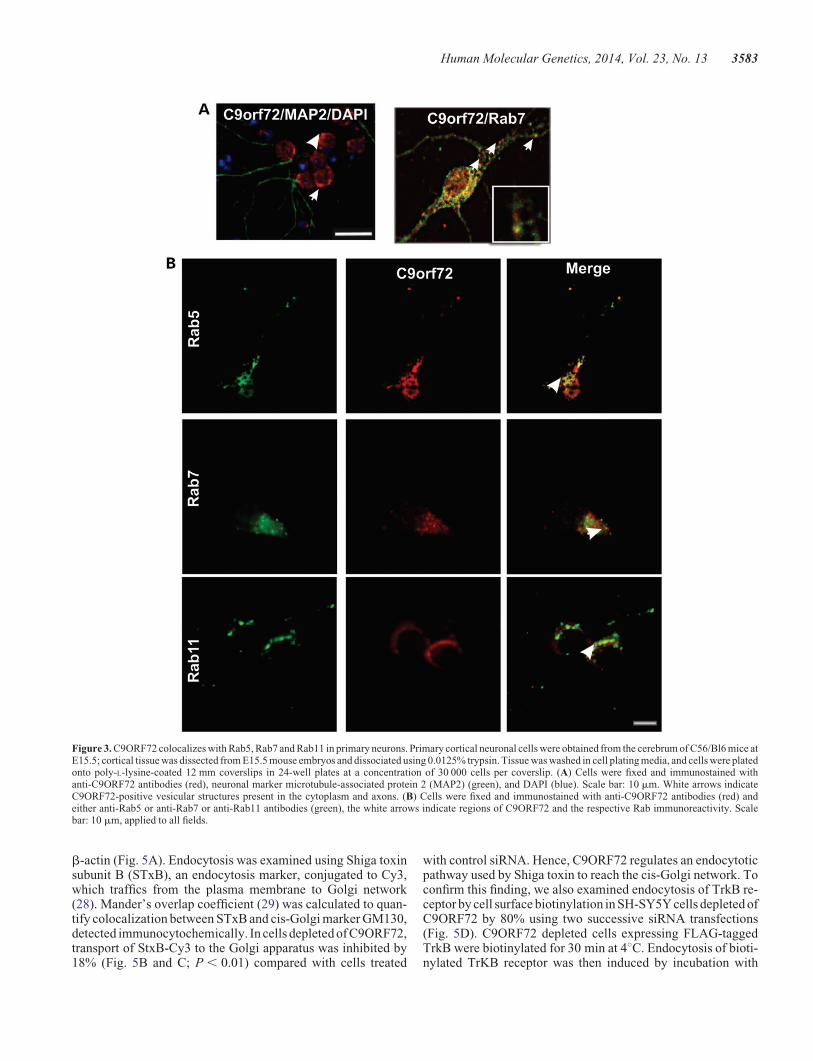
We examined the expression of C9ORF72 further in pri-mary cortical neurons obtained from C57Bl/6 mice. C9ORF72was present both in the nucleus and in vesicles, similar to itsexpression in neuronal cell lines (Fig. 3A). Immunocytochem-istry using Rab5, Rab7 and Rab11 antibodies revealed thatC9ORF72-positive vesicles frequently colocalized with Rab5,Rab7 and Rab11 (Fig. 3B). Interestingly, we detected colocaliza-tion of C9ORF72 with Rab7 in the axon and the cell body. Thesedata provide further evidence that C9ORF72 has a role in proteintrafficking with multiple Rab proteins (Fig. 3A and B). To furthervalidate the interaction between Rab proteins and C9ORF72,C9ORF72-GFP was expressed in neuro2a cells and immunos-taining with Rab5 was performed. Rab5 and GFP-tagged fluor-escent C9ORF72 vesicles were clearly colocalized in thesecells (Supplementary Material, Fig. S2).
Rab7 and Rab11 colocalize in C9ORF72 humanspinal cord motor neurons
These findings were investigated further using immunohis-tochemistry of human spinal cord sections. Quantitative analy-sis of 30+ motor neurons in control patients revealed thatC9ORF72 colocalized with Rab5 (60%), Rab7 (70%) andRab11 (60%), consistent with the findings obtained from cell
3580 Human Molecular Genetics, 2014, Vol. 23, No. 13

Figure 1. C9ORF72 expression and secretion in neuronal cell lines. (A) Murine Neuro2a cells were fixed and immunostained with anti-C9ORF72 antibodies (green)and DAPI (blue); scale bar: 10 mm. White arrows indicate C9ORF72-positive vesicular-type structures present in the cytoplasm; expression of C9ORF72 is diffuse inthe nucleus. (B) Human SH-SY5Y cells were fixed and immunostained with anti-C9ORF72 antibodies (green) and DAPI (blue), expression of C9ORF72 is similar to(A). Scale bar: 10 mm. White arrows indicate C9ORF72-positive vesicular-type structures present in the cytoplasm. (C) Overexpression of C9ORF72-Variant 1tagged with GFP in SH-SY5Y cells forms cytoplasmic vesicles. (D) Subcellular fractionation of human SH-SY5Y neuroblastoma cells; immunoblotting ofnuclear and cytoplasmic fractions. Histone H3 and GAPDH were used as subcellular markers and loading controls for nucleus and cytoplasm, respectively.C9ORF72 is expressed primarily as the 50 kDa isoform in the nucleus, additional bands are present in the cytoplasmic fraction at 25 and 36 kDa. (E) Quantificationof endogenous C9ORF72 present in the nuclear and cytoplasmic fraction by densitometry of immunoblots. Data are represented as mean+SEM; ∗P , 0.05, n ¼ 3nuclear versus cytoplasmic by unpaired t-test. (F) C9ORF72 isoforms (50 and 25 kDa) are present in conditioned medium of Neuro2a cells immunoprecipitated usingan anti-C9ORF72 antibody; cell lysate fraction shown as a control. (G) C9ORF72 is secreted in human CSF. Immunoblotting with C9ORF72 antibody detects bothC9ORF72 isoforms, corresponding to 50 and 25 kDa proteins.
Human Molecular Genetics, 2014, Vol. 23, No. 13 3581

lines (Fig. 4A and B), confirming that C9ORF72 colocalizes withRab proteins. Quantitation of motor neurons from an ALSpatient with the C9ORF72 hexanucleotide repeat expansionrevealed an increased proportion of cells with colocalization ofC9ORF72 with Rab7 or Rab11 compared with controls (P ,0.05) (Fig. 4B). These data suggest possible dysregulation ofendosomal trafficking in ALS patients with the C9ORF72 hexa-nucleotide repeat expansion.
C9ORF72 regulates endocytosis in neuronal cell lines
Next, to examine if C9ORF72 regulates endocytosis, humanneuronal SH-SY5Y cells were treated with short interferingRNA (siRNA) duplexes to silence human C9ORF72 expression.Using immunoblotting, C9ORF72 levels were depleted by 30%,i.e. to 70% of the original expression level, without obviousoff-target effects, as indicated by no change in the levels of
Figure 2. C9ORF72 colocalizes with endosomal Rabs; Rab1, Rab5, Rab7 and Rab11 in neuronal cell lines. (A) Neuro2a cells were fixed and immunostained withanti-C9ORF72 antibodies (red) and either anti-Rab7 or anti-Rab1 antibodies (green), or transfected with constructs encoding either GFP-Rab5 or GFP-Rab11 for 48 h,followed by DAPI staining (blue). The white arrows indicate regions of colocalization between C9ORF72 and the respective Rabs. Scale bar: 10 mm, applied to allfields. Insets demonstrate higher magnification (×100) of the areas highlighted to illustrate colocalization with vesicular structures. (B) Co-immunoprecipitationfollowed by western blotting revealed that Rab7 and Rab11 are pulled down using anti-C9ORF72 antibodies, and that C9ORF72 is pulled down using anti-Rab1antibodies. Control immunoprecipitations using an isotype-matched, irrelevant IgG antibody were negative, indicating no cross-reactivity with the antibodiesused. 1% input also shown.
3582 Human Molecular Genetics, 2014, Vol. 23, No. 13
b-actin (Fig. 5A). Endocytosis was examined using Shiga toxinsubunit B (STxB), an endocytosis marker, conjugated to Cy3,which traffics from the plasma membrane to Golgi network(28). Mander’s overlap coefficient (29) was calculated to quan-tify colocalization between STxB and cis-Golgi marker GM130,detected immunocytochemically. In cells depleted of C9ORF72,transport of StxB-Cy3 to the Golgi apparatus was inhibited by18% (Fig. 5B and C; P , 0.01) compared with cells treated
with control siRNA. Hence, C9ORF72 regulates an endocytoticpathway used by Shiga toxin to reach the cis-Golgi network. Toconfirm this finding, we also examined endocytosis of TrkB re-ceptor by cell surface biotinylation in SH-SY5Y cells depleted ofC9ORF72 by 80% using two successive siRNA transfections(Fig. 5D). C9ORF72 depleted cells expressing FLAG-taggedTrkB were biotinylated for 30 min at 48C. Endocytosis of bioti-nylated TrKB receptor was then induced by incubation with
Figure 3. C9ORF72 colocalizes with Rab5, Rab7 and Rab11 in primary neurons. Primary cortical neuronal cells were obtained from the cerebrum of C56/Bl6 mice atE15.5; cortical tissue was dissected from E15.5 mouse embryos and dissociated using 0.0125% trypsin. Tissue was washed in cell plating media, and cells were platedonto poly-L-lysine-coated 12 mm coverslips in 24-well plates at a concentration of 30 000 cells per coverslip. (A) Cells were fixed and immunostained withanti-C9ORF72 antibodies (red), neuronal marker microtubule-associated protein 2 (MAP2) (green), and DAPI (blue). Scale bar: 10 mm. White arrows indicateC9ORF72-positive vesicular structures present in the cytoplasm and axons. (B) Cells were fixed and immunostained with anti-C9ORF72 antibodies (red) andeither anti-Rab5 or anti-Rab7 or anti-Rab11 antibodies (green), the white arrows indicate regions of C9ORF72 and the respective Rab immunoreactivity. Scalebar: 10 mm, applied to all fields.
Human Molecular Genetics, 2014, Vol. 23, No. 13 3583
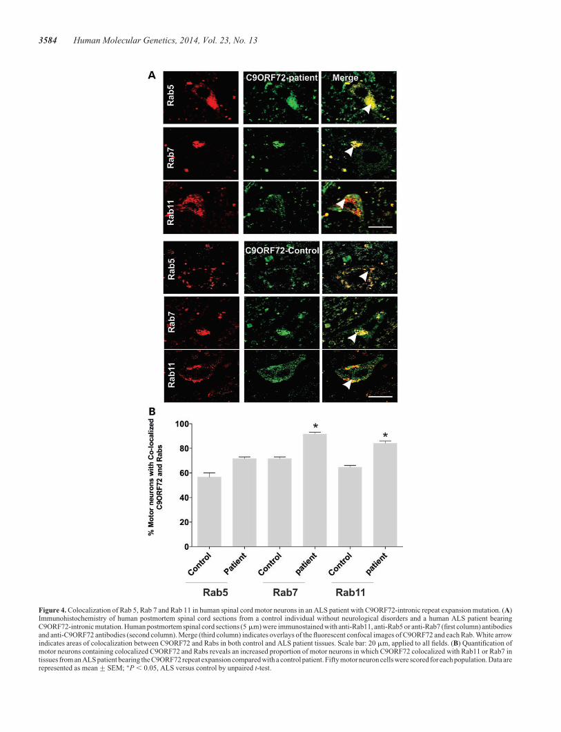
Figure 4. Colocalization of Rab 5, Rab 7 and Rab 11 in human spinal cord motor neurons in an ALS patient with C9ORF72-intronic repeat expansion mutation. (A)Immunohistochemistry of human postmortem spinal cord sections from a control individual without neurological disorders and a human ALS patient bearingC9ORF72-intronic mutation. Human postmortem spinal cord sections (5 mm) were immunostained with anti-Rab11, anti-Rab5 or anti-Rab7 (first column) antibodiesand anti-C9ORF72 antibodies (second column). Merge (third column) indicates overlays of the fluorescent confocal images of C9ORF72 and each Rab. White arrowindicates areas of colocalization between C9ORF72 and Rabs in both control and ALS patient tissues. Scale bar: 20 mm, applied to all fields. (B) Quantification ofmotor neurons containing colocalized C9ORF72 and Rabs reveals an increased proportion of motor neurons in which C9ORF72 colocalized with Rab11 or Rab7 intissues from an ALS patient bearing the C9ORF72 repeat expansion compared with a control patient. Fifty motor neuron cells were scored for each population. Data arerepresented as mean+SEM; ∗P , 0.05, ALS versus control by unpaired t-test.
3584 Human Molecular Genetics, 2014, Vol. 23, No. 13

BDNF ligand. Internalized TrkB receptor was precipitated withstreptavidin beads and quantified by immunoblotting with ananti-FLAG antibody. In cells 80% depleted of C90RF72, endo-cytosis of TrkB was inhibited by 87% compared with controlcells, confirming that C9ORF72 regulates endocytosis (Fig. 5Eand F).
C9ORF72 regulates autophagy in neuronal cell lines
To examine if C9ORF72 regulates autophagy, cells werecotransfected with constructs-encoding C9ORF72-GFP andDsRed-tagged microtubule-associated protein 1 light chain 3(LC3), a marker for autophagosomes, in neuro2a cells. The
Figure 5. C9ORF72 mediates endocytosis of Shiga toxin-CY3 and TrkB receptor. (A) SH-SY5Y cells were transfected with C9ORF72-targeted siRNA and controlsiRNA for 72 h. Cell lysates were harvested and immunoblotting followed by densitometry quantification revealed that C9ORF72 expression was reduced by 30% incells treated with C9ORF72 siRNA compared with control siRNA-treated cells. (B) Purified Shiga toxin conjugated to CY3 (SxTB-CY3) (red) was added to themedium of cells treated with C9ORF72 and control siRNA for 30 min. Endocytosis was examined after 60 min using immunocytochemistry for Golgi markerGM130 (green). (C) The colocalization between C9ORF72 and GM130 was quantified using Mander’s coefficient, revealing 18% inhibition of endocytosis ofShiga toxin in C9ORF72-siRNA-treated cells. For each of two replicate experiments, 50 cells were scored for each population. Data are represented as mean+SEM; ∗∗P , 0.001, using unpaired t-test. (D) SH-SY5Y cells were transfected with C9ORF72-targeted siRNA and control siRNA followed by a second transfection24 h later. Cell lysateswere harvestedand immunoblotting followed by densitometryquantification revealed that C9ORF72expressionwas reduced by 80%comparedwith control siRNA-treated cells. (E) C90RF72 depleted and control cells expressing FLAG-tagged TrKB receptor were treated with biotin NHS at 48C to biotinylatecell surface proteins. The cells were then incubated at 378C and medium containing BDNF was added to induce endocytosis of TrKB receptor. The rate of TrKBreceptor endocytosis was examined after 2 h by precipitating biotinylated and internalized TrKB receptor from cell lysates with strepatividin beads and immunoblot-ting with an anti-FLAG antibody. (F) Densitometric quantification of immunoblots reveals inhibition of endocytosis of TrkB receptor by 87% in cells depleted ofC9ORF72 compared with control cells. Biotin precipitation and immunoblotting were performed in triplicate. Data are represented as mean+SEM; ∗P , 0.05,using unpaired t-tests.
Human Molecular Genetics, 2014, Vol. 23, No. 13 3585

appearance of C9ORF72 as punctate structures that colocalizedwith DsRed-LC3 in 23% of cells (counted from at least 100 cells),suggested that C9ORF72 associates with autophagosome-likestructures under basal conditions (Fig. 6A). Blockage of autop-hagosome fusion to the lysosome by bafilomycin yielded a sig-nificantly increased proportion of cells with LC3-positivestructures, suggestive of autophagosomes (73%, P , 0.01), pro-viding further evidence that C9ORF72 associates with autopha-gosomes (Fig. 6B–D). Immunoblotting for LC3I relative to itsphosphatidylethanolamine-modified product, LC3II, was inves-tigated in human SH-SY5Y cells depleted of C9ORF72 by 75%using siRNA (Fig. 6E). The LC3II:LC3I ratio was increased by45% (P , 0.001) in siRNA-treated cells, indicating dysregu-lated autophagosome formation. In contrast, there was nochange in LC3II:LC3I ratio in cells treated with control siRNA(Fig. 6F) compared with untreated cells. Similarly, in the pres-ence of bafilomycin, LC3II levels increased in SH-SY5Y cells,indicating efficient autophagic flux in this cell line (Fig. 6D),thus confirming the specificity of the findings with C9ORF72 de-pletion. C9ORF72-positive vesicles also colocalized with Lyso-tracker, a marker of lysosomes, in every cell examined (Fig. 6G),and co-migrated with Lysotracker-stained vesicles in live cells,indicating that C9ORF72 was present in acidic vesicles of theendolysomal system (Supplementary Material, Fig. S2). Hence,taken together, these data suggest that C9ORF72 regulatesautophagy-mediated trafficking.
Endogenous C9ORF72 interacts with ubiquilin-2,hnRNPA1 and hnRNPA2/B1
To gain further insight into the function of C9ORF72, we usedmass spectrometry to identify novel proteins that interact withC9ORF72 in immunoprecipitated cell lysates. These studiesidentified five additional interacting partners of C9ORF72;ubiquilin-1, ubiquilin-2, hnRNA1, hnRNA2/B1 and actin (cyto-plasmic), with Mascot scores 284.78, 579.71, 155.58, 185.47 and621.91, respectively (Supplementary Material, Table S1). Theinteraction between C9ORF72 and ubiquilin-2 was investigatedfurther using immunocytochemistry and immunoprecipitation.Immunocytochemistry and subsequent quantification revealedthat C9ORF72 and ubiquilin-2 colocalized in 25% of neuro2acells (Fig. 7A and C) under basal conditions. Also, ubiquilin-2was coprecipitated from neuro2a cell lysates using C9ORF72antibodies: control reactions containing buffer only or anisotype-matched control antibody were negative (Fig. 7D).The autophagy and proteasome systems are closely linked;therefore, we treated cells with an inhibitor of the 26S prote-asome, lactacystin, to increase autophagic flux (Fig. 7B). Thistreatment significantly increased the proportion of cells withubiquilin-2 and C9ORF72 colocalization (Fig. 7B; P ,0.001). Hence these data suggest that C9ORF72 and ubiquilin-2interact, and hence may function together in autophagy.
Using immunocytochemistry, C9ORF72 also colocalizedwith both hnRNPA1 and hnRNPA2/B1 in SH-SY5Y cells(Fig. 7E), and C9ORF72 was co-immunoprecipitated usinghnRNA1 and hnRNA2/B1 antibodies in cell lysates, confirmingthe mass spectrometry findings (Fig. 7F). Similarly, immuno-cytochemistry revealed that C9ORF72 colocalized with actin(Fig. 7G) thus providing further evidence linking C9ORF72 tocellular trafficking pathways.
Inhibition of the proteasome in cells overexpressingC9ORF72 induces the formation of SGs
We next transfected neuro2a cells with a construct-encodingC9ORF72 tagged with GFP. The distribution appeared similarto endogenous C9ORF72, primarily nuclear with cytoplasmicvesicles. However, we also observed the presence of fluorescentaggregate structures containing C9ORF72 in the nucleus in 40%of cells (Fig. 8A), suggesting possible disturbance to proteindegradation pathways in cells overexpressing C9ORF72. Thiswas investigated further using lactacystin treatment to inhibitthe 26S proteasome; the proportion of cells overexpressingC9ORF72 with nuclear C9ORF72 aggregates significantlyincreased to 74% (Fig. 8B; P , 0.001) and the number ofnuclear aggregates per cell significantly increased in treatedcells (Fig. 8C; P , 0.001). Hence, the nuclear aggregates arelinked to proteasome function, indicating possible cellularstress. This was investigated by examining the formation of cyto-plasmic SGs using immunocytochemistry for hnRNPA1 andhnRNPA2/B as SG markers. SGs were not present in untrans-fected cells or control GFP cells with or without lactacystin treat-ment. However, in cells overexpressing C9ORF72, cytoplasmicSGs were present, identified by hnRNPA1 (12% cells) andhnRNPA2/B1 (7% cells) staining, respectively (Fig. 8D), andthe proportion of SGs stained by hnRNPA2/B1 was significantlyincreased with proteasome inhibition (Fig. 8E; P , 0.05).Hence, proteasome inhibition promotes the formation ofnuclear C9ORF72-aggregates and cytoplasmic SGs in cellsoverexpressing C9ORF72.
DISCUSSION
Despite the importance of C9ORF72 in neurodegeneration, itsnormal cellular function remains undefined. Clearly, under-standing this function is necessary for elucidating its role indisease. In this study, we demonstrate that C9ORF72 regulatesintracellular trafficking processes in the endosomal andautophagy-lysosomal compartments. We observed that deple-tion of C9ORF72 using siRNA inhibited endocytosis andincreased the ratio of LC3II:LC3I, suggesting dysregulation ofautophagosome formation. C9ORF72 also colocalized withautophagosomes, lysosomes and ubiquilin-2, implicatingC9ORF72 in trafficking essential for autophagy.
Rabs function as molecular switches that alternate betweentwo conformational states: the activated GTP-bound form andthe GDP-bound inactive form. Exchange of GDP with GTP iscatalyzed by RabGEFs which facilitate GDP release and thusactivate Rabs. One would expect that C9ORF72 would colo-calize with Rabs in the appropriate compartments and to phys-ically interact with Rab proteins if C9ORF72 possessesRabGEF activity. Consistent with this hypothesis, weobserved colocalization and coprecipitation of C9ORF72with four Rab proteins implicated in endolysosomal traffick-ing in both neuronal cell lines and primary cortical neurons:Rab1, Rab5, Rab7 and Rab11. Hence, this study is consistentwith previous bioinformatics studies that reported the pres-ence of a DENN domain in C9ORF72, part of a hitherto un-detected group of DENN proteins that mediate autophagyand vascular–vesicle interactions (13,14). This raises thepossibility that C9ORF72 functions as a RabGEF that
3586 Human Molecular Genetics, 2014, Vol. 23, No. 13

Figure 6. C9ORF72 regulates autophagy in neuronal cell lines. (A) Neuro2a cells were cotransfected with DsRed-LC3 for 48 h. Fluorescence microscopy revealedthat C9ORF72 appeared as punctate structures that colocalized with DsRed-LC3, indicating autophagosomes, inset demonstrates higher magnification (×100) of theareas highlighted to illustrate autophagosomes, Scale bar: 10 mm. (B) Cells were treated with 100 nM bafilomycin for 4 h to inhibit fusion of autophagosomes to thelysosome. White arrow represents C9ORF72 colocalization with autophagosomes. Scale bar: 10 mm. (C) Quantification of the percentage of cells in which C9ORF72colocalized with DsRed-LC3 revealed elevation in the numbers of LC3-positive structures, indicating autophagosomes, colocalizing with C9ORF72 in bafilomycintreated cells. Data are represented as mean+SEM; ∗P , 0.05 versus untreated cells by unpaired t-test, 50 cells were scored, n ¼ 2. (D) Human SH-SY5Y cells weretreated with 100 nM bafilomycin for 4 h to test examine autophagic flux by LC3 immunoblotting. (E) Human SH-SY5Y cells were transfected with human C9ORF72-targeting or control siRNA for 72 h. Immunoblotting revealed depletion of C9ORF72 by 75% compared with control siRNA-treated cells. Immunoblotting for LC3was performed on lysates harvested from cells treated with control or C9ORF72-targetd siRNA, indicating the formation of autophagosomes. (F) Quantification of theratio of phosphatidylethanolamine-modified product of LC3II, relative to LC3I by densitometry of immunoblots, demonstrated a reduction in this ratio by 45% inC9ORF72-siRNA-treated cells, whereas there was no change in control siRNA-treated cells compared with untreated cells. Data are represented as mean+SEM; ∗∗P , 0.001 versus untreated cells by unpaired t-test, n ¼ 3. (G) C9ORF72 is associated with lysosomes in Neuro 2A cells. Cells were transfected withC9ORF72-GFP and treated with Lysotracker for 20 min, Scale bar: 10 mm.
Human Molecular Genetics, 2014, Vol. 23, No. 13 3587

regulates Rab-dependent intracellular trafficking. This studytherefore provides the first experimental evidence to supportthis notion.
Each of the Rab proteins we investigated has a specific functionin endocytosis and/or autophagy. Endocytic cargo is initiallypresent in Rab5-containing early endosomes that undergo
Figure 7. C9ORF72 colocalizes with hnRNPA1, hnRNPA2/B1, ubiquilin-2 and actin. (A) Neuro2a cells were treated with 10 mM lactacystin for 16 h. Both treated anduntreated cells were immunostained with anti-C9ORF72 (red) and anti-ubiquillin-2 antibodies (green) and stained with DAPI to locate the nucleus (blue). White arrowin the merge image shows areas of colocalization of ubiquilin-2 and C9ORF72. Scale bar: 10 mm, applied to all fields. Lac, lactacystin. (B) Inhibition of the proteasomeby lactacystin promotes the colocalization of C9ORF72 and ubiquillin-2. Manders coefficient was used to calculate the degree of colocalization between C9ORF72and ubiquillin-2. Data are represented as mean+SEM; ∗∗P , 0.001 versus untreated cells by unpaired t-test. (C) Ubiquilin-2 coprecipitates using anti-C9ORF72antibodies in Neuro2a cells, revealed by immunoblotting for ubiquillin-2. Control immunoprecipitations using buffer only or irrelevant, isotype-matched control IgGantibody indicates there is no non-specific cross-reactivity. (D) Immunocytochemistry of SH-SY5Y cells using anti-C9ORF72 antibodies (green), anti-hnRNPA1 oranti-hnRNPA2/B1 antibodies (red). White arrow indicates the colocalization between C9ORF72 and hnRNPA2/B1 and hnRNPA1; scale bar: 10 mm. (E) hnRNPScoprecipitate using anti-C9ORF72 antibodies in SH-SY5Y cells, revealed by immunoblotting with anti-hnRNPA1 or anti-hnRNPA2/B1 antibodies. Control immu-noprecipitations using buffer only or irrelevant, isotype-matched control IgG antibody indicate there is no non-specific cross-reactivity. (F) Colocalization ofC9ORF72 (red) and actin (green) in neuro 2a cells and stained with DAPI to locate the nucleus (blue), white arrows indicate areas of colocalization. Inset demonstrateshigher magnification (×100) of the areas highlighted to illustrate colocalization. Scale bar: 10 mm.
3588 Human Molecular Genetics, 2014, Vol. 23, No. 13

Figure 8. Inhibition of the proteasome in cells overexpressing C9ORF72 promotes the formation of nuclear aggregates and cytoplasmic SG’s. (A) Neuro2a cells weretransfected with C9ORF72-GFP for 48 h, followed by 10 mM lactacystin treatment for 16 h and DAPI staining to locate the nucleus. C9ORF72-positive nuclear ag-gregate structures are present in the nucleus, with and without lactacystin treatment. Scale bar: 10 mm. (B) Quantification demonstrates that lactacystin increased thepercentage of cells bearing nuclear C9ORF72 aggregates from 37% to 70%. Data are represented as mean+SEM; ∗∗P , 0.001, n ¼ 2 treated versus untreated byunpaired t-test. (C) The number of nuclear aggregates per cell increased by 60% in lactacystin treated versus untreated cells. For each of three replicate experiments,50 cells were scored for each population. Data are represented as mean+SEM; ∗P , 0.005 versus untreated by one-way unpaired t-test. (D) SH-SY5Y cells weretransfected with C9ORF72-EGFP and immunostained using either hnRNPA1 or hnRNPA2/B1 antibodies (red) and DAPI (blue) to locate the nucleus. Arrows indicatehnRNPs-positive SGs formed in the cytoplasm. Scale bar: 10 mm. (E) The percentage of cells displaying hnRNPA1 or hnRNPA2/B1-positive SGs was quantified, foreach of two replicate experiments, 50 cells were scored for each population. Cytoplasmic SGs positive for hnRNPA1 (12%) and hnRNPA2/B1 (7%) were present inC9ORF72-expressing cells. This was significantly increased in the propotion of hnRNPA2/B1-positive SGs treated by proteasome inhibition, lactacystin. Data arerepresented as mean+SEM; ∗P , 0.005 versus untreated by unpaired t-test.
Human Molecular Genetics, 2014, Vol. 23, No. 13 3589

maturation to become Rab7-containing late endosomes (18).Rab7 is a critical regulatory component that regulates transportfrom early-to-late endosomes, biogenesis of lysosomes and mat-uration of autophagosomes (30–33). Similarly, Rab1 is necessaryfor the formation of the autophagosome (18), and Rab11 regulatesrecycling of endocytosed proteins via recycling endosomes andpromotes autophagy by providing the autophagosome membrane(17). Hence, association with these four Rab proteins suggests thatC9ORF72 may have a broad function in facilitating endocytictrafficking events. Our study is therefore consistent with previousreports that Rab effectors exhibit broad specificity for Rab pro-teins and interact with multiple Rab isoforms (34). However,whilst our findings support the notion that C9ORF72 is aRabGEF, more specific studies are required to determine this con-clusively.
Ubiquilin-2 is part of a family of ubiquitin-like proteinsinvolved in protein degradation that bind and transport ubiquiti-nated cargo to the proteasome and autophagosome (35). Massspectrometry, immunoprecipitation and immunocytochemistryidentified ubiquilin-2 as an interacting partner of C9ORF72,and this interaction was enhanced by proteasome inhibition, thusproviding further evidence that the function of C9ORF72 islinked to protein degradation. These data therefore imply thatC9ORF72 mediates trafficking of cargo to the autophagosomeand/or proteasome in conjunction with ubiquilin-2. Consistentwith this notion, ALS patients with the C9ORF72 repeat expan-sion also possess p62/ubiquitin-positive pathology (20,36,37).
It remains unclear how the hexanucleotide repeat expansionsin C9ORF72 are linked to ALS, although reduced levels ofC9ORF72 have been reported in C9ORF72-patient motorneurons (19). Our finding that endocytosis and autophagy areinhibited in cells depleted of C9ORF72 therefore identifies cel-lular processes affected by reduction in C9ORF72 expression.Membrane trafficking processes are vital for cellular mainten-ance and viability, hence inhibition of these events would signifi-cantly impact on cellular functions. In addition, motor neuronsmaybe particularly vulnerable to disruption in cellular transportdue to their large size and long axons. We also found that inmotor neurons in tissue obtained from ALS patients bearingthe C9ORF72 GGGGCC mutation, the association ofC9ORF72 with Rab7 and Rab11 was increased, implying pos-sible dysregulation of endosomal trafficking inC9ORF72-ALS. Defects in Rab-mediated trafficking are previ-ously described in diseases affecting motor neurons. Mutationsin Rab7 cause Charcot-Marie-Tooth disease type 2B (38), andRab7-positive endosome abnormalities impair trafficking inWobbler mice (39). Mutations in Alsin, a RabGEF for Rab5,cause juvenile onset ALS and primary lateral sclerosis (40)and enlarged Rab5-positive endosomes are present in spinalALS motor neurons (41). Dysfunction in dynein, whichpowers endocytic trafficking, has been described in ALS (42)and depletion of dynein also results in endocytic Rab accumula-tion and endosomal pathology (43). We also recently demon-strated that mutant SOD1 induces dysfunction in thetrafficking between the ER and Golgi compartments, anothertrafficking process regulated by Rab1 (44).
Similarly, disturbance to protein degradation pathways,including autophagy, are increasingly implicated in ALS (19).Autophagosomes accumulate in the spinal cord of sporadicALS patients (45) and enhancement of autophagy and decrease
in autophagic flux is present in ALS mouse and cell lines expres-sing mutant SOD1 (46). Multiple ALS causative or associatedgenes are also linked to autophagy: SQSTM1, DCTN1, VCP,FIG4 and Rab7A (47). We also identified actin as an interactingpartner of C9ORF72 by mass spectrometry, another vesiculartrafficking protein that provides the structural unit of microfila-ments. Moreover, actin plays a role at the early stage of autopha-gosome formation (48), and autophagosomes are surrounded byan actin filament network that is required for autophagosome/lysosome fusion (49), providing additional evidence for a rolefor C9ORF72 in autophagy. Furthermore, mutations in profilin,which regulates actin polymerization, were also recently linkedto ALS (50). We also provide evidence that C9ORF72 is presentextracellularly in neuronal cell lines, human CSF and within theaxons of primary neurons. Similarly, C9ORF72 is highly con-centrated in synaptic terminals in brains of ALS/FTD patients(51). Hence together, these data imply a possible role forC9ORF72 in synaptic vesicle trafficking.
In this study, we also demonstrate a relationship betweenC9ORF72 and hnRNPA1 and hnRNPA2B1. Whereas a recentreport found that C9ORF72 RNA with the repeat expansionbound to three different hnRNPs, hnRNPA1, hnRNPA2/B1and hnRNPA3 (10), here we show that wild-type C9ORF72also interacts with hnRNPs. Hence, this interaction is independ-ent of the presence of the C9ORF72 GGGGCC mutation.hnRNPs shuttle continuously between the nucleus and cyto-plasm and during cellular stress they accumulate in cytoplasmicSGs (52). Hence, it is possible that C9ORF72 may facilitate thetransport of hnRNPs between the nucleus and cytoplasm, al-though we did not examine nuclear-cytoplasmic transport inthis study. Alternatively, another intriguing possibility is thatC9ORF72 may have a functional role with hnRNPs, as an RNA-binding protein with possible roles in RNA metabolism or stabil-ity, as proteasome inhibition modifies RNA splicing (26).
The UPS plays an active role in the nucleus at the qualitycontrol and transcriptional levels. We observed that expressionof C9ORF72 induces cytoplasmic SG’s, and this was enhancedby proteasome inhibition, suggesting that wild-type C9ORF72may induce cellular stress. This may occur via a direct effecton C9ORF72 misfolding or indirectly via other cellular proteinsor pathways. Elevated expression of wild-type versions of otherproteins linked to ALS, SOD1 and TDP-43, trigger ALS-likepathology (53–55). Targets of the nuclear proteasome formnuclear aggregates reminiscent of aggresomes when the prote-asome is inhibited (56). We also identified the presence ofnuclear aggregate structures in cells expressing C9ORF72,although these aggregates remain to be characterized. However,TDP-43 and FUS form nuclear foci and also interact withhnRNPs (57), TDP-43-positive pathology is present inC9ORF72 patients (58), and sequesteration of nuclear RNA-binding proteins leads to cytoplasmic TDP-43 aggregates inALS patients with the C9ORF72 mutation (59).
Hence,weshow in this study thatC9ORF72 is involved in intra-cellular trafficking pathways linked to protein degradation. Wesummarize a hypothetical scenario to illustrate convergence ofC9ORF72 function in proteindegradation (Fig. 9). C9ORF72 nor-mally mediates endocytic trafficking to facilitate both autophagyand endosomal transport, at least partially in conjunction withubiquilin-2. C9ORF72 also mediates shuttling of hnRNPA2/B1and hnRNPA1 between the nucleus and cytoplasm, and may
3590 Human Molecular Genetics, 2014, Vol. 23, No. 13

also be linked to RNA metabolism. Further experiments in thisarea are necessary as understanding the normal cellular functionof C9ORF72 is an important step in understanding how dysregu-lation of this function may lead to both ALS and FTD.
MATERIALS AND METHODS
Constructs
A construct C9ORF72-Variant 1 tagged with AC-GFP-tagged(NM_018325) was obtained from OriGene, encoding the fulllength of human C9ORF72 cDNA and constructs-encodingGFP-tagged Rab5, Rab7, Rab11 and DsRed-LC3, were as previ-ously described (60,61). FLAG-tagged TrkB receptor was a kindgift from Drs Junhua Xiao and Simon Murray from the Depart-ment of Anatomy and Cell Biology, University of Melbourne.
Cell culture and transfection
Neuro2a or SH-SY5Y neuroblastoma cells were subcultured in24-well plates at a density of 2 × 10⁴ cells/well and were trans-fected transiently with plasmids (1 mg DNA per well) using lipo-fectamine reagent. Cells were examined with an invertedfluorescent microscope (Olympus, NSW, Australia) 72 h aftertransfection unless otherwise stated. Cells were transfected withC9ORF72-Variant 2 and DsRed-LC3 (61) using lipofectamine2000 (Invitrogen/Life Technologies) according to the manufac-turer’s instructions. For proteosomal inhibition, cells were treatedwith lactacystin (Sigma, L6785) for 16 h before analysis. Fixedcells were immunostained with DAPI to visualize the nucleus,and at least 50 cells were analyzed in each category. To knockdownendogenous human C9ORF72 expression, human SH-SY5Y cellswere transfected with C9ORF72 siRNA (Dharma; L-013341–01),following the manufacturers instruction’s.
Subcellular fractionation
Subcellular fractionation of SH-SY5Y neuroblastoma cells wasperformed after lysis of cells using 500 ml of fractionation buffer(250 mM sucrose, 20 mM HEPES (7.4), 10 mM KCl, 1.5 mM
MgCl2, 1 mM EDTA, 1 mM EGTA with protease inhibitors).
Cell lysates were passed through a 25 G needle 10 times andleft on ice for 20 min. Homogenates were centrifuged at 720 gfor 5 min to isolate the nuclear fraction, and washed by adding500 ml of fractionation buffer. The pellet was passed through a25 G needle 10 times and centrifuged at 720 g for 10 min.After washing, the pellet was resuspended in standard TN lysisbuffer and stored as the nuclear fraction. The supernatant wasspun at 100 000 g at 48C, to obtain the cytosolic fraction.Protein measurements of the obtained fractions were determinedand analyzed by immunoblotting, as described.
Primary cells from the central nervous system
Primary neurons were harvested from the cells from the cortex ofC57BL/6 mouse embryos at embryonic (E) day 15.5. The pro-cedure for the culture of primary neurons was as described pre-viously (62,63). Briefly, cortical tissue was dissected from E15.5mouse embryos and dissociated using 0.0125% trypsin (5 min at378C). Tissue was washed in cell plating media (consisting ofNeurobasalTM medium (Gibco), 2% B27 supplement, 10%fetal calf serum (Gibco), 0.5 mM glutamine, 25 mM glutamateand 1% antibiotic/antimycotic (Gibco), followed by mechanicaltituration using a 1 ml pipette. Cell viability and density wereassessed using a trypan blue exclusion assay. Cells were platedonto poly-L-lysine-coated 12 mm coverslips (Marienfeld) in24-well plates at a concentration of 30 000 cell per coverslip.The following day, media were changed to cell maintenancemedia consisting of plating media without the fetal calf serumand glutamate. Cells were grown in 5% CO2 at 378C. Allanimal experiments were performed under the approval of theUniversity of Tasmania animal ethics committee (Ethics #A12780). The purity of primary cortical neurons was confirmedby immunostaining using specific antibodies: neuronal markermicrotubule-associated protein 2, MAP2 (Millipore, MAB3418clone AP20, 1:1000). Immunocytochemistry staining was per-formed to show the colocalization of C9ORF72 and Rabs byusing anti-C9ORF antibody and anti-Rabs antibodies usingstandard methods as described below.
Immunocytochemistry and endocytosis
Neuro2a or SH-SY5Y neuroblastoma cells were grown on cov-erslips, washed with PBS and fixed with 4% paraformaldehydein PBS for 10 min. Cells were permeabilized in 0.1% TritonX-100 in PBS for 2 min, blocked for 30 min with 1% BSA inPBS, and incubated with primary anti-C9ORF72 (1:100, Santa-Cruz, sc-138763), anti-Rab 1 (1:100, SantaCruz sc-28566),anti-Rab 7, (Abcam, ab50533, 1:50), anti-hnPNP-A1 (Sigma,R4528) or anti hnRNP-A2/B1 (Sigma, R4653), for 16 h at48C. Secondary AlexaFluor-594 conjugated anti-mouse or anti-rabbit antibodies (1:200, Molecular Probes) were then incubatedfor 1 h at room temperature, and cells were counter-stained withDAPI and mounted. Images were acquired using an Olympusinverted confocal laser-scanning microscope or an Olympusinverted fluorescence microscope. To examine endocytosis,purified Shiga toxin conjugated to CY3 (SxTB-CY3) as previ-ously described (28) was added to the medium of cells treatedwith C9ORF72 and control siRNA for 30 min. Shiga toxin wasadded for 30 min, and then examined after 60 min by immuno-cytochemistry for Golgi marker GM130. Endocytosis was
Figure 9. Possible functions of C9ORF72. C9ORF72 mediates endocytictrafficking to facilitate autophagy and proteasome function, at least partiallywith ubiquilin-2. However, during conditions of cellular stress and SGs,C9ORF72 interacts with hnRNPA2/B1 and hnRNPA1 that regulate splicingand RNA metabolism and may cause RNA instability in ALS.
Human Molecular Genetics, 2014, Vol. 23, No. 13 3591
confirmed by quantifying the internalization of TrkB receptor bycell surface biotinylation as described previously (64–66) .Briefly, SH-SY5Y cells were transfected with C9ORF72siRNA, and 24 h later transfected again with siRNA to increasethe efficiency of knockdown. The C9ORF72 depleted cells werethen transfected with a construct-encoding FLAG-tagged TrKBreceptor for 48 h. Cells were then washed twice with ice-coldPBS, and incubated with 0.5 mg/ml biotin NHS (Thermo Scien-tific) in PBS for 30 min at 48C to biotinylate all cell surface pro-teins. Unreacted biotin was quenched and removed with 50 mM
NH4Cl for 5 min. Biotinylated cells were then transferred to pre-warmed medium containing 50 ng/ml BDNF (Millipore) for 2 hto induce endocytosis of TrkB receptor, and the cells were imme-diately lysed in TN buffer with 0.1% SDS. Internalized biotiny-lated TrKB receptor was then precipitated from cell lysates with10 ml of streptavidin-agarose beads (Cell signaling) and quanti-fied by immunoblotting with anti-FLAG antibody (Sigma).
Live cell imaging
Neuro 2A cells transfected with C9ORF72-GFP were stained for20 min with 1 mM LysoTracker (L7528, Molecular Probes) andthe movement of C9ORF72 vesicles was examined by time-lapse imaging. Images were acquired every 7 s for 15 minusing a Zeiss LSM 510 confocal microscope.
Protein extraction
Cells were lysed in Tris–NaCl (TN) buffer (50 mM Tris–HCl,pH 7.5, 150 mM NaCl) with 0.1% (v/v) NP-40, 0.1% (w/v)SDS and 1% (v/v) protease inhibitor mixture (Sigma) for10 min on ice. Cellular lysates were clarified by centrifugationat 16g for 10 min. Proteins were quantified using the BCAassay kit (Pierce).
Immunoprecipitation and immunoblotting
For immunoprecipitation, cell lysates were incubated withanti-C9ORF72 antibody, and 30 ml of 50% (w/v) proteinA-Sepharose CL-4B (Amersham Biosciences) in Tris buffer[50 mM Tris–HCl, pH 7.5, 0.02% (w/v) NaN3] on a rotatingwheel overnight at 48C. Proteins eluted from the beads wereseparated on 10% tris–glycine polyacrylamide gels. Forwestern blotting, protein measurements of all obtained fractionswere determined by BCA assay. Between 10 and 20 mg ofprotein was loaded and separated in 8–15% SDS–PAGEgels. Proteins were then electrophoretically transferred ontonitrocellulose membranes (Bio-Rad). These membranes weresubsequently blocked with 5% (w/v) non-fat milk powderin Tris-buffered saline (TBS) or with 3% bovine serumalbumin (BSA) in TBS containing 0.1% Tween 20 (TBS-T) atroom temperature for 1 h, followed by incubation with theprimary antibodies diluted in 1% BSA/TBS-T overnight at 48Cwith shaking. C9ORF72 (1:500, SantaCruz sc-138763), or Rab5 (1:500, Abcam ab50523), or Rab 11 (1:500, BD TransductionLabs, 610657), or Rab 7 (1:500, ab50533), or LC3 antibody(1:1000, Novus Biologicals; NB100–2220), ubiquilin-2 antibody(1:700, Abnova, H00029978), mouse anti-glyceraldehyde-3-phosphate dehydrogenase (GAPDH, 1:10,000, AmbionAM4300), Histone H3 96C10, Cell Signalling, 36385) and
b-actin (1:5000, Sigma). Membranes were washed with TBS-Tand then incubated for 1 h at room temperature with secondaryantibodies (1:4000, HRP-conjugated goat anti-rabbit or goat anti-mouse antibodies, Chemicon). Protein bands were detected byusing enhanced chemiluminescence reagents (Roche), asdescribed by the manufacturer.
Immunohistochemistry
Human postmortem cervical spinal cord sections (5 mm) froma male fALS patients carrying the hexanucleotide GGGGCCrepeat expansion was obtained. This patient developed limbonset ALS at 74 years old, with a disease duration of 13 months.He was not diagnosed with FTD, but deterioration of memorywas reported. A sex and age matched neurologically normal indi-vidual was used as a control. Formalin-fixed, paraffin-embeddedspinal cord tissues deparaffinised with xylene, and rehydratedwith a descending series of diluted ethanol and water. Antigenretrieval was conducted by boiling the sections for 20 mins in10 mM citric buffer (pH 6.0). Sections were blocked using 3%goat serum/0.3% Tween 20 for 1 h and then immunostainedwith different Rabs (1:50) and C9ORF72 (1:100) antibodiesand left overnight at 48C. Sections were washed twice in PBSTween 20 (0.1%) and secondary antibodies were added; anti-rabbit/mouse Alexa Fluor (1:200, Molecular Probes, Invitro-gen, VIC, Australia), for 1 h at RT. Sections were againwashed twice in PBS Tween 20 and then a cover slip wasapplied using Prolong Gold antifade reagent (Invitrogen, VIC,Australia). Images were acquired using an Leica confocallaser-scanning microscope.
Mass spectrometry
Untransfected Neuro 2A cell lysates (500 ug) were immunopre-cipitated using anti-C9ORF72 antibody (SantaCruz, sc-138763)and 30 ml of 50% (w/v) protein A-Sepharose CL-4B (AmershamBiosciences), on a rotating wheel overnight at 48C. Sampleswere centrifuged at 15 800 g for 1 min, and Sepharose pelletswere washed twice in Tris buffer for 10 min each. Proteinsamples for mass spectrometry were eluted from the beads byheating for 5 min, followed by centrifugation to remove cellulardebris, then separated on 10% SDS–PAGE gel, and stainedusing Coomassie blue. Gel lanes were cut into slices, reduced,alkylated and digested with trypsin: only the five most abundantprotein bands were excised. The resulting peptides were injectedonto a trapping column (Dionex Acclaim Pepmap100, Nano trap100 mm × 2 cm, C18, 5 mm, 100 A) using A-buffer (2% aceto-nitrile in 0.1% formic acid (aqeous); Sigma-Aldrich). Follow-ing a 6 min wash, the sample was transferred onto a resolvingcolumn (Dionex Acclaim Pepmap RSLC, 75 mm × 15 cm,C18, 5 mm, 100 A) and eluted with a gradient of B-buffer(98% acetonitrile in A-buffer) over 70 min. The eluent fromthe column was directly electrosprayed into the microTof-Q-MS instrument (Bruker-Daltonics, Germany). Mass datawere continuously acquired and for each MS spectrum threeMS/MS were recorded of the most intense peaks. The scorefor each protein reflects the combined scores of all observedpeptide mass spectra that can be matched to the amino acidsequences within that protein. The peptide score reflectsthe probability of matching the experimental spectra to the
3592 Human Molecular Genetics, 2014, Vol. 23, No. 13

swiss data base. The data were annotated and deconvolutedusing the Data Analysis program (Bruker-Daltonics). Theacquired MS/MS spectra of peptides were searched proteindatabase from SwissProt (ExPASy, http://www.expasy.org/proteomics) using the Mascot search engine (Matrix Science,London, UK).
Statistical analysis
All analyses were performed using GraphPad Prism 5 soft-ware. Statistical significance was calculated using one-wayANOVA followed by Tukey’s post-test or two-tailed ‘un-paired t-test’, from two or three independent experiments. P ,0.05 was considered significant. Data are presented as mean+SEM.
SUPPLEMENTARY MATERIAL
Supplementary Material is available at HMG online.
ACKNOWLEDGEMENTS
The C9ORF72 construct was kindly provided by Dr JustinYerbury, University of Wollongong, and the human CSF waskindly provided by Professor Malcolm Horne, Florey Instituteof Neuroscience and Mental Health. We thanks Prof. Gleesonfor providing all the GFP-Rab constructs, and Dr Xiao andDr Murray for providing Flag-tagged TrkB-FL. We also thankDr Gert Hoy Talbo, Head of the mass spectrometry facility atLa Trobe University.
Conflict of Interest statement. None declared.
FUNDING
This work was supported by funding from National Health andMedical Research Council of Australia (project grants1006141, 1004670 and 1030513), MND Research Institute ofAustralia and Bethlehem Griffiths Research Council. Fundingto pay the Open Access publication charges for this article wasprovided by LaTrobe University.
REFERENCES
1. Josephs, K.A., Hodges, J.R., Snowden, J.S., Mackenzie, I.R., Neumann, M.,Mann, D.M. and Dickson, D.W. (2011) Neuropathological background ofphenotypical variability in frontotemporal dementia. Acta Neuropathol.,122, 137–153.
2. Dobson-Stone, C., Hallupp, M., Bartley, L., Shepherd, C.E., Halliday, G.M.,Schofield, P.R., Hodges, J.R. and Kwok, J.B.J. (2012) C9ORF72 repeatexpansion in clinical and neuropathologic frontotemporal dementia cohorts.Neurology, 79, 995–1001.
3. Sha, S.J., Takada, L.T., Rankin, K.P., Yokoyama, J.S., Rutherford, N.J.,Fong, J.C., Khan, B., Karydas, A., Baker, M.C., Dejesus-Hernandez, M.et al. (2012) Frontotemporal dementia due to C9ORF72 mutations: clinicaland imaging features. Neurology, 79, 1002–1011.
4. van Swieten, J.C. and Grossman, M. (2012) FTD/ALS families are no longerorphaned: the C9ORF72 story. Neurology, 79, 962–964.
5. Renton, A.E., Majounie, E., Waite, A., Simon-Sanchez, J., Rollinson, S.,Gibbs, J.R., Schymick, J.C., Laaksovirta, H., van Swieten, J.C.,Myllykangas, L. et al. (2011) A hexanucleotide repeat expansion in
C9ORF72 is the cause of chromosome 9p21-linked ALS-FTD. Neuron, 72,257–268.
6. DeJesus-Hernandez, M., Mackenzie, I.R., Boeve, B.F., Boxer, A.L., Baker,M., Rutherford, N.J., Nicholson, A.M., Finch, N.A., Flynn, H., Adamson, J.et al. (2011) Expanded GGGGCC hexanucleotide repeat in noncodingregion of C9ORF72 causes chromosome 9p-linked FTD and ALS. Neuron,72, 245–256.
7. Majounie, E., Renton, A.E., Mok, K., Dopper, E.G., Waite, A., Rollinson, S.,Chio, A., Restagno, G., Nicolaou, N., Simon-Sanchez, J. et al. (2012)Frequency of the C9orf72 hexanucleotide repeat expansion in patients withamyotrophic lateral sclerosis and frontotemporal dementia: a cross-sectionalstudy. Lancet Neurol., 11, 323–330.
8. Fratta, P., Mizielinska, S., Nicoll, A.J., Zloh, M., Fisher, E.M., Parkinson, G.and Isaacs, A.M. (2012) C9orf72 hexanucleotide repeat associated withamyotrophic lateral sclerosis and frontotemporal dementia forms RNAG-quadruplexes. Sci. Rep., 2, 1016.
9. Ash, P.E., Bieniek, K.F., Gendron, T.F., Caulfield, T., Lin, W.L.,Dejesus-Hernandez, M., van Blitterswijk, M.M., Jansen-West, K., Paul,J.W. III, Rademakers, R. et al. (2013) Unconventional translation ofC9ORF72 GGGGCC expansion generates insoluble polypeptides specific toc9FTD/ALS. Neuron, 77, 639–646.
10. Mori, K., Weng, S.M., Arzberger, T., May, S., Rentzsch, K., Kremmer, E.,Schmid, B., Kretzschmar, H.A., Cruts, M., Van Broeckhoven, C. et al.
(2013) The C9orf72 GGGGCC repeat is translated into aggregatingdipeptide-repeat proteins in FTLD/ALS. Science, 339, 1335–1338.
11. Pfeffer, S. and Aivazian, D. (2004) Targeting Rab GTPases to distinctmembrane compartments. Nat. Rev. Mol. Cell Biol., 5, 886–896.
12. Cherfils, J. and Zeghouf, M. (2013) Regulation of small GTPases by GEFs,GAPs, and GDIs. Physiol. Rev., 93, 269–309.
13. Levine, T.P., Daniels, R.D., Gatta, A.T., Wong, L.H. and Hayes, M.J.(2013) The product of C9orf72, a gene strongly implicated inneurodegeneration, is structurally related to DENN Rab-GEFs.Bioinformatics, 29, 499–503.
14. Zhang, D., Iyer, L.M., He, F. and Aravind, L. (2012) Discovery of novelDENN proteins: implications for the evolution of eukaryotic intracellularmembrane structures and human disease. Front. Genet., 3, 283.
15. Korolchuk, V.I., Menzies, F.M. and Rubinsztein, D.C. (2010) Mechanismsof cross-talk between the ubiquitin-proteasome and autophagy-lysosomesystems. FEBS Lett., 584, 1393–1398.
16. Nishida, Y., Arakawa, S., Fujitani, K., Yamaguchi, H., Mizuta, T., Kanaseki,T., Komatsu, M., Otsu, K., Tsujimoto, Y. and Shimizu, S. (2009) Discoveryof Atg5/Atg7-independent alternative macroautophagy. Nature, 461,654–U699.
17. Longatti, A., Lamb, C.A., Razi, M., Yoshimura, S., Barr, F.A. and Tooze,S.A. (2012) TBC1D14 regulates autophagosome formation viaRab11-and ULK1-positive recycling endosomes. J. Cell Biol., 197,659–675.
18. Zoppino, F.C., Militello, R.D., Slavin, I., Alvarez, C. and Colombo, M.I.(2010) Autophagosome formation depends on the small GTPase Rab1 andfunctional ER exit sites. Traffic, 11, 1246–1261.
19. Thomas, M., Alegre-Abarrategui, J. and Wade-Martins, R. (2013) RNAdysfunction and aggrephagy at the centre of an amyotrophic lateralsclerosis/frontotemporal dementia disease continuum. Brain, 136,1345–1360.
20. Deng, H.X., Chen, W., Hong, S.T., Boycott, K.M., Gorrie, G.H., Siddique,N., Yang, Y., Fecto, F., Shi, Y., Zhai, H. et al. (2011) Mutations in UBQLN2cause dominant X-linked juvenile and adult-onset ALS and ALS/dementia.Nature, 477, 211–215.
21. Rothenberg, C. and Monteiro, M.J. (2010) Ubiquilin at a crossroads inprotein degradation pathways. Autophagy, 6, 979–980.
22. Bennett, E.J., Bence, N.F., Jayakumar, R. and Kopito, R.R. (2005) Globalimpairment of the ubiquitin-proteasome system by nuclear or cytoplasmicprotein aggregates precedes inclusion body formation. Mol. Cell, 17,351–365.
23. Li, Y.R., King, O.D., Shorter, J. and Gitler, A.D. (2013) Stress granulesas crucibles of ALS pathogenesis. J. Cell Biol., 201, 361–372.
24. Kim, H.J., Kim, N.C., Wang, Y.D., Scarborough, E.A., Moore, J., Diaz, Z.,Maclea, K.S., Freibaum, B., Li, S., Molliex, A. et al. (2013) Mutations inprion-like domains in hnRNPA2B1 and hnRNPA1 cause multisystemproteinopathy and ALS. Nature, 495, 467–473.
25. Bekenstein, U. and Soreq, H. (2013) Heterogeneous nuclearribonucleoprotein A1 in health and neurodegenerative disease: from
Human Molecular Genetics, 2014, Vol. 23, No. 13 3593

structural insights to post-transcriptional regulatory roles. Mol. Cell.
Neurosci., 56, 436–446.26. Bieler, S., Hammer, E., Gesell-Salazar, M., Volker, U., Stangl, K. and
Meiners, S. (2012) Low dose proteasome inhibition affects alternativesplicing. J. Proteome Res., 11, 3947–3954.
27. Bendtsen, J.D., Jensen, L.J., Blom, N., Von Heijne, G. and Brunak, S. (2004)Feature-based prediction of non-classical and leaderless protein secretion.Protein Eng. Des. Sel., 17, 349–356.
28. Fuchs, E., Haas, A.K., Spooner, R.A., Yoshimura, S., Lord, J.M. and Barr,F.A. (2007) Specific Rab GTPase-activating proteins define the Shigatoxin and epidermal growth factor uptake pathways. J. Cell Biol., 177,1133–1143.
29. Manders, E.M.M., Verbeek, F.J. and Aten, J.A. (1993) Measurement ofcolocalization of objects in dual-color confocal images. J. Microsc-Oxford,169, 375–382.
30. Harrison, R.E., Bucci, C., Vieira, O.V., Schroer, T.A. and Grinstein, S.(2003) Phagosomes fuse with late endosomes and/or lysosomes by extensionof membrane protrusions along microtubules: role of Rab7 and RILP. Mol.
Cell. Biol., 23, 6494–6506.31. Jager, S., Bucci, C., Tanida, I., Ueno, T., Kominami, E., Saftig, P. and
Eskelinen, E.L. (2004) Role for Rab7 in maturation of late autophagicvacuoles. J. Cell Sci., 117, 4837–4848.
32. Gutierrez, M.G., Munafo, D.B., Beron, W. and Colombo, M.I. (2004) Rab7is required for the normal progression of the autophagic pathway inmammalian cells. J. Cell Sci., 117, 2687–2697.
33. Bucci, C., Thomsen, P., Nicoziani, P., McCarthy, J. and van Deurs, B. (2000)Rab7: a key to lysosome biogenesis. Mol. Biol. Cell, 11, 467–480.
34. Fukuda, M., Kanno, E., Ishibashi, K. and Itoh, T. (2008) Large scalescreening for novel rab effectors reveals unexpected broad Rab bindingspecificity. Mol. Cell. Proteomics, 7, 1031–1042.
35. Rothenberg, C., Srinivasan, D., Mah, L., Kaushik, S., Peterhoff, C.M.,Ugolino, J., Fang, S., Cuervo, A.M., Nixon, R.A. and Monteiro, M.J. (2010)Ubiquilin functions in autophagy and is degraded by chaperone-mediatedautophagy. Hum. Mol. Genet., 19, 3219–3232.
36. Brettschneider, J., Van Deerlin, V.M., Robinson, J.L., Kwong, L., Lee, E.B.,Ali, Y.O., Safren, N., Monteiro, M.J., Toledo, J.B., Elman, L. et al.
(2012) Pattern of ubiquilin pathology in ALS and FTLD indicatespresence of C9ORF72 hexanucleotide expansion. Acta Neuropathol., 123,825–839.
37. Troakes, C., Maekawa, S., Wijesekera, L., Rogelj, B., Siklos, L., Bell, C.,Smith, B., Newhouse, S., Vance, C., Johnson, L. et al. (2012) An MND/ALS phenotype associated with C9orf72 repeat expansion: abundantp62-positive, TDP-43-negative inclusions in cerebral cortex, hippocampusand cerebellum but without associated cognitive decline. Neuropathology,32, 505–514.
38. Meggouh, F., Bienfait, H.M.E., Weterman, M.A.J., de Visser, M. and Baas,F. (2006) Charcot-Marie-Tooth disease due to a de novo mutation of theRAB7 gene. Neurology, 67, 1476–1478.
39. Palmisano, R., Golfi, P., Heimann, P., Shaw, C., Troakes, C., Schmitt-John,T. and Bartsch, J.W. (2011) Endosomal accumulation of APP in wobblermotor neurons reflects impaired vesicle trafficking: implications for humanmotor neuron disease. BMC Neurosci., 12, 24.
40. Topp, J.D., Gray, N.W., Gerard, R.D. and Horazdovsky, B.F. (2004) Alsin isa Rab5 and Rac1 guanine nucleotide exchange factor. J. Biol. Chem., 279,24612–24623.
41. Hadano, S., Benn, S.C., Kakuta, S., Otomo, A., Sudo, K., Kunita, R.,Suzuki-Utsunomiya, K., Mizumura, H., Shefner, J.M., Cox, G.A. et al.
(2006) Mice deficient in the Rab5 guanine nucleotideexchange factor ALS2/alsin exhibit age-dependent neurological deficits and altered endosometrafficking. Hum. Mol. Genet., 15, 233–250.
42. LaMonte, B.H., Wallace, K.E., Holloway, B.A., Shelly, S.S., Ascano, J.,Tokito, M., Van Winkle, T., Howland, D.S. and Holzbaur, E.L.F.(2002) Disruption of dynein/dynactin inhibits axonal transport inmotor neurons causing late-onset progressive degeneration. Neuron, 34,715–727.
43. Kimura, N., Inoue, M., Okabayashi, S., Ono, F. and Negishi, T. (2009)Dynein dysfunction induces endocytic pathology accompanied by anincrease in Rab GTPases: a potential mechanism underlying age-dependentendocytic dysfunction. J. Biol. Chem., 284, 31291–31302.
44. Sundaramoorthy, V., Walker, A.K., Yerbury, J., Soo, K.Y., Farg, M.A.,Hoang, V., Zeineddine, R., Spencer, D. and Atkin, J.D. (2013) Extracellularwildtype and mutant SOD1 induces ER-Golgi pathology characteristic of
amyotrophic lateral sclerosis in neuronal cells. Cell. Mol. Life Sci., 70,4181–4195.
45. Sasaki, S. (2011) Autophagy in spinal cord motor neurons insporadic amyotrophic lateral sclerosis. J. Neuropathol. Exp. Neurol., 70,349–359.
46. Ferraiuolo, L., Kirby, J., Grierson, A.J., Sendtner, M. and Shaw, P.J. (2011)Molecular pathways of motor neuron injury in amyotrophic lateral sclerosis.Nat. Rev. Neurol., 7, 616–630.
47. Otomo, A., Pan, L. and Hadano, S. (2012) Dysregulation of theautophagy-endolysosomal system in amyotrophic lateral sclerosis andrelated motor neuron diseases. Neurol. Res. Int., 2012, 498428.
48. Aguilera, M.O., Beron, W. and Colombo, M.I. (2012) The actin cytoskeletonparticipates in the early events of autophagosome formation upon starvationinduced autophagy. Autophagy, 8, 1590–1603.
49. Lee, J.Y., Koga, H., Kawaguchi, Y., Tang, W., Wong, E., Gao, Y.S., Pandey,U.B., Kaushik, S., Tresse, E., Lu, J. et al. (2010) HDAC6 controlsautophagosome maturation essential for ubiquitin-selective quality-controlautophagy. EMBO J., 29, 969–980.
50. Wu, C.H., Fallini, C., Ticozzi, N., Keagle, P.J., Sapp, P.C., Piotrowska, K.,Lowe, P., Koppers, M., McKenna-Yasek, D., Baron, D.M. et al. (2012)Mutations in the profilin 1 gene cause familial amyotrophic lateral sclerosis.Nature, 488, 499–503.
51. Snowden, J.S., Rollinson, S., Thompson, J.C., Harris, J.M., Stopford, C.L.,Richardson, A.M., Jones, M., Gerhard, A., Davidson, Y.S., Robinson, A.et al. (2012) Distinct clinical and pathological characteristics offrontotemporal dementia associated with C9ORF72 mutations. Brain, 135,693–708.
52. Guil, S., Long, J.C. and Caceres, J.F. (2006) hnRNP A1 relocalization to thestress granules reflects a role in the stress response. Mol. Cell. Biol., 26,5744–5758.
53. Bosco, D.A., Morfini, G., Karabacak, N.M., Song, Y., Gros-Louis, F.,Pasinelli, P., Goolsby, H., Fontaine, B.A., Lemay, N., McKenna-Yasek, D.et al. (2010) Wild-type and mutant SOD1 share an aberrantconformation and a common pathogenic pathway in ALS. Nat. Neurosci.,13, 1396–1403.
54. Janssens, J., Wils, H., Kleinberger, G., Joris, G., Cuijt, I., Ceuterick-deGroote, C., Van Broeckhoven, C. and Kumar-Singh, S. (2013)Overexpression of ALS-associated P.M337V human TDP-43 in miceworsens disease features compared to wild-type human TDP-43 mice. Mol.
Neurobiol., 48, 22–35.55. Armakola, M., Higgins, M.J., Figley, M.D., Barmada, S.J., Scarborough,
E.A., Diaz, Z., Fang, X., Shorter, J., Krogan, N.J., Finkbeiner, S. et al. (2012)Inhibition of RNA lariat debranching enzyme suppresses TDP-43 toxicity inALS disease models. Nat. Genet., 44, 1302–1309.
56. Latonen, L., Moore, H.M., Bai, B., Jaamaa, S. and Laiho, M. (2011)Proteasome inhibitors induce nucleolar aggregation of proteasome targetproteins and polyadenylated RNA by altering ubiquitin availability.Oncogene, 30, 790–805.
57. Tsuiji, H., Iguchi, Y., Furuya, A., Kataoka, A., Hatsuta, H., Atsuta, N.,Tanaka, F., Hashizume, Y., Akatsu, H., Murayama, S. et al. (2013)Spliceosome integrity is defective in the motor neuron diseases ALS andSMA. EMBO Mol. Med., 5, 221–234.
58. Boeve, B.F., Boylan, K.B., Graff-Radford, N.R., DeJesus-Hernandez, M.,Knopman, D.S., Pedraza, O., Vemuri, P., Jones, D., Lowe, V., Murray, M.E.et al. (2012) Characterization of frontotemporal dementia and/oramyotrophic lateral sclerosis associated with the GGGGCC repeatexpansion in C9ORF72. Brain, 135, 765–783.
59. Hsiung, G.Y., DeJesus-Hernandez, M., Feldman, H.H., Sengdy, P.,Bouchard-Kerr, P., Dwosh, E., Butler, R., Leung, B., Fok, A., Rutherford,N.J. et al. (2012) Clinical and pathological features of familialfrontotemporal dementia caused by C9ORF72 mutation on chromosome9p. Brain, 135, 709–722.
60. Derby, M.C., Lieu, Z.Z., Brown, D., Stow, J.L., Goud, B. and Gleeson, P.A.(2007) The trans-Golgi network golgin, GCC185, is required forendosome-to-Golgi transport and maintenance of Golgi structure. Traffic, 8,758–773.
61. Tung, Y.T., Hsu, W.M., Lee, H., Huang, W.P. and Liao, Y.F. (2010) Theevolutionarily conserved interaction between LC3 and p62 selectivelymediates autophagy-dependent degradation of mutant huntingtin. Cell. Mol.Neurobiol., 30, 795–806.
62. Hosie, K.A., King, A.E., Blizzard, C.A., Vickers, J.C. and Dickson, T.C.(2012) Chronic excitotoxin-induced axon degeneration in a compartmentedneuronal culture model. ASN Neuro., 4, pii: e00076.
3594 Human Molecular Genetics, 2014, Vol. 23, No. 13
63. King, A.E., Chung, R.S., Vickers, J.C. and Dickson, T.C. (2006)Localization of glutamate receptors in developing cortical neurons in cultureand relationship to susceptibility to excitotoxicity. J. Comp. Neurol., 498,277–294.
64. Chen, Z.Y., Ieraci, A., Tanowitz, M. and Lee, F.S. (2005) A novel endocyticrecycling signal distinguishes biological responses of Trk neurotrophinreceptors. Mol. Biol. Cell, 16, 5761–5772.
65. Rajagopal, R., Chen, Z.Y., Lee, F.S. and Chao, M.V. (2004) Transactivationof Trk neurotrophin receptors by G-protein-coupled receptor ligands occurson intracellular membranes. J. Neurosci., 24, 6650–6658.
66. Arancibia-Carcamo, I.L., Fairfax, B.P., Moss, S.J. and Kittler, J.T. (2006)Chapter 6. Frontiers in Neuroscience Kittler, J.T. and Moss, S.J. (eds), In TheDynamic Synapse: Molecular Methods in Ionotropic Receptor Biology,Boca Raton, FL, CRC Press.
Human Molecular Genetics, 2014, Vol. 23, No. 13 3595