Difficulties in Detecting Behavioral Symptoms of Frontotemporal Lobar Degeneration Across Cultures
r Human Brain Mapping 00:000–000 (2011) r
Frontal and Posterior Cingulate MetabolicImpairment in the Behavioral Variant ofFrontotemporal Dementia With Impaired
Autonoetic Consciousness*
Christine Bastin,1* Dorothee Feyers,1 Celine Souchay,2
Benedicte Guillaume,3 Jean-Louis Pepin,4 Christian Lemaire,1
Christian Degueldre,1 Fabienne Collette,1,5 and Eric Salmon1,3
1Cyclotron Research Centre, University of Liege, Belgium2Leeds Memory Group, Department of Psychology, University of Leeds, United Kingdom
3Memory Clinic, University Hospital of Liege, Belgium4Regional Hospital Centre La Citadelle, Liege, Belgium
5Department of Cognitive Sciences, University of Liege, Belgium
r r
Abstract: Although memory dysfunction is not a prominent feature of the behavioral variant of fronto-temporal dementia (bv-FTD), there is evidence of specific deficits of episodic memory in these patients.They also have problems monitoring their memory performance. The objective of the present studywas to explore the ability to consciously retrieve own encoding of the context of events (autonoeticconsciousness) and the ability to monitor memory performance using feeling-of-knowing (FOK) in bv-FTD. Analyses of the patients’ cerebral metabolism (FDG-PET) allowed an examination of whetherimpaired episodic memory in bv-FTD is associated with the frontal dysfunction characteristic of thepathology or a dysfunction of memory-specific regions pertaining to Papez’s circuit. Data wereobtained from eight bv-FTD patients and 26 healthy controls. Autonoetic consciousness was evaluatedby Remember responses during the recognition memory phase of the FOK experiment. As a group,bv-FTD patients demonstrated a decline in autonoetic consciousness and FOK accuracy at the chancelevel. While memory monitoring was impaired in most (seven) patients, four bv-FTD participants hadindividual impairment of autonoetic consciousness. They specifically showed reduced metabolism inthe anterior medial prefrontal cortex, the left dorsolateral prefrontal cortex (near the superior frontalsulcus), parietal regions, and the posterior cingulate cortex. These findings were tentatively interpretedby considering the role of the metabolically impaired brain regions in self-referential processes, sug-gesting that the bv-FTD patients’ problem consciously retrieving episodic memories may stem at leastpartly from deficient access to and maintenance/use of information about the self. Frontal and poste-rior cingulate metabolic impairment in the behavioral variant of frontotemporal dementia withimpaired autonoetic consciousness Hum Brain Mapp 00:000–000, 2011. VC 2011 Wiley-Liss, Inc.
Contract grant sponsor: Inter-University Attraction Pole; Contractgrant number: P6/29; Contract grant sponsors: Belgian NationalFunds for Scientific Research, the University of Liege, and theEuropean Community projects Nest-DD and EC—FP6-projectDiMI; Contract grant number: LSHB-CT-2005-512146.The work was performed at the Cyclotron Research Centre,University of Liege, Belgium.
*Correspondence to: Christine Bastin, Cyclotron Research Centre,University of Liege, Allee du 6 Aout, B30, 4000 Liege, Belgium.E-mail: [email protected]
Received for publication 19 July 2010; Revised 29 November 2010;Accepted 18 January 2011
DOI: 10.1002/hbm.21282Published online in Wiley Online Library (wileyonlinelibrary.com).
VC 2011 Wiley-Liss, Inc.

Keywords: frontotemporal dementia; episodic memory; autonoetic consciousness; monitoring; FOK;FDG-PET; cingulate cortex
r r
INTRODUCTION
Frontotemporal dementia (FTD) is a heterogeneous spec-trum of diseases. The most frequent presentations are thebehavioral variant (bv-FTD), progressive aphasia, semanticdementia and corticobasal degeneration [Kertesz et al.,2007; McKhann et al., 2001]. At the cognitive level, the bv-FTD profile mainly consists of a disruption of attentionand executive functions, as well as decision-making diffi-culties [Hodges and Miller, 2001]. Although this does notconstitute a prominent deficit, there is also evidence of animpairment of episodic memory function in bv-FTDpatients [Mendez and Cummings, 2003].
Episodic memory is defined as memory for events thathave been personally experienced in a particular spatio-temporal context, the retrieval of which is accompaniedby the subjective feeling of travelling in time to relive theevents. This particular feeling, which implies the aware-ness of the self as a continuous entity across time, iscalled autonoetic consciousness [Tulving, 2002]. Autono-etic consciousness is frequently assessed by means of theRemember/Know procedure [Gardiner, 1988], in whichparticipants report whether memory retrieval is accompa-nied by the recollection of the encoding context (Remem-ber judgments, reflecting autonoetic consciousness) or bya mere feeling of knowing that the information is old(Know judgments, based on noetic consciousness). Evi-dence of a decreased ability to re-experience past eventsin bv-FTD comes from studies showing a lack of autono-etic consciousness in the retrieval of autobiographicalmemories [Matuszweski et al., 2006; Piolino et al., 2007].To date, only one study has explored the consciousnessstates associated with memory for recently learned itemsin a mixed group of FTD patients [Soderlund et al., 2008].In this study, FTD patients and healthy controls werepresented with humorous definitions and correspondingwords in the auditory or visual modality. At test, thewords had to be retrieved in a cued recall test (with thedefinition as the cue) and in a recognition memory test.For recognized items, participants further indicatedwhether the item had previously been heard or read(source memory) and what their subjective experiencewas when retrieving the item (Remember/Know judg-ments). The results showed that, in addition to cuedrecall, recognition and source memory deficits, FTDpatients gave fewer Remember responses than controls.Moreover, in order to relate the decline in Rememberresponses to structural changes in the patients’ brains,Soderlund et al. [2008] searched for correlations betweenRemember scores and grey matter volume in regions ofinterest. They found that the grey matter volume of the
left medial temporal lobe was positively correlated withthe number of Remember judgments in the patients,whereas the grey matter volume of the left inferior parie-tal cortex was positively correlated with familiarity judg-ments. Thus, this study demonstrated that FTD patientsfind it difficult to consciously re-experience an earlier en-counter with a stimulus, and that this impairment isrelated to atrophy in the medial temporal lobe.
Autonoetic consciousness is a complex phenomenoninvolving the retrieval of events with self-experiencedcontextual details [Tulving, 2002]. Metamemory refers toa domain of high-order processing involving self-knowl-edge, self-awareness, self-monitoring, and control ofone’s own memory functioning [Dunlosky and Bjork,2008]. Given the lack of insight observed early in thecourse of bv-FTD [Neary et al., 1998; Salmon et al., 2008],the investigation of the different facets of metamemory inbv-FTD should clarify the mechanisms responsible forthis prominent symptom. In bv-FTD, the ability to moni-tor one’s own performance in a memory task (self-moni-toring) has been measured by the accuracy of judgmentsabout performance when they are made after exposure tothe material (for instance, to predict, after studying a listof words, how many words will be subsequently remem-bered, or to assess how well a particular task was per-formed after its completion). Previous findings suggestedthat bv-FTD patients have difficulty monitoring theirmemory performance, in post-study predictions of recallperformance [Souchay et al., 2003] and in post-taskassessments [Banks and Weintraub, 2008; Eslinger et al.,2005]. More recently, Rosen et al. [2010] found that a het-erogeneous group, including patients with probable Alz-heimer’s disease (AD), patients with Mild CognitiveImpairment and FTD patients, overestimated their cogni-tive abilities in post-task assessments, and that cognitiveself-appraisal was correlated with grey matter volume ofthe ventromedial prefrontal cortex, independently of thediagnosis. However, self-monitoring measures based onglobal predictions or post-task performance assessmentshas been criticized because normal participants often failto predict their performance accurately, due to lack of ex-perience with the experimental tasks and a tendency toanchor predictions near the midpoint of the possiblerange of performance [Connor et al., 1997]. To avoid thisconfounding factor, methods requesting item-by-item pre-dictions, such as the feeling-of-knowing (FOK) paradigm,have been emphasized in cognitive psychology [Dunloskyand Bjork, 2008]. In the episodic FOK procedure, partici-pants study cue-target word pairs and then try to recallthe target associated with each cue. Moreover, they haveto predict whether they will recognize it later. Using this
r Bastin et al. r
r 2 r

paradigm, participants’ self-monitoring is measured bytheir ability to make accurate FOK predictions for thenon-recalled items. Indeed, when making predictions, itis thought that participants monitor the contents of theirmemory, assessing how familiar the cue is or whetherpartial information remains accessible in order to deter-mine whether they feel they can recognize the target. Toour knowledge, the FOK paradigm has never been usedin bv-FTD.
The purpose of the present study was to explore autono-etic consciousness and self-monitoring in patients with thebehavioral variant of FTD and to examine the neural corre-lates of conscious recollection by comparing the brain me-tabolism of bv-FTD patients with that of healthy controlsusing 18Ffluorodeoxyglucose (FDG) and PET. So bv-FTDpatients and healthy participants performed an episodicFOK task, which assessed recall, prediction of recognitionand recognition performance for word pairs. The states ofconsciousness accompanying recognition were exploredvia the Remember/Know procedure. Self-monitoring abil-ities were assessed by the accuracy of FOK judgments.Based on previous work [Banks and Weintraub, 2008;Eslinger et al., 2005; Soderlund et al., 2008; Souchay et al.,2003], bv-FTD patients were expected to show a decreasein Remember responses and inaccurate item-by-item pre-dictions. Moreover, if the impairment of episodic memoryin bv-FTD reflects a dysfunction of strategic encoding andretrieval processes [Mendez and Cummings, 2003], bv-FTD with poor autonoetic consciousness may present withreduced metabolic activity in the frontoparietal regionssupporting executive functions [Collette and Van der Lin-den, 2002]. Alternatively, the decrease in Rememberresponses may be related to altered memory representa-tions as a result of the dysfunction of memory-specificregions such as the medial temporal lobes, as Soderlundet al. [2008] found.
METHODS
Participants
The data reported here were part of a multidisciplinaryevaluation of self-awareness (including questionnairesabout behavioral and cognitive functioning, and an FOKtask, see below) and cognitive functioning (includingmeasures of episodic memory and executive function) inneurodegenerative diseases. The whole evaluation wasperformed in two sessions of about 2 hours each, with anFDG-PET scan after the first session and a structural MRIscan, when applicable, after the second one. The FOK taskdescribed in the present study was administered at the be-ginning of the first session. The first session also includedexecutive tasks and the Memory Awareness Rating Scale[Clare et al., 2002].
A group of 15 patients (4 women and 11 men) consecu-tively referred by neurologists and presenting with a clini-cal diagnosis of FTD [McKhann et al., 2001] underwent
this evaluation. After 2 years of follow-up, hospital recordsconfirmed progressive deterioration in all patients andclarified the diagnosis: 2 patients were eventually found tohave AD, 11 patients met the criteria for bv-FTD, 1showed a dysexecutive syndrome together with aphasicfeatures, and 1 had clinical characteristics compatible withcorticobasal degeneration. The patients with AD, languagedisorders and corticobasal degeneration were excludedfrom the study, so as to retain only patients with bv-FTD[McKhann et al., 2001; Neary et al., 1998]. Upon inclusion,behavioral disturbances were confirmed by the presence ofat least one symptom on the Neuropsychiatric Inventory[NPI; Cummings et al., 1994] and by significant changes inbehavior reported by an informant on a questionnairerequesting behavior prediction in social and emotional sit-uations [Ruby et al., 2007] (see Table I). On average, thedisease duration of the bv-FTD patients was 2.7 years �1.3. The study also included 26 healthy control participants(17 women and 9 men). During an interview with eachparticipant and a close relative, we ensured that controlparticipants had no psychiatric or neurological problems,
TABLE I. Participants’ demographic and clinical
characteristics and scores on neuropsychological tests
(mean and standard deviation)
Bv-FTD patients(n ¼ 11) Controls
Age 64.6 (12.0)* 72.8 (7.2)Years of education 11.6 (3.5)a 12.8 (2.8)Mill Hill vocabulary
test (max. 33)21.6 (5.5) 24.8 (7.2)
Mattis DRS 125.0 (13.1)** 138.1 (5.9)Neuropsychiatric inventory 17.4 (10.5) Not doneBehavioral changesb 46.9% (18.9)** 20.3% (17.7)Reading span
(no. correct responses)11.8 (8.7)* 16.4 (6.0)
Hayling test (errors) 14.7 (8.1)* 8.6 (4.9)Cognitive estimation
(deviation score)9.45 (3.8)*** 7.2 (3.2)
Memory AwarenessRating Scalec
7.5 (12.2)**** 0.2 (4.9)
aSignificant difference between the four bv-FTD patients withpoor Remember scores (8.4 years of education on average) andthe four bv-FTD patients with high Remember scores (13.2 yearsof education on average, P < 0.05).bPercent of divergence between the informant’s assessment of thepatient’s past (10 years ago) and present behavior in social andemotional situations [see Ruby et al., 2007, for details on the ques-tionnaire]. Divergence scores of the control group were compara-ble to those of the healthy elderly participants in Ruby et al.[2007].cDiscrepancy score between self-rating and relative’s rating ofmemory abilities (a high positive score indicates overestimation ofmemory functioning).*P < 0.01; **P < 0.001; ***P < 0.08; ****P < 0.05.
r Episodic Memory in bv-FTD r
r 3 r
were free of medication that could affect cognitive func-tioning, and were in good health. According to the Decla-ration of Helsinki BMJ 1991;302:1194, all participants andtheir relatives gave their written consent to participate inthe study, which was approved by the ethics committee ofthe University Hospital of Liege.
The demographic and clinical characteristics of the bv-FTD and control groups are presented in Table I. Thegroups were matched in terms of education and vocabu-lary performance [Mill Hill test; Deltour, 1993]. However,the patients were younger than the controls. On the MattisDementia Rating Scale [DRS; Mattis, 1973], the bv-FTDpatients performed worse than the controls.
Neuropsychological tests assessing aspects of executivefunctioning included a dual task in working memory withthe Reading span [Daneman and Carpenter, 1980; Desm-ette et al., 1995], inhibition using the Hayling test [Andresand Van der Linden, 2000; Burgess and Shallice, 1996] andcognitive estimation [Levinoff et al., 2006]. The bv-FTDpatients were impaired on the Hayling task and showedmarginally poorer performance on the Reading span andcognitive estimation tasks. When comparing the partici-pant’s and the relative’s answers on the Memory Aware-ness Rating Scale, the discrepancy scores revealedunawareness of memory impairment in bv-FTD patients(Table I).
Three patients had incomplete data for the experimen-tal task that is the focus of the current investigation.Thus, the analyses were performed on the remainingeight bv-FTD patients. The excluded patients did not dif-fer from those who were included in terms of age, educa-tion, dementia severity, behavioral disturbances,vocabulary, executive performance or anosognosia forcognitive impairment.
Materials and Procedure
The episodic FOK task was adapted from Souchay et al.[2007]. The stimuli consisted of 40 target French words,each paired with a weakly associated word, which servedlater as a cue. The stimuli were randomly divided intotwo sets of 20 items, in order to create two versions of thetask. All participants were tested individually and wererandomly assigned one version of the task. Stimuli werepresented in the centre of a computer screen.
The FOK procedure consisted of a study phase, a cued-recall/FOK judgment phase and a recognition phase. Aftera practice illustrating the whole procedure with three cue-target pairs, participants were told that the main taskwould involve 20 pairs.
In the study phase, participants were presented with 20cue-target pairs. The cue word was printed in lowercaseletters next to the target word, which was printed in capi-tal letters. Participants were instructed to try and remem-ber the pairs because their memory for the second (target)word would later be tested by using the first word as a
cue. The pairs were shown in random order and eachremained on the screen for 5 seconds.
After a short delay filled with instructions, the cuedrecall phase began. The cues were presented in randomorder. The participants were asked to recall the targetword that was associated with each cue during the studyphase. Whatever their response (correct target word, incor-rect answer or omission), they had next to give a feeling-of-knowing judgment. More specifically, they were askedwhether they thought they would be able to recognize thetarget in a later forced-choice recognition test. The feeling-of-knowing judgment was made with a ‘‘yes’’ or ‘‘no’’response.
Finally, a five-alternative forced-choice recognition phasewas administered. Each of the 20 target words was pre-sented with four semantically related distracter words.The participants had to indicate which word they hadseen in the study phase. Moreover, for each response, theywere asked to give a Remember/Know/Guess judgment.Participants were instructed that a Remember responsecorresponded to the recollection of specific information rel-ative to the stimulus encoded at the study phase; that aKnow response referred to recognition on the basis of fa-miliarity without recollection; and that a Guess responsecould be used when they were unsure about theirresponse. In order to check the validity of Remember,Know and Guess judgments, participants were asked tojustify each answer and were reminded of the distinctionbetween the types of judgments when necessary.
FDG-PET Image Acquisition and Analysis
PET images were acquired on an ECAT EXACT HRþSiemens scanner during quiet wakefulness with eyesclosed and ears unplugged after intravenous injection of 2-[18F]fluoro-2-deoxy-D-glucose (130–265 MBq) [Lemaireet al., 2002]. Images of tracer distribution in the brain wereused for analysis: scan start time was 30 min after tracerinjection and scan duration was 20 min. Images werereconstructed using filtered backprojection including cor-rection for measured attenuation and scatter using stand-ard software. FDG-PET image analyses were performedusing SPM8 (Wellcome Department of Cognitive Neurol-ogy, London, UK). The PET data were subjected to anaffine and nonlinear spatial normalisation onto the SPM8PET brain template. Images were then smoothed with a12-mm full width at half-maximum filter. A correction ofpartial volume effect could not be applied because two bv-FTD patients were unable to undergo a structural MRIscan due to metallic implants. As a first step, PET imagesof all patients were compared with those of controls usingproportional scaling by cerebral global mean values tocontrol for individual variation in global FDG uptake.Age, number of years of education and gender were intro-duced as confounding variables. Brain regions with pre-served metabolism in bv-FTD patients were identified by a
r Bastin et al. r
r 4 r

contrast showing areas with relatively increased activity inbv-FTD patients compared to controls [Yakushev et al.,2008]. In this contrast, the region with the highest t-valuewas the sensorimotor cortical region (P < 0.05 FWE cor-rected for multiple comparisons at the voxel level). Rawindividual FDG uptake values in this cluster wereextracted using MarsBar [Brett et al., 2002]. Then, SPManalyses were performed on images that were proportion-ally scaled to the sensorimotor activity. These analyses aredescribed in the following section.
Statistical Analyses
Behavioral analyses
Behavioral data from the experimental task were ana-lyzed using parametric statistical procedures because thevariables were normally distributed. Given that the groupsdiffered in terms of age, the scores were entered into anal-yses of variance with age introduced as a covariable. FOKaccuracy, that is, the relationship between FOK judgmentsand recognition performance, was measured by theGamma index [G ¼ (ad � bc)/(ad þ bc)] and the Hamannindex [H ¼ ((a þ d) � (b þ c))/((a þ d) þ (b þ c))] calcu-lated for omissions, where ‘‘a’’ is the proportion of correctyes predictions, ‘‘d’’ is the proportion of correct no predic-tions, ‘‘b’’ is the proportion of incorrect yes predictions,and ‘‘c’’ is the proportion of incorrect no predictions[Schraw, 1995]. Moreover, we corrected the proportions of‘‘a,’’ ‘‘b,’’ ‘‘c,’’ and ‘‘d’’ by adding 0.5 to each and thendividing them by N þ 1 (N is the number of yes or nojudgments) so that the Gamma index is not undeterminedif one of the four possible outcomes is not observed in asubject [Snodgrass and Corwin, 1988].
To examine the relationship between the ability to makeaccurate FOK judgments and conscious recollection of theencoding context, a correlation was computed betweenFOK indices and Remember responses. In the controlgroup, Pearson correlations were calculated, whereas inthe bv-FTD group, we used a nonparametric Spearmancorrelation, which is more appropriate for small samples.In the bv-FTD group, we also computed Spearman correla-tions between Remember responses and FOK indices onthe one hand and anosognosia for memory (as measuredby the patient-relative discrepancy score on the MemoryAwareness Rating Scale) on the other hand.
Cerebral metabolic analyses
To explore the brain changes specifically associated withimpaired autonoetic consciousness in bv-FTD, we createda factorial design including the PET images of the controlgroup, a subgroup of four bv-FTD patients with poorRemember scores (as measured by low Remember scorescompared to controls, i.e., 1.5 standard deviations from thecontrols’ mean), and a subgroup of four bv-FTD patientswith high Remember scores (within the normal range). A
similar analysis could not be done for self-monitoring abil-ity, as the feeling-of-knowing measure was deficient in allpatients except one. Age was introduced as a covariable,as were the number of years of education (because thetwo bv-FTD subgroups differed in terms of education) andgender (because the women/men ratio differed betweenthe control and patient groups). Since the two subgroupsdid not differ significantly in terms of disease duration,this variable was not introduced as a confounding covari-able. Three types of analyses were planned. First, the brainmetabolism of all bv-FTD patients was compared to thatin healthy controls in order to identify a set of hypometa-bolic regions associated with bv-FTD in general. Second,brain changes specifically associated to decreased autono-etic consciousness in bv-FTD were explored via the‘‘patients with poor Remember scores < controls’’ contrast,masked (exclusive mask at P < 0.05 uncorrected) by the‘‘patients with high Remember scores < controls’’ contrast.Third, the specificity of the results was further checked bylooking at the reverse contrast: ‘‘Patients with highRemember scores < controls,’’ masked exclusively by‘‘patients with poor Remember scores < controls.’’ For allcomparisons, the probability threshold was set at P < 0.05FWE corrected for multiple comparisons at the voxel level,with a threshold for minimum spatial extent of 20 contigu-ous voxels.
RESULTS
Memory and metamemory performance
Memory performance
Cued recall phase. For the bv-FTD and control groups, theproportions of words correctly recalled on the basis of thecue are presented in Table II, together with the propor-tions of missing responses (omissions) and incorrectresponses (intrusions). Bv-FTD patients recalled fewerwords than the controls, F(1,31) ¼ 6.03, P < 0.05, and com-mitted more intrusion errors, F(1,31) ¼ 7.52, P < 0.05. In
TABLE II. Memory and metamemory performance
expressed in mean proportions
(and standard deviations) as a function of group
bv-FTD Controls
Cued recall phaseCorrect cued recall 0.18 (0.26)* 0.33 (0.22)Omissions 0.59 (0.26) 0.56 (0.21)Intrusions 0.23 (0.17)* 0.11 (0.10)
Recognition phaseCorrect recognition 0.65 (0.17)* 0.77 (0.20)
FOKGamma index 0.09 (0.39) 0.17 (0.57)Hamann index 0.14 (0.39) 0.25 (0.48)
*Significant difference at P < 0.05.
r Episodic Memory in bv-FTD r
r 5 r

contrast, they said they could not recall the target (omis-sions) as often as healthy controls did, F(1,31) ¼ 0.89,P > 0.35.
Recognition phase. Table II shows the proportions of cor-rect recognitions of bv-FTD patients and control partici-pants. Bv-FTD patients correctly recognized fewer wordsthan the controls, F(1,31) ¼ 4.59, P < 0.05. The judgmentsassociated with correct recognition in each group are pre-sented in Figure 1. The analyses of Remember/Know/Guess responses indicated that the bv-FTD patients pro-duced fewer Remember responses, F(1,31) ¼ 5.79, P < 0.05.No group difference appeared for Know responses (P >0.61). Patients also tended to give more Guess responsesthan the controls, F(1,31) ¼ 3.74, P < 0.063. With regard tofalse recognitions, there was no significant group differ-ence in the proportions of Remember and Know responses(Ps > 0.38), and only a trend for patients to make moreGuess judgments for false recognitions than controls (P <0.086). Most false recognitions were associated with aGuess (bv-FTD: 0.26 � 0.19, controls: 0.17 � 0.18) or aKnow judgment (bv-FTD: .06 � 0.08, controls: 0.04 � 0.05).There were very few false Remember responses (bv-FTD:0.02 � 0.03, controls: 0.02 � 0.04).
Feeling-of-knowing accuracy
The Gamma and Hamann indices obtained by eachgroup are presented in Table II. No group difference wasfound on any index (Gamma: F(1,31) ¼ 0.04, P > 0.82;Hamann: F(1,31) ¼ 0.37, P > 0.54). However, in the bv-FTDgroup, neither index was significantly different from zero(Gamma, t(7) ¼ 0.71, P > 0.49; Hamann, t(7) ¼ 1.03, P >0.33), which corresponds to a lack of association betweenthe predictions and the recognition performance. In con-trast, in the control group, although the Gamma index didnot differ from zero, the Hamann index was significantly
greater than zero, t(25) ¼ 2.61, P < 0.05, suggesting thatcontrol participants were able to accurately predict theirfuture recognition performance.
Correlations. The correlation between the Hamann indexand Remember responses was positive in the controlgroup (r ¼ 0.46, P < 0.05), but not significant in the bv-FTD group.
In the bv-FTD group, FOK indices indicating predictionof recognition (but not Remember responses) were nega-tively correlated with the level of anosognosia for dailymemory impairment obtained with the Memory Aware-ness Rating Scale (r ¼ �0.72, P < 0.05).
PET Image Analyses
Comparison between the bv-FTD group and the
control group
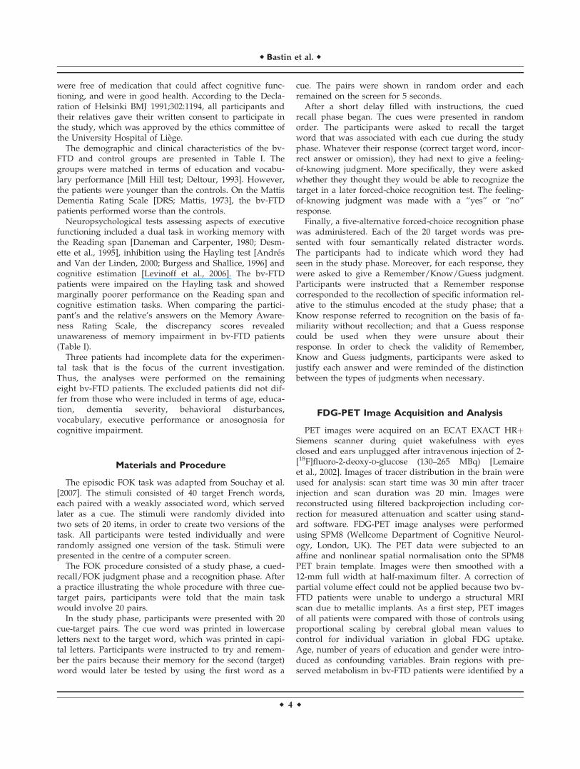
Bv-FTD patients showed hypometabolism in bilateralfrontal regions as well as in the anterior cingulate/medialfrontal cortex, the right inferior parietal gyrus and the pos-terior cingulate cortex (see Fig. 2, peak coordinates arereported in Table III). Given that impaired metabolic activ-ity in the posterior cingulate cortex is unusual in bv-FTD,we checked whether this finding was due to an outlierwho dragged the value of this region down. In fact, mostpatients had reduced activity in this region.
Figure 1.
Proportions of Remember, Know, and Guess responses given to
old words in the bv-FTD patients and the control group.
Figure 2.
Results of the SPM analysis comparing the brain glucose metabo-
lism of the bv-FTD group and the control group rendered on a
single subject brain (top left: sagittal view, bottom left: frontal
view; right: posterior coronal view).
r Bastin et al. r
r 6 r
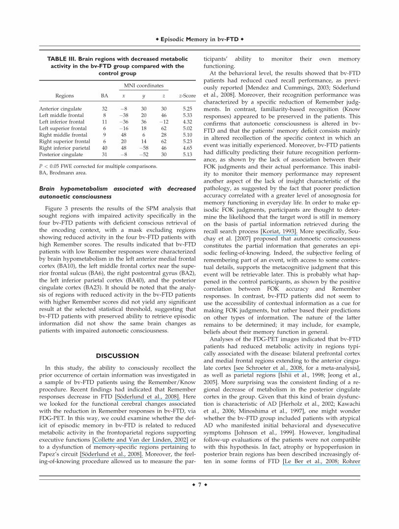
Brain hypometabolism associated with decreased
autonoetic consciousness
Figure 3 presents the results of the SPM analysis thatsought regions with impaired activity specifically in thefour bv-FTD patients with deficient conscious retrieval ofthe encoding context, with a mask excluding regionsshowing reduced activity in the four bv-FTD patients withhigh Remember scores. The results indicated that bv-FTDpatients with low Remember responses were characterizedby brain hypometabolism in the left anterior medial frontalcortex (BA10), the left middle frontal cortex near the supe-rior frontal sulcus (BA6), the right postcentral gyrus (BA2),the left inferior parietal cortex (BA40), and the posteriorcingulate cortex (BA23). It should be noted that the analy-sis of regions with reduced activity in the bv-FTD patientswith higher Remember scores did not yield any significantresult at the selected statistical threshold, suggesting thatbv-FTD patients with preserved ability to retrieve episodicinformation did not show the same brain changes aspatients with impaired autonoetic consciousness.
DISCUSSION
In this study, the ability to consciously recollect theprior occurrence of certain information was investigated ina sample of bv-FTD patients using the Remember/Knowprocedure. Recent findings had indicated that Rememberresponses decrease in FTD [Soderlund et al., 2008]. Herewe looked for the functional cerebral changes associatedwith the reduction in Remember responses in bv-FTD, viaFDG-PET. In this way, we could examine whether the def-icit of episodic memory in bv-FTD is related to reducedmetabolic activity in the frontoparietal regions supportingexecutive functions [Collette and Van der Linden, 2002] orto a dysfunction of memory-specific regions pertaining toPapez’s circuit [Soderlund et al., 2008]. Moreover, the feel-ing-of-knowing procedure allowed us to measure the par-
ticipants’ ability to monitor their own memoryfunctioning.
At the behavioral level, the results showed that bv-FTDpatients had reduced cued recall performance, as previ-ously reported [Mendez and Cummings, 2003; Soderlundet al., 2008]. Moreover, their recognition performance wascharacterized by a specific reduction of Remember judg-ments. In contrast, familiarity-based recognition (Knowresponses) appeared to be preserved in the patients. Thisconfirms that autonoetic consciousness is altered in bv-FTD and that the patients’ memory deficit consists mainlyin altered recollection of the specific context in which anevent was initially experienced. Moreover, bv-FTD patientshad difficulty predicting their future recognition perform-ance, as shown by the lack of association between theirFOK judgments and their actual performance. This inabil-ity to monitor their memory performance may representanother aspect of the lack of insight characteristic of thepathology, as suggested by the fact that poorer predictionaccuracy correlated with a greater level of anosognosia formemory functioning in everyday life. In order to make ep-isodic FOK judgments, participants are thought to deter-mine the likelihood that the target word is still in memoryon the basis of partial information retrieved during therecall search process [Koriat, 1993]. More specifically, Sou-chay et al. [2007] proposed that autonoetic consciousnessconstitutes the partial information that generates an epi-sodic feeling-of-knowing. Indeed, the subjective feeling ofremembering part of an event, with access to some contex-tual details, supports the metacognitive judgment that thisevent will be retrievable later. This is probably what hap-pened in the control participants, as shown by the positivecorrelation between FOK accuracy and Rememberresponses. In contrast, bv-FTD patients did not seem touse the accessibility of contextual information as a cue formaking FOK judgments, but rather based their predictionson other types of information. The nature of the latterremains to be determined; it may include, for example,beliefs about their memory function in general.
Analyses of the FDG-PET images indicated that bv-FTDpatients had reduced metabolic activity in regions typi-cally associated with the disease: bilateral prefrontal cortexand medial frontal regions extending to the anterior cingu-late cortex [see Schroeter et al., 2008, for a meta-analysis],as well as parietal regions [Ishii et al., 1998; Jeong et al.,2005]. More surprising was the consistent finding of a re-gional decrease of metabolism in the posterior cingulatecortex in the group. Given that this kind of brain dysfunc-tion is characteristic of AD [Herholz et al., 2002; Kawachiet al., 2006; Minoshima et al., 1997], one might wonderwhether the bv-FTD group included patients with atypicalAD who manifested initial behavioral and dysexecutivesymptoms [Johnson et al., 1999]. However, longitudinalfollow-up evaluations of the patients were not compatiblewith this hypothesis. In fact, atrophy or hypoperfusion inposterior brain regions has been described increasingly of-ten in some forms of FTD [Le Ber et al., 2008; Rohrer
TABLE III. Brain regions with decreased metabolic
activity in the bv-FTD group compared with the
control group
Regions BA
MNI coordinates
z-Scorex y z
Anterior cingulate 32 �8 30 30 5.25Left middle frontal 8 �38 20 46 5.33Left inferior frontal 11 �36 36 �12 4.32Left superior frontal 6 �16 18 62 5.02Right middle frontal 9 48 6 28 5.10Right superior frontal 6 20 14 62 5.23Right inferior parietal 40 48 �58 46 4.65Posterior cingulate 31 �8 �52 30 5.13
P < 0.05 FWE corrected for multiple comparisons.BA, Brodmann area.
r Episodic Memory in bv-FTD r
r 7 r

et al., 2010; Whitwell et al., 2009a], and in particular, in asubtype of FTD involving ubiquitin and TDP-43 (TARDNA-binding protein 43) immunoreactive neuronal inclu-sions [Mackenzie et al., 2008]. Recently, Whitwell et al.[2009b] identified a subtype of patients with grey matterloss in the temporal and parietal lobes, posterior cingulategyrus and medial frontal regions. At the neuropathologicallevel, half of these patients were diagnosed with TDP-43pathology. Consequently, it may be that several patients in
our sample belonged to this temporofrontoparietal subtypeof FTD.
This study is the first attempt to relate the bv-FTDpatients’ deficit affecting autonoetic consciousness forrecently learned information to metabolic brain function.The only previous study to examine Remember responsesin an anterograde memory task found a positive correla-tion with grey matter volume in a left medial temporalregion of interest [Soderlund et al., 2008]. In the present
Figure 3.
Results of the SPM analysis examining brain changes associated
with decreased autonoetic consciousness in bv-FTD (P < 0.05
FWE corrected): ‘‘patients with poor Remember scores < con-
trols,’’ masked exclusively by ‘‘patients with high Remember
scores < controls’’ on the SPM glass brain. Regions with
reduced activity in patients with decreased autonoetic con-
sciousness are shown on coronal sections of the average of six
patients’ MRI images (A: left anterior frontal region; B: left lat-
eral frontal region; C: posterior cingulate cortex).
r Bastin et al. r
r 8 r
study, in which the functional changes in the brain activityof patients with decreased Remember responses were ana-lyzed by voxel-wise statistical parametric mapping, themain finding was that bv-FTD patients with impairedautonoetic consciousness specifically showed reduced me-tabolism in the left anterior medial frontal cortex, the leftmiddle frontal cortex near the superior frontal sulcus, theright postcentral gyrus, the left inferior parietal cortex, andthe posterior cingulate cortex. Despite the small sample,the results were highly significant.
The anterior medial prefrontal cortex has been claimedto be related to the introspective evaluation of internalmental states [Christoff and Gabrieli, 2000]. This process isat the core of the Remember/Know procedure, whichrequires a subjective judgment of the mental states accom-panying recognition. Bv-FTD patients with impairedautonoetic consciousness also had reduced metabolic activ-ity in the left superior frontal sulcus and parietal regionsincluding the right postcentral gyrus. These regions havebeen found to be related to self-referential processing[Wicker et al., 2003] and first-person perspective [Vogeleyet al., 2004], and may form a frontoparietal network sup-porting the manipulation and monitoring of internallygenerated information [Christoff and Gabrieli, 2000]. Moregenerally, bv-FTD patients seem to lose the ability toapprehend mental states, either their own or others, asindicated by their deficit in Theory-of-Mind tasks[Adenzato et al., 2010]. Such a deficit may underlie someof the clinical features of bv-FTD, such as the difficultyunderstanding social situations [Kipps and Hodges, 2006].Consistently, bv-FTD patients failed to adequately perceivetheir memory functioning in everyday life and tended tooverestimate their memory abilities on the MemoryAwareness Rating Scale [Clare et al., 2002].
The most innovative aspect of the findings concerns theposterior cingulate hypometabolism in the bv-FTDpatients with impaired autonoetic consciousness. In thebrain organisation, the posterior cingulate cortex seems tooccupy a special position given its high level of connectiv-ity with parietal, frontal and temporal cortices [Buckneret al., 2009; Greicius et al., 2009]. It is involved in the re-trieval of information from episodic memory [Skinner andFernandes, 2007] and in self-referential processing [North-off and Bermpohl, 2004], and more generally in thedefault mode network [Buckner et al., 2009]. Recently, ithas been suggested that the posterior cingulate cortexmay be more involved in processes implying reference toone’s own person than in episodic memory per se [Cav-anna and Trimble, 2006; Sajonz et al., 2010]. In Rememberresponses, both dimensions are interconnected as theseresponses are given when participants consciously reacti-vate a personally experienced episode in all its richness.The difficulty some bv-FTD patients have in consciouslyretrieving a past event may stem at least partly from theiraltered sense of self [Miller et al., 2001], which deprivestheir memory retrieval of any elaborated personalengagement.
Altogether, the findings are consistent with the impactof a dysfunction of a frontoparietal network on the bv-FTD patients’ episodic memory, but also showed the cru-cial role of the posterior cingulate cortex, suggesting thatdeficient access to, monitoring and maintenance of infor-mation about the self contributed to impaired autonoeticconsciousness.
At a more clinical level, FDG-PET has been consideredas an important biomarker in diagnosing bv-FTD versusAD, with bv-FTD being characterized by hypometabolismin anterior regions (frontal and anterior cingulate),whereas AD is typically accompanied by posterior abnor-malities (posterior temporoparietal association cortex andposterior cingulate cortex) [Foster et al., 2007]. The currentresults call for caution in considering posterior cingulatehypometabolism as a specific marker of AD.
One limitation of the analyses of the PET data is thelack of correction for partial volume effects due to the MRIincompatibility of some patients, so that it is not clear howmuch atrophy contributed to the dysfunction of the frontaland posterior cortical regions in bv-FTD.
In conclusion, our small sample included patients withclinical features of bv-FTD, showing specific recollectiondisturbances, and posterior medial cortex hypometabolismcoexisting with the typical frontal alteration. Although theinvolvement of the posterior cingulate cortex is not typicalof bv-FTD, it has been reported in some forms of the dis-ease. We have proposed the existence of a relationshipbetween episodic memory disturbances and posterior cin-gulate and frontoparietal self-related dysfunction in bv-FTD.
REFERENCES
Adenzato M, Cavallo M, Enrici I (2010): Theory of mind ability inthe behavioural variant of frontotemporal dementia: An analy-sis of the neural, cognitive, and social levels. Neuropsycholo-gia 48:2–12.
Andres P, Van der Linden M (2000): Age-related differences in su-pervisory attentional system functions. J Gerontol B PsycholSci Soc Sci 55:373–380.
Banks S, Weintraub S (2008): Self-awareness and self-monitoringof cognitive and behavioral deficits in behavioral variant fron-totemporal dementia, primary progressive aphasia and proba-ble Alzheimer’s disease. Brain Cogn 67:58–68.
Brett M, Anton JL, Valabreque R, Poline JB (2002): Region of inter-est analysis using an SPM toolbox. Abstract presented at the8th International Conference on Functional Mapping of theHuman Brain; June 2–6; Sendai, Japan. Neuroimage 16:2 (CD-ROM).
Buckner RL, Sepulcre J, Talukdar T, Krienen FM, Liu H, HeddenT, Andrews-Hanna JR, Sperling RA, Johnson KA (2009): Corti-cal hubs revealed by intrinsic functional connectivity: Map-ping, assessment of stability, and relation to Alzheimer’sdisease. J Neurosci 29, 1860–1873.
Burgess PW, Shallice T (1996): Response suppression, initiationand strategy use following frontal lobe lesions. Neuropsycho-logia 34:263–273.
r Episodic Memory in bv-FTD r
r 9 r

Cavanna AE, Trimble MR (2006): The precuneus: A review of itsfunctional anatomy and behavioural correlates. Brain 129:564–583.
Christoff K, Gabrieli JDE (2000): The frontopolar cortex andhuman cognition: Evidence for a rostrocaudal hierarchical or-ganization within the human prefrontal cortex. Psychobiology28:168–186.
Clare L, Wilson BA, Carter G, Roth I, Hodges JR (2002): Assessingawareness in early-stage Alzheimer’s disease: Developmentand piloting of the Memory Awareness Rating Scale. Neuro-psychol Rehabil 12:341–362.
Collette F, Van der Linden M (2002): Brain imaging of the centralexecutive component of working memory. Neurosci BiobehavRev 26:105–125.
Connor LT, Dunlosky J, Hertzog C (1997): Age-related differencesin absolute but not relative metamemory accuracy. PsycholAging 12:50–71.
Cummings JL, Mega M, Gray K, Rosenberg-Thompson S, CarusiDA, Gornbein J (1994): The Neuropsychiatric Inventory: com-prehensive assessment of psychopathology in dementia. Neu-rology 44:2308–2314.
Daneman M, Carpenter PA (1980): Individual differences in work-ing memory and reading. J Verb Learn Verb Behav 19:450–466.
Deltour JJ (1993): Echelle de vocabulaire de Mill Hill de J.C.Raven. Adaptation francaise et normes comparees du Mill Hillet du Standard Progressive Matrices (PM 38). Manuel. Braine-le-Chateau: Editions l’Application des Techniques Modernes.
Desmette D, Hupet M, Van der Linden M (1995): Adaptation enlangue francaise du ‘Reading span test’ de Daneman et Car-penter (1980). L’Annee Psychologique 95:459–482.
Dunlosky J, Bjork RA (2008): Handbook of Metamemory andMemory. New York: Psychology Press.
Eslinger PJ, Dennis K, Moore P, Antani S, Hauck R, Grossman M(2005): Metacognitive deficits in frontotemporal dementia. JNeurol Neurosurg Psychiatry 76:1630–1635.
Foster NL, Heidebrink JL, Clark CM, Jagust WJ, Arnold SE, Bar-bas NR, DeCarli CS, Turner RS, Koeppe RA, Higdon R, Min-oshima S (2007): FDG-PET improves accuracy indistinguishing frontotemporal dementia and Alzheimer’s dis-ease. Brain 130:2616–2635.
Gardiner JM (1988): Functional aspects of recollective experience.Mem Cogn 16:309–313.
Greicius MD, Supekar K, Menon V, Dougherty RF (2009): Resting-state functional connectivity reflects structural connectivity inthe default mode network. Cereb Cortex 19:72–78.
Herholz K, Salmon E, Perani D, Baron JC, Holthoff V, Frolich L,Schonknecht P, Ito K, Mielke R, Kalbe E, Zundorf G, DelbeuckX, Pelati O, Anchisi D, Fazio F, Kerrouche N, Desgranges B,Eustache F, Beuthien-Baumann B, Menzel C, Schroder J, KatoT, Arahata Y, Henze M, Heiss WD (2002): Discriminationbetween Alzheimer dementia and controls by automated anal-ysis of multicenter FDG PET. Neuroimage 17:302–316.
Hodges JR, Miller B (2001): The neuropsychology of frontal vari-ant frontotemporal dementia and semantic dementia. Introduc-tion to the special topic papers: Part II. Neurocase 7:113–121.
Ishii K, Sakamoto S, Sasaki M, Kitagaki H, Yamaji S, HashimotoM, Imamura T, Shimomura T, Hirono N, Mori E (1998): Cere-bral glucose metabolism in patients with frontotemporal de-mentia. J Nucl Med 39:1875–1878.
Jeong Y, Cho SS, Park JM, Kang SJ, Lee JS, Kang E, Na DL, KimSE (2005): 18F-FDG PET findings in frontotemporal dementia:an SPM analysis of 29 patients. J Nucl Med 46:233–239.
Johnson JK, Head E, Kim R, Starr A, Cotman CW (1999): Clinicaland pathological evidence for a frontal variant of Alzheimer’sdisease. Arch Neurol 56:1233–1239.
Kawachi T, Ishii K, Sakamoto S, Sasaki M, Mori T, Yamashita F,Matsuda H, Mori E (2006): Comparison of the diagnostic per-formance of FDG-PET and VBM-MRI in very mild Alzheimer’sdisease. Eur J Nucl Med Mol Imaging 33:801–809.
Kertesz A, Blair M, McMonagle P, Munoz DG (2007): The diagno-sis and course of frontotemporal dementia. Alzheimer DisAssoc Disord 21:155–163.
Kipps CM, Hodges JR (2006): Theory of mind in frontotemporaldementia. Soc Neurosci 1:235–244.
Koriat A (1993): How do we know that we know? The accessibil-ity model of the feeling of knowing. Psychol Rev 100:609–639.
Le Ber I, Camuzat A, Hannequin D, Pasquier F, Guedj E, Rovelet-Lecrux A, Hahn-Barma V, van der Zee J, Clot F, Bakchine S,Puel M, Ghanim M, Lacomblez L, Mikol J, Deramecourt V,Lejeune P, de la Sayette V, Belliard S, Vercelletto M, MeyrignacC, Van Broeckhoven C, Lambert JC, Verpillat P, Campion D,Habert MO, Dubois B, Brice A;French research network onFTD/FTD-MND (2008): Phenotype variability in progranulinmutation carriers: A clinical, neuropsychological, imaging andgenetic study. Brain 131:732–746.
Lemaire C, Damhaut P, Lauricella B, Mosdzianowski C, MorelleJL, Monclus M, Van Naemen J, Mulleneers E, Aerts J, Plene-vaux A, Brihaye C, Luxen A (2002): Fast [18 F]FDG synthesisby alkaline hydrolysis on a low polarity solid phase support. JLab Compd Radiopharm 45:435–447.
Levinoff EJ, Phillips NA, Verret L, Babins L, Kelner N, Akerib V,Chertkow H (2006): Cognitive estimation impairment in Alz-heimer disease and mild cognitive impairment. Neuropsychol-ogy 20:123–132.
Mackenzie IRA, Foti D, Woulfe J, Hurwitz TA (2008): Atypicalfrontotemporal lobar degeneration with ubiquitin-positive,TDP-43-negative neuronal inclusions Brain 131:1282–1293.
Mattis S (1973): Dementia Rating Scale. Windsor, UK: NFER-Nelson.
Matuszweski V, Piolino P, De La Sayette V, Lalevee C, Pelerin A,Dupuy B, Viader F, Eustache F, Desgranges B (2006): Retrievalmechanisms for autobiographical memories: Insights from thefrontal variant of frontotemporal dementia. Neuropsychologia44:2386–2397.
McKhann GM, Albert MS, Grossman M, Miller B, Dickson D, Tro-janowski JQ (2001): Clinical and pathological diagnosis of fron-totemporal dementia. Report of the work group onfrontotemporal dementia and Pick’s disease. Arch Neurol58:1803–1809.
Mendez MF, Cummings JL (2003): Dementia: A Clinical Approach.Philadelphia, PA: Butterworth-Heinemann (Elsevier).
Miller BL, Seeley WW, Mychack P, Rosen HJ, Mena I, Boone K(2001): Neuroanatomy of the self: Evidence from patients withfrontotemporal dementia. Neurology 57:817–821.
Minoshima S, Giordani B, Berent S, Frey KA, Foster NL, Kuhl DE(1997): Metabolic reduction in the posterior cingulate cortex invery early Alzheimer’s disease. Ann Neurol 42:85–94.
Neary D, Snowden JS, Gustafson L, Passant U, Stuss D, Black S,Freedman M, Kertesz A, Robert PH, Albert M, Boone K, MillerBL, Cummings J, Benson DF (1998): Frontotemporal lobardegeneration: A consensus on clinical diagnostic criteria. Neu-rology 51:1546–1554.
Northoff G, Bermpohl F (2004): Cortical midline structures andthe self. Trends Cogn Sci 8:102–107.
r Bastin et al. r
r 10 r
Piolino P, Chetelat G, Matuszweski V, Landeau B, Mezenge F, ViaderF, De La Sayette V, Eustache F, Desgranges B (2007): In search ofautobiographical memories: A PET study in the frontal variant offrontotemporal dementia. Neuropsychologia 45:2730–2743.
Rohrer JD, Ridgway GR, Modat M, Ourselin S, Mead S, Fox NC,Rossor MN, Warren JD (2010): Distinct profiles of brain atro-phy in frontotemporal lobal degeneration caused by progranu-lin and tau mutations. Neuroimage 53:1070–1076.
Rosen HJ, Alcantar O, Rothlind J, Sturm V, Kramer JH, Weiner M,Miller BL (2010): Neuroanatomical correlates of cognitive self-ap-praisal in neurodegenerative disease. Neuroimage 49:3358–3364.
Ruby P, Schmidt C, Hogge M, D’Argembeau A, Collette F,Salmon E (2007): Social mind representation: Where does it failin frontotemporal dementia? J Cogn Neurosci 19:1–13.
Sajonz B, Kahnt T, Margulies D, Park SQ, Wittmann A, Stoy M,Strohle A, Heinz A, Northoff G, Bermpohl F (2010): Delineat-ing self-referential processing from episodic memory retrieval:Common and dissociable networks. Neuroimage 50:1606–1617.
Salmon E, Perani D, Collette F, Feyers D, Kalbe E, Holthoff V,Sorbi S, Herholz K (2008): A comparison of unawareness infrontotemporal dementia and Alzheimer’s disease. J NeurolNeurosurg Psychiatry 79:176–179.
Schraw G (1995): Measures of feeling of knowing accuracy: A newlook at an old problem. Appl Cogn Psychol 9:321–332.
Schroeter ML, Raczka K, Neumann J, von Cramon DY (2008):Neural networks in frontotemporal dementia: A meta-analysis.Neurobiol Aging 29:418–426.
Skinner EI, Fernandes MA (2007): Neural correlates of recollectionand familiarity: A review of neuroimaging and patient data.Neuropsychologia 45:2163–2179.
Snodgrass JG, Corwin J (1988): Pragmatics of measuring recogni-tion memory: Applications to dementia and amnesia. J ExpPsychol Gen 117:34–50.
Soderlund H, Black SE, Miller BL, Freedman M, Levine B (2008):Episodic memory and regional atrophy in frontotemporal lobardegeneration. Neuropsychologia 46:127–136.
Souchay C, Isingrini M, Pillon B, Gil R (2003): Metamemory accu-racy in Alzheimer’s disease and frontotemporal lobe dementia.Neurocase 9:482–492.
Souchay C, Moulin CJA, Clarys D, Taconnat L, Isingrini M (2007):Diminished episodic memory awareness in older adults: Evi-dence from feeling-of-knowing and recollection. ConsciousCogn 16:769–784.
Tulving E (2002): Episodic memory: From mind to brain. Ann RevPsychol 53:1–53.
Vogeley K, May M, Ritzl A, Falkai P, Zilles K, Fink GR (2004):Neural correlates of first-person perspective as one constituentof human self-consciousness. J Cogn Neurosci 16:817–827.
Whitwell JL, Jack CR, Boeve BF, Senjem ML, Baker M, Rade-makers R, Ivnik RJ, Knopman DS, Wszolek ZK, Petersen RC,Josephs KA (2009a) Voxel-based morphometry patterns of at-rophy in FTLD with mutations in MAPT or PGRN. Neurology72:813–820.
Whitwell JL, Przybelski SA, Weigand SD, Ivnik RJ, Vemuri P,Gunter JL, Senjem ML, Shiung MM, Boeve BF, Knopman DS,Parisi JE, Dickson DW, Petersen RC, Jack CR Jr, Josephs KA(2009b) Distinct anatomical subtypes of the behavioural variantof frontotemporal dementia: A cluster analysis study. Brain132:2932–2946.
Wicker B, Ruby P, Royet JP, Fonlupt P (2003): A relation betweenrest and the self in the brain? Brain Res Rev 43:224–230.
Yakushev I, Landvogt C, Buchholz HG, Fellgiebel A, Hammers A,Scheurich A, Schmidtmann I, Gerhard A, Schreckenberger M,Bartenstein P (2008): Choice of reference area in studies of Alz-heimer’s disease using positron emission tomography with flu-orodeoxyglucose-F18. Psychiatry Res Neuroim 164:143–153.
r Episodic Memory in bv-FTD r
r 11 r
Copyright © 2022 FDOKUMEN