Editing of CD1d-Bound Lipid Antigens by Endosomal Lipid Transfer Proteins
10
Editing of CD1d-Bound Lipid Antigens by Endosomal Lipid Transfer Proteins Dapeng Zhou 1,* , Carlos Cantu III 2,* , Yuval Sagiv 1 , Nicolas Schrantz 2 , Ashok B. Kulkarni 3 , Xiaoyang Qi 4 , Don J. Mahuran 5 , Carlos R. Morales 6 , Gregory A. Grabowski 4 , Kamel Benlagha 1 , Paul Savage 7 , Albert Bendelac 1,† , and Luc Teyton 2,† 1 Department of Pathology, University of Chicago, Chicago, IL 60637, USA 2 Department of Immunology, Scripps Research Institute, La Jolla, CA 92037, USA 3 National Institute of Dental and Craniofacial Research, National Institutes of Health, Bethesda, MD 20892, USA 4 Children Hospital Medical Center, Cincinnati, OH 45229–3039, USA 5 Department of Medicine and Pathobiology, University of Toronto, Toronto, ON M5G 1X8, Canada 6 Department of Anatomy and Cell Biology, McGill University, Montreal, QC H3A 2B2, Canada 7 Department of Chemistry and Biochemistry, Brigham Young University, Provo, UT 84602–5700, USA Abstract It is now established that CD1 molecules present lipid antigens to T cells, although it is not clear how the exchange of lipids between membrane compartments and the CD1 binding groove is assisted. We report that mice deficient in prosaposin, the precursor to a family of endosomal lipid transfer proteins (LTP), exhibit specific defects in CD1d-mediated antigen presentation and lack Vα14 NKT cells. In vitro, saposins extracted monomeric lipids from membranes and from CD1, thereby promoting the loading as well as the editing of lipids on CD1. Transient complexes between CD1, lipid, and LTP suggested a “tug-of-war” model in which lipid exchange between CD1 and LTP is on the basis of their respective affinities for lipids. LTPs constitute a previously unknown link between lipid metabolism and immunity and are likely to exert a profound influence on the repertoire of self, tumor, and microbial lipid antigens. CD1 molecules have evolved a unique hydrophobic binding groove that binds lipid antigens in both the secretory and endosomal compartments for presentation to T lymphocytes (1). In mice, the main population of CD1-restricted T cells, called Vα14 NKT cells, express a semi- invariant Vα14-Jα18/Vβ8 T cell receptor (TCR). These cells exhibit reactivity against CD1d in combination with endogenous ligands (2) that can be mimicked by α-galactosylceramide (αGC) (3). This population is conserved among mammalian species and regulates immune responses (4,5). Like human CD1b-restricted T cells specific for mycobacterial glycolipids † These authors contributed equally to this work. To whom correspondence should be addressed. [email protected] (A.B.); [email protected] (L.T.). * These authors contributed equally to this work. Supporting Online Material www.sciencemag.org/cgi/content/full/1092009/DC1 Materials and Methods Figs. S1 to S4 PubMed Central CANADA Author Manuscript / Manuscrit d'auteur Science. Author manuscript; available in PMC 2010 August 9. Published in final edited form as: Science. 2004 January 23; 303(5657): 523–527. doi:10.1126/science.1092009. PMC Canada Author Manuscript PMC Canada Author Manuscript PMC Canada Author Manuscript
-
Upload
independent -
Category
Documents
-
view
0 -
download
0
Transcript of Editing of CD1d-Bound Lipid Antigens by Endosomal Lipid Transfer Proteins

Editing of CD1d-Bound Lipid Antigens by Endosomal LipidTransfer Proteins
Dapeng Zhou1,*, Carlos Cantu III2,*, Yuval Sagiv1, Nicolas Schrantz2, Ashok B. Kulkarni3,Xiaoyang Qi4, Don J. Mahuran5, Carlos R. Morales6, Gregory A. Grabowski4, KamelBenlagha1, Paul Savage7, Albert Bendelac1,†, and Luc Teyton2,†1 Department of Pathology, University of Chicago, Chicago, IL 60637, USA2 Department of Immunology, Scripps Research Institute, La Jolla, CA 92037, USA3 National Institute of Dental and Craniofacial Research, National Institutes of Health, Bethesda,MD 20892, USA4 Children Hospital Medical Center, Cincinnati, OH 45229–3039, USA5 Department of Medicine and Pathobiology, University of Toronto, Toronto, ON M5G 1X8,Canada6 Department of Anatomy and Cell Biology, McGill University, Montreal, QC H3A 2B2, Canada7 Department of Chemistry and Biochemistry, Brigham Young University, Provo, UT 84602–5700,USA
AbstractIt is now established that CD1 molecules present lipid antigens to T cells, although it is not clearhow the exchange of lipids between membrane compartments and the CD1 binding groove isassisted. We report that mice deficient in prosaposin, the precursor to a family of endosomal lipidtransfer proteins (LTP), exhibit specific defects in CD1d-mediated antigen presentation and lackVα14 NKT cells. In vitro, saposins extracted monomeric lipids from membranes and from CD1,thereby promoting the loading as well as the editing of lipids on CD1. Transient complexesbetween CD1, lipid, and LTP suggested a “tug-of-war” model in which lipid exchange betweenCD1 and LTP is on the basis of their respective affinities for lipids. LTPs constitute a previouslyunknown link between lipid metabolism and immunity and are likely to exert a profound influenceon the repertoire of self, tumor, and microbial lipid antigens.
CD1 molecules have evolved a unique hydrophobic binding groove that binds lipid antigensin both the secretory and endosomal compartments for presentation to T lymphocytes (1). Inmice, the main population of CD1-restricted T cells, called Vα14 NKT cells, express a semi-invariant Vα14-Jα18/Vβ8 T cell receptor (TCR). These cells exhibit reactivity against CD1din combination with endogenous ligands (2) that can be mimicked by α-galactosylceramide(αGC) (3). This population is conserved among mammalian species and regulates immuneresponses (4,5). Like human CD1b-restricted T cells specific for mycobacterial glycolipids
†These authors contributed equally to this work. To whom correspondence should be addressed. [email protected] (A.B.);[email protected] (L.T.).*These authors contributed equally to this work.Supporting Online Materialwww.sciencemag.org/cgi/content/full/1092009/DC1Materials and MethodsFigs. S1 to S4
PubMed Central CANADAAuthor Manuscript / Manuscrit d'auteurScience. Author manuscript; available in PMC 2010 August 9.
Published in final edited form as:Science. 2004 January 23; 303(5657): 523–527. doi:10.1126/science.1092009.
PMC
Canada Author M
anuscriptPM
C C
anada Author Manuscript
PMC
Canada Author M
anuscript
(6), Vα14 NKT cells are dependent on endosomal trafficking of CD1d for natural antigenrecognition (7–10). Other endogenous or exogenous antigens do not require endosomaltrafficking, however, suggesting that loading may be achieved in distinct cellularcompartments depending on the nature of the antigen (9,11,12). CD1 endosomal traffickingis tightly controlled by cytoplasmic tail–encoded tyrosine-containing motifs binding adaptorprotein 2 and 3 (AP-2 and AP-3) complexes, as well as by association with the invariantchain (Ii) or Ii/major histocompatibility (MHC) class II complexes (13–16).
Because lipids are integral membrane components that might require lipid transfer proteins(LTP) (17) for extraction, we investigated whether various families of LTP might assistantigen presentation. We focused on the Vα14 NKT cell endosomal-dependent system totest the possible involvement of LTPs localized in the endocytic pathway, such as saposins(18) and GM2 activator protein (19). Saposins A, B, C, and D are the four members of afamily of small dimeric proteins produced by endosomal proteolytic cleavage of prosaposin(18) that regulate various aspects of lipid metabolism. GM2 activator protein is structurallydifferent from saposins but extracts lipids with the use of similar conformational changes(20). CD1d-αGC tetramers stained Vα14 NKT cells with a CD44high memory phenotype inthe thymus and peripheral tissues of wild-type mice, but in SAP−/− littermates (21), thesecells were undetectable as in CD1d−/− mice (Fig. 1A). The level and pattern of CD1dexpression, however, were conserved in SAP−/− mice (Fig. 1B) (22). SAP−/− mice developneurological symptoms starting between days 25 and 30 and show retarded growth.Although the thymuses and spleens of the adult animals were relatively small, percentagesof naïve (CD44low) and memory (CD44high) CD4+ and CD8+ T cells, B cells, natural killer(NK) cells, and γδ T cells were normal (Fig. 1C) (22). In fetal thymic organ culture, SAP−/−
thymic lobes had the same number of cells as those of the wild type but selectively lackedVα14 NKT cells (Fig. 1D). These findings demonstrate a selective defect in thedevelopment of Vα14 NKT cells in the absence of prosaposin.
Many NKT cell hybridomas can be activated in vitro to release interleukin-2 (IL-2) bycoculture with fresh CD1d-expressing cells, such as thymocytes, which are thought to be thespecific antigen-presenting cells (APCs) that positively select Vα14 NKT cells (23). Thisfeature is attributed to the recognition of natural lipid ligands loaded on CD1d in these cells.Unlike wild-type thymocytes, those from SAP−/− animals failed to stimulate the self-reactive Vα14 NKT cell hybridoma DN32.D3. In contrast, the non-Vα14 CD1d-restrictedhybridomas TCB11 and 1C8DC1 were able to recognize the SAP−/− thymocytes (Fig. 2A)even in serum-free culture conditions (22). These results, which are consistent with theendosomal trafficking requirement for CD1d to stimulate Vα14 NKT cells (9), suggest thatendosomal saposins are selectively involved in the presentation of the Vα14 NKT cellligands by CD1d.
We examined the presentation of αGC and Galα1,2αGC, which are partially and completelydependent on endosomal functions, respectively (10,24). Although stimulation of DN32.D3by SAP−/− thymocytes pulsed with αGC was partially decreased, stimulation by dendriticcells (DCs) was normal (Fig. 2B). In contrast, presentation of Galα1,2αGC, whichabsolutely requires endosomal processing into αGC, was abolished in all cell types (Fig. 2B)(22). These results demonstrate requirements for saposins in the presentation of exogenousglycolipids.
We next performed two sets of control experiments to exclude the possibility that the lipidpresentation defects might be the result of broad nonspecific disruptions of endosomalfunction in the absence of SAP. SAP−/− APCs were comparable to those of wild type forpresentation of intact ovalbumin, hen egg lysozyme, keyhole limpet hemocyanin, andcollagen II to various I-Aq– or I-Ab–restricted T cell hybridomas, which require complex
Zhou et al. Page 2
Science. Author manuscript; available in PMC 2010 August 9.
PMC
Canada Author M
anuscriptPM
C C
anada Author Manuscript
PMC
Canada Author M
anuscript

processing within an intact endosomallysosomal compartment (25) (fig. S1). Consistent withthe preservation of MHC class II and antigen-presenting functions, the Lamp-1+compartment of SAP−/− cells appeared morphologically normal (Fig. 3), and lysosomalacidification, as assayed by Lysotracker (Materials and Methods) staining, was comparableto that seen in wild type (fig. S1). We also found that mutant mice deficient in α-galactosidase A (26), galactosylceramidase (27), or mucopolysaccharidase (28) hadconserved thymic NKT cells and normally presented natural CD1d ligands to NKT cells(fig. S2), indicating that accumulation of lipids or mucopolysaccharides as seen in variouslysosomal storage diseases is not accompanied by nonspecific alterations of CD1d-mediatedantigen presentation.
Both CD1d and saposins have been reported to preferentially localize in late endosomal/lysosomal compartments (15,29). We found that in DCs, as well as in thymocytes, saposinand CD1d colocalized in the Lamp-1+ late endosome/lysosome (Fig. 3), These resultsdemonstrate colocalization of CD1d and saposin in the subcellular compartment whereglycolipid processing and loading occur.
Because no evidence exists that CD1d itself can function as a LTP, we directly testedwhether saposins might be responsible for the transfer of lipids onto CD1d. To examine this,we devised a cell-free system in which lipid exchange between, on one hand, purifiedcomplexes of CD1d bound to the acidic trisialoganglioside GT1b (CD1-GT) and, on theother hand, liposomes made of phosphatidylserine (PS) could be measured by nativeisoelectrofocusing (IEF) (30). In the absence of LTP, PS could not be loaded. However,each of the four saposins readily promoted exchange (Fig. 4A). Incubation of purified CD1-GT with individual saposins alone did not promote the removal of GT1b, suggesting that, inthis particular experimental condition, saposins could extract lipids from membranes but notfrom CD1d. The loading of PS was verified by repurifying CD1d and analyzing the lipidextract by mass spectrometry (22). In the case of PS, dose-effect studies and kinetic analysisdetermined that saposin A was the most effective transfer protein. By comparison, Cdisplayed intermediate activity, and B and D were relatively inefficient (fig. S3).Importantly, the titration experiments revealed that saposins were not enzymatic in theiractivity, because they needed to be present at an equimolar ratio with CD1d to carry theirtransfer function. This property clearly differentiates saposins, in their lipid-editing function,from the peptide editor for MHC class II, HLA-DM (31). It is consistent with thenonenzymatic mechanism of saposin function in the hydrolysis of cerebrosides (32). Thefine specificity of saposins was further examined by comparing the transfer of sulfatide(SFT) onto CD1d (fig. S3). In this case, saposins A and C were the most efficient, saposin Dhad no activity, and B was intermediate. All saposins were most active at 37°C and highlypH-dependent, with maximal activity at pH values below 5.5 (fig. S3). However, saposin Aand C, but not B and D, had substantial function at pH = 6.0, suggesting possible differentfunctions of saposins in endosomal subcompartments and during the maturation process ofdendritic cells.
To better understand the nature of molecular interactions leading to lipid exchange, weattempted to visualize a potential physical interaction between saposins and CD1d. In orderto do so, anion exchange–purified CD1-GT, CD1-SFT, and CD1-αGC complexes wereincubated with an equimolar amount of saposin A at room temperature for 1 hour (Fig. 4B).Saposin A ran to the anodic front because of its low pKa (4.9) and stained very poorly withCoomassie Blue on native IEF. When CD1-GT or CD1-SFT complexes were incubated withsaposin A, a band comigrating with CD1d was detected by a serum against saposin A (anti-saposin) as well as by an antibody against CD1d (anti-CD1) in Western blot (Fig. 4B). Nocomplex formation could be observed between saposin A and classical MHC class Imolecules (H-2Kb) or albumin. The absence of complex formation with CD1-αGC strongly
Zhou et al. Page 3
Science. Author manuscript; available in PMC 2010 August 9.
PMC
Canada Author M
anuscriptPM
C C
anada Author Manuscript
PMC
Canada Author M
anuscript
suggested that the interaction was highly lipid head group– dependent, and it also correlatedwith the inability of saposin A to transfer αGC onto CD1d (see below). We note that theassociation between CD1-GT and saposin A was not accompanied by any notice-ableunloading of GT1b from CD1d, which would have appeared as neutral bands of emptyCD1d.
We considered the possibility that other LTPs might reveal that capacity to extract lipidsbound to CD1 molecules. Indeed, we found that the endosomal GM2 activator protein(GM2A) (33) could remove the GT1b from CD1-GT. Purified CD1-GT complexes wereincubated in the absence of exogenous lipids with increasing concentrations of recombinantGM2A (Fig. 4C). The appearance of a lower band corresponding to neutral, empty CD1ddemonstrated the loss of GT1b on both the Coomassie Blue–stained gel and the anti-CD1Western blot. Concomitantly, GM2A-GT complexes were revealed by a correspondingchange in IEF on the anti-GM2A Western blot. Importantly, in contrast with CD1–GT–saposin A, we did not detect CD1-GT-GM2A trimolecular complexes. These findingssupport an affinity-based “tug-of-war” model of lipid exchange between CD1d and LTP.Transient complexes would not be detected when unloading is rapid, whereas slow orinefficient unloading would result in more stable complexes.
None of the saposins could readily transfer αGC or Galα1,2αGC onto CD1d in vitro (22),whereas GM2A efficiently assisted the loading of αGC onto CD1d (fig. S4), explaining whypresentation of αGC by SAP−/− cells was subnormal (Fig. 2). Neither saposins nor GM2Aassisted the loading of Galα1,2αGC (22). However, because Galα1,2αGC needs processinginto αGC by α-glycosidase A, a saposin-dependent enzyme (34), defective presentation ofGalα1,2αGC by SAP−/− cells (Fig. 2) is likely a result of defective processing. Together, ourfindings suggest that defective presentation of glycolipid antigens by SAP−/− cells mayresult from single or combined defects in lipid processing, loading, and unloading. Further,because of the individual specificities of LTPs, it is likely that different LTPs cooperate togenerate and edit the glycolipid repertoire presented by CD1 molecules. Also, we canassume that LTP dependency for loading will vary for various lipid families.
In conclusion, it appears that endosomal LTPs, such as the saposin family and GM2A, fill amajor gap between CD1-mediated antigen presentation and lipid metabolism. Saposins areinvolved in the generation and/or loading of the natural thymic antigens that determineselection of Vα14 NKT cells. Our biochemical studies suggest an affinity-based tug-of-warmodel of lipid exchange between LTPs and CD1 molecules (Fig. 5). LTPs extract lipidsfrom membranes and CD1d by interacting specifically with their polar heads. On the basisof their relative affinities for the polar head and the fatty acid portion of a given lipid,respectively, LTP and CD1d form transient complexes that lead to lipid exchange anddetermine the repertoire of glycolipids presented by CD1d molecules.
In vivo, such lipid-editing function is likely to have an impact on CD1-restricted T cellssimilar to that of HLA-DM editing of MHC class II– bound peptides on the selection andexpansion of CD4− T cells. Studies of the differential distribution and lipid specificity ofindividual LTPs, which allow for considerable flexibility in regulating the presentation oflipid antigens, will be critical to improve the rational design of lipid vaccines and adjuvants.
AcknowledgmentsWe thank R. Brown, E. Berger, K. Sandhoff, and B. Jabri for helpful suggestions and E. Rosloniec and A.Rudensky for I-Ab– and I-Aq–restricted T cell hybridomas. Supported by NIH grants (PO1 AI53725 to P.S., A.B.,and L.T. and AI50867 and AI38339 to A.B.) and the Cancer Research Institute (D.Z. and Y.S.).
Zhou et al. Page 4
Science. Author manuscript; available in PMC 2010 August 9.
PMC
Canada Author M
anuscriptPM
C C
anada Author Manuscript
PMC
Canada Author M
anuscript
References and Notes1. Moody DB, Porcelli SA. Nat Rev Immunol 2003;3:11. [PubMed: 12511872]2. Bendelac A, et al. Science 1995;268:863. [PubMed: 7538697]3. Kawano T, et al. Science 1997;278:1626. [PubMed: 9374463]4. Godfrey DI, Hammond KJ, Poulton LD, Smyth MJ, Baxter AG. Immunol Today 2000;21:573.
[PubMed: 11094262]5. Bendelac A, Bonneville M, Kearney JF. Nat Rev Immunol 2001;1:177. [PubMed: 11905826]6. Moody DB, et al. J Exp Med 2000;192:965. [PubMed: 11015438]7. Sugita M, et al. Science 1996;273:349. [PubMed: 8662520]8. Jackman RM, et al. Immunity 1998;8:341. [PubMed: 9529151]9. Chiu YH, et al. J Exp Med 1999;189:103. [PubMed: 9874567]10. Chiu YH, et al. Nat Immunol 2002;3:55. [PubMed: 11731798]11. Shamshiev A, et al. Immunity 2000;13:255. [PubMed: 10981968]12. Moody DB, et al. Nat Immunol 2002;3:435. [PubMed: 11938350]13. Sugita M, et al. Immunity 2002;16:697. [PubMed: 12049721]14. Briken V, Jackman RM, Dasgupta S, Hoening S, Porcelli SA. EMBO J 2002;21:825. [PubMed:
11847129]15. Jayawardena-Wolf J, Benlagha K, Chiu YH, Mehr R, Bendelac A. Immunity 2001;15:897.
[PubMed: 11754812]16. Kang SJ, Cresswell P. EMBO J 2002;21:1650. [PubMed: 11927549]17. Rueckert DG, Schmidt K. Chem Phys Lipids 1990;56:1. [PubMed: 2091833]18. Sandhoff K, Kolter T, Van Echten-Deckert G. Ann NY Acad Sci 1998;845:139. [PubMed:
9668348]19. Wright CS, Zhao Q, Rastinejad F. J Mol Biol 2003;331:951. [PubMed: 12909021]20. Ahn VE, Faull KF, Whitelegge JP, Fluharty AL, Prive GG. Proc Natl Acad Sci USA 2003;100:38.
[PubMed: 12518053]21. Fujita N, et al. Hum Mol Genet 1996;5:711. [PubMed: 8776585]22. Zhou D, et al. unpublished observations.23. Bendelac A, Rivera MN, Park SH, Roark JH. Annu Rev Immunol 1997;15:535. [PubMed:
9143699]24. Prigozy TI, et al. Science 2001;291:664. [PubMed: 11158680]25. Harding CV, Collins DS, Slot JW, Geuze HJ, Unanue ER. Cell 1991;64:393. [PubMed: 1899049]26. Ohshima T, et al. Proc Natl Acad Sci USA 1997;94:2540. [PubMed: 9122231]27. Sakai N, et al. J Neurochem 1996;66:1118. [PubMed: 8769874]28. Vogler C, et al. Pediatr Res 2001;49:342. [PubMed: 11228259]29. Zhao Q, Morales CR. J Biol Chem 2000;275:24829. [PubMed: 10818106]30. Cantu C III, Benlagha K, Savage PB, Bendelac A, Teyton L. J Immunol 2003;170:4673. [PubMed:
12707346]31. Denzin LK, Cresswell P. Cell 1995;82:155. [PubMed: 7606781]32. Qi X, Leonova T, Grabowski GA. J Biol Chem 1994;269:16746. [PubMed: 8206997]33. Wu YY, et al. J Biol Chem 1994;269:16276. [PubMed: 8206933]34. Kase R, et al. FEBS Lett 1996;393:74. [PubMed: 8804427]
Zhou et al. Page 5
Science. Author manuscript; available in PMC 2010 August 9.
PMC
Canada Author M
anuscriptPM
C C
anada Author Manuscript
PMC
Canada Author M
anuscript

Fig. 1.SAP−/− mice selectively lack Vα14 NKT cells. (A) Vα14 NKT cells, identified as positivefor CD1d-αGC tetramer (y axis) and negative for empty CD1d tetramer and CD8 (x axis) byflow cytometry of thymocytes, splenocytes, and liver lymphocytes, are indicated in theboxed areas with their corresponding % values. Data representative of four individual adultFVB.SAP−/− mice compared to wild-type (WT) littermates and B6.CD1−/−. (B) Histogramsof CD1d surface thymocyte and splenocyte expression in WT and mutant mice. (C) T cellsubsets identified by CD4 and CD8 or CD4 and CD44 in the indicated tissues, withcorresponding % values as indicated. (D) Vα14 NKT cells in fetal thymuses after 14 days ofculture. Data representative of two individual B6.SAP−/− fetal thymuses and their WTlittermate controls.
Zhou et al. Page 6
Science. Author manuscript; available in PMC 2010 August 9.
PMC
Canada Author M
anuscriptPM
C C
anada Author Manuscript
PMC
Canada Author M
anuscript
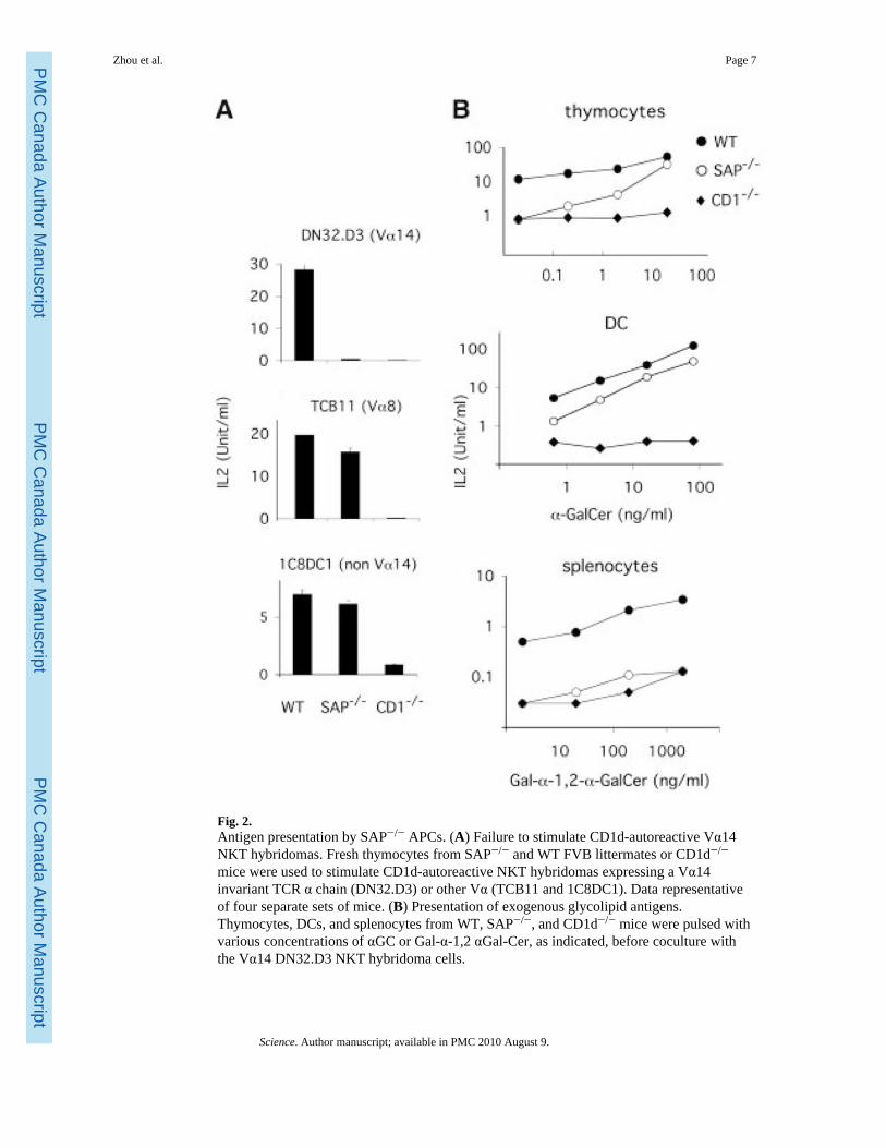
Fig. 2.Antigen presentation by SAP−/− APCs. (A) Failure to stimulate CD1d-autoreactive Vα14NKT hybridomas. Fresh thymocytes from SAP−/− and WT FVB littermates or CD1d−/−
mice were used to stimulate CD1d-autoreactive NKT hybridomas expressing a Vα14invariant TCR α chain (DN32.D3) or other Vα (TCB11 and 1C8DC1). Data representativeof four separate sets of mice. (B) Presentation of exogenous glycolipid antigens.Thymocytes, DCs, and splenocytes from WT, SAP−/−, and CD1d−/− mice were pulsed withvarious concentrations of αGC or Gal-α-1,2 αGal-Cer, as indicated, before coculture withthe Vα14 DN32.D3 NKT hybridoma cells.
Zhou et al. Page 7
Science. Author manuscript; available in PMC 2010 August 9.
PMC
Canada Author M
anuscriptPM
C C
anada Author Manuscript
PMC
Canada Author M
anuscript
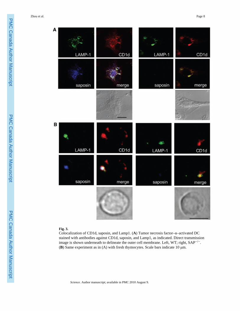
Fig. 3.Colocalization of CD1d, saposin, and Lamp1. (A) Tumor necrosis factor–α–activated DCstained with antibodies against CD1d, saposin, and Lamp1, as indicated. Direct transmissionimage is shown underneath to delineate the outer cell membrane. Left, WT; right, SAP−/−.(B) Same experiment as in (A) with fresh thymocytes. Scale bars indicate 10 μm.
Zhou et al. Page 8
Science. Author manuscript; available in PMC 2010 August 9.
PMC
Canada Author M
anuscriptPM
C C
anada Author Manuscript
PMC
Canada Author M
anuscript
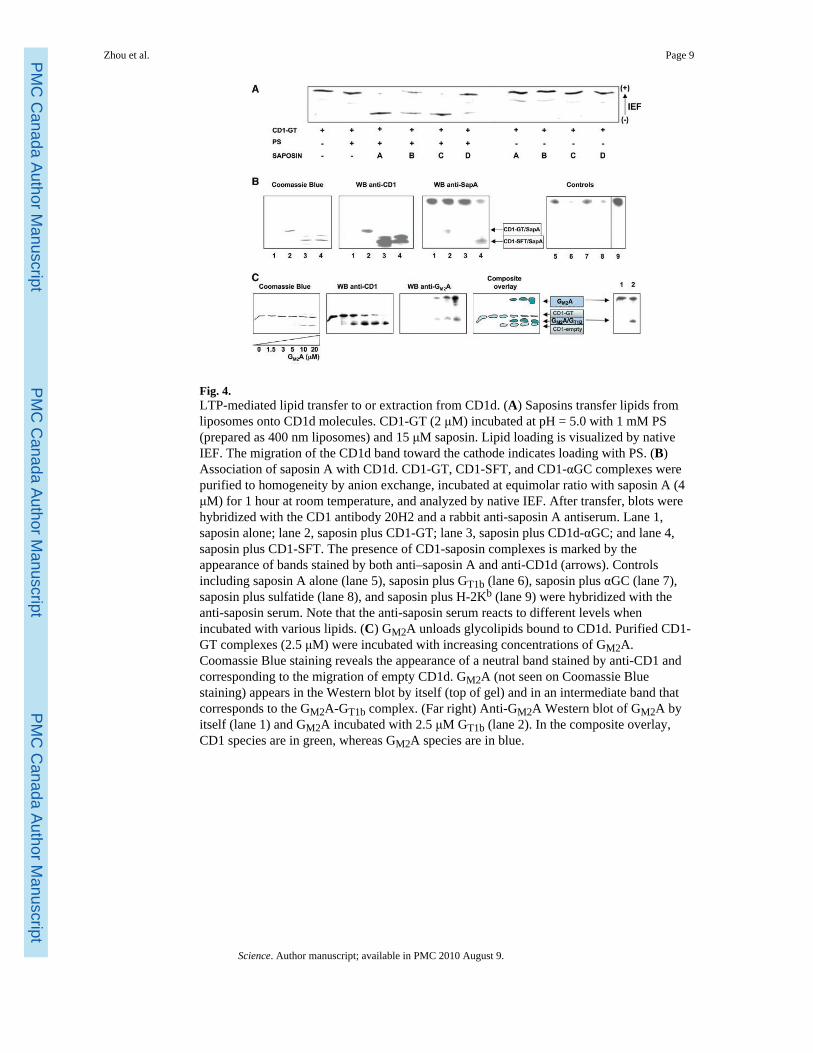
Fig. 4.LTP-mediated lipid transfer to or extraction from CD1d. (A) Saposins transfer lipids fromliposomes onto CD1d molecules. CD1-GT (2 μM) incubated at pH = 5.0 with 1 mM PS(prepared as 400 nm liposomes) and 15 μM saposin. Lipid loading is visualized by nativeIEF. The migration of the CD1d band toward the cathode indicates loading with PS. (B)Association of saposin A with CD1d. CD1-GT, CD1-SFT, and CD1-αGC complexes werepurified to homogeneity by anion exchange, incubated at equimolar ratio with saposin A (4μM) for 1 hour at room temperature, and analyzed by native IEF. After transfer, blots werehybridized with the CD1 antibody 20H2 and a rabbit anti-saposin A antiserum. Lane 1,saposin alone; lane 2, saposin plus CD1-GT; lane 3, saposin plus CD1d-αGC; and lane 4,saposin plus CD1-SFT. The presence of CD1-saposin complexes is marked by theappearance of bands stained by both anti–saposin A and anti-CD1d (arrows). Controlsincluding saposin A alone (lane 5), saposin plus GT1b (lane 6), saposin plus αGC (lane 7),saposin plus sulfatide (lane 8), and saposin plus H-2Kb (lane 9) were hybridized with theanti-saposin serum. Note that the anti-saposin serum reacts to different levels whenincubated with various lipids. (C) GM2A unloads glycolipids bound to CD1d. Purified CD1-GT complexes (2.5 μM) were incubated with increasing concentrations of GM2A.Coomassie Blue staining reveals the appearance of a neutral band stained by anti-CD1 andcorresponding to the migration of empty CD1d. GM2A (not seen on Coomassie Bluestaining) appears in the Western blot by itself (top of gel) and in an intermediate band thatcorresponds to the GM2A-GT1b complex. (Far right) Anti-GM2A Western blot of GM2A byitself (lane 1) and GM2A incubated with 2.5 μM GT1b (lane 2). In the composite overlay,CD1 species are in green, whereas GM2A species are in blue.
Zhou et al. Page 9
Science. Author manuscript; available in PMC 2010 August 9.
PMC
Canada Author M
anuscriptPM
C C
anada Author Manuscript
PMC
Canada Author M
anuscript
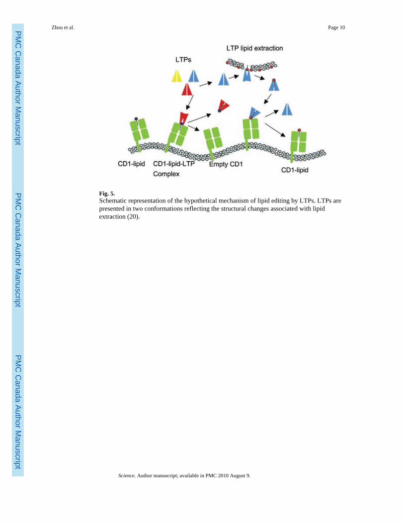
Fig. 5.Schematic representation of the hypothetical mechanism of lipid editing by LTPs. LTPs arepresented in two conformations reflecting the structural changes associated with lipidextraction (20).
Zhou et al. Page 10
Science. Author manuscript; available in PMC 2010 August 9.
PMC
Canada Author M
anuscriptPM
C C
anada Author Manuscript
PMC
Canada Author M
anuscript