Requirements for CD1d Recognition by Human Invariant V a 24 1 CD4 2 CD8 2 T Cells
12
109 J. Exp. Med. The Rockefeller University Press • 0022-1007/97/07/109/12 $2.00 Volume 186, Number 1, July 7, 1997 109–120 Requirements for CD1d Recognition by Human Invariant Va24 1 CD4 2 CD8 2 T Cells By Mark Exley,* Jorge Garcia,* Steven P. Balk,* and Steven Porcelli ‡ From the *Cancer Biology Program, Hematology/Oncology Division, Beth Israel Deaconess Medical Center and Harvard Medical School, Boston, Massachusetts 02215; and ‡ Lymphocyte Biology Section, Division of Rheumatology, Immunology, and Allergy, Brigham and Women’s Hospital and Harvard Medical School, Boston, Massachusetts 02115 Summary A subset of human CD4 2 CD8 2 T cells that expresses an invariant Va24-JaQ T cell receptor (TCR)-a chain, paired predominantly with Vb11, has been identified. A series of these Va24 Vb11 clones were shown to have TCR-b CDR3 diversity and express the natural killer (NK) locus–encoded C-type lectins NKR-P1A, CD94, and CD69. However, in contrast to NK cells, they did not express killer inhibitory receptors, CD16, CD56, or CD57. All invariant Va24 1 clones recognized the MHC class I–like CD16 molecule and discriminated between CD1d and other closely related human CD1 proteins, indicating that recognition was TCR- mediated. Recognition was not dependent upon an endosomal targeting motif in the cytoplas- mic tail of CD1d. Upon activation by anti-CD3 or CD1d, the clones produced both Th1 and Th2 cytokines. These results demonstrate that human invariant Va24 1 CD4 2 CD8 2 T cells, and presumably the homologous murine NK1 1 T cell population, are CD1d reactive and functionally distinct from NK cells. The conservation of this cell population and of the CD1d ligand across species indicates an important immunological function. T he CD1 locus encodes a family of conserved nonpoly- morphic proteins structurally related to MHC class I and II proteins (1–5). Human and murine CD1-restricted T cell lines and clones that recognize lipid antigens (6–8) or hydrophobic peptide antigens (9) have been identified, in- dicating that CD1 proteins can function as specialized anti- gen-presenting molecules. A distinct function for murine CD1d appears to be as a ligand or antigen-presenting mol- ecule recognized by a population of T cells that express the NKR-P1C (NK1) cell surface C-type lectin (10–14). NK1 is otherwise restricted to NK cells and these NK1 1 T cells have also been referred to as NK T cells or natural T cells (15, 16). Phenotypically, these cells are either CD4 1 CD8 2 or CD4 2 CD8 2 (double negative, DN 1 ), and forced ex- pression of CD8 in transgenic mice results in the deletion of this population (12, 17). Most strikingly, the majority of these cells use an invariant TCR-a chain (Va14-Ja281) that pairs preferentially with Vb8, 7 or 2 (17–20). NK1 1 T cells appear to play a role in regulating immune responses, based upon their ability to rapidly produce large amounts of IL-4 after stimulation with anti-CD3 in vivo (21–24). However, these cells also produce large amounts of IFN-g, and production of this cytokine can be specifically induced by stimulation through NK1 (25). The immunological functions of these cells and the physiologically relevant CD1d-presenting cells mediating their activation remain to be determined. Analyses of the CD1 genes in humans and other species indicate that the proteins fall into two groups, CD1a-, b-, and c-like (group 1), and CD1d-like (group 2) (3, 26). The murine CD1 locus appears unique in that it has deleted the group 1 genes, and contains only a duplicated group 2 gene (27, 28). This observation suggests that there may be im- portant functional differences between the CD1 proteins in mice and other species. Nonetheless, a human invariant TCR-a chain closely related to the murine invariant Va14- Ja281 TCR has been identified as a predominant TCR used by TCR-a/b DN T cells from multiple normal do- nors (29). This human invariant TCR-a is generated by a rearrangement between Va24 (TCRAV24) and JaQ with no N-region diversity. Subsequent studies have shown that the human invariant Va24-JaQ TCR-a chain associates preferentially with Vb11 (TCRBV11) (30–32), which is homologous to murine Vb8. This invariant TCR may also be expressed by a small proportion of CD4 1 T cells, but not CD8 1 T cells (31). These observations suggest that hu- man invariant Va24 1 T cells are homologous to murine NK1 1 T cells. 1 Abbreviations used in this paper: CHO, Chinese hamster ovary; DN, dou- ble negative; KIR, killer cell inhibitory receptor; RT, reverse tran- scriptase.
-
Upload
independent -
Category
Documents
-
view
0 -
download
0
Transcript of Requirements for CD1d Recognition by Human Invariant V a 24 1 CD4 2 CD8 2 T Cells
109
J. Exp. Med.
The Rockefeller University Press • 0022-1007/97/07/109/12 $2.00Volume 186, Number 1, July 7, 1997 109–120
Requirements for CD1d Recognition by Human Invariant V
a
24
1
CD4
2
CD8
2
T Cells
By Mark Exley,
*
Jorge Garcia,
*
Steven P. Balk,
*
and Steven Porcelli
‡
From the
*
Cancer Biology Program, Hematology/Oncology Division, Beth Israel Deaconess Medical Center and Harvard Medical School, Boston, Massachusetts 02215; and
‡
Lymphocyte Biology Section, Division of Rheumatology, Immunology, and Allergy, Brigham and Women’s Hospital and Harvard Medical School, Boston, Massachusetts 02115
Summary
A subset of human CD4
2
CD8
2
T cells that expresses an invariant V
a
24-J
a
Q T cell receptor(TCR)-
a
chain, paired predominantly with V
b
11, has been identified. A series of these V
a
24V
b
11 clones were shown to have TCR-
b
CDR3 diversity and express the natural killer (NK)locus–encoded C-type lectins NKR-P1A, CD94, and CD69. However, in contrast to NKcells, they did not express killer inhibitory receptors, CD16, CD56, or CD57. All invariantV
a
24
1
clones recognized the MHC class I–like CD16 molecule and discriminated betweenCD1d and other closely related human CD1 proteins, indicating that recognition was TCR-mediated. Recognition was not dependent upon an endosomal targeting motif in the cytoplas-mic tail of CD1d. Upon activation by anti-CD3 or CD1d, the clones produced both Th1 andTh2 cytokines. These results demonstrate that human invariant V
a
24
1
CD4
2
CD8
2
T cells,and presumably the homologous murine NK1
1
T cell population, are CD1d reactive andfunctionally distinct from NK cells. The conservation of this cell population and of the CD1dligand across species indicates an important immunological function.
T
he CD1 locus encodes a family of conserved nonpoly-morphic proteins structurally related to MHC class I
and II proteins (1–5). Human and murine CD1-restrictedT cell lines and clones that recognize lipid antigens (6–8) orhydrophobic peptide antigens (9) have been identified, in-dicating that CD1 proteins can function as specialized anti-gen-presenting molecules. A distinct function for murineCD1d appears to be as a ligand or antigen-presenting mol-ecule recognized by a population of T cells that express theNKR-P1C (NK1) cell surface C-type lectin (10–14). NK1is otherwise restricted to NK cells and these NK1
1
T cellshave also been referred to as NK T cells or natural T cells(15, 16). Phenotypically, these cells are either CD4
1
CD8
2
or CD4
2
CD8
2
(double negative, DN
1
), and forced ex-pression of CD8 in transgenic mice results in the deletionof this population (12, 17). Most strikingly, the majority ofthese cells use an invariant TCR-
a
chain (V
a
14-J
a
281)that pairs preferentially with V
b
8, 7 or 2 (17–20). NK1
1
Tcells appear to play a role in regulating immune responses,based upon their ability to rapidly produce large amountsof IL-4 after stimulation with anti-CD3 in vivo (21–24).However, these cells also produce large amounts of IFN-
g
,
and production of this cytokine can be specifically inducedby stimulation through NK1 (25). The immunologicalfunctions of these cells and the physiologically relevantCD1d-presenting cells mediating their activation remain tobe determined.
Analyses of the CD1 genes in humans and other speciesindicate that the proteins fall into two groups, CD1a-, b-,and c-like (group 1), and CD1d-like (group 2) (3, 26). Themurine CD1 locus appears unique in that it has deleted thegroup 1 genes, and contains only a duplicated group 2 gene(27, 28). This observation suggests that there may be im-portant functional differences between the CD1 proteins inmice and other species. Nonetheless, a human invariantTCR-
a
chain closely related to the murine invariant V
a
14-J
a
281 TCR has been identified as a predominant TCRused by TCR-
a
/
b
DN T cells from multiple normal do-nors (29). This human invariant TCR-
a
is generated by arearrangement between V
a
24 (TCRAV24) and J
a
Q withno N-region diversity. Subsequent studies have shown thatthe human invariant V
a
24-J
a
Q TCR-
a
chain associatespreferentially with V
b
11 (TCRBV11) (30–32), which ishomologous to murine V
b
8. This invariant TCR may alsobe expressed by a small proportion of CD4
1
T cells, butnot CD8
1
T cells (31). These observations suggest that hu-man invariant V
a
24
1
T cells are homologous to murineNK1
1
T cells.
1
Abbreviations used in this paper:
CHO, Chinese hamster ovary; DN, dou-ble negative; KIR, killer cell inhibitory receptor; RT, reverse tran-scriptase.
110
CD1d Recognition by Human T Cells
To better understand the function of these cells and theirrequirements for specific activation, a series of human in-variant V
a
24
1
V
b
11
1
DN T cell clones were establishedand characterized. TCR-
b
sequence analysis demonstratedthat these cells were derived from a polyclonal populationwith no evidence of a shared
b
chain CDR3 motif. Pheno-typically, the clones expressed high levels of NKR-P1A,the only known human homologue of rodent NK1 (33).They also expressed CD94 and CD69, two other C-typelectins closely linked to NKR-P1A in a chromosomal re-gion referred to as the NK locus. However, these cells didnot express the NK cell-associated p58 or p70 HLA class Ikiller cell inhibitory receptors (KIRs; 34, 35) or othermarkers of NK cells including CD16, CD56, or CD57.Upon stimulation, the clones secreted cytokines associatedwith both Th1 and Th2 cells, including IFN-
g
and IL-4,respectively. Of the four characterized human CD1 pro-teins, each clone specifically recognized only CD1d ex-pressed on human or hamster cell transfectants. CD1d rec-ognition was not dependent upon the specific TCR-
b
chain CDR3 sequence. Moreover, deletion of an endoso-mal targeting sequence motif in the cytoplasmic tail ofCD1d (4, 5, 36) did not effect recognition, suggesting thatrecognition by these cells was not dependent upon efficienttargeting of CD1d to a specialized endosomal compartmentinvolved in antigen processing. These results demonstratethat human invariant V
a
24
1
V
b
11
1
DN T cells are a spe-cialized population of CD1d-specific T cells. The resultsalso indicate that the similarities between this T cell popu-lation, and presumably the homologous murine NK1
1
T cellpopulation, to NK cells may be limited to the expression ofcertain closely linked NK locus–encoded C-type lectins.
Materials and Methods
Cell Lines and Clones.
T cell lines and clones were derived andphenotypic analyses performed essentially as described (32). Inbrief, DN V
a
24
1
V
b
11
1
human peripheral blood T cell lines andclones were established by sequential negative (CD4CD8) andpositive (V
a
24V
b
11) magnetic bead and FACS
sorting, respec-tively, of human peripheral blood T cells followed by stimulationwith PHA-P (reconstituted according to the supplier’s instruc-tions and used at a final dilution of 1:2,000; Difco, Detroit, MI)and IL-2 (1.5 nM; Ajinomoto, Yokohama, Japan) in the presenceof irradiated (5,000 rads) peripheral blood mononuclear cell feed-ers. Clones were established by limiting dilution. Clones and lineswere maintained by restimulation every 3–6 wk as above withphenotype monitored by FACS
. A human IL-2–dependent NKcell line, NKL, (37) was provided by Drs. M. Robertson (IndianaUniversity Medical Center, Indianapolis, IN) and J. Ritz (DanaFarber Cancer Institute, Boston, MA).
Antibodies and Phenotypic Analyses of T Cells.
The following anti-bodies were obtained from the fifth Leukocyte Workshop unlessotherwise indicated: anti-V
a
24 (C15B2) and anti-V
b
11 (C21D2)(provided by Dr. A. Lanzavecchia [Basel Institute for Immunol-ogy, Basel, Switzerland]); anti–TCR-
a
/
b
(BMA031; provided byDr. R.G. Kurrle, Boehringwerke, Marburg, Germany); anti-CD3(SPV-T3b and OKT3; provided by Dr. H. Spits [Netherlands Can-cer Center, Amsterdam, Netherlands] and American Type CultureCollection [Rockville, MD], respectively); anti-CD4 (OKT4;
American Type Culture Collection); anti-CD8
a
(OKT8; Ameri-can Type Culture Collection); anti-CD8
b
(2ST8.5H7; from Dr.E. Reinherz [Dana Farber Cancer Institute]); anti-CD16 (ABA2B1);anti-CD28 (9.3); anti-CD56 (MEM188, MOC-1, 0218); anti-CD57 (TB01, TB02); anti-CD69 (PharMingen, San Diego, CA);anti-CD94 (HP-3D9; PharMingen); anti-NKR-P1A (DX1 andDX12, provided by Dr. L. Lanier (DNAX, Palo Alto, CA); HP-3G10 and 191B8); anti-p58 KIR (NK workshop mAbs GL183,EB6, CH-L, and HP-3E4); and anti-p70 KIR (DX9; provided byDr. L. Lanier). Isotype control mAbs were P3 (IgG
1
), 4A7.6(IgG
2a
, provided by Dr. D. Olive [Institut National de la Sante etde la Recherche Medicale, Marseille, France]), and MPC11(IgG
2b
).CD1d-specific mAbs were raised from mice immunized with
CD1d–IgG fusion proteins. Further characterization of theseCD1d proteins and antibodies will be reported (Balk, S., and S.Porcelli, manuscript in preparation). CD1d mAbs were purifiedfrom low IgG serum (GIBCO BRL, Gaithersburg, MD) contain-ing tissue culture medium by protein G (Pharmacia, Piscataway,NJ) chromatography. FACS
analysis was by indirect immuno-fluorescence as described (32). Positive controls included an NKcell line as above and other cell lines and clones derived from pe-ripheral blood. TCR transcripts were amplified by reverse tran-scriptase (RT)-PCR using variable and constant region-specificprimers as described previously (29, 38). Sequences were deter-mined directly from the PCR products on an automated DNAsequencer (ABI 373A).
CD1 Transfectants.
Chinese hamster ovary (CHO) and C1Rcells were transfected with a CD1d cDNA (3) in the pSR
a
-neoexpression vector (39), followed by G418 selection and FACS
to generate lines stably expressing CD1d. C1R cells stably trans-fected with CD1a, b, and c in the pSR
a
-neo vector were de-scribed previously (38). The CD1d-CD1a chimeric protein wasgenerated using an Xba1 restriction site located at the 3
9
end ofthe
a
3 domain in CD1a and CD1d. The C1R line establishedwith this chimera construct in the pSR
a
-neo vector was uni-formly CD1d positive after G418 selection and was used in theseexperiments without further enrichment for CD1d
1
cells.
Functional Analysis of T Cells.
T cell activation for cytokineanalysis was done using 10
5
T cells/well in 96-well flat-bottomedplates coated with anti-CD3 at 10
m
g/ml for 48 h. Stimulationwith CD1 was done using equal numbers of T cells and stimulatorcells (10
5
/well) for 48 h, unless otherwise indicated. For CHOcells, a 30-s glutaraldehyde fixation (0.05% in PBS) was used tobypass the need for costimulatory molecules (40). For stimulationby CHO and C1R transfectants, PMA was included at 1 ng/ml,except where stated otherwise. PHA control stimulations werecarried out using a 2,000-fold dilution as described above. Forantibody blocking experiments, CD1d transfectants were mixedwith mAb dilutions immediately before addition of T cells.
Released cytokine levels were determined by ELISA withmatched antibody pairs in relation to cytokine standards. The an-tibodies for IFN-
g
were from Endogen, Inc. (Boston, MA) andthe others were from PharMingen. T cell proliferation was deter-mined by incorporation of [
3
H]thymidine (0.5 mCi/well) usingglutaraldehyde fixed (0.05%) or irradiated (5,000 rads) stimulatorcells where indicated. Results shown are means of triplicate sam-ples with error bars representing standard deviations.
Results
Isolation and TCR Analysis of Invariant V
a
24-J
a
Q DN TCells.
Circulating DN T cells were isolated from the pe-
111
Exley et al.
ripheral blood of two healthy donors (DN1 and DN2) byanti-CD4 and -CD8 depletion (29, 32). DN Va241 T cellswere then positively selected from these populations usingthe C15B12 mAb, specific for Va24 (41), and clones wereestablished by limiting dilution. Va241 T cell lines fromthese populations were also established by bulk stimulationwith PHA, and the majority of the cells in these lines (75–95%) were found to be Vb111 using the C21D2 mAb(31). DN and single positive T cells from a third healthydonor (DN3 and SP3, respectively) were isolated by posi-tive selection with the Va24 and Vb11 mAbs and clonedby limiting dilution. As reported previously, the majority ofthese latter clones were CD41, but only the DN clonesfrom this donor expressed JaQ (32).
The TCR structure of a series of eight DN Va241
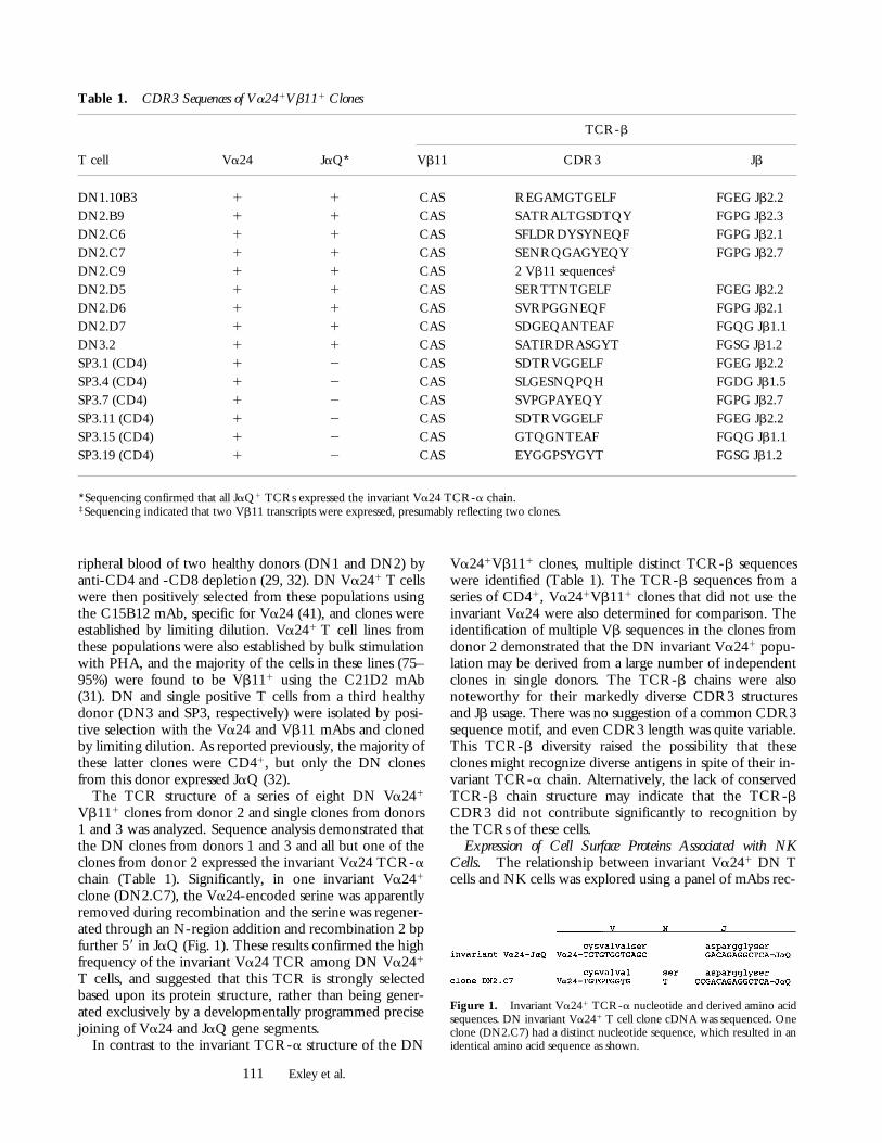
Vb111 clones from donor 2 and single clones from donors1 and 3 was analyzed. Sequence analysis demonstrated thatthe DN clones from donors 1 and 3 and all but one of theclones from donor 2 expressed the invariant Va24 TCR-achain (Table 1). Significantly, in one invariant Va241
clone (DN2.C7), the Va24-encoded serine was apparentlyremoved during recombination and the serine was regener-ated through an N-region addition and recombination 2 bpfurther 59 in JaQ (Fig. 1). These results confirmed the highfrequency of the invariant Va24 TCR among DN Va241
T cells, and suggested that this TCR is strongly selectedbased upon its protein structure, rather than being gener-ated exclusively by a developmentally programmed precisejoining of Va24 and JaQ gene segments.
In contrast to the invariant TCR-a structure of the DN
Va241Vb111 clones, multiple distinct TCR-b sequenceswere identified (Table 1). The TCR-b sequences from aseries of CD41, Va241Vb111 clones that did not use theinvariant Va24 were also determined for comparison. Theidentification of multiple Vb sequences in the clones fromdonor 2 demonstrated that the DN invariant Va241 popu-lation may be derived from a large number of independentclones in single donors. The TCR-b chains were alsonoteworthy for their markedly diverse CDR3 structuresand Jb usage. There was no suggestion of a common CDR3sequence motif, and even CDR3 length was quite variable.This TCR-b diversity raised the possibility that theseclones might recognize diverse antigens in spite of their in-variant TCR-a chain. Alternatively, the lack of conservedTCR-b chain structure may indicate that the TCR-bCDR3 did not contribute significantly to recognition bythe TCRs of these cells.
Expression of Cell Surface Proteins Associated with NKCells. The relationship between invariant Va241 DN Tcells and NK cells was explored using a panel of mAbs rec-
Table 1. CDR3 Sequences of Va241Vb111 Clones
T cell Va24 JaQ*
TCR-b
Vb11 CDR3 Jb
DN1.10B3 1 1 CAS REGAMGTGELF FGEG Jb2.2DN2.B9 1 1 CAS SATRALTGSDTQY FGPG Jb2.3DN2.C6 1 1 CAS SFLDRDYSYNEQF FGPG Jb2.1DN2.C7 1 1 CAS SENRQGAGYEQY FGPG Jb2.7DN2.C9 1 1 CAS 2 Vb11 sequences‡
DN2.D5 1 1 CAS SERTTNTGELF FGEG Jb2.2DN2.D6 1 1 CAS SVRPGGNEQF FGPG Jb2.1DN2.D7 1 1 CAS SDGEQANTEAF FGQG Jb1.1DN3.2 1 1 CAS SATIRDRASGYT FGSG Jb1.2SP3.1 (CD4) 1 2 CAS SDTRVGGELF FGEG Jb2.2SP3.4 (CD4) 1 2 CAS SLGESNQPQH FGDG Jb1.5SP3.7 (CD4) 1 2 CAS SVPGPAYEQY FGPG Jb2.7SP3.11 (CD4) 1 2 CAS SDTRVGGELF FGEG Jb2.2SP3.15 (CD4) 1 2 CAS GTQGNTEAF FGQG Jb1.1SP3.19 (CD4) 1 2 CAS EYGGPSYGYT FGSG Jb1.2
*Sequencing confirmed that all JaQ1 TCRs expressed the invariant Va24 TCR-a chain.‡Sequencing indicated that two Vb11 transcripts were expressed, presumably reflecting two clones.
Figure 1. Invariant Va241 TCR-a nucleotide and derived amino acidsequences. DN invariant Va241 T cell clone cDNA was sequenced. Oneclone (DN2.C7) had a distinct nucleotide sequence, which resulted in anidentical amino acid sequence as shown.
112 CD1d Recognition by Human T Cells
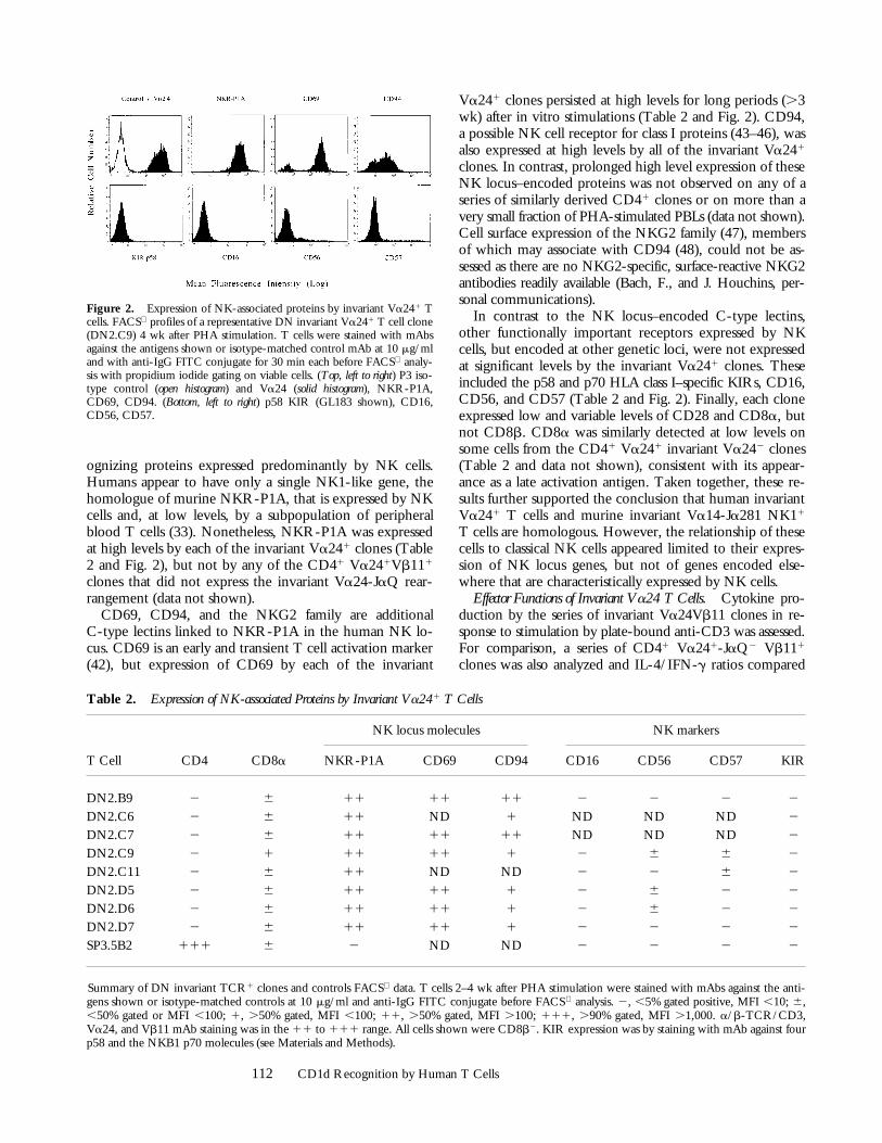
ognizing proteins expressed predominantly by NK cells.Humans appear to have only a single NK1-like gene, thehomologue of murine NKR-P1A, that is expressed by NKcells and, at low levels, by a subpopulation of peripheralblood T cells (33). Nonetheless, NKR-P1A was expressedat high levels by each of the invariant Va241 clones (Table2 and Fig. 2), but not by any of the CD41 Va241Vb111
clones that did not express the invariant Va24-JaQ rear-rangement (data not shown).
CD69, CD94, and the NKG2 family are additionalC-type lectins linked to NKR-P1A in the human NK lo-cus. CD69 is an early and transient T cell activation marker(42), but expression of CD69 by each of the invariant
Va241 clones persisted at high levels for long periods (.3wk) after in vitro stimulations (Table 2 and Fig. 2). CD94,a possible NK cell receptor for class I proteins (43–46), wasalso expressed at high levels by all of the invariant Va241
clones. In contrast, prolonged high level expression of theseNK locus–encoded proteins was not observed on any of aseries of similarly derived CD41 clones or on more than avery small fraction of PHA-stimulated PBLs (data not shown).Cell surface expression of the NKG2 family (47), membersof which may associate with CD94 (48), could not be as-sessed as there are no NKG2-specific, surface-reactive NKG2antibodies readily available (Bach, F., and J. Houchins, per-sonal communications).
In contrast to the NK locus–encoded C-type lectins,other functionally important receptors expressed by NKcells, but encoded at other genetic loci, were not expressedat significant levels by the invariant Va241 clones. Theseincluded the p58 and p70 HLA class I–specific KIRs, CD16,CD56, and CD57 (Table 2 and Fig. 2). Finally, each cloneexpressed low and variable levels of CD28 and CD8a, butnot CD8b. CD8a was similarly detected at low levels onsome cells from the CD41 Va241 invariant Va242 clones(Table 2 and data not shown), consistent with its appear-ance as a late activation antigen. Taken together, these re-sults further supported the conclusion that human invariantVa241 T cells and murine invariant Va14-Ja281 NK11
T cells are homologous. However, the relationship of thesecells to classical NK cells appeared limited to their expres-sion of NK locus genes, but not of genes encoded else-where that are characteristically expressed by NK cells.
Effector Functions of Invariant Va24 T Cells. Cytokine pro-duction by the series of invariant Va24Vb11 clones in re-sponse to stimulation by plate-bound anti-CD3 was assessed.For comparison, a series of CD41 Va241-JaQ2 Vb111
clones was also analyzed and IL-4/IFN-g ratios compared
Figure 2. Expression of NK-associated proteins by invariant Va241 Tcells. FACS profiles of a representative DN invariant Va241 T cell clone(DN2.C9) 4 wk after PHA stimulation. T cells were stained with mAbsagainst the antigens shown or isotype-matched control mAb at 10 mg/mland with anti-IgG FITC conjugate for 30 min each before FACS analy-sis with propidium iodide gating on viable cells. (Top, left to right) P3 iso-type control (open histogram) and Va24 (solid histogram), NKR-P1A,CD69, CD94. (Bottom, left to right) p58 KIR (GL183 shown), CD16,CD56, CD57.
Table 2. Expression of NK-associated Proteins by Invariant Va241 T Cells
NK locus molecules NK markers
T Cell CD4 CD8a NKR-P1A CD69 CD94 CD16 CD56 CD57 KIR
DN2.B9 2 6 11 11 11 2 2 2 2
DN2.C6 2 6 11 ND 1 ND ND ND 2
DN2.C7 2 6 11 11 11 ND ND ND 2
DN2.C9 2 1 11 11 1 2 6 6 2
DN2.C11 2 6 11 ND ND 2 2 6 2
DN2.D5 2 6 11 11 1 2 6 2 2
DN2.D6 2 6 11 11 1 2 6 2 2
DN2.D7 2 6 11 11 1 2 2 2 2
SP3.5B2 111 6 2 ND ND 2 2 2 2
Summary of DN invariant TCR1 clones and controls FACS data. T cells 2–4 wk after PHA stimulation were stained with mAbs against the anti-gens shown or isotype-matched controls at 10 mg/ml and anti-IgG FITC conjugate before FACS analysis. 2, ,5% gated positive, MFI ,10; 6,,50% gated or MFI ,100; 1, .50% gated, MFI ,100; 11, .50% gated, MFI .100; 111, .90% gated, MFI .1,000. a/b-TCR/CD3,Va24, and Vb11 mAb staining was in the 11 to 111 range. All cells shown were CD8b2. KIR expression was by staining with mAb against fourp58 and the NKB1 p70 molecules (see Materials and Methods).
113 Exley et al.
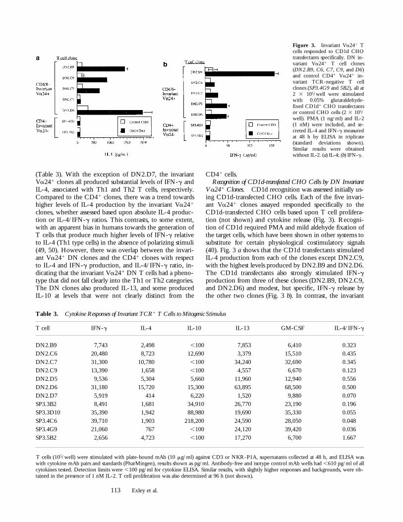
(Table 3). With the exception of DN2.D7, the invariantVa241 clones all produced substantial levels of IFN-g andIL-4, associated with Th1 and Th2 T cells, respectively.Compared to the CD41 clones, there was a trend towardshigher levels of IL-4 production by the invariant Va241
clones, whether assessed based upon absolute IL-4 produc-tion or IL-4/IFN-g ratios. This contrasts, to some extent,with an apparent bias in humans towards the generation ofT cells that produce much higher levels of IFN-g relativeto IL-4 (Th1 type cells) in the absence of polarizing stimuli(49, 50). However, there was overlap between the invari-ant Va241 DN clones and the CD41 clones with respectto IL-4 and IFN-g production, and IL-4/IFN-g ratio, in-dicating that the invariant Va241 DN T cells had a pheno-type that did not fall clearly into the Th1 or Th2 categories.The DN clones also produced IL-13, and some producedIL-10 at levels that were not clearly distinct from the
CD41 cells.Recognition of CD1d-transfected CHO Cells by DN Invariant
Va241 Clones. CD1d recognition was assessed initially us-ing CD1d-transfected CHO cells. Each of the five invari-ant Va241 clones assayed responded specifically to theCD1d-transfected CHO cells based upon T cell prolifera-tion (not shown) and cytokine release (Fig. 3). Recogni-tion of CD1d required PMA and mild aldehyde fixation ofthe target cells, which have been shown in other systems tosubstitute for certain physiological costimulatory signals(40). Fig. 3 a shows that the CD1d transfectants stimulatedIL-4 production from each of the clones except DN2.C9,with the highest levels produced by DN2.B9 and DN2.D6.The CD1d transfectants also strongly stimulated IFN-gproduction from three of these clones (DN2.B9, DN2.C9,and DN2.D6) and modest, but specific, IFN-g release bythe other two clones (Fig. 3 b). In contrast, the invariant
Figure 3. Invariant Va241 Tcells responded to CD1d CHOtransfectants specifically. DN in-variant Va241 T cell clones(DN2.B9, C6, C7, C9, and D6)and control CD41 Va241 in-variant TCR-negative T cellclones (SP3.4G9 and 5B2), all at2 3 105/well were stimulatedwith 0.05% glutaraldehyde-fixed CD1d1 CHO transfectantsor control CHO cells (2 3 105/well). PMA (1 ng/ml) and IL-2(1 nM) were included, and se-creted IL-4 and IFN-g measuredat 48 h by ELISA in triplicate(standard deviations shown).Similar results were obtainedwithout IL-2. (a) IL-4; (b) IFN-g.
Table 3. Cytokine Responses of Invariant TCR1 T Cells to Mitogenic Stimulus
T cell IFN-g IL-4 IL-10 IL-13 GM-CSF IL-4/IFN-g
DN2.B9 7,743 2,498 ,100 7,853 6,410 0.323DN2.C6 20,480 8,723 12,690 3,379 15,510 0.435DN2.C7 31,300 10,780 ,100 34,240 32,690 0.345DN2.C9 13,390 1,658 ,100 4,557 6,670 0.123DN2.D5 9,536 5,304 5,660 11,960 12,940 0.556DN2.D6 31,180 15,720 15,300 63,895 68,500 0.500DN2.D7 5,919 414 6,220 1,520 9,880 0.070SP3.3B2 8,491 1,681 34,910 26,770 23,190 0.196SP3.3D10 35,390 1,942 88,980 19,690 35,330 0.055SP3.4C6 39,710 1,903 218,200 24,590 28,050 0.048SP3.4G9 21,060 767 ,100 24,120 39,420 0.036SP3.5B2 2,656 4,723 ,100 17,270 6,700 1.667
T cells (105/well) were stimulated with plate-bound mAb (10 mg/ml) against CD3 or NKR-P1A, supernatants collected at 48 h, and ELISA waswith cytokine mAb pairs and standards (PharMingen), results shown as pg/ml. Antibody-free and isotype control mAb wells had ,610 pg/ml of allcytokines tested. Detection limits were ,100 pg/ml for cytokine ELISA. Similar results, with slightly higher responses and backgrounds, were ob-tained in the presence of 1 nM IL-2. T cell proliferation was also determined at 96 h (not shown).
114 CD1d Recognition by Human T Cells
Va242 clones did not respond specifically to the CD1dtransfectants (Fig. 3, a and b, and data not shown). The fail-ure of the DN2.C9 clone to produce significant IL-4 in re-sponse to the CD1d transfectant was consistent with therelatively low ratio of IL-4/IFN-g produced by this clonein response to anti-CD3 stimulation (Table 3). Indeed, therelative levels of IL-4 and IFN-g release in response toCD1d from each of the clones were comparable to thoseobserved with anti-CD3 (Table 3).
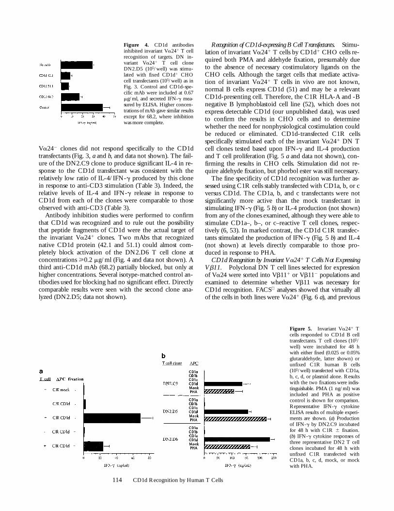
Antibody inhibition studies were performed to confirmthat CD1d was recognized and to rule out the possibilitythat peptide fragments of CD1d were the actual target ofthe invariant Va241 clones. Two mAbs that recognizednative CD1d protein (42.1 and 51.1) could almost com-pletely block activation of the DN2.D6 T cell clone atconcentrations >0.2 mg/ml (Fig. 4 and data not shown). Athird anti-CD1d mAb (68.2) partially blocked, but only athigher concentrations. Several isotype-matched control an-tibodies used for blocking had no significant effect. Directlycomparable results were seen with the second clone ana-lyzed (DN2.D5; data not shown).
Recognition of CD1d-expressing B Cell Transfectants. Stimu-lation of invariant Va241 T cells by CD1d1 CHO cells re-quired both PMA and aldehyde fixation, presumably dueto the absence of necessary costimulatory ligands on theCHO cells. Although the target cells that mediate activa-tion of invariant Va241 T cells in vivo are not known,normal B cells express CD1d (51) and may be a relevantCD1d-presenting cell. Therefore, the C1R HLA-A and -Bnegative B lymphoblastoid cell line (52), which does notexpress detectable CD1d (our unpublished data), was usedto confirm the results in CHO cells and to determinewhether the need for nonphysiological costimulation couldbe reduced or eliminated. CD1d-transfected C1R cellsspecifically stimulated each of the invariant Va241 DN Tcell clones tested based upon IFN-g and IL-4 productionand T cell proliferation (Fig. 5 a and data not shown), con-firming the results in CHO cells. Stimulation did not re-quire aldehyde fixation, but phorbol ester was still necessary.
The fine specificity of CD1d recognition was further as-sessed using C1R cells stably transfected with CD1a, b, or cversus CD1d. The CD1a, b, and c transfectants were notsignificantly more active than the mock transfectant instimulating IFN-g (Fig. 5 b) or IL-4 production (not shown)from any of the clones examined, although they were able tostimulate CD1a–, b–, or c–reactive T cell clones, respec-tively (6, 53). In marked contrast, the CD1d C1R transfec-tants stimulated the production of IFN-g (Fig. 5 b) and IL-4(not shown) at levels directly comparable to those pro-duced in response to PHA.
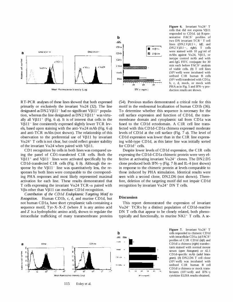
CD1d Recognition by Invariant Va241 T Cells Not ExpressingVb11. Polyclonal DN T cell lines selected for expressionof Va24 were sorted into Vb111 or Vb112 populations andexamined to determine whether Vb11 was necessary forCD1d recognition. FACS analyses showed that virtually allof the cells in both lines were Va241 (Fig. 6 a), and previous
Figure 5. Invariant Va241 Tcells responded to CD1d B celltransfectants. T cell clones (105/well) were incubated for 48 hwith either fixed (0.025 or 0.05%glutaraldehyde, latter shown) orunfixed C1R human B cells(105/well) transfected with CD1a,b, c, d, or plasmid alone. Resultswith the two fixations were indis-tinguishable. PMA (1 ng/ml) wasincluded and PHA as positivecontrol is shown for comparison.Representative IFN-g cytokineELISA results of multiple experi-ments are shown. (a) Productionof IFN-g by DN2.C9 incubatedfor 48 h with C1R 6 fixation.(b) IFN-g cytokine responses ofthree representative DN2 T cellclones incubated for 48 h withunfixed C1R transfected withCD1a, b, c, d, mock, or mockwith PHA.
Figure 4. CD1d antibodiesinhibited invariant Va241 T cellrecognition of targets. DN in-variant Va241 T cell cloneDN2.D5 (105/well) was stimu-lated with fixed CD1d1 CHOcell transfectants (105/well) as inFig. 3. Control and CD1d-spe-cific mAb were included at 0.67mg/ml, and secreted IFN-g mea-sured by ELISA. Higher concen-trations of mAb gave similar resultsexcept for 68.2, where inhibitionwas more complete.
115 Exley et al.
RT-PCR analyses of these lines showed that both expressedprimarily or exclusively the invariant Va24 (32). The linedesignated as DN2.Vb112 had no significant Vb111 popula-tion, whereas the line designated as DN2.Vb111 was virtu-ally all Vb111 (Fig. 6 a). It is of interest that cells in theVb112 line consistently expressed slightly lower TCR lev-els, based upon staining with the anti-Va24 mAb (Fig. 6 a)and anti-TCR mAbs (not shown). The relationship of thisobservation to the preferential use of Vb11 by invariantVa241 T cells is not clear, but could reflect greater stabilityof the invariant Va24 when paired with Vb11.
CD1 recognition by cells in both lines was compared us-ing the panel of CD1-transfected C1R cells. Both theVb111 and Vb112 lines were activated specifically by theCD1d-transfected C1R cells (Fig. 6 b). Although the re-sponse by the Vb112 line was quantitatively less, the re-sponses by both lines were comparable to the correspond-ing PHA responses and most likely represented maximalactivation for each line. These results demonstrated thatT cells expressing the invariant Va24 TCR-a paired withVbs other than Vb11 can mediate CD1d recognition.
Contribution of the CD1d Endoplasmic Targeting Motif toRecognition. Human CD1b, c, d, and murine CD1d, butnot human CD1a, have short cytoplasmic tails containing asequence motif, Tyr-X-X-Z (where X is any amino acidand Z is a hydrophobic amino acid), shown to regulate theintracellular trafficking of many transmembrane proteins
(54). Previous studies demonstrated a critical role for thismotif in the endosomal localization of human CD1b (36).To determine whether this sequence is necessary for thecell surface expression and function of CD1d, the trans-membrane domain and cytoplasmic tail from CD1a wasfused to the CD1d ectodomain. A C1R cell line trans-fected with this CD1d-CD1a chimera expressed moderatelevels of CD1d at the cell surface (Fig. 7 a). The level ofCD1d expression was lower than in the C1R line express-ing wild-type CD1d, as this latter line was initially sortedfor CD1d1 cells.
Despite lower levels of CD1d expression, the C1R cellsexpressing the CD1d-CD1a chimeric protein were very ef-fective at activating invariant Va241 clones. The DN2.D5clone produced both IFN-g (Fig. 7 b) and IL-4 (not shown)in response to the chimeric protein at levels comparable tothose induced by PHA stimulation. Identical results wereseen with a second clone, DN2.D6 (not shown). There-fore, deletion of the targeting motif did not impair CD1drecognition by invariant Va241 DN T cells.
Discussion
This report demonstrated the expression of invariantVa241 TCRs by a distinct population of CD1d-reactiveDN T cells that appear to be closely related, both pheno-typically and functionally, to murine NK11 T cells. A se-
Figure 6. Invariant Va241 Tcells that did not express Vb11responded to CD1d. (a) Repre-sentative FACS profiles oftwo DN invariant TCR1 T celllines (DN2.Vb111, left; andDN2.Vb112, right). T cellswere stained with 10 mg/ml ofmAbs against Va24, Vb11, orisotype control mAb and withanti-IgG FITC conjugate for 30min each before FACS analysisof viable cells. (b) T cell lines(105/well) were incubated withunfixed C1R human B cells(105/well) transfected with CD1a,b, c, d, mock, or mock withPHA as in Fig. 5 and IFN-g pro-duction results are shown.
Figure 7. Invariant Va241 Tcells responded to chimeric CD1dwith intracellular CD1a. (a) FACS
profiles of C1R CD1d (left) andCD1d/a chimera (right) transfec-tants stained with normal mouseserum (open histogram) or 42.1CD1d-specific mAb (solid histo-gram). (b) DN2.D6 T cell clone(105/well) was incubated withunfixed C1R human B cellCD1d/a chimera or mock trans-fectants (105/well) and IFN-gcytokine ELISA results obtained.
116 CD1d Recognition by Human T Cells
ries of invariant Va241Vb111 DN T cell clones wereshown to recognize CD1d expressed either by a hamster(CHO) or human B cell (C1R) line. The fine specificity ofthis recognition was demonstrated by the failure of theseclones to recognize CD1a, b, or c transfectants. Moreover,antibody blocking studies indicated that intact CD1d,rather than peptides derived from this protein, were recog-nized by these clones. These results provided strong evi-dence that CD1d recognition by these clones was mediateddirectly by their TCRs.
The Vb11 chains from these clones were sequenced todetermine whether this population was monoclonal orpolyclonal and whether there were structural constraints onthe CDR3 region. This analysis revealed a unique Vb11chain with extensive N-region diversity in each of theclones isolated from a single donor, demonstrating thatmultiple independently derived clones contributed to thispopulation. Sequence analysis of the Va24-JaQ chains alsorevealed a clone with distinct codon usage in the V-J junc-tion, indicative of N-region addition. These observationssuggest that the TCRs used by these clones are generatedthrough V-(D)-J recombination mechanisms and subse-quent positive selection as with conventional T cells.
The Vb11 sequence analysis also revealed the lack of ap-parent structural constraints on the CDR3 region, sincethere was marked variability in CDR3 length, Jb usage,and sequence. There was no suggestion of a sequence motifthat distinguished the Vb11 CDR3 regions associated withthe invariant Va24 chain from those associated with othernoninvariant Va24 chains. This pattern of CDR3 indepen-dent recognition by one or several Vb chains could beconsistent with selection or expansion by a superantigen(possibly CD1d associated). However, conventional super-antigen recognition is not dependent upon a Va chainCDR3. It also appeared unlikely that these heterogeneousVb11 chains mediated recognition of distinct CD1d-pre-sented antigens since each clone recognized CD1d ex-pressed by both hamster and human cells without the de-liberate addition of an antigen. Therefore, the current datasuggest that Vb11 may be structurally favored as an invari-ant Va24 partner and/or mediate direct contacts withCD1d. However, pairing of the invariant Va24 with Vb11was not an absolute requirement for CD1d recognitionsince analysis of an invariant Va241Vb112 DN T cell linedemonstrated that the invariant Va24 can pair with otherVbs to generate CD1d-reactive TCRs.
Although CD1d recognition by multiple clones wasdemonstrated without the deliberate addition of an antigen,this did not rule out CD1d presentation of one or a smallnumber of conserved ubiquitous endogenous or serum-derived antigens. CD1 has been shown to present lipid an-tigens (6–8, 53) and hydrophobic peptide antigens (9).Consistent with these observations, the crystal structure ofmurine CD1d reveals a potential deep hydrophobic anti-gen-binding cavity (55). The pathway(s) through whichCD1 may acquire such hydrophobic or other antigens arenot clear, but appear distinct from the MHC class I pathway(6, 56–58). Significantly, the cytoplasmic tails of human
CD1b, c, and d, and murine CD1d contain a short tyrosine-based signal that has been implicated in trafficking from theplasma membrane to endosomal compartments (54), and arecent immunogold electron microscopy study demon-strated that a large fraction of CD1b molecules were lo-cated intracellularly in an endosomal compartment (36).Based upon these observations, one hypothesis has beenthat CD1 proteins are targeted to an acidic endosomalcompartment and are there loaded with antigen.
To address this hypothesis in the case of CD1d, the cyto-plasmic tail of CD1d was replaced with the cytoplasmic tailof CD1a, which lacks recognizable targeting motifs. CD1aproteins appear to traffic to the cell surface through the de-fault secretory pathway and do not enter an endosomalcompartment (Sugita, M., M. Brenner, and S. Porcelli, un-published data). The chimeric protein was recognized byinvariant Va241 DN T cell clones despite the loss of theendosomal targeting signal, indicating that endosomal traf-ficking mediated by this signal was not necessary for CD1drecognition by this cell population. Taken together, thedata in this report indicate that T cell recognition of CD1dmediated by the invariant Va24 TCR may not involve aspecific antigen, although presentation of a conserved cel-lular antigen acquired through a pathway that is indepen-dent of the endosomal targeting motif cannot be excluded.Moreover, it remains possible that CD1d presents specificforeign antigens in vivo and that the in vitro responses re-flect relatively low affinity interactions mediated by CD1dbinding to diverse nonspecific antigens.
In addition to specific antigen recognition by the TCR,the activation of conventional T cells is dependent uponthe recruitment of p56lck to the TCR complex by the CD4or CD8 accessory proteins. The invariant Va241 DNclones express p56lck (data not shown) and it is very likelythat there are other accessory proteins that couple it to theTCR in these cells. The consistent high level expression ofNKR-P1A by human invariant Va241 DN T cell clones,the expression of NK1 (NKR-P1C) by the homologousmurine cell population, and the presence of a p56lck bind-ing motif in the cytoplasmic tails of the murine NKR-P1proteins make these clear candidate accessory proteins (59,60). However, the presence of invariant Va1 T cells inmouse strains that do not express NK1 or other NKR-P1proteins argues against such a critical role for NKR-P1. Itshould also be noted that the human NKR-P1A proteindoes not contain the putative p56lck binding site (33) and,in preliminary biochemical studies, we have been unable todemonstrate an association between human NKR-P1A andp56lck (Exley, M., unpublished data).
The human invariant Va241 T cell clones also expressedtwo other C-type lectins, CD69 and CD94, encoded in achromosomal region that has been termed the NK locus.CD94 may heterodimerize with NKG2 proteins (48), an-other family of C-type lectins encoded in the NK locus(47), and this complex may be an MHC class I receptor(44, 48). CD69 is an early and transient T cell activationantigen (42), but its expression by invariant Va241 DN Tcell clones was persistent. This suggests that invariant
117 Exley et al.
Va241 DN T cells may remain in an activated state longerthan conventional T cells. Alternatively, given the highlevel of expression of other NK locus–encoded proteinsobserved on invariant Va241 T cells, CD69 expressionmay reflect constitutive transcriptional activity of the NKlocus in these cells which is not directly related to T cell ac-tivation.
Although the expression of NKR-P1 and CD94 at highlevels suggests some relationship to NK cells, the invariantVa241 DN T cells did not express a number of other mol-ecules that play roles in NK cell function, such as CD16,CD56, and CD57. In particular, the invariant Va241 DNT cells did not express p58 or p70 KIRs, although expres-sion of family members not recognized by the multiple an-tibodies used here remains possible. Consistent with themAb data, preliminary RT-PCR amplification experi-ments with consensus KIR primers have similarly failed todetect KIR expression (data not shown). This is in contrastto recent data showing that KIRs may be expressed inother T cell subpopulations (61–64). These observationsindicate that the link between invariant Va241 DN T cellsand NK cells may be transcriptional activation of the NKlocus, with limited functional overlap between these cellpopulations.
Cytokine production by invariant Va241 DN T cellswas also analyzed and the results supported conclusionsreached in the mouse that these cells can produce signifi-cant levels of IL-4 in response to activation (20–23). How-ever, they also produced other cytokines, particularly IFN-g,at substantial levels, and their regulatory functions in vivo
are probably complex. Murine NK11 T cells are responsi-ble for the acute production of IL-4 in response to anti-CD3 in vivo (23), but this type of stimulus is clearly non-physiological. It is unlikely that the function of this cellpopulation is to determine systemic levels of IL-4 or othercytokines, and more likely that the IL-4 produced in re-sponse to anti-CD3 stimulation reflects the primed activa-tion state of these cells in vivo. A reasonable alternative hy-pothesis is that invariant Va241 T cells function throughcell–cell interactions to provide individual CD1d1 targetcells with IL-4, IFN-g, or other cytokines that in turn di-rect the further proliferation and/or differentiation of thesetarget cells.
B cells express CD1d (51) and represent one possible tar-get cell for invariant Va241 T cells. However, CD1d mayalso be widely expressed (51, 65) and invariant Va241 Tcells may, therefore, have functionally important interac-tions with a number of different cell types. In particular,the large fraction of T cells in murine bone marrow andliver that are NK11 (66, 67) presumably interact locallywith CD1d1 cells and may play roles in regulating B cellmaturation, myeloid development, or hepatic immunefunction. Finally, recent reports indicate that loss of invari-ant Va241 T cells in humans or of the homologous NK11
T cell population in mice is associated with disease progres-sion in several autoimmune diseases (68–70). Although theprecise functions of invariant Va241 T cells and their mu-rine homologues remain to be clarified, the conservation ofthis cell population and of the CD1d ligand across speciessuggests an important immunological function.
For antibodies and cell reagents we wish to thank Drs. L. Lanier, A. Lanzavecchia, M. Robertson, J. Ritz, H.Spits, E. Reinherz, D. Olive, and R.G. Kurrle. We also thank other members of the Lymphocyte BiologySection and Geoffrey Sunshine for advice, and Alexis Fertig for technical assistance.
This work was supported by National Institutes of Health grant R01-AI33911 to S.P. Balk and National In-stitutes of Health grant R01 AI40135 to S. Porcelli. S. Porcelli was also supported by an Investigator Awardfrom the Arthritis Foundation.
Address correspondence to Steven Porcelli, Lymphocyte Biology Section, Division of Rheumatology, Im-munology, and Allergy, Brigham and Women’s Hospital and Harvard Medical School, 250 Longwood Ave.,Boston, MA 02115; Phone: 617-432-4984; FAX: 617-667-0610; or Steven P. Balk, Hematology/OncologyDivision, Beth Israel Deaconess Medical Center, 330 Brookline Ave., Boston, MA 02215. Phone: 617-667-0600; FAX: 617-667-0610.
Received for publication 7 March 1997 and in revised form 21 April 1997.
References1. Martin, L.H., F. Calabi, F.A. Lefebvre, C.A. Bilsland, and C.
Milstein. 1987. Structure and expression of the human thy-mocyte antigens CD1a, CD1b, and CD1c. Proc. Natl. Acad.Sci. USA. 84:9189–9193.
2. Aruffo, A., and B. Seed. 1989. Expression of cDNA clonesencoding the thymocyte antigens CD1a, b, c demonstrates ahierarchy of exclusion in fibroblasts. J. Immunol. 143:1723–1730.
3. Balk, S.P., P.A. Bleicher, and C. Terhorst. 1989. Isolationand characterization of a cDNA and gene coding for a fourthCD1 molecule. Proc. Natl. Acad. Sci. USA. 86:252–256.
4. Blumberg, R.S., D. Gerdes, A. Chott, S.A. Porcelli, and S.P.Balk. 1995. Structure and function of the CD1 family ofMHC-like cell surface proteins. Immunol. Rev. 147:5–29.
5. Porcelli, S.A. 1995. The CD1 family: a third lineage of anti-gen-presenting molecules. Adv. Immunol. 59:1–98.
118 CD1d Recognition by Human T Cells
6. Porcelli, S., C.T. Morita, and M.B. Brenner. 1992. CD1b re-stricts the response of human CD4282 T lymphocytes to amicrobial antigen. Nature (Lond.). 360:593–597.
7. Beckman, E.M., S.A. Porcelli, C.T. Morita, S.M. Behar, S.T.Furlong, and M.B. Brenner. 1994. Recognition of a lipid an-tigen by CD1-restricted ab1 T cells. Nature (Lond.). 372:691–694.
8. Sieling, P.A., D. Chatterjee, S.A. Porcelli, T.I. Prigozy, R.J.Mazzaccaro, T. Soriano, B.R. Bloom, M.B. Brenner, M.Kronenberg, P.J. Brennan, et al. 1995. CD1-restricted T cellrecognition of microbial lipoglycan antigens. Science (Wash.DC). 269:227–230.
9. Castano, A.R., S. Tangri, J.E. Miller, H.R. Holcombe, M.R.Jackson, W.D. Huse, M. Kronenberg, and P.A. Peterson.1995. Peptide binding and presentation by mouse CD1. Sci-ence (Wash. DC). 269:223–226.
10. Coles, M.C., and D.H. Raulet. 1994. Class I dependence ofthe development of CD41 CD82 NK1.11 thymocytes. J.Exp. Med. 180:395–399.
11. Ohteki, T., and H.R. MacDonald. 1994. Major histocom-patibility complex class I related molecules control the devel-opment of CD4182 and CD4282 subsets of natural killer1.11 T cell receptor-a/b1 cells in the liver of mice. J. Exp.Med. 180:699–704.
12. Bendelac, A., N. Killeen, D.R. Littman, and R.H. Schwartz.1994. A subset of CD41 thymocytes selected by MHC class Imolecules. Science (Wash. DC). 263:1774–1778.
13. Adachi, Y., H. Koseki, M. Zijlstra, and M. Taniguchi. 1995.Positive selection of invariant Va141 T cells by non-majorhistocompatibility complex–encoded class I–like moleculesexpressed on bone marrow–derived cells. Proc. Natl. Acad.Sci. USA. 92:1200–1204.
14. Bendelac, A., O. Lantz, M.E. Quimby, J.W. Yewdell, J.R.Bennink, and R.R. Brutkiewicz. 1995. CD1 recognition bymouse NK11 T lymphocytes. Science (Wash. DC). 268:863–865.
15. MacDonald, H.R. 1995. NK1.11 T cell receptor-a/b1 cells:new clues to their origin, specificity, and function. J. Exp.Med. 182:633–638.
16. Bix, M., and R.M. Locksley. 1995. Natural T cells. Cells thatco-express NKRP-1 and TCR. J. Immunol. 155:1020–1022.
17. Lantz, O., and A. Bendelac. 1994. An invariant T cell recep-tor a chain is used by a unique subset of major histocompati-bility complex class I–specific CD41 and CD4282 T cells inmice and humans. J. Exp. Med. 180:1097–1106.
18. Koseki, H., H. Asano, T. Inaba, N. Miyashita, K. Moriwaki,K.F. Lindahl, Y. Mizutani, K. Imai, and M. Taniguchi. 1991.Dominant expression of a distinctive V141 T-cell antigen re-ceptor a chain in mice. Proc. Natl. Acad. Sci. USA. 88:7518–7522.
19. Arase, H., N. Arase, K. Ogasawara, R.A. Good, and K.Onoe. 1992. An NK1.11 CD4182 single-positive thymocytesubpopulation that expresses a highly skewed T-cell antigenreceptor V b family. Proc. Natl. Acad. Sci. USA. 89:6506–6510.
20. Hayakawa, K., B.T. Lin, and R.R. Hardy. 1992. Murinethymic CD41 T cell subsets: a subset (Thy0) that secretes di-verse cytokines and overexpresses the Vb8 T cell receptorgene family. J. Exp. Med. 176:269–274.
21. Zlotnik, A., D.I. Godfrey, M. Fischer, and T. Suda. 1992.Cytokine production by mature and immature CD42CD82
T cells. ab–T cell receptor1 CD42CD82 T cells produce IL-4.J. Immunol. 149:1211–1215.
22. Arase, H., N. Arase, K. Nakagawa, R.A. Good, and K.Onoe. 1993. NK1.11 CD41 CD82 thymocytes with specificlymphokine secretion. Eur. J. Immunol. 23:307–310.
23. Yoshimoto, T., and W.E. Paul. 1994. CD4pos, NK1.1pos Tcells promptly produce interleukin 4 in response to in vivochallenge with anti-CD3. J. Exp. Med. 179:1285–1295.
24. Yoshimoto, T., A. Bendelac, C. Watson, J. Hu-Li, and W.E.Paul. 1995. Role of NK1.11 T cells in a TH2 response and inimmunoglobulin E production. Science (Wash. DC). 270:1845–1847.
25. Arase, H., N. Arase, and T. Saito. 1996. Interferon g produc-tion by natural killer (NK) cells and NK1.11 T cells uponNKR-P1 cross-linking. J. Exp. Med. 183:2391–2396.
26. Calabi, F., J.M. Jarvis, L. Martin, and C. Milstein. 1989. Twoclasses of CD1 genes. Eur. J. Immunol. 19:285–292.
27. Bradbury, A., K.T. Belt, T.M. Neri, C. Milstein, and F. Cal-abi. 1988. Mouse CD1 is distinct from and co-exists with TLin the same thymus. EMBO (Eur. Mol. Biol. Organ.) J. 7:3081–3086.
28. Balk, S.P., P.A. Bleicher, and C. Terhorst. 1991. Isolationand expression of cDNA encoding the murine homologuesof CD1. J. Immunol. 146:768–774.
29. Porcelli, S., C.E. Yockey, M.B. Brenner, and S.P. Balk.1993. Analysis of T cell antigen receptor (TCR) expressionby human peripheral blood CD4282 a/b T cells demon-strates preferential use of several V b genes and an invariantTCR b chain. J. Exp. Med. 178:1–16.
30. Dellabona, P., G. Casorati, B. Friedli, L. Angman, F. Sallusto,A. Tunnacliffe, E. Roosneek, and A. Lanzavecchia. 1993. Invivo persistence of expanded clones specific for bacterial anti-gens within the human T cell receptor a/b CD4282 subset.J. Exp. Med. 177:1763–1771.
31. Dellabona, P., E. Padovan, G. Casorati, M. Brockhaus, andA. Lanzavecchia. 1994. An invariant Va24-JaQ/Vb11 T cellreceptor is expressed in all individuals by clonally expandedCD4282 T cells. J. Exp. Med. 180:1171–1176.
32. Porcelli, S., D. Gerdes, A. Fertig, and S.P. Balk. 1996. Hu-man T cells expressing an invariant Va24-JaQ TCRa areCD42 and heterogeneous with respect to TCRb expression.Hum. Immunol. 48:63–67.
33. Lanier, L.L., C. Chang, and J.H. Phillips. 1994. HumanNKR-P1A. A disulfide-linked homodimer of the C-typelectin superfamily expressed by a subset of NK and T lym-phocytes. J. Immunol. 153:2417–2428.
34. Raulet, D.H. and W. Held. 1995. Natural killer cell recep-tors: the offs and ons of NK cell recognition. Cell. 82:697–700.
35. Lanier, L.L., and J.H. Phillips. 1996. Inhibitory MHC class Ireceptors on NK cells and T cells. Immunol. Today. 17:86–91.
36. Sugita, M., R.M. Jackman, E. van Donselaar, S.M. Behar,R.A. Rogers, P.J. Peters, M.B. Brenner, and S.A. Porcelli.1996. Cytoplasmic tail–dependent localization of CD1b anti-gen-presenting molecules to MICs. Science (Wash. DC). 273:349–352.
37. Robertson, M.J., K.J. Cochran, C. Cameron, J.M. Le, R.Tantravahi, and J. Ritz. 1996. Characterization of a cell line,NKL, derived from an aggressive human natural killer cellleukemia. Exp. Hematol. 24:406–415.
38. Balk, S.P., E.C. Ebert, R.L. Blumenthal, F.V. McDermott,K.W. Wucherpfennig, S.B. Landau, and R.S. Blumberg.1991. Oligoclonal expansion and CD1 recognition by humanintestinal intraepithelial lymphocytes. Science (Wash. DC).253:1411–1415.
119 Exley et al.
39. Takebe, Y., M. Seiki, J. Fujisawa, P. Hoy, K. Yokota, K.Arai, M. Yoshida, and N. Arai. 1988. SRa promoter: an effi-cient and versatile mammalian cDNA expression system com-posed of the simian virus 40 early promoter and the R-U5 seg-ment of human T-cell leukemia virus type 1 long terminalrepeat. Mol. Cell. Biol. 8:466–472.
40. Rhodes, J., H. Chen, S.R. Hall, J.E. Beesley, D.C. Jenkins,P. Collins, and B. Zheng. 1995. Therapeutic potentiation ofthe immune system by costimulatory Schiff–base-formingdrugs. Nature (Lond.). 377:71–75.
41. Padovan, E., G. Casorati, P. Dellabona, S. Meyer, M. Brock-haus, and A. Lanzavecchia. 1993. Expression of two T cellreceptor a chains: dual receptor T cells. Science (Wash. DC).262:422–424.
42. Hamann, J., H. Fiebig, and M. Strauss. 1993. Expressioncloning of the early activation antigen CD69, a type II inte-gral membrane protein with a C-type lectin domain. J. Immu-nol. 150:4920–4927.
43. Aramburu, J., M.A. Balboa, A. Ramirez, A. Silva, A. Ace-vedo, F. Sanchez-Madrid, M.O. De Landazuri, and M. Lo-pez-Botet. 1990. A novel functional cell surface dimer(Kp43) expressed by natural killer cells and T cell receptor-g/d1 T lymphocytes. I. Inhibition of the IL-2–dependentproliferation by anti-Kp43 monoclonal antibody. J. Immunol.144:3238–3247.
44. Moretta, A., M. Vitale, S. Sivori, C. Bottino, L. Morelli, R.Augugliaro, M. Barbaresi, D. Pende, E. Ciccone, and M. Lo-pez-Botet. 1994. Human natural killer cell receptors forHLA–class I molecules. Evidence that the Kp43 (CD94)molecule functions as receptor for HLA-B alleles. J. Exp.Med. 180:545–555.
45. Chang, C., A. Rodriguez, M. Carretero, M. Lopez-Botet,J.H. Phillips, and L.L. Lanier. 1995. Molecular characteriza-tion of human CD94: a type II membrane glycoprotein re-lated to the C-type lectin superfamily. Eur. J. Immunol. 25:2433–2437.
46. Phillips, J.H., C. Chang, J. Mattson, J.E. Gumperz, P. Par-ham, and L.L. Lanier. 1996. CD94 and a novel associatedprotein (94AP) form a NK cell receptor involved in the rec-ognition of HLA-A, HLA-B, and HLA-C allotypes. Immu-nity. 5:163–172.
47. Houchins, J.P., T. Yabe, C. McSherry, and F.H. Bach. 1991.DNA sequence analysis of NKG2, a family of related cDNAclones encoding type II integral membrane proteins on hu-man natural killer cells. J. Exp. Med. 173:1017–1020.
48. Lazetic, S., C. Chang, J.P. Houchins, L.L. Lanier, and J.H.Phillips. 1996. Human natural killer cell receptors involved inMHC class I recognition are disulfide-linked heterodomers ofCD94 and NKG2 subunits. J. Immunol. 157:4741–4745.
49. Webb, L.M., and M. Feldmann. 1995. Critical role ofCD28/B7 costimulation in the development of human Th2cytokine-producing cells. Blood. 86:3479–3486.
50. Brinkmann, V., B. Kinzel, and C. Kristofic. 1996. TCR-independent activation of human CD41 45RO2 T cells byanti-CD28 plus IL-2: induction of clonal expansion andpriming for a Th2 phenotype. J. Immunol. 156:4100–4106.
51. Blumberg, R.S., C. Terhorst, P. Bleicher, F.V. McDermott,C.H. Allan, S.B. Landau, J.S. Trier, and S.P. Balk. 1991. Ex-pression of a nonpolymorphic MHC class I–like molecule,CD1D, by human intestinal epithelial cells. J. Immunol. 147:2518–2524.
52. Zemmour, J., A.M. Little, D.J. Schendel, and P. Parham.1992. The HLA-A,B “negative” mutant cell line C1R ex-
presses a novel HLA-B35 allele, which also has a point muta-tion in the translation initiation codon. J. Immunol. 148:1941–1948.
53. Beckman, E.M., A. Melian, S.M. Behar, P.A. Sieling, D.Chatterjee, S.T. Furlong, R. Matsumoto, J.P. Rosat, R.L.Modlin, and S.A. Porcelli. 1996. CD1c restricts responses ofmycobacteria-specific T cells. Evidence for antigen presenta-tion by a second member of the human CD1 family. J. Immu-nol. 157:2795–2803.
54. Sandoval, I.V., and O. Bakke. 1994. Targeting of membraneproteins to endosomes and lysosomes. Trends Cell Biol. 4:292–297.
55. Zeng, Z.H., A.R. Castano, B. Segelke, E.A. Stura, P.A.Peterson, and I.A. Wilson. 1997. The crystal structure of mu-rine CD1: an MHC-like fold but with a large hydrophobicantigen binding groove. Science (Wash. DC). In press.
56. Hanau, D., D. Fricker, T. Bieber, M.E. Esposito-Farese, H.Bausinger, J.P. Cazenave, L. Donato, M.M. Tongio, and H.de la Salle. 1994. CD1 expression is not affected by humanpeptide transporter deficiency. Hum. Immunol. 41:61–68.
57. Brutkiewicz, R.R., J.R. Bennink, J.W. Yewdell, and A.Bendelac. 1995. TAP-independent, b2-microglobulin–depen-dent surface expression of functional mouse CD1.1. J. Exp.Med. 182:1913–1919.
58. Joyce, S., I. Negishi, A. Boesteanu, A.D. DeSilva, P. Sharma,M.J. Chorney, D.L. Loh, and L. van Kaer. 1996. Expansionof natural (NK11) T cells that express ab T cell receptors inTAP-1 null and thymus leukemia antigen positive mice. J.Exp. Med. 184:1579–1584.
59. Turner, J.M., M.H. Brodsky, B.A. Irving, S.D. Levin, R.M.Perlmutter, and D.R. Littman. 1990. Interaction of theunique N-terminal region of tyrosine kinase p56lck with cyto-plasmic domains of CD4 and CD8 is mediated by cysteinemotifs. Cell. 60:755–765.
60. Giorda, R., and M. Trucco. 1991. Mouse NKR-P1. A fam-ily of genes selectively coexpressed in adherent lymphokine-activated killer cells. J. Immunol. 147:1701–1708.
61. Falk, C.S., A. Steinle, and D.J. Schendel. 1995. Expression ofHLA-C molecules confers target cell resistance to some non–major histocompatibility complex–restricted T cells in a man-ner analogous to allospecific natural killer cells. J. Exp. Med.182:1005–1018.
62. Nakajima, H., H. Tomiyama, and M. Takiguchi. 1995. Inhi-bition of gd T cell recognition by receptors for MHC class Imolecules. J. Immunol. 155:4139–4142.
63. Phillips, J.H., J.E. Gumperz, P. Parham, and L.L. Lanier.1995. Superantigen-dependent, cell-mediated cytotoxicity in-hibited by MHC class I receptors on T lymphocytes. Science(Wash. DC). 268:403–405.
64. Mingari, M.C., C. Vitale, A. Cambiaggi, F. Schiavetti, G.Melioli, S. Ferrini, and A. Poggi. 1995. Cytolytic T lympho-cytes displaying natural killer (NK)-like activity: expression ofNK-related functional receptors for HLA class I molecules(p58 and CD94) and inhibitory effect on the TCR-mediatedtarget cell lysis or lymphokine production. Int. Immunol. 7:697–703.
65. Canchis, P.W., A.K. Bhan, S.B. Landau, L. Yang, S.P. Balk,and R.S. Blumberg. 1993. Tissue distribution of the non-polymorphic major histocompatibility complex class I–likemolecule, CD1d. Immunology. 80:561–565.
66. Watanabe, H., C. Miyaji, Y. Kawachi, T. Iiai, K. Ohtsuka,T. Iwanage, H. Takahashi-Iwanaga, and T. Abo. 1995. Rela-tionships between intermediate TCR cells and NK1.11 T
120 CD1d Recognition by Human T Cells
cells in various immune organs. NK1.11 T cells are presentwithin a population of intermediate TCR cells. J. Immunol.155:2972–2983.
67. Makino, Y., R. Kanno, T. Ito, K. Higashino, and M. Tan-iguchi. 1995. Predominant expression of invariant Va141
TCR a chain in NK1.1+ T cell populations. Int. Immunol. 7:1157–1161.
68. Sumida, T., A. Sakamoto, H. Murata, Y. Makino, H. Taka-hashi, S. Yoshida, K. Nishioka, I. Iwamoto, and M. Tanigu-chi. 1995. Selective reduction of T cells bearing invariantVa24JaQ antigen receptor in patients with systemic sclerosis.
J. Exp. Med. 182:1163–1168.69. Mieza, M.A., T. Itoh, J. Q. Cui, Y. Makino, T. Kawano, K.
Tsuchida, T. Koike, T. Shirai, H. Yagita, A. Matsuzawa, etal. 1996. Selective reduction of Va141 NK T cells associatedwith disease development in autoimmune-prone mice. J. Im-munol. 156:4035–4040.
70. Gombert, J.M., A. Herbelin, E. Tancrede-Bohin, M. Dy, L.Chatenoud, C. Carnaud, and J.F. Bach. 1996. Early defect ofimmunoregulatory T cells in autoimmune diabetes. C. R.Acad. Sci. III. 319:125–129.