CD8 � Endows CD8 with Efficient Coreceptor Function by Coupling T Cell Receptor/CD3 to...
11
J. Exp. Med. The Rockefeller University Press • 0022-1007/2001/11/1485/11 $5.00 Volume 194, Number 10, November 19, 2001 1485–1495 http://www.jem.org/cgi/content/full/194/10/1485 1485 CD8 Endows CD8 with Efficient Coreceptor Function by Coupling T Cell Receptor/CD3 to Raft-associated CD8/p56 lck Complexes Alexandre Arcaro, 1 Claude Grégoire, 2 Talitha R. Bakker, 3 Lucia Baldi, 4 Martin Jordan, 4 Laurence Goffin, 1 Nicole Boucheron, 1 Florian Wurm, 4 P. Anton van der Merwe, 3 Bernard Malissen, 2 and Immanuel F. Luescher 1 1 Ludwig Institute for Cancer Research, Lausanne Branch, University of Lausanne, 1066 Epalinges, Switzerland 2 Centre d’Immunologie de Marseille-Luminy, Institut National de la Sante et de la Recherche Medicale, Centre National de la Recherche Scientifique, University of Marseille, Marseille 13288 Cedex 9, France 3 Sir William Dunn School of Pathology, University of Oxford, Oxford OX1 3RE, United Kingdom 4 Swiss Federal Institute of Technology, Lausanne 1015, Switzerland Abstract The extraordinary sensitivity of CD8 T cells to recognize antigen impinges to a large extent on the coreceptor CD8. While several studies have shown that the CD8 chain endows CD8 with efficient coreceptor function, the molecular basis for this is enigmatic. Here we report that cell-associated CD8, but not CD8 or soluble CD8, substantially increases the avidity of T cell receptor (TCR)-ligand binding. To elucidate how the cytoplasmic and transmem- brane portions of CD8 endow CD8 with efficient coreceptor function, we examined T1.4 T cell hybridomas transfected with various CD8 constructs. T1.4 hybridomas recognize a pho- toreactive Plasmodium berghei circumsporozoite (PbCS) peptide derivative (PbCS (4-azidobe- zoic acid [ABA])) in the context of H-2K d , and permit assessment of TCR-ligand binding by TCR photoaffinity labeling. We find that the cytoplasmic portion of CD8 , mainly due to its palmitoylation, mediates partitioning of CD8 in lipid rafts, where it efficiently associates with p56 lck . In addition, the cytoplasmic portion of CD8 mediates constitutive association of CD8 with TCR/CD3. The resulting TCR-CD8 adducts exhibit high affinity for major histocom- patibility complex (MHC)-peptide. Importantly, because CD8 partitions in rafts, its interac- tion with TCR/CD3 promotes raft association of TCR/CD3. Engagement of these TCR/ CD3-CD8/lck adducts by multimeric MHC-peptide induces activation of p56 lck in rafts, which in turn phosphorylates CD3 and initiates T cell activation. Key words: T lymphocytes • cytotoxicity • phosphorylation • protein-tyrosine kinase • plasmon resonance Introduction The coreceptor CD8 plays a key role in the activation of mature CD8 T cells. In the absence of CD8, or upon blocking of CD8, activation of CD8 T cells requires con- siderably higher and longer TCR engagement (1–4). While CD8 on peripheral T cell consists of a disulfide-linked and chains, intestinal T cells, T cells, and NK cells ex- press homodimeric CD8 (1, 2). Heterodimeric CD8 is a much more potent coreceptor than homodimeric CD8, but there is considerable controversy on what do- mains of CD8 and by what mechanism CD8 endows CD8 with efficient coreceptor function (1–8). Using K d -restricted CD8 T cells, which recognize a photoreactive derivative of the Plasmodium berghei cir- cumsporozoite (PbCS)* peptide 252–260 (SYIPSAEKI) Address correspondence to I.F. Luescher, Ludwig Institute, Lausanne Branch, Chemin des Boveresses 155, 1066 Epalinges, Switzerland. Phone: 41-21-692-5988; Fax: 41-21-653-4474; E-mail: [email protected] *Abbreviations used in this paper: CHO, Chinese hamster ovary; DIM, de- tergent insoluble microdomains; LAT, linker for activation of T cells; lck, p56 lck ; M, detergent soluble membrane fraction; PbCS, Plasmodium berghei circumsporozoite; pY, phospho-tyrosine. on August 28, 2014 jem.rupress.org Downloaded from Published November 19, 2001
Transcript of CD8 � Endows CD8 with Efficient Coreceptor Function by Coupling T Cell Receptor/CD3 to...
J. Exp. Med.
The Rockefeller University Press • 0022-1007/2001/11/1485/11 $5.00Volume 194, Number 10, November 19, 2001 1485–1495http://www.jem.org/cgi/content/full/194/10/1485
1485
CD8
�
Endows CD8 with Efficient Coreceptor Function by Coupling T Cell Receptor/CD3 to Raft-associatedCD8/p56
lck
Complexes
Alexandre Arcaro,
1
Claude Grégoire,
2
Talitha R. Bakker,
3
Lucia Baldi,
4
Martin Jordan,
4
Laurence Goffin,
1
Nicole Boucheron,
1
Florian Wurm,
4
P. Anton van der Merwe,
3
Bernard Malissen,
2
and Immanuel F. Luescher
1
1
Ludwig Institute for Cancer Research, Lausanne Branch, University of Lausanne, 1066Epalinges, Switzerland
2
Centre d’Immunologie de Marseille-Luminy, Institut National de la Sante et de la Recherche Medicale, Centre National de la Recherche Scientifique, University of Marseille, Marseille 13288 Cedex 9, France
3
Sir William Dunn School of Pathology, University of Oxford, Oxford OX1 3RE, United Kingdom
4
Swiss Federal Institute of Technology, Lausanne 1015, Switzerland
Abstract
The extraordinary sensitivity of CD8
�
T cells to recognize antigen impinges to a large extenton the coreceptor CD8. While several studies have shown that the CD8
�
chain endows CD8with efficient coreceptor function, the molecular basis for this is enigmatic. Here we report thatcell-associated CD8
��
, but not CD8
��
or soluble CD8
��
, substantially increases the avidityof T cell receptor (TCR)-ligand binding. To elucidate how the cytoplasmic and transmem-brane portions of CD8
�
endow CD8 with efficient coreceptor function, we examined T1.4 Tcell hybridomas transfected with various CD8
�
constructs. T1.4 hybridomas recognize a pho-toreactive
Plasmodium berghei
circumsporozoite (PbCS) peptide derivative (PbCS (4-azidobe-zoic acid [ABA])) in the context of H-2K
d
, and permit assessment of TCR-ligand binding byTCR photoaffinity labeling. We find that the cytoplasmic portion of CD8
�
, mainly due to itspalmitoylation, mediates partitioning of CD8 in lipid rafts, where it efficiently associates withp56
lck
. In addition, the cytoplasmic portion of CD8
�
mediates constitutive association of CD8with TCR/CD3. The resulting TCR-CD8 adducts exhibit high affinity for major histocom-patibility complex (MHC)-peptide. Importantly, because CD8
��
partitions in rafts, its interac-tion with TCR/CD3 promotes raft association of TCR/CD3. Engagement of these TCR/CD3-CD8/lck adducts by multimeric MHC-peptide induces activation of p56
lck
in rafts,which in turn phosphorylates CD3 and initiates T cell activation.
Key words: T lymphocytes • cytotoxicity • phosphorylation • protein-tyrosine kinase • plasmon resonance
Introduction
The coreceptor CD8 plays a key role in the activation ofmature CD8
�
T cells. In the absence of CD8, or uponblocking of CD8, activation of CD8
�
T cells requires con-siderably higher and longer TCR engagement (1–4). WhileCD8 on peripheral T cell consists of a disulfide-linked
�
and
�
chains, intestinal T cells,
��
T cells, and NK cells ex-press homodimeric CD8
��
(1, 2). Heterodimeric CD8
��
is a much more potent coreceptor than homodimericCD8
��
, but there is considerable controversy on what do-mains of CD8
�
and by what mechanism CD8
�
endowsCD8 with efficient coreceptor function (1–8).
Using K
d
-restricted CD8
�
T cells, which recognize aphotoreactive derivative of the Plasmodium berghei cir-cumsporozoite (PbCS)
*
peptide 252–260 (SYIPSAEKI)
Address correspondence to I.F. Luescher, Ludwig Institute, LausanneBranch, Chemin des Boveresses 155, 1066 Epalinges, Switzerland. Phone:41-21-692-5988; Fax: 41-21-653-4474; E-mail: [email protected]
*
Abbreviations used in this paper:
CHO, Chinese hamster ovary; DIM, de-tergent insoluble microdomains; LAT, linker for activation of T cells; lck,p56
lck
; M, detergent soluble membrane fraction; PbCS,
Plasmodium berghei
circumsporozoite; pY, phospho-tyrosine.
on August 28, 2014
jem.rupress.org
Dow
nloaded from
Published November 19, 2001
1486
CD8
�
Mediates Association of CD8 with TCR/CD3
(PbCS (4-azidobezoic acid [ABA])) and allow detectionof TCR-ligand interactions by TCR photoaffinity label-ing, we have shown that on cells CD8
��
, but notCD8
��
, substantially strengthens TCR-ligand binding(4, 9–11). Similar findings have been obtained by stain-ing of CD8
�
T cells with fluorescent labeled MHC-peptide multimers (12). Thus, the extraordinary sensitiv-ity of CD8
�
CTL to recognize target cells expressingvery few MHC-peptide complexes or low affinityMHC-peptide variants is attributed to, at least in part,the ability of CD8
��
to enhance the avidity of TCR-ligand binding.
Binding studies with soluble recombinant CD8 haveshown that CD8
��
and CD8
��
bind class I moleculeswith similar affinities (13, 14). By contrast, cell-associatedCD8
��
seems to bind MHC class I molecules more avidlythan CD8
��
(3–5). According to one study, solubleCD8
��
enhances the binding of soluble TCR to MHC-peptide (13). However, another study indicated that solu-ble CD8
��
has no effect on TCR-ligand binding, i.e., thatbinding of soluble CD8 and TCR to MHC-peptide aretwo independent events (15). Here we report that the ex-tracellular portion of CD8
��
in soluble form does notstrengthen the interaction of the T1 TCR with K
d
-PbCS(ABA), indicating that the CD8-mediated increase in T1TCR-ligand binding observed on cells (4, 10) is attributedto the transmembrane and/or cytoplasmic portions ofCD8
�
.The tail of CD8
�
is 30 residues long and contains twovicinal cysteines, which interact with the Src kinase p56
lck
(lck) by means of a zinc chelate complex (1, 2). The tail ofCD8
�
contains 19 residues and seems to play an importantrole for the positive selection and activation of CD8
�
Tcells (5, 6–8, 16). We have shown previously that theCD8
�
tail is palmitoylated at a membrane-proximal cys-teine and mediates CD8 partitioning in lipid rafts (6). Rafts,also called detergent-insoluble membranes (DIMs), are or-dered microdomains, enriched in sphingolipids and choles-terol, which include molecules containing short saturatedfatty acids, such as GPI-linked proteins, or on the innermembrane leaflet palmitoylated molecules, like CD8, lck,p59fyn (fyn), and the linker for activation of T cells (LAT)(6, 17–22). Other molecules are excluded from rafts,namely phosphatases (e.g., CD45), which makes rafts privi-leged sites for the induction of TCR signaling. Raft local-ization of CD8 increases its association with lck, which isimportant, because cross-linking-mediated lck activation inrafts is a crucial initial event in T cell activation (6, 20). TheCD8
�
tail has also been implicated in association of CD8with LAT (5, 23).
In this study we prepared T1 T cell hybridomas express-ing CD8
�
and various CD8
�
variants. We show that oncells, the CD8
�
cytoplasmic portion mediates associationof CD8 with TCR/CD3. The resulting TCR/CD3 ad-ducts with CD8/lck are raft associated, exhibit increasedavidity for MHC-peptide, and upon cross-linking, inducephosphorylation of lck and CD3 and mobilization of intra-cellular calcium
Materials and Methods
Antibodies and Cell Culture.
The following antibodies wereobtained from American Type Culture Collection: anti-CD8
�
mAb 53.6.72, anti-CD8
�
mAb H35–17, anti-CD8
�
mAb53.5.81, anti-TCRC
�
mAb H57, anti-TCRC
�
mAb H28, andanti-CD3
�
mAb HAM146. Anti-phospho-tyrosine (pY) mAb4G10 and anti-lck mAb 3A5 were from Upstate Biologicals.Anti-LAT, anti-lck, and anti-CD3
�
polyclonal antibodies werefrom Santa Cruz Biotechnology, Inc. Transfected Chinese ham-ster ovary (CHO) cells were maintained in Glasgow minimumessential medium (Life Technologies) supplemented with 25
Mmethionine sulfoximine and 5% dialyzed FCS, as described (14).T cell hybridomas were cultured in DMEM (Life Technologies)supplemented with 5% FCS (Life Technologies), penicillin/streptomycin/neomycin (PSN; Life Technologies), and 50
M
�
-mercaptoethanol.
Soluble CD8
��
, T1 TCR, and Biotinlyated K
d
-PbCS(ABA).
Soluble T1 TCR and CD8 were prepared essentially as described(14). In brief, the cDNA encoding the extracellular portions ofthe T1 TCR
�
and
�
chains were fused to the sequences encod-ing the basic and acidic units of a leucine zipper, respectively.The constructs were cloned into the pEE14GS vector using theEcoRI site and stably transfected into CHO-K1 cells with yieldsof
�
8 mg/l. sT1 TCR was purified on H28-Sepharose, usingacid elution (pH 3.1). The extracellular portions of murineCD8
�
(residues 1–156) and CD8
�
(residues 1–146) were fusedto basic and acidic leucine zipper, cloned into pEE14GS, and sta-bly expressed in CHO-K1 cells, with yields of
�
10 mg/l. sCD8was purified on H35–17 Sepharose, using acid elution (pH 3.1).Correct folding of the purified sCD8, was confirmed by ELISAusing five different anti-CD8 monoclonal antibodies (anti-CD8
�
mAb 53.6.72, 19/178 and anti-CD8
�
mAb 53.5.8, KT112,H35–17). Monomeric covalent K
d
-
125
”iodo-4-azidosalicylic acid(IASA)”-YIPSAEK (ABA) I complexes (
�
2,000 Ci/mMol) wereprepared as described (6, 9, 20). Biotinylated K
d-
SYIP-SAEK(ABA)I was obtained by refolding of K
d
heavy chain andhuman
�
2-microglobulin from
Escherichia coli
as described (20).The monomers were biotinylated, purified, and oligomerized byreaction with streptavidin (Molecular Probes) as described (6, 20).
CD8 Transfections of T1.4 Hybridomas.
T1.4 T cell hybrid-omas and their CD8
�
transfectants have been described previ-ously (6, 10). The CD8
�
cDNA, cloned in the pMI vector,which contains human tailless CD2 (24), was mutated at position
178 (Tyr
→
Stop) or 179 (Cys
→
Ala) resulting in CD8
�
andCD8
�
, respectively, using the Quick-Change Mutagenesis Kit(Stratagene). For the chimeric CD8
�
construct
�
�
the CD8
�
ex-tracellular and transmembrane cDNA (residues 1–190) was fusedwith the cytoplasmic CD8
�
cDNA (residues 176–194). ThecDNA of the chimeric CD8
�
construct
�
� was obtained by fus-ing the CD8� extracellular and transmembrane coding regions(residues 1–175) with the cytoplasmic CD8� cDNA (residues191–220). The different cDNA were cloned in the pMI vector(24) and transfected into CD8��, CD8�� T1.4 hybridomas byretroviral gene transfer using the Phoenix packaging cell line (24).Phoenix cells were transfected using calcium phosphate precipita-tion. After 4 d the cells were sorted and then cloned for highCD2 expression using PE-labeled anti-CD2 mAb (BD PharMin-gen) and a FACStar™ (Becton Dickinson).
Calcium Measurements and TCR Photoaffinity Labeling. P815cells (106/ml) were pulsed or not with 1 M of IASA-YIP-SAEK(ABA)I or IASA-YISSAEK(ABA)I (P255S) and then UVirradiated at �350 nm as described (6, 25). T cell hybridomas(106/ml) were incubated with 5 M Indo-1/AM (Sigma-Aldrich)
on August 28, 2014
jem.rupress.org
Dow
nloaded from
Published November 19, 2001

1487 Arcaro et al.
at 37 C for 45 min, washed in DMEM, and incubated with P815cells at an E/T ratio of 1/3 for 2 min. Calcium-dependent Indo-1fluorescence was measured on a FACStar™ as described (25).TCR photoaffinity labeling of T1.4 hybridomas was performedas described (9–11, 20). In brief, hybridomas (107/ml) were incu-bated with Kd-125IASA-YIPSAEK(ABA)I for 2 h at 0–4 C.After UV-cross-linking at 312 � 20 nm the cells were washedand lysed in RIPA buffer (50 mM HEPES [pH 7.4], 150 mMNaCl, 1% Triton X-100, 0.5% sodium deoxycholate, 0.1% SDS,2 mM EDTA, 1 mM PMSF, 10 g/ml leupeptin, 10 M pepsta-tin A, 5 mM benzamidine, 2 g/ml aprotinin) for 30 min on ice.The detergent soluble fractions were immunoprecipitated withH57-Sepharose and the samples analyzed by SDS-PAGE (10%,reducing) and PhosphorImaging using a Fuji BAS1000 Phosphor-Imager.
Isolation of DIM. T cell hybridomas (5 � 107) were lysed for30 min on ice in MNE buffer (25 mM MES [pH 6.5], 150 mMNaCl, 5 mM EDTA) containing 1% Triton X-100 (Sigma-Aldrich). Alternatively, 0.5% Brij58 (Fluka) without EDTA wasused (21). The lysates were homogenized with a Dounce ho-mogenizer (10 strokes) and fractionated on sucrose density gradi-ents as described (6). The gradients were fractionated from thetop in 10 fractions. In some experiments the fractions 2–4 and 6–9were pooled and referred to as DIM and detergent solublemembrane (M) fractions, respectively. Surface biotinylation wasperformed as described (6). For immunoprecipitation the frac-tions were incubated with Sepharose-conjugated anti-CD8�mAb 53.6.72, anti-CD8� mAb H35–17, or anti-TCR mAbH57 for 3 h at 4 C. The immunoprecipitates were resolved onSDS-PAGE (10%, reducing), Western blotted with streptavidin-horseradish peroxidase (HRP; GIBCO BRL) or anti-CD8� an-tiserum and revealed by enhanced chemoluminescence (ECL) asdescribed (6, 20).
Coimmunoprecipitations. To assess association of CD8 withlck, hybridomas (107) were lysed in lysis buffer (1 ml; 50 mMHEPES [pH 7.4], 150 mM NaCl, 1% Brij96 [Fluka], and proteaseinhibitors) for 60 min on ice. After centrifugation for 20 min at12,000 g and 4 C, the supernatants were incubated for 3 h at 4 Cwith anti-CD8 mAb 53.6.72 or H35–17 conjugated to Sepharose4B (6). After two washes with lysis buffer, the samples were ana-lyzed by SDS-PAGE (10%, reducing) and Western blotting usinganti-lck mAb 3A5. To detect association of CD8 with TCR/CD3, cells (2 � 107) were lysed likewise in 1 ml lysis buffer con-taining 0.3% NP-40 (Sigma-Aldrich) instead of Brij96. After onewash with lysis buffer, the immunoprecipitates were analyzed bySDS-PAGE (10%, reducing) and Western blotting using anti-CD8� antiserum.
Analysis of lck and CD3� Tyrosine Phosphorylation. T1.4 hy-bridomas (5 � 107) were incubated in DMEM (5 ml), supple-mented with 1% BSA and 10 mM HEPES (pH 7.4) with 50 nMof Kd �SYIPSAEK(ABA)I tetramer for 20 min at 26 C followedby 2 min at 37 C. The cells were lysed in 1 ml of MNE buffercontaining 1% Triton X-100, protease inhibitors (see above), 1mM Na3VO4, and 10 mM NaF for 30 min on ice. The DIMfractions were lysed in 1 ml RIPA buffer supplemented withprotease and phosphatase inhibitors. The soluble fractions wereincubated for 3 h at 4 C with anti-CD3� HAM146-Sepharose orProtein A Sepharose, preincubated with anti-lck antiserum. Af-ter two washes with lysis buffer, the immunoprecipitates wereanalyzed by SDS-PAGE and Western blotting with anti-pYmAb 4G10.
BIAcore Measurements. Surface plasmon resonance (SPR)studies were performed on a BIAcore 2000 (BIAcore AB) as de-
scribed previously (15, 26). In brief, streptavidin (Sigma-Aldrich) was covalently coupled to Research Grade CM5 sensorchips using the Amine Coupling Kit (BIAcore). Coupling levelsranged from 6,000 to 8,000 response units (RU). BiotinylatedKd-SYIPSAEK(ABA) or, as control, soluble human CD4(sCD4), were immobilized via streptavidin. To eliminate aggre-gates, sCD8�� and sT1 TCR were purified by gel-filtration ona Superdex S-200 column (Amersham Pharmacia Biotech) andused within 24 h. Experiments were performed at 25 C usingHBS buffer (10 mM HEPES [pH 7.4], 150 mM NaCl, 3.4 mMEDTA, and 0.005% Surfactant P20; BIAcore) and a flow-rate of10 l/ml.
ResultsSoluble CD8�� and T1 TCR Bind Independently to Kd-
PbCS(ABA). To assess whether the extracellular portionof CD8�� affects TCR-ligand binding, we prepared solu-ble CD8�� and T1 TCR. Purified soluble sT1 TCR wastested for ligand binding by TCR photoaffinity labelingwith soluble Kd-125“IASA”-YIPSAEK(ABA)I (9–11). AfterUV irradiation it exhibited on reducing SDS-PAGE gelsone labeled specie of 80–90 kD corresponding to the com-plex of sKd-PbCS (ABA) and one chain of the T1 TCR.This material was immunoprecipitated with anti-TCRC�mAb H57 or anti-TCRC� mAb H28, confirming the het-erodimeric nature of the sT1 TCR. To ascertain the bio-logical activity of the soluble CD8��, it was tested as in-hibitor in a cytolytic assay using PbCS(ABA) pulsed P815cells as targets. Increasing concentrations of sCD8�� inhib-ited the lysis of P815 cells by cloned PbCS(ABA)-specificS14 CTL, with a half maximal inhibition at 0.66 mg/ml(10 M), and maximal inhibition of 80% at 2 mg/ml (datanot shown), indicating that sCD8�� competes with cell-associated CD8��.
SPR studies were performed to assess the interactions ofsT1 TCR and sCD8�� with immobilized Kd-PbCS(ABA).As shown in Fig. 1 A, sT1 �� bound to Kd-PbCS(ABA) ina dose-dependent manner with an equilibrium constant KD
of 4 � 0.4 M. No binding was detectable when HLA-A2-flu matrix peptide complexes or CD4 were used in-stead of Kd-PbCS(ABA) or sT1 TCR (data not shown).sCD8�� bound also in a dose-dependent manner to im-mobilized Kd-PbCS(ABA) (Fig. 1 B), but with low affinity(KD 99 � 10 M), which is in agreement with publishedvalues (13, 14). To determine whether CD8�� affectsTCR-ligand binding, sT1 TCR was tested for binding toimmobilized Kd-PbCS(ABA) in the presence or absence of80 M of sCD8��. As shown in Fig. 1 C, sCD8 did notincrease the binding of sT1 TCR to Kd-PbCS(ABA). Con-sistent with this we found that sCD8�� has no effect onTCR photoaffinity labeling on CD8� T1.4 cell hybrid-omas (data not shown). These findings demonstrate thatsCD8�� does not affect the interaction of T1 TCR withKd-PbCS(ABA). This is in accordance with a study show-ing that CD8�� has no effect on TCR-ligand binding(15), but not with another study claiming that solubleCD8�� does (13).
on August 28, 2014
jem.rupress.org
Dow
nloaded from
Published November 19, 2001

1488 CD8� Mediates Association of CD8 with TCR/CD3
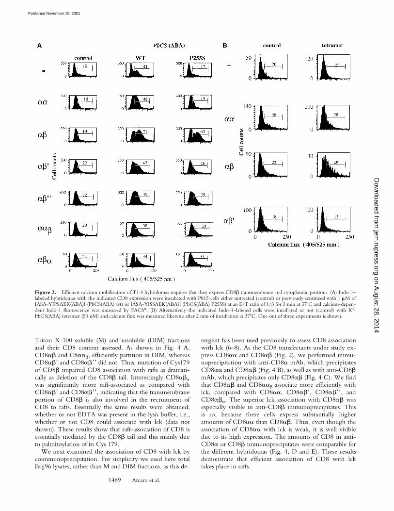
The CD8� Transmembrane and Cytoplasmic Portions AreRequired for Efficient Intracellular Calcium Mobilization inCD8-transfected T1.4 Hybridomas. To evaluate the role ofCD8� transmembrane and cytoplasmic portions for CD8coreceptor function, we transfected CD8�� T1.4 hybrid-omas (6, 10) with CD8� or the CD8� variants shown inFig. 2. While the T1 CTL clone expresses only CD8��,
the hybridomas express substantially more CD8� thanCD8�, i.e., express homodimeric CD8�� and hetero-dimeric CD8��. The different hybridomas were testedfor intracellular calcium mobilization upon incubationwith P815 cells sensitized with IASA-YIPSAEK(ABA)I.CD8�� cells exhibited higher calcium mobilization thanthe other cells (Fig. 3 A). More striking differences amongthe different cells emerged, when the peptide variantIASA-YISSAEK(ABA)I (P255S) was used, which is a weakagonist for T1 CTL (25). In this case, the CD8��� cellsdisplayed nearly the same calcium mobilization as observedfor the wild-type peptide, whereas no change was observedin CD8� or CD8��� cells. Hybridomas expressingCD8�� or CD8��� responded less efficiently than thoseexpressing CD8�� or CD8���.
We also measured intracellular calcium mobilization ofthe indicated hybridomas after incubation with Kd-SYIP-SAEK(ABA)I tetramer. As shown in Fig. 3 B, only cellsexpressing CD8��, but not those expressing no CD8,CD8��, or CD8�� exhibited significant increase in in-tracellular calcium upon incubation with tetramer. As as-sessed by staining with Cy5 labeled tetramer, binding wasmaximal on all cells under the indicated conditions (datanot shown).
The CD8� Tail Is Essential for CD8 Partitioning in Raftsand Efficient Association with lck. As CD8 association withlck seems to take place preferentially in rafts (6), we exam-ined the partitioning of the different CD8 molecules inrafts. To this end the different cells were fractionated in
Figure 1. Soluble CD8�� binds to Kd-PbCS(ABA) without affectingits interaction with T1 TCR. For affinity measurements sT1 TCR (A)and sCD8�� (B) were injected for 30 s at the indicated concentrationsover surfaces expressing Kd-SYIPSAEK(ABA)I (4,500 RU) or sCD4(4,200 RU). Binding was calculated as the difference in the observedequilibrium response between the Kd-SYIPSAEK(ABA)I and the controlsCD4 flow cells. The solid lines represent nonlinear fits of the Langmuirbinding isotherm to the data, which yielded the indicated KD values. Themaximal binding was 2,600 RU for sT1 TCR and 1,200 RU forsCD8��. The insets show Scatchard transformations of the same data. (C)The indicated concentrations of sT1 TCR were injected for 30 s overKd-SYIPSAEK(ABA)I (2,000 RU) or CD4 (2,300 RU) coated sensorchips in the absence (�) or presence (�) of added sCD8�� (80 M).The calculated difference in the responses observed with sT1 alone versussT1 plus sCD8�� are also shown (�). The binding level observed withsCD8�� alone at 80 M is shown as dashed line, indicating that the ef-fect of adding sCD8�� on sT1 binding was never more than additive.
Figure 2. TCR and CD8 expression of cells under study.
on August 28, 2014
jem.rupress.org
Dow
nloaded from
Published November 19, 2001
1489 Arcaro et al.
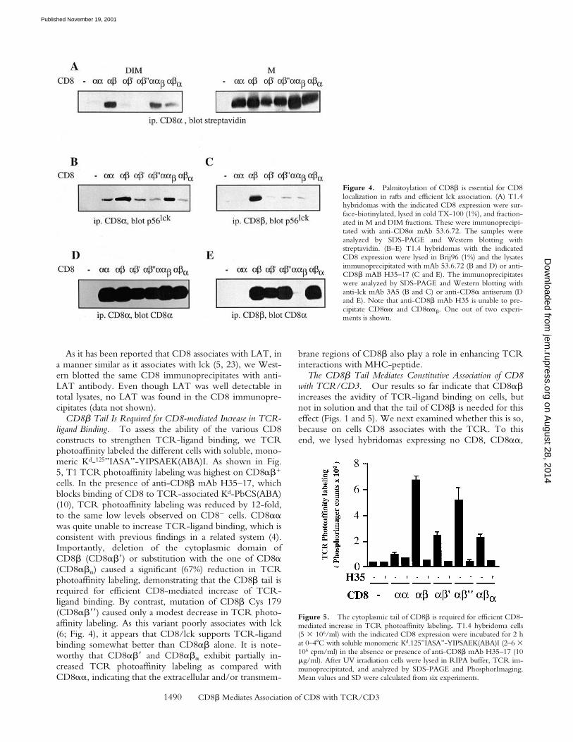
Triton X-100 soluble (M) and insoluble (DIM) fractionsand their CD8 content assessed. As shown in Fig. 4 A,CD8�� and C8���, efficiently partition in DIM, whereasCD8�� and CD8�� did not. Thus, mutation of Cys179of CD8� impaired CD8 association with rafts as dramati-cally as deletion of the CD8� tail. Interestingly CD8���
was significantly more raft-associated as compared withCD8�� and CD8��, indicating that the transmembraneportion of CD8� is also involved in the recruitment ofCD8 to rafts. Essentially the same results were obtained,whether or not EDTA was present in the lysis buffer, i.e.,whether or not CD8 could associate with lck (data notshown). These results show that raft-association of CD8 isessentially mediated by the CD8� tail and this mainly dueto palmitoylation of its Cys 179.
We next examined the association of CD8 with lck bycoimmunoprecipitation. For simplicity we used here totalBrij96 lysates, rather than M and DIM fractions, as this de-
tergent has been used previously to assess CD8 associationwith lck (6–8). As the CD8 transfectants under study ex-press CD8�� and CD8�� (Fig. 2), we performed immu-noprecipitation with anti-CD8� mAb, which precipitatesCD8�� and CD8�� (Fig. 4 B), as well as with anti-CD8�mAb, which precipitates only CD8�� (Fig. 4 C). We findthat CD8�� and CD8��� associate more efficiently withlck, compared with CD8��, CD8��, CD8��, andCD8���. The superior lck association with CD8�� wasespecially visible in anti-CD8� immunoprecipitates. Thisis so, because these cells express substantially higheramounts of CD8�� than CD8��. Thus, even though theassociation of CD8�� with lck is weak, it is well visibledue to its high expression. The amounts of CD8 in anti-CD8� or CD8� immunoprecipitates were comparable forthe different hybridomas (Fig. 4, D and E). These resultsdemonstrate that efficient association of CD8 with lcktakes place in rafts.
Figure 3. Efficient calcium mobilization of T1.4 hybridomas requires that they express CD8� transmembrane and cytoplasmic portions. (A) Indo-1–labeled hybridomas with the indicated CD8 expression were incubated with P815 cells either untreated (control) or previously sensitized with 1 M ofIASA-YIPSAEK(ABA)I (PbCS(ABA) wt) or IASA-YISSAEK(ABA)I (PbCS(ABA) P255S) at an E/T ratio of 1/3 for 3 min at 37 C and calcium-depen-dent Indo-1 fluorescence was measured by FACS®. (B) Alternatively the indicated Indo-1–labeled cells were incubated or not (control) with Kd-PbCS(ABA) tetramer (50 nM) and calcium flux was measured likewise after 2 min of incubation at 37 C. One out of three experiments is shown.
on August 28, 2014
jem.rupress.org
Dow
nloaded from
Published November 19, 2001
1490 CD8� Mediates Association of CD8 with TCR/CD3
As it has been reported that CD8 associates with LAT, ina manner similar as it associates with lck (5, 23), we West-ern blotted the same CD8 immunoprecipitates with anti-LAT antibody. Even though LAT was well detectable intotal lysates, no LAT was found in the CD8 immunopre-cipitates (data not shown).
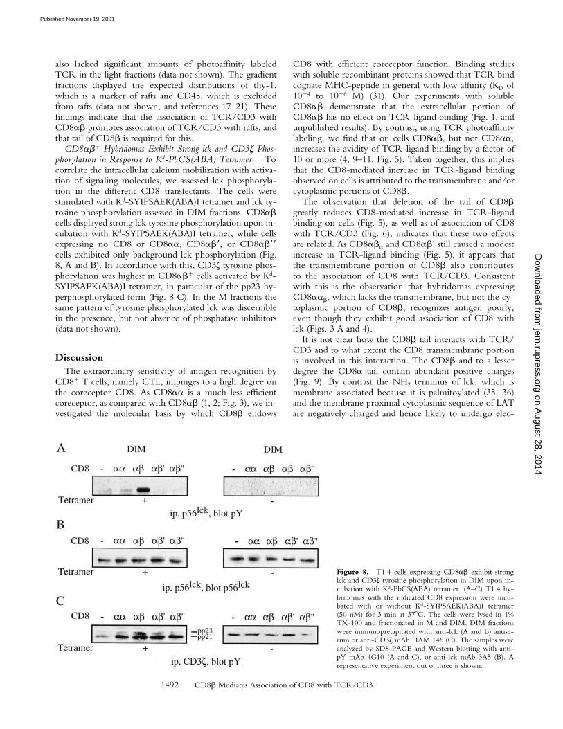
CD8� Tail Is Required for CD8-mediated Increase in TCR-ligand Binding. To assess the ability of the various CD8constructs to strengthen TCR-ligand binding, we TCRphotoaffinity labeled the different cells with soluble, mono-meric Kd-125”IASA”-YIPSAEK(ABA)I. As shown in Fig.5, T1 TCR photoaffinity labeling was highest on CD8���
cells. In the presence of anti-CD8� mAb H35–17, whichblocks binding of CD8 to TCR-associated Kd-PbCS(ABA)(10), TCR photoaffinity labeling was reduced by 12-fold,to the same low levels observed on CD8� cells. CD8��was quite unable to increase TCR-ligand binding, which isconsistent with previous findings in a related system (4).Importantly, deletion of the cytoplasmic domain ofCD8� (CD8��) or substitution with the one of CD8�(CD8���) caused a significant (67%) reduction in TCRphotoaffinity labeling, demonstrating that the CD8� tail isrequired for efficient CD8-mediated increase of TCR-ligand binding. By contrast, mutation of CD8� Cys 179(CD8��) caused only a modest decrease in TCR photo-affinity labeling. As this variant poorly associates with lck(6; Fig. 4), it appears that CD8/lck supports TCR-ligandbinding somewhat better than CD8�� alone. It is note-worthy that CD8�� and CD8��� exhibit partially in-creased TCR photoaffinity labeling as compared withCD8��, indicating that the extracellular and/or transmem-
brane regions of CD8� also play a role in enhancing TCRinteractions with MHC-peptide.
The CD8� Tail Mediates Constitutive Association of CD8with TCR/CD3. Our results so far indicate that CD8��increases the avidity of TCR-ligand binding on cells, butnot in solution and that the tail of CD8� is needed for thiseffect (Figs. 1 and 5). We next examined whether this is so,because on cells CD8 associates with the TCR. To thisend, we lysed hybridomas expressing no CD8, CD8��,
Figure 4. Palmitoylation of CD8� is essential for CD8localization in rafts and efficient lck association. (A) T1.4hybridomas with the indicated CD8 expression were sur-face-biotinylated, lysed in cold TX-100 (1%), and fraction-ated in M and DIM fractions. These were immunoprecipi-tated with anti-CD8� mAb 53.6.72. The samples wereanalyzed by SDS-PAGE and Western blotting withstreptavidin. (B–E) T1.4 hybridomas with the indicatedCD8 expression were lysed in Brij96 (1%) and the lysatesimmunoprecipitated with mAb 53.6.72 (B and D) or anti-CD8� mAB H35–17 (C and E). The immunoprecipitateswere analyzed by SDS-PAGE and Western blotting withanti-lck mAb 3A5 (B and C) or anti-CD8� antiserum (Dand E). Note that anti-CD8� mAb H35 is unable to pre-cipitate CD8�� and CD8���. One out of two experi-ments is shown.
Figure 5. The cytoplasmic tail of CD8� is required for efficient CD8-mediated increase in TCR photoaffinity labeling. T1.4 hybridoma cells(5 � 106/ml) with the indicated CD8 expression were incubated for 2 hat 0–4 C with soluble monomeric Kd
-125”IASA”-YIPSAEK(ABA)I (2–6 �106 cpm/ml) in the absence or presence of anti-CD8� mAb H35–17 (10g/ml). After UV irradiation cells were lysed in RIPA buffer, TCR im-munoprecipitated, and analyzed by SDS-PAGE and PhosphorImaging.Mean values and SD were calculated from six experiments.
on August 28, 2014
jem.rupress.org
Dow
nloaded from
Published November 19, 2001

1491 Arcaro et al.
CD8��, CD8��, or CD8�� in 0.3% NP-40 and im-munoprecipitated TCR. As shown in Fig. 6 A, the amountof CD8� coimmunoprecipitated with TCR was substan-tially higher on CD8��� or CD8��� cells, than onCD8��� or CD8��� cells. These differences were notaccounted for by variations of TCR or CD8 expression, asall cells expressed comparable levels of CD3�, CD8�, andCD8� (Fig. 6, B and C).
To find out whether Kd-PbCS(ABA) complexesstrengthen association of CD8 with TCR, CD8���
cells were photo–cross-linked with soluble Kd-SYIP-SAEK(ABA)I complexes and analyzed as described in theprevious paragraph. No significant increase in CD8 associa-tion with TCR/CD3 was observed (Fig. 6 D), indicatingthat the interaction of CD8�� with TCR/CD3 is consti-tutive and not induced by MHC-peptide. Moreover, thecoimmunoprecipitation was not impaired by the SH2 pep-tide pYEEI (data not shown), which has been shown todisrupt activation-induced coupling of CD8 to TCR/CD3via lck and ZAP-70 (27). Constitutive association of CD8(and CD4) with TCR/CD3 has been reported previously
and low concentrations of NP-40 or Triton X-100 havebeen recommended to preserve these interactions (28–30).Essentially the same findings where obtained when Brij78was used, which unlike NP-40 and Triton X-100 solubi-lizes lipid rafts, indicating that raft integrity was not re-quired for this interaction (data not shown).
CD8�� Mediates Raft-association of TCR/CD3. As theprevious experiments indicated that CD8� mediates associ-ation of CD8 with lck in rafts (Fig. 4), as well as withTCR/CD3 (Fig. 6), we examined whether thereforeCD8� induces raft-association of TCR/CD3. To this endwe TCR photoaffinity labeled T1.4 T cell hybridomas thatexpressed CD8��, CD8��, CD8�� or no CD8 withsoluble Kd-125”IASA”-YIPSAEK(ABA)I and fractionatedtheir Brij58 lysates on sucrose density gradients. Brij58 wasused rather than Triton X-100, because it better preservesweak molecular interaction of membrane proteins (21).This difference becomes especially apparent after long peri-ods of incubation, such as fractionation on sucrose densitygradients. As shown in Fig. 7, the DIM-containing lightfractions exhibited substantial amounts of photoaffinity la-beled TCR on CD8��� cells, but not on cells expressingCD8��, CD8��, or no CD8. Cells expressing CD8���
Figure 6. The CD8� tail mediates CD8 association with TCR/CD3.(A–C) T1.4 hybridomas with the indicated CD8 expression were lysed in0.3% NP-40, and lysates immunoprecipitated with anti-TCR mAb H57(A and B), anti-CD8� mAb 53.6.72 (C, left), or with anti-CD8� mAbH35–17 (C, right). The samples were analyzed by SDS-PAGE and West-ern blotting with anti-CD8� antiserum (A and C) or anti-CD3� antise-rum (B). (D) T1.4 T cell hybridomas expressing CD8�� or CD8�� wereincubated with monomeric Kd-SYIPSAEK(ABA)I complexes (1.16M) for 2 h at 0–4 C. After UV irradiation the cells were lysed in 0.3%NP-40. Lysates were immunoprecipitated with anti-TCR mAb H57 oranti-CD8� mAb 53.6.72, as indicated and the immunoprecipitates ana-lyzed by SDS-PAGE and Western blotting with anti-CD8� antiserum.
Figure 7. CD8�� mediates raft association of TCR/CD3. T1.4 hy-bridomas (5 � 107) expressing no CD8 (A), CD8�� (B), CD8�� (C), orCD8�� (D) were photoaffinity labeled at 26 C with Kd
-125”IASA”-YIPSAEK(ABA)I (0.5–1.5 � 108 cpm/7 ml). After UV irradiation thewashed cells were lysed in 0.5% Brij58 and the lysates fractionated on su-crose density gradients. Fractions were collected from the top and immu-noprecipitated with anti-TCRC� mAb H57. The immunoprecipitateswere analyzed by SDS-PAGE and PhosphorImaging.
on August 28, 2014
jem.rupress.org
Dow
nloaded from
Published November 19, 2001
1492 CD8� Mediates Association of CD8 with TCR/CD3
also lacked significant amounts of photoaffinity labeledTCR in the light fractions (data not shown). The gradientfractions displayed the expected distributions of thy-1,which is a marker of rafts and CD45, which is excludedfrom rafts (data not shown, and references 17–21). Thesefindings indicate that the association of TCR/CD3 withCD8�� promotes association of TCR/CD3 with rafts, andthat tail of CD8� is required for this.
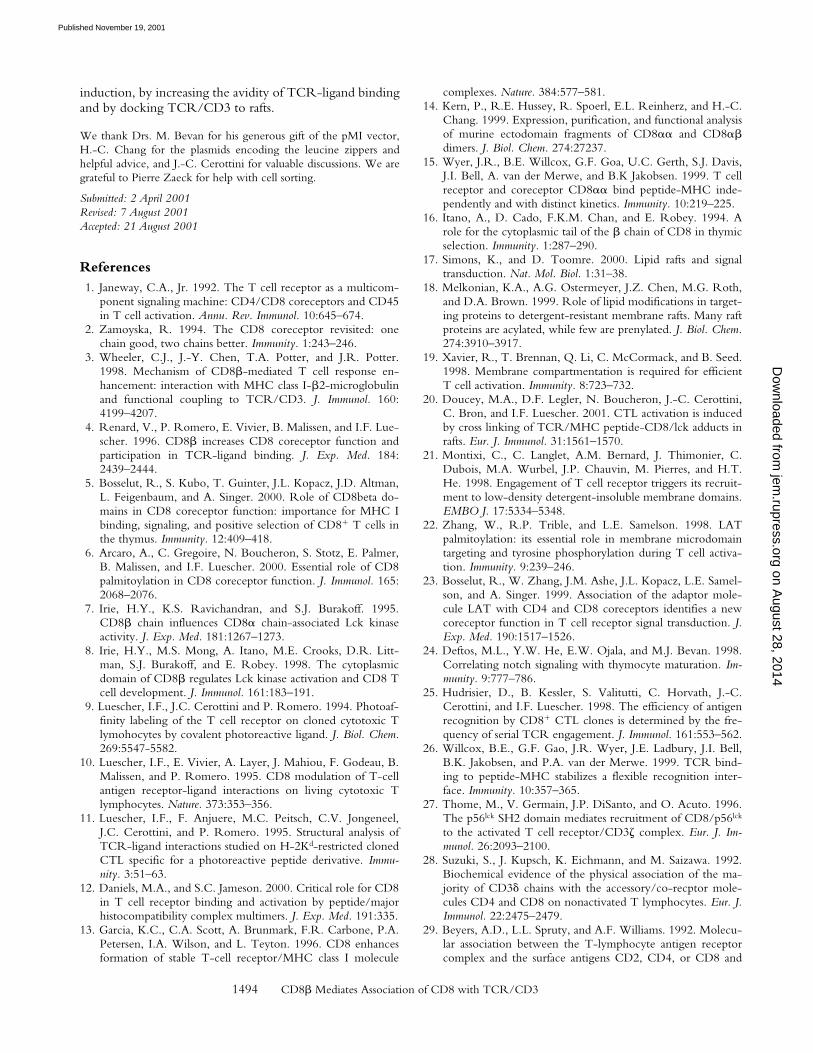
CD8��� Hybridomas Exhibit Strong lck and CD3� Phos-phorylation in Response to Kd-PbCS(ABA) Tetramer. Tocorrelate the intracellular calcium mobilization with activa-tion of signaling molecules, we assessed lck phosphoryla-tion in the different CD8 transfectants. The cells werestimulated with Kd-SYIPSAEK(ABA)I tetramer and lck ty-rosine phosphorylation assessed in DIM fractions. CD8��cells displayed strong lck tyrosine phosphorylation upon in-cubation with Kd-SYIPSAEK(ABA)I tetramer, while cellsexpressing no CD8 or CD8��, CD8��, or CD8��cells exhibited only background lck phosphorylation (Fig.8, A and B). In accordance with this, CD3� tyrosine phos-phorylation was highest in CD8��� cells activated by Kd-SYIPSAEK(ABA)I tetramer, in particular of the pp23 hy-perphosphorylated form (Fig. 8 C). In the M fractions thesame pattern of tyrosine phosphorylated lck was discerniblein the presence, but not absence of phosphatase inhibitors(data not shown).
DiscussionThe extraordinary sensitivity of antigen recognition by
CD8� T cells, namely CTL, impinges to a high degree onthe coreceptor CD8. As CD8�� is a much less efficientcoreceptor, as compared with CD8�� (1, 2; Fig. 3), we in-vestigated the molecular basis by which CD8� endows
CD8 with efficient coreceptor function. Binding studieswith soluble recombinant proteins showed that TCR bindcognate MHC-peptide in general with low affinity (KD of10�4 to 10�6 M) (31). Our experiments with solubleCD8�� demonstrate that the extracellular portion ofCD8�� has no effect on TCR-ligand binding (Fig. 1, andunpublished results). By contrast, using TCR photoaffinitylabeling, we find that on cells CD8��, but not CD8��,increases the avidity of TCR-ligand binding by a factor of10 or more (4, 9–11; Fig. 5). Taken together, this impliesthat the CD8-mediated increase in TCR-ligand bindingobserved on cells is attributed to the transmembrane and/orcytoplasmic portions of CD8�.
The observation that deletion of the tail of CD8�greatly reduces CD8-mediated increase in TCR-ligandbinding on cells (Fig. 5), as well as of association of CD8with TCR/CD3 (Fig. 6), indicates that these two effectsare related. As CD8��� and CD8��’ still caused a modestincrease in TCR-ligand binding (Fig. 5), it appears thatthe transmembrane portion of CD8� also contributesto the association of CD8 with TCR/CD3. Consistentwith this is the observation that hybridomas expressingCD8���, which lacks the transmembrane, but not the cy-toplasmic portion of CD8�, recognizes antigen poorly,even though they exhibit good association of CD8 withlck (Figs. 3 A and 4).
It is not clear how the CD8� tail interacts with TCR/CD3 and to what extent the CD8 transmembrane portionis involved in this interaction. The CD8� and to a lesserdegree the CD8� tail contain abundant positive charges(Fig. 9). By contrast the NH2 terminus of lck, which ismembrane associated because it is palmitoylated (35, 36)and the membrane proximal cytoplasmic sequence of LATare negatively charged and hence likely to undergo elec-
Figure 8. T1.4 cells expressing CD8�� exhibit stronglck and CD3� tyrosine phosphorylation in DIM upon in-cubation with Kd-PbCS(ABA) tetramer. (A–C) T1.4 hy-bridomas with the indicated CD8 expression were incu-bated with or without Kd-SYIPSAEK(ABA)I tetramer(50 nM) for 3 min at 37 C. The cells were lysed in 1%TX-100 and fractionated in M and DIM. DIM fractionswere immunoprecipitated with anti-lck (A and B) antise-rum or anti-CD3� mAb HAM 146 (C). The samples wereanalyzed by SDS-PAGE and Western blotting with anti-pY mAb 4G10 (A and C), or anti-lck mAb 3A5 (B). Arepresentative experiment out of three is shown.
on August 28, 2014
jem.rupress.org
Dow
nloaded from
Published November 19, 2001

1493 Arcaro et al.
trostatic interactions with the CD8 tails (Fig. 9). Remark-ably, this LAT sequence becomes highly negativelycharged upon activation-dependent phosphorylation of itstyrosines and serine/threonines (37–39) and thus is ex-pected to undergo strong electrostatic interactions withCD8. Differences in LAT phosphorylation may explainwhy we were unable to detect association of LAT withCD8, as has been observed in other systems (5, 23). Themembrane proximal cytoplasmic sequences of CD3�,CD3�, and CD3�, similarly become acidic upon phos-phorylation of their serines/threonines.
Association of CD8 (and CD4) with TCR/CD3 hasbeen previously observed in different systems (28–30). Ofparticular interest is a study showing that anti-CD8 immu-noprecipitates contain a high proportion of CD3�, suggest-ing that CD8 associates with TCR via CD3� (28). Thismay explain why mice lacking CD3� (32), or expressing avariant TCR which fails to associate with CD3� (33), ex-hibit severely impaired activation and positive selection ofCD8� T cells. It is important to note that the association ofCD8 with TCR/CD3 described here (Fig. 6) and in previ-ous studies (28–30) is constitutive, i.e., is not, or little, in-duced by MHC-peptide.
Moreover, the tail of CD8� mediates raft localization ofCD8 (6, 20; Fig. 4). The finding that point mutation ofCD8� Cys 179 (CD8��) abolishes this (Fig. 4 A), dem-onstrates that this is attributed essentially to palmitoylationof the CD8� tail. As lck is myristoylated and dipalmitoy-lated, it also efficiently partitions in rafts (17–19, 35, 36).Thus, the observation that efficient association of CD8with lck takes place in rafts (6; Fig. 4), is most likely ex-plained by increased concentrations of both molecules inmicrodomains, which constitute only a small fraction of thecell membrane (17). By contrast, association of CD8 withTCR/CD3 and hence TCR-ligand binding were greatlyimpaired upon deletion of the CD8� tail, but hardly bymutation of CD8� Cys 179 (Figs. 5 and 6). This demon-strates that the tail of CD8� exerts two different functions:(a) it mediates raft localization of CD8 and hence efficient
association of CD8 with lck, and (b) it mediates associationof CD8 with TCR/CD3. Because CD8� exerts bothfunctions at the same time, it promotes raft-association ofTCR/CD3 (Fig. 7).
While TCR/CD3 per se do not partition significantly inrafts (19–22), this study indicates that they become raft-associated by interacting with raft-resident CD8-lck com-plexes. Small fractions of TCR/CD3 have been shown tobe raft-associated in resting cells in a fractionation study(21) and by confocal microscopy (40). Although CD3components seem to bind weakly to raft-associated Src ki-nases in resting cells, which may be important for corecep-tor independent T cell activation (41), we show here thatCD8 substantially strengthens raft-association of TCR/CD3 (Fig. 7). As association of TCR/CD3 with CD8/lckalso enhances the avidity of TCR-ligand binding (20; Fig.5), it appears that raft-associated TCR have an increasedavidity for MHC-peptide, i.e., are those that are engagedfirst by MHC-peptide, particularly when these are scant orof low affinity. Thus, for the induction of TCR signalingthis constitutive TCR/CD3 association with CD8/lck isimportant, as it provides a significant increase in TCR-ligand binding and at the same time raft association of en-gaged TCR/CD3 (Figs. 5–8).
Because phosphatases are excluded from rafts, they areprivileged sites for the induction of TCR signaling (19–22). Indeed we find that tetrameric Kd-PbCS(ABA) com-plexes efficiently induce tyrosine phosphorylation of lckand CD3� in rafts on CD8��� hybridoma, but much lessso on hybridomas expressing no CD8, CD8��, CD8��,or CD8�� (Fig. 8). As lck is activated by cross-linking,even when Y505 is phosphorylated (6–8, 20, 42), our datapropose that TCR signaling is induced by engagement ofraft-associated TCR/CD3 adducts with CD8/lck by mul-timeric MHC-peptide complexes. While it is clear thatonce TCR signaling is induced, SH2-mediated interactionsprovide additional coupling of the TCR/CD3 with CD8,LAT, ZAP-70, and other molecules (1, 27, 34), the presentstudy shows that CD8� significantly facilitates TCR signal
Figure 9. Membrane proxi-mal cytoplasmic sequences ofCD3, CD8, lck and LAT. Theindicated sequences were takenfrom Swiss-Prot (http://www.expasy.ch/cgi-bin/sprotsearch-de).Cysteines labeled with an asteriskare known to be palmitoylatedand hence are membrane inte-grated. The numbers of the dis-played residues are indicated inparenthesis. Basic residues areshown in oval, acidic ones inboxes, tyrosines in diamonds,and serines and threonines inshaded gray.
on August 28, 2014
jem.rupress.org
Dow
nloaded from
Published November 19, 2001
1494 CD8� Mediates Association of CD8 with TCR/CD3
induction, by increasing the avidity of TCR-ligand bindingand by docking TCR/CD3 to rafts.
We thank Drs. M. Bevan for his generous gift of the pMI vector,H.-C. Chang for the plasmids encoding the leucine zippers andhelpful advice, and J.-C. Cerottini for valuable discussions. We aregrateful to Pierre Zaeck for help with cell sorting.
Submitted: 2 April 2001Revised: 7 August 2001Accepted: 21 August 2001
References1. Janeway, C.A., Jr. 1992. The T cell receptor as a multicom-
ponent signaling machine: CD4/CD8 coreceptors and CD45in T cell activation. Annu. Rev. Immunol. 10:645–674.
2. Zamoyska, R. 1994. The CD8 coreceptor revisited: onechain good, two chains better. Immunity. 1:243–246.
3. Wheeler, C.J., J.-Y. Chen, T.A. Potter, and J.R. Potter.1998. Mechanism of CD8�-mediated T cell response en-hancement: interaction with MHC class I-�2-microglobulinand functional coupling to TCR/CD3. J. Immunol. 160:4199–4207.
4. Renard, V., P. Romero, E. Vivier, B. Malissen, and I.F. Lue-scher. 1996. CD8� increases CD8 coreceptor function andparticipation in TCR-ligand binding. J. Exp. Med. 184:2439–2444.
5. Bosselut, R., S. Kubo, T. Guinter, J.L. Kopacz, J.D. Altman,L. Feigenbaum, and A. Singer. 2000. Role of CD8beta do-mains in CD8 coreceptor function: importance for MHC Ibinding, signaling, and positive selection of CD8� T cells inthe thymus. Immunity. 12:409–418.
6. Arcaro, A., C. Gregoire, N. Boucheron, S. Stotz, E. Palmer,B. Malissen, and I.F. Luescher. 2000. Essential role of CD8palmitoylation in CD8 coreceptor function. J. Immunol. 165:2068–2076.
7. Irie, H.Y., K.S. Ravichandran, and S.J. Burakoff. 1995.CD8� chain influences CD8� chain-associated Lck kinaseactivity. J. Exp. Med. 181:1267–1273.
8. Irie, H.Y., M.S. Mong, A. Itano, M.E. Crooks, D.R. Litt-man, S.J. Burakoff, and E. Robey. 1998. The cytoplasmicdomain of CD8� regulates Lck kinase activation and CD8 Tcell development. J. Immunol. 161:183–191.
9. Luescher, I.F., J.C. Cerottini and P. Romero. 1994. Photoaf-finity labeling of the T cell receptor on cloned cytotoxic Tlymohocytes by covalent photoreactive ligand. J. Biol. Chem.269:5547-5582.
10. Luescher, I.F., E. Vivier, A. Layer, J. Mahiou, F. Godeau, B.Malissen, and P. Romero. 1995. CD8 modulation of T-cellantigen receptor-ligand interactions on living cytotoxic Tlymphocytes. Nature. 373:353–356.
11. Luescher, I.F., F. Anjuere, M.C. Peitsch, C.V. Jongeneel,J.C. Cerottini, and P. Romero. 1995. Structural analysis ofTCR-ligand interactions studied on H-2Kd-restricted clonedCTL specific for a photoreactive peptide derivative. Immu-nity. 3:51–63.
12. Daniels, M.A., and S.C. Jameson. 2000. Critical role for CD8in T cell receptor binding and activation by peptide/majorhistocompatibility complex multimers. J. Exp. Med. 191:335.
13. Garcia, K.C., C.A. Scott, A. Brunmark, F.R. Carbone, P.A.Petersen, I.A. Wilson, and L. Teyton. 1996. CD8 enhancesformation of stable T-cell receptor/MHC class I molecule
complexes. Nature. 384:577–581.14. Kern, P., R.E. Hussey, R. Spoerl, E.L. Reinherz, and H.-C.
Chang. 1999. Expression, purification, and functional analysisof murine ectodomain fragments of CD8�� and CD8��dimers. J. Biol. Chem. 274:27237.
15. Wyer, J.R., B.E. Willcox, G.F. Goa, U.C. Gerth, S.J. Davis,J.I. Bell, A. van der Merwe, and B.K Jakobsen. 1999. T cellreceptor and coreceptor CD8�� bind peptide-MHC inde-pendently and with distinct kinetics. Immunity. 10:219–225.
16. Itano, A., D. Cado, F.K.M. Chan, and E. Robey. 1994. Arole for the cytoplasmic tail of the � chain of CD8 in thymicselection. Immunity. 1:287–290.
17. Simons, K., and D. Toomre. 2000. Lipid rafts and signaltransduction. Nat. Mol. Biol. 1:31–38.
18. Melkonian, K.A., A.G. Ostermeyer, J.Z. Chen, M.G. Roth,and D.A. Brown. 1999. Role of lipid modifications in target-ing proteins to detergent-resistant membrane rafts. Many raftproteins are acylated, while few are prenylated. J. Biol. Chem.274:3910–3917.
19. Xavier, R., T. Brennan, Q. Li, C. McCormack, and B. Seed.1998. Membrane compartmentation is required for efficientT cell activation. Immunity. 8:723–732.
20. Doucey, M.A., D.F. Legler, N. Boucheron, J.-C. Cerottini,C. Bron, and I.F. Luescher. 2001. CTL activation is inducedby cross linking of TCR/MHC peptide-CD8/lck adducts inrafts. Eur. J. Immunol. 31:1561–1570.
21. Montixi, C., C. Langlet, A.M. Bernard, J. Thimonier, C.Dubois, M.A. Wurbel, J.P. Chauvin, M. Pierres, and H.T.He. 1998. Engagement of T cell receptor triggers its recruit-ment to low-density detergent-insoluble membrane domains.EMBO J. 17:5334–5348.
22. Zhang, W., R.P. Trible, and L.E. Samelson. 1998. LATpalmitoylation: its essential role in membrane microdomaintargeting and tyrosine phosphorylation during T cell activa-tion. Immunity. 9:239–246.
23. Bosselut, R., W. Zhang, J.M. Ashe, J.L. Kopacz, L.E. Samel-son, and A. Singer. 1999. Association of the adaptor mole-cule LAT with CD4 and CD8 coreceptors identifies a newcoreceptor function in T cell receptor signal transduction. J.Exp. Med. 190:1517–1526.
24. Deftos, M.L., Y.W. He, E.W. Ojala, and M.J. Bevan. 1998.Correlating notch signaling with thymocyte maturation. Im-munity. 9:777–786.
25. Hudrisier, D., B. Kessler, S. Valitutti, C. Horvath, J.-C.Cerottini, and I.F. Luescher. 1998. The efficiency of antigenrecognition by CD8� CTL clones is determined by the fre-quency of serial TCR engagement. J. Immunol. 161:553–562.
26. Willcox, B.E., G.F. Gao, J.R. Wyer, J.E. Ladbury, J.I. Bell,B.K. Jakobsen, and P.A. van der Merwe. 1999. TCR bind-ing to peptide-MHC stabilizes a flexible recognition inter-face. Immunity. 10:357–365.
27. Thome, M., V. Germain, J.P. DiSanto, and O. Acuto. 1996.The p56lck SH2 domain mediates recruitment of CD8/p56lck
to the activated T cell receptor/CD3� complex. Eur. J. Im-munol. 26:2093–2100.
28. Suzuki, S., J. Kupsch, K. Eichmann, and M. Saizawa. 1992.Biochemical evidence of the physical association of the ma-jority of CD3� chains with the accessory/co-recptor mole-cules CD4 and CD8 on nonactivated T lymphocytes. Eur. J.Immunol. 22:2475–2479.
29. Beyers, A.D., L.L. Spruty, and A.F. Williams. 1992. Molecu-lar association between the T-lymphocyte antigen receptorcomplex and the surface antigens CD2, CD4, or CD8 and
on August 28, 2014
jem.rupress.org
Dow
nloaded from
Published November 19, 2001

1495 Arcaro et al.
CD5. Proc. Natl. Acad. Sci. USA. 89:2945–2949.30. Gallagher, P.F., B. Fazekas de St. Groth, and J.A.A.P. Miller.
1989. CD4 and CD8 molecules can physically associate withthe same T cell receptor. Proc. Natl. Acad. Sci. USA. 86:10044-10048.
31. Fremont, D.H., W.A. Rees, and H. Kozono. 1996. Biophys-ical studies of T-cell receptors and their ligands. Curr. Opin.Immunol. 8:93–100.
32. Delgado, P., E. Fernandez, V. Dave, D. Kappas, and B. Alar-con. 2000. CD3� couples T-cell receptor signaling to ERKactivation and thymocyte positive selection. Nature. 406:426–430.
33. Guy, W., B. Hausmann, and E. Palmer. 2000. A motif in theT-cell receptor controls positive selection by modulatingERK activity. Nature. 406:422–426.
34. Qian, D., and A. Weiss A. 1997. T cell antigen receptor sig-nal transduction. Curr. Opin. Cell Biol. 9:205-212.
35. Webb, Y., L. Hermida-Matsumoto, and M.D. Resh. 2000.Inhibition of protein palmitoylation, raft localization, and Tcell signaling by 2-bromopalmitate and poyunsaturated fattyacids. J. Biol. Chem. 275:261–270.
36. Yasuda, K., A. Kosugi, F. Hayashi, S. Saitoh, M. Nagafuku,Y. Mori, M. Ogata, and T. Hamaoka. 2000. Serine 6 of Lck
tyrosine kinase: a critical site for Lck myristoylation, mem-brane localization, and function in T lymphocytes. J. Immu-nol. 165:3226–3231.
37. Lin, J., A. Weiss, and T.S. Finco. 1999. Localization of LATin glycolipid-enriched microdomains is required for T cellactivation. J. Biol. Chem. 274:28861–28864.
38. Zhang, W., and L.E. Samelson. 2000. The role of mem-brane-associated adaptors in T cell receptor signalling. Semin.Immunol. 12:35–41.
39. Zhang, W., J. Sloan-Lancaster, J. Kitchen, R.P. Trible, andL.E. Samelson. 1998. LAT: the ZAP-70 tyrosine kinase sub-strate that links T cell receptor to cellular activation. Cell. 92:83–92.
40. Janes, P.W., S.C. Ley, and A.I. Magee. 1999. Aggregation oflipid rafts accompanies signaling via the T cell antigen recep-tor. J. Cell Biol. 147:447–461.
41. van’t Hof, W., and M.D. Resh. 1999. Dual fatty acylation ofp59(Fyn) is required for association with the T cell receptorzeta chain through phosphotyrosine-Src homology domain-2interactions. J. Cell Biol. 145:377–389.
42. Yamaguchi, H., and W.A. Hendrickson. 1996. Structural ba-sis for activation of human lymphocyte kinase Lck upon tyro-sine phosphorylation. Nature. 384:484–489.
on August 28, 2014
jem.rupress.org
Dow
nloaded from
Published November 19, 2001