Cytotoxicity and Anti-Inflammatory Activity of Methylsulfanyl-triazoloquinazolines
Upload
khangminh22Category
view
2download
0
Role of LAT in the Cytotoxicity and Memory Response
of CD8 T Cells Following Microbial Infection
by
Chihwen (Claudia) Ouyang
Department of Immunology
Duke University
Date:_______________________
Approved:
___________________________
Weiguo Zhang, Supervisor
___________________________
Yuan Zhuang, Chair
___________________________
Michael Krangel
___________________________
Qi-Jing Li
___________________________
Jeffrey Rathmell
Dissertation submitted in partial fulfillment of the
requirements for the degree of Doctor of
Philosophy in the Department of
Immunology in the Graduate School
of Duke University
2013

ABSTRACT
Role of LAT in the Cytotoxicity and Memory Response
of CD8 T Cells Following Microbial Infection
by
Chihwen (Claudia) Ouyang
Department of Immunology
Duke University
Date:_______________________
Approved:
___________________________
Weiguo Zhang, Supervisor
___________________________
Yuan Zhuang, Chair
___________________________
Michael Krangel
___________________________
Qi-Jing Li
___________________________
Jeffrey Rathmell
An abstract of a dissertation submitted in partial
fulfillment of the requirements for the degree
of Doctor of Philosophy in the Department
of Immunology in the Graduate School
of Duke University
2013

Copyright by
Chihwen Ouyang
2013

iv
Abstract
Linker for activation of T cells (LAT) is a transmembrane adaptor protein that is
crucial in linking TCR engagement to downstream signaling events, such as calcium flux
and Ras-MAPK pathway. Following TCR engagement, LAT is phosphorylated at its
membrane-distal tyrosine residues, which mediates the binding of Grb2/Sos, PLC-γ1,
and GADS/SLP-76 complexes. Formation of this multi-protein signaling complex
initiates signaling cascades eventually leading to the activation of transcription factors
that regulate the genes required for T cell proliferation and effector functions. The
indispensable role of LAT in thymocyte development has been evidenced as LAT-
deficient mice completely lack peripheral T cells. To study the function of LAT in mature
T cells, our lab previously generated a conditional knock-in mouse line in which the lat
gene can be deleted by Cre recombinase. Deletion of LAT in mature T cells reveals the
critical role of LAT in T cell activation. Here, we used this inducible LAT deletion mouse
line crossed with the OT-I transgenic mice which express CD8 T cells that can be
activated by ovalbumin (Ova) to study the role of LAT in mature CD8 T cells.
To analyze the contribution of LAT in CD8 T cells during the course of pathogen
infection, we infected mice with Listeria monocytogenes-expressing Ova to elicit activation
of antigen-specific CD8 T cells, and then inducibly deleted LAT in these cells at different
stages of infection by tamoxifen treatment. We show that LAT is important for
maintaining CD8 T cell expansion during the priming phase; however, it is not required
v
for CD8 T cell contraction. In addition, memory CD8 T cell can persist in the absence of
LAT, suggesting that LAT-mediated signaling is not necessary for memory maintenance.
Nonetheless, these LAT-deficient memory T cells were unable to proliferate or produce
cytokines upon secondary infection. Moreover, LAT deficiency accelerates memory
differentiation during the effector-to-memory transition, leading to a higher frequency
of KLRG1lowIL-7RhighCD62Lhigh memory T cells. Together, these data demonstrate that,
while it is dispensable for contraction and memory maintenance, LAT-signaling
regulates CD8 T cell memory differentiation and is essential for the memory response
against pathogens.
The fundamental activity of CD8 T cells is to elicit cytotoxicity toward target cells
that express foreign antigens, and this is mediated through granule-dependent and Fas
ligand-dependent mechanisms. The signaling events that regulate these processes
remain unclear. We show that LAT-deficient cytotoxic T cells (CTLs) failed to upregulate
FasL and produce IFN-γ after engagement with target cells. Moreover, they displayed
reduced granule-mediated killing. We further dissected the effect of the LAT deletion on
each step of granule exocytosis. LAT-deficiency led to altered synapse formation,
subsequently causing unstable T cell:APC conjugates. MTOC polarization and granule
reorientation were also impaired by LAT-deficiency, leading to reduced granule
delivery. Despite these defects, granule release was still observed in LAT-deficient CTLs
due to residual calcium flux and PLC activity. This reveals an unexpected finding that
vi
CTL function is not entirely dependent on LAT. Collectively, these data indicate that the
signaling circuits governing CTLs are programmed to adopt multiple pathways,
allowing CTLs to effectively eliminate various pathogens during adaptive immune
responses.
vii
Contents
Abstract ......................................................................................................................................... iv
List of Figures ............................................................................................................................... xi
Acknowledgements .................................................................................................................. xiii
1. Introduction........................................................................................................................... 1
1.1 Antigen-specific CD8 T cell response ............................................................................ 1
1.2 Granule exocytosis ...................................................................................................... 6
1.2.1 Lytic granules ............................................................................................................... 7
1.2.2 Cytotoxic immunological synapse ............................................................................ 9
1.2.3 MTOC and granule polarization ............................................................................. 13
1.3 T Memory CD8 T cell formation .................................................................................. 17
1.3.1 Memory precursor effector cell (MPEC) vs Short-lived effector cell (SLEC) .... 17
1.3.2 Central memory T cell (TCM) vs Effector memory T cell (TEM)............................. 18
1.3.3 Memory Stem Cells (TSCM) ........................................................................................ 20
1.3.4 Extrinsic programming of T cell differentiation ................................................... 21
1.3.5 Intrinsic programming of T cell differentiation .................................................... 23
1.4 LAT-mediated signaling ........................................................................................... 26
1.4.1 Early events in TCR signaling ................................................................................. 26
1.4.2 Linker for activation of T cells (LAT) ..................................................................... 28
1.4.3 PLC-γ1 and calcium mobilization ........................................................................... 32
1.4.4 Inducible LAT deletion system ............................................................................... 34
viii
2. Materials and Methods........................................................................................................... 36
2.1 Mouse models ................................................................................................................. 36
2.2 Inducible deletion of LAT ............................................................................................. 36
2.2.1 Deletion of LAT in vivo ............................................................................................. 36
2.2.2 Generation of LAT-deficient effector CD8 T cells in vitro .................................... 37
2.3 Infection ........................................................................................................................... 37
2.4 Antibodies ....................................................................................................................... 37
2.4.1 Antibodies for flow cytometry ................................................................................ 37
2.4.2 Antibodies for biochemical studies ......................................................................... 38
2.4.3 Antibodies for immunofluorescence ...................................................................... 38
2.5 Flow cytometry ............................................................................................................... 39
2.5.1 Cell surface staining .................................................................................................. 39
2.5.2 Intracellular staining ................................................................................................. 39
2.6 T cell activation and Western blot analysis ................................................................. 40
2.7 FACS-based cytotoxicity assay ..................................................................................... 41
2.8 Conjugate assay .............................................................................................................. 42
2.9 Granzyme B substrate assay ......................................................................................... 42
2.10 Calcium mobilization ................................................................................................... 43
2.11 Immunofluorescence imaging and analysis ............................................................. 43
2.12 Multichannel live cell microscopy and data analysis .............................................. 44
2.12.1 Calcium imaging in living cells ............................................................................. 44

ix
2.12. (PH)Akt imaging in living cells ............................................................................... 45
3. The requirement of LAT in the primary and memory responses of CD8 T cells
following microbial infection .................................................................................................... 47
3.1 Introduction ..................................................................................................................... 47
3.2 Results .............................................................................................................................. 50
3.2.1 Inducible deletion of LAT in OT-I cells .................................................................. 50
3.2.2 LAT mediated-signaling is essential for optimal CD8 T cell expansion ............ 51
3.2.3 LAT mediated-signaling is not required for CD8 T cell contraction .................. 56
3.2.4 LAT function during the effector-to-memory transition ..................................... 60
3.2.5 LAT deficiency affects T-bet expression in memory T cells ................................ 63
3.2.6 LAT-mediated signaling is dispensable for memory CD8 T cell maintenance 68
3.2.7 LAT-mediated signaling is indispensable for a memory recall response ......... 71
3.3 Discussion ........................................................................................................................ 78
4. The role of LAT in the granule-mediated cytotoxicity of CD8 T cells ............................. 86
4.1 Introduction ..................................................................................................................... 86
4.2 Results .............................................................................................................................. 88
4.2.1 Ablation of LAT impairs CTL killing in vivo ......................................................... 88
4.2.2 Generation of LAT-deficient CTLs in vitro ............................................................. 89
4.2.4 Unstable CTL-target conjugates and immunological synapse in LATKO CTLs
............................................................................................................................................... 97
4.2.5 Impaired MTOC and lytic granule polarization in LATKO CTLs ................... 103
4.2.6 Defective TCR signaling in LATKO CTLs ........................................................... 107

x
4.2.7 Residual PLC activity and calcium flux are necessary to drive cytotoxicity of
LATKO CTLs .................................................................................................................... 115
4.3 Discussion ...................................................................................................................... 119
5. General Discussion and Future Directions ........................................................................ 126
5.1 LAT-mediated signaling in CD8 T cell expansion ................................................... 126
5.2 LAT-mediated signaling in CD8 T cell contraction ................................................. 130
5.3 LAT-mediated signaling in memory CD8 T cell maintenance .............................. 131
5.4 The potential role of LAT signaling in chronic infection ........................................ 136
5.5 LAT-mediated signaling in CTL cytotoxicity ........................................................... 139
References .................................................................................................................................. 145
Biography ................................................................................................................................... 158

xi
List of Figures
Figure 1: Antigen-specific CD8 T cell response ........................................................................ 2
Figure 2: CD8 T cell cytotoxicity ................................................................................................. 4
Figure 3: Granule exocytosis ....................................................................................................... 8
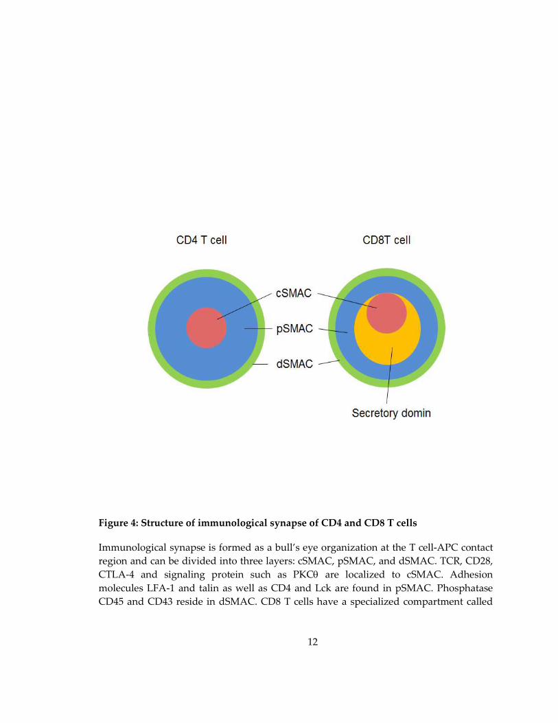
Figure 4: Structure of immunological synapse of CD4 and CD8 T cells ............................. 12
Figure 5: CD8 T cell differentiation .......................................................................................... 19
Figure 6: TCR-mediated signaling ............................................................................................ 30
Figure 7: The LAT knock-in construct...................................................................................... 35
Figure 8: LAT in the expansion of CD8 T cells upon Lm-Ova infection. ............................ 53
Figure 9: LAT in the proliferation and cell death of CD8 T cells.......................................... 55
Figure 10: LAT in the contraction of CD8 T cells .................................................................... 58
Figure 11: LAT in the memory differentiation of CD8 T cells. ............................................. 61
Figure 12: LAT in the formation of memory precursor CD8 T cells. ................................... 65
Figure 13: Deletion of LAT affects T-bet expression. ............................................................. 67
Figure 14: LAT in the maintenance of memory CD8 T cells. ................................................ 69
Figure 15: LAT in the cytokine production by memory CD8 T cells. .................................. 74
Figure 16: LAT in the recall response by memory CD8 T cells. ........................................... 76
Figure 17: LAT in CTL cytotoxicity in vivo. ............................................................................. 90
Figure 18: Deletion of LAT in vitro. .......................................................................................... 93
Figure 19: LAT in CTL cytotoxicity in vitro. ............................................................................ 96
Figure 20: LAT in the granule-mediated killing. .................................................................... 98

xii
Figure 21: LAT in the formation of CTL-target cell conjugate and immunological
synapse. ...................................................................................................................................... 101
Figure 22: LAT in the MTOC and lytic granule polarization. ............................................ 105
Figure 23: Defective TCR signaling in LATKO CTLs. ......................................................... 109
Figure 24: Reduced PI3K/Akt activity in LATKO CTLs. ..................................................... 110
Figure 25: Impaired but not abolished calcium signaling in LATKO CTLs. .................... 113
Figure 26: The residual activation of PLC and calcium signaling enabled LATKO CTLs
to kill target cells. ...................................................................................................................... 116
Figure 27: PLC activity is not necessary for MTOC polarization in CTLs. ....................... 118
Figure 28: The models of LAT-mediated signaling in antigen-specific CD8 T cell
response during acute and chronic infections. ..................................................................... 138
Figure 29: Models of impaired CTL cytotoxicity by LAT deficiency. ............................... 142
xiii
Acknowledgements
When I was giving my defense talk, most of those who came to support me were
friends with whom I have shared the past 5 years at Duke. It finally occurred to me that
an incredibly important chapter of my life, graduate school, had come to an end.
Without the help of these people, I wouldn’t have made it through the many challenges
to stand at the finish line of graduate school with great achievements. First and
foremost, I would like to give my deepest gratitude to my lovely parents, my sister, and
my brother. Although they are far away in Taiwan, they have continued to be my
biggest support. Visiting them in Taiwan every year has always refilled my energy, and
pushed me to keep going. Moreover, dearest thanks to my husband and my best friend,
Richard, who constantly offers me positive feedback whenever I need it. Talking with
him after work has been the remedy to release my stress and rebuild my confidence over
the years.
I would also like to thank my Ph.D. advisor, Dr. Weiguo Zhang, for his great
mentorship. He gave me freedom to explore and grow, yet offered advice when I
encountered difficulties. I will always remember how patient and nice he was when he
helped me with molecular cloning techniques. Although I was unable to inherit his
genus in cloning, the knowledge and skills he has passed on to me are an invaluable

xiv
treasure that I could not have gained anywhere else. Another important person in my
lab that I am truly grateful for is Ming. Not only is she a brilliant scientist who could
always provide all the solutions I asked for, she is also a lab mom who truly cares about
me. I have also been very fortunate to be surrounded by many bright and smart girls in
the lab. Deirdre, Mariana, and Shudan are former graduate students with whom I have
shared many valuable experiences. Sarah, Sarah2, Cat, and Gabby have been great
company to share joys and tears with for this past year. I also want to thank the girls in
my year, Mingxiao, Sruti, Ebeth, and Jia for going through highs and lows with me for
these past 5 years. Moreover, Hanyu, Kueiying, Alex, and Guang serve as great sources
of positive energy and friendship. I have been so lucky to have these wonderful friends
in my life and I am sure that we will continue caring about each other after graduate
school. I will miss these people very very much.
1
1. Introduction
1.1 Antigen-specific CD8 T cell response
CD8 T lymphocytes are an essential component of adaptive immune response.
They possess cytotoxic killing ability that can destroy any cells displaying foreign
antigens, such as virally infected cells and cells from a foreign tissue graft. After exiting
the thymus, naïve CD8 T cells migrate throughout the secondary lymphoid organs and
peripheral tissues to keep the immune system under surveillance. When naïve CD8 T
cells recognize foreign peptide-MHC complexes presented by antigen presenting cells
(APCs), CD8 T cells and APCs interact for about 12-24 h in vivo to drive T cell activation
(Cui and Kaech 2010). Subsequently, CD8 T cells dissociate from APCs and undergo
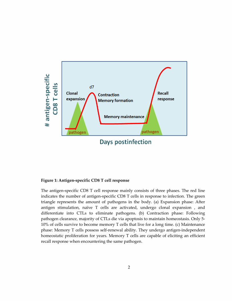
robust clonal expansion (Figure 1). Meanwhile, they differentiate into effector CD8 T
cells, also called cytotoxic T lymphocytes (CTLs). CTLs migrate to inflamed areas and
acquire effector functions essential to the elimination of pathogen-infected cells through
granule-dependent and Fas ligand (FasL)-dependent pathways (Kagi, Vignaux et al.
1994). The granule pathway relies on directed granule exocytosis, a perforin-mediated
directional transfer of granzymes from CTLs to target cells. The FasL pathway is
triggered by the interaction between FasL on CTLs and Fas on target cells, leading to
caspase activation and apoptotic cell death. Additionally, CTLs produce type I
cytokines, such as interferon-gamma (IFN-γ) and tumor necrosis factor-alpha (TNF-α), to
suppress replication of pathogen and trigger apoptosis of infected cells (Figure 2).
2
Figure 1: Antigen-specific CD8 T cell response
The antigen-specific CD8 T cell response mainly consists of three phases. The red line
indicates the number of antigen-specific CD8 T cells in response to infection. The green
triangle represents the amount of pathogens in the body. (a) Expansion phase: After
antigen stimulation, naïve T cells are activated, undergo clonal expansion , and
differentiate into CTLs to eliminate pathogens. (b) Contraction phase: Following
pathogen clearance, majority of CTLs die via apoptosis to maintain homeostasis. Only 5-
10% of cells survive to become memory T cells that live for a long time. (c) Maintenance
phase: Memory T cells possess self-renewal ability. They undergo antigen-independent
homeostatic proliferation for years. Memory T cells are capable of eliciting an efficient
recall response when encountering the same pathogen.
3
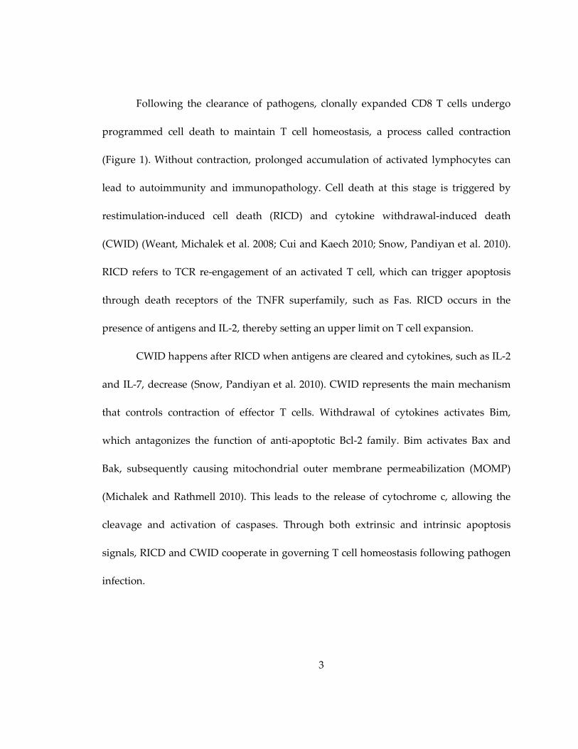
Following the clearance of pathogens, clonally expanded CD8 T cells undergo
programmed cell death to maintain T cell homeostasis, a process called contraction
(Figure 1). Without contraction, prolonged accumulation of activated lymphocytes can
lead to autoimmunity and immunopathology. Cell death at this stage is triggered by
restimulation-induced cell death (RICD) and cytokine withdrawal-induced death
(CWID) (Weant, Michalek et al. 2008; Cui and Kaech 2010; Snow, Pandiyan et al. 2010).
RICD refers to TCR re-engagement of an activated T cell, which can trigger apoptosis
through death receptors of the TNFR superfamily, such as Fas. RICD occurs in the
presence of antigens and IL-2, thereby setting an upper limit on T cell expansion.
CWID happens after RICD when antigens are cleared and cytokines, such as IL-2
and IL-7, decrease (Snow, Pandiyan et al. 2010). CWID represents the main mechanism
that controls contraction of effector T cells. Withdrawal of cytokines activates Bim,
which antagonizes the function of anti-apoptotic Bcl-2 family. Bim activates Bax and
Bak, subsequently causing mitochondrial outer membrane permeabilization (MOMP)
(Michalek and Rathmell 2010). This leads to the release of cytochrome c, allowing the
cleavage and activation of caspases. Through both extrinsic and intrinsic apoptosis
signals, RICD and CWID cooperate in governing T cell homeostasis following pathogen
infection.
4
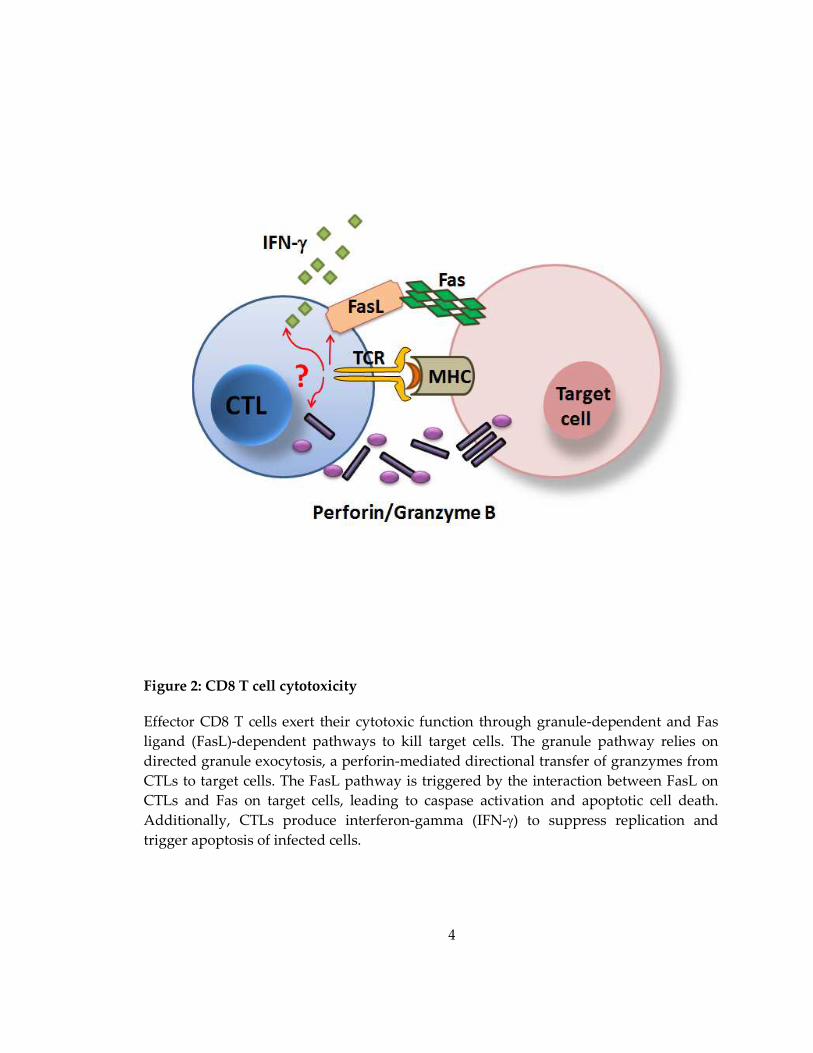
Figure 2: CD8 T cell cytotoxicity
Effector CD8 T cells exert their cytotoxic function through granule-dependent and Fas
ligand (FasL)-dependent pathways to kill target cells. The granule pathway relies on
directed granule exocytosis, a perforin-mediated directional transfer of granzymes from
CTLs to target cells. The FasL pathway is triggered by the interaction between FasL on
CTLs and Fas on target cells, leading to caspase activation and apoptotic cell death.
Additionally, CTLs produce interferon-gamma (IFN-γ) to suppress replication and
trigger apoptosis of infected cells.

5
Whereas the majority of CTLs terminally differentiate into end-stage effectors
that die via programmed cell death, a small subset (5-10%) of cells survive and convert
into memory precursors that eventually become long-lived memory T cells (Williams
and Bevan 2007; Harty and Badovinac 2008; Ahmed, Bevan et al. 2009; Jameson and
Masopust 2009). The fate of a particular T cell to become a terminally differentiated
effector cell or a memory T cell is determined by multiple factors and occurs early in the
process of clonal expansion (Williams and Bevan 2007; Cui and Kaech 2010). For
example, the magnitude of antigenic stimulation, the extent of co-stimulation, the
severity of inflammation, the availability of CD4 T cell help, and the cytokine conditions,
all can influence the expression of transcriptional factors that regulate the differentiation
of CD8 T cells. Importantly, heterogeneity of IL-7R expression on CD8 T cells at early
stages of immune response destines cells with distinct developmental potentials. IL-7Rlo
T cells tend to die and IL-7Rhi T cells preferentially give rise to memory precursors and
memory T cells (Kaech, Tan et al. 2003).
Cells that survive through the contraction phase, called memory CD8 T cells, are
distinguished by the ability to undergo long-term, antigen-independent proliferative
self-renewal. This process is heavily dependent on IL-7 and IL-15, two crucial cytokines
that regulate the survival and homeostatic proliferation of memory CD8 T cells (Hand
and Kaech 2009). In addition to their capacity for self-renewal, another unique feature of

6
memory CD8 T cells is their plasticity to differentiate into secondary effector cells that
have the ability to rapidly proliferate and reiterate their cytotoxic function upon re-
encounter with the same pathogen. The generation and maintenance of memory CD8 T
cells has been the focus of strategies to improve vaccination and immunotherapy.
1.2 Granule exocytosis
The main characteristic of CTLs is their capacity to elicit cytotoxicity toward target
cells. The granule-dependent pathway, which involves granule exocytosis, a directional
release of perforin and granzymes from CTLs into target cells, is a primary mechanism
that mediates CTL killing.
The process of granule exocytosis by CTLs has been well-characterized
(Stinchcombe and Griffiths 2007; de Saint Basile, Menasche et al. 2010) (Figure 3). When
CTLs interact with target cells, the engagement of TCR and MHC-peptide initiates the
formation of an immunological synapse (IS) at the T-target cell contact zone. The IS
consists of the TCR and additional signaling proteins that accumulate in the center of the
supramolecular activation cluster (cSMAC) that are surrounded by a peripheral ring
enriched in adhesion molecules (pSMAC) (Monks, Freiberg et al. 1998; Grakoui, Bromley
et al. 1999; Potter, Grebe et al. 2001; Stinchcombe, Majorovits et al. 2006). Within minutes
of TCR recognition, the microtubule organizing center (MTOC, also called the
centrosome) of the T cell polarizes toward the target cell and remains positioned beneath
7
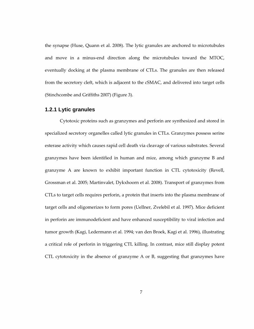
the synapse (Huse, Quann et al. 2008). The lytic granules are anchored to microtubules
and move in a minus-end direction along the microtubules toward the MTOC,
eventually docking at the plasma membrane of CTLs. The granules are then released
from the secretory cleft, which is adjacent to the cSMAC, and delivered into target cells
(Stinchcombe and Griffiths 2007) (Figure 3).
1.2.1 Lytic granules
Cytotoxic proteins such as granzymes and perforin are synthesized and stored in
specialized secretory organelles called lytic granules in CTLs. Granzymes possess serine
esterase activity which causes rapid cell death via cleavage of various substrates. Several
granzymes have been identified in human and mice, among which granzyme B and
granzyme A are known to exhibit important function in CTL cytotoxicity (Revell,
Grossman et al. 2005; Martinvalet, Dykxhoorn et al. 2008). Transport of granzymes from
CTLs to target cells requires perforin, a protein that inserts into the plasma membrane of
target cells and oligomerizes to form pores (Uellner, Zvelebil et al. 1997). Mice deficient
in perforin are immunodeficient and have enhanced susceptibility to viral infection and
tumor growth (Kagi, Ledermann et al. 1994; van den Broek, Kagi et al. 1996), illustrating
a critical role of perforin in triggering CTL killing. In contrast, mice still display potent
CTL cytotoxicity in the absence of granzyme A or B, suggesting that granzymes have
8
redundant roles and that loss of one granzyme can be compensated by other isoforms
(Ebnet, Hausmann et al. 1995; Smyth, Street et al. 2003).
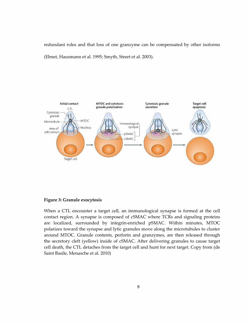
Figure 3: Granule exocytosis
When a CTL encounter a target cell, an immunological synapse is formed at the cell
contact region. A synapse is composed of cSMAC where TCRs and signaling proteins
are localized, surrounded by integrin-enriched pSMAC. Within minutes, MTOC
polarizes toward the synapse and lytic granules move along the microtubules to cluster
around MTOC. Granule contents, perforin and granzymes, are then released through
the secretory cleft (yellow) inside of cSMAC. After delivering granules to cause target
cell death, the CTL detaches from the target cell and hunt for next target. Copy from (de
Saint Basile, Menasche et al. 2010)
9
One interesting question that rose from studying cytotoxicity of CTLs was how
they protect themselves from the potential lethal effect of perforin. Perforin is
synthesized with an inhibitory propeptide which keeps itself in an inactive state. Once
transported to the acidic lytic granules (pH 5.1-5,4), the propeptide of perforin is cleaved
(Kataoka, Togashi et al. 1997). However, perforin is unable to exert its function in the
acidic environment of granules, thus it only acquires the pore-forming activity after
release from CTLs (Kuta, Bashford et al. 1991). Additionally, other molecules stored
within granules can function to inhibit perforin activity. For instance, calreticulin
deprives calcium from perforin (Fraser, Karimi et al. 2000) and serglycan complexs with
perforin to prevent its multimerization (Metkar, Wang et al. 2002). With multiple layers
of inhibitory regulation, CTLs ensure that perforin only elicit its cytotoxic function upon
secretion from CTLs.
1.2.2 Cytotoxic immunological synapse
Immunological synapse formed at the T cell-APC contact zone is an antigen-
specific cell-cell junction that allows for cellular communication between T cells and
APCs. The synapse was first described in CD4 T cells by Kupfer et al. (Monks, Freiberg
et al. 1998), which consists of the TCR and additional signaling proteins that accumulate
in a cSMAC and adhesion molecules LFA-1 and talin enriched in a pSMAC to stabilize

10
the synapse (Figure 4). Upon antigen stimulation, both TCR and adhesion proteins
concentrate at the center of the T cell-APC contact zone, and then segregate to form a
bull’s eye organization in which signaling molecules stay in the center and adhesion
molecules exclude from the center to form a surrounding ring. Some signaling proteins,
such as the phosphatase CD45, are excluded from the synapse to form a distal
supramolecular activation complex (dSMAC).
Similar to CD4 T cells, the cytotoxic synapse in CD8 T cells is also comprised of
cSMAC and pSMAC (Dustin and Long 2010). Beyond that, CD8 T cells have a
specialized secretory domain, which locates adjacent to the cSMAC, allowing for the
delivery of lytic granules (Figure 4). The secretory domain is surrounded by pSMAC
which can prevent leakage of cytolytic cargo and decrease the nonspecific killing of
bystander cells (Stinchcombe and Griffiths 2007; Huse 2012).
cSMAC is assembled by microclusters, transient structures composed of 30-300
TCRs as well as kinases and adaptor proteins measured by lipid bilayers (Yokosuka,
Sakata-Sogawa et al. 2005). As a T cell encounters an APC, microclusters are generated
and associate with dynein, a microtubule motor protein, which helps to move the
microclusters along microtubules toward the center of the cell contact region
(Hashimoto-Tane, Yokosuka et al. 2011). In addition to dynein, myosin IIA, an actin-
based motor protein, also plays an important role in microcluster assembly and

11
movement (Ilani, Vasiliver-Shamis et al. 2009). With the help of these motor proteins,
hundreds of microclusters move along the cytoskeleton and merge to become a cSMAC.
It was thought that cSMAC functioned to sustain TCR signaling required for T cell
activation by APCs (Grakoui, Bromley et al. 1999). Nonetheless, more recent studies
reveal that cSMACs do not emanate tyrosine kinase signaling during the T cell-APC
interaction. Rather, TCR signaling occurs in microclusters that merge to the edge of
cSMAC and stops when the cSMAC is formed, indicating that cSMAC actually functions
to downregulate TCR signaling (Varma, Campi et al. 2006).
pSMAC, enriched with adhesion molecules such as LFA-1 and ICAM-1, provides
structurally stabilization for cell-cell contact. Integrin is maintained in an inactive state
with a low affinity for its ligand. Engagement of the TCR triggers an inside-out
signaling, which causes the conformational change of integrin and augments its affinity
toward the ligands on APCs, allowing T cells to form stable conjugates with APCs. The
synapse formed between CD4 T cells and APCs is sustained for a long time (hours to
days) to maintain the maximal efficiency of cytokine secretion and T cell proliferation
(Huppa, Gleimer et al. 2003). As opposed to CD4 T cells, the synapse formed between
CD8 T cells and APCs is relatively transient, only lasting for less than 20-30 minutes
(Stinchcombe and Griffiths 2007). How CTLs know to dissociate from target cells after
they are programmed for lysis is still not clear.
12
Figure 4: Structure of immunological synapse of CD4 and CD8 T cells
Immunological synapse is formed as a bull’s eye organization at the T cell-APC contact
region and can be divided into three layers: cSMAC, pSMAC, and dSMAC. TCR, CD28,
CTLA-4 and signaling protein such as PKCθ are localized to cSMAC. Adhesion
molecules LFA-1 and talin as well as CD4 and Lck are found in pSMAC. Phosphatase
CD45 and CD43 reside in dSMAC. CD8 T cells have a specialized compartment called
13
secertory domain located adjacent to the cSMAC, which functions to deliver lytic
granules. Adapted from (Stinchcombe and Griffiths 2007).
It has been demonstrated that formation of a mature immunological synapse,
which requires persistent TCR signaling, is necessary for T cell activation; however, it is
not a prerequisite for CD8 T cells to exert cytotoxicity. Furthermore, approximately 10
peptides are required to form a stable, mature immunological synapse but only 3 Ova
peptides are needed in the synapse to elicit OT-I T cell killing (Purbhoo, Irvine et al.
2004). Collectively, these observations indicate that the triggering of CTL killing can be
very sensitive and does not require the formation of a mature synapse.
1.2.3 MTOC and granule polarization
MTOC and granule polarization is a critical step in the process of granule
exocytosis. When encountering an APC, a CD8 T cell undergoes a drastic structural
conformation that is associated with cytoskeleton remodeling of both actin and
microtubule networks (Huse 2012). Actin polymerization drives the leading edge of a T
cell into a radially symmetric lamellipodium that spreads over the surface of an APC. In
addition, the MTOC in a T cell polarizes toward to the T cell-APC contact region and
stays beneath the synapse with microtubules rotated toward its direction. This guides
the movement of lytic granules along the microtubules toward the synapse, thus
enabling the directional transfer of cytolytic molecules toward the target cells.

14
MTOC polarization is initiated by TCR signaling. Studies show that several
proximal signaling proteins downstream of TCR, including Lck, Fyn, Zap-70, LAT, SLP-
76, and Vav, are central for MTOC reorientation (Ardouin, Bracke et al. 2003; Kuhne, Lin
et al. 2003; Combs, Kim et al. 2006; Martin-Cofreces, Sancho et al. 2006). However, the
downstream signaling event that controls MTOC polarization, such as calcium flux, is
still controversial. While researchers observed that MTOC movement correlates with
higher intracellular calcium mobilization (Kuhne, Lin et al. 2003), another group
reported an opposite finding. They revealed that calcium-independent diacylglycerol
(DAG) accumulation at the synapse recruits dynein to reorient the MTOC (Quann,
Merino et al. 2009). DAG is a lipid secondary messenger generated by phospholipase C-γ
(PLC-γ), and transduces signals by recruiting proteins that contains DAG-binding C1
domains to the cell membrane. This discovery immediately suggested that the PKC
family which contain C1 domains, play a role in MTOC reorientation (Huse 2012).
Interestingly, TCR stimulation induced a robust synapse recruitment of not only
PKCθ, but also PKCε and PKCη. The subsequent siRNA experiments demonstrated that
all three PKCs participate in MTOC reorientation, but PKCε and PKCη can functionally
compensate for each other (Quann, Liu et al. 2011). In line with this observation, one
study demonstrated that granules can be polarized to the T cell:APC contact region

15
when calcium was depleted, further suggesting that MTOC and granule polarization is
not guided by calcium signaling (Lyubchenko, Wurth et al. 2001).
Several cytoskeletal regulators downstream of TCR signaling have been linked to
the regulation of MTOC movement toward the synapse. Among those, dynein, a
microtubule motor protein, is recruited to the synapse upon TCR engagement (Combs,
Kim et al. 2006). Therefore, it is thought that the dynein–dynactin complex in the
synapse can pull microtubules to reorient the MTOC. Indeed, knockdown of dynein by
siRNA impaired MTOC polarization in Jurkat T cells (Martin-Cofreces, Robles-Valero et
al. 2008). How dynein is recruited to the synapse is not well understood.
Since dynein associates with the cytoskeletal adaptor molecule ADAP, it has
been proposed that dynein can be recruited to the synapse by the SLP-76-ADAP
complex (Combs, Kim et al. 2006). Knockdown of ADAP indeed blocks polarization of
both dynein and MTOC in Jurkat T cells. However, this phenomenon failed to be seen in
mouse primary T cells, suggesting that other ADAP-independent mechanisms are
responsible for driving dynein and MTOC reorientation in primary T cells. While it is
generally thought that the dynein-dynactin complex plays a critical role in MTOC
polarization in T cells, other cytoskeletal regulators of actin, such as formin diaphanous
1 (Dia) and the scaffolding molecule IQ-motif-containing GTPase activating protein
homolog (IQGAP), have been also shown to regulate MTOC polarization.

16
As the microtubule network polarizes, the lytic granules move along the
microtubules toward the MTOC, subsequently resulting in an accumulation of the
granules at the contact region of T cell and APC. Lytic granules dock with the plasma
membrane and release their cytolytic contents through the secretory cleft in the cSMAC,
thus triggering rapid death of the target cells. Studies indicate that the secretion of
granules requires calcium mobilization (Lyubchenko, Wurth et al. 2001), and molecules
involved in this process include Rab27a, Munc13-4, and several other proteins
(Stinchcombe and Griffiths 2007). In Rab27a-deficient patients and mouse model, the
granules polarize at the MTOC but fail to dock at the synapse in CTLs and NK cells,
suggesting that Rab27a is required for the granules to dissociate from the microtubules
(Tolmachova, Anders et al. 2004).
Munc13-4 is a protein that associates with Rab27a and colocalizes with perforin
when the granules are polarized. In the absence of Munc13-4, the granules polarize and
dock at the synapse but are unable to secret their cytolytic factors, indicating that
Munc13-4 is required for granule fusion and secretion (Feldmann, Callebaut et al. 2003;
Menager, Menasche et al. 2007). Altogether, these findings demonstrate that granule
exocytosis involves many steps, including synapse and conjugate formation, MTOC and
granule polarization, and lytic granule secretion, and each step is tightly regulated to
ensure an efficient and specific CTL killing.
17
1.3 T Memory CD8 T cell formation
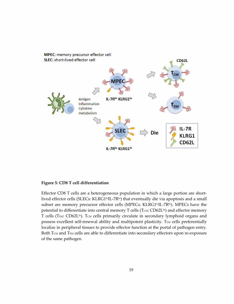
1.3.1 Memory precursor effector cell (MPEC) vs Short-lived effector cell (SLEC)
Following antigen stimulation, activated CD8 T cells consist of a heterogeneous
population in which a large portion are short-lived effector cells (SLECs) that eventually
die via apoptosis, and the remaining small subset are memory precursor effector cells
(MPECs) that have the potential to become memory T cells (Figure 5) (Kaech and
Wherry 2007). A single naïve T cell can differentiate into a diverse population composed
of both SLECs and MPECs, indicating that their fate specification is not pre-
programmed but occurs after T cell activation (Stemberger, Huster et al. 2007).
Memory differentiation is a process where the phenotypic and functional
properties of memory T cells are acquired over a period of time (Sarkar, Kalia et al.
2008). Expression of several biomarkers has been shown to correlate with the memory
potential of CD8 effector T cells. IL-7R (CD127) was the first marker identified to
differentiate MPECs from SLECs (Kaech, Tan et al. 2003). IL-7R is expressed on naïve T
cells, down-regulated on effector T cells, and up-regulated again once becoming
memory T cells. IL-7 signaling provides survival for T cells, thus IL-7Rhi cells
preferentially become MPECs following pathogen infection. Other molecules, such as
Bcl-2, CXCR3, CD27, and CD28, are also highly expressed in MPECs (Joshi, Cui et al.
18
2007; Kaech and Wherry 2007), providing additional markers to distinguish MPECs
from SLECs.
In contrast, enhanced expression of KLRG1 (NK cell inhibitory receptor) and
CD57 are associated with terminally differentiated and senescent CD8 effector T cells. It
has been demonstrated that KLRG1hi effector cells are committed to downregulating IL-
7R, guiding their fate to be short-lived. Therefore, based on expression of KLRG1 and IL-
7R, KLRG1loIL-7Rhi CD8 T cells are referred as MPECs and KLRG1hiIL-7Rlo CD8 T cells
are referred as SLECs (Joshi, Cui et al. 2007; Kaech and Wherry 2007).
1.3.2 Central memory T cell (TCM) vs Effector memory T cell (TEM)
MPECs eventually develop into long-lived memory T cells. Similar to effector T
cells, CD8 memory T cells are also heterogeneous and can be classified into
subpopulations (TCM & TEM ) based on tissue distribution, the expression of lymph node
homing molecules, and functional properties (Figure 5) (Sallusto, Lenig et al. 1999; Cui
and Kaech 2010). Central memory T (TCM) cells express CD62L and CCR7 and primarily
circulate in secondary lymphoid organs. They are capable of producing IL-2 and
undergo IL-7/IL-15-dependent, and antigen-independent homeostatic proliferation.
Upon encountering re-infection, TCM cells expand vigorously and differentiate into
secondary effectors to provide immediate protection. Due to their superior capacity for
19
Figure 5: CD8 T cell differentiation
Effector CD8 T cells are a heterogeneous population in which a large portion are short-
lived effector cells (SLECs: KLRG1hiIL-7Rlo) that eventually die via apoptosis and a small
subset are memory precursor effector cells (MPECs: KLRG1loIL-7Rhi). MPECs have the
potential to differentiate into central memory T cells (TCM: CD62Lhi) and effector memory
T cells (TEM: CD62Llo). TCM cells primarily circulate in secondary lymphoid organs and
possess excellent self-renewal ability and multipotent plasticity. TEM cells preferentially
localize in peripheral tissues to provide effector function at the portal of pathogen entry.
Both TCM and TEM cells are able to differentiate into secondary effectors upon re-exposure
of the same pathogen.
20
self-renewal, TCM cells eventually become a dominant memory population in lymphoid
tissues.
Unlike TCM cells, effector memory T (TEM) cells lack the expression of CD62L and
CCR7 and preferentially localize in peripheral tissues (Masopust, Vezys et al. 2001).
They are unable to make IL-2, have limited self-renewal potential and poor proliferative
ability upon recall challenge. However, they express cytotoxic molecules and are able to
exert potent cytotoxicity during the recall response. In general, most experimental
observations are pointing toward the idea that tissue–resident TEM cells provide effector
function at the portal of pathogen entry, while TCM cells serve as the stem cell-like
population that maintain lifelong immunological memory.
1.3.3 Memory Stem Cells (TSCM)
Recently, a new population of memory CD8 T cells has been identified in mouse
and human with superior self-renewal and multipotent plasticity compared to TCM and
TEM cells. These memory T cells were designated memory stem T (TSCM) cells because of
their stem-cell like properties. Mouse TSCM cells express CD44lo CD62Lhi Sca-1hi Bcl-2hi IL-
2Rβhi CXCR3hi, and this population can be efficiently generated in vitro by triggering
Wnt signaling during T cell priming (Gattinoni, Zhong et al. 2009). Human TSCM cells
were identified as a population that is CD45ROlo CCR7hi CD45RAhi CD62Lhi CD27hi
CD28hi IL-7Rhi. In addition to enhanced self-renewal and multipotency, human TSCM cells

21
exhibit better proliferation, survival, and antitumor ability than TCM and TEM cells
(Gattinoni, Lugli et al. 2011). Comparing the transcriptome of TSCM cells with naïve (TN)
and memory T cell subsets, it was found that the expression of effector-associated genes
gradually increased and the expression of memory-associated genes progressively
decreased in the order of TN >TSCM >TCM >TEM (Gattinoni, Lugli et al. 2011). This
observation indicates that TSCM cells are the least differentiated memory T cell
population, thus enabling them to have superior plasticity to differentiate into other cell
types, such as TCM, TEM and secondary effector T cells.
1.3.4 Extrinsic programming of T cell differentiation
The fate of a particular cell to become a SLEC or MPEC (and TCM or TEM) is
determined by multiple factors. Several lines of evidence have suggested that CD8 T cell
differentiation is primarily destined during the encounter of a naïve T cell with antigens
in the process of T cell expansion (Cui and Kaech 2010). This event can be influenced by
multiple environmental factors, such as the magnitude of antigenic stimulation, extent of
co-stimulation, inflammation, cytokine conditions, and the availability of CD4 T cell
help (Figure 5).
Weak signals derived from antigenic stimulation are sufficient to trigger the
differentiation from naïve to effector and memory T cells (Zehn, Lee et al. 2009);
however, the strength of stimulation controls the decision of memory cell fate (Sarkar,
22
Teichgraber et al. 2007; Jung and Kaech 2010). Studies demonstrate that cells receiving
strong antigenic stimulation or continued stimulation during the later stage of infection
are more likely to become terminally differentiated and die (Sarkar, Kalia et al. 2008). In
contrary, decreasing the amount, duration, and affinity of antigenic stimulation
enhances the generation of memory T cells (van Faassen, Saldanha et al. 2005). Based on
these findings, a model has been proposed in which the initial antigenic stimulation
creates a threshold of signaling strength that controls T cell differentiation (Jung and
Kaech 2010). T cells that receive insufficient stimulation become nonresponsive, while T
cells that encounter excessive stimulation undergo terminal differentiation and cell
death. In between these two extreme scenarios are T cells that receive intermediate
stimulation which have the potential to differentiate into memory precursors.
In addition to antigen stimulation, CD4 T cell represents another extrinsic factor
that regulates the differentiation, survival, and effector function of memory CD8 T cells
(Kaech and Ahmed 2001). The primary response of CD8 T cell occurs normally in the
absence of CD4 T cells; however, the recall response is severely compromised (Belz,
Wodarz et al. 2002; Sun and Bevan 2003). Selectively depleting CD4 T cells during the
recall challenge allowed for normal secondary CD8 T cell response, indicating that CD4
T cell help is required prior to recall response (Sun and Bevan 2003). To further dissect
the timing of the requirement of CD4 T cells, CD8 T cells were adoptively transferred

23
into the CD4 T cell-deficient mice at different stages. The subsequent results revealed
that the presence of CD4 T cells during the formation and maintenance of memory CD8
T cells is essential to maintain the functionality and quantity of these CD8 T cells (Sun,
Williams et al. 2004).
Similar to CD4 T cells, the absence of IL-2 during the priming phase does not
influence the generation of effector and memory CD8 T cells; however, it drastically
affects the magnitude of secondary immune responses (Williams, Tyznik et al. 2006).
This observation is not due to a requirement of IL-2 in CD8 T cell activation and
expansion during the recall response. Instead, the presence of IL-2 during the primary
response is essential for the development of functional memory CD8 T cells. Recently,
the roles of CD4 T cell help and IL-2 signaling in CD8 T cell differentiation have been
linked via CD25 upregulation (Obar, Molloy et al. 2010). Researchers observed that the
expression of IL-2R, CD25, on antigen-specific CD8 T cells peaked 3-4 days after initial
priming, and this event was dependent on CD4 T cell help. Uptake of IL-2 during the
process of T cell expansion is important for the survival of SLECs and the generation of
functional memory CD8 T cells.
1.3.5 Intrinsic programming of T cell differentiation
In addition to environmental stimuli, memory CD8 T cell differentiation is
tightly controlled by intrinsic programming, including transcriptional regulators,

24
metabolic switches, and the strength of TCR signaling (Cui and Kaech 2010). Just as how
Th differentiation is regulated in CD4 T cells, transcriptional regulation plays an
imperative role in effector CD8 T cell differentiation. T-bet, the first transcription factor
known to modulate CD8 T cell fate decision, regulates gene expression of IFN-γ and the
cytotoxic molecules, perforin and granzyme B. T-bet expression is induced in effector
CD8 T cells and can be enhanced by the inflammatory cytokine, IL-12 (Joshi, Cui et al.
2007). IL-12 induces a gradient of T-bet expression, which decides the fate of a CD8 T
cell to become SLEC or MPEC (Joshi, Cui et al. 2007). Higher amounts of T-bet promote
SLEC formation, whereas lower amounts of T-bet instruct MPEC development (Joshi,
Cui et al. 2007).
Another T-box factor expressed in effector CD8 T cells is Eomesodermin (Eomes),
which also controls the expression of perforin in effector T cells in a cooperative manner
with T-bet (Pipkin, Sacks et al. 2010). However, in contrast to T-bet, Eomes expression
preferentially increases as memory CD8 T cells form. Furthermore, while T-bet
expression is augmented by IL-12 and mTOR pathways, Eomes expression is decreased
under the same conditions (Takemoto, Intlekofer et al. 2006; Rao, Li et al. 2010),
suggesting that these two transcription factors have reciprocal roles in the regulation of
CD8 T cell differentiation. Collectively, these studies demonstrate that the balance
between T-bet and Eomes is a key to determine effector versus memory CD8 T cell fate.

25
In addition to T-bet, Blimp-1 is another transcriptional repressor that functions to
promote the differentiation of CD8 T cells into SLECs. Blimp-1-deficient CD8 T cells
develop into MPECs more rapidly than their WT counterparts (Rutishauser, Martins et
al. 2009), but this mechanism was not well understood at the time. Recently, studies of
E-protein transcription factors in T cell differentiation have begun uncovering this
mystery relationship. Specifically, an association between Blimp-1 and inhibitor of DNA
binding (Id) family was identified, and Id3 is required for the generation of long-lived
memory CD8 T cells (Ji, Pos et al. 2011; Yang, Best et al. 2011). Moreover, Blimp-1 and a
number of cytokines known to promote effector T cell development such as IL-2, IL-12,
and IL-21, are shown to downregulate Id3 and upregulate Id2 expression, which in turn
regulate genes involved in genome stability.
Metabolism is another intrinsic factor that determines the fate of effector CD8 T
cells. The metabolic states of resting cells and activated cells are quite different. Upon
infection, effector CD8 T cells undergo clonal expansion, and they increase glucose
uptake and utilize anabolic metabolism that is characterized by enhanced mammalian
target of rapamycin (mTOR) activity and glycolysis. After pathogen clearance, effector T
cells halt proliferation and enter a quiescent stage, where the mTOR pathway is
inhibited and they primarily consume energy through oxidative phosphorylation in
mitochondria. Studies have revealed that the regulation of the metabolic state of effector

26
CD8 T cells plays an important role in deciding their effector versus memory fates (Chi
2012). For example, blockage of mTOR activity by rapamycin enhances the quantity and
quality of memory T cells during LCMV infection (Araki, Turner et al. 2009). Further
investigations revealed that mTOR determines effector versus memory CD8 T cell fate
by regulating the expression of T-bet and Eomes in a Foxo1-dependent manner (Rao, Li
et al. 2010; Rao, Li et al. 2012).
In conjunction with these findings, another study reported that in the absence of
TNF receptor-associated factor 6 (TRAF6), a critical factor for induction of fatty acid
oxidation (FAO), the generation of memory CD8 T cell was defective following Listeria
infection (Pearce, Walsh et al. 2009). Therefore, it appears that the regulation between
mTOR-mediated cell proliferation (anabolism) and FAO-mediated cell homeostasis
(catabolism) is one of the intrinsic factors that control memory T cell differentiation.
1.4 LAT-mediated signaling
1.4.1 Early events in TCR signaling
Immune cells, such as T and B cells, rely on signal transduction to activate
transcription factors that regulate genes required for development, differentiation,
activation, and effector functions. Initiation of signaling cascades in T cells occurs mainly
through the interaction between the T cell receptor (TCR) on T cells and the MHC-
peptide complex on antigen-presenting cells (Samelson 2002). Based on their receptors, T
27
cells can be divided into αβ and γδ T cells. αβ T cells are further sub-categorized as CD4
and CD8 T cells according to the co-receptor expression. The TCR of αβ T cells is a
complex consisting of an α and β chain noncovalently associated with the CD3 proteins,
including γε, δε, and ζζ subunits. Following the engagement of the TCR, coreceptor-
associated Lck (lymphocyte-specific protein tyrosine kinase), a Src family kinase, is
brought in proximity of the TCR complex and phosphorylates the immunoreceptor
tyrosine-based activation motifs (ITAMs) of TCRζ (Reth 1989; Cambier 1992; Cambier
1995). Phosphorylated ITAMs of TCRζ recruit Zap-70 (ζ-chain-associated protein kinase
of 70 kDa), which is a Syk family kinase, by associating with the tandem SH2 domains of
Zap-70. The binding of TCRζ causes the conformational change of Zap-70, exposing its
regulatory domains for Lck-mediated phosphorylation or auto-phosphorylation by Zap-
70 itself, which in turn promotes the catalytic function of Zap-70 (Salojin, Zhang et al.
2000).
Subsequently, a number of signaling scaffold proteins, such as the linker for
activation of T cells (LAT) and SH2-domain-containing leukocyte protein of 76 kDa
(SLP-76), are phosphorylated by Zap-70 and then act as scaffold proteins to recruit
numerous signaling molecules within close proximity to the TCR (Wang, Kadlecek et al.
2010). Phosphorylated LAT binds directly to Grb2, Gads, and PLC-γ1, as well as
indirectly associates with SLP-76, Vav, and other signaling proteins (Liu and McGlade

28
1998; Zhang, Sloan-Lancaster et al. 1998; Jordan, Singer et al. 2003). LAT binding to Grb2
leads to the membrane recruitment of Sos, a guanine nucleotide exchange factor
(RasGEF), which activates Ras. Interestingly, TCR-mediated Ras activation also requires
RasGRP1 (Ebinu, Stang et al. 2000). The interaction of LAT with PLC-γ1 and the Gads-
SLP-76 complex is critical for the activation of PLC-γ1 which is essential for initiating
calcium signaling (Figure 6).
1.4.2 Linker for activation of T cells (LAT)
Transmembrane adaptor proteins (TRAPs) do not possess intrinsic kinase
activity but contain many tyrosine-based signaling motifs that are phosphorylated by
catalytically-active Syk kinases. TRAPs function as scaffolds by recruiting signaling and
effector molecules. Several TRAPs including LAT, LAB (linker for activation of B
cells)/NTAL (non-T cell activation linker), and LAX (linker for activation of X cells), have
been identified in T cells and their roles in TCR-mediated signaling have been
highlighted in studies using cell lines and mouse models (Zhu, Granillo et al. 2005; Zhu,
Koonpaew et al. 2006). Studies from collective work show that these TRAPs are able to
exert both positive and negative effects on TCR signaling pathways that regulate
immune responses.
LAT, a TRAP that plays an indispensable role in bridging TCR engagement to
downstream signaling events, is essential for T cell development and function. LAT is a
29
membrane bound protein expressed in thymocytes, mature T cells, NK cells, mast cells,
platelets/megakaryocytes, and pre-B cells. It is a 36-38 KDa protein that is heavily
phosphorylated upon TCR stimulation, and contains a short extracellular domain,
transmembrane domain, and cytoplasmic tail. Of the nine conserved tyrosine residues
within the cytoplasmic domain, the distal four tyrosines are required for binding of
Grb2, Gads, phospholipase C-γ1 (PLC), and the p85 subunit of phosphatidylinositol 3-
kinase (PI3K) (Liu and McGlade 1998; Zhang, Sloan-Lancaster et al. 1998; Jordan, Singer
et al. 2003). Moreover, two cysteine residues localized in the juxtamembrane region of
LAT are crucial for LAT palmitoylation and raft localization (Zhang, Trible et al. 1998).
While it remains a point of controversy whether LAT localization to lipid rafts has any
physiological relevance to T cell responses (Zhu, Shen et al. 2005), the significance of
palmitoylation in regulating LAT function has been determined (Zhang, Trible et al.
1998).
The indispensable function of LAT in TCR-mediated signaling was initially
identified in LAT-deficient Jurkat T cells. While proximal TCR signaling is unaffected,
these cells exhibit defective calcium mobilization, Erk activation, CD69 upregulation,
and AP (activator protein)-1- and NFAT (nuclear factor of activated T cells)-dependent
gene transcription (Finco, Kadlecek et al. 1998; Zhang, Irvin et al. 1999). Because LAT

does not have intrinsic enzymatic activity, the ability of LAT to transmit signals relies
heavily upon its phosphorylation.
Figure 6: TCR-mediated signaling
TCR engagement leads to the recruitment of Zap
LAT and SLP-76. Phosphorylated LAT recruits Grb2, Gad, and PLC
Sos to initiate Ras activat
SLP-76 is able to cleave PIP
induces calcium flux.
30
does not have intrinsic enzymatic activity, the ability of LAT to transmit signals relies
heavily upon its phosphorylation.
mediated signaling
TCR engagement leads to the recruitment of Zap-70 which phosphorylates and activates
76. Phosphorylated LAT recruits Grb2, Gad, and PLC-γ
Sos to initiate Ras activation. PLC-γ1 which is structurally stabilized by LAT, Gads, and
76 is able to cleave PIP2 into DAG and IP3. DAG activates Ras-Erk pathway while IP
does not have intrinsic enzymatic activity, the ability of LAT to transmit signals relies
70 which phosphorylates and activates
γ1. Grb2 binds to
1 which is structurally stabilized by LAT, Gads, and
Erk pathway while IP3
31
Mutating the four distal tyrosines (human Y132, Y171, Y191, and Y226) of LAT renders
Jurkat T cells completely unresponsive to TCR ligation.
Additionally, it was identified that Grb2 binds to Y171, 191, and 226, Gads binds
to Y171 and 191, and PLC-γ1 associates with Y132, further demonstrating that the
recruitment of Grb2, Gads, and PLC-γ1 is critical for LAT-mediated signaling. The role
of LAT in thymocyte development was evaluated in LAT-deficient mice. Analysis of
these mice revealed a lack of double-positive (DP) and single-positive (SP) thymocytes,
and consequently no peripheral T cells were generated, indicating that thymocyte
development is completely blocked at the double-negative (DN) stage in the absence of
LAT (Zhang, Sommers et al. 1999). Knock-in mice harboring mutations at the four distal
tyrosine residues have an identical phenotype as LAT-deficient mice, emphasizing the
importance of LAT phosphorylation in thymocyte development (Sommers, Menon et al.
2001).
To further determine the role of LAT beyond the DN3 stage, our lab generated
LAT knock-in mice in which the lat gene can be deleted by the Cre recombinase (Shen,
Zhu et al. 2009). Mice crossed onto a CD4Cre background allow the deletion of LAT to
occur at the late DN3 stage. Analysis of these mice showed that DP thymocytes were
formed but the transition from DP to SP was drastically blocked. Collectively, these
32
studies demonstrate that LAT is imperative during both early and late stages of
thymocyte development.
1.4.3 PLC-γγγγ1 and calcium mobilization
Upon TCR engagement, PLC-γ1 is one of the most important molecules
immediately downstream of LAT phosphorylation. PLC-γ1 is recruited to the proximal
TCR siganlsome by LAT and SLP-76 which in turn brings Itk (IL2-inducible T cell
kinase) to phosphorylate and activate PLC-γ1. Activated PLC-γ1 is able to hydrolyze the
membrane lipid phospholipid phosphatidylinositol 4,5 biphosphate (PIP2) to inositol
triphosphate (IP3) and diacylglycerol (DAG) (Rhee 2001). These two second messengers
play essential roles in T cell function. While DAG serves to activate the Ras-MAPK
pathway, IP3 binds to the IP3 receptor on the endoplasmic reticulum (ER) membrane,
triggering the release of calcium.
The depletion of calcium from the ER lumen results in the clustering of the Ca2+
sensors stromal interaction molecules (STIM) on the ER membrane, subsequently
causing the rise in store-operated Ca2+ entry (SOCE) (Liou, Kim et al. 2005; Roos,
DiGregorio et al. 2005). Moreover, the oligomerization of STIM1 triggers the opening of
Orai1 channels, also known as Ca2+ release-activated Ca2+ (CRAC) channels, resulting in
a sustained Ca2+ flux into the cytoplasm (Feske, Gwack et al. 2006; Luik, Wu et al. 2006;
Luik, Wang et al. 2008). The elevated Ca2+ concentration in T cells activates the

33
calcineurin-NFAT pathway, leading to the translocation of NFAT into the nucleus
(Hogan, Chen et al. 2003). In conjunction with other transcription factors, NFAT is able
to mediate gene transcription required for T cell development, differentiation, and
function.
The critical role of PLC-γ1 in T cell signaling has been investigated using animal
models. Since the ablation of PLC-γ1 is embryonic lethal, studies using PLC-γ1
conditional knockout mice that allow for the generation of PLC-γ1-deficient T cells,
demonstrate that these T cells fail to flux calcium, activate MAPKs, produce IL-2, and
proliferate in response to TCR stimulation (Fu, Chen et al. 2010). As a result of these
defects, these mice have impaired thymocyte development and develop an autoimmune
disease with age. Interestingly, a recent study from the same group reported that PLC-
γ2, which is known to be involved in B cell signaling, also plays a role in T cell signaling.
They observed that PLC-γ1/PLC-γ2-double deficient T cells display a more severe defect
in TCR signal transduction than PLC-γ1-deficient T cells (Fu, Chen et al. 2012).
Moreover, the PLC-γ1/PLC-γ2-double deficient mice demonstrate a further block in
thymocyte development. Altogether, these findings indicate that PLC-γ1 and PLC-γ2
cooperate in governing TCR signaling.

34
1.4.4 Inducible LAT deletion system
The function of LAT in mature T cells was unable to be determined since no
peripheral T cells are formed in LAT-deficient mice. To study the role of LAT in mature
T cells, our lab crossed LAT floxed knock-in mice onto an ERCre background in which
the lat gene can be inducibly deleted upon tamoxifen administration. In LATf/f mice, the
functional exons 7-11 of the lat gene, which encode the region containing critical tyrosine
residues that bind Grb2, Gads, and PLC-γ1, are flanked by two Loxp sites (Shen, Zhu et
al. 2009). Cre-mediated deletion results in the expression of a non-functional LAT-GFP
fusion protein, thus accurately marking T cells with the lat gene deleted (Figure 7).
LAT deficiency in mature CD4 T cells dramatically diminishes TCR-mediated
signaling, rendering T cells unresponsive to TCR stimulation (Shen, Chuck et al. 2010).
Correspondingly, these cells display defective homeostatic proliferation and long term
survival. Furthermore, deletion of LAT in mature regulatory T cells (Tregs) leads to
reduced expression of Foxp3, CTLA-4, and CD25, and subsequently impairs their
suppressive function (Chuck, Zhu et al. 2010). Collectively, these findings indicate that
LAT-mediated signaling is essential for T cell activation and survival, as well as the
suppressive function mediated by Tregs.
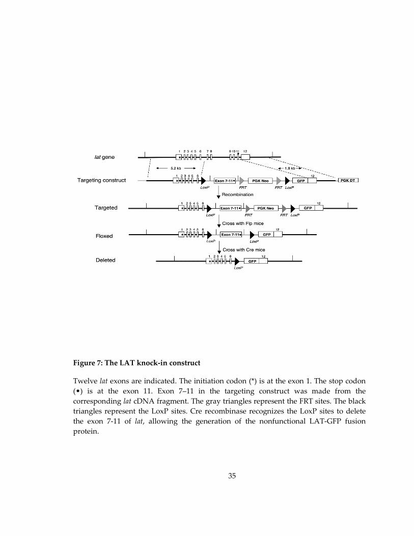
Figure 7: The LAT knock
Twelve lat exons are indicated. The initiation codon (*) is at the exon 1. The stop codon
(•) is at the exon 11. Exon 7
corresponding lat cDNA fragment. The gray triangles represent the FRT sites. The black
triangles represent the LoxP sites.
the exon 7-11 of lat, allowing the generation of the nonfun
protein.
35
The LAT knock-in construct
exons are indicated. The initiation codon (*) is at the exon 1. The stop codon
(•) is at the exon 11. Exon 7–11 in the targeting construct was made from the
cDNA fragment. The gray triangles represent the FRT sites. The black
epresent the LoxP sites. Cre recombinase recognizes the LoxP sites to delete
, allowing the generation of the nonfunctional LAT
exons are indicated. The initiation codon (*) is at the exon 1. The stop codon
11 in the targeting construct was made from the
cDNA fragment. The gray triangles represent the FRT sites. The black
Cre recombinase recognizes the LoxP sites to delete
ctional LAT-GFP fusion
36
2. Materials and Methods
2.1 Mouse models
LAT-/- and LATf/f mice were generated as previously described (Zhang, Sommers
et al. 1999; Shen, Zhu et al. 2009). LAT-/- mice were crossed with ERCre transgenic mice
(kindly provided by Dr. Thomas Ludwig, Columbia University, New York, NY) to
produce ERCre+LAT-/- mice. LATf/f mice were crossed with OT-I transgenic mice (Jackson
lab, Bar Harbor, ME) to generate OT-I/LATf/f mice, which were then mated with
ERCre+LAT-/- mice to obtain ERCre+LATf/-OT-I or LATf/-OT-I mice. These mice have
been backcrossed onto the C57/Bl6 background for more than ten generations. All mice
were used in accordance with the NIH guidelines. The procedures in this study were
approved by the Duke University IACUC. Mice were housed in a specific pathogen-free
facility.
2.2 Inducible deletion of LAT
2.2.1 Deletion of LAT in vivo
To delete the flexed lat allele in vivo, 1.5mg of tamoxifen (Sigma) was dissolved in
150μl corn oil. Mice were injected intraperitoneally for two consecutive days. Following
the initial deletion, mice were administered the same amount of tamoxifen once every
other week.
37
2.2.2 Generation of LAT-deficient effector CD8 T cells in vitro
Lymphocytes from OT-I mice were stimulated with 1 μM OVA peptide (257-264)
for 48 hours. After peptide stimulation, cells were washed to remove peptides and
cultured in the presence of IL-2 (10 ng/ml). To delete the flexed lat allele in vitro, cells
were incubated with 50 nM 4-hydroxytamoxifen (4-OHT, Sigma) for 72-96 hours
following activation with OVA peptide.
2.3 Infection
Mice received 1.5*104 CFU of Lm-Ova intravenously via retro-orbital injection to
initiate the acute infection. For recall challenge, 1.5*105 CFUs of Lm-Ova were injected
into the same mice with the same method.
2.4 Antibodies
2.4.1 Antibodies for flow cytometry
For use in flow cytometry, the cell viability marker 7AAD was purchased from
Invitrogen (Carlsbad, CA). Live/dead Violet Viability kit was purchased from Life
Technology. The following antibodies were used to stain cell surface markers and
intracellular molecules and were acquired from eBioscience and Biolegend (San Diego,
CA): CD4, CD8α, Th1.1, Th1.2, TCRVα2, FasL, TCRβ, CD25, CD69, CD62L, CD44,
KLRG1, CD127, CD122, CXCR3, CD27, Bcl-2, T-bet, Eomes, Perforin, IFN-γ, TNF-α, and
IL-2.
38
2.4.2 Antibodies for biochemical studies
Anti-LAT Ab#1 is homemade anti-LAT rabbit sera (#3023) and polyclonal anti-
LAT Ab#2 was purchased from Cell Signaling (#9166). Anti-perforin antibody was
obtained from Kamiya Biomedical and anti-granzyme B antibody was acquired from
R&D systems. Monoclonal anti-phosphotyrosine antibody (4G10) was purchased from
Upstate Biotechnology. Anti-pLAT (Y191), pERK, ERK2, pAkt (Ser473, Thr308), pan Akt,
pPLCγ1, PLCγ1, pPKCθ, and PKCθ antibodies were obtained from Cell signaling.
Secondary antibodies, such as goat anti-mouse and anti-rabbit Ig conjugated with Alexa
Fluor 680, were purchased from Molecular Probes. IRDye anti-GFP antibody was
bought from Rockland.
2.4.3 Antibodies for immunofluorescence
Anti-LAT (#9166) and anti-PKCθ antibodies were obtained from Cell Signaling.
Anti-LFA-1 and APC conjugated anti-granzyme B antibodies were purchased from
eBioscience. Anti-tubulin antibody was bought from Sigma. Secondary antibodies, such
as anti-mouse and anti-rabbit Ig conjugated with Alexa Fluor 488 were acquired from
Molecular Probes. Cy3-conjugated anti-rabbit and Cy5-conjugated anti-rat IgG were
purchased from Jackson Laboratory.

39
2.5 Flow cytometry
2.5.1 Cell surface staining
Single cell suspensions were prepared by homogenizing spleens and lymph
nodes. Splenocytes and blood were resuspended in ACK lysis buffer (0.15M NH4CL,
10mM KHCO3, 0.1mM EDTA pH7.4) for few minutes to lyse red blood cells. Cell
suspensions were filtered through mesh to remove debris and adjusted to the desired
concentration. To stain cell surface markers, cells were incubated with 2.4G2 (anti-
FcγII/III receptor) for 15 minutes on ice followed by staining with a mixture of different
fluorochrome-conjugated antibodies for another 15 minutes on ice. Cells were then
washed with FACS buffer (2% FBS in PBS) and collected on FACSCanto II (BD
Biosciences). FACS plots were analyzed with FlowJo software (Ashland, OR).
2.5.2 Intracellular staining
To stain intracellular molecules and transcription factors, cells were first stained
for surface markers as described above, and fixed and permeabilized using BD
Cytofix/Cytoperm kit according to the manufacturer’s instructions. Cells were then
stained on ice for 20-30 minutes with fluorochrome-conjugated antibodies. For
intracellular staining of cytokines, splenocytes were stimulated with Ova peptide (257-
264, 1 μM) or with PMA (20ng/mL) and ionomycin (0.5μg/mL) for 4 hours in the
presence of Golgi-stop. Cells were then fixed, permeabilized, and stained with

40
fluorochrome-conjugated antibodies against IFN-γ, TNF-α, and IL-2. To detect
upregulation of FasL, 5x10⁵ T cells were incubated with 5x10⁵ EL4 cells preloaded with
Ova peptide and PE-conjugated anti-FasL antibody for 5 hours. Cells were then stained
with anti-CD8 and analyzed.
2.6 T cell activation and Western blot analysis
For detecting LAT, perforin and gzmB, CTLs treated with 4-OHT for 3-4 days
were lysed, resolved by SDS-PAGE, and blotted with anti-perforin (Kamiya Biomedical),
anti-gzmB (R&D systems), and two different anti-LAT antibodies. Anti-LAT (Ab#1)
recognizes the cytoplasmic domain of LAT (Zhang, Sloan-Lancaster et al. 1998), and
anti-LAT (Ab#2) recognizes a peptide in the central region of LAT, which is deleted in
LATKO T cells (Cell signaling #9166). For detection of phosphorylated proteins, CTLs
were rested for 5-6 hours (0.5% FBS in RPMI) before being incubated with biotin-anti-
CD3 (5μg/ml) and biotin-anti-CD8 (1μg/ml) and stimulated by cross-linking with
streptavidin (25μg/ml). The cells were lysed at the indicated time points in RIPA lysis
buffer. Cell lysates were resolved using SDS-PAGE and transferred onto nitrocellulose
membranes (BioRad). Membranes were blotted with the following antibodies: pTyr
(4G10), pLAT (Y191), pERK, ERK2, pAkt (Ser473, Thr308), pan Akt, pPLCγ1, PLCγ1,
pPKCθ, and PKCθ (Cell signaling). The membranes were scanned using the LI-COR
Odyssey infrared imaging system.

41
2.7 FACS-based cytotoxicity assay
For in vivo cytotoxicity, 5x10⁵ ERCre+LATf/-OT-I or LATf/-OT-I T cells (Thy1.2+)
were adoptively transferred into Thy1.1+ Thy1.2+ mice at day 0. One day after transfer,
these mice were infected with 1.5x10⁴ Listeria-Ova intravenously. At days 7 and 8, they
received 1.5 mg tamoxifen (10 mg/ml in corn oil) intraperitoneally. At day 11,
splenocytes from Thy1.1+ mice were loaded with 1 μM Ova peptide (SIINFEKL) and 6
μM Cell Tracker Orange (Molecular Probes) as target cells or unlabeled as negative
controls. These cells were mixed at a ratio of 1:1 and 1.5x10⁷ cells were transferred into
mice that received OT-I cells. One hour later, splenocytes were analyzed by FACS.
For in vitro cytotoxicity assays, splenocytes from ERCre+LATf/-OT-I and LATf/-
OT-I mice were primed with 1 μM Ova peptide. Two days after priming, T cells were
cultured in the presence of 50 nM 4-hydroxytamoxifen (4-OHT) to induce LAT deletion
and IL-2 (10 ng/ml) for another 3-5 days. EL4 and L1210/Kb were used as target cells.
L1210/Kb cells (kindly provided by Dr. Hanne Ostergaard) are Fas-deficient (He and
Ostergaard 2007). Target cells loaded with 10 μM Ova peptide were labeled with 6 μM
Cell Tracker Orange. Cells labeled with only 1 μM Cell Tracker Orange were used as
negative controls. CTLs were mixed with 105 target cells at different effector:target ratios
of 4:1, 2:1, and 1:1 in a 96-well round-bottom plate at 37°C for 4 hr. For FACS analysis,
live target cells were gated as Pacific Blue negative and PE (Cell Tracker Orange)

42
positive cells. Specific lysis was determined as 100-100* (% of peptide-loaded targets/%
of control targets in the presence of CTLs)/ (% of peptide-loaded targets/% of control
targets in the absence of CTLs).
For the cytotoxicity assay in the presence of the PLC inhibitor, CTLs were pre-
treated with U73122 (Sigma) for 30 min at 37°C prior to incubation with L1210/Kb cells
for 45 min. Inhibitors were kept at the same concentration during the killing assay.
2.8 Conjugate assay
OT-I CTLs were labeled with 5 μM Cell Tracker Violet (Molecular Probes) at
37°C for 20 min. L1210/Kb cells were labeled with 5 μM Cell Tracker Orange and were
either pulsed with 10 μM Ova peptide or left untreated. 105 CTLs were mixed with 105
L1210/Kb cells in a 96-well round-bottom plate and incubated at 37°C. At different time
points, cells were pipetted to disrupt the nonspecific binding, fixed with 2%
paraformaldehyde, and analyzed by FACS. Conjugates were defined as cells that were
positive for both dyes.
2.9 Granzyme B substrate assay
This assay was done according to the GranToxiLux killing protocol
(OncoImmunin) with some modifications. Briefly, L1210/Kb target cells were labeled
with 5 μM Cell Tracker Orange and loaded with or without 10 μM Ova peptide. 3x10⁵
OT-I CTLs were mixed with 1x10⁵ L1210/Kb cells, centrifugated in a 96-well round-

43
bottom plate for 2 min, and then incubated at 37°C for 40 min in the presence of 40 μl
fluoregenic gzmB substrate solution. The gzmB activity was indicated by increased
green fluorescence in target cells.
2.10 Calcium mobilization
T cells were loaded with Indo-1 for 30 minutes and then stained with PE-anti-
CD8 antibody. Calcium flux was initiated by the addition of biotin-anti-CD3 (5μg/ml)
and biotin-anti-CD8 antibodies (1μg/ml) followed by crosslinking with streptavidin (25
μg/ml). Calcium flux was determined by monitoring the fluorescence emission ratio at
405/525 nm.
2.11 Immunofluorescence imaging and analysis
For LAT staining, 2x10⁵ CTLs were dropped onto poly-L-lysine coated cover
slides, fixed with 2% paraformaldehyde for 15 min at RT, permeabilized with ice-cold
methanol for 10 min, and placed at -20°C for an additional 30 minutes. Cells were
blocked with 2.4G2 with 5% donkey serum for 1 hr at RT. LAT was detected by rabbit
anti-mouse LAT antibody (Cell signaling #9166) followed by Cy3-conjugated donkey
anti-rabbit IgG (Jackson Laboratory). For conjugate immunofluorescence staining,
1.5x10⁵ CTLs were mixed with 1.5x10⁵ L1210/Kb cells preloaded with Ova peptide. The
conjugates were formed by spinning down the cell mixture at 5,000 rpm for 30 seconds
and incubated at 37°C for various time points. After incubation, the cells were dropped

44
onto poly-L-lysine coated cover slides, fixed, permeabilized, and blocked with 2.4G2
with 5% goat serum or donkey serum for 1 hr at RT. For immunological synapse
staining, the conjugates were fixed at 15 and 30 minutes. PKCθ was detected by rabbit
anti-mouse PKCθ antibody (Cell Signaling) followed by Alexa 488-conjugated goat anti-
rabbit IgG (Molecular Probes). LFA-1 was detected by rat anti-mouse LFA-1 antibody
(eBioscience) followed by Cy5-conjugated goat anti-rat IgG (Jackson Laboratory). For
MTOC and granule staining, conjugates were fixed at 15, 30, and 45 minutes. MTOCs
were detected using mouse anti-α-tubulin antibody (Sigma) followed by Alexa 488-
conjugated goat anti-mouse IgG (Molecular Probes). Granules were detected by APC-
conjugated anti-GzmB antibody (eBioscience). Samples were examined with the 100x
objective on a Zeiss Observer D1 station with a CoolSNAPHQ CCD camera using filter
sets Ex:485/20 and Em:520/35 for Alexa 488 and Ex:650/13 and Em:684/24 for APC
(Semrock). Synapse images were recorded at 1um increment for the Z dimension and
displayed as a projection view after 3D reconstruction. MTOC and granule images are
displayed as xy stack and z-projections.
2.12 Multichannel live cell microscopy and data analysis
2.12.1 Calcium imaging in living cells
The calcium imaging and (PH)Akt-GFP imaging experiments were performed
with the 40x objective on a Zeiss Observer D1 station equipped with a humidified

45
environmental chamber, a CoolSNAPHQ CCD camera (Roper Scientific), and driven by
MetaMorph Software. CTLs were loaded with Fura-2 (5 μM) for 30 min and washed
with minimal imaging buffer before incubation with target cells. Calcium images were
recorded at intervals of 30s for 30 min with a filter set of Ex1:340/26, Ex2:387/11, and
Em:510/84 (Semrock). For calcium elevation analysis in activated T cells, time 0 was
defined as the time point prior to contact with target cells. The base line (value = 1.00)
was then drawn by averaging the ratio of the fluorescent intensity (340/380 nm) before
time 0. The integrated value of cytosolic calcium elevation was generated by summing
the changes in relative calcium concentration at each time point within the first 10 min
(time frame 1-21) of the calcium response in CTLs.
2.12. (PH)Akt imaging in living cells
Expression of the (PH)Akt-GFP probe in activated T cells by retroviral
transduction was performed as previously described(Li, Chau et al. 2007). CTLs were
washed with minimal imaging buffer (HBSS, 2mM CaCl2, 2mM MgCl2, 2%FBS), and
transferred to the chamber slide at 37°C. L1210/Kb cells preloaded with Ova peptide
were added into the chamber. The (PH)Akt imaging was recorded at intervals of 20s for
20 min, with filter set Ex:485/20 and Em:520/35. For analysis of PI3K activation in CTLs,
time 0 was defined as the time point prior to the first contact with target cells observed
in DIC channel. The (PH)Akt-GFP intensity in CTLs was background subtracted and
46
threshold by MetaMorph software. The integrated value of PI3K accumulation at each
time point was calculated by dividing the fluorescent intensity at the synapse by the
intensity in the rest of the cell.

47
3. The requirement of LAT in the primary and memory responses of CD8 T cells following microbial infection
3.1 Introduction
Due to their ability to self-renew and differentiate into effector cells upon antigen
re-exposure, memory CD8 T cells are essential to mounting effective immune responses
against pathogen infections. After an initial pathogen infection, naïve CD8 T cells
undergo a three-phase response comprised of expansion, contraction, and memory
formation (Cui and Kaech 2010). Upon recognition of MHC class I-peptide complexes,
antigen-specific CD8 T cells proliferate rapidly and acquire effector functions that are
essential to the elimination of pathogen-infected cells. Following pathogen clearance, the
majority of CD8 T cells undergo contraction by apoptosis; however, a small subset (5-
10%) survives and converts into memory precursors. These precursor cells eventually
become long-lived memory T cells that are able to rapidly respond to infection by the
same pathogen.
The differentiation of memory CD8 T cells is a process during which the
phenotypic and functional properties of memory T cells are acquired over time (Sarkar,
Kalia et al. 2008). After initial pathogen infection, activated CD8 T cells consist of a
heterogeneous population that includes short-lived effector cells (SLECs: KLRG1highIL-
7Rlow) and long-lived memory precursor cells (MPECs: KLRG1lowIL-7Rhigh). The fate of a
particular cell to become a SLEC or MPEC is determined by the amount of inflammatory
48
cytokines, transcriptional regulators, metabolic switches, and the strength of TCR (Cui
and Kaech 2010). As MPECs develop into memory CD8 T cells, they fall into one of two
subsets based on the expression of lymph node homing molecules: central memory T
cells (TCM: CD62L+ CCR7+) and effector memory T cells (TEM: CD62L- CCR7-). It is thought
that tissue–resident TEM cells provide effector function at the portal of pathogen entry,
and TCM cells serve as the stem cell-like population that maintain lifelong immunological
memory.
IL-7 and IL-15 are two crucial cytokines that regulate the survival and
homeostatic proliferation of memory CD8 T cells (Hand and Kaech 2009). In the absence
of these two cytokines, there are no memory CD8 T cells after infection. In addition to
the important role of these two cytokines, many studies have been done to evaluate the
role of TCR signaling in the homeostasis of memory T cells. Although naïve T cells
require tonic TCR signaling for long-term (Takeda, Rodewald et al. 1996; Tanchot,
Lemonnier et al. 1997), homeostatic proliferation of memory CD8 T cells is independent
of persistent TCR-MHC engagement. The generation and maintenance of CD8 and CD4
memory T cells are observed in MHC class I- and MHC class II-deficient mice,
respectively (Murali-Krishna, Lau et al. 1999; Swain, Hu et al. 1999). In addition, deletion
of the TCR or essential signaling molecules, such as Lck and SLP-76, does not seem to
impair the persistence of memory T cells (Tewari, Walent et al. 2006; Leignadier, Hardy

49
et al. 2008; Wiehagen, Corbo et al. 2010). Two recent studies show that although SLP-76
is dispensable for memory maintenance, increased frequencies of MPECs and TCM cells
were observed when SLP-76 signaling was attenuated (Smith-Garvin, Burns et al. 2010;
Wiehagen, Corbo et al. 2010). The accelerated memory generation observed in SLP-76-
deficient or SLP-76-mutant mice could be caused by two distinct mechanisms. First,
memory generation could be directly affected by altered TCR signaling in CD8 T cells.
Second, enhanced CD8 memory generation could be an indirect effect of mutant CD4 T
cells as it has been well-characterized that CD4 T cells provide a vital role in the
differentiation, survival, and effector function of memory CD8 T cells (Kaech and
Ahmed 2001). The mechanism by which TCR-mediated signaling regulates CD8 T cell
memory needs to be further studied.
LAT is an integral adaptor protein that is essential in linking TCR engagement to
downstream signaling pathways. LAT deficiency in Jurkat T cells leads to defective
calcium flux and Erk activation, rendering them completely unresponsive to TCR
stimulation (Finco, Kadlecek et al. 1998). Furthermore, studies using LAT-deficient and
conditional LAT knock-in mouse models reveal the indispensable role of LAT in
thymocyte development and T cell activation (Zhang, Sommers et al. 1999; Shen, Chuck
et al. 2010). Nonetheless, the role of LAT in the function of antigen-specific CD8 T cells
remains largely unknown. In this study, we sought to enhance our understanding of the

50
role of TCR signaling in CD8 T cell-mediated immune responses following Listeria
monocytogenes (Lm-Ova) infection. To accomplish this, we performed a mixed adoptive
transfer of wildtype and LAT-floxed OT-I TCR transgenic CD8 T cells to assess the
requirement of LAT in CD8 T cell priming, contraction, differentiation, maintenance,
and memory response.
3.2 Results
3.2.1 Inducible deletion of LAT in OT-I cells
To study the function of LAT in CD8 T cells during in vivo immune responses
against pathogens, we utilized LAT conditional knock-in mice (LATf/f) to inducibly
delete the lat gene in mature T cells at different stages of the immune response. To
achieve an inducible gene deletion and unified antigen specificity, we crossed
ERCreLATf/f, OT-I TCR transgenic, and LAT-/- mice to generate ERCre+LATf/-OT-I mice in
which only one floxed allele needs to be deleted. LAT can be deleted in OT-I T cells in
vivo by injection of tamoxifen or in vitro by treatment with hydroxytamoxifen (4-OHT).
LATf/-OT-I mice were used as controls. For simplicity, “LATKO” will be used to
represent OT-I cells from ERCre+LATf/-OT-I mice and “WT” used to describe OT-I cells
from LATf/-OT-I controls after tamoxifen administration. The inducible deletion of lat in
T cells has been proven to be very efficient in our previous publication. Two days after
tamoxifen injection, more than 95% of LATKO cells are GFP positive. Furthermore, the
51
expression of LAT protein in these cells is undetectable by Western blotting 3-4 days
following tamoxifen administration (Shen, Chuck et al. 2010).
3.2.2 LAT mediated-signaling is essential for optimal CD8 T cell expansion
We previously demonstrated that LAT is required for the activation of naïve T
cells; however, whether continuous LAT mediated-signaling is needed to maintain T cell
proliferation during T cell priming remains unclear. To assess the role of LAT in CD8 T
cell expansion upon pathogen infection, a dual adoptive transfer experiment followed
by Listeria monocytogenes (Lm-Ova) infection was carried out (Figure 8A). A mixture of
1x10⁵ naïve LATf/- OT-I cells (CD45.2+) and 1x10⁵ naïve ERCre+LATf/- OT-I cells (CD45.1+)
were transferred into wildtype (CD45.1+ CD45.2+) mice (day 0). On the following day
(day 1), these recipients were infected intravenously with 1.5x10⁴ CFU Lm-Ova. To
delete the floxed lat gene, recipient mice were treated with tamoxifen intraperitoneally
starting at day 4 for two consecutive days. CD8 T cell expansion was monitored at day 5,
6, 7, and 8 post-infection. As shown in Figure 8B, more than 95% of LATKO T cells were
GFP+ one day after tamoxifen injection. The percentages of LATKO and WT OT-I T cells
among total splenic CD8 T cells were similar at day 5. By day 6, the CD8 T cell
population was composed of 13% WT T cells and only 2.5% LATKO T cells (Figure 8B).
The difference in percentages between WT and LATKO OT-I cells was more drastic by
day 7 and 8. Whereas the number of WT OT-I cells increased ~60-fold from day 5 to day
52
7, LATKO OT-I cells only expanded 7-fold (Figure 8C, D). Recipient mice without
tamoxifen treatment showed comparable expansion of WT and LATKO OT-I cells
(Figure 8C, E). These data demonstrate that the expansion of CD8 T cells following Lm-
Ova infection is severely perturbed in the absence of LAT.
The reduced expansion of LATKO OT-I cells could be a consequence of either
impaired proliferation or increased death. We examined the proliferative capacity of
CD8 T cells using BrdU incorporation and expression of Ki-67, a marker that is
positively associated with cell proliferation. Mice were treated as described in Figure 8A,
injected with BrdU intraperitoneally at day 6, and sacrificed 24h later. As shown in
Figure 8F, total splenic CD8 T cells were divided into three populations: LATKO
(CD45.1+), WT (CD45.2+), and endogenous CD8 T cells (CD45.1+CD45.2+). Compared
with WT OT-I cells, LATKO OT-I cells had less BrdU incorporation compared to WT
OT-I cells (Figure 9). Moreover, the expression of Ki-67 in LATKO OT-I cells was
reduced compared to that seen in WT OT-I cells, further supporting a role for LAT in
continuous T cell expansion during the priming phase (Figure 9).
To determine if LATKO T cells exhibited accelerated cell death, we quantified
dying and dead cells among LATKO and WT OT-I cells by Annexin V and 7AAD
staining at day 7. The frequency of dying and dead cells was similar between WT and
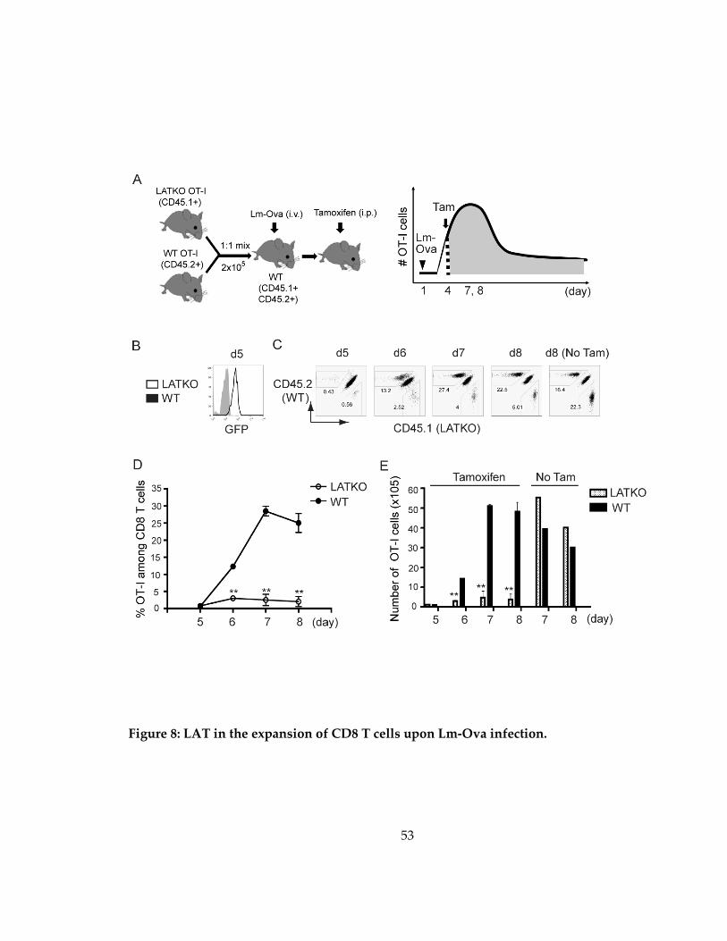
53
Figure 8: LAT in the expansion of CD8 T cells upon Lm-Ova infection.

54
Figure 8:
(A) A schematic diagram of the experimental procedure. A mixture of 1x10⁵ WT OT-I
cells (CD45.2+) and 1x10⁵ LATKO OT-I cells (CD45.1+) were adoptively transferred into
WT (CD45.1+ CD45.2+) recipient mice. On the following day, these recipients were
infected intravenously with Lm-Ova at day 1. The mice then received tamoxifen to delete
lat on day 4 and 5 during the T cell expansion phase. (B) Deletion efficiency. GFP
expression in WT and LATKO cells were examined on day 5. Flow cytometry of WT
cells is gated live CD8+CD45.2+ and LATKO cells is gated live CD8+ CD45.1+. (C) CD8 T
cell expansion. Live CD8 T cells in the spleen were analyzed at the indicated times after
infection. Numbers on the plots indicate the percentage of LATKO (CD45.1+) and WT
(CD45.2+) OT-I cells in the total CD8 T cell population. (D) The percentages of OT-I cells
among total CD8 T cells in the spleen after Lm-Ova infection. Data are shown as
mean±SEM. (E) The number of OT-I cells in the spleen after infection with or without
tamoxifen administration. *, P<0.05; **, P<0.005. Data represent four (B-E) independent
experiments (n=3-4 mice per time point)

55
Figure 9: LAT in the proliferation and cell death of CD8 T cells
Representative FACS plots of BrdU incorporation, expression of Ki-67, AnnexinV,
7AAD, and Bcl-2 in splenic CD8 T cells at day 7 post-infection. “Endo” is defined as the
endogenous CD8 T cells that were CD8+CD45.1+CD45.2+. Data are representative of 2
independent experiments using 2-4 mice per genotype.

56
LATKO OT-I cells (Figure 9). Moreover, Bcl-2 expression was downregulated similarly
in both LATKO and WT OT-I cells (Figure 9), indicating that reduced expansion of
LATKO T cells in recipient mice was not due to augmented cell death during the
priming phase. Collectively, our data indicate that continuous LAT mediated-signaling
is essential for optimal CD8 T cell proliferation during the T cell expansion phase.
3.2.3 LAT mediated-signaling is not required for CD8 T cell contraction
After the expansion phase and pathogen clearance, 90-95% of effector CD8 T cells
undergo programmed cell death to re-establish homeostasis. To investigate whether
LAT-mediated signaling regulates cell death during the contraction phase, we
performed a similar transfer experiment as described above. A mixture of 1x10⁴ LATf/-
OT-I cells (CD45.1+) and 1x10⁴ ERCre+LATf/- naïve OT-I cells (CD45.2+) were adoptively
transferred into wildtype (CD45.1+ CD45.2+) recipients at day 0. Mice were infected with
Lm-Ova at day 1 and treated with tamoxifen at the peak of CD8 T cell expansion, day 7
and 8 (Figure 10A). LATKO OT-I cells became GFP+ one day after tamoxifen
administration, as described previously. To further confirm that LAT was deleted, and
consequently that LAT mediated-signaling was abrogated, we stimulated T cells in vitro
with the Ova peptide at day 13 and examined their ability to produce IFN-γ. As
expected, LATKO cells were unable to produce IFN-γ upon stimulation (Figure 10B).
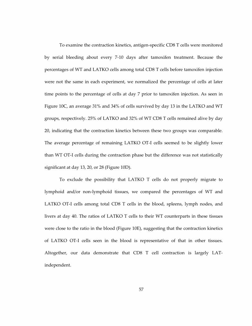
57
To examine the contraction kinetics, antigen-specific CD8 T cells were monitored
by serial bleeding about every 7-10 days after tamoxifen treatment. Because the
percentages of WT and LATKO cells among total CD8 T cells before tamoxifen injection
were not the same in each experiment, we normalized the percentage of cells at later
time points to the percentage of cells at day 7 prior to tamoxifen injection. As seen in
Figure 10C, an average 31% and 34% of cells survived by day 13 in the LATKO and WT
groups, respectively. 25% of LATKO and 32% of WT CD8 T cells remained alive by day
20, indicating that the contraction kinetics between these two groups was comparable.
The average percentage of remaining LATKO OT-I cells seemed to be slightly lower
than WT OT-I cells during the contraction phase but the difference was not statistically
significant at day 13, 20, or 28 (Figure 10D).
To exclude the possibility that LATKO T cells do not properly migrate to
lymphoid and/or non-lymphoid tissues, we compared the percentages of WT and
LATKO OT-I cells among total CD8 T cells in the blood, spleens, lymph nodes, and
livers at day 40. The ratios of LATKO T cells to their WT counterparts in these tissues
were close to the ratio in the blood (Figure 10E), suggesting that the contraction kinetics
of LATKO OT-I cells seen in the blood is representative of that in other tissues.
Altogether, our data demonstrate that CD8 T cell contraction is largely LAT-
independent.
58
Figure 10: LAT in the contraction of CD8 T cells
59
Figure 10:
(A) 1x10⁴ WT OT-I cells (CD45.1+) and 1x10⁴ LATKO OT-I cells (CD45.2+) were
adoptively transferred into hosts (CD45.1+ CD45.2+) that were subsequently infected
with Lm-Ova. The recipient mice received tamoxifen to delete lat on day 7 and 8 at the
peak of T cell expansion and the CD8 T cell response was monitored every 7-10 days. (B)
IFN-γ production by splenic OT-I cells at day 13. Splenocytes were stimulated with
(open) or without (filled) 1μM Ova peptide for 4 h before intracellular staining of IFN-γ.
Data are representative of three independent experiments, n=3-4 mice. (C, D)
Contraction curves following Lm-Ova infection. The percentage of OT-I cells in the
blood at the indicated time points was normalized by their percentage at day 7 before
tamoxifen treatment. The percentage of WT OT-I is gated on live CD8+CD45.2+ and the
percentage of LATKO OT-I is gated on live CD8+CD45.1+. Each dot represents one
mouse and the horizontal lines depict the mean of the population. NS=not significant; *,
P<0.05. Data are representative of three independent experiments (n=5-11 per time
point). (E) Ratio of the percentages of LATKO to WT OT-I cells in blood, spleen, liver,
and lymph nodes on day 40. Data are shown as mean±SEM. Data represent two
independent experiments (n=3 per group).

60
3.2.4 LAT function during the effector-to-memory transition
During the effector-to-memory transition phase, a small portion of effector cells
survive and convert into functional memory cells that persist for a long time. To assess
whether LAT-mediated signaling regulates memory formation, we deleted LAT at day 7
and 8 during the effector-to-memory transition and characterized the phenotype of
LATKO and WT memory CD8 T cells at day 36-40 post-infection (Figure 11A).
Before tamoxifen treatment at day 7, LATKO OT-I cells remained GFP negative
and they upregulated KLRG1 and downregulated CD62L similar to WT OT-I cells
(Figure 11B). By day 40 after tamoxifen treatment, in comparison with their WT
counterparts, LATKO memory OT-I cells acquired phenotypic properties that are
associated with a better memory potential (Figure 11C, D, E). LATKO memory OT-I cells
demonstrated increased expression of surface markers that are important for memory
formation and maintenance, such as IL-7R, CD27, CD122, and CXCR3 (Figure 11C, E).
Conversely, expression of markers that are associated with T cell effector function, such
as KLRG1, perforin, T-bet, and Eomes, was decreased in LATKO memory OT-I cells
(Figure 11E).
We also wanted to evaluate the ability of LATKO T cells to form central memory
cells, which express CD62L and are thought to be the stem cell-like subset that
undergoes self-renewal and exhibits multipotency. 55% of LATKO memory T cells
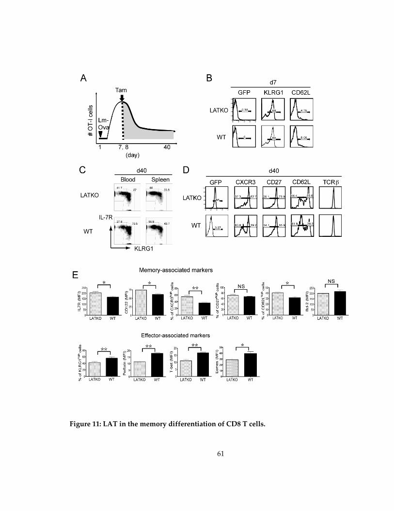
61
Figure 11: LAT in the memory differentiation of CD8 T cells.
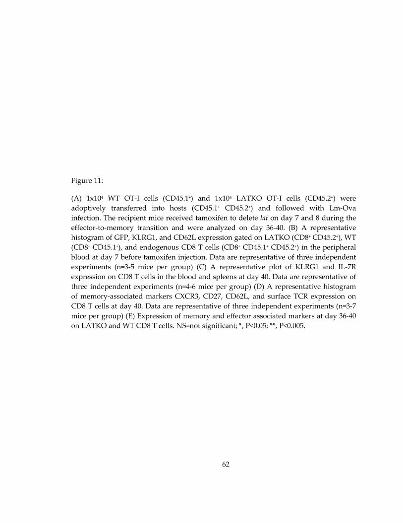
62
Figure 11:
(A) 1x10⁴ WT OT-I cells (CD45.1+) and 1x10⁴ LATKO OT-I cells (CD45.2+) were
adoptively transferred into hosts (CD45.1+ CD45.2+) and followed with Lm-Ova
infection. The recipient mice received tamoxifen to delete lat on day 7 and 8 during the
effector-to-memory transition and were analyzed on day 36-40. (B) A representative
histogram of GFP, KLRG1, and CD62L expression gated on LATKO (CD8+ CD45.2+), WT
(CD8+ CD45.1+), and endogenous CD8 T cells (CD8+ CD45.1+ CD45.2+) in the peripheral
blood at day 7 before tamoxifen injection. Data are representative of three independent
experiments (n=3-5 mice per group) (C) A representative plot of KLRG1 and IL-7R
expression on CD8 T cells in the blood and spleens at day 40. Data are representative of
three independent experiments (n=4-6 mice per group) (D) A representative histogram
of memory-associated markers CXCR3, CD27, CD62L, and surface TCR expression on
CD8 T cells at day 40. Data are representative of three independent experiments (n=3-7
mice per group) (E) Expression of memory and effector associated markers at day 36-40
on LATKO and WT CD8 T cells. NS=not significant; *, P<0.05; **, P<0.005.

63
upregulated CD62L while only 38% of WT memory T cells re-expressed CD62L at day
40 post-infection (Figure 11D, E), demonstrating that a higher frequency of central
memory T cells was generated in the absence of LAT. Collectively, these results indicate
that LAT deficiency during the effector-to-memory transition accelerates CD8 T cell
memory differentiation.
3.2.5 LAT deficiency affects T-bet expression in memory T cells
We sought to investigate the mechanism by which LAT deletion modulates
memory differentiation. Published studies indicate that cells that continue to receive
stimulation during the later stage of infection are more likely to become terminally
differentiated (Sarkar, Kalia et al. 2008). Therefore, in our study, it is possible that some
residual antigens are present during the contraction phase (day 7 to 20) and continue to
stimulate T cells. We hypothesize that LATKO T cells are unable to receive stimulation
at this stage, and they preferentially become memory precursors. To test this possibility,
we closely monitored the kinetics of memory precursor formation after tamoxifen
treatment from day 7 to day 48 in the peripheral blood. Based on expression of KLRG1
and IL-7R, KLRG1loIL-7Rhi CD8 T cells are referred as MPECs and KLRG1hiIL-7Rlo CD8 T
cells as SLECs (Kaech and Wherry 2007). The percentages of MPECs among LATKO OT-
I cells remained comparable to those among WT OT-I cells at early time points, day 7

64
and 13 (Figure 12A). By day 20, more LATKO MPECs than WT ones were observed and
this increase was maintained over time.
We also monitored the kinetics of CD62L upregulation as an indicator of TCM
formation. In line with the kinetics of MPEC differentiation, LATKO OT-I cells displayed
more CD62Lhi cells at day 20 and later time points in comparison to WT OT-I cells
(Figure 12B), demonstrating that the generation of TCM cells occurred more rapidly in the
absence of LAT.
To assess the transcriptional mechanism driving TCM development, we analyzed
T-bet expression in LATKO and WT cells. T-bet is a transcription factor that is induced
in effector CD8 T cells and plays a critical role in determining CD8 T cell fate.. Higher
amounts of T-bet promote SLEC formation, whereas lower amounts of T-bet instruct
MPEC development (Joshi, Cui et al. 2007). To determine if T-bet expression was
decreased at earlier time points, we depleted LAT at day7 and analyzed T-bet expression
in OT-I cells 5-6 days after deletion. Indeed, T-bet expression was decreased in LATKO
OT-I cells but not in the control group that did not receive tamoxifen treatment (Figure
13A, B). This observation suggests that LAT deficiency leads to diminished T-bet
expression, which consequently promotes the formation of MPECs.

65
Figure 12: LAT in the formation of memory precursor CD8 T cells.
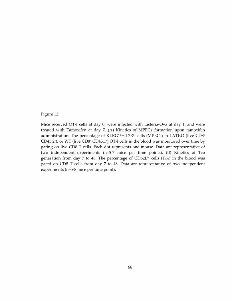
66
Figure 12:
Mice received OT-I cells at day 0, were infected with Listeria-Ova at day 1, and were
treated with Tamoxifen at day 7. (A) Kinetics of MPECs formation upon tamoxifen
administration. The percentage of KLRG1lowIL7Rhi cells (MPECs) in LATKO (live CD8+
CD45.2+), or WT (live CD8+ CD45.1+) OT-I cells in the blood was monitored over time by
gating on live CD8 T cells. Each dot represents one mouse. Data are representative of
two independent experiments (n=5-7 mice per time points). (B) Kinetics of TCM
generation from day 7 to 48. The percentage of CD62Lhi cells (TCM) in the blood was
gated on CD8 T cells from day 7 to 48. Data are representative of two independent
experiments (n=5-8 mice per time point).
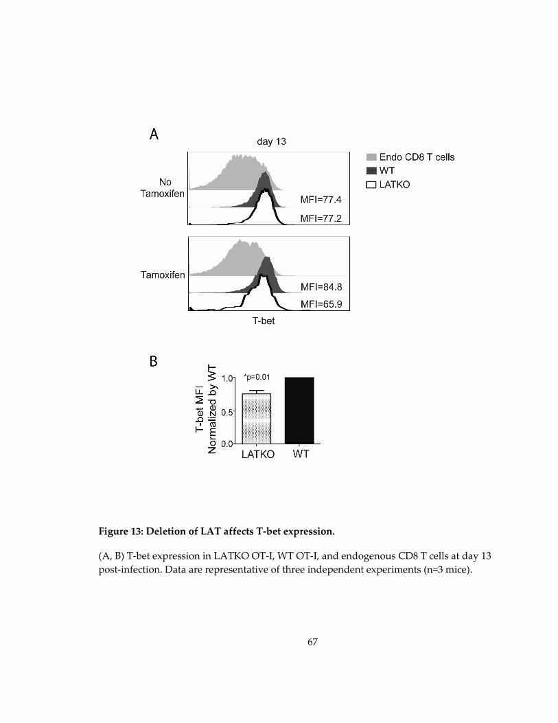
67
Figure 13: Deletion of LAT affects T-bet expression.
(A, B) T-bet expression in LATKO OT-I, WT OT-I, and endogenous CD8 T cells at day 13
post-infection. Data are representative of three independent experiments (n=3 mice).

68
3.2.6 LAT-mediated signaling is dispensable for memory CD8 T cell maintenance
Since LAT-mediated signaling is important in naïve T cell survival (Shen, Chuck
et al. 2010), we were interested in determining whether LAT is also essential for memory
T cell maintenance. To study the role of LAT in the homeostasis of memory CD8 T cells,
we performed the same adoptive transfer experiment described previously in which
LAT was deleted in memory OT-I cells starting at day 32 post-infection (Figure 14A). We
analyzed CD8 T cells at day 48, 60, 80, and 100 in the peripheral blood and normalized
the percentages of T cells to their percentages at day 32. As depicted in Figure 14C and
D, the frequency of LATKO and WT memory OT-I cells was comparable after tamoxifen
administration. 31.8% of LATKO memory OT-I cells and40% of WT memory OT-I cells
were observed by day 80 (Figure 14C, D).
By day 100, 29% and 37% of LATKO and WT memory OT-I cells, respectively,
remained. The average percentage of LATKO T cells was slightly lower than their WT
counterparts but the difference was not statistically significant (Figure 14D). We also
observed that few mice (4 out of 26) had significantly lower percentages of LATKO T
cells in other set of experiments and we speculated that the depletion of LATKO OT-I
cells in these mice might be due to the rejection of GFP+ cells over time.
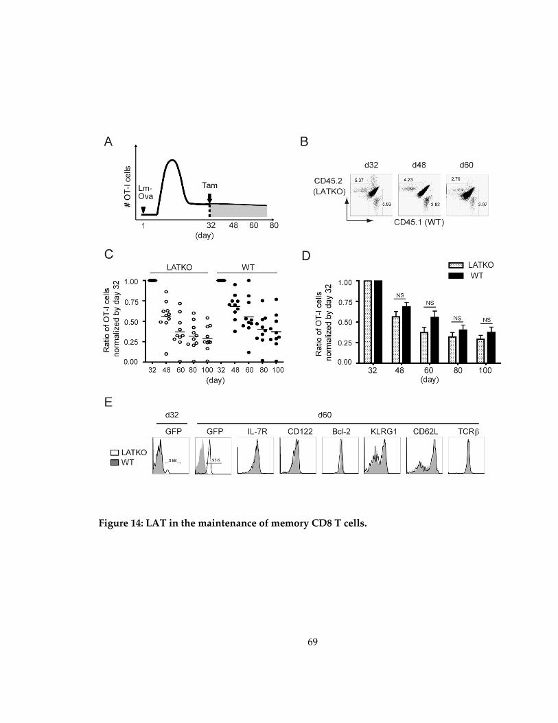
69
Figure 14: LAT in the maintenance of memory CD8 T cells.
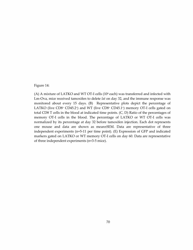
70
Figure 14:
(A) A mixture of LATKO and WT OT-I cells (104 each) was transferred and infected with
Lm-Ova, mice received tamoxifen to delete lat on day 32, and the immune response was
monitored about every 15 days. (B) Representative plots depict the percentage of
LATKO (live CD8+ CD45.2+) and WT (live CD8+ CD45.1+) memory OT-I cells gated on
total CD8 T cells in the blood at indicated time points. (C, D) Ratio of the percentages of
memory OT-I cells in the blood. The percentage of LATKO or WT OT-I cells was
normalized by its percentage at day 32 before tamoxifen injection. Each dot represents
one mouse and data are shown as mean±SEM. Data are representative of three
independent experiments (n=5-11 per time point). (E) Expression of GFP and indicated
markers gated on LATKO or WT memory OT-I cells on day 60. Data are representative
of three independent experiments (n=3-5 mice).

71
Except for these 4 outliers, memory CD8 T cells persisted in the absence of LAT,
demonstrating that the maintenance of memory CD8 T cells was largely LAT-
independent.
We also investigated the expression of IL-7 and IL-15 receptors because these two
cytokines are essential for the survival and homeostasis of memory T cells. LATKO
memory T cells displayed similar levels of IL-7R and IL-15R as WT memory T cells
(Figure 14E). Moreover, the expression of Bcl-2, a cell-intrinsic survival factor
upregulated in memory cells to support their maintenance, was equivalent among WT
and LATKO memory T cells (Figure 14E). Collectively, these observations suggest that
LAT-mediated signaling is dispensable for the persistence of memory CD8 T cells.
3.2.7 LAT-mediated signaling is indispensable for a memory recall response
We next compared the functional potency of LATKO and WT memory OT-I cells
by evaluating their ability to produce effector cytokines upon in vitro stimulation. Mice
were treated with tamoxifen at day 7 and at day 48 splenocytes were cultured in the
presence of Ova peptide or PMA and ionomycin (P+I) for 4 h before cytokine production
by CD8 T cells was examined. As shown in Figure 15A and B, WT memory OT-I cells
produced robust amounts of IFN-γ, TNF-α, and IL-2; however, LATKO memory OT-I
cells produced insignificant amounts of cytokines upon Ova peptide stimulation. When
cells were stimulated with P+I, which bypasses the TCR signaling complex, both LATKO
72
and WT cells demonstrated a similar capability to produce cytokines (Figure 15A and
C).
We also examined the ability of LATKO memory OT-I cells to mobilize calcium.
WT OT-I cells displayed strong calcium flux initiated by anti-CD3 and anti-CD8
crosslinking; however, calcium mobilization by LATKO memory OT-I cells was
abrogated (Figure 15D). The defects in cytokine production and calcium mobilization
were not due to a defect in TCR surface expression. Our previous study demonstrated
that depletion of LAT in naïve T cells leads to a drastic downregulation of surface TCR
(Shen, Chuck et al. 2010). In contrast to naïve T cells, depletion of LAT did not affect
TCR expression on memory OT-I cells (Figure 11D and 14E). Here, we show that LAT-
mediated signaling is imperative for cytokine production and calcium flux in memory
CD8 T cells.
To examine whether LAT is also required for secondary immune responses in
vivo, we directly re-infected recipient mice at day 60 with 1.5x10⁵ Lm-Ova and
monitored the T cell response at day 63 and 65 (Figure 16A). Compared with WT
memory OT-I cells, LATKO memory OT-I cells failed to proliferate in response to re-
challenge with Lm-Ova in both lymphoid organs and peripheral tissues (Figure 16B and
C). Moreover, while WT memory OT-I cells converted to an effector phenotype
characterized by CD62LlowIL-7RlowKLRG1high, LATKO memory cells remained
73
CD62LhighIL-7Rhigh KLRG1low, indicating their inability to be reactivated (Figure 16D). In
addition, LATKO memory T cells failed to upregulate T-bet and downregulate Bcl-2 like
WT T cells (Figure 16D). Collectively, our data demonstrate that LATKO memory OT-I
cells are unable to proliferate and produce cytokines in response to a recall challenge,
indicating that LAT is required to trigger a secondary CD8 T cell immune response.
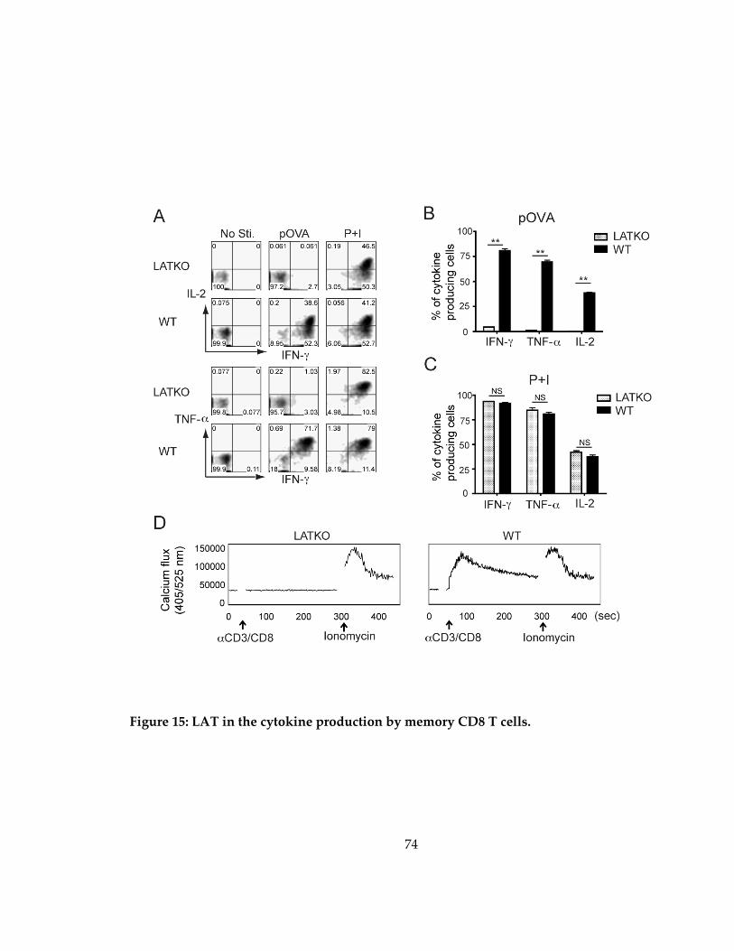
74
Figure 15: LAT in the cytokine production by memory CD8 T cells.

75
Figure 15:
(A, B, C) Cytokine production by LATKO and WT memory OT-I cells upon Ova peptide
(A) or P+I (B) stimulation. Splenocytes from day 48 were stimulated with 1 μM Ova
peptide or P+I for 4 hr in vitro before ICS staining. Representative plots show IFNγ,
TNFα, and IL-2 production by LATKO and WT OT-I cells. Data are representative of
two independent experiments (n=3-4 mice). (D) Calcium mobilization by memory OT-I
cells. Splenocytes from day 48 post-infection were loaded with the calcium indicator,
Indo-1. Calcium flux was induced by anti-CD3 and anti-CD8 cross-linking followed by
ionomycin stimulation and analyzed by flow cytometry.
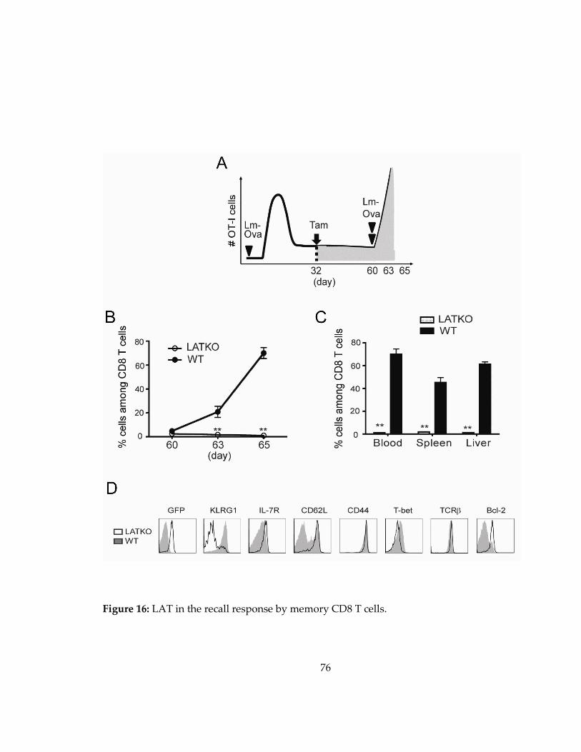
76
Figure 16: LAT in the recall response by memory CD8 T cells.

77
Figure 16:
(A) Mice received mixed OT-I cells and 1.5x10⁴ Lm-Ova were injected with tamoxifen on
day 32. To induce a secondary response, the same mice were challenged with 1.5x10⁵
Lm-Ova on day 60. The recall response was monitored on day 63 and 65. (B) T cell
expansion upon re-infection of Lm-Ova. Percentage of LATKO and WT OT-I cells
among total CD8 T cells in the spleen after infection. Data are shown as mean±SEM.
Data are representative of three independent experiments (n=3 mice per time points). (C)
Percentage of OT-I cells among total CD8 T cells in blood, spleen, and liver. Data are
representative of three independent experiments (n=3 mice per time points). (D)
Expression of indicated markers gated on LATKO or WT OT-I cells on day 65. Data are
representative of three independent experiments (n=3 mice per group). NS=not
significant; *, P<0.05; **, P<0.005

78
3.3 Discussion
Using an inducible LAT deletion system, we investigated the requirement for
TCR-mediated signaling in each step of the CD8 T cell response elicited by acute Listeria
infection. We adoptively transferred LATKO and WT OT-I cells into the same hosts to
ensure that OT-I cells received the same environmental stimuli, including the number of
pathogens, CD4 T cell help, and inflammatory cytokines. This system allowed us to
compare the quantitative and qualitative changes that occur during the immune
response. Our data show that LAT-mediated signaling is essential for the continuous
expansion of CD8 T cells during T cell priming but is not required for contraction and
memory maintenance of CD8 T cells. Deletion of LAT during the effector-to-memory
transition results in enhanced development of memory precursors likely due to the
downregulation of T-bet expression. Our data further indicated that LAT is essential for
the recall response as LATKO memory CD8 T cells are unable to produce cytokines or
respond to a secondary challenge.
When naïve T cells recognize foreign peptide-MHC complexes presented by
antigen presenting cells, T cells and APCs interact for about 12-24 h in vivo to drive T cell
activation (Cui and Kaech 2010). Afterward, T cells dissociate from APCs and undergo
proliferation and differentiation. A single cell clone can undergo ~15 rounds of division
in 7-8 days in a mouse model. Our data demonstrated that after the initial contact with

79
APCs, CD8 T cell expansion is perturbed by LAT deficiency, indicating that sustained
TCR signaling during the priming phase is required for optimal primary response
magnitude. Although it was reported that the expression of ERCre might have a toxic
effect on T cells after tamoxifen injection (Higashi, Ikawa et al. 2009), impaired T cell
expansion during priming is unlikely caused by the toxic effect of ERCre. The dose of
tamoxifen we used in our experiments was much lower compared with the dose used in
the published study. Moreover, thymocyte development and T cell activation are normal
in ERCreLATf/+ mice treated with tamoxifen at the same dose (Chuck, Zhu et al. 2010).
Our results are well in line with previous findings showing that varying the antigen
dose or affinity can affect CTL expansion (Kaech and Ahmed 2001; Zehn, Lee et al. 2009).
For example, depletion of epitope-presenting DCs during T cell primary responses
dampens the magnitude of the responding CTL population (van Stipdonk, Lemmens et
al. 2001). It has also been shown that the extent of CTL proliferation is proportional to
the amount of Ova epitope produced by Vaccinia virus (Wherry, Puorro et al. 1999).
Together with these studies, our results provide additional evidence that the degree of
CTL expansion correlates with the strength of TCR signaling.
Manipulating the amount and duration of external stimuli affects cell expansion
in addition to influencing memory differentiation of CD8 T cells (Sarkar, Teichgraber et
al. 2007; Sarkar, Kalia et al. 2008). These data suggest that the strength of TCR signaling
80
during the expansion phase plays a role in memory formation. Recently, analyses of
SLP-76-deficient or –mutant knock-in mice showed that altered intrinsic TCR signaling
affects the differentiation and function of endogenous memory CD8 T cells (Smith-
Garvin, Burns et al. 2010; Wiehagen, Corbo et al. 2010). SLP-76-deficient or -mutated
CD8 T cells differentiate into memory precursors and TCM cells at a faster rate than WT
cells. Notably, a caveat of these systems is that CD4 T cells in these mice also have
altered SLP-76 signaling and CD4 T cells play an important role in the differentiation of
CD8 T cells during pathogen infection (Khanolkar, Badovinac et al. 2007). Weak TCR
signaling alters the differentiation of CD4 T cells from Th1 to Th2, thereby skewing the
cytokines production (Shen, Chuck et al. 2010). Furthermore, an additional study
showed that the depletion of Treg cells leads to enhanced formation of memory CD8 T
cells, and, similar to LAT-deficient Treg cells, SLP-76-deficient Treg cells seem to lose
their inhibitory function (Chuck, Zhu et al. 2010). Therefore, the altered memory
differentiation observed in SLP-76-deficient or -mutated CD8 T cells could be an indirect
effect of altered CD4 T cells.
In our study, by transferring LATKO OT-I cells into WT recipients, we were able
to exclusively focus on LAT signaling in CD8 memory T cell formation while CD4 T cells
remained unaffected. Our data revealed that LATKO memory CD8 T cells expressed
higher levels of memory associated markers, such as IL-7R, CD122, CD27, CXCR3, and

81
CD62L, indicating that dampened TCR signaling drives CD8 T cell development into
memory precursors and TCM cells. It is thought that cells that receive a higher amount of
antigen or are stimulated for longer periods of time during expansion are more likely to
become terminally differentiated cells (Sarkar, Kalia et al. 2008). Previous studies
suggest that the decision for a CD8 T cell to become a short-lived effector or a self-
renewing memory T cell occurs early in the stage of clonal expansion (Cui and Kaech
2010). In our study, the deletion of LAT happened at and after the peak of T cell
expansion, allowing a normal primary response to occur. Our findings indicated that
diminishing TCR signaling after the expansion phase still affected memory formation.
Thus, our data suggested that intrinsic TCR signaling can still control fate of CD8 T cell
differentiation after initial expansion.
To gain insight into the mechanism by which LAT-mediated signaling regulates
memory differentiation, we examined the expression of T-bet, which controls the
balance between MPEC and SLEC formation (Joshi, Cui et al. 2007). Indeed, decreased
expression of T-bet in LATKO OT-I cells was detected several days after LAT deletion
(day 13), and this reduction occurred before the skewing of SLEC to MPEC was
observed (day 20), suggesting that reduced T-bet expression in LATKO T cells was one
of the driving forces that promote MPEC development. It is not yet clear how LAT
regulates T-bet expression. One possibility is that the downstream signaling pathways of
82
LAT, such as the mTOR pathway, regulate T-bet expression. mTOR has also been shown
to modulate the balance between T-bet and Eomes expression through regulation of
Foxo1 (Rao, Li et al. 2012). Blockade of mTOR activity diminishes T-bet and enhances
Eomes expression (Rao, Li et al. 2010). However, since both T-bet and Eomes expression
were reduced in LATKO memory OT-I cells, other LAT-dependent pathways are likely
to be involved in controlling or mediating this balance. Another possible mechanism by
which LAT regulates T-bet is through crosstalk between LAT and cytokine pathways.
Since the production of IFN-γ is impaired by LAT deficiency, it is possible that IFN-γ-
driven STAT1-mediated synthesis of T-bet is not as efficient in LATKO cells as it is in
WT T cells.
Additionally, IL-12 and IL-2 signaling are involved in determining CD8 T cell
fate via regulation of T-bet expression. Prolonged IL-2 signals or higher expression of
CD25 on CD8 T cells during the early expansion phase (day 3-5) favor the differentiation
of CD8 T cells into SLECs (Obar, Molloy et al. 2010). CD8 T cells convert to a central
memory phenotype faster in the absence of IL-2 signaling (Williams, Tyznik et al. 2006).
Therefore, it is possible that LAT deficiency affects IL-12 or IL-2 signaling; however,
when we deleted LAT at day 7, CD25 expression had already decreased in both LATKO
and WT CD8 T cells (data not shown). Hence, it is not likely that the altered memory
differentiation observed in LATKO OT-I cells was caused by altered IL-2 signaling.

83
Following pathogen clearance, the majority of antigen-specific CD8 T cells
undergo programmed cell death in order to maintain cellular homeostasis. In our study,
we observed that when LAT was depleted in CD8 T cells at the peak of T cell expansion
(day 7 and 8), CTL contraction still occurred at a similar rate as WT CD8 T cells,
indicating that the regulation of programmed cell death at this stage was independent of
LAT-signaling. Effector CD8 T cell death during the contraction phase is triggered by
restimulation-induced cell death (RICD) and cytokine withdrawal-induced death
(CWID). RICD occurs in the presence of antigens and IL-2, and is induced upon TCR re-
engagement of activated T cells. This then triggers extrinsic apoptotic signals such TNFR
and Fas. Mice deficient in TNFRI and TNFRII exhibit severely compromised contraction
of virus-specific CD8 T cells, thus illustrating the critical role of TNFR signaling in CTL
contraction (Suresh, Singh et al. 2005).
When antigens and inflammation diminish, CWID is initiated and triggered
through the intrinsic apoptotic pathway. Withdrawal of cytokines and growth factors
leads to inactivation of PI3K/Akt pathway. (Michalek and Rathmell 2010). As a result,
Bim is activated, which in turn activates Bax and Bak, leading to mitochondrial outer
membrane permeabilization (MOMP) and cytochrome c release, ultimately causing
cellular atrophy and apoptosis. In line with this, Bim-deficient, as well as Bax/Bak
double-deficient mice, showed delayed or defective CTL contraction and antigen
84
receptor-induced cell death (Hildeman, Zhu et al. 2002; Rathmell, Lindsten et al. 2002;
Pellegrini, Belz et al. 2003).
Since RICD is induced by TCR engagement, it was hypothesized that LAT
would be important in the regulation of RICD. Yet, our results showed that the kinetics
of cell death during contraction was comparable between WT and LATKO CD8 T cells.
One possible explanation for our observation is that LATKO T cells had already
upregulated TNF receptors at day 7. After tamoxifen administration, LATKO CD8 T
cells failed to produce TNF-α, while WT CD8 T cells and other cell types in the same
environment were able to do so, thus inducing the death of LATKO cells to a similar
extent as the WT counterparts. Furthermore, RICD mainly occurs during the T cell
expansion phase while CWID represents the critical pathway to induce cell death after
antigens are cleared. This suggests that the cell death seen during contraction for both
LATKO and WT T cells is heavily dependent on cytokine signaling, instead of TCR
signaling. To support this notion, our data showed that Bcl-2 expression was
comparable in LATKO and WT CD8 T cells during contraction, further demonstrating
that LAT is dispensable for controlling cell death at this stage.
Memory CD8 T cells are capable of mounting a robust and efficient recall
immune response upon secondary exposure to re-infection. Our results here showed
that although LATKO memory T cells acquire phenotypic markers associated with a
85
high memory potential, they completely fail to be reactivated and produce IFN-γ, TNF-
α, and IL-2 upon re-exposure to Listeria. This impaired memory response was not due
to a lack of CD4 T cell help as LATKO OT-I cells were transferred to WT recipients
containing normal CD4 T cells. One explanation is that LAT-mediated signaling is
essential for T cell activation and cytokine production by memory CD8 T cells. Another
possibility is that persistent LAT-mediated signaling is vital to maintain the
functionality of memory CD8 T cells over time. We favor the former explanation because
LATKO memory T cells were still fully capable of producing cytokines, especially IL-2,
upon TCR-independent stimulation.
By assessing the role of LAT in the immunological response following microbial
infection, we have demonstrated that continuous TCR-mediated signaling is essential to
promote T cell expansion and cytokine production during both primary and secondary
immune responses. However, contraction and maintenance of memory CD8 T cells do
not require the presence of TCR signaling.
86
4. The role of LAT in the granule-mediated cytotoxicity of CD8 T cells
4.1 Introduction
CD8 cytotoxic T lymphocytes (CTLs) have the capacity to eliminate virally
infected or tumor cells by two pathways: the Fas pathway and the granule pathway
(Kagi, Ledermann et al. 1996; Russell and Ley 2002). The Fas pathway is triggered by the
interaction between Fas ligand (FasL) on CTLs and Fas on target cells, leading to caspase
activation and apoptotic cell death. The granule pathway relies on directed granule
exocytosis, a perforin-mediated directional transfer of granzymes from CTLs into target
cells. This process involves the initial contact of CTLs and target cells, the formation of
an immunological synapse, the polarization of MTOC and lytic granules, and eventually
the delivery of perforin and granzyme B.
Although the molecular mechanisms of granule exocytosis have been well
described, the signaling events that control each step of the process are still under
investigation. Published data show that calcium is critical for granule exocytosis and
killing of target cells (Esser, Haverstick et al. 1998; Lyubchenko, Wurth et al. 2001).
However, the role of calcium in MTOC and granule polarization is still controversial.
While Kuhne et al. observed that MTOC movement correlates with a higher intracellular
calcium mobilization (Kuhne, Lin et al. 2003), Quann et al. reported an opposite finding
that diacylglycerol (DAG) accumulation at the synapse recruits dynein to reorient
87
MTOC, and this phenomenon is calcium-independent (Quann, Merino et al. 2009).
Studies using pharmacological inhibitors also demonstrate that signaling proteins in the
TCR signaling pathway, such as PI3K, PKC, and MAPK, play important roles in CTL-
mediated cytotoxicity (Juszczak and Russell 1989; Pardo, Buferne et al. 2003; Robertson,
Mireau et al. 2005). Recent data uncover that stronger TCR signals lead to increased
death of target cells. In contrast, weak TCR stimuli fail to induce the recruitment of
granules to the synapse, despite proper synapse formation and MTOC reorientation
(Beal, Anikeeva et al. 2009; Jenkins, Tsun et al. 2009), suggesting that MTOC
polarization, granule transportation, and granule release are differentially regulated.
To investigate the role of TCR signaling in CTL cytotoxicity, we took advantage
of the inducible LAT deletion system to deplete LAT in effector CD8 T cells as described
in the previous chapter. Since T cell activation and expansion require LAT, we deleted
LAT after antigen-induced expansion and differentiation of naive CD8 T cells into CTLs
to examine the capability of LAT-deficient CTLs to exert cytotoxicity and produce
cytokines. We also dissected the effect of LAT deletion on each step of granule
exocytosis, including conjugate formation, immunological synapse formation, MTOC
polarization, and lytic granule reorientaion. Moreover, using a series of pharmacological
inhibitors, we aimed to identify the signaling events that regulate granule exocytosis.
88
4.2 Results
4.2.1 Ablation of LAT impairs CTL killing in vivo
We first tested whether LAT is required for CTL-mediated killing in vivo using
the procedure as illustrated in Figure 17A. OT-I cells from ERCre+LATf/-OT-I and
LATf/-OT-I mice (Thy1.2+) were adoptively transferred into Thy1.1+Thy1.2+ mice. On
the following day, these mice were infected with Listeria expressing ovalbumin to
stimulate the proliferation and further differentiation of OT-I T cells. Mice without the
transfer of OT-I T cells were used as controls to measure the cytotoxicity exhibited by
endogenous Ova-specific T cells. Since LAT is required for T cell activation, we chose to
delete LAT after the T cell expansion phase. Mice that received OT-I T cells were injected
with tamoxifen on both day 7 and 8 after infection.
As shown in Figure 17B, the lat gene was efficiently deleted after tamoxifen
injection as indicated by the expression of GFP in donor CD8 T cells (Thy1.2+). At day 11,
95% of ERCre+LATf/-OT-I cells were GFP+. The percentages of donor T cells were similar
between LATKO and WT (Figure 17C). We performed an in vivo cytotoxicity assay at
day 11. Splenocytes from Thy1.1+ mice were used as target cells and thus were loaded
with 6 μM Cell Tracker Orange and 1 μM Ova peptide. Splenocytes without Cell Tracker
labeling and the Ova peptide, which should not be killed by OT-I cells, were used as a
negative control. These cells were mixed at a ratio of 1:1 and transferred into mice that

89
had received OT-I cells. One hour after transfer, splenocytes from these mice were
analyzed by FACS and the percentage of specific killing was calculated accordingly.
As shown in Figure 17D and 1E, endogenous T cells could respond to Listeria
infection, resulting in ~34.8% lysis of target cells. While 72.5% of target cells were killed
in mice receiving WT OT-I cells, only 58.1% of them were lysed in mice receiving
LATKO OT-I cells, indicating impaired killing by LATKO CTLs.
4.2.2 Generation of LAT-deficient CTLs in vitro
To investigate the cytotoxic mechanism of LATKO OT-I cells, we generated LAT-
deficient OT-I CTLs in vitro. Splenocytes from LATKO and WT mice were cultured in
the presence of 1 µM Ova peptide for 2 days to activate OT-I CTLs; this was followed by
treatment with 4-OHT for an additional 3-5 days to delete LAT. Two days after 4-OHT
treatment, more than 99% of LATKO CTLs expressed GFP (Figure 18A). Deletion of LAT
exons 7-11 leads to the expression of a LAT-GFP fusion protein (Shen, Zhu et al. 2009).
To confirm LAT deletion, we used two different antibodies: one (Ab #1) that recognizes
both WT and LAT-GFP proteins and the other one (Ab #2) that detects only WT LAT. At
day 3 after 4-OHT treatment, WT LAT protein was undetectable by Western blotting in
LATKO samples (Figure 18B), indicating that deletion of lat in vitro was efficient.
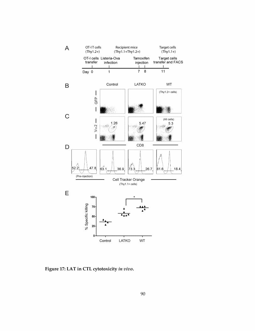
90
Figure 17: LAT in CTL cytotoxicity in vivo.

91
Figure 17:
(A) Experimental protocol of the in vivo killing assay. (B) Deletion efficiency of LAT. GFP
expression in Thy1.2+CD8+ T cells was analyzed by FACS. (C) Expansion of OT-I CTLs.
The percentage of donor CD8+ T cells (Thy1.2+) at day 11 after Listeria infection. (D)
Cytotoxicity. As target cells, 1x10⁷ splenocytes from Thy1.1+ mice were loaded with 6 µM
dye (Cell Tracker Orange) and 1µM Ova peptide or left unloaded, mixed 1:1, and
injected into mice that received OT-I CTLs. One hour later, splenocytes were analyzed
by FACS. Plots were gated on live cells. (E) Percentage of specific killing as calculated in
the Methods section. Data shown are one representative of two independent
experiments (Control=4, LATKO=6, WT=5). Each dot represents one mouse; *p<0.05;
horizontal bars indicate the mean.
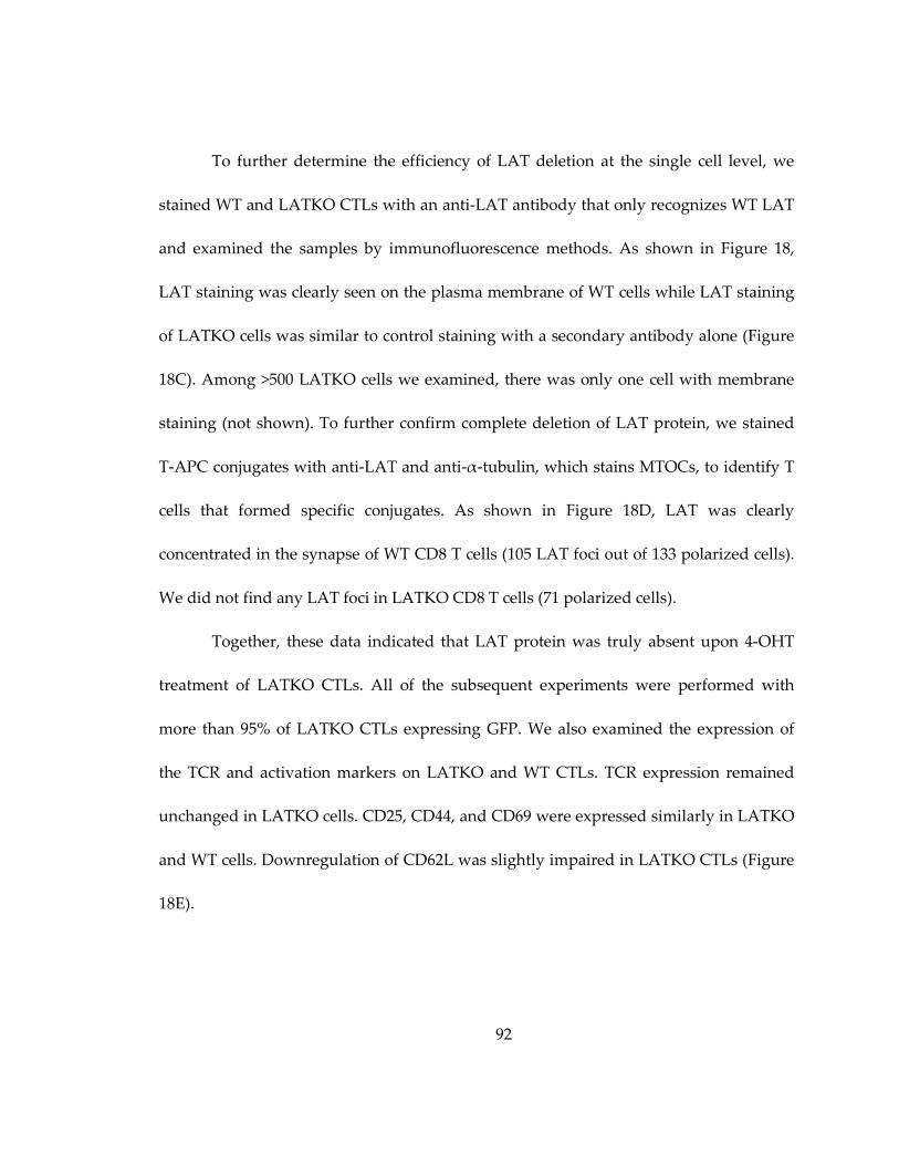
92
To further determine the efficiency of LAT deletion at the single cell level, we
stained WT and LATKO CTLs with an anti-LAT antibody that only recognizes WT LAT
and examined the samples by immunofluorescence methods. As shown in Figure 18,
LAT staining was clearly seen on the plasma membrane of WT cells while LAT staining
of LATKO cells was similar to control staining with a secondary antibody alone (Figure
18C). Among >500 LATKO cells we examined, there was only one cell with membrane
staining (not shown). To further confirm complete deletion of LAT protein, we stained
T-APC conjugates with anti-LAT and anti-α-tubulin, which stains MTOCs, to identify T
cells that formed specific conjugates. As shown in Figure 18D, LAT was clearly
concentrated in the synapse of WT CD8 T cells (105 LAT foci out of 133 polarized cells).
We did not find any LAT foci in LATKO CD8 T cells (71 polarized cells).
Together, these data indicated that LAT protein was truly absent upon 4-OHT
treatment of LATKO CTLs. All of the subsequent experiments were performed with
more than 95% of LATKO CTLs expressing GFP. We also examined the expression of
the TCR and activation markers on LATKO and WT CTLs. TCR expression remained
unchanged in LATKO cells. CD25, CD44, and CD69 were expressed similarly in LATKO
and WT cells. Downregulation of CD62L was slightly impaired in LATKO CTLs (Figure
18E).
93
Figure 18: Deletion of LAT in vitro.

94
Figure 18:
Splenocytes from ERCre+LATf/-OT-I and LATf/-OT-I mice were primed with Ova
peptide for 2 days before being treated with 4-OHT for an additional 3-5 days. (A)
Deletion efficiency of LAT in vitro. GFP expression in CD8+ T cells was analyzed by
FACS. (B) The absence of LAT protein in LATKO CTLs. WT and LAT KO lysates were
analyzed by Western blotting using two polyclonal antibodies. Ab #1 recognizes the
entire cytoplasmic domain, while Ab#2 only recognizes a peptide that is located in the
deleted region. (C) The absence of LAT at the single cell level. CTLs (WT and LATKO)
were fixed, permeabilized, and stained with anti-LAT antisera and Cy3-conjugated anti-
rabbit secondary antibody. The negative control was LATKO staining with the
secondary antibody only. (D) The absence of LAT at the synapse of LATKO CTLs. CTLs
were incubated with L1210/Kb cells (green) preloaded with the Ova peptide for 30
minutes to allow for the formation of conjugates. Conjugates were stained with anti-α-
tubulin to represent MTOC (white) as well as anti-LAT antibody (red). (E) TCR
expression and activation markers on CTLs. WT and LATKO CTLs were stained with
anti-TCRβ, anti-CD25, anti-CD69, anti-CD62L, and anti-CD44 antibodies and analyzed
by FACS. Data shown are one representative of three independent experiments.
95
EL4 cells were loaded with Cell Tracker Orange and Ova peptide as described
previously. Consistent with the reduced killing observed in vivo, LATKO CTLs clearly
showed a diminished cytotoxic capability toward the peptide-loaded EL4 cells (Figure
19A). We then analyzed IFN-γ production upon stimulation with EL4 cells with or
without the Ova peptide. Compared with WT cells, LATKO CTLs failed to produce
significant amounts of IFN-γ (Figure 19B). In addition to IFN-γ pathway, CTLs can also
kill target cells through the Fas-mediated pathway in which triggering of the Fas
pathway in target cells relies upon the upregulation of FasL on T cells. FasL was
upregulated on WT CTLs upon re-encountering target cells. However, this upregulation
was largely reduced in LATKO CTLs (Figure 19B). To further determine whether
granule-mediated killing is affected by LAT deficiency, we used a Fas-deficient cell line,
L1210 expressing H2-Kb, as target cells. The cytotoxicity of LATKO CTLs was
significantly decreased compared with WT CTLs, indicating that LAT is also important
in granule-mediated killing (Figure 20A).
Next, we examined the expression of perforin and granzyme B (gzmB), two
effector molecules that are crucial for granule-mediated killing. The expression of gzmB
was comparable between LATKO and WT CTLs, whereas perforin expression was
slightly reduced in LATKO CTLs (Figure 20B). To determine whether the granules in
LATKO CTLs were delivered to target cells, we performed a FACS-based assay using a
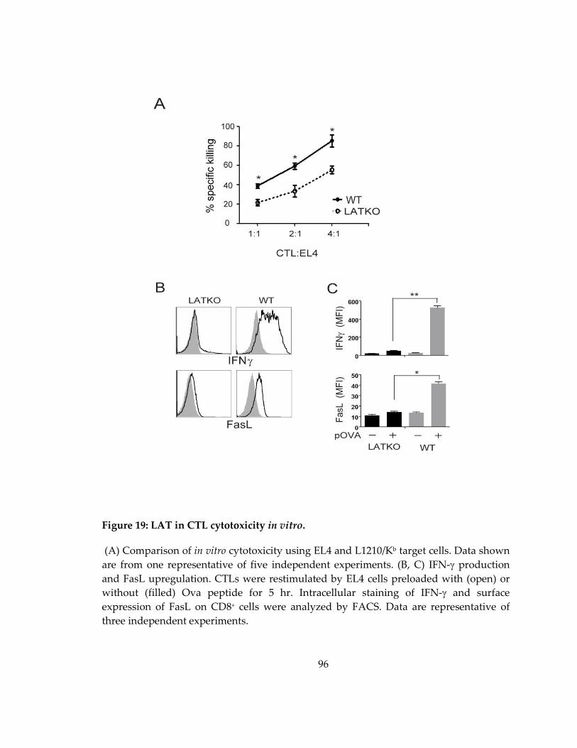
96
Figure 19: LAT in CTL cytotoxicity in vitro.
(A) Comparison of in vitro cytotoxicity using EL4 and L1210/Kb target cells. Data shown
are from one representative of five independent experiments. (B, C) IFN-γ production
and FasL upregulation. CTLs were restimulated by EL4 cells preloaded with (open) or
without (filled) Ova peptide for 5 hr. Intracellular staining of IFN-γ and surface
expression of FasL on CD8+ cells were analyzed by FACS. Data are representative of
three independent experiments.

97
cell-permeable fluorogenic substrate of gzmB. This substrate can be cleaved in target
cells in the presence of gzmB, which leads to the emission of fluorescent light for
quantification of gzmB activity. Although gzmB activity was still detected in target cells
incubated with LATKO CTLs, the intensity was much lower than the WT CTL-treated
group (Figure 20C). Altogether, these data demonstrated that LAT regulates IFN-γ
production, FasL upregulation, and granule delivery.
4.2.4 Unstable CTL-target conjugates and immunological synapse in LATKO CTLs
Our data showed that LATKO CTLs were capable of delivering granules to
target cells albeit at a reduced level. Granule-dependent cytotoxicity of T cells involves
several critical steps: conjugate formation, synapse formation, MTOC reorientation,
granule polarization, and secretion. We sought to examine the roles of LAT in each step
of CTL granule exocytosis. We first determined the efficiency of conjugate formation
between LATKO CTLs and target cells. CTLs and target cells were labeled with two
different dyes. Upon the formation of CTL-target cell conjugates, CTLs should stain for
both dyes. As shown in Figure 21A, LATKO and WT CTLs showed similar kinetics in
the formation of conjugates at early time points and both peaked at 20 minutes,
indicating that LATKO CTLs are capable of forming proper conjugates with target cells.
Twenty minutes after mixing, T cells gradually dissociated from target cells before or
after killing, leading to a decline in the number of conjugates.

98
Figure 20: LAT in the granule-mediated killing.

99
Figure 20:
(A) Comparison of in vitro cytotoxicity against L1210/Kb target cells. Data shown are
from one representative of three independent experiments. (B) Granzyme B and perforin
expression by Western blotting. Data are representative of three independent
experiments. (C) The delivery of granules. L1210/Kb cells were labeled with Cell Tracker
Orange with or without Ova peptide. CTLs were incubated with L1210/Kb cells in the
presence of a cell-permeable FITC-conjugated gzmB substrate for 40 minutes. The
numbers in the figure indicate the percentage of L1210/Kb cells that received gzmB from
CTLs. Data are representative of two independent experiments. n.s., not significant;
*p<0.05, **p<0.005; error bars represent means ± s.e.m.

100
For WT CTLs, 30.4%, 24.6%, and 13.7% of conjugates were maintained at 30 min, 45 min
and 60 min, respectively. However, only 18.9%, 9%, and 3.3% of conjugates were
maintained for LATKO CTLs at those time points, suggesting that LAT is required for
the maintenance of stable conjugates. These data indicated that the initial conjugate
formation between CTLs and APCs is LAT-independent; however, LAT is necessary for
the formation of stable conjugates that remain intact for an extended period of time.
Previous studies demonstrate that synapse maintenance requires continuous
TCR-mediated signaling (Huppa, Gleimer et al. 2003). Therefore, next we examined
whether synapse formation is affected by LAT deletion. We chose PKCθ as a marker for
the cSMAC and LFA-1 as a marker for the pSMAC (Monks, Freiberg et al. 1998).
Conjugates were examined in the xy and xz planes to view the synapse from the side
and enface, respectively. To evaluate the efficiency of cSMAC formation in the synapse,
we calculated the accumulation of cSMAC as the ratio of the fluorescence intensity at the
synapse to the fluorescence intensity in the cytoplasm at different time points. The
intensity of cSMAC accumulation at the synapse of LATKO (average=2.35, n=46) and
WT CTLs (average=2.49, n=44) was comparable 15 minutes after T cell-target cell
engagement, indicating that cSMACs can form in the absence of LAT (Figure 21B).
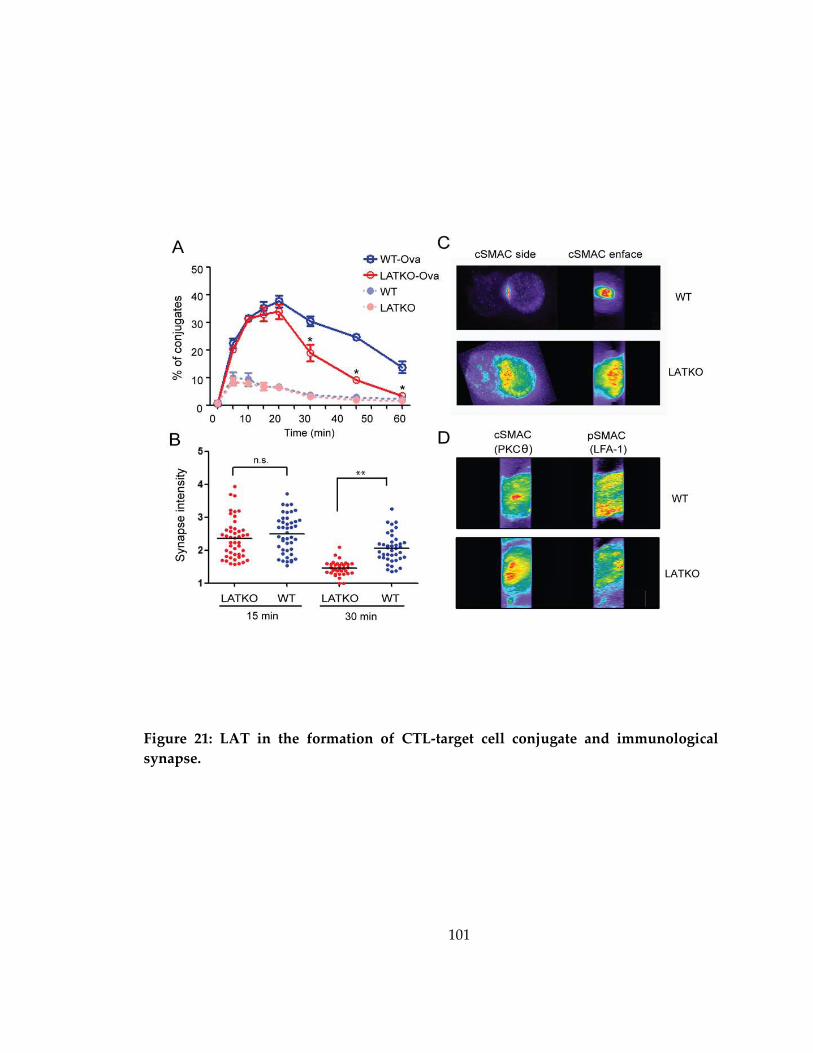
101
Figure 21: LAT in the formation of CTL-target cell conjugate and immunological
synapse.
102
Figure 21:
(A) Conjugate formation. CTLs were labeled with Cell Tracker Violet and mixed at a
ratio of 1:1 with L1210/Kb cells labeled with Cell Tracker Orange with or without Ova
peptide. At various time points, the cells were fixed and analyzed by FACS.CTL-target
cell conjugates were defined as cells labeled with both fluorescent dyes. The graph
shows one representative of three independent experiments. *p < 0.05; error bars
represent means ± s.e.m. (B) Quantitative analysis of cSMAC accumulation at the
synapse of WT and LATKO CTLs. CTLs were incubated with L1210/Kb cells preloaded
with the Ova peptide for 15 and 30 minutes to allow for the formation of conjugates. The
conjugates were fixed, permeabilized, and stained with anti-PKCθ antibody to define
cSMAC and anti-LFA1 antibody to represent pSMAC. cSMAC accumulation was
calculated as the ratio of the synaptic fluorescence intensity to the cytoplasmic
fluorescence intensity. Each dot represents one CTL; horizontal bars indicate the mean;
n.s., not significant; **p < 0.005. Data shown are from two independent experiments. (C,
D) Immunological synapse patterns. The representative cells depicted here show the
side and enface of cSMAC (C) and excluded pSMAC (D) of WT and LATKO CTLs.
103
Notably, by 30 minutes, the intensity of cSMAC accumulation significantly
decreased in LATKO CTLs (average=1.45, n=34) compared with WT CTLs (average=2.06,
n=40) (Figure 21B). Further analysis of cSMAC fluorescence at the synapse of LATKO
CTLs revealed a phenotype distinguished by multiple centers (Figure 21C), indicating
that synapse formation was altered by LAT-deficiency. We further examined pSMAC
exclusion from the cSMAC. The percentage of pSMAC exclusion was similar between
LATKO and WT CTLs (Figure 21D). These observations indicated that LATKO CTLs can
form a mature immunological synapse; however, LAT is essential for the maintenance of
a stable synapse.
4.2.5 Impaired MTOC and lytic granule polarization in LATKO CTLs
The synaptic release of lytic granules in CTLs is guided by the reorientation of
the MTOC. We next examined whether the MTOC in LATKO CTLs could be polarized
toward the synapse upon TCR engagement. OT-I CTLs were mixed with Ova peptide-
loaded L1210/Kb cells, fixed, and stained with anti-α tubulin and anti-gzmB to identify
MTOCs and granules, respectively. To quantitate MTOC polarization, we evenly
divided conjugated T cells into four zones as described previously where zone 1 is the
area closest to the target cell (Quann, Merino et al. 2009) (Figure 22A). In WT CTLs,
MTOCs gradually moved to zone 1 after interacting with target cells, increasing from
26.7% to 72.5% to 81% at 15, 30, and 45 min, respectively (n ≥ 60 for each group, Fig.
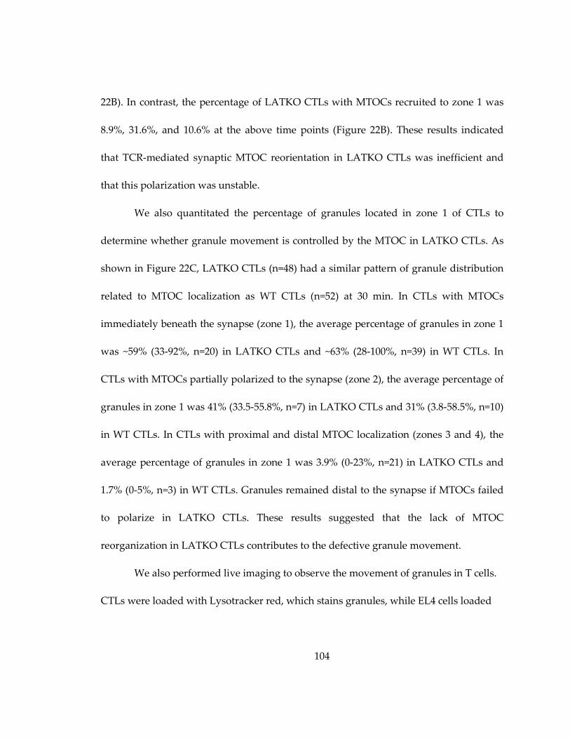
104
22B). In contrast, the percentage of LATKO CTLs with MTOCs recruited to zone 1 was
8.9%, 31.6%, and 10.6% at the above time points (Figure 22B). These results indicated
that TCR-mediated synaptic MTOC reorientation in LATKO CTLs was inefficient and
that this polarization was unstable.
We also quantitated the percentage of granules located in zone 1 of CTLs to
determine whether granule movement is controlled by the MTOC in LATKO CTLs. As
shown in Figure 22C, LATKO CTLs (n=48) had a similar pattern of granule distribution
related to MTOC localization as WT CTLs (n=52) at 30 min. In CTLs with MTOCs
immediately beneath the synapse (zone 1), the average percentage of granules in zone 1
was ~59% (33-92%, n=20) in LATKO CTLs and ~63% (28-100%, n=39) in WT CTLs. In
CTLs with MTOCs partially polarized to the synapse (zone 2), the average percentage of
granules in zone 1 was 41% (33.5-55.8%, n=7) in LATKO CTLs and 31% (3.8-58.5%, n=10)
in WT CTLs. In CTLs with proximal and distal MTOC localization (zones 3 and 4), the
average percentage of granules in zone 1 was 3.9% (0-23%, n=21) in LATKO CTLs and
1.7% (0-5%, n=3) in WT CTLs. Granules remained distal to the synapse if MTOCs failed
to polarize in LATKO CTLs. These results suggested that the lack of MTOC
reorganization in LATKO CTLs contributes to the defective granule movement.
We also performed live imaging to observe the movement of granules in T cells.
CTLs were loaded with Lysotracker red, which stains granules, while EL4 cells loaded
105
Figure 22: LAT in the MTOC and lytic granule polarization.

106
Figure 22:
CTL-target cell conjugates were stained with anti-α tubulin and anti-gzmB to represent
MTOCs (white) and granules (green), respectively. (A) The representative conjugates
depicted here show MTOCs and granules localized at zones 1-4 in CTLs. (B)
Quantitative analysis of MTOC localization in different zones of CTLs upon engagement
of L1210/Kb cells for 15, 30, and 45 minutes. (C) Granule movement is controlled by
MTOC polarization. The percentage of granules in zone 1 was quantitated in each CTL
with MTOCs at different zones at 30 minutes. Each dot represents one CTL and the
value represents the percentage of granules localized to zone 1; horizontal bars indicate
the mean (LATKO=48, WT=52). Data shown are from two independent experiments.

107
with Ova peptide were labeled with calcein, a dye that is retained in live cells and lost
upon membrane permeabilization. Following CTL-target cell contact, the lytic granules
in WT CTLs reoriented toward and were subsequently delivered into the target cells. In
contrast, the granules in some LATKO CTLs remained distal to the synapse for more
than 10 minutes, resulting in a failure of granule delivery to the target cell. Collectively,
these data suggested that LAT functions to promote MTOC reorientation and,
consequently, granule movement towards the synapse.
4.2.6 Defective TCR signaling in LATKO CTLs
Our data demonstrated that LAT plays a significant role in each step of granule
exocytosis, thereby LATKO CTLs are defective in their ability to kill target cells;
however, they are not devoid of cytotoxicity. To analyze the TCR-mediated signaling
events that regulate this residual cytotoxicity, we performed biochemical analyses of
TCR-mediated signaling pathways in LATKO CTLs. LATKO and WT CTLs were
stimulated with anti-CD3 plus anti-CD8 to assess the overall tyrosine phosphorylation
of proteins. No significant difference was observed except that phosphorylated LAT was
absent in LATKO CTLs (Figure 23A). The lack of phosphorylated LAT was also
supported by Western blotting with anti-pY191 of LAT (Figure 23B), further confirming
complete deletion of LAT. PLC-γ1 and MAPK activation, two important signaling events
downstream of LAT, were dramatically diminished, as predicted, though they were not
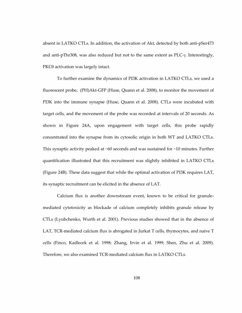
108
absent in LATKO CTLs. In addition, the activation of Akt, detected by both anti-pSer473
and anti-pThr308, was also reduced but not to the same extent as PLC-γ. Interestingly,
PKCθ activation was largely intact.
To further examine the dynamics of PI3K activation in LATKO CTLs, we used a
fluorescent probe, (PH)Akt-GFP (Huse, Quann et al. 2008), to monitor the movement of
PI3K into the immune synapse (Huse, Quann et al. 2008). CTLs were incubated with
target cells, and the movement of the probe was recorded at intervals of 20 seconds. As
shown in Figure 24A, upon engagement with target cells, this probe rapidly
concentrated into the synapse from its cytosolic origin in both WT and LATKO CTLs.
This synaptic activity peaked at ~60 seconds and was sustained for ~10 minutes. Further
quantification illustrated that this recruitment was slightly inhibited in LATKO CTLs
(Figure 24B). These data suggest that while the optimal activation of PI3K requires LAT,
its synaptic recruitment can be elicited in the absence of LAT.
Calcium flux is another downstream event, known to be critical for granule-
mediated cytotoxicity as blockade of calcium completely inhibits granule release by
CTLs (Lyubchenko, Wurth et al. 2001). Previous studies showed that in the absence of
LAT, TCR-mediated calcium flux is abrogated in Jurkat T cells, thymocytes, and naive T
cells (Finco, Kadlecek et al. 1998; Zhang, Irvin et al. 1999; Shen, Zhu et al. 2009).
Therefore, we also examined TCR-mediated calcium flux in LATKO CTLs.
109
Figure 23: Defective TCR signaling in LATKO CTLs.
(A, B) TCR-mediated protein phosphorylation. CTLs were restimulated with anti-CD3 (5
μg/ml) and anti-CD8 (1 μg/ml) for the indicated time points before lysis. Cell lysates
were analyzed by Western blotting with anti-pTyr (A), phospho-specific, or pan
antibodies against LAT, Erk, PLC-γ1, PKCθ, and Akt (B). Data shown are from one
representative of three independent experiments.

110
Figure 24: Reduced PI3K/Akt activity in LATKO CTLs.

111
Figure 24:
(A) The recruitment of PH(Akt) probe to the synapse upon engagement with target cells.
Splenocytes were primed with Ova peptide before being transduced with retroviruses
expressing (PH)Akt-GFP construct. For live cell imaging, CTLs were incubated with
L1210/Kb cells and the movement of the probe was recorded at intervals of 20 seconds.
(B) Comparison of the recruitment of (PH)Akt to the synapse in LATKO CTLs (n=14)
and WT CTLs (n=15) as a function of time. The accumulation of (PH)Akt was calculated
as the ratio of the fluorescence intensity at the synapse versus the non-synapse area of
the cells at different time points. Data shown are from two independent experiments

112
CTLs were loaded with indo-1 and calcium flux was initiated by anti-CD3 and anti-CD8
crosslinking. As shown in Figure 25A, calcium flux in GFP+ LATKO CTLs was
dramatically diminished but not abrogated.
To analyze calcium signaling at the single cell level, CTLs were loaded with
Fura-2 and mixed with Ova peptide-loaded L1210/Kb cells for live cell imaging.
Consistent with the FACS data (Figure 25B), there was a detectable increase of Fura-2
fluorescence in LATKO CTLs, although it was much weaker than that seen in WT CTLs
(Figure 25B). We also quantitated Fura-2 fluorescent intensity in these CTLs. As shown
in Figure 25C, the integrated value of calcium mobilization into the cytosol within the
first 10 minutes upon T-target cell interaction was much lower in LATKO CTLs (~3,
n=24) than that in WT CTLs (~8, n=19).
This residual calcium flux could be a result of co-stimulation or signaling
through adhesion molecules (Smith-Garvin, Koretzky et al. 2009). To examine this
possibility, we pretreated target cells with CTLA4-Ig and anti-ICAM-1 antibody before
imaging. With the blockade, calcium flux was significantly decreased in WT CTLs (~5,
n=27) (Figure 25C). However, it remained unchanged in LATKO CTLs (~3, n=24),
suggesting that the residual calcium was not mediated by CD28 or LFA-1-mediated
signaling.

113
Figure 25: Impaired but not abolished calcium signaling in LATKO CTLs.

114
Figure 25:
(A) Calcium mobilization. CTLs were loaded with the calcium indicator, indo-1.
Calcium flux was induced by anti-CD3 and anti-CD8 crosslinking and analyzed by
FACS. Data are representative of three independent experiments. (B) Time-lapse
montages of both ratiometric Fura-2 images and DIC of CTLs. CTLs were preloaded
with Fura-2, mixed with Ova peptide-loaded L1210/Kb cells, and imaged at intervals of
30 seconds. The first time point (0’00”) marked the initial contact of a T cell with a target
cell. Fura images are pseudo-colored with warmer colors indicating higher intracellular
calcium concentrations. Upper rows, WT CTLs. Bottom rows, LATKO CTLs. (C)
Quantitative analysis of calcium flux by live cell imaging. CTLs were pre-treated with or
without CTLA4-Ig and anti-ICAM-1 antibody for 30 minutes before incubation with
target cells. The integrated value of calcium mobilized into the cytosol for each cell was
generated by summing the changes in the relative calcium ratio (340/380 nm) at each
time point within the first 10 minutes. Each dot represents one CTL; horizontal bars
indicate the mean; n.s., not significant; *p<0.05, **p<0.005. Data shown are from two
independent experiments.
115
4.2.7 Residual PLC activity and calcium flux are necessary to drive cytotoxicity of LATKO CTLs
Our data showed that LAT-deficiency severely impaired TCR-mediated
signaling in CTLs; however, activation of Akt and PKCθ was relatively normal. It is
possible that these residual signaling events are importance for the cytotoxicity of
LATKO CTLs. To determine which pathways are required, we examined the killing
capability of CTLs in the presence of a series of pharmacological inhibitors targeting
different signaling modules. CTLs were pretreated with inhibitors for only 30 minutes to
minimize the toxicity and were used in in vitro killing assay. The PKC inhibitor Gö6976
and three MAP kinase inhibitors, PD98059 (MEK), SP600125 (JNK), and SB203580 (p38),
had only minimal effects on the cytotoxicity of both WT and LATKO CTLs (Figure 26A
and data not shown). In contrast, the PI3K inhibitor LY294002 had a dramatic effect. It
reduced the killing by LATKO CTLs and WT CTLs by 56% and 42%, respectively.
Furthermore, the PLC inhibitor U73122 had the most impact (Figure 26A). In the
presence of U73122, the percentage of killing by LATKO CTLs and WT CTLs was
reduced by 77% and 67%, respectively. The inhibition by U73122 was dose-dependent
(data not shown). At the concentration we used (0.5 µM), U73122 completely abrogated
calcium flux in LATKO CTLs (Figure 26B); however, there was still residual calcium flux
in WT CTLs.

116
Figure 26: The residual activation of PLC and calcium signaling enabled LATKO
CTLs to kill target cells.
(A) The effect of different inhibitors on CTL-mediated killing. CTLs were pre-treated
with PD98059 (MEK inhibitor, 50 μM), LY294002 (PI3K inhibitor, 50 μM), Gö6976 (PKC
inhibitor, 30 μM), U73122 (PLC inhibitor, 0.5 μM), or DMSO for 30 minutes and then
incubated with target cells in the presence of these inhibitors for 45 minutes before FACS
analysis. Data shown are from one representative of three independent experiments. (B)
Calcium mobilization in the presence of U73122 (0.5 μM).
117
Since this inhibitor can abrogate MTOC polarization in CD4 T cells (Quann,
Merino et al. 2009), we next examined whether it also affects MTOC reorientation in
CTLs. In our experimental setup, the percentages of MTOCs polarized to zone 1 in
LATKO CTLs was reduced from 27% (n=146) to 13% (n=166) by U73122; however, this
inhibitor had no effect on MTOC polarization in WT CTLs (n=138 each) (Figure 27A).
On the other hand, LY294002 impaired MTOC reorientation of both LATKO and WT
CTLs. The percentage of MTOCs polarized to zone 1 was reduced from 38% (n=140) to
20% (n=157) in LATKO CTLs, and from 68% (n=166) to 44% (n=150) in WT CTLs (Figure
27B). Together, our results suggested that cytotoxicity is regulated by multiple signaling
events. While PI3K activation is one of the driving forces positioning MTOC
polarization, PLC activation is imperative for cytotoxicity, but not necessarily for MTOC
reorientation in CTLs.

118
Figure 27: PLC activity is not necessary for MTOC polarization in CTLs.
(A, B) Quantitative analysis of MTOC localization in the presence of U73122 and
LY294002. CTLs were treated with inhibitors for 30 minutes and incubated with target
cells for another 30 minutes in the presence of inhibitors.

119
4.3 Discussion
The primary feature of CTLs is their ability to kill cells that present foreign
antigens. CTLs exhibit cytotoxicity by three mechanisms: IFN-γ production, FasL-
mediated and lytic granule-mediated pathways. Our data showed that the cytotoxicity
triggered by LATKO CTLs was reduced compared to WT CTLs, and this defective
killing was contributed by impaired IFN-γ production, FasL upregulation, as well as
reduced granule delivery. Notably, these defects are not caused by reduced expression
of the TCR at the cell surface, which is a phenomenon previously observed with long-
term deletion of LAT (Mingueneau, Roncagalli et al. 2009; Shen, Zhu et al. 2009{; Shen,
Chuck et al. 2010). In this study, deletion of LAT by treatment with tamoxifen for less
than a week did not cause any change in TCR surface expression. Therefore, it suggests
that signaling emanating from LAT is critical to activate transcription factors that
regulate IFN-γ production and FasL upregulation.
Every step of granule exocytosis was also affected by LAT-deficiency, from
immunological synapse maintenance to MTOC polarization, yet how LAT affect these
cellular events is not known. When a CTL encounters a target cell, a mature cytotoxic
immunological synapse is formed at the CTL:target contact zone, allowing the MTOC
and granules to reorient and move toward the target cell. It has been shown that
continuous TCR-mediated signaling is required for stabilizing cSMAC and pSMAC

120
organization into the synapse, as well as polarizing the MTOC toward the synapse
(Huppa, Gleimer et al. 2003). Our conjugation assay showed that the initial conjugate
formation between CTLs and target cells was not affected by LAT-deficiency; however,
the conjugates formed between target cells and LATKO CTLs were not as stable as those
formed with WT CTLs. Our synapse data also showed that LAT-deficiency affected
synapse maintenance in CTLs. In addition, MTOC polarization in CTLs was unstable
and inefficient in the absence of LAT, possibly due to the weak stability of conjugate and
synapse formation. These observations suggest that mature synapses and polarized
MTOCs could form without LAT; however, the maintenance of stable conjugates and
synapses requires LAT-mediated signaling.
These defects are most likely due to the inactivation of downstream effector
molecules. For examples, integrin, an adhesion molecule enriched in the pSMAC, is
maintained in an inactive conformation with a low affinity for its ligand. “Inside-out”
signals that originate upon engagement of the TCR or chemokine receptors activate
signaling molecules, such as Rap1, which associates with ADAP and SKAP55, to induce
conformational changes of LFA-1, thereby increasing their affinity to the ligands
(Menasche, Kliche et al. 2007; Smith-Garvin, Koretzky et al. 2009). It is possible that
inside-out signals in LATKO CTLs were impaired and the affinity between integrins and
their ligands remained weak, resulting in unstable conjugate and synapse formation
121
between T cells and target cells. Furthermore, we observed that the cSMACs in LATKO
CTLs were altered, with a phenotype distinguished by multiple centers. It is possible
that the activity of motor proteins involved in synapse formation and maintenance is
affected by LAT-deficiency. For instance, myosin IIA, an actin-based motor protein, is
essential for the movement of microclusters and synapse persistence (Ilani, Vasiliver-
Shamis et al. 2009). Dynein, a microtubule motor protein, is shown to associate with TCR
and helps to bring the microclusters toward the center of the cell contact region
(Hashimoto-Tane, Yokosuka et al. 2011). Accessing the activity of these motor proteins
in LATKO CTLs will help to identify the mechanisms by which LAT regulates granule
delivery from CD8 T cells.
Interestingly, although LATKO CTLs demonstrated multiple defects in
cytotoxicity, their granule release was not totally blocked (~30% granule release). One
possibility is that there was a very small amount of undeleted LAT, which was sufficient
to trigger the residual CTL killing. Our knock-in strategy allows us to accurately mark T
cells with LAT exons deleted since LAT-GFP fusion protein is expressed from the LAT
knock-in allele. As such, GFP+ CTLs should all have successfully deleted the LAT gene.
The killing capability of the sorted GFPhigh LATKO cells was comparable to that of
non-sorted T cells. Moreover, our Western blot analysis and immunofluorescence
staining of LATKO CTLs showed undetectable levels of LAT or phospho-LAT,

122
suggesting that in addition to gene deletion, there was no residual LAT protein in these
cells. Nevertheless, despite that no LAT protein was detected in LATKO CTLs, it is
difficult to prove experimentally that there is absolutely no functional LAT in these cells.
Regardless, it is intriguing to observe that CTLs with extremely weak TCR signaling, as
shown in our biochemical and calcium data, still retained cytotoxic ability (~25%
compared to WT cells). Given that T cell activation and interferon production were
nearly abolished under the same inducible LAT-deletion system, this finding suggests
that the signaling strength governing CTL cytotoxicity differs from that required for
other effector functions.
Previous studies using inhibitors indicate that PI3K activity is critical for the
cytotoxicity of CTLs and NK cells (Fuller, Ravichandran et al. 1999; Robertson, Mireau et
al. 2005). Our biochemical data showing that Akt phosphorylation was only slightly
reduced in LATKO cells indicated that PI3K can be activated in the absence of LAT,
although LAT is necessary for its optimal activation. A previous report also
demonstrated the activation of PI3K/Akt in the absence of LAT in activated CD4 T cells.
Even though LAT does not have a canonical p85-binding motif, phosphorylated LAT
has been shown to bind the p85 subunit of PI3K (Zhang, Sloan-Lancaster et al. 1998). p85
also binds the cytoplasmic domain of CD28. However, the role of CD28-mediated PI3K
activation in CTLs is limited. CTLs are able to eliminate target cells that do not express
123
B7 molecules, indicating that T cell killing does not require the interaction of CD28 with
B7 molecules. Blockade of CD28-mediated costimulation by CTLA4-Ig had no effect on
the cytotoxic ability of LATKO CTLs, further supporting a limited role of CD28 in CTL
killing. Therefore, it is unlikely that CD28 signaling can be counted as the major source
providing LAT-independent PI3K activation. It is unclear how PI3K is activated in the
absence of LAT after TCR engagement. One possibility is that PI3K binds to adaptor
proteins that contain YXXM motifs, such as TRIM (TCR-interacting molecule). It is also
possible that p85 can be recruited to the membrane through the interaction between its
proline-rich region and the SH3 domains of Lck or Fyn (Okkenhaug and
Vanhaesebroeck 2003).
PLC-γ1-mediated signaling has been implicated in regulating the molecular
mechanisms of granule exocytosis (de Saint Basile, Menasche et al. 2010). Our data
showed that PLC-γ1 phosphorylation and activity, albeit diminished, was still detected
in LATKO CTLs and that this residual PLC activity appears to be important for
cytotoxicity. Blockade of PKC or MAPK activity, the signaling pathways downstream of
DAG, had only minor effects on the killing ability of LATKO CTLs, suggesting that the
residual cytotoxicity mediated by LATKO CTLs relies heavily on calcium signaling. Our
data further demonstrated that inhibition of PLC by U73122 completely abrogated the
residual calcium flux in LATKO CTLs and abolished their residual cytotoxicity. This
124
provides a cohesive explanation that the residual PLC activity generates a minimal
amount of cytosolic calcium elevation, which is sufficient to support the observed CTL
killing.
Calcium signaling is directly involved in lytic granule release. One study
determined that granule movement can occur in Ca2+-free medium; however, the release
of granules and the killing of target cells can only happen after the addition of calcium
(Lyubchenko, Wurth et al. 2001). Whether intracellular Ca2+ regulates MTOC
polarization has been a controversial issue. The minus end-directed motor protein
dynein, which can bind calmodulin, has been shown to function in MTOC orientation in
Jurkat cells (Combs, Kim et al. 2006). Kuhne’s data demonstrated a correlation between
calcium flux and MTOC polarization in Jurkat cells (Kuhne, Lin et al. 2003). However,
recent data using single-cell photoactivation of the TCR revealed a different result where
blocking calcium signaling has no effect on MTOC reorientation (Quann, Merino et al.
2009).
Our data here showed that, although TCR-mediated calcium mobilization was
severely impaired in LATKO CTLs, their MTOC reorientation was only partially
affected. The residual activity of PLC in LATKO CTLs might generate a small amount of
calcium flux that is sufficient to activate downstream signaling molecules, including
classical and novel PKC, protein kinase D, and the chimaerins. This is supported by our

125
data showing that, in LATKO CTLs, the activity of PKCθ, a downstream signaling
molecule of DAG, was relatively normal.
Early studies using LAT-deficient Jurkat cells indicate that LAT is crucial for
TCR-mediated PLCγ1 activation and calcium mobilization. It is not clear why TCR-
mediated PLC-γ1 activation and calcium flux are not totally dependent on LAT in CTLs.
It is likely that other adaptor molecules, such as LAB, compensate for the loss of LAT in
these OT-I CTLs. Our unpublished data showed that LAB is indeed expressed in CTLs.
Whether it functions in these cells remains to be investigated using LAB- or LAT/LAB-
deficient mice. It is also possible that residual PLC-γ1 activation and calcium flux are
initiated through other signaling components, such as adhesion molecules and growth
factors. LAT-independent Ras-MAPK activation has been documented previously (Chau
and Madrenas 1999). Similarly, ERK activation was observed in ZAP-70-deficient Jurkat
cells (Shan, Balakir et al. 2001). PLC-γ1 might be recruited to the plasma membrane
through molecules other than LAT or other LAT-independent PLC isozymes may
compensate for the residual activity of PLC-γ1.
In conclusion, our data demonstrate that in the absence of LAT, CTLs failed to
produce IFN-γ and upregulate FasL, as well as exhibiting partial defects at each step of
granule-mediated cytotoxicity. All of these results indicated the importance of LAT in
CTL function. However, LATKO CTLs were still able to kill target cells at a reduced

126
level, suggesting that CTLs may be programmed to adopt multiple pathways to perform
their cytolytic function.
5. General Discussion and Future Directions
Upon pathogen infection, the antigen-specific CD8 T cell population undergoes
several changes in a short timeframe, including clonal expansion, contraction, memory
formation and maintenance. These processes must be tightly regulated in order to
properly defend the hosts against rapidly proliferating pathogens, efficiently resolve
inflammation once the pathogens are cleared, and prevent autoimmunity and
immunopathology. Through our studies, we were able to demonstrate that LAT
signaling is indispensable for activation and continuous expansion of CD8 T cells during
both primary and memory responses. On the contrary, CD8 T cell contraction and the
maintenance of memory CD8 T cells can occur normally when LAT is absent. This
chapter will discuss the potential rationale and mechanisms by which LAT signaling,
and thereby TCR-mediated signaling in general, play distinct regulatory roles during
different stages of T cell response to Listeria monocytogenes infection.
5.1 LAT-mediated signaling in CD8 T cell expansion
As described in Figure 8, we showed that sustained TCR signaling during the
priming phase is crucial for an optimal primary response. This is supported by several
findings that persistent antigens and TCR-MHC engagement are essential for CD8 T cell
127
expansion (Kaech and Ahmed 2001; Tewari, Walent et al. 2006; Zehn, Lee et al. 2009).
Additionally, the magnitude of T cell expansion during priming is proportional to the
amount and the affinity of antigens (Wherry, Puorro et al. 1999; Badovinac, Porter et al.
2002; Zehn, Lee et al. 2009).
Interestingly, some studies argue against the notion that antigen persistence is
required for CD8 T cell expansion. For example, mice that received antibiotic treatment
at 24 hours post-infection with Listeria monocytogenes still displayed a normal CD8 T cell
expansion (Mercado, Vijh et al. 2000), indicating that the initial antigen stimulation is
sufficient to drive subsequent T cell proliferation. Moreover, after exposure of APCs and
antigens for as short as 2 hours, CD8 T cells can continue to undergo several rounds of
division in the absence of antigenic stimulation. This antigen-independent proliferation
is driven by IL-2 and can be augmented by IL-7 or IL-15 (Kaech and Ahmed 2001; van
Stipdonk, Lemmens et al. 2001; Wong and Pamer 2001). As a result of these
observations, it was suggested that antigen stimulation is only required for a short
period of time during the initial stage of priming, whereas continuous T cell expansion
can proceed independently of antigens (Condotta, Richer et al. 2012).
This discrepancy between our study and others might have resulted from
different experimental methods employed to detect the response of CD8 T cells. After
revisiting their publications, it appears that most of their experiments were performed to

128
study the ability of CD8 T cells to “proliferate” in vitro, rather than their capacity to
mount an immune response in vivo as a read out for T cell expansion. In these studies,
CD8 T cells were incubated with antigen-pulsed APCs for a short period of time and
then taken away from APCs. Subsequently, purified CD8 T cells were cultured in vitro or
injected back into naïve mice to measure their proliferative ability in the absence of
antigens by CFSE dilution. Since it is well-established that activated T cells undergo
proliferation in response to IL-2 (Toribio, Gutierrez-Ramos et al. 1989), it is not
surprising that these CD8 T cells were able to divide in an IL-2-dependent manner after
antigen withdrawal. In fact, our result showed that activated LATKO T cells were able to
proliferate normally in vitro with the presence of exogenous IL-2, demonstrating that
TCR signaling is not required for activated T cells to undergo proliferation in vitro.
Nonetheless, this does not imply that IL-2-dependent T cell proliferation is capable of
mounting an effective immune response in vivo.
In our study, when mice were treated with tamoxifen to delete LAT at day 4
post-infection, LATKO CD8 T cells expanded ~7 fold in three days, which was a
drastically impairment in comparison with the ~60-fold expansion by WT CD8 T cells. It
is possible that this ~7-fold expansion could be due to residual LAT protein or an
antigen-independent, IL-2-mediated cell proliferation. Therefore, it can be inferred that

129
antigen-independent CD8 T cell expansion plays a minimal role in the overall
proliferation seen during the primary response in a mouse model.
Since activation of naïve CD8 T cells is not synchronized, it is also possible that
the majority of LATKO CD8 T cells were not activated or not ready to proliferate when
tamoxifen was administrated, thus preventing their ability to expand. Moreover, it is
also plausible that LAT regulates CD8 T cell expansion through TCR signaling-
independent mechanisms. For example, deletion of LAT might affect cytokine signaling
which affects proliferation of CD8 T cells. It could also be explained that LAT might be
involved in the modulation of cell cycling.
Our data suggest that sustained TCR signaling is crucial to maintain CD8 T cell
expansion; however, TCR signaling alone may not be sufficient to elicit an efficient
primary response. Evidence supporting this point comes from the observation that the
extent of primary CD8 T cell expansion was partially affected by IL-2 deficiency
(D'Souza and Lefrancois 2003). Similarly, Bcl-2 transgenic mice carrying a deletion of
common gamma chain, which abolishes IL-2, IL-7, IL-15, and IL-21 signals, had a slight
reduction in CD8 T cell expansion (Decaluwe, Taillardet et al. 2010), suggesting that
these cytokines play a role in the maintenance of cell survival and/or proliferation
during the priming stage. Collectively, these data indicate that TCR signaling is the

130
primary factor driving T cell expansion, and that together with cytokine signals,
maximal magnitude of T cell expansion is achieved synergistically.
5.2 LAT-mediated signaling in CD8 T cell contraction
CD8 T cell contraction is caused by multiple mechanisms including TCR
stimulation, inflammation, and cytokine withdrawal. Cytokine withdrawal has a
paramount role in the regulation of contraction. When growth factors and cytokines,
such as IL-2, IL-7, and IL-15, become limited, PI3/Akt pathway is inactivated.
Consequently, Glut1 and amino acid transporters are internalized and degraded, thus
preventing T cells from using extracellular energy. Moreover, pro-apoptotic Bcl-2 family
proteins are activated, resulting in cellular atrophy and apoptosis.TCR engagement
induces TNFR expression on the surface of T cells, which triggers extrinsic apoptotic
signals when it binds to its ligand.
In addition to cytokine signaling, TCR stimulation induces T cells to produce
IFN-γ, which has been shown to regulate cell contraction (Badovinac, Tvinnereim et al.
2000). Interestingly, our studies showed that CD8 T cell contraction can be independent
of LAT, suggesting that triggering of programmed cell death at this stage does not
require TCR engagement. One possible explanation is that although LATKO CD8 T cells
were unable to produce TNF-α and IFN-γ, these cytokines were produced by WT T cells
and other cell types within the secondary lymphoid organs in the same environment,

131
and were able to bind the TNFR and IFNR of LATKO T cells, thus inducing apoptosis.
Therefore, it appears that TCR signals function to program cell death during the
expansion phase; however, they can be dispensable when contraction is occurring.
5.3 LAT-mediated signaling in memory CD8 T cell maintenance
Immunological memory can be maintained for a long time, which is mainly
attributed to the ability of memory CD8 T cells to undergo antigen-independent self-
renewal. IL-7 and IL-15 are two cytokines that are crucial in supporting cell survival and
homeostatic proliferation of memory T cells, respectively (Lodolce, Boone et al. 1998;
Yajima, Nishimura et al. 2002; Ma, Koka et al. 2006; Surh, Boyman et al. 2006). As
described in Figure 14, our data showed that memory CD8 T cells are able to survive for
months after deletion of LAT, demonstrating that LAT signaling is dispensable for the
maintenance of memory CD8 T cells. This is supported by several other studies showing
that deletion of TCR or downstream signaling proteins, such as Lck and SLP-76, had no
effect on the persistence of memory T cells (Tewari, Walent et al. 2006; Leignadier,
Hardy et al. 2008; Wiehagen, Corbo et al. 2010). These experiments further suggest that
memory CD8 T cells do not need tonic TCR signaling for maintenance.
In contrast to memory T cells, naïve T cells require both IL-7 signaling and tonic
TCR signaling pathways for long-term survival (Takeda, Rodewald et al. 1996; Tanchot,
Lemonnier et al. 1997). Constant recognition of self-peptide/MHC complex induces a

132
basal level of TCRζ phosphorylation and Zap-70 association, which are the early
signaling events that regulates naïve T cell survival (Witherden, van Oers et al. 2000). In
line with this concept, previous data from our laboratory revealed that deletion of LAT
in naive T cells abolished their survival and homeostatic proliferation (Shen, Chuck et al.
2010). Nonetheless, there still exists a need to determine whether survival and
homeostatic proliferation are differentially regulated, and how LAT signaling may be
involved in these two processes.
These discoveries have led us to question why LAT signaling, specifically tonic
TCR signaling, is necessary for T cell homeostasis in naïve T cells but not memory T
cells. This discrepancy might reflect the differences of biological requirements between
naïve and memory T cells. The primary function of naïve CD8 T cells is surveillance.
They migrate throughout the secondary lymphoid organs and peripheral tissues to
constantly scan for APCs that may carry foreign pathogens. It is possible that the ability
of antigen recognition is so critical for naïve T cells that tonic TCR signaling is
evolutionally integrated with the survival signals in this cell population. On the other
hand, memory CD8 T cells have already seen their antigens, thus their primary goal is to
survive long term until they encounter the same pathogens. Consequently, tonic TCR
signaling might be uncoupled from the survival pathway or can be compensated by

133
other survival/proliferation mechanisms in memory T cells, such as IL-7 and IL-15
signaling pathways.
The crosstalk between IL-7 and TCR signals has been demonstrated to play an
important role in naïve T cell survival (Carrette and Surh 2012). Mechanistically, binding
of IL-7 or IL-15 triggers JAK1/JAK3-mediated Stat5 dimerization. Activated Stat5, and
together with TCR signaling, induce the expression of Bcl-2 and Mcl-1, which
antagonize Bim and Bid-mediated intrinsic apoptotic pathways (Surh and Sprent 2008).
In addition, TCR engagement by self-peptide/MHC complexes can enhance the
sensitivity of IL-7R (CD127) by inducing the formation of lipid rafts at the plasma
membrane, and then recruiting IL-7R to the lipid rafts. Moreover, IL-7R expression can
also be tuned by tonic TCR signaling through Foxo1 or Foxp1-mediated transcriptional
regulation (Kerdiles, Beisner et al. 2009).
Since these studies demonstrated that tonic TCR signaling has the potential to
crosstalk with IL-7 signaling through multiple mechanisms, it will be interesting if we
can compare the effect of LAT deletion on the function of IL-7 and IL-15 signaling in
both naïve and memory T cells, such as the expression of IL-7R, IL-15R, Bcl-2, Mcl-2, and
Bim, as well as Stat5 activation. Our preliminary data showed that CD127, CD122, and
Bcl-2 expression were similar between LATKO and WT memory CD8 T cells, suggesting
that the level of these markers in memory CD8 T cells was not affected by LAT
134
deficiency. Since it has been shown that deletion of LAT in naïve T cells results in
reduced cell numbers over time, perhaps tonic LAT signaling in naïve cells effects IL-7
and/or IL-15 signaling. This may provide insight into the interplay between TCR and
cytokine signals in both naïve and memory T cells, and possibly explain why tonic TCR
signaling is more important in naïve T cells than in memory T cells.
Another explanation of why memory T cells do not require tonic TCR signaling
for homeostasis could be that memory T cells adopt a different survival mechanism
when TCR signals are abrogated. One such example is Bcl-2, a molecule that is critical
for naïve T cell survival, yet, several studies questioned the significance of Bcl-2 in
memory T cell maintenance. Interestingly, transgenic expression of Bcl-2 failed to rescue
memory T cell formation when IL-7 and IL-15 signals are deficient, suggesting that
memory T cells employ Bcl-2-independent pathways for survival (Decaluwe, Taillardet
et al. 2010). This point is further supported by a report showing that, while the numbers
of Bcl-2-deficient naïve T cells were drastically diminished in mice, the numbers of Bcl-2-
deficient memory T cells after Lymphocytic Choriomeningitis Virus (LCMV) infection
remained relatively normal (Wojciechowski, Tripathi et al. 2007), suggesting that
memory T cell generation and/or maintenance is not entirely dependent on Bcl-2.
Further investigations revealed that memory T cells still require Bcl-2 for IL-7/IL-15-
135
driven survival in vitro, but their capacity for homeostatic proliferation is augmented
when Bcl-2 is absent.
Therefore, it is plausible that LAT signaling is required for memory T cell
survival; however, the ability for self-renewal is enhanced in LAT-deficient memory
CD8 cells. Consequently, the survival defect is compensated and these cells persist over
time. This hypothesis is supported by data demonstrating that cells receiving weak TCR
signals preferentially become TCM cells, which is a cell population that has superior self-
renewal capability. Further study to dissect survival vs. homeostatic proliferation in
LAT-deficient memory T cells will be helpful to differentiate the role of LAT-signaling in
these two events.
In addition to memory CD8 T cells, it will be interesting to study the role of LAT
signaling in CD4 T cell memory formation and maintenance. Previous data in our
laboratory demonstrated that LAT has both positive and negative roles in the regulation
of naïve CD4 T cell proliferation. Transfer of LAT-deficient naïve CD4 T cells into
lymphopenic hosts exhibited impaired homeostatic proliferation. Unexpectedly,
inducible deletion of LAT will result in mice developing a lymphoproliferative
syndrome, featured by an uncontrolled expansion of CD4 T cells that produce large
amounts of Th2 cytokines, though less severe than that seen in LATf/Y136F mutant mice
(Shen, Chuck et al. 2010). Whether LAT plays a positive or negative role in the

136
homeostatic proliferation of antigen-specific memory CD4 T cells remains unsolved. The
ERCre-LAT conditional knockout system can provide us with a tool to further explore
these interesting questions.
5.4 The potential role of LAT signaling in chronic infection
Through our studies, we conclude that the requirement for LAT-mediated
signaling corresponds to the presence of antigens in an acute infection model elicited by
Listeria monocytogenes (Figure 28). This finding leads us to question, under a chronic
infection model where antigens are persistent, what is the role of LAT-mediated
signaling in CD8 T cells? It would be interesting to elucidate if LATKO T cells would
undergo a similar contraction as WT T cells and if LATKO memory T cells would persist
over time or be able to exert their effector function. Our data demonstrate that the
occurrence of programmed cell death during contraction can be independent of TCR
signals, implying that the presence of antigens is not a determining factor during this
process. Indeed, CD8 T cell contraction still occurred during chronic infection. This leads
us to hypothesize that LATKO CTLs would undergo a relatively normal contraction
during both acute and chronic infections.
Another interesting area to explore is CD8 T cell exhaustion, which is the main
feature of chronic infection. Due to constant antigen stimulation, CD8 T cells become
dysfunctional and lose the capacity to produce cytokines and exhibit cytotoxicity, which
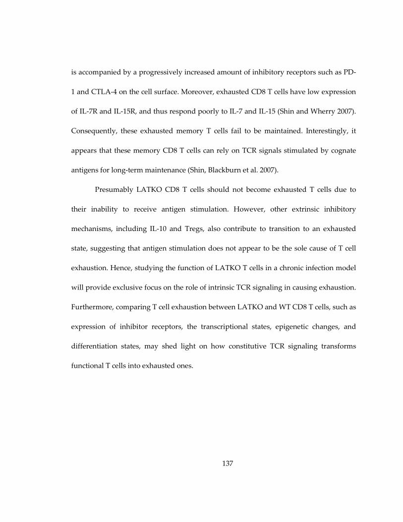
137
is accompanied by a progressively increased amount of inhibitory receptors such as PD-
1 and CTLA-4 on the cell surface. Moreover, exhausted CD8 T cells have low expression
of IL-7R and IL-15R, and thus respond poorly to IL-7 and IL-15 (Shin and Wherry 2007).
Consequently, these exhausted memory T cells fail to be maintained. Interestingly, it
appears that these memory CD8 T cells can rely on TCR signals stimulated by cognate
antigens for long-term maintenance (Shin, Blackburn et al. 2007).
Presumably LATKO CD8 T cells should not become exhausted T cells due to
their inability to receive antigen stimulation. However, other extrinsic inhibitory
mechanisms, including IL-10 and Tregs, also contribute to transition to an exhausted
state, suggesting that antigen stimulation does not appear to be the sole cause of T cell
exhaustion. Hence, studying the function of LATKO T cells in a chronic infection model
will provide exclusive focus on the role of intrinsic TCR signaling in causing exhaustion.
Furthermore, comparing T cell exhaustion between LATKO and WT CD8 T cells, such as
expression of inhibitor receptors, the transcriptional states, epigenetic changes, and
differentiation states, may shed light on how constitutive TCR signaling transforms
functional T cells into exhausted ones.
138
Figure 28: The models of LAT-mediated signaling in antigen-specific CD8 T cell
response during acute and chronic infections.
(A) LAT-mediated signaling is required for the maintenance of CD8 T cell expansion
during the primary response, and the initiation of memory response. Also, LAT-
mediated signaling is dispensable for CTL contraction and maintenance of memory CD8
T cells. During chronic infection where antigens are persistent (B), the role of LAT-
mediated signaling in exhausted CD8 T cells is still unknown.

139
5.5 LAT-mediated signaling in CTL cytotoxicity
One of our novel findings is that, although not at optimal levels, CTL cytotoxicity
can still be triggered in the absence of LAT. Previous data in our laboratory, together
with studies shown here, demonstrate that the LAT-mediated signaling pathway is
indispensable for thymocyte development, T cell activation, proliferation, TCR-
dependent cytokine production, and memory response, indicating that LAT functions to
govern TCR signaling. Unexpectedly, TCR-mediated CTL killing is not entirely
dependent on LAT. This reveals that the signaling strength required for cytotoxicity is
relatively lower, thus the instigation of CTL killing is very sensitive.
Since cytotoxicity is a “kiss of death” process, why is the signaling threshold
governing this life-threatening event lower than that required for cytokine production?
Cytokines, when released by cells, diffuse to the surrounding areas, either locally or
systemically, to trigger a cellular response that potentially affects an abundant amount
of cells. Therefore, having a higher threshold ensures that responses with far-reaching
effects are less likely to occur under the wrong conditions. In contrast, CTLs attack an
individual target cell one at a time. Therefore, the outcome of mistaken action in this
case is less deleterious, and setting a lower signaling threshold to instigate CTL killing
enables an efficient immune response of CD8 T cells.
140
Since most of the signaling pathways downstream of TCR engagement, such as
MAPK activation and calcium flux, are severely diminished by LAT deficiency, how do
LATKO CTLs execute their effector function? Deletion of LAT indeed impaired IFN-γ
production and FasL upregulation, and also affected each step of granule exocytosis.
Further investigations revealed that LAT deficiency impaired the stability of the
CTL:target cell conjugate formation and the immunological synapse formation.
Moreover, MTOC polarization and lytic granule reorientation in CTLs were unstable
and inefficient in the absence of LAT (Figure 29). These observations suggested that
mature synapses and polarized MTOCs could form without LAT; however, the
maintenance of stable conjugates and synapses requires LAT-mediated signaling.
An obvious question to be asked is that, with all these defects, why are LAT-
deficient CTLs still able to kill target cells? Naturally, CTLs executed an efficient killing
process by forming relatively transient conjugates (in comparison to CD4 T cells) with
target cells, delivering granules, and then quickly dissociating from target cells. This
phenomenon was described at a quantitative level by Mark Davis’s group. They showed
that only 3 Ova peptides are needed in the synapse to elicit OT-I T cell killing; however,
approximately 10 peptides are required to form a stable, mature immunological synapse
(Purbhoo, Irvine et al. 2004). In contrast to CD4 helper T cells, which must form a stable
synapse to maximize their proliferation and cytokine secretion, it might be an intrinsic
141
advantage for CTLs to have brief contact with target cells. This transient interaction
enables them to switch back to the hunting phase for the next target after each successful
mission. This could explain why, although LATKO CTLs formed unstable conjugates
and synapses with target cells, some lytic granules were delivered before CTLs
dissociated from target cells.
As demonstrated in Figure 23, 25 and 26, we were able to show that the residual
PLC activity in LATKO CTLs generates a minimal amount of cytosolic calcium
elevation, which is sufficient to support the observed CTL killing. Since PLC-γ1
activation and calcium mobilization are abolished in LAT-deficient Jurkat T cells
(Zhang, Irvin et al. 1999), this leads us to question why these two events are not totally
dependent on LAT in CTLs. One possibility is that other adaptor molecules compensate
for the loss of LAT, such as LAB, an adaptor protein that is expressed in activated T cells
and also binds to Gads and Grb2 (Zhu, Koonpaew et al. 2006). To test this, we crossed
conditional LAT knockin mice with LAB knockout mice to obtain ERCre+LATf/-LAB-/-OT-
I mice. Unfortunately, the LAT/LAB double-deficient CD8 T cells still displayed cytolytic
function at a similar level as LATKO T cells, indicating that the residual PLC activity
was not attributed by LAB-mediated signaling.

142
Figure 29: Models of impaired CTL cytotoxicity by LAT deficiency.
(A) A model of CTL killing in the absence of LAT. LAT-deficient CTLs display impaired
IFN-γ production and FasL upregulation. Moreover, they demonstrate a reduced
granule release. (B) A model of defective granule exocytosis seen in LAT-deficient CTLs.
LAT deficiency impaired the stability of the CTL:target cell conjugate formation and the
immunological synapse formation. Furthermore, MTOC polarization and lytic granule
reorientation in CTLs were unstable and inefficient in the absence of LAT, leading to a
decreased granule delivery.

143
Another possibility is that other PLC isozymes in LATKO CTLs are able to
generate this minimal amount of calcium to elicit the killing. To test this hypothesis, we
can treat cells with Itk inhibitors to specifically block PLC-γ1 activity, and then
determine the cytotoxicity mediated by LATKO CTLs. If the killing is not abolished, it
indicates that other PLC isozymes compensate for the loss of PLC-γ1 in these LATKO T
cells. On the other hand, if the residual killing can be completely blocked by Itk
inhibitors, this would suggest that Itk-involved signalsomes are still formed to support
the activation of PLC-γ1 in the absence of LAT.
Moreover, it is also possible that the calcium mobilization seen in LATKO CTLs
is initiated through other signaling components, such as adhesion molecules and growth
factors. Although we showed that blockade of LFA-1/ICAM-1 and CD28/B7-mediated
signals failed to abrogate the residual calcium flux, other co-stimulatory molecules, such
as 2B4 and ICOS, are known to couple to calcium signaling pathways (Bida, Upshaw
Neff et al. 2011).
Furthermore, a recent report revealed that inflammation can directly integrate
with TCR signaling to increase the sensitivity of the T cell response. Specifically, IL-12-
and type I interferon-mediated pathways enhance the signaling events downstream of
TCR, including activation of PLCγ1 and Erk, subsequently leading to a more effective
cytotoxicity (Richer, Nolz et al. 2012). This may explain our findings that the specific

144
killing by LATKO CTLs was ~25% compared to that mediated by WT CTLs in vitro, yet
in vivo cytotoxicity elicited by LATKO CTLs was only slightly reduced compared to WT.
The inducible LAT deletion system can provide us with a tool to explore the interplay
between TCR signals and inflammatory signals in immune cells.
To summarize, this dissertation enhanced our understanding about the role of
TCR signaling in the regulation of antigen-specific immune responses. We demonstrate
that during infection TCR signaling is essential for the maintenance of CD8 T cell
expansion during the priming phase, and the initiation of memory responses. On the
contrary, CD8 T cell contraction and the persistence of memory CD8 T cells occur
independent of TCR signaling. We went on to further analyze the role of TCR signaling
in the effector function of cytotoxic T cells and observed that deletion of LAT affected
each step of granule exocytosis. Interestingly, CD8 T cell cytotoxicity can still be
triggered in the absence of LAT, suggesting that signaling circuits governing CTLs are
programmed to adopt multiple pathways. Together with cytokine and inflammatory
signals, LAT-mediated TCR signaling in the immune system coordinates highly
controlled and regulated networks to ensure protection against invading pathogens.
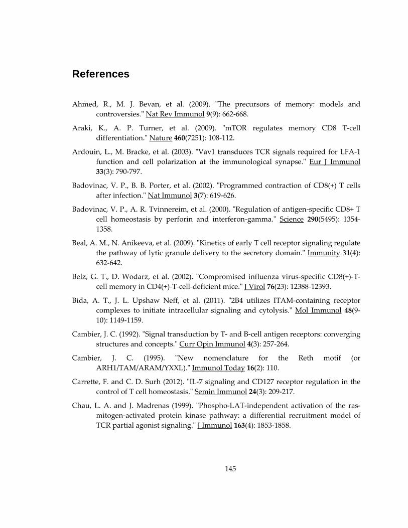
145
References
Ahmed, R., M. J. Bevan, et al. (2009). "The precursors of memory: models and
controversies." Nat Rev Immunol 9(9): 662-668.
Araki, K., A. P. Turner, et al. (2009). "mTOR regulates memory CD8 T-cell
differentiation." Nature 460(7251): 108-112.
Ardouin, L., M. Bracke, et al. (2003). "Vav1 transduces TCR signals required for LFA-1
function and cell polarization at the immunological synapse." Eur J Immunol
33(3): 790-797.
Badovinac, V. P., B. B. Porter, et al. (2002). "Programmed contraction of CD8(+) T cells
after infection." Nat Immunol 3(7): 619-626.
Badovinac, V. P., A. R. Tvinnereim, et al. (2000). "Regulation of antigen-specific CD8+ T
cell homeostasis by perforin and interferon-gamma." Science 290(5495): 1354-
1358.
Beal, A. M., N. Anikeeva, et al. (2009). "Kinetics of early T cell receptor signaling regulate
the pathway of lytic granule delivery to the secretory domain." Immunity 31(4):
632-642.
Belz, G. T., D. Wodarz, et al. (2002). "Compromised influenza virus-specific CD8(+)-T-
cell memory in CD4(+)-T-cell-deficient mice." J Virol 76(23): 12388-12393.
Bida, A. T., J. L. Upshaw Neff, et al. (2011). "2B4 utilizes ITAM-containing receptor
complexes to initiate intracellular signaling and cytolysis." Mol Immunol 48(9-
10): 1149-1159.
Cambier, J. C. (1992). "Signal transduction by T- and B-cell antigen receptors: converging
structures and concepts." Curr Opin Immunol 4(3): 257-264.
Cambier, J. C. (1995). "New nomenclature for the Reth motif (or
ARH1/TAM/ARAM/YXXL)." Immunol Today 16(2): 110.
Carrette, F. and C. D. Surh (2012). "IL-7 signaling and CD127 receptor regulation in the
control of T cell homeostasis." Semin Immunol 24(3): 209-217.
Chau, L. A. and J. Madrenas (1999). "Phospho-LAT-independent activation of the ras-
mitogen-activated protein kinase pathway: a differential recruitment model of
TCR partial agonist signaling." J Immunol 163(4): 1853-1858.
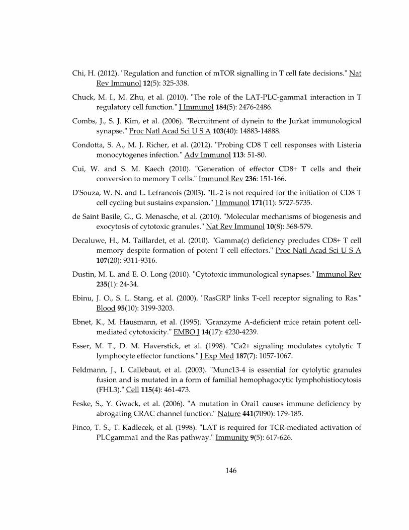
146
Chi, H. (2012). "Regulation and function of mTOR signalling in T cell fate decisions." Nat
Rev Immunol 12(5): 325-338.
Chuck, M. I., M. Zhu, et al. (2010). "The role of the LAT-PLC-gamma1 interaction in T
regulatory cell function." J Immunol 184(5): 2476-2486.
Combs, J., S. J. Kim, et al. (2006). "Recruitment of dynein to the Jurkat immunological
synapse." Proc Natl Acad Sci U S A 103(40): 14883-14888.
Condotta, S. A., M. J. Richer, et al. (2012). "Probing CD8 T cell responses with Listeria
monocytogenes infection." Adv Immunol 113: 51-80.
Cui, W. and S. M. Kaech (2010). "Generation of effector CD8+ T cells and their
conversion to memory T cells." Immunol Rev 236: 151-166.
D'Souza, W. N. and L. Lefrancois (2003). "IL-2 is not required for the initiation of CD8 T
cell cycling but sustains expansion." J Immunol 171(11): 5727-5735.
de Saint Basile, G., G. Menasche, et al. (2010). "Molecular mechanisms of biogenesis and
exocytosis of cytotoxic granules." Nat Rev Immunol 10(8): 568-579.
Decaluwe, H., M. Taillardet, et al. (2010). "Gamma(c) deficiency precludes CD8+ T cell
memory despite formation of potent T cell effectors." Proc Natl Acad Sci U S A
107(20): 9311-9316.
Dustin, M. L. and E. O. Long (2010). "Cytotoxic immunological synapses." Immunol Rev
235(1): 24-34.
Ebinu, J. O., S. L. Stang, et al. (2000). "RasGRP links T-cell receptor signaling to Ras."
Blood 95(10): 3199-3203.
Ebnet, K., M. Hausmann, et al. (1995). "Granzyme A-deficient mice retain potent cell-
mediated cytotoxicity." EMBO J 14(17): 4230-4239.
Esser, M. T., D. M. Haverstick, et al. (1998). "Ca2+ signaling modulates cytolytic T
lymphocyte effector functions." J Exp Med 187(7): 1057-1067.
Feldmann, J., I. Callebaut, et al. (2003). "Munc13-4 is essential for cytolytic granules
fusion and is mutated in a form of familial hemophagocytic lymphohistiocytosis
(FHL3)." Cell 115(4): 461-473.
Feske, S., Y. Gwack, et al. (2006). "A mutation in Orai1 causes immune deficiency by
abrogating CRAC channel function." Nature 441(7090): 179-185.
Finco, T. S., T. Kadlecek, et al. (1998). "LAT is required for TCR-mediated activation of
PLCgamma1 and the Ras pathway." Immunity 9(5): 617-626.

147
Fraser, S. A., R. Karimi, et al. (2000). "Perforin lytic activity is controlled by calreticulin." J
Immunol 164(8): 4150-4155.
Fu, G., Y. Chen, et al. (2012). "Phospholipase Cgamma2 Plays a Role in TCR Signal
Transduction and T Cell Selection." J Immunol 189(5): 2326-2332.
Fu, G., Y. Chen, et al. (2010). "Phospholipase C{gamma}1 is essential for T cell
development, activation, and tolerance." J Exp Med 207(2): 309-318.
Fuller, C. L., K. S. Ravichandran, et al. (1999). "Phosphatidylinositol 3-kinase-dependent
and -independent cytolytic effector functions." J Immunol 162(11): 6337-6340.
Gattinoni, L., E. Lugli, et al. (2011). "A human memory T cell subset with stem cell-like
properties." Nat Med 17(10): 1290-1297.
Gattinoni, L., X. S. Zhong, et al. (2009). "Wnt signaling arrests effector T cell
differentiation and generates CD8+ memory stem cells." Nat Med 15(7): 808-813.
Grakoui, A., S. K. Bromley, et al. (1999). "The immunological synapse: a molecular
machine controlling T cell activation." Science 285(5425): 221-227.
Hand, T. W. and S. M. Kaech (2009). "Intrinsic and extrinsic control of effector T cell
survival and memory T cell development." Immunol Res 45(1): 46-61.
Hand, T. W., M. Morre, et al. (2007). "Expression of IL-7 receptor alpha is necessary but
not sufficient for the formation of memory CD8 T cells during viral infection."
Proc Natl Acad Sci U S A 104(28): 11730-11735.
Haring, J. S., X. Jing, et al. (2008). "Constitutive expression of IL-7 receptor alpha does
not support increased expansion or prevent contraction of antigen-specific CD4
or CD8 T cells following Listeria monocytogenes infection." J Immunol 180(5):
2855-2862.
Harty, J. T. and V. P. Badovinac (2008). "Shaping and reshaping CD8+ T-cell memory."
Nat Rev Immunol 8(2): 107-119.
Hashimoto-Tane, A., T. Yokosuka, et al. (2011). "Dynein-driven transport of T cell
receptor microclusters regulates immune synapse formation and T cell
activation." Immunity 34(6): 919-931.
He, J. S. and H. L. Ostergaard (2007). "CTLs contain and use intracellular stores of FasL
distinct from cytolytic granules." J Immunol 179(4): 2339-2348.
Higashi, A. Y., T. Ikawa, et al. (2009). "Direct hematological toxicity and illegitimate
chromosomal recombination caused by the systemic activation of CreERT2." J
Immunol 182(9): 5633-5640.
148
Hildeman, D. A., Y. Zhu, et al. (2002). "Activated T cell death in vivo mediated by
proapoptotic bcl-2 family member bim." Immunity 16(6): 759-767.
Hogan, P. G., L. Chen, et al. (2003). "Transcriptional regulation by calcium, calcineurin,
and NFAT." Genes Dev 17(18): 2205-2232.
Huppa, J. B., M. Gleimer, et al. (2003). "Continuous T cell receptor signaling required for
synapse maintenance and full effector potential." Nat Immunol 4(8): 749-755.
Huse, M. (2012). "Microtubule-organizing center polarity and the immunological
synapse: protein kinase C and beyond." Front Immunol 3: 235.
Huse, M., E. J. Quann, et al. (2008). "Shouts, whispers and the kiss of death: directional
secretion in T cells." Nat Immunol 9(10): 1105-1111.
Ilani, T., G. Vasiliver-Shamis, et al. (2009). "T cell antigen receptor signaling and
immunological synapse stability require myosin IIA." Nat Immunol 10(5): 531-
539.
Jameson, S. C. and D. Masopust (2009). "Diversity in T cell memory: an embarrassment
of riches." Immunity 31(6): 859-871.
Jenkins, M. R., A. Tsun, et al. (2009). "The strength of T cell receptor signal controls the
polarization of cytotoxic machinery to the immunological synapse." Immunity
31(4): 621-631.
Ji, Y., Z. Pos, et al. (2011). "Repression of the DNA-binding inhibitor Id3 by Blimp-1
limits the formation of memory CD8+ T cells." Nat Immunol 12(12): 1230-1237.
Jordan, M. S., A. L. Singer, et al. (2003). "Adaptors as central mediators of signal
transduction in immune cells." Nat Immunol 4(2): 110-116.
Joshi, N. S., W. Cui, et al. (2007). "Inflammation directs memory precursor and short-
lived effector CD8(+) T cell fates via the graded expression of T-bet transcription
factor." Immunity 27(2): 281-295.
Jung, Y. W. and S. M. Kaech (2010). "Like parent, like child: inheritance of effector CD8+
T cell traits." Immunity 33(3): 296-298.
Juszczak, R. J. and J. H. Russell (1989). "Inhibition of cytotoxic T lymphocyte-mediated
lysis and cellular proliferation by isoquinoline sulfonamide protein kinase
inhibitors. Evidence for the involvement of protein kinase C in lymphocyte
function." J Biol Chem 264(2): 810-815.

149
Kaech, S. M. and R. Ahmed (2001). "Memory CD8+ T cell differentiation: initial antigen
encounter triggers a developmental program in naive cells." Nat Immunol 2(5):
415-422.
Kaech, S. M., J. T. Tan, et al. (2003). "Selective expression of the interleukin 7 receptor
identifies effector CD8 T cells that give rise to long-lived memory cells." Nat
Immunol 4(12): 1191-1198.
Kaech, S. M. and E. J. Wherry (2007). "Heterogeneity and cell-fate decisions in effector
and memory CD8+ T cell differentiation during viral infection." Immunity 27(3):
393-405.
Kagi, D., B. Ledermann, et al. (1994). "Cytotoxicity mediated by T cells and natural killer
cells is greatly impaired in perforin-deficient mice." Nature 369(6475): 31-37.
Kagi, D., B. Ledermann, et al. (1996). "Molecular mechanisms of lymphocyte-mediated
cytotoxicity and their role in immunological protection and pathogenesis in
vivo." Annu Rev Immunol 14: 207-232.
Kagi, D., F. Vignaux, et al. (1994). "Fas and perforin pathways as major mechanisms of T
cell-mediated cytotoxicity." Science 265(5171): 528-530.
Kataoka, T., K. Togashi, et al. (1997). "Inactivation and proteolytic degradation of
perforin within lytic granules upon neutralization of acidic pH." Immunology
91(3): 493-500.
Kerdiles, Y. M., D. R. Beisner, et al. (2009). "Foxo1 links homing and survival of naive T
cells by regulating L-selectin, CCR7 and interleukin 7 receptor." Nat Immunol
10(2): 176-184.
Khanolkar, A., V. P. Badovinac, et al. (2007). "CD8 T cell memory development: CD4 T
cell help is appreciated." Immunol Res 39(1-3): 94-104.
Kuhne, M. R., J. Lin, et al. (2003). "Linker for activation of T cells, zeta-associated protein-
70, and Src homology 2 domain-containing leukocyte protein-76 are required for
TCR-induced microtubule-organizing center polarization." J Immunol 171(2):
860-866.
Kuta, A. E., C. L. Bashford, et al. (1991). "Characterization of non-lytic cytolysin-
membrane intermediates." Mol Immunol 28(11): 1263-1270.
Leignadier, J., M. P. Hardy, et al. (2008). "Memory T-lymphocyte survival does not
require T-cell receptor expression." Proc Natl Acad Sci U S A 105(51): 20440-
20445.
150
Li, Q. J., J. Chau, et al. (2007). "miR-181a is an intrinsic modulator of T cell sensitivity and
selection." Cell 129(1): 147-161.
Liou, J., M. L. Kim, et al. (2005). "STIM is a Ca2+ sensor essential for Ca2+-store-
depletion-triggered Ca2+ influx." Curr Biol 15(13): 1235-1241.
Liu, S. K. and C. J. McGlade (1998). "Gads is a novel SH2 and SH3 domain-containing
adaptor protein that binds to tyrosine-phosphorylated Shc." Oncogene 17(24):
3073-3082.
Lodolce, J. P., D. L. Boone, et al. (1998). "IL-15 receptor maintains lymphoid homeostasis
by supporting lymphocyte homing and proliferation." Immunity 9(5): 669-676.
Luik, R. M., B. Wang, et al. (2008). "Oligomerization of STIM1 couples ER calcium
depletion to CRAC channel activation." Nature 454(7203): 538-542.
Luik, R. M., M. M. Wu, et al. (2006). "The elementary unit of store-operated Ca2+ entry:
local activation of CRAC channels by STIM1 at ER-plasma membrane junctions."
J Cell Biol 174(6): 815-825.
Lyubchenko, T. A., G. A. Wurth, et al. (2001). "Role of calcium influx in cytotoxic T
lymphocyte lytic granule exocytosis during target cell killing." Immunity 15(5):
847-859.
Ma, A., R. Koka, et al. (2006). "Diverse functions of IL-2, IL-15, and IL-7 in lymphoid
homeostasis." Annu Rev Immunol 24: 657-679.
Martin-Cofreces, N. B., J. Robles-Valero, et al. (2008). "MTOC translocation modulates IS
formation and controls sustained T cell signaling." J Cell Biol 182(5): 951-962.
Martin-Cofreces, N. B., D. Sancho, et al. (2006). "Role of Fyn in the rearrangement of
tubulin cytoskeleton induced through TCR." J Immunol 176(7): 4201-4207.
Martinvalet, D., D. M. Dykxhoorn, et al. (2008). "Granzyme A cleaves a mitochondrial
complex I protein to initiate caspase-independent cell death." Cell 133(4): 681-692.
Masopust, D., V. Vezys, et al. (2001). "Preferential localization of effector memory cells in
nonlymphoid tissue." Science 291(5512): 2413-2417.
Menager, M. M., G. Menasche, et al. (2007). "Secretory cytotoxic granule maturation and
exocytosis require the effector protein hMunc13-4." Nat Immunol 8(3): 257-267.
Menasche, G., S. Kliche, et al. (2007). "Regulation of T-cell antigen receptor-mediated
inside-out signaling by cytosolic adapter proteins and Rap1 effector molecules."
Immunol Rev 218: 82-91.

151
Mercado, R., S. Vijh, et al. (2000). "Early programming of T cell populations responding
to bacterial infection." J Immunol 165(12): 6833-6839.
Metkar, S. S., B. Wang, et al. (2002). "Cytotoxic cell granule-mediated apoptosis: perforin
delivers granzyme B-serglycin complexes into target cells without plasma
membrane pore formation." Immunity 16(3): 417-428.
Michalek, R. D. and J. C. Rathmell (2010). "The metabolic life and times of a T-cell."
Immunol Rev 236: 190-202.
Mingueneau, M., R. Roncagalli, et al. (2009). "Loss of the LAT adaptor converts antigen-
responsive T cells into pathogenic effectors that function independently of the T
cell receptor." Immunity 31(2): 197-208.
Monks, C. R., B. A. Freiberg, et al. (1998). "Three-dimensional segregation of
supramolecular activation clusters in T cells." Nature 395(6697): 82-86.
Murali-Krishna, K., L. L. Lau, et al. (1999). "Persistence of memory CD8 T cells in MHC
class I-deficient mice." Science 286(5443): 1377-1381.
Obar, J. J., M. J. Molloy, et al. (2010). "CD4+ T cell regulation of CD25 expression controls
development of short-lived effector CD8+ T cells in primary and secondary
responses." Proc Natl Acad Sci U S A 107(1): 193-198.
Okkenhaug, K. and B. Vanhaesebroeck (2003). "PI3K in lymphocyte development,
differentiation and activation." Nat Rev Immunol 3(4): 317-330.
Pardo, J., M. Buferne, et al. (2003). "Differential implication of protein kinase C isoforms
in cytotoxic T lymphocyte degranulation and TCR-induced Fas ligand
expression." Int Immunol 15(12): 1441-1450.
Pearce, E. L., M. C. Walsh, et al. (2009). "Enhancing CD8 T-cell memory by modulating
fatty acid metabolism." Nature 460(7251): 103-107.
Pellegrini, M., G. Belz, et al. (2003). "Shutdown of an acute T cell immune response to
viral infection is mediated by the proapoptotic Bcl-2 homology 3-only protein
Bim." Proc Natl Acad Sci U S A 100(24): 14175-14180.
Pipkin, M. E., J. A. Sacks, et al. (2010). "Interleukin-2 and inflammation induce distinct
transcriptional programs that promote the differentiation of effector cytolytic T
cells." Immunity 32(1): 79-90.
Potter, T. A., K. Grebe, et al. (2001). "Formation of supramolecular activation clusters on
fresh ex vivo CD8+ T cells after engagement of the T cell antigen receptor and
CD8 by antigen-presenting cells." Proc Natl Acad Sci U S A 98(22): 12624-12629.

152
Purbhoo, M. A., D. J. Irvine, et al. (2004). "T cell killing does not require the formation of
a stable mature immunological synapse." Nat Immunol 5(5): 524-530.
Quann, E. J., X. Liu, et al. (2011). "A cascade of protein kinase C isozymes promotes
cytoskeletal polarization in T cells." Nat Immunol 12(7): 647-654.
Quann, E. J., E. Merino, et al. (2009). "Localized diacylglycerol drives the polarization of
the microtubule-organizing center in T cells." Nat Immunol 10(6): 627-635.
Rao, R. R., Q. Li, et al. (2012). "Transcription factor Foxo1 represses T-bet-mediated
effector functions and promotes memory CD8(+) T cell differentiation."
Immunity 36(3): 374-387.
Rao, R. R., Q. Li, et al. (2010). "The mTOR kinase determines effector versus memory
CD8+ T cell fate by regulating the expression of transcription factors T-bet and
Eomesodermin." Immunity 32(1): 67-78.
Rathmell, J. C., T. Lindsten, et al. (2002). "Deficiency in Bak and Bax perturbs thymic
selection and lymphoid homeostasis." Nat Immunol 3(10): 932-939.
Reth, M. (1989). "Antigen receptor tail clue." Nature 338(6214): 383-384.
Revell, P. A., W. J. Grossman, et al. (2005). "Granzyme B and the downstream granzymes
C and/or F are important for cytotoxic lymphocyte functions." J Immunol 174(4):
2124-2131.
Rhee, S. G. (2001). "Regulation of phosphoinositide-specific phospholipase C." Annu Rev
Biochem 70: 281-312.
Richer, M. J., J. C. Nolz, et al. (2012). "Pathogen-Specific Inflammatory Milieux Tune the
Antigen Sensitivity of CD8(+) T Cells by Enhancing T Cell Receptor Signaling."
Immunity.
Robertson, L. K., L. R. Mireau, et al. (2005). "A role for phosphatidylinositol 3-kinase in
TCR-stimulated ERK activation leading to paxillin phosphorylation and CTL
degranulation." J Immunol 175(12): 8138-8145.
Roos, J., P. J. DiGregorio, et al. (2005). "STIM1, an essential and conserved component of
store-operated Ca2+ channel function." J Cell Biol 169(3): 435-445.
Russell, J. H. and T. J. Ley (2002). "Lymphocyte-mediated cytotoxicity." Annu Rev
Immunol 20: 323-370.
Rutishauser, R. L., G. A. Martins, et al. (2009). "Transcriptional repressor Blimp-1
promotes CD8(+) T cell terminal differentiation and represses the acquisition of
central memory T cell properties." Immunity 31(2): 296-308.
153
Sallusto, F., D. Lenig, et al. (1999). "Two subsets of memory T lymphocytes with distinct
homing potentials and effector functions." Nature 401(6754): 708-712.
Salojin, K. V., J. Zhang, et al. (2000). "ZAP-70 is essential for the T cell antigen receptor-
induced plasma membrane targeting of SOS and Vav in T cells." J Biol Chem
275(8): 5966-5975.
Samelson, L. E. (2002). "Signal transduction mediated by the T cell antigen receptor: the
role of adapter proteins." Annu Rev Immunol 20: 371-394.
Sarkar, S., V. Kalia, et al. (2008). "Functional and genomic profiling of effector CD8 T cell
subsets with distinct memory fates." J Exp Med 205(3): 625-640.
Sarkar, S., V. Teichgraber, et al. (2007). "Strength of stimulus and clonal competition
impact the rate of memory CD8 T cell differentiation." J Immunol 179(10): 6704-
6714.
Shan, X., R. Balakir, et al. (2001). "Zap-70-independent Ca(2+) mobilization and Erk
activation in Jurkat T cells in response to T-cell antigen receptor ligation." Mol
Cell Biol 21(21): 7137-7149.
Shen, S., M. I. Chuck, et al. (2010). "The importance of LAT in the activation,
homeostasis, and regulatory function of T cells." J Biol Chem 285(46): 35393-
35405.
Shen, S., M. Zhu, et al. (2009). "The essential role of LAT in thymocyte development
during transition from the double-positive to single-positive stage." J Immunol
182(9): 5596-5604.
Shin, H., S. D. Blackburn, et al. (2007). "Viral antigen and extensive division maintain
virus-specific CD8 T cells during chronic infection." J Exp Med 204(4): 941-949.
Shin, H. and E. J. Wherry (2007). "CD8 T cell dysfunction during chronic viral infection."
Curr Opin Immunol 19(4): 408-415.
Smith-Garvin, J. E., J. C. Burns, et al. (2010). "T-cell receptor signals direct the
composition and function of the memory CD8+ T-cell pool." Blood 116(25): 5548-
5559.
Smith-Garvin, J. E., G. A. Koretzky, et al. (2009). "T cell activation." Annu Rev Immunol
27: 591-619.
Smyth, M. J., S. E. Street, et al. (2003). "Cutting edge: granzymes A and B are not
essential for perforin-mediated tumor rejection." J Immunol 171(2): 515-518.
154
Snow, A. L., P. Pandiyan, et al. (2010). "The power and the promise of restimulation-
induced cell death in human immune diseases." Immunol Rev 236: 68-82.
Sommers, C. L., R. K. Menon, et al. (2001). "Knock-in mutation of the distal four
tyrosines of linker for activation of T cells blocks murine T cell development." J
Exp Med 194(2): 135-142.
Stemberger, C., K. M. Huster, et al. (2007). "A single naive CD8+ T cell precursor can
develop into diverse effector and memory subsets." Immunity 27(6): 985-997.
Stinchcombe, J. C. and G. M. Griffiths (2007). "Secretory mechanisms in cell-mediated
cytotoxicity." Annu Rev Cell Dev Biol 23: 495-517.
Stinchcombe, J. C., E. Majorovits, et al. (2006). "Centrosome polarization delivers
secretory granules to the immunological synapse." Nature 443(7110): 462-465.
Sun, J. C. and M. J. Bevan (2003). "Defective CD8 T cell memory following acute infection
without CD4 T cell help." Science 300(5617): 339-342.
Sun, J. C., M. A. Williams, et al. (2004). "CD4+ T cells are required for the maintenance,
not programming, of memory CD8+ T cells after acute infection." Nat Immunol
5(9): 927-933.
Suresh, M., A. Singh, et al. (2005). "Role of tumor necrosis factor receptors in regulating
CD8 T-cell responses during acute lymphocytic choriomeningitis virus infection."
J Virol 79(1): 202-213.
Surh, C. D., O. Boyman, et al. (2006). "Homeostasis of memory T cells." Immunol Rev
211: 154-163.
Surh, C. D. and J. Sprent (2008). "Homeostasis of naive and memory T cells." Immunity
29(6): 848-862.
Swain, S. L., H. Hu, et al. (1999). "Class II-independent generation of CD4 memory T
cells from effectors." Science 286(5443): 1381-1383.
Takeda, S., H. R. Rodewald, et al. (1996). "MHC class II molecules are not required for
survival of newly generated CD4+ T cells, but affect their long-term life span."
Immunity 5(3): 217-228.
Takemoto, N., A. M. Intlekofer, et al. (2006). "Cutting Edge: IL-12 inversely regulates T-
bet and eomesodermin expression during pathogen-induced CD8+ T cell
differentiation." J Immunol 177(11): 7515-7519.
Tanchot, C., F. A. Lemonnier, et al. (1997). "Differential requirements for survival and
proliferation of CD8 naive or memory T cells." Science 276(5321): 2057-2062.
155
Tewari, K., J. Walent, et al. (2006). "Differential requirement for Lck during primary and
memory CD8+ T cell responses." Proc Natl Acad Sci U S A 103(44): 16388-16393.
Tolmachova, T., R. Anders, et al. (2004). "A general role for Rab27a in secretory cells."
Mol Biol Cell 15(1): 332-344.
Toribio, M. L., J. C. Gutierrez-Ramos, et al. (1989). "Interleukin-2-dependent autocrine
proliferation in T-cell development." Nature 342(6245): 82-85.
Uellner, R., M. J. Zvelebil, et al. (1997). "Perforin is activated by a proteolytic cleavage
during biosynthesis which reveals a phospholipid-binding C2 domain." EMBO J
16(24): 7287-7296.
van den Broek, M. E., D. Kagi, et al. (1996). "Decreased tumor surveillance in perforin-
deficient mice." J Exp Med 184(5): 1781-1790.
van Faassen, H., M. Saldanha, et al. (2005). "Reducing the stimulation of CD8+ T cells
during infection with intracellular bacteria promotes differentiation primarily
into a central (CD62LhighCD44high) subset." J Immunol 174(9): 5341-5350.
van Stipdonk, M. J., E. E. Lemmens, et al. (2001). "Naive CTLs require a single brief
period of antigenic stimulation for clonal expansion and differentiation." Nat
Immunol 2(5): 423-429.
Varma, R., G. Campi, et al. (2006). "T cell receptor-proximal signals are sustained in
peripheral microclusters and terminated in the central supramolecular activation
cluster." Immunity 25(1): 117-127.
Wang, H., T. A. Kadlecek, et al. (2010). "ZAP-70: an essential kinase in T-cell signaling."
Cold Spring Harb Perspect Biol 2(5): a002279.
Weant, A. E., R. D. Michalek, et al. (2008). "Apoptosis regulators Bim and Fas function
concurrently to control autoimmunity and CD8+ T cell contraction." Immunity
28(2): 218-230.
Wherry, E. J., K. A. Puorro, et al. (1999). "The induction of virus-specific CTL as a
function of increasing epitope expression: responses rise steadily until
excessively high levels of epitope are attained." J Immunol 163(7): 3735-3745.
Wiehagen, K. R., E. Corbo, et al. (2010). "Loss of tonic T-cell receptor signals alters the
generation but not the persistence of CD8+ memory T cells." Blood 116(25): 5560-
5570.
Williams, M. A. and M. J. Bevan (2007). "Effector and memory CTL differentiation."
Annu Rev Immunol 25: 171-192.
156
Williams, M. A., A. J. Tyznik, et al. (2006). "Interleukin-2 signals during priming are
required for secondary expansion of CD8+ memory T cells." Nature 441(7095):
890-893.
Witherden, D., N. van Oers, et al. (2000). "Tetracycline-controllable selection of CD4(+) T
cells: half-life and survival signals in the absence of major histocompatibility
complex class II molecules." J Exp Med 191(2): 355-364.
Wojciechowski, S., P. Tripathi, et al. (2007). "Bim/Bcl-2 balance is critical for maintaining
naive and memory T cell homeostasis." J Exp Med 204(7): 1665-1675.
Wong, P. and E. G. Pamer (2001). "Cutting edge: antigen-independent CD8 T cell
proliferation." J Immunol 166(10): 5864-5868.
Yajima, T., H. Nishimura, et al. (2002). "Overexpression of IL-15 in vivo increases
antigen-driven memory CD8+ T cells following a microbe exposure." J Immunol
168(3): 1198-1203.
Yang, C. Y., J. A. Best, et al. (2011). "The transcriptional regulators Id2 and Id3 control
the formation of distinct memory CD8+ T cell subsets." Nat Immunol 12(12):
1221-1229.
Yokosuka, T., K. Sakata-Sogawa, et al. (2005). "Newly generated T cell receptor
microclusters initiate and sustain T cell activation by recruitment of Zap70 and
SLP-76." Nat Immunol 6(12): 1253-1262.
Zehn, D., S. Y. Lee, et al. (2009). "Complete but curtailed T-cell response to very low-
affinity antigen." Nature 458(7235): 211-214.
Zhang, W., B. J. Irvin, et al. (1999). "Functional analysis of LAT in TCR-mediated
signaling pathways using a LAT-deficient Jurkat cell line." Int Immunol 11(6):
943-950.
Zhang, W., J. Sloan-Lancaster, et al. (1998). "LAT: the ZAP-70 tyrosine kinase substrate
that links T cell receptor to cellular activation." Cell 92(1): 83-92.
Zhang, W., C. L. Sommers, et al. (1999). "Essential role of LAT in T cell development."
Immunity 10(3): 323-332.
Zhang, W., R. P. Trible, et al. (1998). "LAT palmitoylation: its essential role in membrane
microdomain targeting and tyrosine phosphorylation during T cell activation."
Immunity 9(2): 239-246.
Zhu, M., O. Granillo, et al. (2005). "Negative regulation of lymphocyte activation by the
adaptor protein LAX." J Immunol 174(9): 5612-5619.
157
Zhu, M., S. Koonpaew, et al. (2006). "Negative regulation of T cell activation and
autoimmunity by the transmembrane adaptor protein LAB." Immunity 25(5):
757-768.
Zhu, M., S. Shen, et al. (2005). "Cutting Edge: Localization of linker for activation of T
cells to lipid rafts is not essential in T cell activation and development." J
Immunol 174(1): 31-35.

158
Biography
I was born on April 21, 1981 in Tainan, Taiwan. I grew up in Tainan and
attended National Tsing Hua University where I earned a Bachelor of Science in May
2003. Following my love of science, I pursued my Master of Science in Microbiology in
National Taiwan University in 2005. During my training at that time, I worked on the
genetic factors and molecular mechanisms of Carbamazepine-induced Stevens Johnson
Syndrome under the supervision of Dr. Yuan-Tsong Chen. In August 2007, I joined the
Ph.D. program in Department of Immunology at Duke University. For the past 5 years, I
have been fortunate to study the role of T cell signaling in the function of cytotoxic T
cells under the guidance of Dr. Weiguo Zhang.

159
Publications:
� Ou-Yang CW, Zhu M, Sullivan SA, Fuller DM, Zhang W. The requirement of LAT in
the primary and memory responses of CD8 T cells. J Immunol 2013.
� Dolled-Filhart MP, Lee M, Ou-Yang CW, Haraksingh RR, Jimmy CH Lin.
Computational and Bioinformatics Frameworks for Next-Generation Whole Exome
and Genome Sequencing. The Scientific World Journal 2013: 730210, 2013
� Ou-Yang CW, Zhu M, Fuller DM, Chuck MI, Li Qj, Zhang W. Role of LAT in the
granule-mediated cytotoxicity of CD8 T cells. Mol Cell Biol 32(14):2674-84, 2012.
� Dolled-Filhart MP, Lordemann A, Dahl W, Haraksingh RR, Ou-Yang CW, Jimmy
CH Lin. Personalizing Rare Disease Research: how genomics is revolutionizing the
diagnosis and treatment of rare disease. Personalized Medicine 9 (8): 805-819, 2012.
� Fuller DM, Zhu M, Song X, Ou-Yang CW, Sullivan SA, Stone JC, Zhang W.
Regulation of RasGRP1 Function in T Cell Development and Activation by Its
Unique Tail Domain. PLoS One 7(6):e38796, 2012.
� Zhu M, Fuller DM, Ou-Yang CW, Sullivan SA, Zhang W. Tyrosine Phosphorylation-
Independent Regulation of Lipopolysaccharide-Mediated Response by the
Transmembrane Adaptor Protein LAB. J Immunol 188(6):2733-41, 2012.
� Markegard E, Trager E, Ou-Yang CW, Zhang W, Weiss A, Roose JP. Basal LAT-
diacylglycerol-RasGRP1 signals in T cells maintain TCRα gene expression. PLoS
One 6(9):e25540, 2011.
� Fuller DM, Zhu M, Ou-Yang CW, Sullivan SA, Zhang W. A tale of two TRAPs: LAT
and LAB in the regulation of lymphocyte development, activation, and
autoimmunity. Immunol Res 49(1-3):97-108, 2011.
� Shen S, Chuck MI, Zhu M, Fuller DM, Ou-Yang CW, Zhang W. The importance of
LAT in the activation, homeostasis, and regulatory function of T cells. J Biol Chem
285(46):35393-405, 2010. Epub Sep 13, 2010.
� Ou-Yang CW, Hung SI, Juo CG, Lin YP, Fang WH, Lu IH, Chen ST, Chen YT. HLA-
B*1502-bound peptides: implications for the pathogenesis of carbamazepine-induced
Stevens-Johnson syndrome. J Allergy Clin Immunol 120:870-877, 2007.
Copyright © 2022 FDOKUMEN