Are spontaneous otoacoustic emissions generated by self-sustained cochlear oscillators
CD86 Has Sustained Costimulatory Effects on CD8 T Cells1
Ian J. Thomas,* Liliana G. Petrich de Marquesini,* Rommel Ravanan,† Richard M. Smith,‡
Sylvie Guerder,§ Richard A. Flavell,¶ David C. Wraith,* Li Wen,� and F. Susan Wong2*
CD80 and CD86 both costimulate T cell activation. Their individual effects in vivo are difficult to study as they are coordinatelyup-regulated on APCs. We have studied mice expressing rat insulin promoter (RIP)-CD80 and RIP-CD86 on the NOD andNOD.scid genetic background to generate in vivo models, using diabetes as a readout for cytotoxic T cell activation. Acceleratedspontaneous diabetes onset was observed in NOD-RIP-CD80 mice and the transfer of diabetes from 6-wk-old NOD mice toNOD.scid-RIP-CD80 mice was greater compared with NOD-RIP-CD86 and NOD.scid-RIP-CD86 mice, respectively. However, thesecondary in vivo response was maintained if T cells were activated through CD86 costimulation compared with CD80. This wasdemonstrated by greater ability to cause recurrent diabetes in NOD-RIP-CD86 diabetic mice transplanted with 6-wk-old NODislets and adoptively transferred diabetes from diabetic NOD-RIP-CD86 mice to NOD.scid mice. In vitro, CD80 costimulationenhanced cytotoxicity, proliferation, and cytokine secretion in activated CD8 T cells compared with CD86 costimulation. Wedemonstrated increased CTLA-4 and programmed death-1 inhibitory molecule expression following costimulation by both CD80and CD86 (CD80 > CD86). Furthermore, T cells stimulated by CD80 were more susceptible to inhibition by CD4�CD25� T cells.Overall, while CD86 does not stimulate an initial response as strongly as CD80, there is greater sustained activity that is seen evenin the absence of continued costimulation. These functions have implications for the engineered use of costimulatory molecules inaltering immune responses in a therapeutic setting. The Journal of Immunology, 2007, 179: 5936–5946.
T cell activation is a multistep process that involves spe-cific recognition of the MHC-peptide complex throughthe TCR (signal 1) as well as costimulatory influences
through a variety of molecules (signal 2) (1). It is now clear thatthe group of molecules which include the B7 as well as the TNFRfamilies are not only responsible for costimulation, but also have crit-ical inhibitory and regulatory activity (2).
Costimulatory interactions are essential for T cells to undergocomplete activation and occur when the constitutively expressed Tcell costimulatory ligand CD28 binds either of the costimulatorymolecules on APCs, CD80 (B7.1), or CD86 (B7.2). In the absenceof signal 2 (and weak signal 3), T cells enter a state of anergy orapoptosis and hence CD28 is essential for T cell activation. How-ever, in addition to the interaction of CD80 and CD86 moleculeswith CD28, CTLA-4, which is up-regulated after T cell activation(3) and reaches maximal cell surface expression 2–3 days afterstimulation, acts as an inhibitory ligand (4). CTLA-4 may functioneither by direct competition with CD28 for CD80/CD86 binding,because CTLA-4 has a stronger affinity for both CD80 and CD86
compared with CD28 (5, 6), or through intracellular signaling.CTLA-4 becomes stabilized at the cell surface through phosphor-ylation and plays a role in initiating dephosphorylation events inthe T cell synapse to down-regulate TCR signaling (reviewed inRef. 7).
In recent years, other costimulatory molecules have beenidentified. ICOS and 4-1BB are expressed after initial T cellactivation (8 –11). ICOS is thought to be involved in effectorphases of T cell function (12), while 4-1BB has been associatedwith proliferation and T cell survival (13). Programmed death-1(PD-1)3 has been identified as another inhibitory costimulator,such that PD-1-deficient animals develop autoimmune disor-ders, such as lupus-like glomerulonephritis (14) and acceleratedautoimmune diabetes (15).
CD80 and CD86 are primarily expressed on APCs with dif-ferent kinetics, although there is expression on activated T cellsas well (16, 17). CD86 is constitutively expressed on restingAPCs, such as DCs and B cells, albeit at low levels, while CD80is not detectable (18, 19). However, following activation, CD86is up-regulated 6-h poststimulation, peaking between 18 and24 h (17, 19). In contrast, CD80 expression on stimulated APCsis not evident for 24 h and demonstrates peak expression be-tween 48 and 72 h (17, 19).
When studied individually, rather than on the same APCs, CD80is a more potent costimulator of T cell activation compared withCD86. Tumors transfected with CD80 stimulated greater CTL ac-tivation compared with CD86 transfectants (20), while tumor-spe-cific CTLs were only generated in mice immunized with irradiatedCD80-transfected tumors and not CD86-transfected tumors (21). Agreater number of CD4 T cells enter cell cycle with quicker ki-netics when stimulated with CD80, compared with CD86 (22).These actions of CD80 have suggested that this molecule may be
*Department of Cellular and Molecular Medicine, University of Bristol, Bristol,United Kingdom; †Nephrology and Transplant Directorate, University Hospital OfWales, Cardiff, United Kingdom; ‡Academic Renal Unit, Southmead Hospital, Bris-tol, United Kingdom; §Centre de physiopathologie de Toulouse Purpan, Institut Na-tional de la Sante et de la Recherche Medicale, Unite 563/Universite Paul Savatier,Centre Hospitalier de l’Universite Purpan, Toulouse, France; ¶Section of Immuno-biology, Howard Hughes Medical Institute, Yale University School of Medicine, NewHaven, CT 06520; and �Section of Endocrinology, Yale University School of Med-icine, New Haven, CT 06520
Received for publication December 12, 2006. Accepted for publication August20, 2007.
The costs of publication of this article were defrayed in part by the payment of pagecharges. This article must therefore be hereby marked advertisement in accordancewith 18 U.S.C. Section 1734 solely to indicate this fact.1 I.J.T. was supported by a Wellcome Trust Prize Studentship, R.A.F. is an investi-gator of the Howard Hughes Medical Institute, and F.S.W. was a Wellcome TrustSenior Fellow in Clinical Science.2 Address correspondence and reprint requests to Dr. F. Susan Wong, Department ofCellular and Molecular Medicine, University of Bristol, Bristol BS8 1TD, U.K.E-mail address: [email protected]
3 Abbreviations used in this paper: PD-1, programmed death-1; RIP, rat insulinpromoter.
Copyright © 2007 by The American Association of Immunologists, Inc. 0022-1767/07/$2.00
The Journal of Immunology
www.jimmunol.org

of use when expressed ectopically in tumors for enhancing im-mune responses. Much less is known of CD86 in this context.
The rat insulin promoter (RIP) has been used to direct expres-sion of CD80 or CD86 costimulatory molecules to insulin-produc-ing islet � cells, which do not normally express any costimulatorymolecules. In conjunction with endogenous MHC class I mole-cules, islet � cells are effectively transformed into APCs for CD8T cells. Model systems using RIP-CD80 and RIP-CD86 have beendeveloped to test whether tissue expression of costimulators onislet � cells would break tolerance and cause diabetes. The exper-iments showed that, by itself, T cell activation with CD80 costimu-lation on a B6 or bm1 genetic background is not sufficient to causedisease (23–25). However, the presence of other factors, such asthe coexpression of a viral protein or MHC class II, IE, couldstimulate diabetes (26–28). Coexpression of cytokines TNF-� orIL-2 also precipitated diabetes (23, 28). Furthermore, the expres-sion of CD80 on the NOD genetic background caused accelerateddiabetes, as early as 3 wk of age (25, 29, 30). The effect of theMHC in this acceleration is particularly important as CD80 ex-pression on the nonobese-resistant and congenic B6g7 mice alsofacilitates spontaneous diabetes in these diabetes-resistant strains(31). CD86 has been less studied in these models. When CD86 wasexpressed on the pancreatic islet � cells on the B6 genetic back-ground, T cell tolerance to � cells was maintained and the mice didnot develop diabetes (32). However, there was peri-islet infiltrationwith IL-4-producing cells in older mice. The effect of CD86 ex-pression on the NOD genetic background was not previouslyreported.
In this study, we directly compared the effects of CD80 andCD86 costimulation on CD8 T cell activation using diabetes onsetas an in vivo read out. We report that CD86 activates CD8 T cellswith less potency during primary activation in vivo compared withCD80. In vitro studies, using CD80- or CD86-expressing islets asAPCs, demonstrated less enhanced proliferation and cytotoxicityof CD86-activated CD8 T cells compared with CD80. However,CD80 costimulation also leads to greater increases of the inhibi-tory molecules CTLA-4 and PD-1 which could down-regulateCD8 T cell function. Furthermore, CD8 T cells activated by CD80are more susceptible to inhibition by CD4�CD25� T cells. Thesecells require continued costimulatory activity for maintenance ofactivation, whereas CD86 costimulation leads to lower but moresustained activity of CD8 T cells.
Materials and MethodsMice
Two founder lines of C57BL/6-RIP-CD86 mice, lines 12 and 46, whichexpressed human CD86 on pancreatic � cells at medium and high levels,respectively (32), were backcrossed to NOD/caj mice (provided by the lateC. Janeway, Yale School of Medicine, New Haven, CT) for at least 14generations and were designated lines 12B and 46K. NOD-RIP-CD80 mice(29) were generated by backcrossing C57BL/6-RIP-CD80 mice to NOD/caj mice for at least three generations. NOD.scid-RIP-CD80 and NOD.scid-RIP-CD86 mice were generated by breeding NOD-RIP-CD80 orNOD-RIP-CD86 mice with NOD.scid mice. Transgene-positive F1 micewere backcrossed to NOD.scid mice and transgene-positive scid homozy-gous F2 mice were generated. These NOD.scid-RIP-CD80 mice were back-crossed nine generations to NOD.scid mice. NOD.scid-RIP-CD86 werederived from RIP-CD86 mice previously backcrossed 14 generations toNOD mice and therefore no further backcross was required.
Insulin-specific TCR-transgenic mice were generated by isolating TCRgenomic DNA from G9C8 cloned T cells (33), which have previously beenshown to have specific reactivity to amino acids 15–23 of the insulin Bchain (34). TCR � (V�18S1 J�18 C�) and � (V�6S1 D�1.1 J�2.3 C�1)chain DNA was purified and cloned into pT�cass and pT�cass constructs,respectively, provided by D. Mathis (Joslin Diabetes Center, Harvard Med-ical School, Boston, MA) (35). The cloned constructs were injected di-rectly into NOD OVA to generate independent TCR � and � founder lines,
which were intercrossed to produce �� TCR-transgenic mice (G9.NOD).G9C��/�.NOD mice were generated by crossing the �� TCR-transgenicmice to NOD.C��/� mice (�20 generations backcross to NOD mice). Themice were specific pathogen free and housed in barrier rooms. All procedureswere performed in accordance with U.K. Home Office-approved protocols.
Genotyping of mice for expression of CD80 or CD86 using PCR
Tail-tip DNA from NOD-RIP-CD80 and NOD-RIP-CD86 DNA was ana-lyzed in a standard PCR. The samples were amplified for 35 cycles of 30 sdenaturing at 95°C, 1 min annealing at 55°C, 1 min extension at 72°C anda final extension of 10 min. Primers B7-311 (TGA AGC CAT GGG CCACAC) and B7-1192 (GAC ACT GTT ATA CAG GG) produced an 880-bpband for CD80 and primers B70–480 (CCC ACA GGA ATG ATT CGCATC) and B70–1009 (TCA CTC TCT TCC CTC TCC ATT GTG) pro-duced a 530-bp band for CD86 in transgene-positive mice. Primers weresynthesized by Invitrogen Life Technologies.
Detection of CD80 and CD86 from islet cDNA
Pancreata from eight NOD-RIP-CD80 and eight NOD-RIP-CD86 mice at6 wk of age were harvested and islets were isolated as described. RNA wasextracted from the islet samples using TRIzol (Invitrogen Life Technolo-gies). cDNA was generated from the RNA samples according to the man-ufacturer’s instructions. Concentrations of cDNA were measured using aspectrophotometer and 1/2 dilutions of cDNA (starting at 2 �g) were am-plified using primers for CD80 and CD86 as described above. GAPDH wasused as a control (GAPDH forward: GGT CAT CAT CTC CGC CCC TTCTGC, GAPDH reverse: GAG TGG GAG TTG CTG TTG AAG TCG;Invitrogen Life Technologies).
Histology
Pancreata, spleen, or kidney was harvested, fixed, and immunohistochem-istry was performed as previously described (29, 30). Sections were incu-bated with specific biotinylated Abs (rat anti-murine CD4, CD8, B220(Caltag Laboratories)), rat anti-human CD80 or CD86 (BD Pharmingen))and detected with streptavidin-alkaline phosphatase (Vector Laboratories)and the AP substrate kit, VectorRed (Vector Laboratories). VectorRedworking solution, made up in 100 mM Tris-HCl (pH 8.4) with the additionof levamisole (Vector Laboratories), was used in accordance with the man-ufacturer’s instructions. Sections were incubated in the dark for a preop-timized time and counterstained with hematoxylin (Vector Laboratories)before mounting under coverslips with Hydromount (National Diagnos-tics). Insulitis was measured in diabetic, 6- and 12-wk-old NOD, NOD-RIP-CD80, and NOD-RIP-CD86 mice. Sections were stained with H&E(Vector Laboratories) as described previously (29, 30) and 30–200 isletsfrom four mice per group were scored for insulitis. Score 0, no infiltration;1, peri-insulitis; 2, �50% infiltration; 3, 50–90% infiltration; and 4, �90%infiltration.
Diagnosis of diabetes
Groups of NOD-RIP-CD86 lines, NOD-RIP-CD80, and female NOD-RIP-CD86 transgene-negative mice were monitored weekly for spontaneousdevelopment of diabetes over a period of 25 wk. Diabetes was detected byinitially testing for glycosuria (Bayer Diastix). Diagnosis was confirmed byblood glucose measurements �14 mM/L. Mice from adoptive transfer andtransplant experiments were tested two to three times weekly for 112 daysor until diabetic.
Adoptive transfer of diabetes
A total of 20 � 106 splenocytes at 108 cells/ml, isolated from diabeticNOD, NOD-RIP-CD80, and NOD-RIP-CD86 mice or 6-wk-old NODmice or 107 purified CD8 T cells from G9C��/�.NOD mice were i.v.injected into 6- to 8-wk-old NOD.scid, NOD.scid-RIP-CD80, orNOD.scid-RIP-CD86-transgenic mice.
Murine islet isolation and transplantation
Perfusion of murine pancreas followed an adapted version based on a mod-ification of the method established by Gotoh et al. (36). In brief, pancreatafrom NOD.scid, NOD.scid-RIP-CD80, NOD.scid-RIP-CD86, and diabetictransgenic mice were perfused with 2–3 ml of collagenase P (1 mg/ml;Boehringer Mannheim). Bile ducts were clamped off at the duodenal in-sertion to allow cannulation and specific perfusion to the pancreas. Per-fused pancreata were digested for a batch-specific optimized time and col-lagenase activity was stopped with ice-cold HBSS, 10 mM HEPES, 5%FCS (Invitrogen Life Technologies). Digested pancreata were passedthrough a �100-�m strainer and centrifuged through layers of 25, 23, 20,
5937The Journal of Immunology
and 11% Ficoll in HBSS-10 mM HEPES. Islets were collected at the in-terface between layers and for in vitro assays were handpicked under adissecting microscope into RPMI complete medium (RPMI 1640–5%FCS, 50 �M 2-ME, 2 mM L-glutamine, 100 U/ml penicillin, 100 �g/mlstreptomycin, 0.25 �g/ml amphotericin; Invitrogen Life Technologies).When used for culture, the islets were irradiated with 4200 rad. For ex-traction of islet-infiltrating cells, the islets were trypsinized and filteredthrough a 70-�m cell strainer before flow cytometric analysis.
Flow cytometry
Single-cell suspensions of 0.1–1 � 106 cells were stained for various cellsurface markers (rat anti-murine CD8-allophycocyanin; CD25, CD62L,CD95L, 4-1BB, ICOS, and PD-1-PE; CD107a and CD95-FITC). FcRswere blocked with anti-CD16 (2.4G2, provided by the late C. A. Janeway)and cells were stained for surface markers and costimulatory molecules.Intracellular staining was performed for CTLA-4 after cell surface stainingfollowing BD Pharmingen’s protocol. Intracellular staining for FoxP3 wasperformed using a FoxP3 staining kit (eBioscience/Insight Technology).The samples were acquired by flow cytometry using a FACSCalibur, re-sults were analyzed using FlowJo software. All Abs had been previouslytitrated to determine optimal concentration and all were purchased fromBD Pharmingen, except anti-murine CD8-allophycocyanin (CaltagLaboratories).
Analysis of cytokine secretion in T cell cultures
Supernatants from cell stimulation assays were harvested and analyzed forthe presence of secreted cytokines using a multiplex fluorescent bead im-munoassay from Bender Medsystems (Caltag Laboratories) (mouse Th1/Th2 cytokines: IFN-�, IL-1�, IL-2, IL-4, IL-5, IL-6, IL-10, IL-17, TNF-�,and GM-CSF). Measurements were performed as described in the manu-facturer’s instructions. Data were analyzed using BMS FlowCytomix Soft-ware, which was based in WinMDI and Microsoft Excel.
Proliferation, cytokine secretion, and cytotoxicity assays
Insulin-reactive CD8 T cells (3 � 105) from 5- to 8-wk-old G9C��/�.NODmice were cocultured with 25 irradiated islets from NOD.scid, NOD.scid-RIP-CD80, or NOD.scid-RIP-CD86 mice in RPMI complete medium. CD8T cells were purified from G9C��/�.NOD splenocytes using positive se-lection beads (Miltenyi Biotec) with �95% purities. Islets and CD8 T cellsalone were used as controls. After 2 days of incubation, supernatants weretaken for cytokine secretion analysis and cells were either pulsed with[3H]thymidine for 14 h to test proliferation or were used in cytotoxicityassays. Stimulated CD8 T cells were harvested and further cocultured with104 51Cr-sodium chromate (Amersham) labeled P815 cells together withinsulin peptide (B15–23) at an E:T ratio of 10:1 for 16 h. Specific lysis wascalculated as ((cytotoxic release � min)/(max � min)) � 100%, where theminimal release (min) corresponds to the spontaneous lysis, and the max-imal lysis corresponds to lysis induced by addition of hydrochloric acid(max).
CD4�CD25� T cell depletion
CD25� T cells were depleted using the CD25 depletion kit (Miltenyi Bio-tec) according to the manufacturer’s instructions. The depletion removed
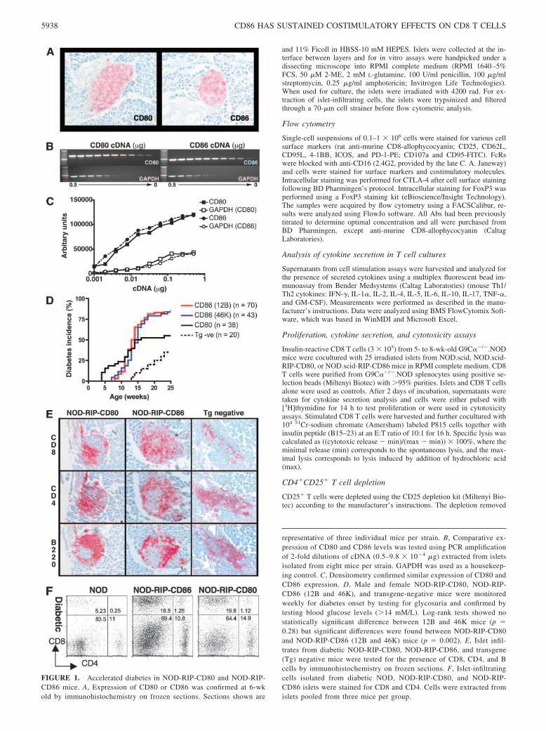
FIGURE 1. Accelerated diabetes in NOD-RIP-CD80 and NOD-RIP-CD86 mice. A, Expression of CD80 or CD86 was confirmed at 6-wkold by immunohistochemistry on frozen sections. Sections shown are
representative of three individual mice per strain. B, Comparative ex-pression of CD80 and CD86 levels was tested using PCR amplificationof 2-fold dilutions of cDNA (0.5–9.8 � 10�4 �g) extracted from isletsisolated from eight mice per strain. GAPDH was used as a housekeep-ing control. C, Densitometry confirmed similar expression of CD80 andCD86 expression. D, Male and female NOD-RIP-CD80, NOD-RIP-CD86 (12B and 46K), and transgene-negative mice were monitoredweekly for diabetes onset by testing for glycosuria and confirmed bytesting blood glucose levels (�14 mM/L). Log-rank tests showed nostatistically significant difference between 12B and 46K mice (p �0.28) but significant differences were found between NOD-RIP-CD80and NOD-RIP-CD86 (12B and 46K) mice (p � 0.002). E, Islet infil-trates from diabetic NOD-RIP-CD80, NOD-RIP-CD86, and transgene(Tg) negative mice were tested for the presence of CD8, CD4, and Bcells by immunohistochemistry on frozen sections. F, Islet-infiltratingcells isolated from diabetic NOD, NOD-RIP-CD80, and NOD-RIP-CD86 islets were stained for CD8 and CD4. Cells were extracted fromislets pooled from three mice per group.
5938 CD86 HAS SUSTAINED COSTIMULATORY EFFECTS ON CD8 T CELLS
all the CD4�CD25high cells and �5% of the low-staining CD4�CD25�
cells remained after the procedure.
Statistical analysis
Statistical analysis was performed as stated using Graphpad Prism soft-ware. Analyses of Kaplan-Meier survival curves used a log-rank test sim-ilar to a Mantel-Haenszel test. Fisher’s exact tests were performed forinsulitis comparisons. For both tests, Bonferroni corrections were made formultiple comparisons.
ResultsTransgene expression is similar in NOD-RIP-CD86 andNOD-RIP-CD80 mice
A comparative study of spontaneous diabetes incidence was per-formed using NOD-RIP-CD80 and two founder lines of NOD-RIP-CD86-transgenic mice. Ab staining of pancreatic sections forthe presence of transgenes was specific in the appropriate mice anddemonstrated similar levels of staining in both NOD-RIP-CD80and NOD-RIP-CD86 mice (Fig. 1A). A 2-fold limiting dilutionPCR of NOD-RIP-CD80 and NOD-RIP-CD86 cDNA demon-strated comparatively similar levels of expression for the 880- and530-bp products, respectively, related to the control products of thehousekeeping gene GAPDH (Fig. 1, B and C).
NOD-RIP-CD86 mice develop accelerated diabetes but lessrapidly than NOD-RIP-CD80 mice
A previous report demonstrated no difference in the onset of au-toimmune diabetes in two founder lines of NOD-RIP-CD80-trans-genic mice with differing levels of CD80 expression or a differencebetween genders (29). Similar to NOD-RIP-CD80 mice, no genderdifference in the onset of diabetes was observed between the me-dium (12B) and high (46K) expressing founder lines of NOD-RIP-CD86-transgenic mice (data not shown). Combined male and fe-male diabetes incidence curves of NOD-RIP-CD86 12B and46K-transgenic mice showed comparable results with an onset ofdiabetes from 7 to 8 wk of age, 50% diabetes incidence between 13and 14 wk and �80% diabetes incidence by 20 wk of age. Therewas no statistically significant difference in the diabetes incidencebetween the two lines of NOD-RIP-CD86 (Fig. 1D). In contrast,NOD-RIP-CD80 mice developed diabetes from an earlier age of 4wk, 50% were diabetic by 13 wk and at 20 wk of age diabetesincidence was 58% (Fig. 1D), which was similar to previouslypublished work (29, 30). In addition, at the point of diabetes onsetin NOD-RIP-CD86-transgenic mice, 15% of NOD-RIP-CD80-transgenic mice had become diabetic (Fig. 1D). Log-rank testscomparing NOD-RIP-CD80 and combined 46K and 12B NOD-RIP-CD86 incidence curves showed that diabetes incidence was
significantly different ( p � 0.002). Both NOD-RIP-CD80 andNOD-RIP-CD86 mice had accelerated onset of diabetes comparedwith 13-wk-old transgene-negative mice.
Expression of RIP-CD80 and RIP-CD86 and acceleration ofautoimmune diabetes was associated with greater levels of isletinfiltration (Table I) and an increased ratio of CD8 T cellswithin the islets (Fig. 1, E and F). At 6 wk of age, islet infil-tration was most pronounced in NOD-RIP-CD80 mice, where�50% of islets had high levels of insulitis (scores 2– 4), com-pared with 10% in NOD-RIP-CD86 mice and only low levels(scores 0 –1) of infiltration were observed in transgene-negativemice (Table I). By 12 wk of age, the degree of insulitis inNOD-RIP-CD80 and NOD-RIP-CD86 islets was comparablebut more insulitis was observed compared with transgene-neg-ative mice. Islet infiltration was similar in all strains after dia-betes development (Table I). Islet infiltrates in both diabeticNOD-RIP-CD80 and NOD-RIP-CD86 mice comprised a highproportion of CD8 T cells compared with transgene-negativemice, while the proportion of CD4 T cells and B cells wascomparable (Fig. 1E). This was confirmed by flow cytometryshowing that isolated islet infiltrates from diabetic mice dem-onstrated a 3-fold increase in the proportion of CD8 T cellsfrom NOD-RIP-CD80 and NOD-RIP-CD86 mice comparedwith transgene-negative mice (Fig. 1F).
Recurrent diabetes occurs at a faster rate in diabeticNOD-RIP-CD86 mice compared with diabetic NOD-RIP-CD80mice transplanted with healthy NOD islets
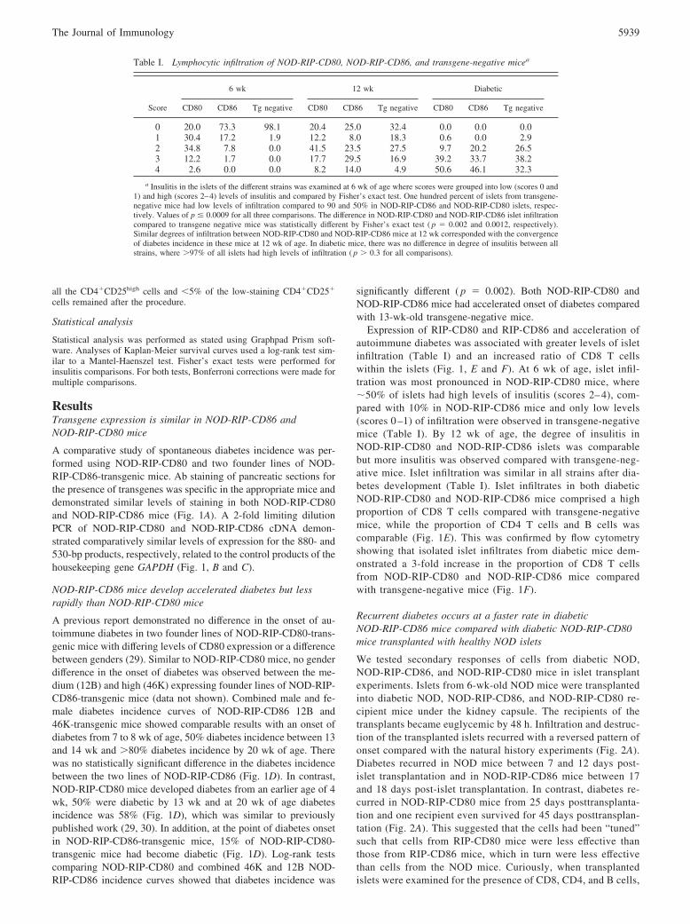
We tested secondary responses of cells from diabetic NOD,NOD-RIP-CD86, and NOD-RIP-CD80 mice in islet transplantexperiments. Islets from 6-wk-old NOD mice were transplantedinto diabetic NOD, NOD-RIP-CD86, and NOD-RIP-CD80 re-cipient mice under the kidney capsule. The recipients of thetransplants became euglycemic by 48 h. Infiltration and destruc-tion of the transplanted islets recurred with a reversed pattern ofonset compared with the natural history experiments (Fig. 2A).Diabetes recurred in NOD mice between 7 and 12 days post-islet transplantation and in NOD-RIP-CD86 mice between 17and 18 days post-islet transplantation. In contrast, diabetes re-curred in NOD-RIP-CD80 mice from 25 days posttransplanta-tion and one recipient even survived for 45 days posttransplan-tation (Fig. 2A). This suggested that the cells had been “tuned”such that cells from RIP-CD80 mice were less effective thanthose from RIP-CD86 mice, which in turn were less effectivethan cells from the NOD mice. Curiously, when transplantedislets were examined for the presence of CD8, CD4, and B cells,
Table I. Lymphocytic infiltration of NOD-RIP-CD80, NOD-RIP-CD86, and transgene-negative micea
Score
6 wk 12 wk Diabetic
CD80 CD86 Tg negative CD80 CD86 Tg negative CD80 CD86 Tg negative
0 20.0 73.3 98.1 20.4 25.0 32.4 0.0 0.0 0.01 30.4 17.2 1.9 12.2 8.0 18.3 0.6 0.0 2.92 34.8 7.8 0.0 41.5 23.5 27.5 9.7 20.2 26.53 12.2 1.7 0.0 17.7 29.5 16.9 39.2 33.7 38.24 2.6 0.0 0.0 8.2 14.0 4.9 50.6 46.1 32.3
a Insulitis in the islets of the different strains was examined at 6 wk of age where scores were grouped into low (scores 0 and1) and high (scores 2–4) levels of insulitis and compared by Fisher’s exact test. One hundred percent of islets from transgene-negative mice had low levels of infiltration compared to 90 and 50% in NOD-RIP-CD86 and NOD-RIP-CD80 islets, respec-tively. Values of p � 0.0009 for all three comparisons. The difference in NOD-RIP-CD80 and NOD-RIP-CD86 islet infiltrationcompared to transgene negative mice was statistically different by Fisher’s exact test ( p � 0.002 and 0.0012, respectively).Similar degrees of infiltration between NOD-RIP-CD80 and NOD-RIP-CD86 mice at 12 wk corresponded with the convergenceof diabetes incidence in these mice at 12 wk of age. In diabetic mice, there was no difference in degree of insulitis between allstrains, where �97% of all islets had high levels of infiltration ( p � 0.3 for all comparisons).
5939The Journal of Immunology
a marked reduction in the CD8:CD4 T cell ratio was observedin NOD-RIP-CD80-transplanted mice (Fig. 2B). The relativenumber of infiltrating CD4 T cells was at least double comparedwith infiltrating CD8 T cells in the NOD-RIP-CD80-trans-planted mice. In contrast, both NOD and NOD-RIP-CD86 dia-betic mice had similar proportions of infiltrating CD8 and CD4T cells although the NOD-RIP-CD86 islets had a greater totalinfiltrate. B cells followed a similar pattern of infiltration intoislet transplants of diabetic NOD and NOD-RIP-CD86 mice.
CD8 T cells from diabetic NOD-RIP-CD86 mice have a greaterability to transfer diabetes compared with CD8 T cells fromdiabetic NOD-RIP-CD80 mice
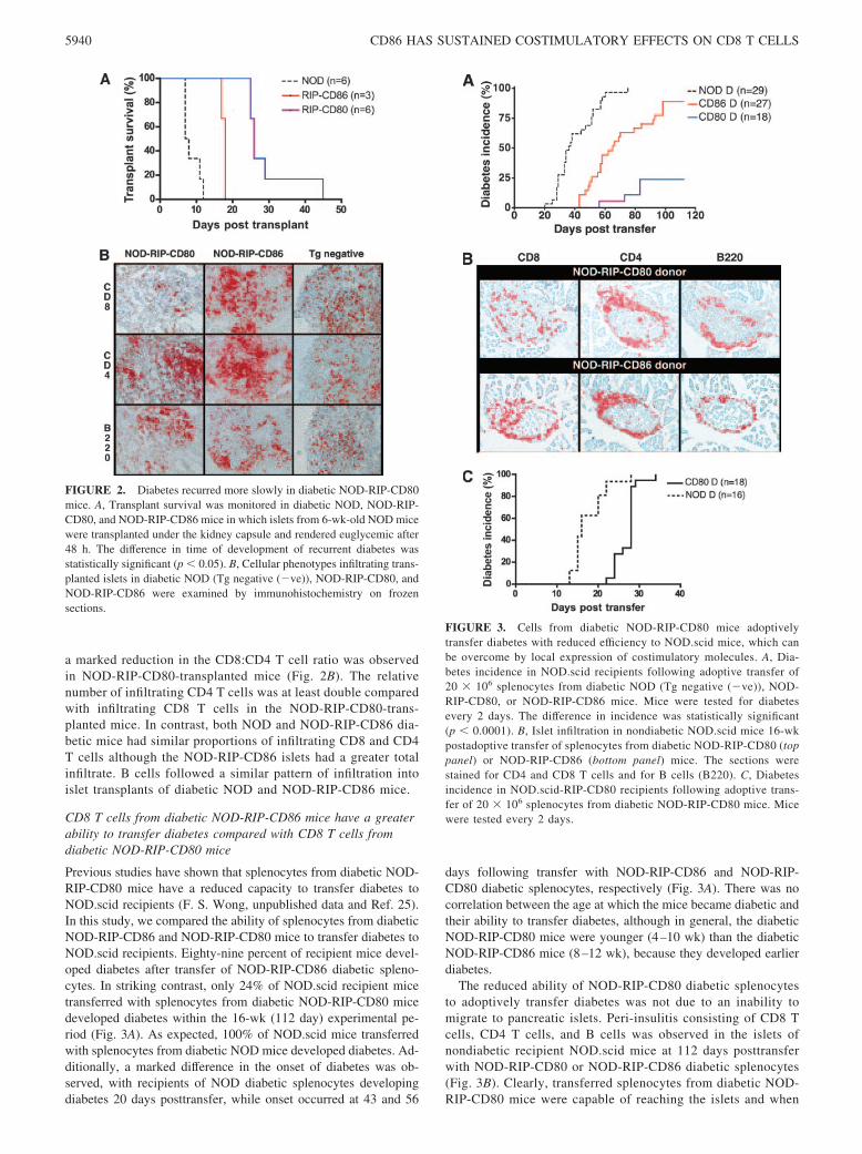
Previous studies have shown that splenocytes from diabetic NOD-RIP-CD80 mice have a reduced capacity to transfer diabetes toNOD.scid recipients (F. S. Wong, unpublished data and Ref. 25).In this study, we compared the ability of splenocytes from diabeticNOD-RIP-CD86 and NOD-RIP-CD80 mice to transfer diabetes toNOD.scid recipients. Eighty-nine percent of recipient mice devel-oped diabetes after transfer of NOD-RIP-CD86 diabetic spleno-cytes. In striking contrast, only 24% of NOD.scid recipient micetransferred with splenocytes from diabetic NOD-RIP-CD80 micedeveloped diabetes within the 16-wk (112 day) experimental pe-riod (Fig. 3A). As expected, 100% of NOD.scid mice transferredwith splenocytes from diabetic NOD mice developed diabetes. Ad-ditionally, a marked difference in the onset of diabetes was ob-served, with recipients of NOD diabetic splenocytes developingdiabetes 20 days posttransfer, while onset occurred at 43 and 56
days following transfer with NOD-RIP-CD86 and NOD-RIP-CD80 diabetic splenocytes, respectively (Fig. 3A). There was nocorrelation between the age at which the mice became diabetic andtheir ability to transfer diabetes, although in general, the diabeticNOD-RIP-CD80 mice were younger (4–10 wk) than the diabeticNOD-RIP-CD86 mice (8–12 wk), because they developed earlierdiabetes.
The reduced ability of NOD-RIP-CD80 diabetic splenocytesto adoptively transfer diabetes was not due to an inability tomigrate to pancreatic islets. Peri-insulitis consisting of CD8 Tcells, CD4 T cells, and B cells was observed in the islets ofnondiabetic recipient NOD.scid mice at 112 days posttransferwith NOD-RIP-CD80 or NOD-RIP-CD86 diabetic splenocytes(Fig. 3B). Clearly, transferred splenocytes from diabetic NOD-RIP-CD80 mice were capable of reaching the islets and when
FIGURE 2. Diabetes recurred more slowly in diabetic NOD-RIP-CD80mice. A, Transplant survival was monitored in diabetic NOD, NOD-RIP-CD80, and NOD-RIP-CD86 mice in which islets from 6-wk-old NOD micewere transplanted under the kidney capsule and rendered euglycemic after48 h. The difference in time of development of recurrent diabetes wasstatistically significant (p � 0.05). B, Cellular phenotypes infiltrating trans-planted islets in diabetic NOD (Tg negative (�ve)), NOD-RIP-CD80, andNOD-RIP-CD86 were examined by immunohistochemistry on frozensections.
FIGURE 3. Cells from diabetic NOD-RIP-CD80 mice adoptivelytransfer diabetes with reduced efficiency to NOD.scid mice, which canbe overcome by local expression of costimulatory molecules. A, Dia-betes incidence in NOD.scid recipients following adoptive transfer of20 � 106 splenocytes from diabetic NOD (Tg negative (�ve)), NOD-RIP-CD80, or NOD-RIP-CD86 mice. Mice were tested for diabetesevery 2 days. The difference in incidence was statistically significant(p � 0.0001). B, Islet infiltration in nondiabetic NOD.scid mice 16-wkpostadoptive transfer of splenocytes from diabetic NOD-RIP-CD80 (toppanel) or NOD-RIP-CD86 (bottom panel) mice. The sections werestained for CD4 and CD8 T cells and for B cells (B220). C, Diabetesincidence in NOD.scid-RIP-CD80 recipients following adoptive trans-fer of 20 � 106 splenocytes from diabetic NOD-RIP-CD80 mice. Micewere tested every 2 days.
5940 CD86 HAS SUSTAINED COSTIMULATORY EFFECTS ON CD8 T CELLS

further costimulation was provided by transfer into NOD.scid-RIP-CD80 recipients, these cells were capable of adoptivelytransferring diabetes in 100% of recipients (Fig. 3C). Thus,cells costimulated by CD80 were CD80 dependent whereasthose stimulated by CD86 were not.
T cell development does not play a role in the difference in Tcell activation by CD86 or CD80
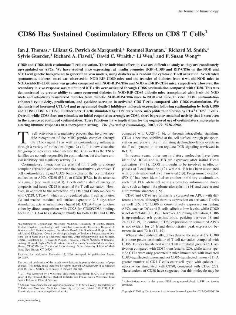
To test that the different outcomes following adoptive transferwere not related to differences in T cell development in thethymus, we used adoptive transfer of splenocytes from 6-wk-old NOD mice into NOD.scid-RIP-CD86 and NOD.scid-RIP-CD80 mice and tested for their ability to develop diabetes (Fig.4A). A similar pattern of development of diabetes comparedwith the natural history of diabetes in NOD-RIP-CD86 andNOD-RIP-CD80 mice was observed. One hundred percent ofNOD.scid-RIP-CD80 recipients had developed diabetes by 40days posttransfer, while the earliest time of diabetes onset oc-curred 45 and 54 days in NOD.scid-RIP-CD86 and NOD.scidmice, respectively. By 70 days after transfer, 90% of NOD.scid-RIP-CD86 recipients had developed diabetes whereas only 45%of NOD.scid recipients had developed disease at this point (Fig.4A). Thus, this experiment removed the possibility that any dif-ferences seen in diabetes onset in the NOD-RIP-CD86 or NOD-RIP-CD80 mice related to expression of the transgenes in thethymus and hence to development of diabetogenic T cells.
Additionally, greater expansion (or recruitment) of CD8 Tcells in islet infiltrates was observed in NOD.scid-RIP-CD80recipients transferred with spleen cells from 6-wk-old NODmice (Fig. 4B). Islet infiltration was similar at 16 days post-transfer, where �5– 6% CD8 and 10 –11% CD4 T cells wereobserved (Fig. 4B). By 28 days, the proportion of CD8 T cellsin NOD-scid-RIP-CD80 recipients had risen to �30%, doublethat compared with NOD.scid.RIP-CD86 recipients. Con-versely, the level of CD4 T cells was increased in the NOD.scid-RIP-CD86 recipient mice (Fig. 4B). Early diabetes onset,increased islet infiltration, and greater numbers of CD8 T cellsall suggest that islet expression of CD80 and CD86 enablesspecific activation of CD8 T cells. The fact that these observa-tions occur at an earlier time point in NOD-RIP-CD80 micestrongly indicates that activation of CD8 T cells through CD80generates enhanced responses, compared with CD86.
To demonstrate that the expression of CD80 or CD86 coulddifferentially stimulate CD8 T cells directly in our model system,rather than mainly through an effect on CD4 T cells, we adoptivelytransferred purified insulin-specific CD8 T cells directly ex vivofrom the splenocytes of G9C��/�. NOD-transgenic mice intoNOD.scid-RIP-CD80 and NOD.scid-RIP-CD86 recipients. Weshowed that the naive CD8 insulin-reactive T cells transfer diabe-tes more rapidly and with a higher final incidence to NOD.scid-RIP-CD80 recipients compared with NOD.scid.RIP-CD86 recipi-ents shown in Fig. 4C.
In vitro stimulation of lymphocytes with islets fromNOD.scid-RIP-CD80 mice induces greater proliferationand cytokine production than islets from NOD.scid-RIP-CD86mice and nontransgenic mice
To test the islet reactivity of T cells of known specificity, CD8 Tcells were isolated from insulin-specific CD8 TCR (G9.C��/�)transgenic mice and in vitro assays were performed using isolatedislets as both a source of CD8 T cell-specific APCs as well asinsulin peptides. Proliferation assays clearly demonstrated thatstimulation of insulin-specific CD8 T cells using islets from NOD.scid-RIP-CD80 mice generated heightened activity, �4-fold
FIGURE 4. Accelerated diabetes in RIP-CD80 and RIP-CD86 mice isnot due to differences in thymic T cell development. A, Adoptive transferof diabetes into NOD.scid, NOD.scid-RIP-CD80, and NOD.scid-RIP-CD86 mice showed a similar pattern of diabetes development to the naturalhistory of diabetes in NOD, NOD-RIP-CD80, and NOD-RIP-CD86 mice.A total of 20 � 106 splenocytes from 6-wk-old mice were transferred intoNOD.scid, NOD.scid-RIP-CD80 (NSCD80), and NOD.scid-RIP-CD86(NSCD86) mice and monitored weekly for diabetes development. The dif-ference in the time taken to develop diabetes and the final incidence ofdiabetes was statistically significant (p � 0.0001). B, Islet expression ofCD80 promotes increased islet infiltration of CD8 T cells in NOD.scid-RIP-CD80 recipients over time. Islet-infiltrating cells isolated from NOD.scid-RIP-CD80 and NOD.scid-RIP-CD86 islets after transfer with spleno-cytes from 6-wk-old mice were stained for CD8 and CD4 T cells. Toppanels, Dot plots for cells from 16 days posttransfer; bottom panels, 28days posttransfer. Islets from three mice per group were used. C, Purifiedinsulin-specific CD8 T cells cause increased incidence of diabetes in NOD.scid-RIP-CD80 recipients compared with NOD.scid-RIP-CD86 recipients.Purified insulin-specific CD8 T cells (107) (�95% pure) were transferredinto NOD.scid-RIP-CD80 (NSCD80) recipients compared with NOD.scid-RIP-CD86 (NSCD86) recipients. The difference in the time taken to de-velop diabetes and the final incidence of diabetes was statistically signif-icant (p � 0.0001). No diabetes is seen when these cells are transferred toNOD.scid recipients (n � 10, data not shown).
5941The Journal of Immunology
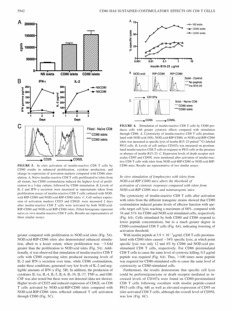
greater compared with proliferation to NOD.scid islets (Fig. 5A).NOD.scid-RIP-CD86 islets also demonstrated enhanced stimula-tion, albeit to a lesser extent, where proliferation was �3-foldgreater than the proliferation to NOD.scid islets (Fig. 5A). Addi-tionally, it was observed that stimulation of insulin-reactive CD8 Tcells with CD80 expressing islets produced increasing levels ofIL-2 and IFN-� secretion over time, while CD86 costimulation,under these conditions, generated very low levels of IL-2 and neg-ligible amounts of IFN-� (Fig. 5B). In addition, the production ofcytokines IL-1�, IL-4, IL-5, IL-6, IL-10, IL-17, TNF-�, and GM-CSF was also tested but these were not detected (data not shown).Higher levels of CD25 and reduced expression of CD62L on CD8T cells activated by NOD.scid-RIP-CD80 islets compared withNOD.scid-RIP-CD86 islets reflected enhanced T cell activationthrough CD80 (Fig. 5C).
In vitro stimulation of lymphocytes with islets fromNOD.scid-RIP-CD80 mice alters the threshold ofactivation of cytotoxic responses compared with islets fromNOD.scid-RIP-CD86 mice and nontransgenic mice
The cytotoxicity of insulin-reactive CD8 T cells after activationwith islets from the different transgenic strains showed that CD80costimulation induced greater levels of effector function with spe-cific target cell lysis reaching a maximum of 68%, compared with34 and 31% for CD86 and NOD.scid stimulated cells, respectively(Fig. 6A). Cells stimulated by both CD80 and CD86 respond tolower peptide concentrations, but to a much greater degree inCD80-costimulated CD8 T cells (Fig. 6A), indicating lowering ofactivation threshold.
With insulin peptide at 3.9 � 10�3�g/ml, CD8 T cells prestimu-lated with CD80 islets caused �34% specific lysis, at which pointspecific lysis was only 12 and 4% by CD86 and NOD.scid pre-stimulated CD8 T cells, respectively. For CD86 prestimulatedCD8 T cells to cause the same level of cytotoxic killing, 0.5 �g/mlpeptide was required (Fig. 6A). Thus, �100 times more peptidewas required for CD86-stimulated cells to cause the same level ofcytotoxicity as CD80-stimulated cells.
Furthermore, the results demonstrate that specific cell lysiscould be perforin/granzyme or death receptor mediated as in-creased levels of CD107a were found on CD80-prestimulatedCD8 T cells following coculture with insulin peptide-coatedP815 cells (Fig. 6B) as well as elevated expression of CD95 onislet-activated CD8 T cells, although the overall level of CD95Lwas low (Fig. 6C).
FIGURE 5. In vitro activation of insulin-reactive CD8 T cells byCD80 results in enhanced proliferation, cytokine production, andchange in expression of activation markers compared with CD86 stim-ulation. A, Naive insulin-reactive CD8 T cells proliferated to islets fromall strains, but CD80 costimulation induced the highest level of prolif-eration in a 3-day culture, followed by CD86 stimulation. B, Levels ofIL-2 and IFN-� secretion were measured in supernatants taken fromproliferation assays of insulin-reactive CD8 T cells cultured with NOD.scid-RIP-CD80 and NOD.scid-RIP-CD86 islets. C, Cell surface expres-sion of activation markers CD25 and CD62L were measured 2 daysafter insulin-reactive CD8 T cells were activated by both NOD.scid-RIP-CD80 and NOD.scid-RIP-CD86 islets. Filled histograms representnaive ex vivo insulin-reactive CD8 T cells. Results are representative ofthree similar assays.
FIGURE 6. Stimulation of insulin-reactive CD8 T cells by CD80 pro-duces cells with greater cytotoxic effects compared with stimulationthrough CD86. A, Cytotoxicity of insulin-reactive CD8 T cells prestimu-lated with NOD.scid (NS), NOD.scid-RIP-CD80, or NOD.scid-RIP-CD86islets was measured as specific lysis of insulin B15–23-pulsed 51Cr-labeledP815 cells. B, Levels of cell surface CD107a was measured on prestimu-lated insulin-reactive CD8 T cells in response to P815 cells in the presenceor absence of insulin B15–23. C, Expression levels of death receptor mol-ecules CD95 and CD95L were monitored after activation of insulin-reac-tive CD8 T cells with islets from NOD.scid-RIP-CD80 or NOD.scid-RIP-CD86 mice. Results are representative of two similar assays.
5942 CD86 HAS SUSTAINED COSTIMULATORY EFFECTS ON CD8 T CELLS

CD86 and CD80 both stimulate up-regulation of CTLA-4and PD-1
Although CD86 did not stimulate CD8 T cells to the same extentas CD80 in terms of increased proliferation, cytotoxicity, andincreased diabetogenicity, we also did not observe the inhibi-tion seen upon secondary stimulation. We therefore tested ex-pression of inhibitory costimulatory ligands. We examined theexpression of CTLA-4 in cells infiltrating islets from adoptivelytransferred diabetic NOD.scid-RIP-CD86 mice and found thatthe inhibitory coreceptor CTLA-4 was increased but less thanwith CD80 stimulation (Fig. 7A). Additionally, PD-1 was alsoexpressed in islet infiltrates from NOD.scid-RIP-CD86 andNOD.scid-RIP-CD80 mice transferred with splenocytes from6-wk-old NOD mice (Fig. 7B). Sixteen days posttransfer, CD8T cells isolated from NOD.scid-RIP-CD80 mice expressed higherlevels of PD-1 but by 28 days expression was similar in CD8 Tcells isolated from both strains (Fig. 7B). Other costimulatory li-gands that include 4-1BB and ICOS were studied but they were notdetected (data not shown). We tested the effect of blocking PD-1and CTLA-4 on stimulation of in vitro cytotoxicity using CD107aassays. However, we did not demonstrate any differences in gen-eration of in vitro cytotoxicity in these assays.
Depletion of CD4�CD25� cells facilitates adoptive transfer ofdiabetes by splenocytes from diabetic NOD.scid-RIP-CD80 mice
We had shown earlier (Fig. 3) a reduced ability of splenocytesfrom diabetic RIP-CD80 mice to transfer diabetes to secondaryNOD.scid recipients. To examine further possible inhibitory mech-anisms for this reduced ability to transfer disease, we performedadoptive transfer of spleen cells from 6-wk-old NOD mice intoNOD.scid-RIP-CD80 and NOD.scid-RIP-CD86 mice, similar tothe experiment shown in Fig. 4A. When the mice developed dia-betes, the spleen cells were harvested and the expression ofCD4�CD25�FoxP3 was examined by flow cytometry in an ali-quot of cells. We found that both sets of spleen cells expressed asimilar number of CD4�CD25�FoxP3� cells in the spleen shownin Fig. 8A.
From these spleen cells in diabetic mice, CD4�CD25� T cellswere depleted immediately, in a proportion of the splenocytes,before secondary adoptive transfer to NOD.scid mice. The deple-tion process removed all the CD4�CD25high T cells. Spleen cellsthat were not depleted of CD4�CD25� cells were used as controls.We found that after depletion of the CD4�CD25� T cells, spleno-
cytes from diabetic NOD.scid-RIP-CD80 were able to transfer di-abetes at a similar rate to cells obtained from diabetic NOD.scid-RIP-CD86 mice (Fig. 8B).
DiscussionCostimulation is essential for the full activation of CD8 T cells. Inthis study, we have aimed first to study the effect of CD80 (B7.1)and CD86 (B7.2) individually, a feature not possible in naturalAPCs as both are coexpressed and regulation is interdependent.Second, we are studying cells that respond to an important autoan-tigen, insulin, that is presented endogenously in the islets. Third,the strength of signal 1 is also an important factor. Using thismodel, we are able to use the ability of the islet to naturally presentthe range of Ag concentrations that the cells would encounter invivo in the islet. The ability of CD8 T cells to cause diabetes is acomplex process and our model allows us to test effects on cellexpansion, cytokine production, cytotoxicity in vitro and in vivo aswell as homing in vivo. We are using diabetes as an in vivo readoutto study the individual effects of costimulatory molecules and notthe development of natural diabetes, as islet � cells do not nor-mally express costimulatory molecules (37).
FIGURE 7. CD80 and CD86 stimulation of CD8 T cells results in up-regulation of inhibitory coreceptors CTLA-4 and PD-1 but CD80 stimu-lation causes earlier and greater expression levels. A, Intracellular CTLA-4expression was examined following in vitro activation of insulin-reactiveCD8 T cells by NOD.scid-RIP-CD80 or NOD.scid-RIP-CD86 islets. B,Cell surface expression of PD-1 was studied on islet infiltrate CD8 T cellsfollowing transfer of splenocytes from 6-wk-old NOD mice into NOD.scid-RIP-CD80 and NOD.scid-RIP-CD86 mice 16 and 28 days posttrans-fer. Filled histograms represent naive ex vivo CD8 T cells (A and B).Results are representative of two similar assays.
FIGURE 8. CD4�CD25� depletion of CD80-stimulated splenocytes re-stores the ability to cause diabetes. A, Splenocytes from 6-wk-old NODmice were adoptively transferred into NOD.scid-RIP-CD80 and NOD.scid-RIP-CD86 mice. When the mice became diabetic, the splenocytes wereharvested and stained for CD4�CD25� and FoxP3� cells. Top panels,Representative cells from NOD.scid-RIP-CD80 mice; lower panels, cellsfrom NOD.scid-RIP-CD86 mice. B, The splenocytes were depleted ofCD4�CD25� T cells (�80% depleted) and then adoptively transferred toNOD.scid mice which were observed for diabetes. These were comparedwith cells that had not been depleted of CD4�CD25� cells. The resultsshown for the transfer from diabetic NOD.scid-RIP-CD80 mice are com-bined from two separate experiments.
5943The Journal of Immunology
In this study, we have demonstrated a clear difference in theactivation of CD8 T cells after CD80 or CD86 costimulation. Invivo, we observed accelerated diabetes in RIP-CD80 mice (4 wk)as noted in previous studies (25, 29, 30) as well as in RIP-CD86mice (7 wk) compared with transgene-negative mice (13 wk). Thegreater acceleration of diabetes incidence in RIP-CD80 mice wasnot likely to be due to a difference in expression levels of CD80compared with CD86. Diabetes development in the medium (12B)and high (46K) expressing NOD-RIP-CD86 mice was almost iden-tical, while previous studies showed a similar result with differentexpression levels in RIP-CD80 mice (29). Additionally, Ab stain-ing of pancreatic sections from NOD-RIP-CD80 and NOD-RIP-CD86 (12B) had similar protein levels, and PCR products fromcDNA isolated from islets of these transgenic mice demonstratedcomparable levels of expression. Hence, we infer that accelerateddiabetes was due to enhanced stimulation of T cells by CD80 com-pared with CD86.
The proportion of CD8 T cells in islet infiltrates was increasedin diabetic NOD-RIP-CD80 and NOD-RIP-CD86 mice comparedwith diabetic transgene-negative littermates. However, proportionsof B and CD4 T cells were similar in all the diabetic mice exam-ined, which suggested that costimulation and MHC class I on islet� cells specifically activated CD8 T cells. We further demonstratedthat CD80 enhanced the proportion of CD8 T cells in islet infil-trates more than CD86 costimulation. NOD.scid-RIP-CD80 andNOD.scid-RIP-CD86 mice i.v. transferred with splenocytes from6-wk-old NOD mice showed similar proportions of CD8 and CD4T cells in islet infiltrates after 16 days but after 28 days, twice theCD8 T cells were present in NOD.scid-RIP-CD80 islets comparedwith NOD.scid-RIP-CD86. This suggested that CD8 T cells werebeing further activated in situ and hence proliferating in the isletsthemselves.
Further evidence that the expression of CD80 and CD86 ex-pressed on the islets directly stimulate CD8 T cells was shown byexperiments using purified, naive low-avidity CD8 T cells recog-nizing insulin peptide B15–23. These cells are unable to transferdiabetes to NOD.scid mice unless they have been preactivated(L. K. Siew, F. S. Wong, unpublished observations). However,they cause diabetes in NOD.scid-RIP-CD80 mice with faster ki-netics than NOD.scid-RIP-CD86 mice. No CD4 T cells were trans-ferred in these experiments indicating that the expression of thecostimulatory molecules on the islets has a direct effect on CD8 Tcells.
When the cells from the diabetic NOD-RIP-CD80 and NOD-RIP-CD86-transgenic mice were subjected to a secondary stimu-lation, CD86-stimulated cells were more effective in causing re-current diabetes in both NOD islet-transplanted mice and inNOD.scid recipients receiving cells from diabetic NOD-RIP-CD86-transgenic mice. Although the cells from diabetic NOD-RIP-CD80-transgenic mice caused delayed diabetes in NOD.scidrecipients or did not transfer diabetes, when the cells from diabeticNOD-RIP-CD80-transgenic mice were retransferred into NOD.scid-RIP-CD80-transgenic mice, diabetes occurred rapidly and inall mice. This indicated that the threshold of activation of theCD80-stimulated cells was altered such that they could respond inthe presence of CD80 but only poorly in the absence of the co-stimulatory molecule.
To examine the effect of the stimulation by islet Ag with eitherCD80 or 86, we performed in vitro studies with T cells of a definedspecificity that are able to react to the islet Ag, insulin. Islets iso-lated from both NOD.scid-RIP-CD86 and NOD.scid-RIP-CD80mice induced higher levels of proliferation in naive insulin-reac-tive CD8 T cells compared with NOD.scid islets but greater pro-liferation was seen with RIP-CD80 islets. CD8 T cells secreted
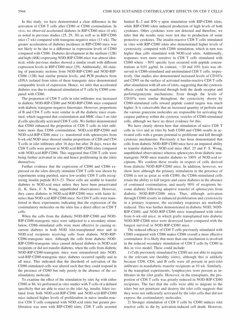
limited IL-2 and IFN-� upon stimulation with RIP-CD86 islets,while RIP-CD80 islets induced production of high levels of bothcytokines. Other cytokines were not detected and therefore, weinfer that the results seen were not due to production of someprotective cytokines. The insulin-reactive CD8 T cells stimulatedin vitro with RIP-CD80 islets also demonstrated higher levels ofcytotoxicity, compared with CD86 stimulation, which in turn washigher than cells stimulated with NOD.scid islets. Additionally,responses were more sensitive in CD8 T cells stimulated withCD80 where �50% specific lysis occurred with peptide concen-trations at 0.01 �g/ml. In contrast, �20 and 10% lysis was ob-served in CD86-stimulated and unstimulated CD8 T cells, respec-tively. Our studies also demonstrated increased levels of CD107aand CD95 on the surface of activated insulin-reactive CD8 T cellsafter stimulation with all sets of islets, suggesting that the cytotoxiceffects could be manifested through both the death receptor andperforin/granzyme mechanisms. Even though the levels ofCD107a were similar throughout, the cytotoxicity shown byCD80-stimulated cells toward peptide coated targets was muchhigher. It is conceivable that an increased quantity of perforin andthe various granzyme molecules are responsible for activating thecaspase pathway within the cytotoxic vesicles of CD80-stimulatedcells, although we have no direct evidence for this.
We have clearly shown here that stimulation of naive CD8 Tcells in vivo and in vitro by both CD80 and CD86 results in ac-tivated cells with a greater potential to proliferate and kill throughcytotoxic mechanisms. Previously it has been demonstrated thatcells from diabetic NOD-RIP-CD80 mice have an impaired abilityto transfer diabetes to NOD.scid mice (Ref. 25 and F. S. Wong,unpublished observations) whereas splenocytes from diabetic non-transgenic NOD mice transfer diabetes to 100% of NOD.scid re-cipients. We confirm these results in respect of cells derivedfrom diabetic NOD-RIP-CD80 mice. In addition, however, weshow here although the primary stimulation in the presence ofCD86 is not as great as with CD80, the CD86-stimulated cellsretain the ability to kill targets and cause diabetes in the absenceof continued costimulation, and nearly 90% of recipients be-come diabetic following adoptive transfer of splenocytes fromdiabetic NOD-RIP-CD86 mice. Thus, although activationthrough CD80 results in enhanced proliferation and cytotoxicityin a primary response, the secondary responses are markedlyreduced. This was further demonstrated in diabetic NOD, NOD-RIP-CD80, and NOD-RIP-CD86 mice transplanted with isletsfrom 6-wk-old mice, in which grafts transplanted into diabeticNOD-RIP-CD86 mice were destroyed faster compared with thelongest survival in NOD-RIP-CD80 mice.
The reduced efficacy of CD8 T cells previously stimulated withCD80 compared with CD86 makes CD86 overall a more effectivecostimulator. It is likely that more than one mechanism is involvedin the reduced secondary stimulation of CD8 T cells by CD80 inthis in vivo model. These could include:
1) Cells previously stimulated by CD80 are not able to migrateto the relevant site (healthy islets), although this is unlikelybecause CD8, CD4, and B cells were all present in peri-isletinfiltrates in nondiabetic transfer recipients at 10 wk. Similarly,in the transplant experiments, lymphocytes were present as in-filtrates in the islet grafts. However, in the transplants, the pro-portion of CD8 T cells was greatly reduced in NOD-RIP-CD80recipients. The fact that the cells were able to migrate to theislets but not penetrate and destroy the islet cells suggests thatthey were not sufficiently activated by the islet cells that did notexpress the costimulatory molecules.
2) Stronger stimulation of CD8 T cells by CD80 induces isletreactive cells to die by activation-induced cell death. However,
5944 CD86 HAS SUSTAINED COSTIMULATORY EFFECTS ON CD8 T CELLS
after in vitro stimulation with islets expressing CD80 or CD86,survival of the cells is similar as shown by similar cell numbersand annexin V staining (data not shown). Furthermore, followingin vitro stimulation with islets and then a short maintenance periodover 3 days in IL-2, a greater proportion of CD80 prestimulatedCD8 T cells remained viable (data not shown).
3) A possible explanation behind the reduced efficacy of CD80-stimulated CD8 T cells is increased and sustained expression ofinhibitory coreceptors, such as CTLA-4 and PD-1, although CD86also up-regulated these inhibitory receptors. It is well known thatCTLA-4 is up-regulated on activation and interacts with CD80 andcells are inhibited as a result of this interaction. However, PD-1up-regulation has not previously been linked to cells activated byCD80. In this study, we have demonstrated that in vivo, PD-1 isup-regulated on the cell surface of CD8 T cells stimulated byCD80 and CD86 but higher expression is evident at an earlier timein CD80-stimulated CD8 T cells. Additionally, intracellular ex-pression of CTLA-4 is apparent in insulin-reactive CD8 T cells ata greater level after activation through CD80 stimulation comparedwith CD86 stimulation. We propose that after the initial aggressiveactivation of CD8 T cells by CD80, the T cells up-regulate inhib-itory coreceptors to a greater degree and potentially for a longerperiod of time, compared with CD86 stimulation. This would en-able T cells to be “turned off” and, depending on the duration ofinhibitory coreceptor expression, prevent or reduce the possibilityof a secondary stimulation. This would most likely be the casewhen peptides of interest are displayed at endogenous concentra-tions, such as in the studies presented here. However, by providingincreased levels of costimulation, this inhibition can be overcome.We performed in vitro blocking experiments using CD8 cells stim-ulated by RIP-CD80 and RIP-CD86 islets with Abs to blockCTLA-4 or PD-1 and tested in vitro cytotoxicity measuringCD107a but did not observe consistent alteration in the in vitroresponses (data not shown). Clearly, it would be interesting tomonitor the kinetics of inhibitory receptor expression before, dur-ing, and after primary and secondary activation of CD8 T cellsthrough CD80 or CD86 stimulation in vivo. Most recently, it hasbeen shown that in addition to binding to the known inhibitoryligand, CTLA-4 on T cells, B7-1 is also able to directly bind toPD-L1 and deliver an inhibitory signal (38). This interaction couldalso explain the inhibition observed after the initial activation. Itwould certainly be of importance also to test for the up-regulationof PD-L1 and the effect of blocking this interaction in this system.
4) A further possibility for the reduced ability of CD80-stimu-lated cells to cause diabetes in secondary adoptive transfer couldbe an increase in regulatory T cell inhibition of cells stimulatedwith CD80 compared with CD86. We measured the percentage ofCD4�CD25� cells in the splenocytes of mice that had becomediabetic after the primary transfer of 6-wk-old NOD splenocytesinto NOD.scid-RIP-CD86 and NOD.scid-RIP-CD80 mice. Thepercentage of these cells in the splenocytes was not different be-tween the two types of mice. However, following depletion of theCD4�CD25� T cells, the cells from the diabetic NOD.scid-RIP-CD80 mice were able to transfer diabetes with similar kinetics tocells from diabetic NOD.scid-RIP-CD86 mice. Thus, this suggeststhat either an increase in the levels of CD4�CD25� T cells in thediabetic RIP-CD80 splenocytes were inhibiting the CD80-stimu-lated CD8 T cells (our evidence did not support this), or the CD8cells were more susceptible to inhibition by these regulatory cells.Removing the CD4�CD25� T cells facilitated the secondaryadoptive transfer of diabetes by the CD80-stimulated splenocytes.
Finally, the use of ectopic expression of costimulatory mole-cules to boost immune responses is a strategy that has been usedfor modulating immune responses to tumors. The fact that CD80
stimulates stronger activating responses also has an impact in vivo.The increased stimulation of cells with more rapid responses andexpansion of CD8 T cells that is not maintained if the costimula-tory molecule is removed may also suggest that more cells areinitially recruited by islets expressing CD80 as previously sug-gested by Allison et al. (25). Diabetes in the NOD mouse is causedby a heterogeneous group of cells and the ability of CD80 to lowerthe threshold of activation of lower avidity cells could allow forrecruitment of these lower avidity cells to the islet and in situexpansion of cells. However, the low-avidity cells, although stim-ulated to full activation in response to CD80 stimulation, are notable to maintain the level of activation important for cytotoxicityin the absence of costimulatory molecules. Thus, on encounter oflow levels of Ag in the absence of the costimulatory molecule,such as in the case of transplantation with islets that do not expressCD80, or in secondary transfer when the recipients do not expressCD80, the low-avidity CD8 T cells are unable to further contributeto the disease process. Our results also suggest that these CD80-stimulated T cells are more susceptible to inhibition byCD4�CD25� regulatory T cells. This may further imply that theislet destructive repertoire is different in the RIP-CD80 mice com-pared with the RIP-CD86 mice and nontransgenic NOD mice.
In conclusion, our studies have shown that when CD80 andCD86 stimulation is separated and tested in vivo, both CD80 andCD86 can stimulate activating and inhibitory responses but CD80induces, overall, stronger responses. Thus, CD80 targeted locallymay be useful in the context where CD80 remains expressed andcontinues to be active (39) to stimulate lower avidity T cells andovercome inhibition by CD4�CD25� T cells. Although once ac-tivated, effector cells may not require continued costimulation un-der all circumstances, this depends on continued lower threshold ofactivation and is likely also to be dependent on the level of Agexpression. Overall, while CD86 does not stimulate an initial re-sponse as strongly as CD80, there is greater sustained activity thatis seen, even in the absence of continued costimulation. Thesefunctions have implications for the engineered use of costimula-tory molecules in altering immune responses in a therapeuticsetting.
AcknowledgmentsWe thank Andrew Herman for assistance in flow cytometry, Irene Visintinfor advice on immunohistochemistry, Stephen Chapman for excellent tech-nical assistance, and Gwen Scott for assistance at the end stages of theexperiments. The original RIP-CD80, and RIP-CD86 mice were generatedby the Yale Diabetes Endocrinology Research Center Transgenic CoreDK45735.
DisclosuresThe authors have no financial conflict of interest.
References1. Greenwald, R. J., G. J. Freeman, and A. H. Sharpe. 2005. The B7 family revis-
ited. Annu. Rev. Immunol. 23: 515–548.2. Watts, T. H. 2005. TNF/TNFR family members in costimulation of T cell re-
sponses. Annu. Rev. Immunol. 23: 23–68.3. Linsley, P. S., J. L. Greene, P. Tan, J. Bradshaw, J. A. Ledbetter, C. Anasetti, and
N. K. Damle. 1992. Coexpression and functional cooperation of CTLA-4 andCD28 on activated T lymphocytes. J. Exp. Med. 176: 1595–1604.
4. Walunas, T. L., D. J. Lenschow, C. Y. Bakker, P. S. Linsley, G. J. Freeman,J. M. Green, C. B. Thompson, and J. A. Bluestone. 1994. CTLA-4 can functionas a negative regulator of T cell activation. Immunity 1: 405–413.
5. Collins, A. V., D. W. Brodie, R. J. Gilbert, A. Iaboni, R. Manso-Sancho,B. Walse, D. I. Stuart, P. A. van der Merwe, and S. J. Davis. 2002. The interactionproperties of costimulatory molecules revisited. Immunity 17: 201–210.
6. Linsley, P. S., J. L. Greene, W. Brady, J. Bajorath, J. A. Ledbetter, and R. Peach.1994. Human B7-1 (CD80) and B7-2 (CD86) bind with similar avidities butdistinct kinetics to CD28 and CTLA-4 receptors. Immunity 1: 793–801.
7. Alegre, M. L., K. A. Frauwirth, and C. B. Thompson. 2001. T-cell regulation byCD28 and CTLA-4. Nat. Rev. Immunol. 1: 220–228.
5945The Journal of Immunology
8. Beier, K. C., A. Hutloff, A. M. Dittrich, C. Heuck, A. Rauch, K. Buchner,B. Ludewig, H. D. Ochs, H. W. Mages, and R. A. Kroczek. 2000. Induction,binding specificity and function of human ICOS. Eur. J. Immunol. 30:3707–3717.
9. Coyle, A. J., S. Lehar, C. Lloyd, J. Tian, T. Delaney, S. Manning, T. Nguyen,T. Burwell, H. Schneider, J. A. Gonzalo, et al. 2000. The CD28-related moleculeICOS is required for effective T cell-dependent immune responses. Immunity 13:95–105.
10. Futagawa, T., H. Akiba, T. Kodama, K. Takeda, Y. Hosoda, H. Yagita, andK. Okumura. 2002. Expression and function of 4-1BB and 4-1BB ligand onmurine dendritic cells. Int. Immunol. 14: 275–286.
11. McAdam, A. J., T. T. Chang, A. E. Lumelsky, E. A. Greenfield, V. A. Boussiotis,J. S. Duke-Cohan, T. Chernova, N. Malenkovich, C. Jabs, V. K. Kuchroo, et al.2000. Mouse inducible costimulatory molecule (ICOS) expression is enhanced byCD28 costimulation and regulates differentiation of CD4� T cells. J. Immunol.165: 5035–5040.
12. Rottman, J. B., T. Smith, J. R. Tonra, K. Ganley, T. Bloom, R. Silva, B. Pierce,J. C. Gutierrez-Ramos, E. Ozkaynak, and A. J. Coyle. 2001. The costimulatorymolecule ICOS plays an important role in the immunopathogenesis of EAE. Nat.Immunol. 2: 605–611.
13. Hurtado, J. C., Y. J. Kim, and B. S. Kwon. 1997. Signals through 4-1BB arecostimulatory to previously activated splenic T cells and inhibit activation-in-duced cell death. J. Immunol. 158: 2600–2609.
14. Nishimura, H., M. Nose, H. Hiai, N. Minato, and T. Honjo. 1999. Developmentof lupus-like autoimmune diseases by disruption of the PD-1 gene encoding anITIM motif-carrying immunoreceptor. Immunity 11: 141–151.
15. Ansari, M. J., A. D. Salama, T. Chitnis, R. N. Smith, H. Yagita, H. Akiba,T. Yamazaki, M. Azuma, H. Iwai, S. J. Khoury, et al. 2003. The programmeddeath-1 (PD-1) pathway regulates autoimmune diabetes in nonobese diabetic(NOD) mice. J. Exp. Med. 198: 63–69.
16. Azuma, M., D. Ito, H. Yagita, K. Okumura, J. H. Phillips, L. L. Lanier, andC. Somoza. 1993. B70 antigen is a second ligand for CTLA-4 and CD28. Nature366: 76–79.
17. Hathcock, K. S., G. Laszlo, C. Pucillo, P. Linsley, and R. J. Hodes. 1994. Com-parative analysis of B7-1 and B7-2 costimulatory ligands: expression and func-tion. J. Exp. Med. 180: 631–640.
18. Freeman, G. J., G. S. Gray, C. D. Gimmi, D. B. Lombard, L. J. Zhou, M. White,J. D. Fingeroth, J. G. Gribben, and L. M. Nadler. 1991. Structure, expression, andT cell costimulatory activity of the murine homologue of the human B lympho-cyte activation antigen B7. J. Exp. Med. 174: 625–631.
19. Lenschow, D. J., G. H. Su, L. A. Zuckerman, N. Nabavi, C. L. Jellis, G. S. Gray,J. Miller, and J. A. Bluestone. 1993. Expression and functional significance of anadditional ligand for CTLA-4. Proc. Natl. Acad. Sci. USA 90: 11054–11058.
20. Fields, P. E., R. J. Finch, G. S. Gray, R. Zollner, J. L. Thomas, K. Sturmhoefel,K. Lee, S. Wolf, T. F. Gajewski, and F. W. Fitch. 1998. B7.1 is a quantitativelystronger costimulus than B7.2 in the activation of naive CD8� TCR-transgenic Tcells. J. Immunol. 161: 5268–5275.
21. Gajewski, T. F., F. Fallarino, C. Uyttenhove, and T. Boon. 1996. Tumor rejectionrequires a CTLA4 ligand provided by the host or expressed on the tumor: supe-riority of B7-1 over B7-2 for active tumor immunization. J. Immunol. 156:2909–2917.
22. Manzotti, C. N., M. K. Liu, F. Burke, L. Dussably, Y. Zheng, and D. M. Sansom.2006. Integration of CD28 and CTLA-4 function results in differential responsesof T cells to CD80 and CD86. Eur. J. Immunol. 36: 1413–1422.
23. Guerder, S., D. E. Picarella, P. S. Linsley, and R. A. Flavell. 1994. CostimulatorB7-1 confers antigen-presenting-cell function to parenchymal tissue and in con-
junction with tumor necrosis factor � leads to autoimmunity in transgenic mice.Proc. Natl. Acad. Sci. USA 91: 5138–5142.
24. Herrera, P. L., D. M. Harlan, L. Fossati, S. Izui, J. Huarte, L. Orci, J. D. Vassalli,and P. Vassalli. 1994. A CD8� T-lymphocyte-mediated and CD4� T-lympho-cyte-independent autoimmune diabetes of early onset in transgenic mice. Diabe-tologia 37: 1277–1279.
25. Allison, J., L. A. Stephens, T. W. Kay, C. Kurts, W. R. Heath, J. F. Miller, andM. F. Krummel. 1998. The threshold for autoimmune T cell killing is influencedby B7-1. Eur. J. Immunol. 28: 949–960.
26. Harlan, D. M., H. Hengartner, M. L. Huang, Y. H. Kang, R. Abe,R. W. Moreadith, H. Pircher, G. S. Gray, P. S. Ohashi, G. J. Freeman, et al. 1994.Mice expressing both B7-1 and viral glycoprotein on pancreatic � cells alongwith glycoprotein-specific transgenic T cells develop diabetes due to a breakdownof T-lymphocyte unresponsiveness. Proc. Natl. Acad. Sci. USA 91: 3137–3141.
27. von Herrath, M. G., S. Guerder, H. Lewicki, R. A. Flavell, and M. B. Oldstone.1995. Coexpression of B7-1 and viral (“self”) transgenes in pancreatic � cells canbreak peripheral ignorance and lead to spontaneous autoimmune diabetes. Im-munity 3: 727–738.
28. Guerder, S., J. Meyerhoff, and R. Flavell. 1994. The role of the T cell costimu-lator B7-1 in autoimmunity and the induction and maintenance of tolerance toperipheral antigen. Immunity 1: 155–166.
29. Wong, S., S. Guerder, I. Visintin, E. P. Reich, K. E. Swenson, R. A. Flavell, andC. A. Janeway, Jr. 1995. Expression of the co-stimulator molecule B7-1 in pan-creatic �-cells accelerates diabetes in the NOD mouse. Diabetes 44: 326–329.
30. Wong, F. S., I. Visintin, L. Wen, J. Granata, R. Flavell, and C. A. Janeway. 1998.The role of lymphocyte subsets in accelerated diabetes in nonobese diabetic-ratinsulin promoter-B7-1 (NOD-RIP-B7-1) mice. J. Exp. Med. 187: 1985–1993.
31. Wong, F. S., W. Du, I. J. Thomas, and L. Wen. 2005. The influence of the majorhistocompatibility complex on development of autoimmune diabetes in RIP-B7.1mice. Diabetes 54: 2032–2040.
32. Guerder, S., E. E. Eynon, and R. A. Flavell. 1998. Autoimmunity without dia-betes in transgenic mice expressing � cell-specific CD86, but not CD80: param-eters that trigger progression to diabetes. J. Immunol. 161: 2128–2140.
33. Wong, F. S., I. Visintin, L. Wen, R. A. Flavell, and C. A. Janeway, Jr. 1996. CD8T cell clones from young nonobese diabetic (NOD) islets can transfer rapid onsetof diabetes in NOD mice in the absence of CD4 cells. J. Exp. Med. 183: 67–76.
34. Wong, F. S., J. Karttunen, C. Dumont, L. Wen, I. Visintin, I. M. Pilip, N. Shastri,E. G. Pamer, and C. A. Janeway, Jr. 1999. Identification of an MHC class I-re-stricted autoantigen in type 1 diabetes by screening an organ-specific cDNAlibrary. Nat. Med. 5: 1026–1031.
35. Kouskoff, V., K. Signorelli, C. Benoist, and D. Mathis. 1995. Cassette vectorsdirecting expression of T cell receptor genes in transgenic mice. J. Immunol.Methods 180: 273–280.
36. Gotoh, M., T. Maki, S. Satomi, J. Porter, and A. P. Monaco. 1986. Immunolog-ical characteristics of purified pancreatic islet grafts. Transplantation 42:387–390.
37. Stephens, L. A., and T. W. Kay. 1995. Pancreatic expression of B7 co-stimulatorymolecules in the non-obese diabetic mouse. Int. Immunol. 7: 1885–1895.
38. Butte, M. J., Keir, M. E., Phamduy, T. B., Sharpe, A. H., and Freeman, G. J.2007. Programmed death-1 Ligand 1 interacts specifically with the B7-1 costimu-latory molecule to inhibit T cell responses. Immunity 27: 111–122.
39. Kaufman, H. L., G. Deraffele, J. Mitcham, D. Moroziewicz, S. M. Cohen,K. S. Hurst-Wicker, K. Cheung, D. S. Lee, J. Divito, M. Voulo, et al. 2005.Targeting the local tumor microenvironment with vaccinia virus expressing B7.1for the treatment of melanoma. J. Clin. Invest. 115: 1903–1912.
5946 CD86 HAS SUSTAINED COSTIMULATORY EFFECTS ON CD8 T CELLS
Copyright © 2022 FDOKUMEN