Frontline: GITR, a member of the TNF receptor superfamily, is costimulatory to mouse T lymphocyte...
10
Frontline: GITR, a member of the TNF receptor superfamily, is costimulatory to mouse T lymphocyte subpopulations Simona Ronchetti, Ornella Zollo, Stefano Bruscoli, Massimiliano Agostini, Rodolfo Bianchini, Giuseppe Nocentini, Emira Ayroldi and Carlo Riccardi Department of Clinical and Experimental Medicine, Section of Pharmacology, Perugia University Medical School, Perugia, Italy GITR (glucocorticoid-induced TNFR family related gene) is a member of the TNFR superfam- ily (TNFRSF) that is expressed in different cell types, including T lymphocytes. Because of a high homology in its cytoplasmic region with other known costimulatory members of the TNFRSF, we investigated whether GITR played a costimulatory role in T lymphocyte subpop- ulations. Our results show that the proliferation response of CD8 + and CD4 + peripheral T cell subpopulations was potentiated when a GITR costimulus was added to an anti-CD3 stimu- lus. Furthermore, expression of the main activation-induced receptor (IL-2R ) and produc- tion of IL-2 and IFN- were increased more with a GITR costimulus than with anti-CD3 alone. GITR stimulation also enhanced anti-CD3-induced ERK phosphorylation, suggesting that GITR is involved in MAPK-pathway activation. Interestingly, CD4 + CD25 + regulatory T cell (Treg cell) proliferation was triggered by the GITR costimulus; Treg cell proliferation was par- alleled by the loss of the anergic phenotype and suppressor activity. Nevertheless, unstimu- lated GITR –/– CD4 + CD25 + and GITR +/+ CD4 + CD25 + cells were equally able to exert suppressor activity on CD4 + CD25 – responder cells. These results indicate a novel function for GITR as costimulatory molecule of T cell subsets. Key words: Costimulation / Tumor necrosis factor receptor superfamily / T cell activation Received 8/12/03 Accepted 15/1/04 [DOI 10.1002/eji.200324804] Abbreviations: GITR: Glucocorticoid-induced TNFR family related gene mGITRL: Murine ligand for GITR TNFRSF: TNFR superfamily Treg cell: Regulatory T cell 1 Introduction GITR (glucocorticoid-induced TNFR family related gene), a member of the TNFR superfamily (TNFRSF), was origi- nally cloned in a glucocorticoid-treated hybridoma T cell line [1]. GITR, like most members of this superfamily, is mainly expressed in lymphoid tissues and is up- regulated upon T lymphocyte activation. It has a high homology in the cytoplasmic region with other TNFRSF members [2], namely 4-1BB, CD27, OX40 and CD40, which all exert costimulatory activity [3–6]. We have pre- viously demonstrated GITR over-expression protects from activation-induced cell death (AICD) [1]. When activated by treatment with anti-CD3 Ab, GITR- knockout (GITR –/– ) T lymphocytes proliferate more than wild-type (GITR +/+ ) T cells, express higher levels of IL-2R and IL-2 and are more sensitive to AICD [7]. All these findings provide evidence that GITR actively contributes to control T cell activation. GITR is highly expressed on CD4 + CD25 + regulatory T cells (Treg cells), and an agonist antibody against GITR has been reported to abrogate their suppressor function [8, 9]. Despite these observations, the role of GITR in the regulation of different T cell subpopulations, including CD8 + , CD4 + CD25 – and CD4 + CD25 + cells, has not yet been completely defined, so we investigated whether GITR plays a costimulatory role on T cells by comparing T lymphocytes from GITR +/+ and GITR –/– mice. This study discovers that GITR is a costimulatory mole- cule for all T cell subpopulations because it increases proliferation in anti-CD3-stimulated CD8 + cells, CD4 + CD25 – cells and CD4 + CD25 + Treg cells. In fact, although triggering GITR alone did not influence T cell activity, cotriggering of GITR and CD3 stimulated expression of activation molecules, such as IL-2R , and production of cytokines, such as IL-2 and IFN- , sug- gesting that GITR acts as a costimulatory molecule. Moreover, GITR costimulation increased anti-CD3- Eur. J. Immunol. 2004. 34: 613–622 GITR as a costimulatory molecule 613 © 2004 WILEY-VCH Verlag GmbH & Co. KGaA, Weinheim www.eji.de
-
Upload
independent -
Category
Documents
-
view
0 -
download
0
Transcript of Frontline: GITR, a member of the TNF receptor superfamily, is costimulatory to mouse T lymphocyte...
Frontline:
GITR, a member of the TNF receptor superfamily,is costimulatory to mouse T lymphocytesubpopulations
Simona Ronchetti, Ornella Zollo, Stefano Bruscoli, Massimiliano Agostini, RodolfoBianchini, Giuseppe Nocentini, Emira Ayroldi and Carlo Riccardi
Department of Clinical and Experimental Medicine, Section of Pharmacology, Perugia UniversityMedical School, Perugia, Italy
GITR (glucocorticoid-induced TNFR family related gene) is a member of the TNFR superfam-ily (TNFRSF) that is expressed in different cell types, including T lymphocytes. Because of ahigh homology in its cytoplasmic region with other known costimulatory members of theTNFRSF, we investigated whether GITR played a costimulatory role in T lymphocyte subpop-ulations. Our results show that the proliferation response of CD8+ and CD4+ peripheral T cellsubpopulations was potentiated when a GITR costimulus was added to an anti-CD3 stimu-lus. Furthermore, expression of the main activation-induced receptor (IL-2R § ) and produc-tion of IL-2 and IFN- + were increased more with a GITR costimulus than with anti-CD3 alone.GITR stimulation also enhanced anti-CD3-induced ERK phosphorylation, suggesting thatGITR is involved in MAPK-pathway activation. Interestingly, CD4+CD25+ regulatory T cell(Treg cell) proliferation was triggered by the GITR costimulus; Treg cell proliferation was par-alleled by the loss of the anergic phenotype and suppressor activity. Nevertheless, unstimu-lated GITR–/– CD4+CD25+ and GITR+/+ CD4+CD25+ cells were equally able to exert suppressoractivity on CD4+CD25– responder cells. These results indicate a novel function for GITR ascostimulatory molecule of T cell subsets.
Key words: Costimulation / Tumor necrosis factor receptor superfamily / T cell activation
Received 8/12/03Accepted 15/1/04
[DOI 10.1002/eji.200324804]
Abbreviations: GITR: Glucocorticoid-induced TNFR familyrelated gene mGITRL: Murine ligand for GITR TNFRSF:TNFR superfamily Treg cell: Regulatory T cell
1 Introduction
GITR (glucocorticoid-induced TNFR family related gene),a member of the TNFR superfamily (TNFRSF), was origi-nally cloned in a glucocorticoid-treated hybridoma T cellline [1]. GITR, like most members of this superfamily, ismainly expressed in lymphoid tissues and is up-regulated upon T lymphocyte activation. It has a highhomology in the cytoplasmic region with other TNFRSFmembers [2], namely 4-1BB, CD27, OX40 and CD40,which all exert costimulatory activity [3–6]. We have pre-viously demonstrated GITR over-expression protectsfrom activation-induced cell death (AICD) [1].
When activated by treatment with anti-CD3 Ab, GITR-knockout (GITR–/–) T lymphocytes proliferate more thanwild-type (GITR+/+) T cells, express higher levels of IL-2R
and IL-2 and are more sensitive to AICD [7]. All thesefindings provide evidence that GITR actively contributesto control T cell activation.
GITR is highly expressed on CD4+CD25+ regulatory Tcells (Treg cells), and an agonist antibody against GITRhas been reported to abrogate their suppressor function[8, 9]. Despite these observations, the role of GITR in theregulation of different T cell subpopulations, includingCD8+, CD4+CD25– and CD4+CD25+ cells, has not yetbeen completely defined, so we investigated whetherGITR plays a costimulatory role on T cells by comparingT lymphocytes from GITR+/+ and GITR–/– mice.
This study discovers that GITR is a costimulatory mole-cule for all T cell subpopulations because it increasesproliferation in anti-CD3-stimulated CD8+ cells,CD4+CD25– cells and CD4+CD25+ Treg cells. In fact,although triggering GITR alone did not influence T cellactivity, cotriggering of GITR and CD3 stimulatedexpression of activation molecules, such as IL-2R § , andproduction of cytokines, such as IL-2 and IFN- + , sug-gesting that GITR acts as a costimulatory molecule.Moreover, GITR costimulation increased anti-CD3-
Eur. J. Immunol. 2004. 34: 613–622 GITR as a costimulatory molecule 613
© 2004 WILEY-VCH Verlag GmbH & Co. KGaA, Weinheim www.eji.de

Fig. 1. A GITR costimulus enhances the proliferation of T lym-phocytes. (A) Lymph node T lymphocytes from both GITR+/+
and GITR–/– mice were cultured on irradiated splenocytes inthe presence of anti-CD3 and/or anti-GITR and/or anti-CD28Ab. Proliferation was measured as [3H]thymidine uptake[counts per minute (cpm)] after 48 and 72 h. *p X 0.05 (column3 vs. column 1) and **p X 0.01 (column 9 vs. column 7). (B)Lymph node T lymphocytes from both GITR+/+ and GITR–/–
mice were cultured with anti-CD3 and/or anti-GITR, whichwere either plate-bound (right) or soluble and in the presenceof irradiated splenocytes (left). Proliferation was measured by[3H]thymidine uptake at 72 h. **p X 0.01 (column 3 vs. column1, in both graphs). (C) Purified lymph node CD4+ and CD8+ Tlymphocytes from GITR+/+ and GITR–/– mice were cultured onirradiated splenocytes for 48 and 72 h in the presence of theindicated Ab. Proliferation was measured by [3H]thymidineuptake. *p X 0.05 (column 3 vs. column 1, CD4 graph) (column3 vs. column 1, CD8 graph), **p X 0.01 (column 11 vs. column9 in both graphs). All the results are representative of threeexperiments and represent the mean ± SD of four counts. (D)Cell cycle phases in the above experimental conditions weremeasured after 72 h of culture by propidium iodide stainingand flow cytometric analysis. *p X 0.05 (column 5 vs. column 2in both graphs).
induced ERK phosphorylation, indicating that GITR maycontribute to TCR/CD3-mediated activation of the MAPKpathway.
Finally, lack of GITR expression in the CD4+CD25+ cellsof GITR–/– mice did not impair their suppressive abilitybut GITR costimulation, with anti-CD3, o CD4+CD25+
cells from GITR+/+ mice increased cell proliferation andweakened suppressor activity. Thus GITR appears toregulate the activity of CD4+CD25+ Treg cells, CD8+ andCD4+CD25– effector cells.
2 Results
2.1 Anti-GITR Ab enhances anti-CD3-inducedproliferation of T lymphocytes
According to the two-signal model, T cell activationrequires signal 1 from the T cell antigen-receptor com-plex (TCR–CD3), and signal 2 from costimulatory mole-cules. To find out whether GITR behaves as a classicalcostimulatory molecule, we activated lymph node T lym-phocytes with a combination of anti-mouse GITR Ab(2 ? g/ml) and anti-CD3 4 Ab at a suboptimal concentra-tion (0.5 ? g/ml), on a layer of irradiated splenocytes. Pro-liferation was measured, by the [3H]thymidine incorpora-tion assay, after stimulation for 48 and 72 h. In all experi-ments, the effect of anti-GITR Ab was assessed usingGITR-null cells, from GITR–/– mice, as a negative controlcompared with cells from GITR+/+ mice.
The results indicate that anti-GITR Ab alone did notaffect GITR+/+ T lymphocyte proliferation. Anti-GITR plusanti-CD3 Ab enhanced proliferation significantly morethan the anti-CD3 stimulus alone (Fig. 1A). The GITR-induced increase in proliferation was observed 48 and at72 h after activation. As expected, cells from GITR–/–
mice, which were as susceptible as cells from GITR+/+ tocostimulation by anti-CD28 Ab, did not respond to anti-GITR costimulation (Fig. 1A). Moreover, our previousresults, indicating a major anti-CD3-induced proliferationresponse at 48 h by whole T lymphocyte populationsfrom GITR–/– mice, were confirmed (Fig. 1A) [7]. We alsoperformed an experiment using plated-bound anti-GITRAb, in the absence of a layer of irradiated splenocytes.The results shown in Fig. 1B indicate that cross-linkedanti-GITR Ab increased anti-CD3-induced cell prolifera-tion (Fig. 1B, right) as well as soluble anti-GITR Ab did(Fig. 1B, left) thus excluding the effect of anti-GITR Ab onfeeder cells.
The above results indicate that GITR augments the pro-liferation of anti-CD3-activated lymphocytes. This effectcould be due to inhibition of Treg cell function [8, 9] or
614 S. Ronchetti et al. Eur. J. Immunol. 2004. 34: 613–622
© 2004 WILEY-VCH Verlag GmbH & Co. KGaA, Weinheim www.eji.de

Fig. 2. GITR costimulation induces the proliferation ofanergic CD4+CD25+ cells and increases proliferation inCD4+CD25– T lymphocytes. (A) CD4+CD25+ and CD4+CD25–
T lymphocytes were purified from lymph nodes of GITR+/+
and GITR–/– mice and cultured on irradiated splenocytes for72 h in the presence of anti-CD3 and/or anti-GITR and/oranti-CD28 Ab. Proliferation was assayed by [3H]thymidineuptake. Results are representative of three experiments andare the mean ± SD of four counts. **p X 0.01 (column 3 vs.column 1, right graph) and ***p X 0.001 (column 3 vs. column1, left graph). (B) GITR expression on T lymphocyte subpop-ulations. Naive T cells were separated from lymph nodes ofGITR+/+ and GITR–/– mice and stained with either an anti-GITR or control rat IgG Ab. Flow cytometric analysis wasperformed on gated live cells. Shaded histograms representthe control (goat IgG) staining, whereas unshaded histo-grams represent GITR staining. Staining with secondary Abalone is omitted for clarity. GITR–/– staining is reported as thenegative control. Histograms are representative of at leasttwo experiments. (C) CD8+, CD4+CD25–, CD4+CD25+ T lym-phocytes, either unstimulated or anti-CD3-stimulated, werecultured for 72 h on CHO cells transfected either with theempty vector (CHO-ev) or with mGITRL (CHO-GITRL). Prolif-eration was assayed by [3H]thymidine uptake. Results arerepresentative of three experiments and represent the mean± SD of four counts. **p X 0.01 (column 4 vs. column 3 ineach graph).
also to a direct effect of GITR Ab on responder cells. Toaddress this question, we first separated CD4+ and CD8+
T lymphocytes and analyzed the proliferation responseto GITR costimuluation at 48 and 72 h. Fig. 1C showsthat the anti-GITR Ab, but not the isotype-control Ab, inassociation with the anti-CD3 Ab, exerted a costimula-tory effect on both CD4+ and CD8+ single-positive T cellsfrom GITR+/+ but not from GITR–/– mice. The same resultswere obtained when the proliferation was assayed bypropidium iodide staining and cell cycle analysis at 72 h(Fig. 1D). Of note, the separated CD4+ but not the CD8+
GITR–/– subpopulation displayed, upon anti-CD3 stimula-tion, a higher proliferation rate than the CD4+ GITR+/+
population (Fig. 1C, D), suggesting that CD4+ cells con-tributed to the increased proliferation of the whole T cellpopulation from GITR–/– mice [7]. These results indicatethat GITR functions as a coaccessory molecule in com-bination with stimulation of the CD3–TCR complex.
2.2 Anti-GITR costimulation induces proliferationin anergic CD4+CD25+ immunoregulatoryT cells and enhances proliferation ofCD4+CD25– responder T lymphocytes
In two previous studies GITR emerged as crucial forCD4+CD25+ Treg cell function, because stimulation withan anti-GITR Ab abrogated suppressive activity [8, 9].CD4+CD25+ Treg cells are anergic. They do not prolifer-ate and do not produce cytokines upon stimulation ofthe CD3–TCR complex in vitro [10–12]. However, they doproliferate in the presence of exogenous IL-2 or, depend-ing on mouse strain or experimental conditions, uponcostimulation with anti-CD28 Ab [8, 13]. To investigatewhether GITR costimulation induces proliferation, wecostimulated CD4+CD25+ cells with anti-CD3 plus anti-GITR Ab. The results confirm the lack of proliferation ofanti-CD3-stimulated CD4+CD25+ and show that addingthe GITR costimulus induces proliferation of anti-CD3-stimulated CD4+CD25+ T cells (Fig. 2A, left panel).
To investigate whether the enhanced proliferation ofCD4+ cells in the presence of GITR costimulation wasdue only to the CD4+CD25+ fraction, we stimulatedCD4+CD25– T cells under the same experimental condi-tions as described for the other cell subpopulations. Asfor CD8+ and CD4+CD25+ T lymphocytes, an enhancedproliferation response was observed in the CD4+CD25–
subpopulation when costimulated with anti-GITR plusanti-CD3 Ab (Fig. 2A, right). Of note, CD4+CD25– cellproliferation was higher than that of CD4+CD25+ cells. Incontrast, triggering of GITR did not provide any costimu-lus to GITR–/– cells. However, GITR–/– and GITR+/+ cellswere equally susceptible to the anti-CD28 Ab costimu-lus.
Eur. J. Immunol. 2004. 34: 613–622 GITR as a costimulatory molecule 615
© 2004 WILEY-VCH Verlag GmbH & Co. KGaA, Weinheim www.eji.de
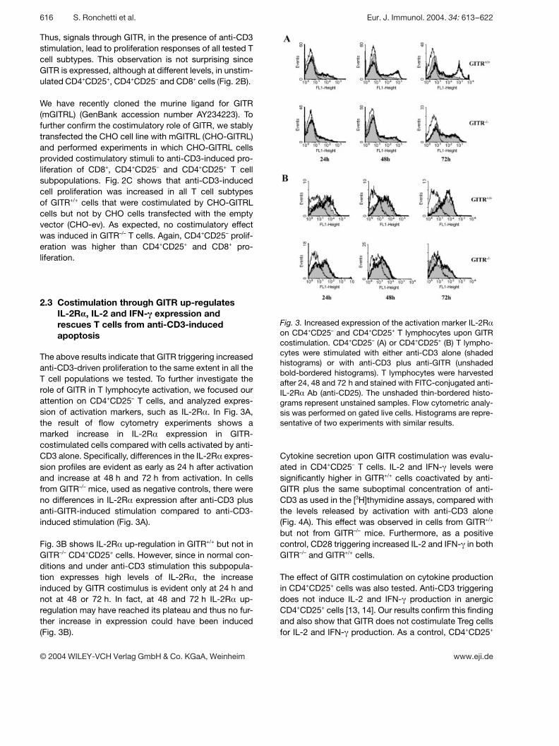
Fig. 3. Increased expression of the activation marker IL-2R §on CD4+CD25– and CD4+CD25+ T lymphocytes upon GITRcostimulation. CD4+CD25– (A) or CD4+CD25+ (B) T lympho-cytes were stimulated with either anti-CD3 alone (shadedhistograms) or with anti-CD3 plus anti-GITR (unshadedbold-bordered histograms). T lymphocytes were harvestedafter 24, 48 and 72 h and stained with FITC-conjugated anti-IL-2R § Ab (anti-CD25). The unshaded thin-bordered histo-grams represent unstained samples. Flow cytometric analy-sis was performed on gated live cells. Histograms are repre-sentative of two experiments with similar results.
Thus, signals through GITR, in the presence of anti-CD3stimulation, lead to proliferation responses of all tested Tcell subtypes. This observation is not surprising sinceGITR is expressed, although at different levels, in unstim-ulated CD4+CD25+, CD4+CD25– and CD8+ cells (Fig. 2B).
We have recently cloned the murine ligand for GITR(mGITRL) (GenBank accession number AY234223). Tofurther confirm the costimulatory role of GITR, we stablytransfected the CHO cell line with mGITRL (CHO-GITRL)and performed experiments in which CHO-GITRL cellsprovided costimulatory stimuli to anti-CD3-induced pro-liferation of CD8+, CD4+CD25– and CD4+CD25+ T cellsubpopulations. Fig. 2C shows that anti-CD3-inducedcell proliferation was increased in all T cell subtypesof GITR+/+ cells that were costimulated by CHO-GITRLcells but not by CHO cells transfected with the emptyvector (CHO-ev). As expected, no costimulatory effectwas induced in GITR–/– T cells. Again, CD4+CD25– prolif-eration was higher than CD4+CD25+ and CD8+ pro-liferation.
2.3 Costimulation through GITR up-regulatesIL-2R > , IL-2 and IFN- q expression andrescues T cells from anti-CD3-inducedapoptosis
The above results indicate that GITR triggering increasedanti-CD3-driven proliferation to the same extent in all theT cell populations we tested. To further investigate therole of GITR in T lymphocyte activation, we focused ourattention on CD4+CD25– T cells, and analyzed expres-sion of activation markers, such as IL-2R § . In Fig. 3A,the result of flow cytometry experiments shows amarked increase in IL-2R § expression in GITR-costimulated cells compared with cells activated by anti-CD3 alone. Specifically, differences in the IL-2R § expres-sion profiles are evident as early as 24 h after activationand increase at 48 h and 72 h from activation. In cellsfrom GITR–/– mice, used as negative controls, there wereno differences in IL-2R § expression after anti-CD3 plusanti-GITR-induced stimulation compared to anti-CD3-induced stimulation (Fig. 3A).
Fig. 3B shows IL-2R § up-regulation in GITR+/+ but not inGITR–/– CD4+CD25+ cells. However, since in normal con-ditions and under anti-CD3 stimulation this subpopula-tion expresses high levels of IL-2R § , the increaseinduced by GITR costimulus is evident only at 24 h andnot at 48 or 72 h. In fact, at 48 and 72 h IL-2R § up-regulation may have reached its plateau and thus no fur-ther increase in expression could have been induced(Fig. 3B).
Cytokine secretion upon GITR costimulation was evalu-ated in CD4+CD25– T cells. IL-2 and IFN- + levels weresignificantly higher in GITR+/+ cells coactivated by anti-GITR plus the same suboptimal concentration of anti-CD3 as used in the [3H]thymidine assays, compared withthe levels released by activation with anti-CD3 alone(Fig. 4A). This effect was observed in cells from GITR+/+
but not from GITR–/– mice. Furthermore, as a positivecontrol, CD28 triggering increased IL-2 and IFN- + in bothGITR–/– and GITR+/+ cells.
The effect of GITR costimulation on cytokine productionin CD4+CD25+ cells was also tested. Anti-CD3 triggeringdoes not induce IL-2 and IFN- + production in anergicCD4+CD25+ cells [13, 14]. Our results confirm this findingand also show that GITR does not costimulate Treg cellsfor IL-2 and IFN- + production. As a control, CD4+CD25+
616 S. Ronchetti et al. Eur. J. Immunol. 2004. 34: 613–622
© 2004 WILEY-VCH Verlag GmbH & Co. KGaA, Weinheim www.eji.de

Fig. 4. Increased cytokine production, and protection fromapoptosis upon GITR costimulation. (A) CD4+CD25– orCD4+CD25+ cells purified from both GITR+/+ and GITR–/– Tlymphocytes were stimulated by the indicated Ab, on irradi-ated splenocytes, for 72 h. Supernatants were harvestedand used for IL-2 and IFN- + ELISA assays (nd indicates thatthe cytokine was not detected). The results represent themean ± SD of three independent experiments. *p X 0.05 (col-umn 3 vs. column 1, IFN- + graph) and ***p X 0.001 (column 3vs. column 1, IL-2 graph). (B) Apoptosis of CD4+CD25– andCD4+CD25+ T lymphocytes was measured by propidiumiodide staining and analyzed by flow cytometry after 72 h ofactivation with the indicated stimuli. Results represent oneof three independent experiments.
Fig. 5. GITR costimulation increases ERK phosphorylation.CD4+CD25– T cells were cultured for 3 h (A) and 24 h (B) inthe presence of anti-CD3 or anti-CD3 plus anti-GITR stimuli.Levels of phosphorylated p44/42 MAPK and total p44/42MAPK were assessed by immunoblotting with the samemembrane. The 42-kDa band, representing phosphorylatedMAPK, and the 44-kDa and 42-kDa bands, representingtotal p44/42 MAPK protein, are indicated by arrows. West-ern blot with g -tubulin was performed to verify that equiva-lent amounts of proteins were loaded in each lane.
cells were also stimulated with anti-CD28 Ab. Results inFig. 4A confirm previous results indicating that costim-uluation with anti-CD28 Ab induces production of IFN- +[13] but not of IL-2 [15].
These results, together with results described in Fig. 1and 2, indicate that GITR contributes to the T lympho-cyte activation process.
GITR, like other costimulatory/coaccesssory molecules[16–18], protects T cells from anti-CD3-induced apopto-
sis. To determine whether GITR regulates survival of theCD4+CD25+ and CD4+CD25– cells we measured apopto-sis by propidium iodide staining at 72 h after adding theactivating stimulus. Anti-GITR costimulation significantlyprotected CD4+CD25– (upper panel in Fig. 4B) andCD4+CD25+ (lower panel in Fig. 4B) cells from anti-CD3-activated apoptosis. This effect was observed in cellsfrom GITR+/+ mice but not in cells from GITR–/– mice.
These data indicate that GITR costimulation, in associa-tion with triggering of the CD3–TCR complex, increasesT cell activation, proliferation, cytokine production andsurvival.
2.4 Signaling events after costimulation of GITR
Many signals participate in T lymphocyte activation [19,20]. Activation by triggering the CD3–TCR complexresults in activation of the ras-dependent MAPK path-way with the sequential activation of raf-1, Mek and ERK.Phosphorylation or ERK, a downstream event of MAPKactivation, is required for ERK activation [21, 22]. Previ-
Eur. J. Immunol. 2004. 34: 613–622 GITR as a costimulatory molecule 617
© 2004 WILEY-VCH Verlag GmbH & Co. KGaA, Weinheim www.eji.de

Fig. 6. GITR–/– CD4+CD25+ T lymphocytes are as suppres-sive as GITR+/+ CD4+CD25+ T lymphocytes. (A) CD4+CD25– Tcells from both GITR+/+ and GITR–/– mice were either culturedalone in the presence of anti-CD3 or co-cultured withCD4+CD25+ suppressor cells from the same genotype in thepresence of anti-CD3 alone or in combination with anti-GITRon a layer of irradiated splenocytes. **p X 0.01 (column 3 vs.column 1; column 4 vs. column 2; column 5 vs. column 3).(B) GITR+/+ CD4+CD25– responder T cells were either cul-tured alone in the presence of anti-CD3 or anti-CD3 plusanti-GITR or co-cultured with GITR–/– CD4+CD25+ suppres-sor cells in the same conditions as in (A). **p X 0.01 (column4 vs. column 2). (C) GITR–/– CD4+CD25– responder T cellswere either cultured alone in the presence of anti-CD3 oranti-CD3 plus anti-GITR or co-cultured with GITR+/+
CD4+CD25+ suppressor cells in the same conditions as in(A). Results are the mean ± SD of four counts and are repre-sentative of two independent experiments.
ous results indicate that coaccessory molecules of theTNFRSF increased ERK phosphorylation [23, 24]. Toanalyze whether GITR also influences this signalingevent, we evaluated ERK protein levels and phosphory-lation. To this end, CD4+CD25– T cells from GITR+/+ andGITR–/– lymph nodes were treated with anti-CD3 or anti-CD3 plus anti-GITR for 3 and 24 h. Activation of P44/42MAPK was monitored in Western blotting with a specificAb that recognizes the phosphorylated form of ERK1/2.Results in Fig. 5 indicate that the level of phosphorylatedp42 MAPK was higher after stimulation with anti-CD3plus anti-GITR than with anti-CD3 alone, both at 3 h(Fig. 5A) and 24 h (Fig. 5B). This was observed in GITR+/+
but not in GITR–/– cells. Moreover, anti-CD3 or anti-CD3plus anti-GITR treatment did not change the protein lev-els of p44/42 MAPK.
Therefore GITR costimulation contributes to CD3/TCRsignaling in T cells, by increasing MAPK pathway activa-tion, as shown by the increase in ERK phosphorylation.
2.5 GITR is dispensable for CD4+CD25+
suppressor activity
Our results indicate that GITR triggering acts as acostimulatory signal in all T cell populations, includingCD4+CD25+ Treg cells. Two independent groups havepreviously demonstrated that anti-GITR Ab abrogatesthe suppressive activity of CD4+CD25+ Treg cells, sug-gesting that GITR may control their activity [8, 9].
We performed experiments to address (1) the possiblerole of GITR expression in the development of Treg cellactivity and (2) the possible effect of GITR triggering inmodulating Treg cell activity. Therefore, we first analyzedthe role of GITR expression in regulating the suppressiveproperty of the CD4+CD25+ cells by comparing the sup-pressor activity of CD4+CD25+ GITR+/+ and ofCD4+CD25+ GITR–/– cells. Results in Fig. 6A demonstratethat GITR+/+ and GITR–/– CD4+CD25+ Treg cells have asimilar ability to suppress anti-CD3-induced proliferationof CD4+CD25– responder cells, suggesting that a lack ofGITR expression does not affect the development of Tregcell suppressor activity in GITR–/– mice.
We also investigated the effect of GITR triggering onsuppressor activity. Costimulation with anti-GITR abro-gates Treg cell suppressor activity of GITR+/+, as alreadydemonstrated [8, 9], but not of GITR–/– cells (Fig. 6A). Torule out that abrogation of suppression was due to theanti-GITR-induced increase of responder cell prolifera-tion, we mixed GITR+/+ with GITR–/– cells (both suppres-sor and responder). The experiment reported in Fig. 6Bwas performed by mixing CD4+CD25+ GITR–/– suppres-
sor cells and CD4+CD25– GITR+/+ responder cells. Theresults show that GITR–/– Treg cells suppressed the anti-CD3-induced proliferation response of CD4+CD25– cells.GITR costimulation increased the proliferative responseof responder cells cultured alone (Fig. 6B, column 2 vs.column 1) but did not abrogate the Treg suppressoractivity (Fig. 6B, column 2 vs. column 4). However, in thepresence of GITR–/– suppressor cells, the proliferativeresponse of responder cells, triggered with anti-GITRplus anti-CD3, was higher than what was induced byanti-CD3 alone (Fig. 6B, column 4 vs. column 3). Thiseffect may not be due to inhibition of GITR–/– Treg sup-pressor activity but to costimulation of GITR+/+ respondercells and the consequently increased proliferation.
618 S. Ronchetti et al. Eur. J. Immunol. 2004. 34: 613–622
© 2004 WILEY-VCH Verlag GmbH & Co. KGaA, Weinheim www.eji.de
When CD4+CD25+ GITR+/+ suppressor cells were co-cultured with CD4+CD25– GITR–/– responder cells(Fig. 6C), abrogation of suppressor activity due to GITRcostimulation was evident, resulting in a statistically sig-nificant increase of proliferation (Fig. 6C, column 4 vs.column 3). These results indicate that GITR triggeringinhibits Treg cell suppressor activity.
These results are similar to earlier evidence showing thatwhen CD4+CD25+ cells proliferate they lose their sup-pressive properties [14, 25]. In fact, we here demonstratethat GITR costimulation increases CD3-activated T cellproliferation, including CD4+CD25+ Treg cell proliferation.So, as has already been observed with anti-CD28costimulation [12], the anti-GITR costimulus increasesTreg cell proliferation (Fig. 2A) and consequently inhibitsits suppressive ability.
These data, although suggesting that GITR expressionmight not be necessary for the development of Treg cellsuppressor activity, support the hypothesis that GITR,when expressed, is involved in control of CD4+CD25+ Tcell homeostasis.
These data also indicate that GITR may have two func-tions: (1) costimulation of CD4+CD25– responder cells;and (2) costimulation and inhibition of suppressor activityof CD4+CD25+ Treg cells.
3 Discussion
The aim of this study was to evaluate the potentialcostimulatory function of GITR engagement on T lym-phocytes. We observed that GITR acts as a costimula-tory molecule of anti-CD3-stimulated cells, includingCD4+ (either CD25+ or CD25–) and CD8+ single-positivecells.
The rationale for this experimental approach was theGITR homology, in the cytoplasmic domain, with othercostimulatory molecules of the TNFRSF (4-1BB, CD27,OX40 and CD40) that are known to participate in T lym-phocyte activation [2–6]. In our experiments weassessed the ability of GITR triggering to modulate CD3/TCR-driven T lymphocyte activation.
GITR costimulation enhanced the anti-CD3-triggeredactivation responses in all the T cell subpopulationswe assayed, i.e. CD4+ (including CD4+CD25– andCD4+CD25+) and CD8+ lymph node T cells. GITR costim-ulation increased anti-CD3-activated cell proliferation,IL-2R § expression, IL-2 and IFN- + production and ERKphosphorylation. These results clearly place GITR in thelarge group of costimulatory/coaccessory moleculesinvolved in regulating T cell activation.
A previous study [9] suggested a costimulatory effect ofan anti-GITR Ab (DTA-1) on CD28-null CD4+CD25– cells,although with no increase in proliferative response inCD4+CD25+ cells. Another study [8] showed a prolifera-tion response of CD4+CD25+ cells to anti-GITR stimulusin the presence of IL-2, but ruled out costimulation by thesame anti-GITR Ab on anti-CD3-induced proliferation inresponder cells. Our evaluation of proliferation, cytokineproduction, activation-receptor expression and intracel-lular signaling indicates that T lymphocyte activationincreases as a consequence of GITR stimulation. Theseobservations appear in part contradictory, but the use ofdifferent experimental procedures and animal strainsmay account for the discrepancies.
We provide evidence that GITR is a costimulatory mole-cule. We minimized the possibility of other nonspecificinteractions with other receptors through the use ofGITR–/– cells as a negative control, which confirmed thatthe anti-GITR Ab used in the study, either in the presenceor in absence of feeder cells, and the GITRL expressedon CHO cells, act through interaction with GITR inGITR+/+ cells. Finally, the coactivating role of GITR is sup-ported by the induction of several activation markerssuch as IL-2R § , IL-2, and IFN- + , by ERK phosphorylationand by the protection from anti-CD3-induced apoptosis,which confirms previously reported results [1, 2].
Previous studies [8, 9] also showed that GITR plays afunctional role in controlling the suppressive activity ofCD4+CD25+ T cells, as GITR stimulation abrogatedCD4+CD25+ T cell mediated suppression, and concludedthat GITR delivers an inhibitory signal to these cells. Ourresults confirm that GITR triggering abrogates suppres-sor activity and suggest that proliferation induced byGITR costimulation in CD4+CD25+ cells correlates withthe loss of their suppressive ability. It has largely beenproven in fact that immunoregulatory T cells lose theirsuppressive ability when they proliferate [14, 25]. GITR,by acting as a costimulatory molecule, increases prolifer-ation and renders CD4+CD25+ Treg cells unable to sup-press responder cell proliferation.
For an in-depth analysis of the role of GITR on the modu-lation of Treg cell activity, we tested the capacity ofGITR–/– CD4+CD25+ lymphocytes to suppress prolifera-tion of CD4+CD25– T cells in vitro. In our experiments weobserved that freshly isolated Treg cells from GITR+/+ andGITR–/– mice exerted similar suppression, suggestingthat GITR expression in itself does not modulate, eitherby increasing or decreasing, development of suppressorcell activity.
Different events may underlie these results. GITR couldbe optional for suppressor function development,
Eur. J. Immunol. 2004. 34: 613–622 GITR as a costimulatory molecule 619
© 2004 WILEY-VCH Verlag GmbH & Co. KGaA, Weinheim www.eji.de
because GITR–/– cells do suppress effectors, but couldbe involved in controlling their anergic status, so thatGITR triggering modulates their activity. The homologybetween GITR and other members of the TNFRSF, suchas 4-1BB, CD27, OX40 and CD40, is evidence for a sub-family of costimulatory molecules with similar propertieswithin the TNFRSF. However, some differences do existamong them — in particular the phenotypic features ofthe lymphocytes that develop when the molecules areknocked out; some of these features are apparently incontrast with their function as costimulatory molecules.T lymphocytes from OX40–/– or CD27–/– mice showimpaired activation and proliferation [26, 27] when stimu-lated with anti-CD3 alone. Unseparated 4-1BB–/– andGITR–/– T lymphocytes are activated better and prolifer-ate more than wild-type cells [7, 28] when stimulatedwith anti-CD3 alone. This also occurs when CD4+ single-positive cells from GITR+/+ and GITR–/– mice are tested,suggesting that CD4+ cells contribute, at least in part, tothe augmented proliferation.
Although the impaired ability of T cells either fromOX40–/– or CD27–/– mice to proliferate correlates with theircostimulatory role, the increased proliferation of 4-1BB–/–
and GITR–/– T lymphocytes to anti-CD3 stimulation sug-gests an unknown role in the control of T lymphocytegrowth and development. GITR could play a role not onlyas a coactivating molecule in anti-CD3-stimulatedmature lymphocytes, but also during T cell developmentin vivo thus generating lymphocytes that are more sus-ceptible to anti-CD3-induced activation. Moreover, com-pensatory mechanisms, induced by a lack of GITR,could be involved in the GITR–/– phenotype. Whether theincreased proliferation of anti-CD3-stimulated GITR–/–
lymphocytes depends on a defect in the number ofperipheral CD4+CD25+ T cells is an open question. Wefound a slight, though not statistically significant, differ-ence in CD4+CD25+ percentages in GITR+/+ T cells andGITR–/– T cells with the latter being slightly fewer in num-ber. Whether this modifies the in vitro response of GITR–/–
T lymphocytes, compared with GITR+/+ cells, remains tobe investigated.
Overall our findings place GITR in an important positionin the T cell response to antigen in the immune system;this molecule has a role in both suppressor andresponder T cell function. If suppressor and responder Tcells do not act together, then either autoreactive T cellscan take over or tumor cells can escape immunosurveill-ance. If GITR control of immunoregulatory T cell anergyis defective, a tolerant immunosurveillance could switchto an autoimmune pathology. Intervening in the GITR/GITRL system in the future could help treat pathologiessuch as autoimmune diseases and cancers. Furtherstudies aimed at defining the nature and distribution of
GITRL in different cell subpopulations might providemore insights into GITR function.
4 Materials and methods
4.1 Mice and cells
Mouse lymph node T lymphocytes were obtained fromSv129 GITR+/+ or GITR–/– mice [2] kept under specificpathogen-free conditions. Mice were sacrificed between 6and 8 weeks of age.
4.2 Cell purification
Purified lymph node T lymphocytes were obtained as previ-ously described [7]. CD4+ and CD8+ lymphocytes were sep-arated using anti-CD4 and anti-CD8 microbeads (MiltenyiBiotec, Bergisch Gladbach, Germany), following the manu-facturer’s instructions. Briefly, T lymphocytes were incu-bated with the appropriate microbeads at 4°C for 15 min.After washing, the cells were loaded on to a MiniMACS MS+
column (Miltenyi Biotec), the flow-through cells were dis-carded and the others were eluted from the column. Thisprocedure was repeated twice to increase the purity of thesubpopulations. Flow cytometry showed that the magneti-cally retained cells were G 98% CD4+ or CD8+ cells.CD4+CD25+ cells were purified from T lymphocytes using aCD4+CD25+ T cell isolation kit (Miltenyi Biotec), following themanufacturer’s instructions. Briefly, after isolation of CD4+
cells, the CD25+ PE-labeled cells were magnetically labeledwith anti-PE microbeads. The cell suspension was loadedon to a column, which was placed in the magnetic field of aMACS Separator. The flow-through cells were collected andused as CD4+CD25– cells whereas the retained cells wereeluted from the column and used as CD4+CD25+ cells. Toachieve the highest purities, two consecutive column-runswere performed. Flow cytometry showed that G 97% ofmagnetically retained cells were CD25+ and G 96% of flow-through cells were CD25–.
4.3 Cell transfection and fixation
CHO cells were stably transfected with FuGENE (Roche,Mannheim, Germany), following the manufacturer’s instruc-tions. Fixation of GITRL-CHO stably transfected cells wasperformed by washing the cells (7×10⁴ cells/well in 96-wellplates) three times with HBSS. Glutaraldehyde (0.05%) wasadded to the cells and removed after 90 sec. An equal vol-ume of 0.2 M glycine in HBSS was added to the cells, whichwere first washed in HBSS and then washed twice in com-plete medium (DMEM plus 10% FCS).
4.4 Proliferation assays
Total lymphocytes and purified T cell subpopulations werecultured in RPMI 1640 supplemented with 10% heat-
620 S. Ronchetti et al. Eur. J. Immunol. 2004. 34: 613–622
© 2004 WILEY-VCH Verlag GmbH & Co. KGaA, Weinheim www.eji.de

inactivated FCS, streptomycin (100 ? g/ml), 10 mM Hepes,0.1% nonessential amino acids, 1 mM sodium pyruvate and50 ? M 2-ME. Soluble or immobilized anti-GITR Ab (goat IgGAF524, R & D Systems, Minneapolis, MN, USA) or isotype-control, normal goat IgG (R & D Systems), was added at afinal concentration of 2.0 ? g/ml; soluble or plated-boundanti-CD3 4 and anti-CD28 Ab (Pharmingen) was added at afinal concentration of 0.5 ? g/ml. Cells (5×10⁴/well) wereplated on a layer of irradiated splenocytes (5×10⁴ cells/well)or transfected CHO cells, or on plate-bound Ab, in 96-wellplates (200 ? l/well) and cultured for the specified time. Co-culture of CD4+CD25+ and CD4+CD25– cells was performedby adding 5×10⁴ cells/well for each subpopulation on a layerof irradiated splenocytes (5×10⁴ cells/well) or CHO cells(7×10⁴ cells/well).
4.5 [3H]Thymidine incorporation assay
Tritiated thymidine (Amersham International, Amersham,GB) was added at 2.5 ? Ci per well to the cultures at thespecified times. After an overnight culture, cells were har-vested with a multiple suction-filtration apparatus (Mash II)on a fiberglass filter (Whittaker Co.) and counted in a g coun-ter (Packard).
4.6 Apoptosis and cell cycle analysis
To study apoptosis and cell cycle, propidium iodide wasadded to each sample as previously described [29]. Apopto-sis was analyzed by flow cytometry with a FACScan andLYSIS II software. The phases of cell cycle were evaluatedby flow-cytometry FACScan and CELLQUEST software.
4.7 ELISA for cytokine production
Cells were stimulated for 72 h and supernatants were har-vested and used for ELISA assays. Cytokines secreted intothe culture fluid were assayed as follows: 96-well plateswere coated with the specific primary Ab (anti-IL-2 or anti-IFN- + ; Pharmingen) overnight at 4°C. Samples and stan-dards were added to the coated wells and kept at room tem-perature for 4 h. After washing, a biotin-conjugated specificAb was added for 1 h at room temperature. After washing,avidin-D coupled to horseradish peroxidase (Vector Labora-tories, Burlingame, CA, USA) was added for 30 min at roomtemperature. Finally the substrate was added for 30 min andthe absorbance read at 550 nm.
4.8 Flow cytometry
Harvested cells were spun down and washed with PBSbefore addition of Ab. Either anti-GITR or isotype-control Abwas added undiluted to the cells (10 ? l/sample) and kept at+4°C for 30 min. The secondary anti-goat FITC-conjugated
Ab (Sigma, St Louis, MI, USA) was added diluted 1:100(10 ? l/sample) and kept at +4°C for 30 min. FITC-conjugatedor PE-conjugated anti-CD25, anti-CD4 or anti-CD8 Ab(Pharmingen) was added to the samples diluted 1:25 (10 ? l/sample) and kept at +4°C for 30 min. Flow cytometric analy-sis was conducted on a Becton Dickinson FACScan runningLYSIS II software.
4.9 Immunoblotting
Anti-CD3 (0.25 ? g/ml) or anti-CD3 plus anti-GITR (2.00 ? g/ml) were coated onto 96-well plates overnight at 4°C. Puri-fied CD4+CD25– T cells were seeded in the precoated wellsat a final concentration of 1×10⁶ cells/ml and cultured at37°C in complete medium for the indicated times. Cells wereharvested, washed and lysed in lysis buffer for 30 min onice. After centrifuging, the cleared lysates were resolved by15% SDS-PAGE. Proteins were transferred to nitrocellulosemembranes, which were sequentially hybridized with rabbitanti-phospho-p44/42 MAPK, anti-p44/42 MAPK and anti-tubulin Ab (Cell Signaling, New England Biolabs, Missis-sauga, ON, Canada). Immunoreactive protein bands werevisualized using horseradish-peroxidase-conjugated goatanti-rabbit-IgG (Pierce Endogen, Woburn, MA, USA) fol-lowed by enhanced chemiluminescence.
4.10 Statistical analysis
The results reported in the histograms are the mean ± SD.Student’s t-test was used for statistical analysis (*p X 0.05,**p X 0.01 and ***p X 0.001).
Acknowledgements: This work was supported by aresearch grant from the Italian Association for CancerResearch (AIRC), Milan.
References
1 Nocentini, G., Giunchi, L., Ronchetti, S., Krausz, L. T., Bartoli,A., Moraca, R., Migliorati, G. and Riccardi, C., A new memberof the tumor necrosis factor/nerve growth factor receptor familyinhibits T cell receptor-induced apoptosis. Proc. Natl. Acad. Sci.USA 1997. 94: 6216–6221.
2 Nocentini, G., Bartoli, A., Ronchetti, S., Giunchi, L., Cupelli ,A., Delfino, D., Migliorati, G. and Riccardi, C., Gene structureand chromosomal assignment of mouse GITR, a member of thetumor necrosis factor/nerve growth factor receptor family. DNACell. Biol. 2000. 19: 207–219.
3 Hurtado, J. C., Kim, Y.-J. and Kwon, B. S., Signals through4–1BB are costimulatory to previously activated splenic T cellsand inhibit activation-induced cell death. J. Immunol. 1997. 158:2600–2609.
4 Gravestein, L. A., Nieland, J. D., Kruisbeek, A. M. and Borst,J., Novel mAbs reveal potent co-stimulatory activity of murineCD27. Int. Immunol. 1995. 4: 551–557.
Eur. J. Immunol. 2004. 34: 613–622 GITR as a costimulatory molecule 621
© 2004 WILEY-VCH Verlag GmbH & Co. KGaA, Weinheim www.eji.de
5 Gramaglia, I., Weinberg, A. D., Lemon, M. and Croft, M., OX40ligand: a potent costimulatory molecule for sustaining primaryCD4 T cell responses. J. Immunol. 1998. 161: 6510–6517.
6 Kennedy, M. K., Mohler, K. M., Shanebeck, K. D., Baum, P. R.,Picha, K. S, Otten-Evans, C. A., Janeway, C. A. Jr. and Grab-stein, K. H., Induction of B cell costimulatory function by recom-binant murine CD40 ligand. Eur. J. Immunol. 1994. 1: 116–123.
7 Ronchetti, S., Nocentini, G., Riccardi, C. and Pandolfi, P. P.,Role of GITR in activation response of T lymphocytes. Blood2002. 100: 350–352.
8 Mc Hugh, R., Whitters, M. J., Piccirillo, C. A., Young, D. A.,Shevach, E. M., Collins, M. and Byrne, M. C., CD4+CD25+
immunoregulatory T cells: gene expression analysis reveals afunctional role for the glucocorticoid-induced TNF receptor.Immunity 2002. 16: 311–323.
9 Shimizu, J., Yamazaki, S., Takahashi, T., Ishida, Y. and Saka-guchi, S., Stimulation of CD25+CD4+ regulatory T cells throughGITR breaks immunological self-tolerance. Nat. Immunol. 2002.3: 135–142.
10 Ermann, J., Szanya, V., Fors, G. S., Paragas, V., Fathman, C. G.and Lejon, K., CD4+CD25+ T cells facilitate the induction of T cellanergy. J. Immunol. 2001. 167: 4271–4275.
11 Shevach, E. M., Certified professionals: CD4+CD25+ suppressorT cells. J. Exp. Med. 2001. 193: F41–F45.
12 Asseman, C. and von Herrath, M., About CD4pos CD25pos regu-latory cells. Autoimmunity Reviews 2002. 1: 190–197.
13 Nakamura, K., Kitani, A. and Strober, W., Cell contact-dependent immunosuppression by CD4+CD25+ regulatory T cellsis mediated by cell surface-bound transforming growth factor g .J. Exp. Med. 2001. 194: 629–644.
14 Thornton, A. M. and Shevach, E. M., CD4+CD25+ immunoregu-latory T cells suppress polyclonal T cell activation in vitro byinhibiting interleukin 2 production. J. Exp. Med. 1998. 188:287–296.
15 Lin, C. and Hunig, T., Efficient expansion of regulatory T cells invitro and in vivo with a CD28 superagonist. Eur. J. Immunol. 2003.33: 626–638.
16 Sharpe, A. H. and Freeman, G. J., The B7-CD28 superfamily.Nat. Rev. Immunol. 2002. 2: 116–126.
17 Ayroldi, E., Migliorati, G., Cannarile, L., Moraca, R., Delfino, D.V. and Riccardi, C., CD2 rescues T cell from T-cell receptor/CD3apoptosis: a role for the Fas/FasL system. Blood 1997. 89:3717–3726.
18 Ayroldi, E., Cannarile, L., Migliorati, G., Bartoli, A., Nicoletti, I.and Riccardi, C., CD44 inhibits CD3 and dexamethasone-induced apoptosis. Blood 1995. 86: 2672–2678.
19 Varga, G., Dreikhausen, U., Kracht, M., Appel, A., Resch, K.and Szamel, M., Molecular mechanisms of T lymphocyte activa-tion: convergence of T cell antigen receptor and IL-1 receptor-induced signaling at the level of IL-2 gene transcription. Int.Immunol. 1999. 11: 1851–1862.
20 Cannons, J. L., Choi, Y. and Watts, T. H., Role of TNF receptor-associated factor 2 and p38 mitogen-activated protein kinaseactivation during 4-1BB-dependent immune response. J. Immu-nol. 2000. 165: 6193–6204.
21 Franklin, R. A., Tordai, A., Patel, H., Gardner, A. M., Johnson,G. L. and Gelfand, E. W., Ligation of the T-cell receptor complexresults in activation of the Ras/Raf-1/Mek/MAPK cascade inhuman T lymphocytes. J. Clin. Invest. 1994. 93: 2134–2140.
22 Rincon, M., MAP-kinase signaling pathways in T cells. Curr.Opin. Immunol. 2001. 13: 339–345.
23 Suttles, J., Milhorn, D. M., Miller, R. W., Poe, J. C., Wahl, L. M.and Stout, R. D., CD40 signaling of monocyte inflammatorycytokine synthesis through an ERK1/2-dependent pathway. Atarget of Il-4 and Il-10 anti-inflammatory action. J. Biol. Chem.1999. 274: 5835–5842.
24 Zheng, B., Fiumara, P., Li, Y. V., Georgakis, G., Snell, V., You-nes, M., Vauthey, J. N., Carbone, A. and Younes, A., MEK/ERKpathway is aberrantly active in Hodgkin disease: a signaling path-way shared by CD30, CD40, and RANK that regulates cell prolif-eration and survival. Blood 2003. 102: 1019–1027.
25 Takahashi, T., Kuniyasu, Y., Toda, M., Sakaguchi, N., Itoh, M.,Iwata, M., Shimizu, J., and Sakaguchi, S., Immunologic self-tolerance maintained by CD25+CD4+ naturally anergic and sup-pressive T cells: induction of autoimmune disease by breakingtheir anergic/suppressive state. Int. Immunol. 1998. 10:1969–1980.
26 Kopf, M., Ruedl, C., Schmitz, N., Gallimore, A., Lefrang, K.,Ecabert, B., Odermatt, B., and Bachmann, M. F., OX40-deficient mice are defective in Th cell proliferation but are compe-tent in generating B cell and CTL responses after virus infection.Immunity 1999. 11: 699–708.
27 Hendriks, J., Gravestein, L. A., Tesselaar, K., van Lier, R. A. W.,Schumacher, T. N. M. and Borst, J., CD27 is required for gener-ation and long-term maintenance of T cell immunity. Nat. Immu-nol. 2000. 1: 433–440.
28 Kwon, B. S., Hurtado, J. C., Lee, Z. H., Kwack, K. B., Seo, S. K,Choi, B. K., Koller, B. H., Wolisi, G., Broxmeyer, H. E. andVinay, D. S., Immune responses in 4-1BB (CD137)-deficientmice. J. Immunol. 2002. 168: 5483–5490.
29 Migliorati, G., Nicoletti, I., Pagliacci, M. C., D’Adamio, L. andRiccardi, C., Interleukin-4 protects double negative and CD4single-positive thymocytes from dexamethasone-induced apop-tosis. Blood 1993. 1: 1352–1358.
Correspondence: Carlo Riccardi, Dipartimento di MedicinaClinica e Sperimentale, Sezione di Farmacologia, Universitadi Perugia, Via del Giochetto, 06100 Perugia, ItalyFax: +39-075-5857405e-mail: riccardi — unipg.it
622 S. Ronchetti et al. Eur. J. Immunol. 2004. 34: 613–622
© 2004 WILEY-VCH Verlag GmbH & Co. KGaA, Weinheim www.eji.de


![Frontline [49] MECH - College of Engineering](https://static.fdokumen.com/doc/165x107/6328a921cedd78c2b50e29e2/frontline-49-mech-college-of-engineering.jpg)