Interferon-β deficiency at asthma exacerbation promotes ... - Nature
Upload
independentCategory
view
1download
0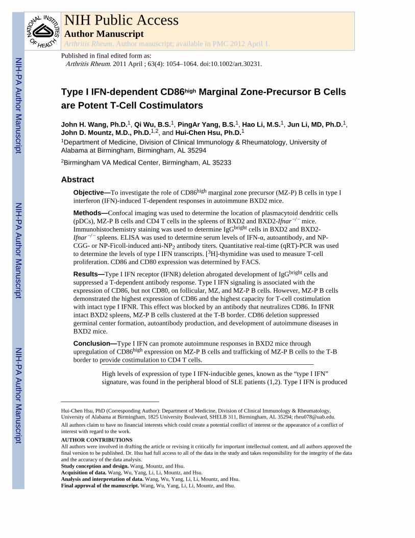
Type I IFN-dependent CD86high Marginal Zone-Precursor B Cellsare Potent T-Cell Costimulators
John H. Wang, Ph.D.1, Qi Wu, B.S.1, PingAr Yang, B.S.1, Hao Li, M.S.1, Jun Li, MD, Ph.D.1,John D. Mountz, M.D., Ph.D.1,2, and Hui-Chen Hsu, Ph.D.11Department of Medicine, Division of Clinical Immunology & Rheumatology, University ofAlabama at Birmingham, Birmingham, AL 352942Birmingham VA Medical Center, Birmingham, AL 35233
AbstractObjective—To investigate the role of CD86high marginal zone precursor (MZ-P) B cells in type Iinterferon (IFN)-induced T-dependent responses in autoimmune BXD2 mice.
Methods—Confocal imaging was used to determine the location of plasmacytoid dendritic cells(pDCs), MZ-P B cells and CD4 T cells in the spleens of BXD2 and BXD2-Ifnar−/− mice.Immunohistochemistry staining was used to determine IgGbright cells in BXD2 and BXD2-Ifnar−/− spleens. ELISA was used to determine serum levels of IFN-α, autoantibody, and NP-CGG- or NP-Ficoll-induced anti-NP2 antibody titers. Quantitative real-time (qRT)-PCR was usedto determine the levels of type I IFN transcripts. [3H]-thymidine was used to measure T-cellproliferation. CD86 and CD80 expression was determined by FACS.
Results—Type I IFN receptor (IFNR) deletion abrogated development of IgGbright cells andsuppressed a T-dependent antibody response. Type I IFN signaling is associated with theexpression of CD86, but not CD80, on follicular, MZ, and MZ-P B cells. However, MZ-P B cellsdemonstrated the highest expression of CD86 and the highest capacity for T-cell costimulationwith intact type I IFNR. This effect was blocked by an antibody that neutralizes CD86. In IFNRintact BXD2 spleens, MZ-P B cells clustered at the T-B border. CD86 deletion suppressedgerminal center formation, autoantibody production, and development of autoimmune diseases inBXD2 mice.
Conclusion—Type I IFN can promote autoimmune responses in BXD2 mice throughupregulation of CD86high expression on MZ-P B cells and trafficking of MZ-P B cells to the T-Bborder to provide costimulation to CD4 T cells.
High levels of expression of type I IFN-inducible genes, known as the “type I IFN”signature, was found in the peripheral blood of SLE patients (1,2). Type I IFN is produced
Hui-Chen Hsu, PhD (Corresponding Author): Department of Medicine, Division of Clinical Immunology & Rheumatology,University of Alabama at Birmingham, 1825 University Boulevard, SHELB 311, Birmingham, AL 35294; [email protected] authors claim to have no financial interests which could create a potential conflict of interest or the appearance of a conflict ofinterest with regard to the work.AUTHOR CONTRIBUTIONSAll authors were involved in drafting the article or revising it critically for important intellectual content, and all authors approved thefinal version to be published. Dr. Hsu had full access to all of the data in the study and takes responsibility for the integrity of the dataand the accuracy of the data analysis.Study conception and design. Wang, Mountz, and Hsu.Acquisition of data. Wang, Wu, Yang, Li, Li, Mountz, and Hsu.Analysis and interpretation of data. Wang, Wu, Yang, Li, Li, Mountz, and Hsu.Final approval of the manuscript. Wang, Wu, Yang, Li, Li, Mountz, and Hsu.
NIH Public AccessAuthor ManuscriptArthritis Rheum. Author manuscript; available in PMC 2012 April 1.
Published in final edited form as:Arthritis Rheum. 2011 April ; 63(4): 1054–1064. doi:10.1002/art.30231.
NIH
-PA Author Manuscript
NIH
-PA Author Manuscript
NIH
-PA Author Manuscript

primarily by CD11clow-expressing dendritic cells (DCs) that express the phenotypic markersB220, Gr-1, and a more specific surface marker, the plasmacytoid dendritic cell antigen(PDCA-1) (3,4). These DCs are known as plasmacytoid dendritic cells (pDCs) (3–6).
T-dependent antibody response requires antigen presentation by major histocompatibilitycomplex II and costimulation via CD80 or CD86 expressed on antigen-presenting cells (7).Studies of human peripheral blood have found increased expression levels of CD80 andCD86 on B cells from SLE patients compared to healthy individuals (8,9). The severity oflupus disease is positively correlated with the expression levels of CD80 and CD86 (9).However, only CD86 expression was significantly elevated in lupus patients with renaldisease, the hallmark of SLE, while differences in CD80 levels were statisticallyinsignificant (10). Other studies have corroborated the importance of CD86 but not CD80 byfinding that only CD86 expression on B cells is elevated in patients with inactive SLE andthat its level is further elevated in conjunction with active disease (11,12).
We previously demonstrated that BXD2 mice spontaneously produce pathogenicautoantibodies that can induce and exacerbate glomerulonephritis and erosive arthritis (13).Blocking of the interaction of B7-CD28 in young BXD2 mice using AdCTLA4-Igdramatically suppressed the expression of activation-induced cytidine deaminase (AID),which is the essential enzyme to promote B-cell somatic hypermutation (SHM) and class-switch recombination (CSR) (14). This treatment also prevented the development of bothnephritis and arthritis in BXD2 mice (14). Although CD86 was found to be increased inBXD2 B cells (14), it has not been specifically determined if the increased expression ofCD86 is associated with the autoimmune pathogenesis in BXD2 mice. It is also unclear as toat what stage(s) of the germinal center (GC) development that CD86high B cells encounterCD28+ CD4 T cells and what mechanisms are involved in driving the encounter of thesecells.
Recently, we have identified a subpopulation of B cells that have the surface expression ofCD1dhighIgMhighCD21highCD23high in BXD2 mice, which are significantly increased in thespleens of BXD2 mice at the expense of reduced marginal zone (MZ) B cell counts (15).This population of CD19+ splenocytes is commonly known as the marginal zone precursor(MZ-P) B cells (16). The immunopathogenesis for MZ-P B cells in BXD2 mice wasdemonstrated by their high-affinity binding for an exogenous antigen, TNP-Ficoll (15).Importantly, our previous study also showed that high levels of type I IFN produced bypDCs in the marginal sinus plays an important role in upregulating CD69 and facilitatingTNP+ MZ-P B-cell migration to the light zone border of GCs (15).
In the current study, we examined the role of type I IFN in regulating the surface expressionof costimulatory molecules, CD80 and CD86, on follicular (FO), MZ, and MZ-P B cells. Wealso determined if type I IFN signaling is required for MZ-P localization at the critical T-Bborder before a spontaneous GC response is initiated. Our present results show that type IIFN-induced upregulation of CD86 on MZ-P B cells and direction of MZ-P migration to theT-B border is important in promoting an IgG antibody response and autoimmune disease.
MATERIALS and METHODSMice
Female homozygous C57BL/6J (B6), BXD2 recombinant inbred, and B6-Cd86−/− micewere obtained from The Jackson Laboratory (Bar Harbor, ME); B6.Ifnar−/− mice wereobtained from Dr. Jocelyn Demengeot (Instituto Gulbenkian de Ciência, Oeiras, Portugal).BXD2-Ifnar−/− and BXD2-Cd86−/− were generated by backcrossing B6-Ifnar−/− and B6-Cd86−/− with BXD2 mice for seven generations. All mice were housed in the University of
Wang et al. Page 2
Arthritis Rheum. Author manuscript; available in PMC 2012 April 1.
NIH
-PA Author Manuscript
NIH
-PA Author Manuscript
NIH
-PA Author Manuscript

Alabama at Birmingham (UAB) Mouse Facility under specific pathogen-free conditions andall procedures were approved by the UAB Institutional Animal Care and Use Committee.Unless, specified, all mice were sacrificed at 8 to 12 weeks of age.
In vivo treatments and ELISA assaysFor induction of IFN-α, 5 µg of CpG-A oligonucleotide was dissolved with 30 µL DOTAPLiposomal Transfection Reagent (F. Hoffman-La Roche, Boulder, CO) in 120 µLphosphate-buffered saline (PBS). The CpG-A:DOTAP mixture was intravenously injectedinto mice. IFN-α ELISA kit (PBL Biomedical, Piscataway, NJ) was used to assay serumIFN-α levels.
Immunizations were carried out by intraperitoneal injection of 50 µg chicken γ-globulinhaptenated with 4-hydroxy-3-nitrophenylacetyl (NP-CGG; BioSearch Technologies)adsorbed onto 1.3 mg of alum (Sigma-Aldrich) in a total volume of 100 µL of PBS or 50 µgNP-Ficoll in PBS into mice. Antibodies to NP2-BSA coated plates were assayed by ELISAusing enzyme HRP-linked anti-mouse IgM (Southern Biotech), anti-mouse IgG2b (SouthernBiotech), and anti-mouse IgG2c (Southern Biotech) antibodies as we previously described(17).
Urinary albumin was analyzed using a competitive Albuwell M ELISA kit (Exocell Inc)according the manufacturer’s protocol (13).
Flow cytometryFor the analysis of costimulatory molecules, PE-anti-CD80 (Biolegend, San Diego, CA) orPE-anti-CD86 (Biolegend) antibodies were used. For the analysis of B-cell subpopulations,Alexa700-anti-CD19 (Biolegend), PerCP-Cy5.5-anti-CD93/AA4 (Biolegend), PE-Cy7-anti-IgM (Southern Biotechnology Associates, Birmingham, AL), FITC-anti-CD21 (Biolegend),and APC-anti-CD23 (Biolegend) antibodies were used. GC B cells were analyzed using PE-anti-Fas (Biolegend) and biotin-peanut agglutinin (PNA) (Vector Laboratories, Burlingame,CA) followed by APC-conjugated streptavidin (Biolegend). All cells are fixed in 1%paraformaldehyde/FACS solution before analysis by flow cytometry using a BD LSR IIflow cytometer (BD Biosciences). The analysis was performed using FlowJo Software (TreeStar, Ashland, OR). Forward-angle light scattering was used to exclude dead and aggregatedcells.
For analyses of FO, MZ, and MZ-P B cells, CD19+ splenocytes were first gated. As shownin Supporting Fig. 1, for FO, MZ, and MZ-P B cells,AA4−IgMlowIgDhighCD21lowCD23high, AA4−IgMhighCD21highCD23low, andAA4−IgMhighCD21highCD23high splenocytes, respectively, were gated using the methoddescribed by Allman and Pillai (18). For dose-response to IFN-α, FACS sorted FO, MZ, andMZ-P B cells were cultured with increasing doses of IFN-α (PBL Biomedical Laboratories)at 0, 200, 400, and 800 U/mL for 12 hours prior to flow cytometry analysis for CD80+ orCD86+ cells.
Histological and confocal imaging analysisFrozen confocal imaging was used for the detection of CD1d, CD23, CD4, IgM, and PNA(15). For the analysis of pDC and marginal sinus location, spleen frozen sections wereblocked with 10% normal rat serum and were stained with anti-PDCA1 (rat IgG2b)(Miltenyi Biotec, Auburn, CA), followed by Alexa 555 dye-conjugated anti-rat IgG(Invitrogen) and Alexa488-anti-MAdCAM-1 (Biolegend). All tissue sections were mountedin Fluormount G (Southern Biotechnology Associates) and viewed with a Leica DM IRBE
Wang et al. Page 3
Arthritis Rheum. Author manuscript; available in PMC 2012 April 1.
NIH
-PA Author Manuscript
NIH
-PA Author Manuscript
NIH
-PA Author Manuscript

inverted Nomarski/epifluorescence microscope outfitted with LeicaTCS NT (LeicaMicrosystems, Bannockburn, IL) laser confocal optics.
For analysis of IgM or IgG deposit in the spleens and IgG deposit in the kidneys,immunohistochemistry was performed on 10% formalin-fixed spleen tissue (4 µM) sections.Endogenous peroxidase quenching was performed by addition of 3% H2O2 followed by0.25% pepsin-antigen retrieval. Tissues were subsequently blocked with 1.5% BSA,followed by incubation with HRP-linked anti-mouse IgM (Southern Biotech) and/or anti-mouse IgG (Southern Biotech) and treated with 3,3',5,5'-tetramethylbenzidine (Sigma).Tissues were imaged under an Olympus BX41 System Microscope. Quantitation of IgG+
glomeruli was carried out using the ImageJ program (developed at the U.S. NationalInstitutes of Health and available on the Internet at http://rsb.info.nih.gov/nih-image/).Background intensity was subtracted for each image.
Costimulatory proliferation assayB cells from the spleens were enriched by positive selection using magnetic anti-CD19microbeads (Miltenyi Biotech). Subsequently, CD19+ B cells were sorted by flow cytometryinto FO, MZ, and MZ-P B cells by the combinations of surface markers specified above viaFACS sorting. For IFN-α stimulation, sorted B cells were incubated with IFN-α (400 U/mL,PBL Biomedical Laboratories) for 12 hours and washed with culture medium twice prior toirradiation. For costimulation, irradiated (3000 Rad) FO, MZ, or MZ-P B cells (5 × 10⁵)were co-cultured with 5 ×10⁵ MACS sorted CD4 T cells derived from the spleens of BXD2mice at a 1:1 ratio for 48 hours in triplicate round-bottom wells in 96-well plates (Costar) inthe presence of anti-CD3 (2.5 µg/mL, clone 145-2C11) plus anti-CD86 (10 µg/mL, cloneGL-1), anti-CD80 (10 µg/mL, clone 16-10A1) or rat IgG2a κ isotype control (10 µg/mL,clone RTK2758). The proliferative response was measured by a standard 3H-thymidineincorporation assay (17) in which 1 µCi of 3H-thymidine (Perkin Elmer) was added to eachwell during the last 12 h of culture
RNA QuantitationThe expression of type I IFN isoforms, Ifna1, Ifna4, Ifna11, and Ifnb in peripheral blood wasdetermined using a quantitative real-time PCR (qRT-PCR) method as we previouslydescribed (15,17). The qRT-PCR mixtures contained SYBR Green PCR Master Mix (Bio-Rad) with the following primers: Ifna1 forward: agtgagctgacccagcagat; Ifna1 reverse:ggtggaggtcattgcagaat; Ifna4 forward: tctgcaatgacctccatcag; Ifna4 reverse:tatgtcctcacagccagcag; Ifna11 forward: cccagcagatcttgaacctc; Ifna11 reverse:ggtggaggtcattgcagaat; Ifnb forward: ctccaccacagccctctc; Ifnb reverse: catcttctccgtcatctccatag;Actin forward: cgttgacatccgta; Actin reverse: ggaaggtggacagtgagg.
Statistical analysisAll results were shown as mean ± standard error of the mean (SEM). A two-tail t test wasused when two groups were compared for statistical differences. ANOVA test was usedwhen more than 2 groups were compared for statistical differences. P values less than 0.05were considered significant.
RESULTSIncreased type I IFN/pDCs in lupus-prone BXD2 mice
There was increased clustering of pDCs (PDCA1+, green) in BXD2 mice located in the MZand outside the FO, whose boundary is demarcated by the marginal sinus, which areMAdCAM-1+ (red) (19) compared to B6 mice (Figure 1A). Further, we observeddeveloping GCs in BXD2 mice that were located inside the FO, with clusters of these pDCs
Wang et al. Page 4
Arthritis Rheum. Author manuscript; available in PMC 2012 April 1.
NIH
-PA Author Manuscript
NIH
-PA Author Manuscript
NIH
-PA Author Manuscript
at the MZ (Figure 1B). Intravenous treatment with CpG-A, a known toll-like receptor 9(TLR9) ligand, in BXD2 mice dramatically increased IFN-α serum levels in the first 2hours, peaking at 4 hours, and dramatically declined at 12 hours (Figure 1C). Thoughsimilarly-treated B6 mice also showed a similar IFN-α level kinetics, these levels weresignificantly lower than those from BXD2 mice (Figure 1C).
Peripheral blood cells from untreated BXD2 and B6 mice at the different ages were assayedfor different type I IFN isoform transcripts (Figure 1D). In BXD2 mice, the age groups <2months, 9–12 months, and >1 year of age showed significantly lower Ifna1, Ifna4, andIfna11 transcript levels than do the 3–6 month age group (Figure 1D). With the exception ofIfnb, all type I IFN transcripts were significantly lower in B6 mice compared to BXD2 micein matched age groups <2 months, and 3–6 months of age (Figure 1D). At older ages (> 9months), we did not detect significant differences (Figure 1D).
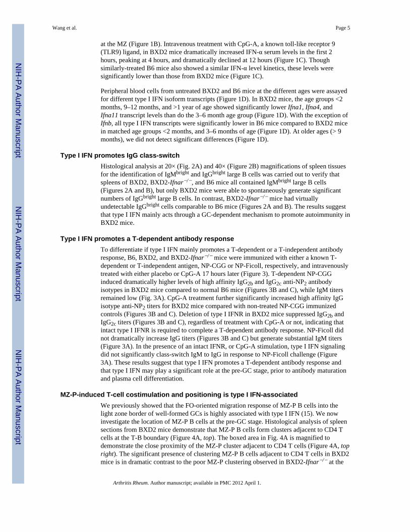
Type I IFN promotes IgG class-switchHistological analysis at 20× (Fig. 2A) and 40× (Figure 2B) magnifications of spleen tissuesfor the identification of IgMbright and IgGbright large B cells was carried out to verify thatspleens of BXD2, BXD2-Ifnar−/−, and B6 mice all contained IgMbright large B cells(Figures 2A and B), but only BXD2 mice were able to spontaneously generate significantnumbers of IgGbright large B cells. In contrast, BXD2-Ifnar−/− mice had virtuallyundetectable IgGbright cells comparable to B6 mice (Figures 2A and B). The results suggestthat type I IFN mainly acts through a GC-dependent mechanism to promote autoimmunity inBXD2 mice.
Type I IFN promotes a T-dependent antibody responseTo differentiate if type I IFN mainly promotes a T-dependent or a T-independent antibodyresponse, B6, BXD2, and BXD2-Ifnar−/− mice were immunized with either a known T-dependent or T-independent antigen, NP-CGG or NP-Ficoll, respectively, and intravenouslytreated with either placebo or CpG-A 17 hours later (Figure 3). T-dependent NP-CGGinduced dramatically higher levels of high affinity IgG2b and IgG2c anti-NP2 antibodyisotypes in BXD2 mice compared to normal B6 mice (Figures 3B and C), while IgM titersremained low (Fig. 3A). CpG-A treatment further significantly increased high affinity IgGisotype anti-NP2 titers for BXD2 mice compared with non-treated NP-CGG immunizedcontrols (Figures 3B and C). Deletion of type I IFNR in BXD2 mice suppressed IgG2b andIgG2c titers (Figures 3B and C), regardless of treatment with CpG-A or not, indicating thatintact type I IFNR is required to complete a T-dependent antibody response. NP-Ficoll didnot dramatically increase IgG titers (Figures 3B and C) but generate substantial IgM titers(Figure 3A). In the presence of an intact IFNR, or CpG-A stimulation, type I IFN signalingdid not significantly class-switch IgM to IgG in response to NP-Ficoll challenge (Figure3A). These results suggest that type I IFN promotes a T-dependent antibody response andthat type I IFN may play a significant role at the pre-GC stage, prior to antibody maturationand plasma cell differentiation.
MZ-P-induced T-cell costimulation and positioning is type I IFN-associatedWe previously showed that the FO-oriented migration response of MZ-P B cells into thelight zone border of well-formed GCs is highly associated with type I IFN (15). We nowinvestigate the location of MZ-P B cells at the pre-GC stage. Histological analysis of spleensections from BXD2 mice demonstrate that MZ-P B cells form clusters adjacent to CD4 Tcells at the T-B boundary (Figure 4A, top). The boxed area in Fig. 4A is magnified todemonstrate the close proximity of the MZ-P cluster adjacent to CD4 T cells (Figure 4A, topright). The significant presence of clustering MZ-P B cells adjacent to CD4 T cells in BXD2mice is in dramatic contrast to the poor MZ-P clustering observed in BXD2-Ifnar−/− at the
Wang et al. Page 5
Arthritis Rheum. Author manuscript; available in PMC 2012 April 1.
NIH
-PA Author Manuscript
NIH
-PA Author Manuscript
NIH
-PA Author Manuscript

T-B boundary (Figure 4A, bottom). Instead, in BXD2-Ifnar−/− mice, the pool of B cellsclustered at the T-B border is biased towards a predominant FO B cells (Figure 4A, bottom).
To test the CD4 T cell costimulatory capability of each of the B-cell subpopulations, wesorted and co-cultured irradiated B-cell subpopulations from BXD2 or BXD2-Ifnar−/− micewith anti-CD3 stimulated CD4 T cells. Consistent with the literature (20), MZ B cells makebetter costimulatory agents than do FO B cells (Figure 4B). MZ-P B cells from BXD2 mice,however, provided the greatest costimulation of CD4 T cells, compared to costimulationprovided by MZ or FO B cells (Figure 4B). MZ-P B cells from BXD2-Ifnar−/− micedemonstrated significantly attenuated T-cell costimulation compared to BXD2 MZ-P B cells(Figure 4B). MZ B cells also displayed attenuated costimulatory capability when type IIFNR was deleted (Figure 4B).
Consistent with these results, there was a significantly increased percentage of CD86high
MZ-P compared to MZ, and FO B cells in BXD2 mice (Figure 4C). BXD2-Ifnar−/− miceexhibited an approximate 50% decrease in CD86high expression by all B-cell subpopulations(Figure 4C). Interestingly, BXD2-Ifnar−/− MZ-P B cells maintained a higher basal CD86surface expression than did their MZ and FO B cell counterparts (Figure 4C). Both MZ andMZ-P B cells maintained higher CD80 expression than did FO B cells (Figure 4D).However, CD80 expression in all 3 subpopulations of B cells was not significantly differentbetween BXD2 and BXD2-Ifnar−/− mice (Figure 4D).
Type I IFN stimulates the costimulatory function of MZ-P via the induction of CD86To test whether IFN-α has a direct effect on CD86 surface expression, splenic MZ-P, MZ,and FO B cells from BXD2 and BXD2-Ifnar−/− were sorted and left unstimulated orstimulated IFN-α. CD86 surface expression on all three B-cell subpopulations wasdetermined 12 hr after stimulation with IFN-α (Figure 5A). MZ-P B cells exhibited thehighest expression of CD86 compared with either FO or MZ B cells in response to IFN-α.Type I IFNR deletion showed significant blunting of CD86 expression, especially on MZ-PB cells (Figure 5A). In contrast, CD80 surface expression on FO, MZ, or MZ-P B cells wasnot dependent on IFN-α signaling (Figure 5A).
Stimulation of FO, MZ, and MZ-P B cells by increasing doses of IFN-α (0, 200, 400, 800 U/mL) demonstrated a dose-related upregulation of CD86 surface expression (Figure 5B, left)for FO, MZ, and MZ-P B cells, but most significantly on MZ-P B cells, whereas noupregulation was observed for CD80 surface expression (Figure 5B, right) on all three B cellsubpopulations.
To test whether type I IFN promotes CD4 T cell costimulatory capability of each of the Bcell subpopulations via increased expression of CD86, sorted B cells were stimulated withIFN-α and then co-cultured with anti-CD3 stimulated CD4 T cells, in the presence of ratIgG2a κ isotype control, anti-CD86, or anti-CD80. IFN-α pre-stimulated MZ-P B cellsexhibited the most dramatic effect in promoting anti-CD3-induced T-cell proliferativeresponse (Figure 5C), with lesser T-cell proliferative response by IFN-α stimulation of eitherMZ or FO B cells (Figure 5C). Anti-CD86 antibody effectively blocked T-cell costimulationby MZ-P B cells that were either with or without IFN-α pre-stimulation (Figure 5C). Incontrast, anti-CD80 did not significantly block T-cell costimulation provided by MZ-P Bcells (Figure 5C).
CD86 is essential for BXD2 autoimmune diseaseTo demonstrate that CD86 is a key pathogenic molecule in the induction of autoimmunedisease, the phenotype of BXD2-Cd86−/− mice was determined. CD86 deletion significantlyreduced spleen weight and cell count (Figure 6A). IgG autoantibodies to histone, BiP, and
Wang et al. Page 6
Arthritis Rheum. Author manuscript; available in PMC 2012 April 1.
NIH
-PA Author Manuscript
NIH
-PA Author Manuscript
NIH
-PA Author Manuscript

DNA, were significantly reduced in BXD2-Cd86−/− mice, compared with that in BXD2mice (Figure 6B). Histological analysis confirms that in BXD2-Cd86−/− mice, there was adramatically reduced generation of IgGbright cells in the spleen (Figure 6C). Flow cytometryand histological analyses showed significantly reduced PNA+ GC B cells in BXD2-Cd86−/−,compared with BXD2 mice (Figure 6D). Consistent with these findings, there weredramatically diminished mesangial proliferation, diminished IgG containing immunecomplexes deposited in the glomeruli of the kidney (Supporting Figure 2A) and significantlydiminished urinary albumin (Supporting Figure 2B) from BXD2-Cd86−/− mice at 10-mo-old, compared with age-matched BXD2 mice. BXD2-Cd86−/− mice also developeddramatically decreased erosive arthritis, characterized by significantly lower levels ofinflammatory infiltration, synovial hyperplasia, and marginal erosion, compared to arthritisdeveloped by wild-type BXD2 mice (Supporting Figure 2C and D).
DISCUSSIONType I IFN is known to be required for autoantibody production (21). Deletion of the type IIFNR eliminated the development of autoantibodies in various murine models of lupus (22–24). Though studies conducted in non-autoimmune hosts have been proposed as pathwaysby which type I IFNs drive antibody responses in lupus (21), the precise mechanism bywhich type I IFNs generate autoantibodies is not clear. The dominant paradigm is that type IIFNs generate autoantibodies in lupus by directly promoting the maturation of autoreactiveB cells into antibody-secreting plasmablasts (4,25). While this paradigm may hold true, ourstudies suggest additional or alternative mechanisms by which type I IFNs generateplasmablasts and/or plasma cells by promoting T-dependent autoantibody responses.
We have found increased pDCs located in clustering layers of cells in the MZ outside theFO, which is demarcated by MAdCAM-1. Consistent with a non-inflammatory phenotype,pDCs in B6 mice exhibited significantly less clustering in the MZ, and a more diffusedistribution inside the FO. However, in BXD2 mice, clustering pDCs were found in the MZ,consistent with an inflammatory environment in which immune complexes carrying TLR-binding deoxynucleoproteins may trigger pDC-induced type I IFNs and pDC clustering inthe MZ (26). Because pDCs are also systemically circulated between the peripheral bloodand the secondary lymphoid organs (27), significant amounts of type IFN transcripts werealso found in the peripheral blood in BXD2 mice. CpG-A induction further confirms thatBXD2 mice have the potential to generate high serum levels of IFN-α. Previously, we havedemonstrated that the spleens of BXD2 mice have increased counts of type I IFN producingpDCs compared to normal B6 mice (15). The mRNA levels of most Ifna species increaseuntil 6 months, at which age, the levels peak, and subsequently decline. Interestingly, thesemRNA levels increase and peak in the same age range as do certain autoantibodies (13),consistent with our findings that type I IFNs can modulate CD4 T-cell costimulation andinduce antibody responses. Declining type I IFN message levels in older BXD2 mice (>9months) may be due to senescing/maturing pDCs in response to antigen exposure (28) andin response to an increasing antigen-autoantibody load in aging BXD2 mice (13).
We observed that intact type I IFN signaling is required for formation of IgGbright large sizeplasmablast or plasma B cell formation. Deletion of type I IFNR in the BXD2 backgroundeliminated the presence of IgGbright B cells, while keeping the presence of IgMbright B cellsintact, a finding that would not be expected if type I IFNs were only responsible formaturing antibody-producing B cells into plasmablasts. Immunization with a T-dependentantigen but not a T-independent antigen shows that type I IFN can produce a high affinityIgG antibody response. Also, the T-dependent response was significantly dependent uponCpG-A induction of IFN-α. Deletion of type I IFNR, in spite of CpG-A treatment,suppressed IgG titers to baseline levels. Our findings suggest that type I IFN can promote
Wang et al. Page 7
Arthritis Rheum. Author manuscript; available in PMC 2012 April 1.
NIH
-PA Author Manuscript
NIH
-PA Author Manuscript
NIH
-PA Author Manuscript

the generation of plasmablasts or plasma B cells in addition to their effects to induce post-GC B cell maturation.
Previous results showed that costimulation of CD4 T cells that is blocked, for instance byCTLA4-Ig, which competitively binds to either CD80 or CD86, anergizes CD4 T cells (29).The finding that B cells from SLE human subjects display high levels of costimulatoryreceptors, especially CD86, corroborates the assertion that the T-dependent humoralresponse plays a prominent role in generating autoantibodies (8–12). BXD2 mice display anautoantibody response that is significantly dependent upon costimulation signaling broughtabout by CD80/86-CD28 interactions as AdCTLA4-Ig treatment in BXD2 mice almostcompletely blocked the expression of the gene encoding AID and autoantibody production(14). As demonstrated in the current study, CD86 expression is especially important in theautoimmune phenotype of BXD2 mice. Deletion of CD86 significantly inhibited GCformation, suppressed the presence of IgGbright class switched cells, attenuated autoantibodytiters, and obviated the natural course of glomerulonephritis and erosive arthritis in BXD2mice.
In the secondary lymphoid organs, such as the spleen, consist of a heterogeneous assortmentof B cells (30–32). Most of these consist of mature FO B cells and a smaller population ofMZ B cells, with a series of “transitional” B cells or “intermediates”, including the MZ-P Bcells (32). We have previously shown that this FO-oriented migration of MZ-P B cells canbe regulated via the type I IFN-induced CD69-dependent S1P1 down-regulation and thatMZ-P B cells can transport antigens to the light zone end of existing GCs (15). In thepresent study, we further demonstrated that MZ-P B cells exhibit close physical contact withCD4 T cells at the T-B border during the pre-GC stage. The deletion of type I IFNRredistributes MZ-P B cells away from this T-B border and towards the MZ. This result,together with our previous finding (15), suggest that type I IFN plays an important role topromote the FO-oriented migration of MZ-P B cells both before and after the onset of thespontaneous GC response is initiated in BXD2 mice.
The present study further shows that an important function of type I IFN-promoted MZ-P Bcells at the T-B border is for the provision of costimulation to CD4 T cells. MZ B cells havebeen reported to express higher levels of CD86 and higher costimulation of CD4 T cellscompared to FO B cells (20). We observed that MZ-P B cells have higher CD86 expressioncompared to either FO or MZ, and MZ-P B cells provide even stronger costimulation toCD4 T cells than MZ B cells. Significantly, CD86 expression on B cells is under theregulation of type I IFN, while CD80 is not. Disruption of type I IFN signaling loweredCD86 expression on FO, MZ, and MZ-P B cells. Although MZ-P B cells retained higherlevels of CD86 in spite of disrupted type I IFN signaling compared with MZ or FO B cells,diminished CD86 expression with type I IFNR deletion significantly decreased MZ-P-induced T-cell costimulation to levels comparable to those induced by FO or MZ B cells.These results suggest that there is a critical CD86 expression threshold below which T cellcostimulation becomes ineffective, and that the signal provided by type I IFN is important toelevate the expression of CD86 to levels above this threshold. Anti-CD28 costimulationnormalized the costimulatory effects of MZ-P B cells, with or without IFN-α pre-stimulation, on CD4 T cell proliferation (data not shown), suggesting IFN-α mainly actthrough the CD86 pathway to enhance the costimulatory function of MZ-P B cells.Interestingly, CD80 expression is not under the regulation of type I IFN, and appears to notoffer as strong a costimulatory molecule compared with CD86, at least in BXD2 mice. Ourobservation that these highly potent CD86high MZ-P costimulators cluster at the splenic T-Bborder in BXD2 mice provides the geographical advantage of providing costimulation in thevery initial stages of GC formation.
Wang et al. Page 8
Arthritis Rheum. Author manuscript; available in PMC 2012 April 1.
NIH
-PA Author Manuscript
NIH
-PA Author Manuscript
NIH
-PA Author Manuscript
In summary, our study proposes that type I IFN can offer a new avenue by which T-dependent antibody responses are generated. First, type I IFN is critical in bringing MZ-P Bcells to the T-B border into the FO interior at the pre-GC stage. Second, type I IFNsignificantly increases the levels of CD86 on MZ-P B cells, providing potent costimulationto activated CD4 T cells. This dual-level function of type I IFN prior to the formation ofGCs offers new insights into how type I IFNs can induce T-dependent autoreactive antibodyresponses in lupus-prone BXD2 mice and ultimately, perhaps, in human SLE.
Supplementary MaterialRefer to Web version on PubMed Central for supplementary material.
AcknowledgmentsWe thank Dr. Jocelyn Demengeot at the Instituto Gulbenkian de Ciência in Oeiras, Portugal to provide B6-Ifnar−/−mice. We thank Ms. Enid Keyser of the Analytic and Preparative Cytometry Facility - UAB Rheumatic DiseasesCore Facility (P30 AR48311) and Mr. Marion L. Spell of the UAB Center for AIDS FACS Core (P30 AI027767)for operating the FACS instrument. Confocal imaging was carried out at the Arthritis and Musculoskeletal DiseaseCenter High Resolution Imaging Facility (P30 AR48311). P30 AI027767-23, P30 AI027767-24, and P30AI27767-22S1 (PI: Michael Saag)
This work was supported by the American College of Rheumatology Research and Education Foundation for theWithin Our Reach: Finding a Cure for Rheumatoid Arthritis campaign, the Alliance for Lupus Research – TargetIdentification in Lupus program, the Department of Veterans Affairs Merit Review Grant 1I01BX000600-01,Daiichi-Sankyo Co., Ltd, the National Institutes of Health Grants 1AI 071110-01A1 and ARRA3RO1AI71110-02S1 (all to J.D.M.), the Lupus Research Institute Novel Research project and the ArthritisInvestigator Award supported by the Arthritis Foundation (to H.-C.H).
REFERENCES1. Bengtsson AA, Sturfelt G, Truedsson L, Blomberg J, Alm G, Vallin H, et al. Activation of type I
interferon system in systemic lupus erythematosus correlates with disease activity but not withantiretroviral antibodies. Lupus. 2000; 9(9):664–671. [PubMed: 11199920]
2. Bennett L, Palucka AK, Arce E, Cantrell V, Borvak J, Banchereau J, et al. Interferon andgranulopoiesis signatures in systemic lupus erythematosus blood. J Exp Med. 2003; 197(6):711–723. [PubMed: 12642603]
3. Nakano H, Yanagita M, Gunn MD. CD11c(+)B220(+)Gr-1(+) cells in mouse lymph nodes andspleen display characteristics of plasmacytoid dendritic cells. J Exp Med. 2001; 194(8):1171–1178.[PubMed: 11602645]
4. Colonna M, Trinchieri G, Liu YJ. Plasmacytoid dendritic cells in immunity. Nat Immunol. 2004;5(12):1219–1226. [PubMed: 15549123]
5. Asselin-Paturel C, Brizard G, Pin JJ, Briere F, Trinchieri G. Mouse strain differences inplasmacytoid dendritic cell frequency and function revealed by a novel monoclonal antibody. JImmunol. 2003; 171(12):6466–6477. [PubMed: 14662846]
6. Siegal FP, Kadowaki N, Shodell M, Fitzgerald-Bocarsly PA, Shah K, Ho S, et al. The nature of theprincipal type 1 interferon-producing cells in human blood. Science. 1999; 284(5421):1835–1837.[PubMed: 10364556]
7. Wang S, Chen L. T lymphocyte co-signaling pathways of the B7-CD28 family. Cell Mol Immunol.2004; 1(1):37–42. [PubMed: 16212919]
8. Wong CK, Lit LC, Tam LS, Li EK, Lam CW. Aberrant production of soluble costimulatorymolecules CTLA-4, CD28, CD80 and CD86 in patients with systemic lupus erythematosus.Rheumatology (Oxford). 2005; 44(8):989–994. [PubMed: 15870153]
9. Dolff S, Wilde B, Patschan S, Durig J, Specker C, Philipp T, et al. Peripheral circulating activated b-cell populations are associated with nephritis and disease activity in patients with systemic lupuserythematosus. Scand J Immunol. 2007; 66(5):584–590. [PubMed: 17868260]
Wang et al. Page 9
Arthritis Rheum. Author manuscript; available in PMC 2012 April 1.
NIH
-PA Author Manuscript
NIH
-PA Author Manuscript
NIH
-PA Author Manuscript

10. Liu W, Li J. Expression of CD80/CD86 and CTLA-4 mRNA in peripheral blood mononuclearcells of the patients with systemic lupus erythematosus. J Huazhong Univ Sci Technolog Med Sci.2004; 24(3):247–249. [PubMed: 15315339]
11. Bijl M, Horst G, Limburg PC, Kallenberg CG. Expression of costimulatory molecules onperipheral blood lymphocytes of patients with systemic lupus erythematosus. Ann Rheum Dis.2001; 60(5):523–526. [PubMed: 11302879]
12. Folzenlogen D, Hofer MF, Leung DY, Freed JH, Newell MK. Analysis of CD80 and CD86expression on peripheral blood B lymphocytes reveals increased expression of CD86 in lupuspatients. Clin Immunol Immunopathol. 1997; 83(3):199–204. [PubMed: 9175908]
13. Hsu HC, Zhou T, Kim H, Barnes S, Yang P, Wu Q, et al. Production of a novel class ofpolyreactive pathogenic autoantibodies in BXD2 mice causes glomerulonephritis and arthritis.Arthritis Rheum. 2006; 54(1):343–355. [PubMed: 16385526]
14. Hsu HC, Wu Y, Yang P, Wu Q, Job G, Chen J, et al. Overexpression of activation-inducedcytidine deaminase in B cells is associated with production of highly pathogenic autoantibodies. JImmunol. 2007; 178(8):5357–5365. [PubMed: 17404321]
15. Wang JH, Li J, Wu Q, Yang P, Pawar RD, Xie S, et al. Marginal zone precursor B cells as cellularagents for type I IFN-promoted antigen transport in autoimmunity. J Immunol. 2010; 184(1):442–451. [PubMed: 19949066]
16. Srivastava B, Quinn WJ 3rd, Hazard K, Erikson J, Allman D. Characterization of marginal zone Bcell precursors. J Exp Med. 2005; 202(9):1225–1234. [PubMed: 16260487]
17. Hsu HC, Yang P, Wang J, Wu Q, Myers R, Chen J, et al. Interleukin 17-producing T helper cellsand interleukin 17 orchestrate autoreactive germinal center development in autoimmune BXD2mice. Nat Immunol. 2008; 9(2):166–175. [PubMed: 18157131]
18. Allman D, Pillai S. Peripheral B cell subsets. Curr Opin Immunol. 2008; 20(2):149–157. [PubMed:18434123]
19. Kraal G, Schornagel K, Streeter PR, Holzmann B, Butcher EC. Expression of the mucosal vascularaddressin, MAdCAM-1, on sinus-lining cells in the spleen. Am J Pathol. 1995; 147(3):763–771.[PubMed: 7677187]
20. Attanavanich K, Kearney JF. Marginal zone, but not follicular B cells, are potent activators ofnaive CD4 T cells. J Immunol. 2004; 172(2):803–811. [PubMed: 14707050]
21. Ronnblom L, Alm GV, Eloranta ML. Type I interferon and lupus. Curr Opin Rheumatol. 2009;21(5):471–477. [PubMed: 19525849]
22. Agrawal H, Jacob N, Carreras E, Bajana S, Putterman C, Turner S, et al. Deficiency of type I IFNreceptor in lupus-prone New Zealand mixed 2328 mice decreases dendritic cell numbers andactivation and protects from disease. J Immunol. 2009; 183(9):6021–6029. [PubMed: 19812195]
23. Nacionales DC, Kelly-Scumpia KM, Lee PY, Weinstein JS, Lyons R, Sobel E, et al. Deficiency ofthe type I interferon receptor protects mice from experimental lupus. Arthritis Rheum. 2007;56(11):3770–3783. [PubMed: 17968932]
24. Santiago-Raber ML, Baccala R, Haraldsson KM, Choubey D, Stewart TA, Kono DH, et al. Type-Iinterferon receptor deficiency reduces lupus-like disease in NZB mice. J Exp Med. 2003; 197(6):777–788. [PubMed: 12642605]
25. Jego G, Palucka AK, Blanck JP, Chalouni C, Pascual V, Banchereau J. Plasmacytoid dendriticcells induce plasma cell differentiation through type I interferon and interleukin 6. Immunity.2003; 19(2):225–234. [PubMed: 12932356]
26. Asselin-Paturel C, Brizard G, Chemin K, Boonstra A, O'Garra A, Vicari A, et al. Type I interferondependence of plasmacytoid dendritic cell activation and migration. J Exp Med. 2005; 201(7):1157–1167. [PubMed: 15795237]
27. Yoneyama H, Matsuno K, Zhang Y, Nishiwaki T, Kitabatake M, Ueha S, et al. Evidence forrecruitment of plasmacytoid dendritic cell precursors to inflamed lymph nodes through highendothelial venules. International Immunology. 2004; 16(7):915–928. [PubMed: 15159375]
28. McKenna K, Beignon AS, Bhardwaj N. Plasmacytoid dendritic cells: linking innate and adaptiveimmunity. J Virol. 2005; 79(1):17–27. [PubMed: 15596797]
Wang et al. Page 10
Arthritis Rheum. Author manuscript; available in PMC 2012 April 1.
NIH
-PA Author Manuscript
NIH
-PA Author Manuscript
NIH
-PA Author Manuscript
29. Mirshahidi S, Huang C-T, Sadegh-Nasseri S. Anergy in Peripheral Memory Cd4+ T Cells Inducedby Low Avidity Engagement of T Cell Receptor. The Journal of Experimental Medicine. 2001;194(6):719–732. [PubMed: 11560989]
30. Lopes-Carvalho T, Kearney JF. Development and selection of marginal zone B cells. ImmunolRev. 2004; 197:192–205. [PubMed: 14962196]
31. Martin F, Kearney JF. Positive selection from newly formed to marginal zone B cells depends onthe rate of clonal production, CD19, and btk. Immunity. 2000; 12(1):39–49. [PubMed: 10661404]
32. Pillai S, Cariappa A. The follicular versus marginal zone B lymphocyte cell fate decision. Nat RevImmunol. 2002; 9(11):767–777. [PubMed: 19855403]
ABBREVIATIONS
AID activation-induced cytidine deaminase
CSR class-switch recombination
FO follicular
GC germinal center
IC immune complexes
MAdCAM-1 mucosal addressin cell adhesion molecule-1
MZ marginal zone
MZ-P marginal zone precursor
NP-CGG 4-hydroxy-3-nitrophenylacetyl chicken gamma globulin
pDCs plasmacytoid dendritic cells
PNA peanut agglutinin
RA rheumatoid arthritis
SHM somatic hypermutation
SLE systemic lupus erythematosus
Wang et al. Page 11
Arthritis Rheum. Author manuscript; available in PMC 2012 April 1.
NIH
-PA Author Manuscript
NIH
-PA Author Manuscript
NIH
-PA Author Manuscript

Figure 1.Activated pDCs and type I IFN in BXD2 mice. A, Images of spleen sections taken from arepresentative BXD2 mouse and a B6 mouse stained with anti-PDCA1 (pDC, green),MAdCAM-1 (red). B, Representative spleen section from a BXD2 mouse with PNA+ GC(blue) inside follicles demarcated with MAdCAM-1 (red) and surrounded by layers of pDCs(green). C, CpG-A:DOTAP was i.v. injected into BXD2 and B6 mice as described inMaterials and Methods. Serum IFN-α levels assayed by ELISA collected at the indicatedtimes (N=3–6, **p<0.01, ***p<0.001 between B6 and BXD2 at the same time point). D,RNA was extracted from peripheral blood cells. Transcripts of Ifna1, Ifna4, Ifna11, and Ifnbisoforms from BXD2 and B6 mice at different age groups were quantitated relative to copycounts of Actin, as described in Materials and Methods (N=6, *p<0.05, **p<0.01,***p<0.001 noted with BXD2 age groups were compared to BXD2 at the 3–6 months ofage, or in the case of B6, **p<0.01, and ***p<0.001 compared to age group-matched BXD2mice).
Wang et al. Page 12
Arthritis Rheum. Author manuscript; available in PMC 2012 April 1.
NIH
-PA Author Manuscript
NIH
-PA Author Manuscript
NIH
-PA Author Manuscript

Figure 2.Type I IFNR deletion abrogates IgG class-switch in 4-mo-old BXD2 mice. Representativespleen sections of BXD2, BXD2-Ifnar−/−, and B6 mice were stained with anti-IgM and anti-IgG antibodies as described in Materials and Methods. A, 20× objective lens magnificationof images of spleen sections showing the presence of follicles and IgMbright or IgGbright Bcells. B, 40× objective lens magnification of images of spleen sections, denoted with respectto the presence of a follicle (FO) whose boundary is denoted by broken lines.
Wang et al. Page 13
Arthritis Rheum. Author manuscript; available in PMC 2012 April 1.
NIH
-PA Author Manuscript
NIH
-PA Author Manuscript
NIH
-PA Author Manuscript

Figure 3.Type I IFNR deletion abrogates the T-dependent antibody response. B6, BXD2, and BXD2-Ifnar−/− mice were immunized with either NP-CGG or NP-Ficoll. Seventeen hours later, themice were either treated with placebo or CpG-A as described in Materials and Methods.Sera were collected on day 17 after immunization. IgM (A), IgG2b (B), IgG2c (C) titers ofanti-NP2, as measured by O.D. units, were assayed by ELISA (N=6, *p<0.05, **p<0.01 forCpG-A treated vs untreated BXD2 mice, *p<0.05, **p<0.01, ***p<0.001 for B6 or BXD2-Ifnar−/− vs. BXD2 mice).
Wang et al. Page 14
Arthritis Rheum. Author manuscript; available in PMC 2012 April 1.
NIH
-PA Author Manuscript
NIH
-PA Author Manuscript
NIH
-PA Author Manuscript

Figure 4.Potent costimulatory MZ-P B cells at T-B border at the pre-GC stage depends on type I IFN.A, Histological analysis of FO (CD23+CD1d−, red), MZ (CD23−CD1d+, green), MZ-P(CD23+CD1d+, yellow) B cells, and CD4 (blue) T cells located on a representative spleentissue section from a representative BXD2 and a BXD2-Ifnar−/− mouse. CD4 and B cellsare denoted as T and B, respectively. The boxed area in the left panels are magnified toshow junction at the T-B border, denoted as white dashed line. B, Tritiated thymidineproliferation assay of irradiated MZ-P, MZ, and FO B cells from BXD2 and BXD2-Ifnar−/−
mice co-cultured with CD4 T cells and an agonistic anti-CD3 antibody for 48 hours (N=3independent experiments from two mice per experiment, * p<0.05, **p<0.01, *** p<0.001between BXD2 and and BXD2-Ifnar−/− or between the indicated comparison). C, D,Representative flow cytometry analyses indicating CD86 (C) and CD80 (D) expression onsplenic MZ-P, MZ, and FO B cells from BXD2 and BXD2-Ifnar−/− mice. Results are shownas average ± SEM of CD86+ or CD80+ B-cell subpopulation from BXD2 (numerator) andBXD2-Ifnar−/− (denominator) mice (**p<0.01, ***p<0.001).
Wang et al. Page 15
Arthritis Rheum. Author manuscript; available in PMC 2012 April 1.
NIH
-PA Author Manuscript
NIH
-PA Author Manuscript
NIH
-PA Author Manuscript

Figure 5.IFN-α-induced costimulatory function of MZ-P B cells was suppressed by CD86neutralization. A, FACS sorted MZ-P, MZ, and FO B cells from BXD2 and BXD2-Ifnar−/−
mice were stimulated with medium or IFN-α (400 U/ml), as described in Materials andMethods. Results are shown as the representative FACS histograms showing the cell surfaceexpression of CD86 and CD80 in the indicated population of B cells. B, Bar graphs showingthe IFN-α-induced dose dependent induction of the expression of CD86 and CD80 on sortedMZ-P, MZ, and FO B cells. C, Tritiated thymidine proliferation assay of irradiated MZ-P,MZ, and FO B cells from BXD2 and BXD2-Ifnar−/− mice co-cultured with CD4 T cells andan agonistic anti-CD3 antibody for 48 hours. Sorted B cells were stimulated with or withoutIFN-α first as described in Materials and Methods. Cells were cultured in the presence of ratIgG2a κ isotype control (10 µg/ml), anti-CD86 (10 µg/ml), anti-CD80 (10 µg/ml). Allresults are shown as mean ± SEM (N=3 independent experiments from two mice perexperiment, * p<0.05, **p<0.01, *** p<0.001 between the indicated comparison).
Wang et al. Page 16
Arthritis Rheum. Author manuscript; available in PMC 2012 April 1.
NIH
-PA Author Manuscript
NIH
-PA Author Manuscript
NIH
-PA Author Manuscript

Figure 6.CD86 is critical for autoantibody and GC formation in BXD2 mice. Spleens from BXD2 andBXD2-Cd86−/− mice (10-mo-old) were harvested. A, Spleen weight and the total cell count,as determined by a hemocytometer, between BXD2, and BXD2-Cd86−/− mice are shown. B,Titers (O.D. absorbance units) of serum autoantibodies to histone, BiP, and DNA from B6,BXD2, and BXD2-Cd86−/− mice. C, Representative (1 of N=6) histological analysis ofspleen tissue sections from BXD2 and BXD2-Cd86−/− immunohistochemically stained asdescribed in Materials and Methods with anti-IgG antibody at 10× and 20× objective lensmagnifications. D, Left: Flow cytometry analysis of splenic GC B cells (Fas+PNA+, gatedon CD19+ B cells first) from BXD2 and BXD2-Cd86−/− mice with indicated percentages ofFas+PNA+ cells. Right: The corresponding representative (1 of N=6) histological analysis ofspleen sections from BXD2 and BXD2-Cd86−/− mice at 20× and 40× objective lensmagnifications. All results are shown as mean ± SEM (** p<0.01, ***p<0.001 betweenBXD2 vs BXD2-Cd86−/−, N=6).
Wang et al. Page 17
Arthritis Rheum. Author manuscript; available in PMC 2012 April 1.
NIH
-PA Author Manuscript
NIH
-PA Author Manuscript
NIH
-PA Author Manuscript
Copyright © 2022 FDOKUMEN