The role of the ICOS-B7h T cell costimulatory pathway in transplantation immunity
10
234 The Journal of Clinical Investigation | July 2003 | Volume 112 | Number 2 Introduction An efficient T cell response requires costimulatory signals delivered by APCs in addition to specific antigen signals acting on the T cell receptor (TCR). The prototypical cos- timulatory molecule CD28 provides a strong mitogenic signal, induces a large array of effector cytokines, and upregulates cell survival genes such as Bcl-x L (1–3). The CD40-CD154 pathway is another key pathway for T cell activation (4–6). However, it is apparent that T cell activation and transplant rejection can proceed in the absence of CD28-B7 or CD40-CD154 signals, since mice lacking CD28 are able to acutely reject cardiac allografts (7, 8), and CD154-deficient mice go on to develop chron- ic rejection (9). In addition, memory T cells (10, 11) and CD8 + T cells (12), both important mediators of allograft loss, are far less dependent on these costimulatory signals than naive T cells, making them less susceptible to co- stimulatory blocking strategies. Inducible costimulatory molecule (ICOS), another member of the CD28 superfamily, has unique roles in T cell activation and differentiation (13–15), splenic germi- nal center formation, and immunoglobulin class switch- ing (14, 16, 17). ICOS is inducible within 48 hours of T cell activation on both CD4 + and CD8 + cells (13) after CD28 signaling (18), whereas cytotoxic T lymphocyte antigen-4 (CTLA-4) ligation prevents its upregulation (19). ICOS binds to its ligand B7h (20–24), which is con- stitutively expressed at low levels on the surface of APCs and is upregulated by TNF-α or lipopolysaccharide (25). ICOS regulates both Th1 and Th2 cell differentiation (3, 15, 18, 26–31). Recent reports have suggested that CD28 and ICOS play disparate roles in T cell differentiation, CD28 signaling being responsible for T cell priming and ICOS signaling regulating effector responses (3, 32, 33). In a model of autoimmune encephalomyelitis, ICOS The role of the ICOS-B7h T cell costimulatory pathway in transplantation immunity Hiroshi Harada, 1 Alan D. Salama, 1,2 Masayuki Sho, 1 Atsushi Izawa, 1 Sigrid E. Sandner, 1 Toshiro Ito, 2 Hisaya Akiba, 3 Hideo Yagita, 3 Arlene H. Sharpe, 4 Gordon J. Freeman, 5 and Mohamed H. Sayegh 1,2 1 Division of Nephrology, Department of Medicine, Children’s Hospital and Harvard Medical School, Boston, Massachusetts, USA 2 Laboratory of Immunogenetics and Transplantation, Brigham and Women’s Hospital, Boston, Massachusetts, USA 3 Department of Immunology, Juntendo University School of Medicine, Tokyo, Japan 4 Department of Pathology, Brigham and Women’s Hospital, Boston, Massachusetts, USA 5 Department of Medical Oncology, Dana-Farber Cancer Institute, Boston, Massachusetts, USA Inducible costimulatory molecule (ICOS) plays a pivotal role in T cell activation and Th1/Th2 differ- entiation. ICOS blockade has disparate effects on immune responses depending on the timing of blockade. Its role in transplantation immunity, however, remains incompletely defined. We used a vas- cularized mouse cardiac allograft model to explore the role of ICOS signaling at different time points after transplantation, targeting immune initiation (early blockade) or the immune effector phase (delayed blockade). In major histocompatibility–mismatched recipients, ICOS blockade prolonged allograft survival using both protocols but did so more effectively in the delayed-treatment group. By contrast, in minor histocompatibility–mismatched recipients, early blockade accelerated rejection and delayed blockade prolonged graft survival. Alloreactive CD4 + T cell expansion and alloantibody pro- duction were suppressed in both treatment groups, whereas only delayed blockade resulted in sup- pression of effector CD8 + T cell generation. After delayed ICOS blockade, there was a diminished fre- quency of allospecific IL-10–producing cells and an increased frequency of both IFN-γ– and IL-4–producing cells. The beneficial effects of ICOS blockade in regulating allograft rejection were seen in the absence of CD28 costimulation but required CD8 + cells, cytotoxic T lymphocyte antigen-4, and an intact signal transducer and activator of transcription–6 pathway. These data define the com- plex functions of the ICOS-B7h pathway in regulating alloimmune responses in vivo. J. Clin. Invest. 112:234–243 (2003). doi:10.1172/JCI200317008. Received for publication September 30, 2002, and accepted in revised form April 22, 2003. Address correspondence to: Mohamed H. Sayegh, Laboratory of Immunogenetics and Transplantation, Brigham and Women’s Hospital, 75 Francis Street, Boston, Massachusetts 02115, USA. Phone: (617) 732-5259; Fax: (617) 732-5254; E-mail: [email protected]. Alan D. Salama, Masayuki Sho, and Atsushi Izawa contributed equally to this work. Conflict of interest: The authors have declared that no conflict of interest exists. Nonstandard abbreviations used: inducible costimulatory molecule (ICOS); T cell receptor (TCR); cytotoxic T lymphocyte antigen-4 (CTLA-4); signal transducer and activator of transcription- 4 –/– (STAT4 –/– ); median survival time (MST); PBS containing 0.05% Tween (PBST); anti-bm12 (ABM); experimental autoimmune encephalomyelitis (EAE).
-
Upload
independent -
Category
Documents
-
view
1 -
download
0
Transcript of The role of the ICOS-B7h T cell costimulatory pathway in transplantation immunity

234 The Journal of Clinical Investigation | July 2003 | Volume 112 | Number 2
IntroductionAn efficient T cell response requires costimulatory signalsdelivered by APCs in addition to specific antigen signalsacting on the T cell receptor (TCR). The prototypical cos-timulatory molecule CD28 provides a strong mitogenicsignal, induces a large array of effector cytokines, andupregulates cell survival genes such as Bcl-xL (1–3). TheCD40-CD154 pathway is another key pathway for Tcell activation (4–6). However, it is apparent that T cellactivation and transplant rejection can proceed in the
absence of CD28-B7 or CD40-CD154 signals, since micelacking CD28 are able to acutely reject cardiac allografts(7, 8), and CD154-deficient mice go on to develop chron-ic rejection (9). In addition, memory T cells (10, 11) andCD8+ T cells (12), both important mediators of allograftloss, are far less dependent on these costimulatory signalsthan naive T cells, making them less susceptible to co-stimulatory blocking strategies.
Inducible costimulatory molecule (ICOS), anothermember of the CD28 superfamily, has unique roles in Tcell activation and differentiation (13–15), splenic germi-nal center formation, and immunoglobulin class switch-ing (14, 16, 17). ICOS is inducible within 48 hours of Tcell activation on both CD4+ and CD8+ cells (13) afterCD28 signaling (18), whereas cytotoxic T lymphocyteantigen-4 (CTLA-4) ligation prevents its upregulation(19). ICOS binds to its ligand B7h (20–24), which is con-stitutively expressed at low levels on the surface of APCsand is upregulated by TNF-α or lipopolysaccharide (25).ICOS regulates both Th1 and Th2 cell differentiation (3,15, 18, 26–31). Recent reports have suggested that CD28and ICOS play disparate roles in T cell differentiation,CD28 signaling being responsible for T cell priming andICOS signaling regulating effector responses (3, 32, 33).In a model of autoimmune encephalomyelitis, ICOS
The role of the ICOS-B7h T cell costimulatory pathway in transplantation immunity
Hiroshi Harada,1 Alan D. Salama,1,2 Masayuki Sho,1 Atsushi Izawa,1 Sigrid E. Sandner,1
Toshiro Ito,2 Hisaya Akiba,3 Hideo Yagita,3 Arlene H. Sharpe,4 Gordon J. Freeman,5
and Mohamed H. Sayegh1,2
1Division of Nephrology, Department of Medicine, Children’s Hospital and Harvard Medical School, Boston, Massachusetts, USA
2Laboratory of Immunogenetics and Transplantation, Brigham and Women’s Hospital, Boston, Massachusetts, USA3Department of Immunology, Juntendo University School of Medicine, Tokyo, Japan4Department of Pathology, Brigham and Women’s Hospital, Boston, Massachusetts, USA5Department of Medical Oncology, Dana-Farber Cancer Institute, Boston, Massachusetts, USA
Inducible costimulatory molecule (ICOS) plays a pivotal role in T cell activation and Th1/Th2 differ-entiation. ICOS blockade has disparate effects on immune responses depending on the timing ofblockade. Its role in transplantation immunity, however, remains incompletely defined. We used a vas-cularized mouse cardiac allograft model to explore the role of ICOS signaling at different time pointsafter transplantation, targeting immune initiation (early blockade) or the immune effector phase(delayed blockade). In major histocompatibility–mismatched recipients, ICOS blockade prolongedallograft survival using both protocols but did so more effectively in the delayed-treatment group. Bycontrast, in minor histocompatibility–mismatched recipients, early blockade accelerated rejection anddelayed blockade prolonged graft survival. Alloreactive CD4+ T cell expansion and alloantibody pro-duction were suppressed in both treatment groups, whereas only delayed blockade resulted in sup-pression of effector CD8+ T cell generation. After delayed ICOS blockade, there was a diminished fre-quency of allospecific IL-10–producing cells and an increased frequency of both IFN-γ– andIL-4–producing cells. The beneficial effects of ICOS blockade in regulating allograft rejection wereseen in the absence of CD28 costimulation but required CD8+ cells, cytotoxic T lymphocyte antigen-4,and an intact signal transducer and activator of transcription–6 pathway. These data define the com-plex functions of the ICOS-B7h pathway in regulating alloimmune responses in vivo.
J. Clin. Invest. 112:234–243 (2003). doi:10.1172/JCI200317008.
Received for publication September 30, 2002, and accepted in revisedform April 22, 2003.
Address correspondence to: Mohamed H. Sayegh, Laboratory ofImmunogenetics and Transplantation, Brigham and Women’sHospital, 75 Francis Street, Boston, Massachusetts 02115, USA.Phone: (617) 732-5259; Fax: (617) 732-5254; E-mail: [email protected] D. Salama, Masayuki Sho, and Atsushi Izawa contributedequally to this work.Conflict of interest: The authors have declared that no conflict ofinterest exists.Nonstandard abbreviations used: inducible costimulatorymolecule (ICOS); T cell receptor (TCR); cytotoxic T lymphocyteantigen-4 (CTLA-4); signal transducer and activator of transcription-4–/– (STAT4–/–); median survival time (MST); PBS containing0.05% Tween (PBST); anti-bm12 (ABM); experimentalautoimmune encephalomyelitis (EAE).

blockade during immune priming exacerbates the dis-ease, but blockade during the effector phase amelioratesit (33). The precise reason for this disparity is unclear, butit is known that ICOS-B7h signal blockade during anti-gen priming impairs Th2 development and may thus leadto Th1 polarization, hence exacerbating autoimmunedisease (33, 34) as well as acute graft-versus-host disease(35). These observations are supported by the findings oflethal autoimmune disease that develops in ICOS-defi-cient mice (15). In transplantation models, ICOS expres-sion was found to be upregulated in cardiac allograftsduring acute rejection, and ICOS-B7h pathway blockadeproduced a modest but significant prolongation of graftsurvival (30). However, the effect of ICOS blockade dur-ing different phases of the alloimmune response was notaddressed. Such understanding is of considerable impor-tance for the potential translation of ICOS blockadestrategies to a clinical setting. In this study, we report onthe role of ICOS-B7h signaling at different time points inthe alloimmune response in vivo and its unique mecha-nism controlling alloimmune responses in vivo.
MethodsAnimals. C57BL/6 (H-2b) and BALB/c (H-2d) mice werepurchased from Taconic Farms (Germantown, New York,USA). WT 129S1/SvImJ (H-2b) and B10.D2 (H-2d) micewere from the Jackson Laboratory (Bar Harbor, Maine,USA). ICOS-deficient (ICOS–/–) 129S4/SvJae (H-2b) mice(14, 29) were maintained in accordance with the institu-tional guidelines of the Brigham and Women’s Hospitaland Harvard Medical School. BALB/c background signaltransducer and activator of transcription-4–/– (STAT4–/–) orSTAT6–/– mice were purchased from the Jackson Labora-tory. Animals were used at 6–10 weeks of age.
Reagents and antibodies. The anti-ICOS mAb 7E.17G9(rat IgG2b isotype)(18), anti-B7h mAb HK5.3 (rat IgG2aisotype) (36), and anti–CTLA-4 mAb 4F10 (a kind giftof J. Bluestone) (37) have all been described previously.These antibodies were produced by Bioexpress Cell Cul-ture Services (West Lebanon, New Hampshire, USA).Anti–CD8-depleting mAbs were prepared from hybrido-ma 2.43 (rat anti-mouse CD8) obtained from AmericanType Culture Collection (Manassas, Virginia, USA).
Cardiac transplantation and treatment protocols. BALB/cmice (WT, STAT4–/– and STAT6–/–) were used as recipientsand C57BL/6 or B10.D2 mice as donors. 129S1/SvImJWT mice and 129S4/SvJae ICOS–/– mice were used asrecipients and fully allogeneic BALB/c WT mice asdonors in some experiments. Vascularized heart graftswere transplanted using microsurgical techniques asdescribed by Corry et al. (38). Briefly, donor and recipientmice were anesthetized with pentobarbital. Donor heartswere harvested and placed in chilled physiological saline,during which time the recipient mice were prepared. Thedonor heart was anastomosed to the recipient abdomi-nal aorta and inferior vena cava using microsurgical tech-niques. Graft function was assessed by daily palpation ofthe abdomen. Rejection was defined as complete ces-sation of cardiac contractility as determined by direct
visualization. Loss of graft function within 48 hours aftertransplantation was considered a technical failure (lessthan 5% on average), and these animals were excludedfrom further analysis. Graft survival is shown as themedian survival time (MST) in days. Recipients receivedanti-ICOS mAb or anti-B7h mAb (early treatment proto-col, 500 µg intraperitoneally on day 0 and 250 µgintraperitoneally on days 2, 4, and 6; delayed treatmentprotocol, 500 µg intraperitoneally on day 4 and 250 µgintraperitoneally on days 6, 8, and 10). In some experi-ments, anti–CTLA-4 mAb was administered (500 µgintraperitoneally on day 0 and 250 µg intraperitoneallyon days 2, 4, and 6) alone or in conjunction with anti-ICOS mAb using early or delayed treatment protocols.CD8+ T cell depletion was achieved by treating mice pre-operatively with 100 µl of ascites containing anti–CD8-depleting mAb (roughly equivalent to 100 µg of purifiedantibody) on days 6, 3, and 1 before transplantation (8).This regimen ensures over 95% depletion of CD8+ cells inthe peripheral blood on the day of transplantation.
ELISPOT assay. The technique for ELISPOT analysishas been described recently by our group and others (8,39–41). Immunospot plates (Cellular Technology Ltd.,Cleveland, Ohio, USA) were coated with 4 µg/ml of ratanti-mouse IFN-γ mAb (R4-6A2), rat anti-mouse IL-4mAb (BVD4-1D11), or rat anti-mouse IL-10 mAb (JES5-2A5)(all from Pharmingen, San Diego, California, USA)in sterile PBS overnight. The plates were then blockedfor 1 hour with sterile PBS containing 1% BSA–fractionV and washed three times with sterile PBS. Splenocytes(106 in 200 µl of HL-1 medium containing 1% L-gluta-mine) were then placed in each well in the presence of106 irradiated (30 Gy) syngeneic or allogeneic spleno-cytes and cultured for 24 hours at 37°C in 5% CO2. Afterwashing with PBS followed by washing with PBS con-taining 0.05% Tween (PBST), 2 µg/ml of biotinylated ratanti-mouse IFN-γ detection mAb (XMG1.2) or 4 µg/mlof biotinylated anti-mouse IL-4 mAb (BVD6-24G2) orIL-10 mAb (SXC-1) (all from Pharmingen) were addedovernight. The plates were then washed four times inPBST, followed by 2 hours of incubation with horse-radish peroxidase–conjugated streptavidin (Dako, Car-penteria, California, USA) diluted at 1:2000 in PBS/1%BSA. After washing three times with PBST followed byPBS, the plates were developed using 3-amino-9-ethyl-carbazole (Sigma-Aldrich, St. Louis, Missouri, USA).The resulting spots were counted on a computer-assist-ed enzyme-linked immunospot image analyzer (Cellu-lar Technology Ltd.), and frequencies were expressed asthe number of cytokine-producing spots per millionsplenocytes. In additional experiments, splenocytes weredepleted of CD8+ cells by incubating splenocyte prepa-rations with anti-CD8 mAb (clone 53-6.7, Pharmingen)followed by magnetic bead depletion (Dynal, Oslo, Nor-way), as per the manufacturer’s instructions.
CD8+ effector T cell enumeration. Recipient spleno-cytes were isolated 10 days after transplantation, andred cells were lysed with AKLysis buffer (Biowhittak-er, Walkersville, Maryland, USA). Cells (106) were
The Journal of Clinical Investigation | July 2003 | Volume 112 | Number 2 235

stained with anti–CD8-FITC, anti–CD62L-APC, andanti–CD44-PE (all from Pharmingen). Flow cytome-try was performed using a FACSCalibur flow cytome-ter system (Beckton Dickinson, San Jose, California,USA) and analyzed using CellQuest software (BectonDickinson). Percentages of effector CD8+ cellsexpressing the CD44highCD62Llow phenotype weremeasured in triplicate, as previously described (42).Results are representative of three experiments.
Measurement of serum alloantibody. Naive splenocytes(106) of donor strain C57BL/6 were incubated for 30minutes at 4°C with 50 µl of serially diluted seraobtained from naive BALB/c or C57BL/6 mice (con-trols), C57BL/6 heart recipients on days 10–14 aftertransplantation with no treatment, and those treatedwith either early or delayed anti-ICOS mAb. Cells werewashed twice, incubated with 50 µl of FITC-conjugat-ed anti-mouse IgG1 or anti-mouse IgG2a (both fromPharmingen) at 4°C for 30 minutes, and analyzed byflow cytometry using a FACSCalibur (Becton Dickin-son) and CellQuest software (Becton Dickinson). Thepercentage of donor cells stained at each serum dilu-tion and the relative median fluorescence was deter-mined (43) and compared with that of control samples.
Adoptive transfer of CD4+ TCR transgenic T cells. WT ornude C57BL/6 mice were adoptively transferred with 2–3million CD4+ T cells from anti-bm12 (ABM) mice carry-ing a TCR transgene reactive to a mutant class II MHC,I-Abm12, expressed in bm12 mice (44–46). Recipients werethen transplanted with bm12 skin grafts as described(45, 46). In this model, the alloreactive CD4+ T cells canbe tracked using specific mAbs to the transgenic TCRVα2 and Vβ8 chains (46). While some transplanted micewere left untreated, others received anti-ICOS mAb
according to either the early- or delayed-treatment pro-tocols described above. Ten days after transplantation,the draining lymph nodes were removed and stainedwith anti–CD4-PerCP, anti–Vα2.1-FITC and anti–Vβ8.1-biotin, followed by streptavidin-APC as well as annexinV–PE (all from Pharmingen) to quantitate in vivo allore-active T cell expansion and apoptosis. Cells were ana-lyzed by flow cytometry using a FACSCalibur (BectonDickinson) and CellQuest software (Becton Dickinson).
Histology. Allogeneic heart grafts were harvested 10days after transplantation. All specimens were fixed in10% buffered formalin and embedded in paraffin. Coro-nal sections were cut and stained with hematoxylin andeosin. Light microscopy was performed to assess overallcellularity and preservation of myocardial tissue.
Statistics. To calculate graft survival, Kaplan-Meiersurvival graphs were constructed, and the log-rankcomparison of the groups was used to calculate P val-ues. Significant differences between experimentalgroups in the ELISPOT assay, transgenic CD4+ T cellmodel, effector CD8+ T cell generation assay, andalloantibody production assay were analyzed usingStudent’s t test. Differences were considered to be sig-nificant when P was less than 0.05.
ResultsICOS-B7h signal blockade with anti-ICOS or anti-B7h mAbprolongs cardiac allograft survival. We used a previouslydescribed blocking anti-ICOS mAb (18) and anti-B7hmAb (36) to study the role of ICOS-B7h inactivation invascularized cardiac allograft rejection. Cardiac graftsfrom C57BL/6 donors (H-2b) were transplanted intofully allogeneic BALB/c (H-2d) recipients. Untreatedrecipients acutely rejected their grafts between 8 and 12
236 The Journal of Clinical Investigation | July 2003 | Volume 112 | Number 2
Figure 1Delayed ICOS-B7h signal blockade more effectively prolongs allograft survival. (a) Vascularized C57BL/6 (H-2b) hearts were transplant-ed into BALB/c (H-2d) recipients and treated with anti-ICOS mAb using two different protocols. Early blockade prolonged graft survival(MST, 16 days; n = 6; P < 0.0001) versus untreated control group (MST, 9 days; n = 6). However, delayed treatment prolonged graft sur-vival more than early treatment (MST, 30 days; n = 14; P < 0.0005 versus early treatment group). (b) Vascularized BALB/c hearts weretransplanted into 129S1/SvImJ WT mice or 129S4/SvJae ICOS–/– mice. Allograft survival in ICOS–/– recipients was significantly prolonged(MST, 14 days; n = 6; P < 0.001) versus that in WT (MST, 8 days; n = 6). (c) Transplant recipients treated with anti-B7h mAb using thesame two protocols also demonstrated that early blockade significantly prolonged graft survival (MST, 20 days; n = 9; P = 0.0001 versuscontrol), while delayed treatment prolonged graft survival even further (MST, > 70 days; n = 10; P < 0.005 versus the early treatment group).(d) Vascularized B10.D2 (H-2d) hearts were transplanted into BALB/c (H-2d) recipients and treated with anti-ICOS mAb according to theabove protocols. Control grafts were rejected with an MST of 26 days. While delayed blockade prolonged graft survival (MST, 100 days;P = 0.049 versus control), early blockade resulted in a trend to accelerated graft rejection (MST, 12 days; P = 0.0008 versus delayed block-ade; P = not statistically significant versus control).

days with an MST of 9 days. Early (days 0, 2, 4, and 6)anti-ICOS mAb treatment significantly extended graftsurvival (MST, 16 days), and delayed (days 4, 6, 8, and10) treatment further prolonged allograft survival(MST, 30 days), with 20% of grafts surviving long-term(more than 60 days) (Figure 1a). Importantly, it was thedelay in starting therapy rather than the duration oftherapy that was critical, since administration of anti-ICOS mAb from day 0 to day 10 every other day did notresult in improved outcome as compared with earlyanti-ICOS mAb treatment (MST, 18 days; n = 4; not sta-tistically significant versus the early-treatment group).
We then compared allograft survival in WT andICOS–/– (129 background) recipients (14). ICOS–/– recip-ients had prolonged cardiac allograft survival (BALB/cdonors) of a magnitude similar to that of the early anti-ICOS mAb treatment group (MST, 14 days) (Figure 1b).
Similarly but more effectively, using the anti-B7hmAb to target ICOS ligand, early blockade extendedgraft survival (MST, 20 days), and delayed treatmentpromoted even greater prolongation of allograft sur-vival (MST, more than 70 days), with 60% of recipientsexhibiting long-term graft survival (Figure 1c). Thishighlights the universal effect of the delayed blockadeof the ICOS-B7h signaling pathway in vivo.
Furthermore, histological assessment of rejectedgrafts mirrored the survival data. In untreated controlrecipients (Figure 2a), diffuse mononuclear cell infil-tration with associated myocyte necrosis and intersti-tial hemorrhage was seen. In the anti-ICOS mAb early-treatment group, there was scattered inflammatory cellinfiltration within the grafts with relatively preservedmyocytes (Figure 2b), whereas in the delayed-treatmentgroup little inflammatory infiltrate was seen, andmyocytes were undamaged (Figure2c).
These findings are in contrast to some of the reportedresults in the experimental autoimmune encepha-lomyelitis (EAE) model, in which ICOS deficiency andearly ICOS blockade exacerbated disease, but late block-ade ameliorated it (33, 34). One important differencebetween auto- and alloimmune responses is the frequen-cy of responding antigen-specific T cells, with signifi-cantly higher frequencies of alloreactive T cells (47).Therefore, in order to investigate whether alloreactive Tcell clone size was important in determining the outcomeof ICOS blockade in vivo, we transplanted minor-mis-matched B10.D2 cardiac allografts into BALB/c recipi-ents and blocked the ICOS pathway with anti-ICOSmAb. We recently reported that the frequency of allore-active T cells is 5–10 times lower in the same strains ofminor versus major-mismatched allograft recipients (48).Although untreated recipients rejected their grafts withan MST of 26 days, delayed ICOS blockade again result-ed in significantly prolonged graft survival, with an MSTof 100 days (P = 0.049 as compared with controls and0.0008 as compared with early blockade). Interestingly,early blockade was associated with decreased graft sur-vival (MST, 12 days), although this was not statistical-ly different from untreated controls (Figure 1d). This
somewhat unusual result, in which an immunomodula-tory strategy is less effective in minor-mismatched versusmajor-mismatched combinations, demonstrates that thealloreactive T cell clone size has a great effect in deter-mining the outcome of ICOS-B7h blockade.
Role of ICOS-B7h in alloreactive CD4+ T cell expansion in vivo.ICOS has been shown to regulate antigen-specific CD4+
T cell activation and cytokine production (18). To inves-tigate the role of ICOS-B7h signaling in expansion ofalloreactive CD4+ T cells in vivo, we made use of a newlydeveloped CD4-dependent alloreactive TCR transgenicmodel (44–46). This model allowed us to track the expan-sion of alloantigen-specific CD4+ T cells in vivo duringthe process of allograft rejection, and we have recentlydescribed the kinetics of such expansion (46). Ten daysafter transplantation, both early and delayed ICOS block-ade resulted in a significant reduction (54% and 53%,respectively) in expansion of alloantigen-specific CD4+Tcells (P < 0.0001 for either as compared with untreatedtransplant controls) (Figure 3a). Moreover, this reductionwas not due to increased CD4+ T cell apoptosis, sincethere was no difference in the percentage of annexin V–stained cells between the groups (Figure 3b).
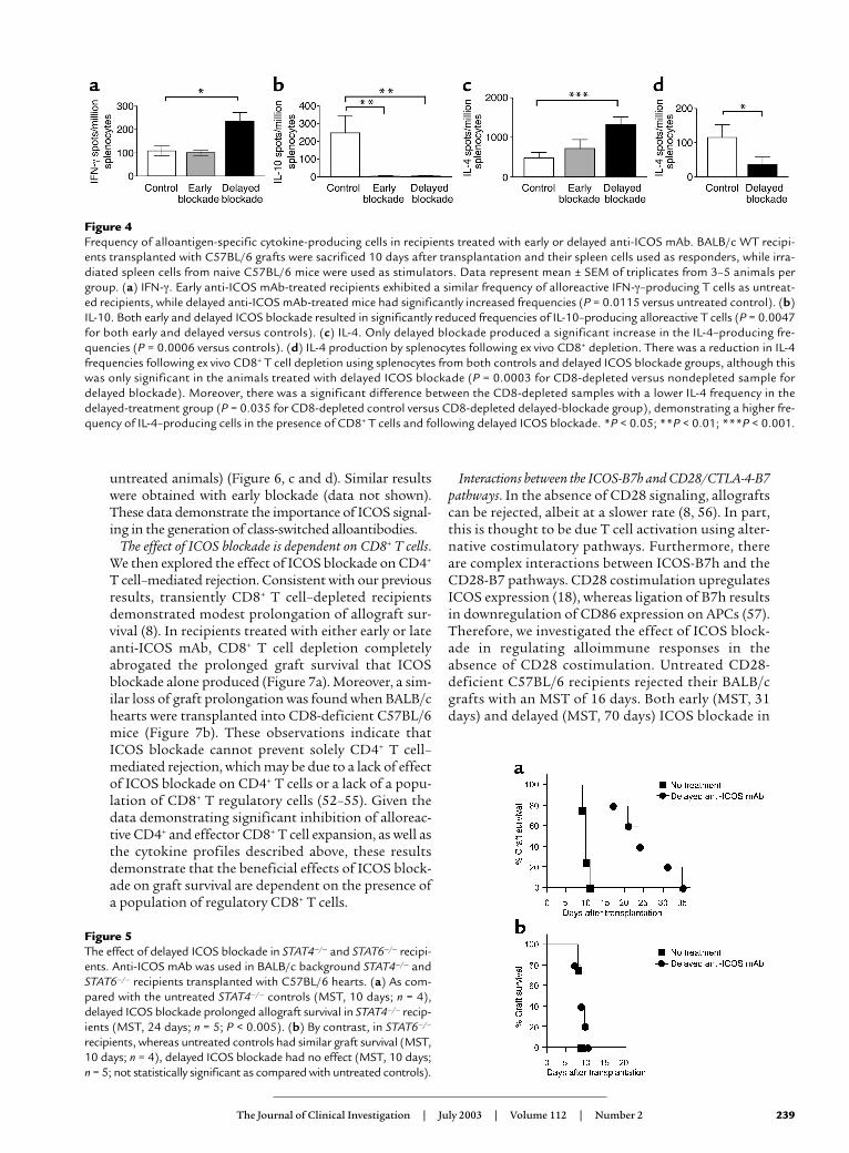
Effect of early and delayed ICOS blockade on alloantigen-spe-cific cytokine production. Since differences in IFN-γ pro-duction after early and delayed ICOS blockade were pre-viously described in experimental autoimmune models(33), and since ICOS is known to influence Th1 andTh2 cell differentiation (28), we studied alloantigen-spe-cific Th1 (IFN-γ) and Th2 (IL-4 and IL-10) cytokine pro-duction in recipients treated with early or delayed ICOSblockade, using a previously published ELISPOT assay(40). Recipient splenocytes were collected 10–14 daysafter transplantation, and the frequency of IFN-γ–, IL-4– and IL-10–producing allospecific cells was meas-ured. In the delayed- but not the early-treatment group,the frequency of alloreactive IFN-γ–producing spleno-cytes was increased as compared with the untreated con-trol recipients (P = 0.0115 as compared with control)(Figure 4a). By contrast, both early and delayed blockade
The Journal of Clinical Investigation | July 2003 | Volume 112 | Number 2 237
Figure 2Histology of murine cardiac allografts from animals treated with ICOS-B7h signal blockade. C57BL/6 grafts were harvested from BALB/crecipients 10 days after cardiac transplantation in animals receiving notreatment and either early or delayed anti-ICOS mAb therapy. Rejectedgrafts from untreated control recipients demonstrated diffuse mononu-clear cell infiltration, myocyte destruction, and interstitial hemorrhage(a). There was a scattered inflammatory cell infiltrate in the grafts treat-ed with the early ICOS-B7h blockade (b), whereas few inflammatorycells and preserved cardiac myocytes were seen in the grafts treated withdelayed therapy (c). Magnification, ×400 (H&E).

significantly decreased the frequency of IL-10–produc-ing cells (P = 0.0047 for both early and delayed treat-ment as compared with controls) (Figure 4b). Interest-ingly, early blockade produced a slight but nonsignificantincrease in the frequency of IL-4–producing cells as com-pared with controls, whereas delayed blockade signifi-cantly increased the IL-4–producing T cell frequency
(P = 0.0006 as compared with controls) (Figure 4c). Fur-thermore, after ex vivo depletion of CD8+ T cells fromsplenocyte preparations, IL-4 production was dimin-ished in both control and delayed ICOS blockadegroups, although only the decline in the delayed ICOSblockade group was significantly greater than in con-trols (P = 0.035 for CD8-depleted ICOS blockade recip-ients as compared with CD8-depleted control recipi-ents) (Figure 4d). These data demonstrate that in thecontext of alloimmune responses, delayed ICOS block-ade significantly augments IFN-γ and IL-4 production,while diminishing IL-10 production, and that induc-tion of IL-4 production is dependent on a subset ofalloreactive CD8+ T cells.
We then investigated the role of the ICOS-B7h path-way in alloreactive Th1/Th2 cell differentiation byusing STAT4- or STAT6-deficient animals with polar-ized T helper cell environments. Consistent with previ-ous reports (40), both STAT4–/– (Th1-defective) andSTAT6–/– (Th2-defective) recipients were able to rejectallografts with a tempo similar to that of WT recipients(MST, 10 days for both groups). In a similar fashion toWT recipients, delayed treatment with anti-ICOS mAbresulted in significant prolongation of allograft sur-vival in STAT4–/– animals (MST, 24 days) (Figure 5a). Bycontrast, in STAT6–/– mice, delayed anti-ICOS mAb didnot extend allograft survival at all as compared with theuntreated control group (Figure 5b). This is not due toan inherent resistance of STAT6–/– recipients to T cellcostimulatory blockade, since CTLA-4 Ig prolongedcardiac allograft survival in these recipients (41). Thesedata suggest that prolongation of allograft survival byICOS blockade requires an intact STAT6 pathway.
The role of ICOS in generation of alloreactive effector CD8+
T cells in vivo. The outcome of ICOS signaling on CD8+
T cell function is unclear (31, 49–51). In order to assessthe effect of ICOS-B7h blockade on alloreactive CD8+
T cells, we measured the percentage of effector CD8+
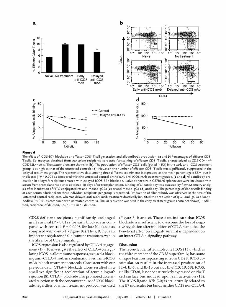
cells (expressing a CD62LlowCD44high phenotype) gen-erated 10 days after transplantation, in untreated con-trol recipients and in recipients treated with early ordelayed ICOS blockade. A significant decrease (34%) inthe percentage of effector CD8+ cells was observed onlyin the delayed-treatment group (P < 0.005 as comparedwith controls) (Figure 6, a and b). These data clearlyshow that ICOS signaling is necessary for the genera-tion and/or maintenance of alloreactive CD8+ effectorT cells, and that this effect occurs after T cell priming.
The role of ICOS in alloantibody production. ICOS signalingplays a major role in immunoglobulin class switching(14, 17). To assess the role of ICOS-B7h on humoralalloimmune responses, we measured the levels of donor-specific alloantibody production by flow cytometry10–14 days after transplantation in recipients treatedwith delayed anti-ICOS mAb. High levels of alloantibod-ies of IgG2a (Figure 6c) and IgG1 (Figure 6d) isotypeswere observed in the sera of the untreated control recipi-ents, which were significantly decreased after delayedICOS blockade (P < 0.01 as compared with control
238 The Journal of Clinical Investigation | July 2003 | Volume 112 | Number 2
Figure 3Expansion and apoptosis of alloreactive CD4+ T cells after ICOS-B7hblockade. After adoptive transfer of transgenic ABM CD4+ T cells intonude mice, the animals were transplanted with bm12 skin and leftuntreated or were given anti-ICOS mAb according to the two proto-cols. Ten days later, draining lymph nodes were removed and the num-ber of alloreactive CD4+ T cells was examined (a). To control for home-ostatic proliferation, some animals were adoptively transferred withcells but received no transplant (AT only), and these demonstratedonly modest proliferation. By comparison, there was a marked expan-sion of CD4+ T cells after transplantation, which was significantlyreduced after early and delayed ICOS-B7h blockade (by 54% and 53%,respectively; ***P < 0.0001 for either as compared with transplantcontrols). (b) By gating on the transgenic CD4+ T cells, the percentageof alloreactive cells undergoing apoptosis was measured by annexin Vstaining. There was no difference in the percentage of cells undergoingapoptosis between controls and early or delayed blockade, demon-strating that the reduction in expansion after ICOS-B7h blockade wasnot simply due to increased apoptosis. FL2-H, annexin V.
untreated animals) (Figure 6, c and d). Similar resultswere obtained with early blockade (data not shown).These data demonstrate the importance of ICOS signal-ing in the generation of class-switched alloantibodies.
The effect of ICOS blockade is dependent on CD8+ T cells.We then explored the effect of ICOS blockade on CD4+
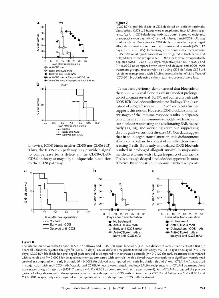
T cell–mediated rejection. Consistent with our previousresults, transiently CD8+ T cell–depleted recipientsdemonstrated modest prolongation of allograft sur-vival (8). In recipients treated with either early or lateanti-ICOS mAb, CD8+ T cell depletion completelyabrogated the prolonged graft survival that ICOSblockade alone produced (Figure 7a). Moreover, a sim-ilar loss of graft prolongation was found when BALB/chearts were transplanted into CD8-deficient C57BL/6mice (Figure 7b). These observations indicate thatICOS blockade cannot prevent solely CD4+ T cell–mediated rejection, which may be due to a lack of effectof ICOS blockade on CD4+ T cells or a lack of a popu-lation of CD8+ T regulatory cells (52–55). Given thedata demonstrating significant inhibition of alloreac-tive CD4+ and effector CD8+ T cell expansion, as well asthe cytokine profiles described above, these resultsdemonstrate that the beneficial effects of ICOS block-ade on graft survival are dependent on the presence ofa population of regulatory CD8+ T cells.
Interactions between the ICOS-B7h and CD28/CTLA-4-B7pathways. In the absence of CD28 signaling, allograftscan be rejected, albeit at a slower rate (8, 56). In part,this is thought to be due T cell activation using alter-native costimulatory pathways. Furthermore, thereare complex interactions between ICOS-B7h and theCD28-B7 pathways. CD28 costimulation upregulatesICOS expression (18), whereas ligation of B7h resultsin downregulation of CD86 expression on APCs (57).Therefore, we investigated the effect of ICOS block-ade in regulating alloimmune responses in theabsence of CD28 costimulation. Untreated CD28-deficient C57BL/6 recipients rejected their BALB/cgrafts with an MST of 16 days. Both early (MST, 31days) and delayed (MST, 70 days) ICOS blockade in
The Journal of Clinical Investigation | July 2003 | Volume 112 | Number 2 239
Figure 4Frequency of alloantigen-specific cytokine-producing cells in recipients treated with early or delayed anti-ICOS mAb. BALB/c WT recipi-ents transplanted with C57BL/6 grafts were sacrificed 10 days after transplantation and their spleen cells used as responders, while irra-diated spleen cells from naive C57BL/6 mice were used as stimulators. Data represent mean ± SEM of triplicates from 3–5 animals pergroup. (a) IFN-γ. Early anti-ICOS mAb-treated recipients exhibited a similar frequency of alloreactive IFN-γ–producing T cells as untreat-ed recipients, while delayed anti-ICOS mAb-treated mice had significantly increased frequencies (P = 0.0115 versus untreated control). (b)IL-10. Both early and delayed ICOS blockade resulted in significantly reduced frequencies of IL-10–producing alloreactive T cells (P = 0.0047for both early and delayed versus controls). (c) IL-4. Only delayed blockade produced a significant increase in the IL-4–producing fre-quencies (P = 0.0006 versus controls). (d) IL-4 production by splenocytes following ex vivo CD8+ depletion. There was a reduction in IL-4frequencies following ex vivo CD8+ T cell depletion using splenocytes from both controls and delayed ICOS blockade groups, although thiswas only significant in the animals treated with delayed ICOS blockade (P = 0.0003 for CD8-depleted versus nondepleted sample fordelayed blockade). Moreover, there was a significant difference between the CD8-depleted samples with a lower IL-4 frequency in thedelayed-treatment group (P = 0.035 for CD8-depleted control versus CD8-depleted delayed-blockade group), demonstrating a higher fre-quency of IL-4–producing cells in the presence of CD8+ T cells and following delayed ICOS blockade. *P < 0.05; **P < 0.01; ***P < 0.001.
Figure 5The effect of delayed ICOS blockade in STAT4–/– and STAT6–/– recipi-ents. Anti-ICOS mAb was used in BALB/c background STAT4–/– andSTAT6–/– recipients transplanted with C57BL/6 hearts. (a) As com-pared with the untreated STAT4–/– controls (MST, 10 days; n = 4),delayed ICOS blockade prolonged allograft survival in STAT4–/– recip-ients (MST, 24 days; n = 5; P < 0.005). (b) By contrast, in STAT6–/–
recipients, whereas untreated controls had similar graft survival (MST,10 days; n = 4), delayed ICOS blockade had no effect (MST, 10 days;n = 5; not statistically significant as compared with untreated controls).
CD28-deficient recipients significantly prolongedgraft survival (P = 0.0122 for early blockade as com-pared with control, P = 0.0008 for late blockade ascompared with control) (Figure 8a). Thus, ICOS is animportant regulator of alloimmune responses even inthe absence of CD28 signaling.
ICOS expression is also regulated by CTLA-4 engage-ment (19). To investigate the effect of CTLA-4 on regu-lating ICOS in alloimmune responses, we used a block-ing anti–CTLA-4 mAb in combination with anti-ICOSmAb in both treatment protocols. Consistent with ourprevious data, CTLA-4 blockade alone resulted in asmall yet significant acceleration of acute allograftrejection (8). CTLA-4 blockade also promoted acceler-ated rejection with the concomitant use of ICOS block-ade, regardless of which treatment protocol was used
(Figure 8, b and c). These data indicate that ICOSblockade is insufficient to overcome the loss of nega-tive regulation after inhibition of CTLA-4 and that thebeneficial effect on allograft survival is dependent onan intact CTLA-4 signaling pathway.
DiscussionThe recently identified molecule ICOS (13), which isthe third member of the CD28 superfamily, has someunique features separating it from CD28. ICOS co-stimulation results in the increased production of IL-4, IL-5, and IL-10 but not IL-2 (13, 18, 58). ICOS,unlike CD28, is not constitutively expressed on the Tcell surface but induced upon cell activation (13).The ICOS ligand B7h (20) is structurally related tothe B7 molecules but binds neither CD28 nor CTLA-4.
240 The Journal of Clinical Investigation | July 2003 | Volume 112 | Number 2
Figure 6The effect of ICOS-B7h blockade on effector CD8+ T cell generation and alloantibody production. (a and b) Percentages of effector CD8+
T cells. Splenocytes obtained from transplant recipients were used for staining of effector CD8+ T cells, characterized as CD8+CD44high
CDD62Llow cells. The scatter plots are shown in (b). The population of effector CD8+ cells (gated in R3) in the early anti-ICOS treatmentgroup is as high as that of the untreated controls (a). However, the number of effector CD8+ T cells was significantly suppressed in thedelayed-treatment group. The representative data among three different experiments is expressed as the mean percentage ± SEM, run intriplicates (*P < 0.005 as compared with the untreated control or the early anti-ICOS mAb treatment group). (c and d) Alloantibody pro-duction in allograft recipients treated with delayed ICOS-B7h blockade. Naive donor strain C57BL/6 splenocytes were incubated withserum from transplant recipients obtained 10 days after transplantation. Binding of alloantibody was assessed by flow cytometry analy-sis after incubation of FITC-conjugated rat anti-mouse IgG2a (c) or anti-mouse IgG1 (d) antibody. The percentage of donor cells bindingat each serum dilution from three individual recipients per group is expressed. Production of alloantibody was observed in the sera of theuntreated control recipients, whereas delayed anti-ICOS mAb treatment drastically inhibited the production of IgG1 and IgG2a alloanti-bodies (P < 0.01 as compared with untreated controls). Similar reduction was seen in the early-treatment group (data not shown). 1/dilu-tion, reciprocal of dilution, i.e., 50 = 1 in 50 dilution.
Likewise, ICOS binds neither CD80 nor CD86 (13).Thus, the ICOS-B7h pathway may provide a signalto compensate for a deficit in the CD28-CD80/CD86 pathway or may play a unique role in additionto the CD28 pathway.
It has been previously demonstrated that blockade ofthe ICOS-B7h signal alone results in a modest prolonga-tion of allograft survival (30, 59), and our results with earlyICOS-B7h blockade confirmed these findings. The obser-vation of allograft survival in ICOS–/– recipients furthersupports this notion. However, ICOS blockade at differ-ent stages of the immune response results in disparateoutcomes in some autoimmune models, with early andlate blockade exacerbating and ameliorating EAE, respec-tively (33, 34), and worsening acute but suppressingchronic graft-versus-host disease (35). Our data suggestthat in solid organ transplantation, this dichotomouseffect occurs only in the context of a smaller clone size ofreacting T cells. Both early and delayed ICOS blockaderesulted in prolonged allograft survival in major-mis-matched recipients with a larger frequency of alloreactiveT cells, although delayed blockade does appear to be moreeffective. By contrast, in minor-mismatched recipients
The Journal of Clinical Investigation | July 2003 | Volume 112 | Number 2 241
Figure 7ICOS-B7h signal blockade in CD8-depleted or -deficient animals.Vascularized C57BL/6 hearts were transplanted into BALB/c recip-ients. (a) Anti–CD8-depleting mAb was administered to recipientspreoperatively on days –6, –3, and –1, whereas anti-ICOS mAb wasused as above. Preoperative CD8 depletion modestly prolongedallograft survival as compared with untreated controls (MST, 12days; n = 9; P < 0.05). Interestingly, the beneficial effects of anti-ICOS mAb on allograft survival were abrogated in both early- anddelayed-treatment groups when CD8+ T cells were preoperativelydepleted (MST, 10 and 10.5 days, respectively; n = 6; P < 0.005 andP < 0.0005 as compared with early and delayed anti-ICOS mAbtreatment groups, respectively). (b) Using CD8-deficient C57BL/6recipients transplanted with BALB/c hearts, the beneficial effects ofICOS-B7h blockade using either treatment protocol were lost.
Figure 8The interaction between the CD28/CTLA-4-B7 pathway and ICOS-B7h signal blockade. (a) CD28-deficient C57BL/6 recipients of a BALB/cheart all ultimately rejected their grafts (MST, 16 days). CD28-deficient recipients treated with early (MST, 31 days) or delayed (MST, 70days) ICOS-B7h blockade had prolonged graft survival as compared with untreated controls (P = 0.0122 for early treatment as comparedwith controls and P = 0.0008 for delayed treatment as compared with controls), with delayed treatment resulting in significantly prolongedsurvival as compared with early blockade (P = 0.0089 for delayed as compared with early blockade). (b and c) Anti–CTLA-4 mAb was usedin conjunction with anti-ICOS mAb. Vascularized C57BL/6 hearts were transplanted into BALB/c recipients. Anti–CTLA-4 treatment aloneaccelerated allograft rejection (MST, 7 days; n = 4; P < 0.005 as compared with untreated control). Anti–CTLA-4 abrogated the prolon-gation of allograft survival in the recipients of early (b) or delayed anti-ICOS mAb (c) treatment (MST, 7 and 6 days; n = 5; P < 0.005 andP < 0.0001, respectively) as compared with recipients of early or delayed anti-ICOS mAb treatment.

characterized by a smaller alloreactive T cell clone size,early blockade accelerated graft rejection, while delayedblockade prolonged survival. However, T cell clone sizeis not the only factor determining the outcome of animmune response in the absence of ICOS signaling.Recent data using an autoimmune arthritis modeldemonstrated that, in contradiction to the EAE model,ICOS-deficient animals are protected from developingcollagen-induced arthritis (33, 60).
ICOS blockade inhibited the effector mechanisms ofallograft rejection, although that was dependent on thetiming of antibody therapy. Alloantibody production wassignificantly suppressed in both early- and delayed-treat-ment groups and is in keeping with the recent datademonstrating an important role for ICOS in humoralimmune responses (60). However, the effect on alloanti-body production cannot alone explain the differences inallograft survival between the early- and delayed-block-ade groups. Importantly, the production of CD8+ T celleffectors was significantly suppressed in the delayed- butnot the early-treatment group, suggesting that the effectof ICOS blockade on effector CD8+ T cell generation invivo is not secondary to inhibiting early CD4+ T cell help.These data are in keeping with an important role forICOS-B7h signaling in enhancing both naive and recallCD8+ T cell responses (50, 51). Taken together, these dataindicate that the contribution of ICOS costimulation toT cell responses and the functional consequences ofICOS blockade may be critically influenced by both thenature of the immune response, the responding T cellclone size, and the timing of intervention (61).
ICOS-B7h signaling plays a role in both Th1 and Th2differentiation (29–32), although it appears to be morecrucial for Th2 development. However, the precise effectof ICOS deficiency or blockade on antigen-specificcytokine responses appears in part to depend on themodels studied and the timing of blockade (30, 33, 60,62). The ICOS signal is necessary for Th2 cell develop-ment after immune priming, so that early blockade mayresult in a skewing to a Th1 response, and delayed block-ade during the effector phase would maintain a Th2response. Our data demonstrate that the frequency ofalloreactive IL-10–producing recipient spleen cells withearly or delayed anti-ICOS mAb treatment was signifi-cantly lower than in the control group, in keeping withfindings from an allergen-induced airway hyperreactivi-ty model (62), whereas the frequency of IFN-γ–producingcells was higher in the delayed-treatment group. Howev-er, we demonstrated that in the delayed-treatment groupthere was a significant increase in IL-4 production, whichwas dependent on the presence of CD8+ T cells (there wasalso an increase in the early-treatment group but this wasnot statistically significant). Dissociation between IL-4and IL-10 production after ICOS blockade has been pre-viously reported (62). Interestingly, we found that ICOSblockade was ineffective in prolonging graft survival inthe absence of CD8+ T cells. Data from our alloreactiveTCR transgenic model indicate that this is not due to alack of effect on the CD4+ T cell population, whose
expansion in vivo was significantly reduced after ICOSblockade. These data suggest that a subset of CD8+ T cellsmay be regulatory in nature. The mechanism by whichthe CD8+ T cells regulate remains uncertain. However,significant loss of IL-4 production after ex-vivo CD8+ Tcell depletion suggests that these cells either directly orindirectly produce IL-4. Furthermore, and confirmingthese findings, our studies in STAT4–/– and STAT6–/– micedemonstrated that delayed ICOS blockade was not effec-tive in STAT6-deficient recipients that are also deficientin Th2 cytokines. The importance and function of CD8+
regulatory T cells have been recently appreciated (52–55).A number of mechanisms of action have been found forsuch cells using different models, including a direct effecton pathogenic CD4+ Th cell phenotype (63) and an effectmediated through alterations in APC function (54).Definitive proof of a role for regulatory CD8+ T cell pop-ulations in allotransplantation after ICOS blockade andtheir characterization and mechanisms of action requirefurther in-depth studies.
Although there are a number of costimulatory mole-cules capable of fully activating T cells, the various expres-sion patterns and resultant effector functions of thesemolecules suggest some degree of compartmentalizationor hierarchy among the costimulatory pathways (64, 65).Interestingly, despite CD28 costimulation optimizingICOS expression, we have shown that in the absence ofCD28 signaling ICOS remains an important mechanismfor T cell activation during alloimmune responses, in partexplaining how grafts are rejected despite an absence ofCD28 costimulation. Furthermore, the beneficial effectsof ICOS blockade are lost in the absence of CTLA-4 sig-naling. These findings further demonstrate the complexinteractions between these two pathways in vivo.
In conclusion, we have shown that ICOS-B7h block-ade induced prolongation of allograft survival moreeffectively during the effector/differentiation phasethan the priming phase of the alloimmune responseand that ICOS-B7h signaling is important for T helpercell differentiation in alloimmunity. Therefore, givenour results, manipulation of the ICOS-B7h signalingpathway in order to prolong graft survival may be bestinitiated after T cell priming.
AcknowledgmentsWe thank Christopher S. Geehan, Karla S. Stenger, andSusan P. Shea for their invaluable technical assistance.This work was supported by NIH grants 1RO1AI51559 and 5 PO1 AI41521 (to M.H. Sayegh) andCA84500 and P01 AI39671 (to G.J. Freeman).
1. Sayegh, M.H., and Turka, L.A. 1998. The role of T-cell costimulatory acti-vation pathways in transplant rejection. N. Engl. J. Med. 338:1813–1821.
2. Salomon, B., and Bluestone, J.A. 2001. Complexities of CD28/B7: CTLA-4costimulatory pathways in autoimmunity and transplantation. Annu. Rev.Immunol. 19:225–252.
3. Coyle, A.J., and Gutierrez-Ramos, J.C. 2001. The expanding B7 super-family: increasing complexity in costimulatory signals regulating T cellfunction. Nat. Immunol. 2:203–209.
4. Hancock, W.W., et al. 1996. Costimulatory function and expression ofCD40 ligand, CD80, and CD86 in vascularized murine cardiac allograftrejection. Proc. Natl. Acad. Sci. U. S. A. 93:13967–13972.
242 The Journal of Clinical Investigation | July 2003 | Volume 112 | Number 2

5. Grewal, I.S., and Flavell, R.A. 1998. CD40 and CD154 in cell-mediatedimmunity. Annu. Rev. Immunol. 16:111–135.
6. Larsen, C.P., et al. 1996. Long-term acceptance of skin and cardiac allo-grafts after blocking CD40 and CD28 pathways. Nature. 381:434–438.
7. Lin, H., et al. 1998. Cytotoxic T lymphocyte antigen 4 (CTLA-4) block-ade accelerates the acute rejection of cardiac allografts in CD28-deficientmice: CTLA-4 can function independently of CD28. J. Exp. Med.188:199–204.
8. Yamada, A., et al. 2001. CD28-independent costimulation of T cells inalloimmune responses. J. Immunol. 167:140–146.
9. Shimizu, K., Schonbeck, U., Mach, F., Libby, P., and Mitchell, R.N. 2000.Host CD40 ligand deficiency induces long-term allograft survival anddonor-specific tolerance in mouse cardiac transplantation but does notprevent graft arteriosclerosis. J. Immunol. 165:3506–3518.
10. London, C.A., Lodge, M.P., and Abbas, A.K. 2000. Functional responsesand costimulator dependence of memory CD4+ T cells. J. Immunol.164:265–272.
11. Valujskikh, A., Pantenburg, B., and Heeger, P.S. 2002. Primed allospecif-ic T cells prevent the effects of costimulatory blockade on prolonged car-diac allograft survival in mice. Am. J. Transplant. 2:501–509.
12. Ensminger, S.M., et al. 2000. CD8+ T cells contribute to the developmentof transplant arteriosclerosis despite CD154 blockade. Transplantation.69:2609–2612.
13. Hutloff, A., et al. 1999. ICOS is an inducible T-cell co-stimulator struc-turally and functionally related to CD28. Nature. 397:263–266.
14. McAdam, A.J., et al. 2001. ICOS is critical for CD40-mediated antibodyclass switching. Nature. 409:102–105.
15. Dong, C., et al. 2001. ICOS co-stimulator receptor is essential for T-cellactivation and function. Nature. 409:97–101.
16. Dong, C., Temann, U.A., and Flavell, R.A. 2001. Cutting edge: critical roleof inducible costimulator in germinal center reactions. J. Immunol.166:3659–3662.
17. Tafuri, A., et al. 2001. ICOS is essential for effective T-helper-cell respons-es. Nature. 409:105–109.
18. McAdam, A.J., et al. 2000. Mouse inducible costimulatory molecule(ICOS) expression is enhanced by CD28 costimulation and regulates dif-ferentiation of CD4+ T cells. J. Immunol. 165:5035–5040.
19. Riley, J.L., et al. 2001. ICOS costimulation requires IL-2 and can be pre-vented by CTLA-4 engagement. J. Immunol. 166:4943–4948.
20. Swallow, M.M., Wallin, J.J., and Sha, W.C. 1999. B7h, a novel costimula-tory homolog of B7.1 and B7.2, is induced by TNFalpha. Immunity.11:423–432.
21. Yoshinaga, S.K., et al. 1999. T-cell co-stimulation through B7RP-1 andICOS. Nature. 402:827–832.
22. Aicher, A., et al. 2000. Characterization of human inducible costimula-tor ligand expression and function. J. Immunol. 164:4689–4696.
23. Brodie, D., et al. 2000. LICOS, a primordial costimulatory ligand? Curr.Biol. 10:333–336.
24. Ling, V., et al. 2000. Cutting edge: identification of GL50, a novel B7-like pro-tein that functionally binds to ICOS receptor. J. Immunol. 164:1653–1657.
25. Gonzalo, J.A., et al. 2001. Cutting edge: the related molecules CD28 andinducible costimulator deliver both unique and complementary signalsrequired for optimal T cell activation. J. Immunol. 166:1–5.
26. Coyle, A.J., et al. 2000. The CD28-related molecule ICOS is required foreffective T cell-dependent immune responses. Immunity. 13:95–105.
27. Sporici, R.A., and Perrin, P.J. 2001. Costimulation of memory T-cells byICOS: a potential therapeutic target for autoimmunity? Clin. Immunol.100:263–269.
28. Sperling, A.I., and Bluestone, J.A. 2001. ICOS costimulation: it’s not justfor TH2 cells anymore. Nat. Immunol. 2:573–574.
29. Greenwald, R.J., McAdam, A.J., Van Der Woude, D., Satoskar, A.R., andSharpe, A.H. 2002. Cutting edge: inducible costimulator protein reg-ulates both Th1 and Th2 responses to cutaneous leishmaniasis. J. Immunol. 168:991–995.
30. Ozkaynak, E., et al. 2001. Importance of ICOS-B7RP-1 costimulation inacute and chronic allograft rejection. Nat. Immunol. 2:591–596.
31. Carreno, B.M., and Collins, M. 2002. The B7 family of ligands and itsreceptors: new pathways for costimulation and inhibition of immuneresponses. Annu. Rev. Immunol. 20:29–53.
32. Gonzalo, J.A., et al. 2001. ICOS is critical for T helper cell-mediated lungmucosal inflammatory responses. Nat. Immunol. 2:597–604.
33. Rottman, J.B., et al. 2001. The costimulatory molecule ICOS plays animportant role in the immunopathogenesis of EAE. Nat. Immunol.2:605–611.
34. Sporici, R.A., et al. 2001. ICOS ligand costimulation is required for T-cellencephalitogenicity. Clin. Immunol. 100:277–288.
35. Ogawa, S., et al. 2001. Opposing effects of anti-activation-inducible lym-phocyte- immunomodulatory molecule/inducible costimulator anti-body on the development of acute versus chronic graft-versus-host dis-ease. J. Immunol. 167:5741–5748.
36. Iwai, H., et al. 2002. Amelioration of collagen-induced arthritis by block-ade of ICOS-B7h costimulation. J. Immunol. 169:4332–4339.
37. Walunas, T.L., et al. 1994. CTLA-4 can function as a negative regulatorof T cell activation. Immunity. 1:405–413.
38. Corry, R.J., Winn, H.J., and Russell, P.S. 1973. Primarily vascularized allo-grafts of hearts in mice. The role of H-2D, H-2K, and non-H-2 antigensin rejection. Transplantation. 16:343–350.
39. Matesic, D., Lehmann, P.V., and Heeger, P.S. 1998. High-resolution char-acterization of cytokine-producing alloreactivity in naive and allograft-primed mice. Transplantation. 65:906–914.
40. Kishimoto, K., et al. 2000. The role of CD154-CD40 versus CD28-B7 co-stimulatory pathways in regulating allogeneic Th1 and Th2 responsesin vivo. J. Clin. Invest. 106:63–72.
41. Sho, M., et al. 2002. New insights into the interactions between T cellcostimulatory blockade and conventional immunosuppressive drugs.Ann. Surg. 236:667–675.
42. Zhai, Y., et al. 2002. The CD154-CD40 T cell costimulation pathway isrequired for host sensitization of CD8(+) T cells by skin grafts via directantigen presentation. J. Immunol. 169:1270–1276.
43. Marsh, J.E., et al. 2001. The allogeneic T and B cell response is stronglydependent on complement components C3 and C4. Transplantation.72:1310–1318.
44. Backstrom, B.T., Muller, U., Hausmann, B., and Palmer, E. 1998. Positiveselection through a motif in the alphabeta T cell receptor. Science.281:835–838.
45. Sayegh, M.H., et al. 2003. Allograft rejection in a new allospecific CD4+TCR transgenic mouse. Am. J. Transplant. 3:381–389.
46. Sandner, S.E., et al. 2003. The first model of tracking allospecific CD4+T cell activation and tolerance in vivo. Am. J. Transplant. In press.
47. Suchin, E.J., et al. 2001. Quantifying the frequency of alloreactive T cellsin vivo: new answers to an old question. J. Immunol. 166:973–981.
48. Sho, M., et al. 2002. Physiological mechanisms of regulating alloimmu-nity: cytokines, CTLA-4, CD25+ cells, and the alloreactive T cell clonesize. J. Immunol. 169:3744–3751.
49. Kopf, M., et al. 2000. Inducible costimulator protein (ICOS) controls Thelper cell subset polarization after virus and parasite infection. J. Exp.Med. 192:53–61.
50. Wallin, J.J., Liang, L., Bakardjiev, A., and Sha, W.C. 2001. Enhancementof CD8+ T cell responses by ICOS/B7h costimulation. J. Immunol.167:132–139.
51. Liu, X., et al. 2001. B7H costimulates clonal expansion of, and cognatedestruction of tumor cells by, CD8(+) T lymphocytes in vivo. J. Exp. Med.194:1339–1348.
52. Jones, H.P., Tabor, L., Sun, X., Woolard, M.D., and Simecka, J.W. 2002.Depletion of CD8+ T cells exacerbates CD4+ Th cell-associatedinflammatory lesions during murine mycoplasma respiratory disease.J. Immunol. 168:3493–3501.
53. Gilliet, M., and Liu, Y.J. 2002. Generation of human CD8 T regulatorycells by CD40 ligand-activated plasmacytoid dendritic cells. J. Exp. Med.195:695–704.
54. Chang, C.C., et al. 2002. Tolerization of dendritic cells by T(S) cells: thecrucial role of inhibitory receptors ILT3 and ILT4. Nat. Immunol.3:237–243.
55. Colovai, A.I., et al. 2003. Regulatory CD8+CD28– T cells in heart trans-plant recipients. Hum. Immunol. 64:31–37.
56. Lin, H., et al. 1998. Cytotoxic T lymphocyte antigen 4 (CTLA-4) block-ade accelerates the acute rejection of cardiac allografts in CD28-deficientmice: CTLA-4 can function independently of CD28. J. Exp. Med.188:199–204.
57. Salama, A.D., et al. 2003. The interaction between ICOS-B7RP1 andCD28-B7 costimulatory pathways in alloimmune responses in vivo.Am. J. Transplant. 3:390–395.
58. Guo, J., et al. 2001. Stimulatory effects of B7-related protein-1 on cellu-lar and humoral immune responses in mice. J. Immunol. 166:5578–5584.
59. Guo, L., et al. 2002. Prolonged survival in rat liver transplantation withmouse monoclonal antibody against an inducible costimulator (ICOS).Transplantation. 73:1027–1032.
60. Nurieva, R.I., Treuting, P., Duong, J., Flavell, R.A., and Dong, C. 2003.Inducible costimulator is essential for collagen-induced arthritis. J. Clin.Invest. 111:701–706. doi:10.1172/JCI200317321.
61. Greenwald, R.J., Latchman, Y.E., and Sharpe, A.H. 2002. Negative co-receptors on lymphocytes. Curr. Opin. Immunol. 14:391–396.
62. Akbari, O., et al. 2002. Antigen-specific regulatory T cells develop via theICOS-ICOS-ligand pathway and inhibit allergen-induced airway hyper-reactivity. Nat. Med. 8:1024–1032.
63. Jiang, H., Braunstein, N.S., Yu, B., Winchester, R., and Chess, L. 2001.CD8+ T cells control the TH phenotype of MBP-reactive CD4+ T cells inEAE mice. Proc. Natl. Acad. Sci. U. S. A. 98:6301–6306.
64. Yamada, A., Salama, A.D., and Sayegh, M.H. 2002. The role of novelT cell costimulatory pathways in autoimmunity and transplantation.J. Am. Soc. Nephrol. 13:559–575.
65. Salama, A.D., and Sayegh, M.H. 2003. Alternative T-cell costimulatorypathways in transplant rejection and tolerance induction: hierarchy orredundancy? Am. J. Transplant. 3:509–511.
The Journal of Clinical Investigation | July 2003 | Volume 112 | Number 2 243