The major players in adaptive immunity
17
455 RESONANCE May 2009 GENERAL ARTICLE The Major Players in Adaptive Immunity 1. Humoral Immunity Asma Ahmed 1 , Banishree Saha 2 , Anand Patwardhan 3 , Shwetha Shivprasad 4 and Dipankar Nandi 5 How do we remain healthy, for the most parts, in the midst of an environment teeming with opportunistic and infec- tious microbes, potential carcinogens and allergens? The fact is that our immune system, by and large, does a fine job in protecting us. It is therefore important to understand the organization of the immune network, which is broadly cat- egorized into two groups: innate and adaptive. Cells in- volved in innate immunity are the first to come into contact with invading microbes, similar to the border security force, and respond rapidly but in a non-specific manner. On the other hand, the cells involved in adaptive immunity are slower to respond but act in a very specific manner. Though the primary response is slow, the secondary response is much faster and demonstrates memory. This article will focus on some important features and key players involved in the adaptive immune response. The first part deals with the humoral immune response mediated mainly by immuno- globulins produced by the B cells. The second part deals with T cells, the Major Histocompatibility Complex (MHC)- encoded molecules, and Recombination Activating Genes (RAG) responsible for generating diverse B-cell receptors (BCR) and T-cell receptors (TCR). With the advent of newer and smarter infectious agents, it is important to understand the working of the immune network as more research in this area may facilitate the development of better protective strategies. The immune system is responsible for protecting us against microbial infections and the spontaneous generation of tumours. If the immune system is compromised, resident microbes gain advan- 1 Asma Ahmed and 2 Banishree Saha are research scholars in Dipankar Nandi’s laboratory. 3 Anand Patwardhan is a final year MSc (Biotech- nology) student in Shantiniketan, WB. 4 Shwetha Shivaprasad is a KVPY scholar and a first year MSc (Biotech- nology) student at the MS University, Baroda. 5 Dipankar Nandi is an Associate Professor in the Department of Biochemistry, IISc, Bangalore. Keywords Immunity, immunoglobulins, im- munodeficiency, B-cell recep- tor, recombinase, antibody di- versity.
-
Upload
independent -
Category
Documents
-
view
0 -
download
0
Transcript of The major players in adaptive immunity

455RESONANCE May 2009
GENERAL ARTICLE
The Major Players in Adaptive Immunity1.Humoral Immunity
Asma Ahmed1, Banishree Saha2, Anand Patwardhan3,Shwetha Shivprasad4 and Dipankar Nandi5
How do we remain healthy, for the most parts, in the midstof an environment teeming with opportunistic and infec-tious microbes, potential carcinogens and allergens? Thefact is that our immune system, by and large, does a fine jobin protecting us. It is therefore important to understand theorganization of the immune network, which is broadly cat-egorized into two groups: innate and adaptive. Cells in-volved in innate immunity are the first to come into contactwith invading microbes, similar to the border security force,and respond rapidly but in a non-specific manner. On theother hand, the cells involved in adaptive immunity areslower to respond but act in a very specific manner. Thoughthe primary response is slow, the secondary response ismuch faster and demonstrates memory. This article willfocus on some important features and key players involvedin the adaptive immune response. The first part deals withthe humoral immune response mediated mainly by immuno-globulins produced by the B cells. The second part dealswith T cells, the Major Histocompatibility Complex (MHC)-encoded molecules, and Recombination Activating Genes(RAG) responsible for generating diverse B-cell receptors(BCR) and T-cell receptors (TCR). With the advent ofnewer and smarter infectious agents, it is important tounderstand the working of the immune network as moreresearch in this area may facilitate the development ofbetter protective strategies.
The immune system is responsible for protecting us againstmicrobial infections and the spontaneous generation of tumours. Ifthe immune systemis compromised, residentmicrobes gain advan-
1Asma Ahmed and2Banishree Saha areresearch scholars in
Dipankar Nandi’slaboratory.
3Anand Patwardhan is afinal year MSc (Biotech-
nology) student inShantiniketan, WB.
4Shwetha Shivaprasad isa KVPY scholar and a
first year MSc (Biotech-nology) student at the
MS University, Baroda.5Dipankar Nandi is anAssociate Professor in
the Department ofBiochemistry, IISc,
Bangalore.
KeywordsImmunity, immunoglobulins, im-munodeficiency, B-cell recep-tor, recombinase, antibody di-versity.

456 RESONANCE May 2009
GENERAL ARTICLE
tage, which may be deleterious to the host. An example of this isevident in patients suffering from acquired immune deficiencysyndrome (AIDS)1 who show high susceptibility to opportunisticinfections leading to greater mortality. Another example is that ofDavid Vetter who suffered from congenital immunodeficiency(lack or reduced numbers of immune protective cells or mecha-nisms) and spent his entire life (thirteen years) inside a sterileplastic bubble; hence, the name ‘Bubble boy’.
How does the immune system work? Broadly, its functiondepends on three ingredients: cells, receptors and effector mol-ecules. Similar to a nation’s defence organization, the immunesystem possesses different cells which play distinct roles duringthe immune response (Figure 1). Immune cells possess key cell-surface receptors that recognize microbial constituents such aslipopolysaccharides (LPS) and flagellin, present on the bacterialcell surface. The immune system can distinguish between self andnon-self and mounta response byrecognizing the unique molecularsignatures present on invading microbes, tumours or foreign cells.Activation of specific receptors on immune cells results in signal-ling events that culminate in the production of various effectormolecules such as free radicals, cytokines, and immunoglobulinsand lead to the elimination of invadingmicrobes, tumours or foreigncells. As the doubling time of microbes is short, multiple strategiesare required to generate a rapid and effective immune response.
1 Acquired Immune DeficienceySyndrome (AIDS): A set of syn-dromes caused by infection withhuman immunodeficiency virus(HIV). The numbers of CD4+ Tcells decrease as the infectionprogresses, resulting in en-hanced susceptibility to oppor-tunistic infections, tumours andother complications.
Figure 1. Cells of the im-mune system. Pluripotentstem cells give rise to theimmunesystem,whichmaybe classified into twogroups: innate and adap-tive. The left-hand sideshows cells involved in in-nate immunity,whichactasthe first line of defence:granulocytes (neutrophils,basophilsandeosoniphils),monocytes and macroph-ages. The right-hand sideshows cells involved inadaptive immunity: B cellssecrete specific Ig whereasT cells are responsible forcell- mediated immune re-sponses. There is plenty ofcross talk between cellsinvolvedinbothformsoftheresponse, e.g., macroph-ages process antigens andpresent peptide/MHC mol-ecules to T cells to initiatethe adaptive immune re-sponse. T cells, upon acti-vation, secrete cytokines,e.g., IFN, which stimulatemacrophage functions.(See text for details)

457RESONANCE May 2009
GENERAL ARTICLE
There are two types of immune responses: innate or inborn andadaptive.
The Innate Immune Response
The components of the innate system are responsible for the firstline of defence and include anatomic, physiologic, phagocytic andinflammatory barriers. This response is quickbut lacks specificity.Duringinfections, thenumbersofblood cellsknownasneutrophils2a
increase and physicians often use this information obtained fromblood tests for confirmatory diagnosis. Increase in the numbers ofeosinophils2b is evident in parasitic helminthic infections, e.g.,infection with Ascaris, a parasitic roundwormwhich resides in thesmall intestine. In addition, eosinophils and basophils2c also accu-mulate at inflammatory sites during allergic reactions induced byexternal agents. Cells belonging to the monocyte/macrophage3
lineage ingest and kill microbes. They are also important inprocessing and presenting antigens4 (substances from invadingmicrobes or tumour cells or allergens that elicit an immuneresponse) to T cells to initiate the adaptive immune response,which is discussed below.
Adaptive Immunity
The adaptive immune response is much slower but is characterisedby specificity and memory. In fact, these two characteristics areimportant hallmarks of this response. Lymphocytes, which consti-tute 20–40% of white blood cells or leukocytes, are the major cellsresponsible for adaptive immunity. These are further grouped intoB and T lymphocytes (Figure 1). B cells are responsible forhumoral (present in fluids) immunity whereas T cells mediatecellular immunity. B cells possess B-cell receptors (BCR), alsoknown as membrane immunoglobulins, on the cell surface which,upon binding toantigen, result in differentiation of Bcells to plasmacells. These plasma cells secrete copious amounts of solubleimmunoglobulins (or Ig, also known as antibodies) which bind toantigens. Therefore, the active ingredient here, i.e., antibodies, canbe separated awayfromcells without any loss in functional activity
2a Neutrophils: Cells of the in-nate immune system that usuallyare the first to migrate to a site ofinfection. Their primary functionis to phagocytose and kill patho-gens2bEosinophils: are a class ofgranulocytes with a bilobednucleus and cytoplasm whichstains with the acidic dye, eosin.They are part of the anti-micro-bial innate defense network andare also involved in allergic re-actions together with basophilsand mast cells. An abnormallyenhanced number of blood eosi-nophils, known as eosinophilia,iis indicative of infections, usu-ally parasitic.2cBasophils: are non-phogocyticgranulocytes which form <1%of the total circulatingwhite bloodcell population. Their nuclei arelobed and their cytoplasm stainswith a basic dye, methyuleneblue. Basophils and mast cellsare closely related and are themain mediators of the allergicresponse. they store histamineand release it during an allergicreaction when their Fc recep-tors are cross-linked by aller-gen coated IgE antibodies.
3 Macrophages : Cells of the in-nate immune system whosemajor functions are phagocyto-sis, antigen processing and pre-sentation.4 Antigen Presenting Cells: Aclass of immune cells which in-cludes dendritic cells, macroph-ages and B cells. They digestcellular or microbial-derived pro-teins to peptides and presentthem in complex with MHC to Tcells.

458 RESONANCE May 2009
GENERAL ARTICLE
(defined as the capacity to bind antigen). Blood serum (liquidcomponentofbloodwithoutanyclottingfactors)containspolyclonalantibodies which are secreted by plasma cells with multiplespecificities. As serum antibodies are produced due to stimulationby antigens, the presence or absence of specific antibodies has apotential clinical value. Diagnostic kits have been developed todetect antigen-specific antibodies and are used to diagnose micro-bial infections. Those infected with Salmonella possess highamounts of serum antibodies that clump or agglutinateSalmonellaantigens and this test is known as the Widal test, which is often usedin the diagnosis of typhoid. Similarly, the presence of antibodiesagainst HIV proteins is the basis for diagnosing HIV infections.
For cellular immunity mediated by T-lymphocytes, cell–cell con-tact is required. T cells express the T-cell receptor (TCR) whichrecognises peptides (short segments of amino acids derived fromself proteins, microbial proteins, tumour5-specific proteins or selfproteins modified by binding to allergens) on presenting moleculesknown as the major histocompatibility complex (MHC)-encodedproteins located on the cell surface of antigen-presenting cells(APC). In addition, there is a separate group of lymphocytesknown as natural killer (NK)6 cells which are the key effectors of‘antibody-dependent cell-mediated cytotoxicity’ (ADCC)7 andare important for immunity against tumours. NK receptors do notundergo rearrangement.
How is the antigenic specificity of the immune response achieved?B and T cells express on their surface, receptor proteins knownas BCR or TCR which determine antigenic specificity. Each B orT cell expresses a unique BCR or TCR (a heterodimer of twoproteins, eitheror); the total number of molecules that theycan recognize is very large as the number of different combinationspossible are large. This concept that each lymphocyte expressesa unique cell surface receptor for antigen recognition is importantas it allows only selected cells containing the appropriate receptorsto proliferate and mount responses during an immune reaction. FMcFarlane Burnet was the first to enunciate this concept known
5 Tumour: Uncontrolled growthof cells due to loss in cell cycleregulation. Plasma cells thatbecome tumorigenic are knownas myeloma.
6 Natural Killer (NK): Lympho-cytes that do not express B cellor T cell antigen receptors butcan kill tumour cells and partici-pate in ADCC.
7 Antibody-Dependent Cell-me-diated Cytotoxicity (ADCC) : Aprocessby whichantibody boundinfectedcells are recognized andkilled by host NK cells, mac-rophages, neutrophils or eosi-nophils which possess recep-tors for lg.

459RESONANCE May 2009
GENERAL ARTICLE
as the ‘clonal selection hypothesis’ for which he was awarded theNobel Prize in 1960 [1]. This concept has practical applications tooand has led to the development of the ‘hybridoma technology’which enables the isolation and purification of antibodies withunique specificity known as ‘monoclonal antibodies’8. We willexamine lymphocytes and important associated molecules neces-sary for their (lymphocytes) optimal function in greater detailbelow.
The Humoral Response
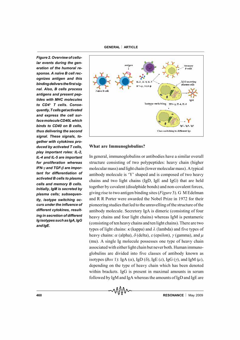
B cells derive their name from ‘Bursa of fabricius’, the organ inbirds in which B cells mature. This was first demonstrated by theobservation that the removal of the Bursa in newly hatchedchickens impairs their ability to generate a humoral response, i.e.,produce antibodies against a specific antigen [2]. Later it wasdiscovered that B cells originate and mature in the bone marrow ofmice and humans. Each B cell expresses a unique antigen-bindingreceptor called the BCR on its cell surface. Circulating B cells thathave not been exposed to antigens are known as naïve B cells9,which get activated upon bindingof their BCR toa cognate antigen.B cells internalize antigens, digest them and present peptidesderived from these antigens together with MHC molecules (seebelow) to T cells. Activated T cells induce the expression of cellsurface proteins (e.g., CD40L) and secrete factors known ascytokines which stimulate the activated B cells to divide rapidlyand differentiate into memory B cells and plasma cells (Figure 2).This process occurs in specialized areas in lymph nodes or spleenknown as germinal centres. Increase in the numbers of germinalcentres in lymph nodes and spleen correlates with an activehumoral response. Plasma cells are the main effector B cells whichsecrete large numbers of immunoglobulin molecules (some esti-mates have it as more than 2000 Ig/second). The first immunoglo-bulin to be secreted in this response is always IgM; subsequently,different cytokines instruct B cells to secrete different classes ofIg, i.e., IgG, IgA and IgE, with the same antigenic specificity (seeFigure 2). Memory cells have a longer life span and they respondquickly upon a second exposure to the same antigen.
8 Monoclonal Antibody: Origi-nating from a single cell and allcells derived from this originalclone possess the identical an-tigenic specificity.
9 Naïve Cell: Mature T or B cellsthat have not encountered anti-gen.
460 RESONANCE May 2009
GENERAL ARTICLE
What are Immunoglobulins?
In general, immunoglobulins or antibodies have a similar overallstructure consisting of two polypeptides: heavy chain (highermolecular mass) and light chain (lower molecular mass). A typicalantibody molecule is ‘Y’ shaped and is composed of two heavychains and two light chains (IgD, IgE and IgG) that are heldtogether by covalent (disulphide bonds) and non-covalent forces,giving rise to two antigen binding sites (Figure 3). G M Edelmanand R R Porter were awarded the Nobel Prize in 1972 for theirpioneering studies that led to the unravelling of the structure of theantibody molecule. Secretory IgA is dimeric (consisting of fourheavy chains and four light chains) whereas IgM is pentameric(consisting of ten heavy chains and ten light chains). There are twotypes of light chains: (kappa) and (lambda) and five types ofheavy chains: (alpha), (delta), (epsilon), (gamma), and (mu). A single Ig molecule possesses one type of heavy chainassociated with either light chain but never both. Human immuno-globulins are divided into five classes of antibody known asisotypes (Box 1): IgA (), IgD (), IgE (), IgG (), and IgM (),depending on the type of heavy chain which has been denotedwithin brackets. IgG is present in maximal amounts in serumfollowed by IgM and IgA whereas the amounts of IgD and IgE are
Figure 2. Overview of cellu-lar events during the gen-eration of the humoral re-sponse. A naïve B cell rec-ognizes antigen and thisbindingdeliversthefirstsig-nal. Also, B cells processantigens and present pep-tides with MHC moleculesto CD4+ T cells. Conse-quently,Tcellsgetactivatedand express the cell sur-facemoleculeCD40Lwhichbinds to CD40 on B cells,thus delivering the secondsignal. These signals, to-gether with cytokines pro-duced by activated T cells,play important roles: IL-2,IL-4 and IL-5 are importantfor proliferation whereasIFN-and TGF-are impor-tant for differentiation ofactivated B cells to plasma
cells and memory B cells.Initially, IgM is secreted byplasma cells; subsequen-
tly, isotype switching oc-curs under the influence ofdifferent cytokines, result-
ing in secretion of differentIg isotypessuchasIgA, IgGand IgE.

461RESONANCE May 2009
GENERAL ARTICLE
low. IgG and IgA are further classified into subclasses dependingon minor amino acid differences. IgG is subdivided into IgG1, IgG2,IgG3, IgG4 based on minor chain differences and numberedaccording to decreasing serum concentration. IgA is subdividedinto IgA1 and IgA2. IgA1 is a monomer and forms the major serumIgA while IgA2 is a dimer and the main component of secretoryIgA.
Each heavy and light chain can be further divided into constant andvariable regions. The first 110 amino acids of different immunoglo-bulins show sequence variability (hence the name) and form theantigen-bindingpocket. This characteristic is important as it allowsflexibility in binding to different antigens. Multiple alignment ofamino acid sequences from different immunoglobulins demon-strate that three regions within the variable region show greatervariability and are termed as ‘complementarity determining re-gions’ (CDR), out of which CDR3 shows the greatest variability.On the other hand, there is not much variability in the ‘constantregion’ of antibody molecules. This region contains the site forcarbohydrate addition, i.e., glycosylation and can bind to Fcreceptors on a number of cell types such as NK cells and directdifferent biological activities (Box 1).
What are the Mechanisms Involved in GeneratingAntibody Diversity?
For a long time, immunologists were puzzled by the ability of theimmune system to generate the vast array of antibodies against alarge number of antigens – it would appear that this potential is
Figure3.Structureofa typi-cal Ig molecule. In mostcases, Ig are heterodimersof two heavy and two lightchains. The number of anti-gen binding sites can varyfrom two (e.g. IgA, IgD, IgEand IgG,) to four (e.g.secre-tory IgA) to ten (e.g. IgM).The aminoterminalsofbothheavy and light chains arevariableandareresponsiblefor antigen binding (Fab).The rest of the chains re-mainconstantandform theFc fragment, which is re-sponsible for Ig-mediatedeffector functions, e.g.opsonization and comple-mentactivation (Seetext fordetails).

462 RESONANCE May 2009
GENERAL ARTICLE
Box 1. Different Human Immunoglobulins and their Properties
Class Subclass Molecular Major biological functions(heavy form
chain)
IgG( IgG1 Monomer Placental transfer to protect the fetus.
IgG3 Can activate complement system.
IgG4 Opsonization.
IgG1 and IgG3 are often produced against protein antigens whereas IgG 2 is
produced against capsular polysaccharides present in some bacteria.
In case of deficiency of any of the 4 subclasses, there is increased risk of
asthma or infections.
IgA() IgA1 Monomer Found in external secretions, e.g. breast milk, saliva, tears and mucus.
IgA2 Dimer is Protects new born babies from infections.
Secretory Important line of mucosal defence against infections.
Some individuals have complete absence of IgA, but normal levels
of other Ig. This is called as selective IgA deficiency which is a
common immunodeficiency disease.
The majority of these cases are asymptomatic although in some cases
cases there are increased incidences of infections, autoimmunity and
allergy.
IgM() – Pentamer First class of Ig to be produced during a primaryimmune response.
Contains 10 antigen binding sites.
Activates complement system more efficiently than IgG.
Deficiency results in susceptibility to infections, especially by encapsu
lated bacteria (e.g. Streptococcus pneumoniae etc.) and increased autoim
mune disorders, cancer etc.
Inability of IgM to switch to other classes causes hyper IgM syndrome with
elevated levels of IgM and reduced levels of other Ig subtypes.
IgE() – Monomer Responsible for allergic reactions.
Binds to Fc receptor and induces mast cell degranulation.
Serum IgE levels increase upon parasitic helminth infections or allergy.
Low IgE levels are often associated with immunodeficiency associated
with low amounts of other Ig isotypes.
Hyper IgE syndrome is characterized by recurrent Staphylococcal infec
tions, skin and pulmonary abscesses and increased numbers of blood
eosinophils.
IgD() – Monomer It is present in very low amounts is serum and not much is know of its
roles.
IgD and IgM are membrane bound Igs on naïve B cells.
IgD deficiency individuals do not show any obvious phenotype.

463RESONANCE May 2009
GENERAL ARTICLE
infinite! S Tonegawa’s group (Nobel laureate 1987) showed forthe first time that the immunoglobulin genes rearrangeduringB celldevelopment. In the germ-line DNA there are multiple genesegments that encode a portion of an immunoglobulin’s heavy orlight chain. The genes encoding these chains are arranged as‘multi-gene families’ situated on different chromosomes. Theseregions contain coding sequences that are separated by non-coding regions which are brought together after gene rearrange-ment [3]. The light chain family contains ~76 variable genes (V)and 5 joining genes (J) and a unique constant (C) gene (Chromo-some 2p). There are ~73–74 Vl genes, 7–11 Jl and 7–11 Cl genesthat may encode the human light chain (Chromosome 22q). On theother hand, there are 123–129 VH, 27 diversity (D) and 9 JH genesand 11 CH genes that can encode the human Ig heavy chain(Chromosome 14q). Hence, the ability of any of the VH genesegments to combine with any of the 27 DH gene segments and anyof the 9 JH segments and 11 CH genes gives rise to large numbersof possible combinations (V DJ C). Similarly forand there will also be large numbers of combinations, and this mecha-nism allows humans to synthesize millions of different types of Ig.
This recombination of V, D and J coding genes of B- and T-cellreceptors are performed by enzymes collectively called ‘VDJrecombinase’. The products of two genes known as Recombina-tion Activating Gene (RAG)-1 and RAG-2 form this recombinase(Figure 4). Apart from VDJ recombination, several other mecha-nisms have been identified in humans that generate additionalantibodydiversity.These include junctional flexibility,P-nucleotideaddition (occurs due to variation in endonucleatic cleavage result-ing in addition of nucleotides to form a palindromic sequence), N-nucleotide addition (addition of nucleotides in the free 3’end ofDNA by terminal deoxynucleotidyl transferase), class switchingand combinatorial association of light and heavy chains. Theseprocesses result in expression of immunoglobulin molecules on theB-cell surface.
Further modifications contributing to Ig diversity occur upon B cell
This recombinationof V, D and J codinggenes of B- and T-cell receptors areperformedbyenzymes collectivelycalled ‘VDJrecombinase’.

464 RESONANCE May 2009
GENERAL ARTICLE
activation. For example, it is well known that IgM is on the B-cellsurface but is secreted by plasma cells. This process involvesRNA splicing and the heavy chains of secreted Ig possess a stretchof 20 hydrophilic amino acids at the C-terminus, whereas mem-brane Ig possess a stretch of 40 amino acids comprised of ahydrophilic segment followed by a hydrophobic transmembrane
Figure 4. Overview of RAG mediated Ig gene rearrangement. Germline DNA contains three Igmultigene families: light chain,light chain and the heavy chain, which are located on differentchromosomes. During B cell maturation, gene segments of these families are rearranged to yieldfunctional Ig molecules. The light chain families possess V, J and C segments whereas the heavychain family has V, D, J and C segments. With the help of RAG proteins V, D and J segments arerearranged to give rise to the variable region of the heavy and light chains. Rearrangement of V, Dand J segments in different permutations and combinations generates Ig diversity. The rearrangedvariable segments then join the constant region fragments to produce the finalheavy and light chaintranscripts. These are translated to form the heavy and light chain proteins which assemble to forma functional Ig molecule.
Germ-lineDNA
VL1 VL2 ….VL40DH1…..DH27 JH1… JH6 Constant µ JL1…. JL6 C? \?
Rearranged DNA in B-cell
Primary mRNA transcript
mRNA
Nascent polypeptideLight chainprocessing
Heavy chainprocessing
Assembly into a
Complete Antibody molecule
DNA
RNA
Protein
AAA(n) AAA(n)
VJ recombinationRAG 1 and 2 + other proteins
Transcription Transcription
VDJ recombination by
splicingsplicing
Translation Translation
VH1 VH2 ….VH51
V D J V J
Dimerization

465RESONANCE May 2009
GENERAL ARTICLE
domain anda short hydrophilic cytoplasmicportion. The hydropho-bic segment is encoded by two exons, M1 and M2, present in theheavy chain encoding DNA of all immunoglobulin isotypes. If themRNA transcript is spliced such that M1 and M2 are included, thenthe translated protein is a membrane immunoglobulin. However, ifM1 and M2 are excluded during splicing then the immunoglobulinmolecules are secreted. Most likely, the process of activation anddifferentiation into plasma cells directs altered splicing of themRNA transcript.
IgM is the first isotype to be secreted by plasma cells and, withtime, other isotypes with the same antigenic specificity appear incirculation. During this process, the variable region remains thesame (hence there is no alteration in antigenic specificity) whereasthe constant region of the heavy chain of the immunoglobulinchanges (known as ‘isotype switching’). The switch to otherisotypes is determined by the cytokines secreted by the TH cells.For example, Interleukin10 (IL)-4 induces switching to IgG1 andIgE whereas IL-5 induces switching to IgA. DNA sequencesknown as ‘switch sites’ are located upstream of the heavy chainconstant regions. The order of heavy chain constant regions in theDNA is: IgM, IgD, IgG, IgE followed by IgA. IgM and IgD do nothave any upstream switch sites and are the first isotypes to beexpressed on the cell surface of naïve B cells. If a switch occursfrom IgM to IgE, then the DNA sequence from IgM to IgG islooped and excised and the functional transcript will now containthe heavy chain constant region of IgE.
During B-cell response it is often found that antibodies producedearly during the immune response bind to the antigen with lowaffinity whereas those produced later bind with stronger affinity.This process is known as ‘affinity maturation’ and the mechanisminvolves ‘somatic hypermutation’. Randommutations occur in thevariable regions of immunoglobulins of individual B cells thatenhance the binding of the antibody with its cognate antigen. Thisprocess results in the selection and secretion of high-affinityantibodies by plasma cells.
10 Interleukins: A group of lowmolecular proteins secreted bya variety of cells that regulatedifferent immune functions: pro-liferation, death, differentiation,antibody secretion, etc. IL-2 in-creases proliferation of T cellswhereas IL-4 regulates IgE pro-duction by B cells.

466 RESONANCE May 2009
GENERAL ARTICLE
In summary, rearrangement of immunoglobulin genes results in thegeneration of large numbers of B cells with different membrane Igmolecules. Upon binding to an antigen, B cells are triggered,followed by secretion of antibodies and isotype switching andbinding affinities are increased further by somatic hypermutation.These multiple strategies, i.e., “first bind, adapt and, subsequently,bind with greater affinity” allows greater flexibility to the immunesystem to generate specific antibodies against a limitless numberof antigens.
Antibodies Play Different Effector Roles
The variable regionsof antibodies bind toantigens via non-covalentinteractions, e.g., hydrogen bonds, ionic bonds, hydrophobic inter-actions and van der Waals interactions. It is this binding thatneutralizes toxins and viruses rendering them biologically non-functional. Attenuated viruses (e.g., small pox, polio) or toxoids(inactivated toxins, e.g., tetanus exotoxin, diphtheria exotoxin) areused to elicit specific antibodies and memory cells by the host viaa process known as‘active immunization’. In essence, vaccinationallows the host to produce high amounts of neutralizing antibodiesthat bind to pathogens and render them non-functional (i.e., theyare not capable of replicating) and prepare the host to counter apossible infection. In fact the most effective vaccines are ones thatgenerate good neutralizing antibodies. Sometimes, preformedantibodies (for example from horse or goat) are administeredagainst rapidly acting toxins or snake/insect bites. Such ‘passiveimmunization’is helpful in conditions when there is not much timefor the host to produce antibodies and neutralize toxins (e.g.,botunilum toxin) or snake venoms. Anti-snake venoms containimmunoglobulins that will neutralize venoms of many commonpoisonous snakes found in India, e.g., cobra, kraits and vipers. Thisaspect is important as there is no time to ascertain the identity ofthe snake that has bitten the patient and quick therapy is a must insuch cases.
Complement Activation: The antigen-antibody complexes acti-vate the complement11 system, which consists of several plasma

467RESONANCE May 2009
GENERAL ARTICLE
proteins that are present in blood in an inactive state. Onceactivated, they clear pathogens by cell lysis. Some antibodies bindto microbes and enhance their phagocytosis (this process is knownas opsonization) by promoting adhesion to neutrophils and mac-rophages. Some antibody-coated target cells are lysed by NK cellsby ADCC, as described earlier. Here the constant region of theantibody is bound to CD1612 receptors on NK cells that getactivated and attack antibody-bound cells leading to their lysis.
Protection of Mucosal Surfaces: Some Ig have specialized roles,e.g., secretory IgA is the major class of Ig present in mucosalsecretions (tears, saliva, intestinal secretions, etc.) and plays a keyrole in protecting such environments in the body. The mechanismby which secretory IgA is present in mucosal secretions is due toan interesting cell biological process. Plasma cells secrete IgAwhich is recognized by polymeric Ig receptor (pIgR) present inepithelial cells. These receptors present on the basolateral surfacebind to IgA and transport these molecules to the luminal surface.Here, there is cleavage of the receptor and IgA is released in thelumen together with a remnant of the pIgR known as the ‘secretorycomponent’.
Ig and allergy: An immunoglobulin isotype known as IgE isimportant for allergic responses. IgE binds to specific receptorspresent on the membranes of tissue mast cells and blood basophils.Once an allergen cross-links a receptor-bound IgE on cells, itinduces degranulation, resulting in release of several effectormolecules, including histamines13, leukotrienes, etc. These com-pounds lead to dilation of blood vessels, increased mucous secre-tions (runny nose) and more severe effects leading to drop in bloodpressure, collapse of the respiratory, circulatory systems, etc.
Immunoglobulins and Autoimmunity: In some cases, antibodiesare formed against the host, leading to autoimmunity (Box 2).Rheumatoid arthritis is one such autoimmune disorder, whereantibodies are formed against the Fc region of IgG. Patientssuffering from rheumatoid arthritis possess high amounts of anIgM known as rheumatoid factor. It binds to the circulating IgG, to
11 Complement: A number ofserum proteins which, uponactivitation, cause lysis of anti-body bound infected cells orpathogens.
12Cluster of Differentiation (CD) :Cell surface molecules usuallypresent on leukocytes that areidentified using monoclonal an-tibodies, e.g. CD3 is present onT cells whereas CD19 is presenton B cells.
13 Histamines: Derivatives ofthe amino acid Histidine that arereleased by mast cells upon ex-posure to allergens. Binding totheir receptors leads to smoothmuscle contraction, mucus se-cretion, etc. Anti-histamines arewidely used to counter the ef-fects of allergy.
468 RESONANCE May 2009
GENERAL ARTICLE
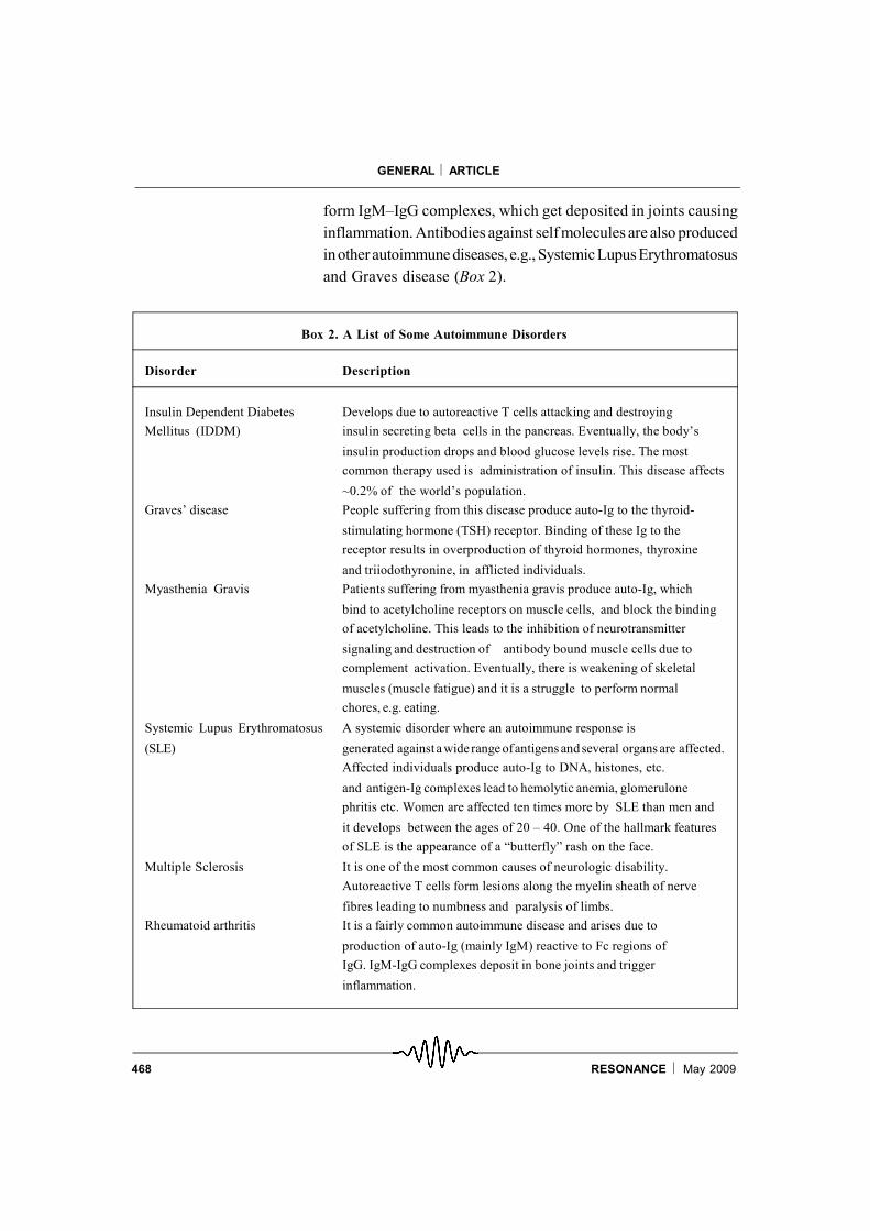
form IgM–IgG complexes, which get deposited in joints causinginflammation. Antibodies against self molecules are also producedinotherautoimmune diseases, e.g., SystemicLupusErythromatosusand Graves disease (Box 2).
Box 2. A List of Some Autoimmune Disorders
Disorder Description
Insulin Dependent Diabetes Develops due to autoreactive T cells attacking and destroyingMellitus (IDDM) insulin secreting beta cells in the pancreas. Eventually, the body’s
insulin production drops and blood glucose levels rise. The mostcommon therapy used is administration of insulin. This disease affects
~0.2% of the world’s population.Graves’ disease People suffering from this disease produce auto-Ig to the thyroid-
stimulating hormone (TSH) receptor. Binding of these Ig to thereceptor results in overproduction of thyroid hormones, thyroxine
and triiodothyronine, in afflicted individuals.Myasthenia Gravis Patients suffering from myasthenia gravis produce auto-Ig, which
bind to acetylcholine receptors on muscle cells, and block the bindingof acetylcholine. This leads to the inhibition of neurotransmitter
signaling and destruction of antibody bound muscle cells due tocomplement activation. Eventually, there is weakening of skeletal
muscles (muscle fatigue) and it is a struggle to perform normalchores, e.g. eating.
Systemic Lupus Erythromatosus A systemic disorder where an autoimmune response is
(SLE) generated against a widerange ofantigens and several organs are affected.Affected individuals produce auto-Ig to DNA, histones, etc.
and antigen-Ig complexes lead to hemolytic anemia, glomerulonephritis etc. Women are affected ten times more by SLE than men and
it develops between the ages of 20 – 40. One of the hallmark featuresof SLE is the appearance of a “butterfly” rash on the face.
Multiple Sclerosis It is one of the most common causes of neurologic disability.Autoreactive T cells form lesions along the myelin sheath of nerve
fibres leading to numbness and paralysis of limbs.Rheumatoid arthritis It is a fairly common autoimmune disease and arises due to
production of auto-Ig (mainly IgM) reactive to Fc regions ofIgG. IgM-IgG complexes deposit in bone joints and trigger
inflammation.

469RESONANCE May 2009
GENERAL ARTICLE
Monoclonal Antibodies: As mentioned previously, serum Ig arepolyclonal as they originate fromdifferent plasma cells. However,as the original B cell is clonal in origin, it is possible to isolate andpropagate such clones using the hybridoma technology developedby G Kohler and C Milstein (Nobel Prize, 1984). The advantage isthat B-cell hybrids grow continuously in culture and produce Ig thatare of single antigenic specificity, knownas monoclonal antibodies.It involves the fusion of antibody-producing plasma cells (Igsecretion) with anon-Igsecreting myeloma cell line (ability to growcontinuously in culture). These hybrid cells are selected for growthand secretion of Ig; subsequently, they are further screened andselected for the production of the desired Ig. Monoclonal antibod-ies are used extensively for research and diagnostics, e.g., devel-opment of ELISA kits for detection of human chorionic gonadot-ropin in the urine of pregnant women, detecting immune cellnumbers (e.g., CD4+ T cells in blood) during pathological condi-tions, e.g., AIDS. In addition, such antibodies are used for therapy,e.g., treatment with an Ig (known as Herceptin) to an oncoprotein(c-erbB-2) is effective against some breast cancers. Also, one ofthe lines of treatment of B-cell lymphomas is using an Ig to CD20known as Rituximab.
Consequences of Reduced or Excess Antibody Produc-tion
As seen in the above section, antibodies play several roles and toevaluate their physiological roles, it is useful to study the effects ofreduced or increased production of antibodies. One such defi-ciency disease is Bruton’s X-linked agammaglobulinemia (XLA),where a male child is unable to produce Ig (Box 3). This defectivegene XLA encodes Bruton’s tyrosine kinase (Btk) which plays arole in B cell maturation. Due to mutations in Btk, patients possessvery low levels of immunoglobulins in blood and suffer fromrecurrent infections duringchildhood. Another immunodeficiencycondition is ‘Common Variable Immunodeficiency’ (CVID) inwhich reduced amounts of antibodies are found. The symptomsobserved are similar to XLA but the reasons for this are multiple,due to mutations in different genes involved in antibodyproduction.
Serum Ig arepolyclonal as theyoriginate fromdifferentplasmacells.
Antibodies playseveral roles and toevaluate theirphysiological roles,it is useful to studythe effects ofreduced orincreasedproduction ofantibodies.

470 RESONANCE May 2009
GENERAL ARTICLE
Box 3. Some Disorders due to Mutations or Deficiencies in Key Immune Response Genes
Disorder Description
Autoimmune polyendocrinopathy Results due to mutations in the autoimmune regulator, AIRE.– candidiasis – ectodermal Studies on AIRE in mice have revealed that it isdystrophy highly expressed in thymic medullary epithelial tissue(APECED) where it regulates expression of peripheral tissue
antigens. Individuals suffering from APECED displaychronic mucocutaneous candidiasis, Type I diabetes andother autoimmune features.
DiGeorge Syndrome Characterized by an absence of the thymus and afflictedindividuals are immunodeficient, suffer from hypoparathyroidism,congenital heart disease and display characteristic facial abnormalities.
SevereCombined It is a broad family of disorders characterized by both BImmunodeficiency and T cell deficiencies that may develop due to a variety(SCID) of reasons. The most common cause of SCID is mutation in the
common gamma chain of the cytokine receptors for IL-2, IL-4, IL-7,IL-9, IL-15 and IL-21. The second most common reason isAdenosine deaminase (ADA) deficiency, which affects purinemetabolism and reduces lymphocyte proliferation. Mutations inRAG affecting VDJ recombination also cause SCID. Most SCIDpatients suffer from recurrent infections and usually die early in lifedue to absence of the adaptive immune response.
X-linked hyper Characterized by abnormally high levels of IgM and absence ofIgM syndrome IgA, IgE and IgG. The defect arises due to inability of T-helper cells
to express CD40L. Therefore, T-dependent B cell activation andclass switching to other antibody isotypes does not occur. Affectedindividuals suffer recurrent infections especially those associatedwith the respiratory tract.
X-linked Agammaglobulinemia Mature B cells fail to develop from pre-B cells due to defects in a(XLA) signal transduction component known as Bruton’s Tyrosine Kinase
(Btk). Characterized by very low levels of IgG and absence of otherisotypes. People suffering from XLA lack peripheral B cells and cannotmake Ig. Consequently, they suffer from recurrent bacterial infections.
Bare Lymphocyte Syndrome Occurs due to low or no surface MHC class I or class II expression.Patients have low numbers of peripheral T lymphocytes andsuffer from recurrent infections, especially diarrhoea and candidiasis.This disorder is usually caused due to defects in a transporter (TAP),which is essential for surface expression of MHC class I, ortranscription factors, e.g. CIITA, required for MHC class II expression.

471RESONANCE May 2009
GENERAL ARTICLE
In general, plasma cells have a short life-span although some long-lived plasma cells have also been found. Plasma cells have a finitelife span; however, in cases when a plasma cell becomes tumori-genic, i.e., keeps on dividing even without antigen stimulation, itgives rise to a condition known as ‘multiple myeloma’. Hereimmortal plasma cells secrete Ig in great excess which are termedas myeloma proteins and constitute about 95% of serum antibod-ies. In some cases, myeloma cells secrete huge amounts of lightchains which are named ‘Bence-Jones proteins’ (after the nameof the physician who made this observation). Interestingly, se-quencing of Bence-Jones proteins played a pivotal role in elucida-tion of the amino acid sequence of immunoglobulin.
The second part of the article will discuss cell-mediated immunity.
Acknowledgments
We thank Profs. R Manjunath, P Sadhale and S Raghavan forpatient reading and comments on the manuscript. The encourage-ment of all members of the DpN laboratory is appreciated. We aregrateful to the Government of India agencies (DBT, CSIR, DSTand ICMR) for providing research funding to the laboratory.
Suggested Reading
[1] F M Burnet, The clonal selection theory of acquired immunity,pp.1–209, Cambridge University Press, Cambridge, 1959.
[2] T S Chang, B Glick, A R Winter, The significance of the bursa ofFabricius of chickens in antibody production., Poultry Sci. Vol.34,p.1187, 1955.
[3] N Hozumi, S Tonegawa, Evidence for somatic rearrangement ofimmunoglobulin genes coding for variable and constant regions.Proc. Natl. Acad. Sci., USA., Vol.73, pp.3628–3632, 1976.
[4] R M Zinkernagel and P C Doherty, Immunological surveillanceagainst altered self components by sensitised T lymphocytes inlymphocytic choriomeningitis, Nature, Vol.251, pp.547–548, 1974.
[5] P Bretscher and M Cohn, A theory of self-nonself discrimination,Science , Vol.169, pp.1042–1049, 1970.
[6] J F Miller, Immunological function of the thymus, Lancet,Vol.2, pp.748–749, 1961.
Address for CorrespondenceDipankar Nandi
Department of BiochemistryIndian Institute of ScienceBangalore 560 012, India.
Email:[email protected]