ICOS Costimulation Requires IL2 and Can Be Prevented by CTLA-4 Engagement1
7
of December 24, 2015. This information is current as Be Prevented by CTLA-4 Engagement ICOS Costimulation Requires IL-2 and Can Katsunari Tezuka, Takashi Tsuji and Carl H. June James L. Riley, Patrick J. Blair, John T. Musser, Ryo Abe, http://www.jimmunol.org/content/166/8/4943 doi: 10.4049/jimmunol.166.8.4943 2001; 166:4943-4948; ; J Immunol References http://www.jimmunol.org/content/166/8/4943.full#ref-list-1 , 22 of which you can access for free at: cites 35 articles This article Subscriptions http://jimmunol.org/subscriptions is online at: The Journal of Immunology Information about subscribing to Permissions http://www.aai.org/ji/copyright.html Submit copyright permission requests at: Email Alerts http://jimmunol.org/cgi/alerts/etoc Receive free email-alerts when new articles cite this article. Sign up at: Print ISSN: 0022-1767 Online ISSN: 1550-6606. Immunologists All rights reserved. Copyright © 2001 by The American Association of 9650 Rockville Pike, Bethesda, MD 20814-3994. The American Association of Immunologists, Inc., is published twice each month by The Journal of Immunology by guest on December 24, 2015 http://www.jimmunol.org/ Downloaded from by guest on December 24, 2015 http://www.jimmunol.org/ Downloaded from
-
Upload
independent -
Category
Documents
-
view
0 -
download
0
Transcript of ICOS Costimulation Requires IL2 and Can Be Prevented by CTLA-4 Engagement1

of December 24, 2015.This information is current as
Be Prevented by CTLA-4 EngagementICOS Costimulation Requires IL-2 and Can
Katsunari Tezuka, Takashi Tsuji and Carl H. JuneJames L. Riley, Patrick J. Blair, John T. Musser, Ryo Abe,
http://www.jimmunol.org/content/166/8/4943doi: 10.4049/jimmunol.166.8.4943
2001; 166:4943-4948; ;J Immunol
Referenceshttp://www.jimmunol.org/content/166/8/4943.full#ref-list-1
, 22 of which you can access for free at: cites 35 articlesThis article
Subscriptionshttp://jimmunol.org/subscriptions
is online at: The Journal of ImmunologyInformation about subscribing to
Permissionshttp://www.aai.org/ji/copyright.htmlSubmit copyright permission requests at:
Email Alertshttp://jimmunol.org/cgi/alerts/etocReceive free email-alerts when new articles cite this article. Sign up at:
Print ISSN: 0022-1767 Online ISSN: 1550-6606. Immunologists All rights reserved.Copyright © 2001 by The American Association of9650 Rockville Pike, Bethesda, MD 20814-3994.The American Association of Immunologists, Inc.,
is published twice each month byThe Journal of Immunology
by guest on Decem
ber 24, 2015http://w
ww
.jimm
unol.org/D
ownloaded from
by guest on D
ecember 24, 2015
http://ww
w.jim
munol.org/
Dow
nloaded from
ICOS Costimulation Requires IL-2 and Can Be Prevented byCTLA-4 Engagement1
James L. Riley,2* Patrick J. Blair, † John T. Musser,* Ryo Abe,‡ Katsunari Tezuka,§
Takashi Tsuji,§ and Carl H. June*
We investigated the relationship between ICOS, CD28, CTLA-4, and IL-2 to gain a better understanding of this family ofcostimulatory receptors in the immune response. Using magnetic beads coated with anti-CD3 and varying amounts of anti-ICOSand anti-CTLA-4 Abs, we show that CTLA-4 ligation blocks ICOS costimulation. In addition to inhibiting cellular proliferation,CTLA-4 engagement prevented ICOS-costimulated T cells from producing IL-4, IL-10, and IL-13. Both an indirect and directmechanism of CTLA-4’s actions were examined. First, CTLA-4 engagement on resting cells was found to indirectly block ICOScostimulation by interferring with the signals needed to induce ICOS cell surface expression. Second, on preactivated cells that hadhigh levels of ICOS expression, CTLA-4 ligation blocked the ICOS-mediated induction of IL-4, IL-10, and IL-13, suggesting aninterference with downstream signaling pathways. The addition of IL-2 not only overcame both mechanisms, but also greatlyaugmented the level of cellular activation suggesting synergy between ICOS and IL-2 signaling. This cooperation between ICOSand IL-2 signaling was explored further by showing that the minimum level of IL-2 produced by ICOS costimulation was requiredfor T cell proliferation. Finally, exogenous IL-2 was required for sustained growth of ICOS-costimulated T cells. These resultsindicate that stringent control of ICOS costimulation is maintained initially by CTLA-4 engagement and later by a requirementfor exogenous IL-2. The Journal of Immunology,2001, 166: 4943–4948.
T he prototypical costimulatory molecule, CD28, provides astrong mitogenic signal (1, 2), induces a large array ofeffector cytokines (3), and up-regulates cell survival
genes such asBcl-xL (4). Equally important for an effective im-mune response, CD28 costimulation promotes long-term growth ofCD4 T cells (5, 6). Whereas CD28’s role is to strongly enhance theT cell response to pathogens, other cell surface molecules serve todown-regulate the immune response. CTLA-4, a CD28 familymember, delivers a negative signal to T cells, limiting their acti-vation (7). The most striking data revealing CTLA-4’s negativerole in T cell proliferation are seen in mice that have a germlinedisruption of the CTLA-4 gene. These mice die of a massive CD4T cell lymphoproliferative disorder (8–10). In vitro, microspherescoated with Abs against CD3, CD28, and CTLA-4 have been use-ful tools for studying the roles these molecules play in the immuneresponse in both human and murine models (11, 12). Using thisapproach, we have shown that CTLA-4 could block CD28-medi-ated proliferation, IL-2 production, and CCR5 down-regulation butnot Bcl-xL induction (13, 14).
ICOS (AILIM) is the third member of the CD28 family whoserole in the immune response remains to be elucidated (15, 16).
Although ICOS possesses costimulatory activity, it differs fromCD28 in at least three important ways: 1) ICOS costimulationresults in the increased production of IL-4, IL-5, and IL-10 but notIL-2 (15). 2) The ICOS ligand, B7RP-1 (B7h, GL50, and B7H2) isstructurally related to the B7 molecules but binds neither CD28 norCTLA-4 (17–20). Likewise, ICOS does not bind either CD80 orCD86. Thus, there is no competition for ligand binding betweenCTLA-4 and ICOS. 3) ICOS, like CTLA-4, is induced on the Tcell surface upon cell activation (15, 21).
ICOS, unlike CTLA-4, does not bind CD80 or CD86 and gen-erates a qualitatively different signal than CD28. Given CTLA-4’sproposed role as the gatekeeper to T cell activation (11), we in-vestigated whether CTLA-4 ligation could prevent ICOS-mediatedcostimulation. CTLA-4 cross-linking can effectively block ICOScostimulation. The addition of exogenous IL-2 not only overcomesthis block, but also greatly augments the effects of ICOS costimu-lation. To further explore the relationship between ICOS costimu-lation and IL-2, we show that IL-2 is necessary for costimulationof CD4 T cells. However, ICOS costimulation is not sufficient tosustain growth, and ICOS-costimulated T cells undergo apoptosisin the absence of exogenous IL-2.
Materials and MethodsCell separation
PBLs were isolated by Percoll (Pharmacia Biotech, Uppsala, Sweden) gra-dient centrifugation from leukopacks obtained following apheresis ofhealthy donors. CD41 T cells were purified by negative selection usingmagnetic beads (Dynal, Lake Success, NY) as described previously (3) andwere routinely.98% CD31, .98% CD281, and,3% CD81 as judgedby flow cytometry.
Abs, bead preparation, and cell stimulation
The mouse anti-human AILIM/ICOS mAb, clone JMAb-52, was generatedby immunizing female BALB/c mice with the membrane fraction of humanAILIM/ICOS-expressing Chinese hamster ovary cells. Anti-CD3 (OKT3)(22), anti-CTLA-4 (ER5.3D6) (23), anti-CD28 (9.3) (24), anti-glycophorin
*Abramson Family Cancer Research Institute and Department of Molecular and Cel-lular Engineering, University of Pennsylvania, Philadelphia, PA 19104;†NationalInstitute of Diabetes and Digestive and Kidney Diseases, Navy Transplantation andAutoimmunity Branch, Naval Medical Research Center, Bethesda, MD 20889;‡ResearchInstitute for Biological Sciences, Science University of Tokyo, Chiba, Japan; and§Pharmaceutical Frontier Research Laboratories, JT Incorporated, Kangawa, Japan
Received for publication November 30, 2000. Accepted for publication February5, 2001.
The costs of publication of this article were defrayed in part by the payment of pagecharges. This article must therefore be hereby markedadvertisementin accordancewith 18 U.S.C. Section 1734 solely to indicate this fact.1 This work was supported by the Abramson Family Cancer Research Institute.2 Address correspondence and reprint requests to Dr. James L. Riley, Department ofMolecular and Cellular Engineering, University of Pennsylvania, 421 Curie Boule-vard, Biomedical Research Building II/III Room 508, Philadelphia, PA 19104-6160.E-mail address: [email protected]
Copyright © 2001 by The American Association of Immunologists 0022-1767/01/$02.00
by guest on Decem
ber 24, 2015http://w
ww
.jimm
unol.org/D
ownloaded from

A (GLY)3 (clone HB-8162; American Type Culture Collection, Manassas,VA), and anti-AILIM/ICOS (JMAb52) covalently attached to polyure-thane-coated tosyl-activated Dynalbeads (Dynal) per the manufacturer’sinstructions. Beads were prepared with a constant, suboptimal amount ofOKT3 (12.5% of total protein coated on the beads), and various ratios(indicated in the text) of anti-ICOS, -CTLA-4, -CD28, and GLY. Poly-clonal goat anti-human IL-2 Abs were obtained from R&D Systems(Minneapolis, MN).
Beads were mixed with CD4 T cells at a 3:1 ratio. Where indicated, 300U/ml of IL-2 (Chiron Therapeutics, Emeryville CA) were added to thecultures. Alternatively, cells were stimulated with 5mg/ml PHA (Sigma,St. Louis, MO) and 300 U/ml IL-2. Cells were cultured at 13 106/ml aspreviously described (14). Cell proliferation assays were conducted in 96-well plates using 13 105 cells. Cultures were pulsed with 1mCi of[3H]thymidine for 18 h before harvest.
Quantitative RT-PCR
RNA was purified and reverse transcribed as described previously (14).Primers and probes to detect IL-2, IL-4, IL-10, IL-13, and 28S rRNA weredesigned using Primer Express software (Applied Biosytems, Foster City,CA), and their sequences are available upon request. Real-time PCR am-plification and product detection was performed using the ABI Prism 7700(Applied Biosystems) as recommended by the manufacturer. Results werenormalized to 28S rRNA levels and relative expression was determined byusing theDDCt method according to the manufacturer’s protocol.
Cell surface staining
A total of 53 105 CD4 T cells were incubated with anti-AILIM (ICOS) oran equivalent amount of an isotype control (BD PharMingen, San Diego,CA) for 30 min at 4°C. After being washed, the cells were incubated witha PE-conjugated goat anti-mouse Ab. Samples were analyzed on a FAC-SCalibur (BD Biosciences, Mountain View, CA) after gating on live lym-phocytes based on a standard light scatter histogram (integral forward scat-ter vs log 90°). Apoptosis assays were conducted by washing 13 106 cellsonce in PBS and incubating with annexin V and propidium iodide per themanufacturer’s protocol (R&D Systems). Data were analyzed using Win-MIDI software (J. Trotter, Scripps Research Institute, La Jolla, CA).
ResultsICOS costimulation delivers a proliferative signal that isblocked by CTLA-4 ligation
To study ICOS, CTLA-4, and CD28 functional effects, we con-structed artificial APCs decorated with Abs to the costimulatoryreceptors. We used Abs rather than natural ligands because B7-1and B7-2 bind to both CD28 and CTLA-4. Therefore, the effect ofCTLA-4 ligation on ICOS-mediated costimulatory signals wasevaluated by stimulating CD4 T cells with magnetic beads con-taining a fixed, suboptimal level of anti-CD3 coupled with varyingamounts of anti-ICOS and anti-CTLA-4. Previous data from otherlaboratories and ours have indicated that a suboptimal level ofCD3 signaling is needed to see the antiproliferative effects ofCTLA-4 engagement (13, 25, 26). To simplify nomenclature, im-munobeads that contain anti-CD3 coupled with equal amounts ofanti-ICOS and anti-CTLA-4 are called 5:5 ICOS/CTLA-4 beads.Likewise, beads that contained three parts anti-ICOS and sevenparts anti-CTLA-4 are referred to as 3:7 ICOS/CTLA-4 beads.Corresponding control sets of immunobeads were made usingGLY. Glycophorin, a nonbinding control, was chosen over a bind-ing control because only minimal ICOS expression is observed onresting cells.
Only minimal proliferation was seen by CD4 T cells stimulatedwith beads containing anti-CD3 and GLY (Fig. 1). The addition ofanti-ICOS to the beads (5:5 ICOS/GLY) greatly augmented themitogenic response. The substitution of anti-CTLA-4 for GLY atan equal ratio to ICOS (5:5 ICOS/CTLA-4 beads) slightly inhib-ited proliferative responses. However, dramatic differences in pro-liferation were observed when cells were stimulated with beads
containing higher ratios of anti-CTLA-4 to anti-ICOS (3:7 ICOS/GLY and 3:7 ICOS/CTLA-4). Similar findings were obtained us-ing 1:9 ICOS/CTLA-4 beads, although the overall level of ICOS-mediated proliferation was reduced. These results indicate that,when present at sufficiently high concentrations, CTLA-4 engage-ment blocked ICOS-mediated proliferation.
CTLA-4 blocks the ICOS-mediated induction of Th2 cytokines
To test the effect of CTLA-4 engagement on ICOS-mediated cy-tokine production, we developed a quantitative RT-PCR assay tomeasure levels of mRNA encoding IL-4, IL-10, and IL-13. Al-though IL-13 was not initially described as a cytokine induced byICOS costimulation (15), in preliminary data we found it to behighly up-regulated by ICOS costimulation. Because stimulationof CD4 T cells with 3:7 ICOS/GLY and 3:7 ICOS/CTLA-4 beadsresulted in vastly different levels of proliferation, we focused ourcytokine transcript analysis on cells stimulated with these beads. Incells costimulated with 3:7 ICOS/GLY beads, we noted a 10- to20-fold increase of the transcripts encoding IL-4, IL-10, and IL-13(Fig. 2). An increase in cytokine RNA levels was not seen in cellsstimulated with 3:7 ICOS/CTLA-4 beads. Similar data were ob-tained by analyzing the levels of IL-10 in the supernatant byELISA (data not shown). These observations indicate that, in ad-dition to inhibiting ICOS-mediated proliferation, CTLA-4 ligationcan block ICOS-directed cytokine production.
We and others have demonstrated that IL-2 addition partiallyoverrides the CTLA-4-mediated blockage of CD28 costimulation(12, 13, 25). When IL-2 was added to cells stimulated with 3:7ICOS/CTLA-4 beads, the inhibitory effects of CTLA-4 were com-pletely abrogated. IL-2 addition greatly augmented both cytokinesecretion and cell proliferation (Fig. 2 and data not shown). Ad-dition of IL-2 to cells stimulated with 3:7 ICOS/GLY beads alsoresulted in increases of cytokine transcripts, suggesting synergybetween ICOS- and IL-2-mediated signal transduction (Fig. 2).Thus, ICOS and IL-2 signals may act cooperatively to activate Tcells in a manner that cannot be inhibited by CTLA-4 engagement.
CTLA-4 engagement prevents the up-regulation of ICOS
Because ICOS is induced after T cell activation (15), we postulatedthat engagement of CTLA-4 early in the cell activation processcould block the actions of ICOS by preventing its cell surface3 Abbreviation used in this paper: GLY, anti-glycophorin A Ab.
FIGURE 1. CTLA-4 ligation can block ICOS-mediated costimulationof CD4 T cells. Purified human CD4 T cells were cultured for 72 h withbeads containing a suboptimal amount of anti-CD3 (12.5%) coupled withvarying ratios of anti-ICOS:anti-CTLA-4 or GLY. The cultures werepulsed with [3H]thymidine for 18 h, harvested, and analyzed for3H incor-poration. The error bars indicate the SD in one experiment, and the data arerepresentative of four independent experiments.
4944 CTLA-4 ENGAGEMENT BLOCKS ICOS COSTIMULATION
by guest on Decem
ber 24, 2015http://w
ww
.jimm
unol.org/D
ownloaded from

up-regulation. To test this hypothesis, we stimulated resting CD4T cells with either 3:7 ICOS/GLY or 3:7 ICOS/CTLA-4 beads for24 h and assayed for ICOS surface expression. Consistent with thishypothesis, ICOS up-regulation was not observed in cells stimu-lated by the 3:7 ICOS/CTLA-4 beads (Fig. 3). Because addition ofIL-2 to cultures stimulated with 3:7 ICOS/CTLA-4 beads not onlyrestored but greatly augmented cytokine expression (Fig. 2), we
reasoned that IL-2 addition would restore ICOS induction in the3:7 ICOS/CTLA-4 stimulated CD4 T cells. However, we observedonly a partial recovery of ICOS expression following IL-2 addi-tion. This is surprising given the enhanced production of IL-4,IL-10, and IL-13 observed upon addition of IL-2 to cultures stim-ulated with 3:7 ICOS/CTLA-4 beads (Fig. 2). Additionally, whileICOS was initially reported to be absent from human resting cells(15), we detected low levels of ICOS expression on the surface ofresting cells, confirming our recent studies in rat T cells (Fig. 3)(21). Despite this low level of expression, we found no evidence ofICOS up-regulating its own expression as both 3:7 ICOS/GLY andbeads just coated with anti-CD3 and GLY up-regulated ICOS ex-pression on CD4 T cells equally (Fig. 3A and data not shown).
CD28 costimulation induces expression of many cell surfacemolecules, including ICOS (15, 21). As shown in Fig. 3B, CD28costimulation induces high levels of ICOS expression on all CD4T cells (Fig. 3B). To test the effects of CTLA-4 ligation on CD28-mediated up-regulation of ICOS, resting CD4 cells were stimu-lated with 1:9 CD28/CTLA-4 beads and a matching 1:9 CD28/MHC I control. Because CD28 is highly expressed on resting cells,we used a binding control Ab (MHC class I). In cells stimulatedwith 1:9 CD28/CTLA-4 beads, no up-regulation of ICOS was ob-served, whereas high levels of ICOS were seen in cells stimulatedwith 1:9 CD28/MHC I beads (Fig. 3B). These results indicate thatCTLA-4 costimulation can block ICOS induction mediated by ei-ther CD3 or CD28 signals. To confirm these results and to gaininsight into the regulation of ICOS expression, we examined ICOSmRNA levels by quantitative PCR and found a very close corre-lation between RNA level and surface expression (data notshown), suggesting that ICOS expression is primarily regulated atthe RNA level.
CTLA-4 can block ICOS-mediated events on preactivated cells
Because CTLA-4 engagement prevents ICOS up-regulation onresting cells, we could not determine whether CTLA-4 blocksICOS signal transduction. Therefore, we stimulated CD4 T cellswith PHA and IL-2, which resulted in large increase in ICOS sur-face expression (Fig. 4,inset). Cells were then washed severaltimes to remove IL-2, restimulated with either 3:7 ICOS/GLY or3:7 ICOS/CTLA-4 beads, and then harvested 6 h later. An increasein the transcripts encoding IL-4 and IL-10 was evident in cellsstimulated with 3:7 ICOS/GLY beads but not in cells stimulatedwith 3:7 ICOS/CTLA-4. CTLA-4 ligation diminished, but did notcompletely prevent, induction of IL-13 by ICOS. Thus, even whenhigh levels of ICOS are present on the cell surface, CTLA-4 en-gagement can block ICOS-mediated costimulation by interferingwith ICOS’s signal transduction pathway.
ICOS costimulation produces low levels of IL-2 that arerequired for initial proliferation but insufficient for sustainedgrowth
The dramatic ability of IL-2 to augment the production of cyto-kines from ICOS-costimulated CD4 T cells prompted us to furtherexplore the relationship between IL-2 and ICOS costimulation.Although one group reported high levels of IL-2 production uponICOS costimulation (20), most groups have reported undetectablelevels of IL-2 in the supernatant (15, 19, 27). We also failed todetect IL-2 in the supernatant of ICOS-stimulated T cells (data notshown). However, because IL-2 is an important factor for CD4 Tcell growth, we speculated that ICOS costimulation was producingsmall levels of IL-2 that were being consumed immediately by theICOS-costimulated T cells and thereby not detectable by ELISA.To test this hypothesis, we isolated RNA from these cells andmeasured IL-2 transcripts using a quantitative real-time RT-PCR
FIGURE 2. Cytokine RNA analysis in ICOS-costimulated CD4 T cells.RNA was collected 24 h after stimulation with the indicated Ab-coatedimmunobeads containing suboptimal levels of anti-CD3. The expression ofIL-4, IL-10, and IL-13 was examined by quantitative RT-PCR as describedin Materials and Methods. Where indicated, 300 U/ml of IL-2 was added.The error bars indicate the SD of three replicates from one experiment, andthe data are representative of three independent experiments.
FIGURE 3. CTLA-4 engagement blocks the induction of cell surfaceICOS. CD4 T cells were stimulated with 3:7 ICOS/GLY or 3:7 ICOS/CTLA-4 beads (A) or 1:9 CD28/GLY and 1:9 CD28/CTLA-4 for 24 h, andcytofluorometric analysis of ICOS expression (shaded) or isotype control(open) was performed. All beads contained suboptimal levels of anti-CD3.Where indicated, 300 U/ml of IL-2 was added. Data are representative ofat least three independent experiments.
4945The Journal of Immunology
by guest on Decem
ber 24, 2015http://w
ww
.jimm
unol.org/D
ownloaded from

assay. The results show that 3:7 ICOS/GLY-costimulated T cellsproduce 30-fold more IL-2 mRNA than CD3 stimulation alone(Fig. 5), suggesting that small amounts (relative to CD28) of IL-2are made by ICOS-costimulated T cells. Interestingly, CTLA-4engagement prevented the induction of IL-2 gene expression.
To determine whether low levels of IL-2 are necessary for ICOScostimulation, we added neutralizing Abs to IL-2 to the culture andmeasured proliferation (Fig. 6). A dose-dependent drop in cellularproliferation was observed in cultures treated with IL-2 Ab,
whereas no effect was seen when a control Ab was added. Similarresults were recently obtained by Yoshinaga and colleagues usinga B7RP-1 fusion protein to stimulate T cells (27). These resultsindicate that ICOS-costimulated T cells produce small amounts ofIL-2 that are immediately consumed by the T cells. Thus, IL-2 isrequired for induction of proliferation by ICOS.
Exogenous IL-2 is needed for sustained growth
CD4 T cells require IL-2 for sustained growth and proliferation(28). The minimum quantities of IL-2 produced by ICOS-costimu-lated T cells prompted us to investigate whether these cells werecapable of sustained proliferation. T cells costimulated by eitheranti-ICOS or anti-CD28 accumulated at equivalent rates for thefirst 6 days after stimulation. However, after that point, the ICOS-costimulated culture stopped growing, whereas the CD28-costimu-lated culture continued to expand in agreement with our previousresults (6) (Fig. 7). We saw no synergy between CD28 and ICOScostimulation, as cells costimulated with ICOS and CD28 (5:5ICOS/CD28) grew as well as those costimulated with CD28 (datanot shown).
FIGURE 4. CTLA-4 ligation blocks ICOS costimulation on preacti-vated cells. CD4 T cells were activated with PHA/IL-2 for 3 days to induceICOS expression. ICOS (shaded) or isotype control (open) surface stainingis shown in the inset. The cultures were washed several times to removeIL-2, and 3:7 ICOS/GLY or 3:7 ICOS/CTLA-4 beads were added for anadditional 6 h before cells were harvested. Quantitative RT-PCR was per-formed to detect expression of IL-4, IL-10, and IL-13 as described inMa-terials and Methods. Data were normalized to the values obtained from theuntreated PHA-stimulated T cell RNA. The error bars indicate the SD fromone experiment, and the data are representative of three independentexperiments.
FIGURE 5. ICOS costimulation induces low IL-2 mRNA levels,whereas CD28 induces high levels of IL-2. CD4 T cells were stimulatedwith beads coated with a suboptimal level of anti-CD3 and the indicatedcostimulatory molecule(s) for 24 h. IL-2 mRNA levels were determined byquantitative RT-PCR. Data were normalized to the values obtained by Tcells costimulated by GLY and are representative of three independentexperiments. Supernatants of cultures costimulated with GLY, 3:7 ICOS/GLY, and 3:7 ICOS/CTLA-4 contained no detectable IL-2 protein,whereas the culture costimulated by CD28 contained 109 ng/ml of IL-2(data not shown).
FIGURE 6. Neutralization of IL-2 prevents ICOS-mediated costimula-tion. CD4 T cells were stimulated with suboptimal levels of CD3 coupledwith either GLY or ICOS. Neutralizing Abs to IL-2 (mg/ml) or an istoypecontrol were added to the indicated cultures. [3H]Thymidine was addedafter 72 h of culture, and the cells were harvested the following day. Dataare representative of two independent experiments.
FIGURE 7. Exogenous IL-2 is required for sustained growth of ICOS-stimulated CD4 T cells. CD4 T cells were activated with beads coated withsuboptimal anti-CD3 and the indicated costimulatory molecule. Where in-dicated, 300 U/ml of IL-2 was maintained in the culture throughout theexperiment. Data are representative of least three independent experiments.
4946 CTLA-4 ENGAGEMENT BLOCKS ICOS COSTIMULATION
by guest on Decem
ber 24, 2015http://w
ww
.jimm
unol.org/D
ownloaded from
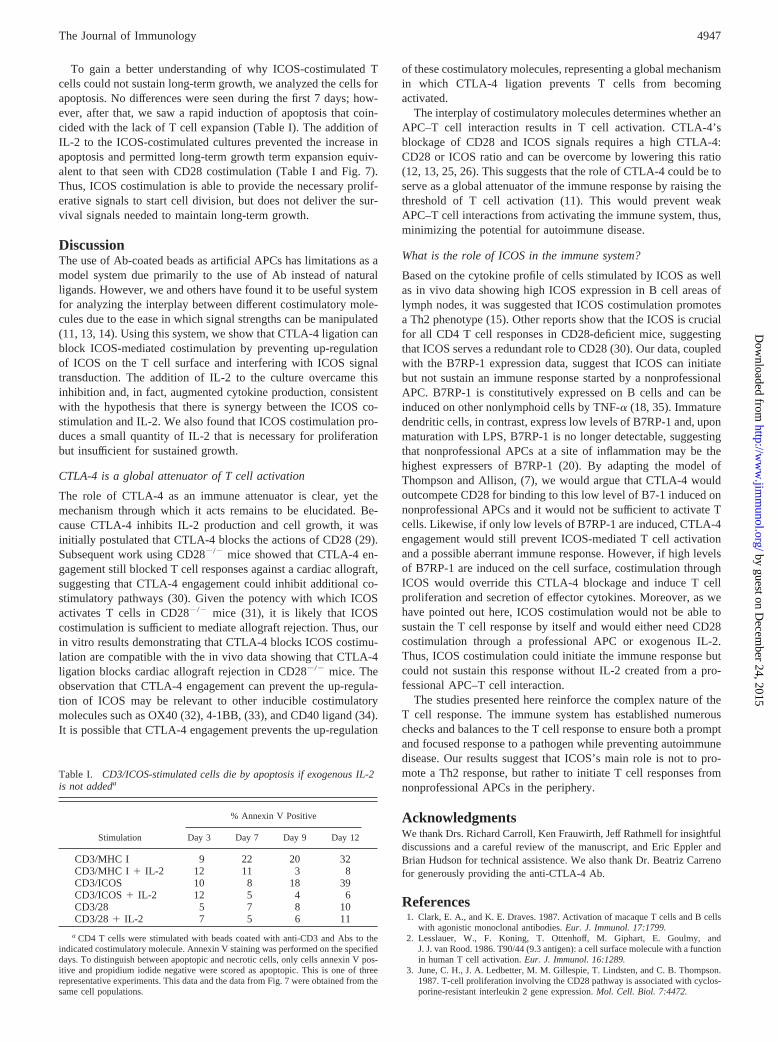
To gain a better understanding of why ICOS-costimulated Tcells could not sustain long-term growth, we analyzed the cells forapoptosis. No differences were seen during the first 7 days; how-ever, after that, we saw a rapid induction of apoptosis that coin-cided with the lack of T cell expansion (Table I). The addition ofIL-2 to the ICOS-costimulated cultures prevented the increase inapoptosis and permitted long-term growth term expansion equiv-alent to that seen with CD28 costimulation (Table I and Fig. 7).Thus, ICOS costimulation is able to provide the necessary prolif-erative signals to start cell division, but does not deliver the sur-vival signals needed to maintain long-term growth.
DiscussionThe use of Ab-coated beads as artificial APCs has limitations as amodel system due primarily to the use of Ab instead of naturalligands. However, we and others have found it to be useful systemfor analyzing the interplay between different costimulatory mole-cules due to the ease in which signal strengths can be manipulated(11, 13, 14). Using this system, we show that CTLA-4 ligation canblock ICOS-mediated costimulation by preventing up-regulationof ICOS on the T cell surface and interfering with ICOS signaltransduction. The addition of IL-2 to the culture overcame thisinhibition and, in fact, augmented cytokine production, consistentwith the hypothesis that there is synergy between the ICOS co-stimulation and IL-2. We also found that ICOS costimulation pro-duces a small quantity of IL-2 that is necessary for proliferationbut insufficient for sustained growth.
CTLA-4 is a global attenuator of T cell activation
The role of CTLA-4 as an immune attenuator is clear, yet themechanism through which it acts remains to be elucidated. Be-cause CTLA-4 inhibits IL-2 production and cell growth, it wasinitially postulated that CTLA-4 blocks the actions of CD28 (29).Subsequent work using CD282/2 mice showed that CTLA-4 en-gagement still blocked T cell responses against a cardiac allograft,suggesting that CTLA-4 engagement could inhibit additional co-stimulatory pathways (30). Given the potency with which ICOSactivates T cells in CD282/2 mice (31), it is likely that ICOScostimulation is sufficient to mediate allograft rejection. Thus, ourin vitro results demonstrating that CTLA-4 blocks ICOS costimu-lation are compatible with the in vivo data showing that CTLA-4ligation blocks cardiac allograft rejection in CD282/2 mice. Theobservation that CTLA-4 engagement can prevent the up-regula-tion of ICOS may be relevant to other inducible costimulatorymolecules such as OX40 (32), 4-1BB, (33), and CD40 ligand (34).It is possible that CTLA-4 engagement prevents the up-regulation
of these costimulatory molecules, representing a global mechanismin which CTLA-4 ligation prevents T cells from becomingactivated.
The interplay of costimulatory molecules determines whether anAPC–T cell interaction results in T cell activation. CTLA-4’sblockage of CD28 and ICOS signals requires a high CTLA-4:CD28 or ICOS ratio and can be overcome by lowering this ratio(12, 13, 25, 26). This suggests that the role of CTLA-4 could be toserve as a global attenuator of the immune response by raising thethreshold of T cell activation (11). This would prevent weakAPC–T cell interactions from activating the immune system, thus,minimizing the potential for autoimmune disease.
What is the role of ICOS in the immune system?
Based on the cytokine profile of cells stimulated by ICOS as wellas in vivo data showing high ICOS expression in B cell areas oflymph nodes, it was suggested that ICOS costimulation promotesa Th2 phenotype (15). Other reports show that the ICOS is crucialfor all CD4 T cell responses in CD28-deficient mice, suggestingthat ICOS serves a redundant role to CD28 (30). Our data, coupledwith the B7RP-1 expression data, suggest that ICOS can initiatebut not sustain an immune response started by a nonprofessionalAPC. B7RP-1 is constitutively expressed on B cells and can beinduced on other nonlymphoid cells by TNF-a (18, 35). Immaturedendritic cells, in contrast, express low levels of B7RP-1 and, uponmaturation with LPS, B7RP-1 is no longer detectable, suggestingthat nonprofessional APCs at a site of inflammation may be thehighest expressers of B7RP-1 (20). By adapting the model ofThompson and Allison, (7), we would argue that CTLA-4 wouldoutcompete CD28 for binding to this low level of B7-1 induced onnonprofessional APCs and it would not be sufficient to activate Tcells. Likewise, if only low levels of B7RP-1 are induced, CTLA-4engagement would still prevent ICOS-mediated T cell activationand a possible aberrant immune response. However, if high levelsof B7RP-1 are induced on the cell surface, costimulation throughICOS would override this CTLA-4 blockage and induce T cellproliferation and secretion of effector cytokines. Moreover, as wehave pointed out here, ICOS costimulation would not be able tosustain the T cell response by itself and would either need CD28costimulation through a professional APC or exogenous IL-2.Thus, ICOS costimulation could initiate the immune response butcould not sustain this response without IL-2 created from a pro-fessional APC–T cell interaction.
The studies presented here reinforce the complex nature of theT cell response. The immune system has established numerouschecks and balances to the T cell response to ensure both a promptand focused response to a pathogen while preventing autoimmunedisease. Our results suggest that ICOS’s main role is not to pro-mote a Th2 response, but rather to initiate T cell responses fromnonprofessional APCs in the periphery.
AcknowledgmentsWe thank Drs. Richard Carroll, Ken Frauwirth, Jeff Rathmell for insightfuldiscussions and a careful review of the manuscript, and Eric Eppler andBrian Hudson for technical assistence. We also thank Dr. Beatriz Carrenofor generously providing the anti-CTLA-4 Ab.
References1. Clark, E. A., and K. E. Draves. 1987. Activation of macaque T cells and B cells
with agonistic monoclonal antibodies.Eur. J. Immunol. 17:1799.2. Lesslauer, W., F. Koning, T. Ottenhoff, M. Giphart, E. Goulmy, and
J. J. van Rood. 1986. T90/44 (9.3 antigen): a cell surface molecule with a functionin human T cell activation.Eur. J. Immunol. 16:1289.
3. June, C. H., J. A. Ledbetter, M. M. Gillespie, T. Lindsten, and C. B. Thompson.1987. T-cell proliferation involving the CD28 pathway is associated with cyclos-porine-resistant interleukin 2 gene expression.Mol. Cell. Biol. 7:4472.
Table I. CD3/ICOS-stimulated cells die by apoptosis if exogenous IL-2is not addeda
Stimulation
% Annexin V Positive
Day 3 Day 7 Day 9 Day 12
CD3/MHC I 9 22 20 32CD3/MHC I 1 IL-2 12 11 3 8CD3/ICOS 10 8 18 39CD3/ICOS1 IL-2 12 5 4 6CD3/28 5 7 8 10CD3/281 IL-2 7 5 6 11
a CD4 T cells were stimulated with beads coated with anti-CD3 and Abs to theindicated costimulatory molecule. Annexin V staining was performed on the specifieddays. To distinguish between apoptopic and necrotic cells, only cells annexin V pos-itive and propidium iodide negative were scored as apoptopic. This is one of threerepresentative experiments. This data and the data from Fig. 7 were obtained from thesame cell populations.
4947The Journal of Immunology
by guest on Decem
ber 24, 2015http://w
ww
.jimm
unol.org/D
ownloaded from

4. Boise, L. H., A. J. Minn, P. J. Noel, C. H. June, M. A. Accavitti, T. Lindsten, andC. B. Thompson. 1995. CD28 costimulation can promote T cell survival byenhancing the expression of Bcl-xL. Immunity 3:87.
5. Vella, A. T., T. Mitchell, B. Groth, P. S. Linsley, J. M. Green, C. B. Thompson,J. W. Kappler, and P. Marrack. 1997. CD28 engagement and proinflammatorycytokines contribute to T cell expansion and long-term survival in vivo.J. Im-munol. 158:4714.
6. Levine, B. L., W. Bernstein, M. Connors, N. Craighead, T. Lindstein,C. B. Thompson, and C. H. June. 1997. Effects of CD28 costimulation on long-term proliferation of CD41 T cells in the absence of exogenous feeder cells.J. Immunol. 159:5921.
7. Thompson, C. B., and J. P. Allison. 1997. The emerging role of CTLA-4 as animmune attenuator.Immunity. 7:445.
8. Waterhouse, P., J. M. Penninger, E. Timms, A. Wakeham, A. Shahinian,K. P. Lee, C. B. Thompson, H. Griesser, and T. W. Mak. 1995. Lymphoprolif-erative disorders with early lethality in mice deficient in Ctla-4.Science 270:985.
9. Tivol, E. A., F. Borriello, A. N. Schweitzer, W. P. Lynch, J. A. Bluestone, andA. H. Sharpe. 1995. Loss of CTLA-4 leads to massive lymphoproliferation andfatal multiorgan tissue destruction, revealing a critical negative regulatory role ofCTLA-4. Immunity 3:541.
10. Chambers, C. A., T. J. Sullivan, and J. P. Allison. 1997. Lymphoproliferation inCTLA-4-deficient mice is mediated by costimulation-dependent activation ofCD41 T cells. Immunity 7:885.
11. Chambers, C. A., M. F. Krummel, B. Boitel, A. Hurwitz, T. J. Sullivan,S. Fournier, D. Cassell, M. Brunner, and J. P. Allison. 1996. The role of CTLA-4in the regulation and initiation of T-cell responses.Immunol. Rev. 153:27.
12. Brunner, M. C., C. A. Chambers, F. K. Chan, J. Hanke, A. Winoto, andJ. P. Allison. 1999. CTLA-4-mediated inhibition of early events of T cell pro-liferation. J. Immunol. 162:5813.
13. Blair, P. J., J. L. Riley, B. L. Levine, K. P. Lee, N. Craighead, T. Francomano,S. J. Perfetto, G. S. Gray, B. M. Carreno, and C. H. June. 1998. CTLA-4 ligationdelivers a unique signal to resting human CD4 T cells that inhibits interleukin-2secretion but allowsBcl-xL induction.J. Immunol. 160:12.
14. Riley, J. L., K. Schlienger, P. J. Blair, B. Carreno, N. Craighead, D. Kim,R. G. Carroll, and C. H. June. 2000. Modulation of susceptibility to HIV-1 in-fection by the cytotoxic T lymphocyte antigen 4 costimulatory molecule.J. Exp.Med. 191:1987.
15. Hutloff, A., A. M. Dittrich, K. C. Beier, B. Eljaschewitsch, R. Kraft,I. Anagnostopoulos, and R. A. Kroczek. 1999. ICOS is an inducible T-cell co-stimulator structurally and functionally related to CD28.Nature 397:263.
16. Tamatani, T., K. Tezuka, and N. Hanzawa-Higuchi. 2000. AILIM/ICOS: a novellymphocyte adhesion molecule.Int. Immunol. 12:51.
17. Ling, V., P. W. Wu, H. F. Finnerty, K. M. Bean, V. Spaulding, L. A. Fouser,J. P. Leonard, S. E. Hunter, R. Zollner, J. L. Thomas, et al. 2000. Cutting edge:identification of GL50, a novel B7-like protein that functionally binds to ICOSreceptor.J. Immunol. 164:1653.
18. Swallow, M. M., J. J. Wallin, and W. C. Sha. 1999. B7h, a novel costimulatoryhomolog of B7.1 and B7.2, is induced by TNFa. Immunity. 11:423.
19. Yoshinaga, S. K., J. S. Whoriskey, S. D. Khare, U. Sarmiento, J. Guo, T. Horan,G. Shih, M. Zhang, M. A. Coccia, T. Kohno, et al. 1999. T-cell co-stimulationthrough B7RP-1 and ICOS.Nature 402:827.
20. Wang, S., G. Zhu, A. I. Chapoval, H. Dong, K. Tamada, J. Ni, and L. Chen. 2000.Costimulation of T cells by B7–H2, a B7-like molecule that binds ICOS.Blood96:2808.
21. Tezuka, K., T. Tsuji, D. Hirano, T. Tamatani, K. Sakamaki, Y. Kobayashi, andM. Kamada. 2000. Identification and characterization of rat AILIM/ICOS, anovel T-cell costimulatory molecule, related to the CD28/CTLA4 family.Bio-chem. Biophys. Res. Commun.276:335.
22. Kung, P., G. Goldstein, E. L. Reinherz, and S. F. Schlossman. 1979. Monoclonalantibodies defining distinctive human T cell surface antigens.Science 206:347.
23. Gribben, J. G., G. J. Freeman, V. A. Boussiotis, P. Rennert, C. L. Jellis,E. Greenfield, M. Barber, V. A. J. Restivo, X. Ke, and G. S. Gray. 1995. CTLA4mediates antigen-specific apoptosis of human T cells.Proc. Natl. Acad. Sci. USA92:811.
24. Hansen, J. A., P. J. Martin, and R. C. Nowinski. 1980. Monoclonal antibodiesidentifying a novel T-cell antigen and Ia antigens of human lymphocytes.Immu-nogenetics 10:247.
25. Krummel, M. F., and J. P. Allison. 1996. CTLA-4 engagement inhibits IL-2accumulation and cell cycle progression upon activation of resting T cells.J. Exp.Med. 183:2533.
26. Griffin, M. D., D. K. Hong, P. O. Holman, K. M. Lee, M. J. Whitters, S. M.O’Herrin, F. Fallarino, M. Collins, D. M. Segal, T. F. Gajewski, D. M. Kranz, andJ. A. Bluestone. 2000. Blockade of T cell activation using a surface-linked single-chain antibody to CTLA-4 (CD152).J. Immunol. 164:4433.
27. Yoshinaga, S. K., M. Zhang, J. Pistillo, T. Horan, S. D. Khare, K. Miner,M. Sonnenberg, T. Boone, D. Brankow, T. Dai, et al. 2000. Characterization ofa new human B7-related protein: B7RP-1 is the ligand to the co-stimulatoryprotein ICOS.Int. Immunol. 12:1439.
28. Ruscetti, F. W., and R. C. Gallo. 1981. Human T-lymphocyte growth factor:regulation of growth and function of T lymphocytes.Blood 57:379.
29. Walunas, T. L., C. Y. Bakker, and J. A. Bluestone. 1996. CTLA-4 ligation blocksCD28-dependent T cell activation.J. Exp. Med. 183:2541.
30. Kopf, M., A. J. Coyle, N. Schmitz, M. Barner, A. Oxenius, A. Gallimore,J. C. Gutierrez-Ramos, and M. F. Bachmann. 2000. Inducible costimulator pro-tein (ICOS) controls T helper cell subset polarization after virus and parasiteinfection.J. Exp. Med. 192:53.
31. Lin, H., J. C. Rathmell, G. S. Gray, C. B. Thompson, J. M. Leiden, andM. L. Alegre. 1998. Cytotoxic T lymphocyte antigen 4 (CTLA4) blockade ac-celerates the acute rejection of cardiac allografts in CD28-deficient mice: CTLA4can function independently of CD28.J. Exp. Med. 188:199.
32. Mallett, S., S. Fossum, and A. N. Barclay. 1990. Characterization of the MRCOX40 antigen of activated CD4 positive T lymphocytes–a molecule related tonerve growth factor receptor.EMBO J. 9:1063.
33. Pollok, K. E., Y. J. Kim, Z. Zhou, J. Hurtado, K. K. Kim, R. T. Pickard, andB. S. Kwon. 1993. Inducible T cell antigen 4-1BB: analysis of expression andfunction.J. Immunol. 150:771.
34. Castle, B. E., K. Kishimoto, C. Stearns, M. L. Brown, and M. R. Kehry. 1993.Regulation of expression of the ligand for CD40 on T helper lymphocytes.J. Im-munol. 151:1777.
35. Aicher, A., M. Hayden-Ledbetter, W. A. Brady, A. Pezzutto, G. Richter,D. Magaletti, S. Buckwalter, J. A. Ledbetter, and E. A. Clark. 2000. Character-ization of human inducible costimulator ligand expression and function.J. Im-munol. 164:4689.
4948 CTLA-4 ENGAGEMENT BLOCKS ICOS COSTIMULATION
by guest on Decem
ber 24, 2015http://w
ww
.jimm
unol.org/D
ownloaded from