PD1 Ligand Expression by Human Colonic Myofibroblasts/Fibroblasts Regulates CD4 + T-Cell Activity

Immunology Letters 132 (2010) 61–68
Contents lists available at ScienceDirect
Immunology Letters
journa l homepage: www.e lsev ier .com/ locate /
IL-2-deprivation and TGF-� are two non-redundant suppressor mechanisms ofCD4+CD25+ regulatory T cell which jointly restrain CD4+CD25− cell activation
Guohua Wanga,b,1, Mithun Khattara,1, Zhiyong Guoa,c,1, Yoshihiro Miyaharaa, Sean P. Linkesa,Zongquan Sunb, Xiaoshun Hec, Stanislaw M. Stepkowskia,∗, Wenhao Chena,∗
a Department of Medical Microbiology and Immunology, University of Toledo College of Medicine, Toledo, OH 43614, United Statesb Department of Cardiovascular Surgery, Union Hospital, Tongji Medical College, Huazhong University of Science and Technology, Wuhan 430022, PR Chinac Organ Transplant Center, 1st Affiliated Hospital, Sun Yat-sen University, Guangzhou 510080, PR China
a r t i c l e i n f o
Article history:Received 18 November 2009Received in revised form 17 May 2010Accepted 1 June 2010Available online 8 June 2010
Keywords:IL-2TGF-�Suppressor mechanismRegulatory T cell
s u m m a r y
The benefits of immunotherapy by regulatory T (Treg) cells are unpredictable partially due to the uncer-tainty of their suppressive mechanism. In fact, various suppressive mechanisms have been proposed buteach remains controversial. To better understand Treg-mediated suppression, we have investigated fac-tors which may influence the suppressive effects. In an in vitro suppression assay, over-expression ofanti-apoptotic Bcl2 enhancing survival of conventional T responder cells (Tconvs) did not subvert Treg-mediated suppression. In contrast, enhancing activation of Tconvs by increasing the potency of calciumsignals completely abrogated Treg-mediated suppression. While Tregs were incapable of suppressingalready activated Tconvs, they prevented expression of activation markers on naïve Tconvs during acti-vation, thereby indicating that Tregs mediate suppression through controlling early activation stage.Interestingly, IL-2 deprivation or TGF-�, two suppressive mechanisms, did not effectively inhibit Tconvactivation and proliferation when applied alone. In contrast, IL-2 deprivation combined with TGF-� sup-
pressed Tconv activation as potently as Tregs. More importantly, in the transwell system, that separatesTregs from Tconvs, TGF-� contributed to Treg suppression under IL-2 depriving condition. In conclusion,echan
1
isipCmateTlr
oT
W
0d
these two suppressive mactivation of Tconvs.
. Introduction
While CD4+ T cells play a central role in establishing and max-mizing various immune responses, they also contain a uniqueubpopulation that suppresses these responses. Sakaguchi et al.dentified CD25 (IL-2R�) as a marker for this CD4+ suppressor cellopulation, named regulatory T cell (Treg). Their study showed thatD4+CD25+ Tregs prevented CD4+CD25− cell-mediated autoim-une diseases in athymic nude mice [1]. In 2003, the same
nd other groups demonstrated that the Foxp3 transcription fac-or was predominantly expressed in CD4+CD25+ cells. The stable
xpression of Foxp3 is critical for establishing and maintainingreg lineage [2–5]. Mutations of the Foxp3 gene can result inethal autoimmunity both in mouse and human [2–5], proving theequirement for Tregs in maintaining self-tolerance.∗ Corresponding authors at: Department of Medical Microbiology and Immunol-gy, University of Toledo-Health Science Campus, 3000 Arlington Avenue, HEB 210,oledo, OH 43614, United States. Tel.: +1 419 383 6673; fax: +1 419 383 3002.
E-mail addresses: [email protected] (S.M. Stepkowski),[email protected] (W. Chen).1 These authors contributed equally to this work.
165-2478/$ – see front matter © 2010 Elsevier B.V. All rights reserved.oi:10.1016/j.imlet.2010.06.001
isms acting in concert may be necessary to effectively restrain the early
© 2010 Elsevier B.V. All rights reserved.
Given its suppressive nature, it is of great interest to explorethe therapeutic effects of Tregs in immune diseases. However, notfully defined suppressive mechanisms of Tregs shroud potentialbarriers in developing Treg-based immunotherapy. In fact, mul-tiple suppressor mechanisms have been proposed but their exactroles are subjects of intense debates. Tregs suppress conventional Tcells (Tconvs) by secreting suppressive cytokines (TGF-�, IL-10, andIL-35) [6–8], by competing with Tconv for IL-2 [9], by expressingsuppressive molecules i.e. galectin-1 on their cell surface [10], orby killing effector T cells [11]. In addition, Tregs have been shownto suppress APCs, which in turn fail to activate Tconvs, as Tregsexpress cell surface molecules (e.g. CTLA-4, LAG-3, CD39, and Nrp-1) that directly influence APC function [8,12]. Not surprisingly,these increasing number of mechanisms has been associated withnumerous controversial issues related to the fact that abrogatingonly one mechanism may not reverse Treg suppression [13–15]. It ispossible and rather likely that two or more timely interacting mech-anisms produce effective Treg suppression. Hence, we explored
different conditions that may limit inhibitory effects and exam-ined how coordinated suppressive factors may produce optimalsuppression.Difficulties had emerged from observations that Tregs in totallyTGF-�1-deficient mice were either protective or nonfunctional in

6 logy Le
tatTdamrosticsiadis
2
2
aJtf
2
as(saDlT
2
uUcPalt0IcsatFc
2
m0
2 G. Wang et al. / Immuno
he same colitis model [13–15]. To avoid these complications, Li etl. generated mice with a T-cell specific deletion of TGF-� produc-ion, and using these mice demonstrated that TGF-� production byregs was absolutely necessary to inhibit the inflammatory bowelisease [6]. More recently, Pesu et al. showed that Tregs deficient inproprotein convertase furin produced little TGF-�, and that theseice were less protective in preventing colitis [7]. Based on these
esults, TGF-� produced by Tregs is continuously considered asne of the important suppressor mechanisms. Another importantuppressive mechanism is IL-2 deprivation wherein Tregs consti-utively expressing IL-2 receptors “consume” local IL-2, therebynhibiting Tconv responses [9]. Our present study examined theoordinated effects of IL-2 deprivation and TGF-� in Treg-mediateduppression. We have demonstrated that these two mechanismsn concert suppress Tconvs, and that the suppressor effects arechieved through restraining their early activation. Although IL-2eprivation alone seems insufficient to inhibit an early activation,
t provides an important prerequisite for effective TGF-�-mediateduppression.
. Materials and methods
.1. Mice
BALB/c, C57BL/6 (B6), C57BL/6-Tg (BCL2)25Wehi/J (Bcl-2 Tg),nd B6.129P2-Il2tm1Hor/J (IL-2 KO) mice were purchased from Theackson Laboratory (Bar Harbor, ME, USA). Animals were main-ained at the University of Toledo specific pathogen-free animalacility according to institutional guidelines.
.2. Reagents
Anti-CD25-PE (clone; PC61), IL-2 neutralizing antibody (S4B6)nd its isotype antibody rat IgG2a were purchased from BD Bio-cience (San Jose, CA, USA). Anti-CD25-APC (PC61.5), anti-GITR-PEDTA-1), and anti-CD4-PE-Cy5 (GK1.5) were obtained from eBio-cience (San Diego, CA, USA). TGF-� neutralizing antibody (1D11)nd its isotype antibody mouse IgG1, and mouse TGF-�1 and IL-2uoSet ELISA kits were purchased from R&D systems (Minneapo-
is, MN, USA). Human TGF-�-1 (hTGF-�) was obtained from Peproech (Rocky Hill, NJ, USA).
.3. FACS analysis for cell surface molecules and CFSE assay
CD4+CD25− and CD4+CD25+ T cells, sorted from BALB/c micesing a FACSAria cell sorter (Becton Dickinson, San Jose, CA,SA), were used as Tconvs and Tregs, respectively. The purity ofells was determined to be over 97%. Tconvs were suspended inBS (1 × 10⁶ cells/ml) containing 1 �M CFSE (Invitrogen, CA, USA)nd incubated for 12 min at 37 ◦C. After cell wash, 5 × 10⁴ CFSE-abeled Tconvs were cultured without or with Treg (1:1 ratio) inhe presence of 1.5 × 10⁵ CD3− syngenic splenocytes (APCs) and.5 �g/ml soluble anti-CD3 mAb. Some cultures were treated withL-2 neutralizing antibody or human (h)TGF-�. To determine theell division and viability, the day-2 or day-3 cultured cells weretained with propidium iodide (PI) followed by flow cytometricnalysis. To determine cell surface marker expression, some cul-ured cells were also stained with anti-GITR-PE and anti-CD25-APC.low cytometric analysis was performed using a FACSCalibur flowytometer (Becton Dickinson).
.4. Proliferation and suppression assay
For proliferation assays, 5 × 10⁴ CD4+CD25− Tconvs from BALB/cice were stimulated by 1.5 × 10⁵ CD3− syngenic APCs and
.5 �g/ml soluble anti-CD3 mAb in 96-well round-bottom plates.
tters 132 (2010) 61–68
Some cultures were treated with various concentrations of IL-2neutralizing mAb, TGF-� neutralizing mAb, isotype control anti-bodies, or hTGF-�. To determine cell proliferation, cultures werepulsed with 1 �Ci of 3H-thymidine for the final 16 h of the 72-hassay and harvested with a Packard harvester. The incorporatedradioactivity was determined by a micro-plate scintillation counter(Packard Ramsey, MN).
In vitro Treg function was measured by culturing 5 × 10⁴ Tconvswith various numbers of Tregs, both of which were isolated fromBALB/c mice. 1.5 × 10⁵ CD3− syngenic APCs and 0.5 �g/ml solu-ble anti-CD3 mAb were used as stimulators. Some cultures weretreated with phorbol-12-myristate-13-acetate (PMA) (3 ng/ml) orionomycin (750 ng/ml). Where indicated, Tconvs, Tregs, or APCswere also obtained from B6 or BCl-2 Tg mice. Cultures were pulsedwith 3H-thymidine and harvested, as described for proliferationassay above.
2.5. Transwell experiments
Tconvs (1 × 10⁵) were isolated from B6 or IL-2 KO mice, andwere cultured in the upper chamber of the 24-well transwell plates(Corning, NY, USA) in the presence of 3 × 10⁵ CD3− syngenic APCsand soluble anti-CD3 (0.5 �g/ml). Tregs (3 × 10⁵) from B6 micewere placed in the bottom chamber of some transwells, and wereactivated with Dynabeads Mouse T-activator CD3/CD28 (Invit-rogen; bead-to-cell ratio = 1:1) and plate-bound anti-CD3 mAb(4 �g/ml). As a control, both Tconvs and Tregs (1 × 10⁵) were addedinto the upper chamber of some transwells. Where indicated, thecultures were added with IL-2 neutralizing antibody or/and TGF-�neutralizing antibody. 3H-thymidine was added to cultures for thefinal 16 h of the 72-h assay. Cells in the upper chambers were trans-ferred to 96-well plates and harvested with a Packard harvester. Thecell proliferation was determined by a scintillation counter.
2.6. ELISA
Tconvs were co-cultured with Tregs in transwell plates asindicated above. Culture supernatants were collect from the topchamber of transwells at 48 h after cultivation. The levels of TGF-�1 and IL-2 in culture supernatants were then assessed by ELISAusing commercial kits following the manufacturer’s instructions.
2.7. Statistical analysis
Statistical analysis and P-value were calculated using anunpaired, 2-tailed, Student’s t-test.
3. Results
3.1. Tregs mediate their suppressive function by inhibiting theearly activation of Tconvs
To better understand the molecular mechanisms of Treg sup-pression, we applied the commonly used in vitro assay in whichCD4+CD25− Tconv responder cells co-cultured with or withoutCD4+CD25+ Treg suppressors (>90% Foxp3+, data not shown) werestimulated by APCs and soluble anti-CD3 mAb. Comparison of CFSElabeled Tconv cells showed that dramatic suppressive effects wereexhibited by Tregs during the first 2 days of cultivation (Fig. 1A).The inhibition was potent as suppressed Tconvs were completelyincapable of proliferation between days 2 and 3, whereas during
the same time non-suppressed Tconvs (cultured without Tregs)continued an extensive proliferation (Fig. 1A; left panels). We alsoidentified a significant percentage of PI+ dead cells (∼70% on day3) within the suppressed Tconvs (Fig. 1A), which is consistent withother reports [9].
G. Wang et al. / Immunology Letters 132 (2010) 61–68 63
Fig. 1. Similar suppressive effects by Tregs on naïve Tconvs and Bcl-2-over-expressing Tconvs but not on activated Tconvs. Purified CD4+CD25− Tconvs were cultured withoutor with CD4+CD25+ Tregs at a 1:1 ratio (except in A). Syngeneic APCs and soluble anti-CD3 mAb were used as stimulators (except in D). In A, B, and D, Tconvs were CFSE-labeled before cultivation. (A) Dot plots show CFSE fluorescence vs. PI staining within the CFSE+ Tconv population at culture day 2 and day 3; Tconv:Treg ratios were indicated.Numbers represent the percentage of PI+ Tconvs. (B) Overlay histograms show the expression of GITR or CD25 on CFSE+PI− Tconvs in the day 2 cultures. The black line and thefi Tregso nels)t . (E) Aw ion. M
ttTwrTIcp[
lled gray area represent Tconvs cultured with and without Tregs, respectively. (C)n day 3 by 3H-thymidine incorporation. (D) APCs together with (top and middle pahe frequency of PI+ cells within the CFSE+ Tconv population in the day 3 culturesere cultured. Cell proliferation was assessed on day 3 by 3H-thymidine incorporat
During the same early activation phase Tregs preventedhe expression of activation markers, GITR and CD25, onhe cell surface of Tconvs in comparison to non-suppressedconvs (Fig. 1B). Most importantly, pre-stimulation of Tconvsith APCs plus anti-CD3 mAb for only 6 h rendered them
esistant to the subsequent suppression by Tregs (Fig. 1C).
hus, Tregs mediate suppression at an early activation phase.nterestingly, enhanced cell proliferation was observed in theo-cultures of Tregs and pre-stimulated Tconvs (Fig. 1C). It isossible that stimulated Tconvs also enhance Treg proliferation16].were added to cultures at the indicated time points. Cell proliferation was assessedor without (bottom panel) anti-CD3 mAb were used as stimulators. Dot plots shows indicated, Tconvs and Tregs isolated from Bcl-2 transgenic or wild-type B6 miceore details are described in Section 2. Figures are representative of 3 experiments.
APC alone (without soluble anti-CD3 mAb) is insufficient to acti-vate the majority of T cells (data not shown). About 80% Tconvswere PI+ in the 3-day cultures in which APCs but not anti-CD3 mAbwere supplemented (Fig. 1D). These data suggested that insuffi-cient activation of Tconvs in the in vitro cultures may lead to theirdeath. Because Tregs suppress the activation of Tconvs, we specu-
lated that the in vitro death of suppressed Tconvs resulted becauseof their failed activation (Fig. 1A). Indeed, despite over-expressingBcl-2 in Tconvs (from Bcl-2 Tg mice) to sustain their viability in vitro[17], Tconvs from Bcl-2 Tg mice were susceptible to Treg-mediatedsuppression (Fig. 1E). Together, our data demonstrate that Tregs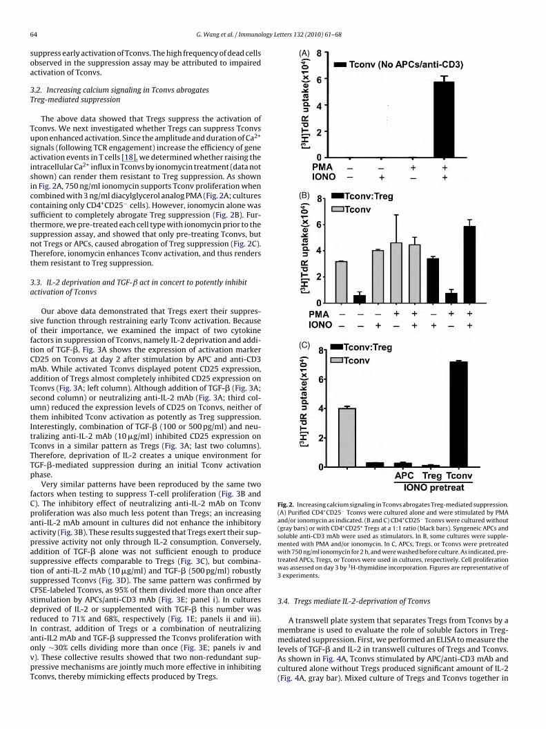
6 logy Letters 132 (2010) 61–68
soa
3T
TusaisiccstsnTt
3a
softCmaTsutItTTTp
fCpaapastsCsdrIaovpT
Fig. 2. Increasing calcium signaling in Tconvs abrogates Treg-mediated suppression.(A) Purified CD4+CD25− Tconvs were cultured alone and were stimulated by PMAand/or ionomycin as indicated. (B and C) CD4+CD25− Tconvs were cultured without(gray bars) or with CD4+CD25+ Tregs at a 1:1 ratio (black bars). Syngeneic APCs andsoluble anti-CD3 mAb were used as stimulators. In B, some cultures were supple-mented with PMA and/or ionomycin. In C, APCs, Tregs, or Tconvs were pretreatedwith 750 ng/ml ionomycin for 2 h, and were washed before culture. As indicated, pre-
4 G. Wang et al. / Immuno
uppress early activation of Tconvs. The high frequency of dead cellsbserved in the suppression assay may be attributed to impairedctivation of Tconvs.
.2. Increasing calcium signaling in Tconvs abrogatesreg-mediated suppression
The above data showed that Tregs suppress the activation ofconvs. We next investigated whether Tregs can suppress Tconvspon enhanced activation. Since the amplitude and duration of Ca2+
ignals (following TCR engagement) increase the efficiency of genectivation events in T cells [18], we determined whether raising thentracellular Ca2+ influx in Tconvs by ionomycin treatment (data nothown) can render them resistant to Treg suppression. As shownn Fig. 2A, 750 ng/ml ionomycin supports Tconv proliferation whenombined with 3 ng/ml diacylglycerol analog PMA (Fig. 2A; culturesontaining only CD4+CD25− cells). However, ionomycin alone wasufficient to completely abrogate Treg suppression (Fig. 2B). Fur-hermore, we pre-treated each cell type with ionomycin prior to theuppression assay, and showed that only pre-treating Tconvs, butot Tregs or APCs, caused abrogation of Treg suppression (Fig. 2C).herefore, ionomycin enhances Tconv activation, and thus rendershem resistant to Treg suppression.
.3. IL-2 deprivation and TGF-ˇ act in concert to potently inhibitctivation of Tconvs
Our above data demonstrated that Tregs exert their suppres-ive function through restraining early Tconv activation. Becausef their importance, we examined the impact of two cytokineactors in suppression of Tconvs, namely IL-2 deprivation and addi-ion of TGF-�. Fig. 3A shows the expression of activation markerD25 on Tconvs at day 2 after stimulation by APC and anti-CD3Ab. While activated Tconvs displayed potent CD25 expression,
ddition of Tregs almost completely inhibited CD25 expression onconvs (Fig. 3A; left column). Although addition of TGF-� (Fig. 3A;econd column) or neutralizing anti-IL-2 mAb (Fig. 3A; third col-mn) reduced the expression levels of CD25 on Tconvs, neither ofhem inhibited Tconv activation as potently as Treg suppression.nterestingly, combination of TGF-� (100 or 500 pg/ml) and neu-ralizing anti-IL-2 mAb (10 �g/ml) inhibited CD25 expression onconvs in a similar pattern as Tregs (Fig. 3A; last two columns).herefore, deprivation of IL-2 creates a unique environment forGF-�-mediated suppression during an initial Tconv activationhase.
Very similar patterns have been reproduced by the same twoactors when testing to suppress T-cell proliferation (Fig. 3B and). The inhibitory effect of neutralizing anti-IL-2 mAb on Tconvroliferation was also much less potent than Tregs; an increasingnti-IL-2 mAb amount in cultures did not enhance the inhibitoryctivity (Fig. 3B). These results suggested that Tregs exert their sup-ressive activity not only through IL-2 consumption. Conversely,ddition of TGF-� alone was not sufficient enough to produceuppressive effects comparable to Tregs (Fig. 3C), but combina-ion of anti-IL-2 mAb (10 �g/ml) and TGF-� (500 pg/ml) robustlyuppressed Tconvs (Fig. 3D). The same pattern was confirmed byFSE-labeled Tconvs, as 95% of them divided more than once aftertimulation by APCs/anti-CD3 mAb (Fig. 3E; panel i). In cultureseprived of IL-2 or supplemented with TGF-� this number waseduced to 71% and 68%, respectively (Fig. 1E; panels ii and iii).n contrast, addition of Tregs or a combination of neutralizing
nti-IL2 mAb and TGF-� suppressed the Tconvs proliferation withnly ∼30% cells dividing more than once (Fig. 3E; panels iv and). These collective results showed that two non-redundant sup-ressive mechanisms are jointly much more effective in inhibitingconvs, thereby mimicking effects produced by Tregs.treated APCs, Tregs, or Tconvs were used in cultures, respectively. Cell proliferationwas assessed on day 3 by 3H-thymidine incorporation. Figures are representative of3 experiments.
3.4. Tregs mediate IL-2-deprivation of Tconvs
A transwell plate system that separates Tregs from Tconvs by amembrane is used to evaluate the role of soluble factors in Treg-mediated suppression. First, we performed an ELISA to measure the
levels of TGF-� and IL-2 in transwell cultures of Tregs and Tconvs.As shown in Fig. 4A, Tconvs stimulated by APC/anti-CD3 mAb andcultured alone without Tregs produced significant amount of IL-2(Fig. 4A, gray bar). Mixed culture of Tregs and Tconvs together in
G. Wang et al. / Immunology Letters 132 (2010) 61–68 65
Fig. 3. IL-2 deprivation cooperates with TGF-� to inhibit early activation and proliferation of Tconvs. CD4+CD25− Tconvs were stimulated by APCs/anti-CD3 mAb. (A) Culturesof CFSE-labeled Tconvs were treated with various concentration of TGF-� (ii), neutralizing anti-IL-2 mAb (iii), or TGF-� (iv, 100 pg/ml; v, 500 pg/ml) together with variousconcentration anti-IL-2 mAb (as indicated). Tconvs were also cultured without treatment or co-cultured with Tregs at a 1:1 ratio (i, middle or bottom panel, respectively).Histograms show the expression of CD25 on CFSE+PI− Tconvs in the day 2 cultures, or on freshly isolated naïve Tconvs (i, top panel). (B–D) Cultures of Tconvs were treatedwithout or with various concentration of anti-IL-2 mAb or TGF-� as indicated (gray bars). Tconvs co-cultured with Tregs were served as a control (black bars). In D, 500 pg/mlTGF-� and 10 �g/ml anti-IL-2 mAb were used. Cell proliferation was assessed on day 3 by 3H-thymidine incorporation. (E) Cultures of CFSE-labeled Tconvs were treatedwith anti-IL-2 mAb (ii, 10 �g/ml) or TGF-� (iii, 500 pg/ml), or anti-IL-2 mAb plus TGF-� (v). Tconvs cultured without treatment (i) or co-cultured with Tregs at a 1:1 ratio (iv)s onv pd
tivreiwc
erved as controls. Histograms show the CFSE fluorescence within the CFSE+PI− Tcivided more than once. Figures are representative of 3 experiments.
he top chamber (but not separated culture of Tregs and Tconvsn two chambers) robustly reduced IL-2 levels (Fig. 4A, white bars. black bar). Therefore, Treg-mediated IL-2 deprivation of Tconvs
equires proximity between Tregs and Tconvs. The same culturesxamined for TGF-� production showed the elevated TGF-� levelsn the presence of Tregs even when Tregs were separately culturedith Tconvs (Fig. 4B). These results indicate that Tregs regulateytokine environment in cultures.
opulation in the day 3 cultures. Numbers represent the percentage of Tconvs that
3.5. IL-2-deprivation creates prerequisite conditions forTGF-ˇ-mediated suppression in transwell system
We applied neutralizing mAbs for TGF-� and IL-2 to furtherinvestigate the roles of IL-2 deprivation and TGF-� in Treg-mediated suppression. Anti-IL-2 mAb, but not its isotype Ab,decreased Tconv proliferation in concentration-dependent fashion(Fig. 5A). In addition, anti-TGF-� mAb, but not its isotype Ab, abro-

66 G. Wang et al. / Immunology Le
Fig. 4. Tregs mediate IL-2 deprivation of Tconvs. Tconvs were stimulated by APCsand anti-CD3 mAb. Tconvs were cultured alone (gray bar), or were co-cultured withTcoF
gno
tpTtasgcestsTc
meItfiso
regs either in the same top chamber (white bar, no transwell) or in the differenthambers (black bar, transwell) of a transwell. At 48 h after cultivation, the levelsf IL-2 (A) and TGF-� (B) in the culture supernatant were assessed by ELISA assay.igures are representative of 3 experiments.
ated TGF-�-mediated suppression of Tconvs (Fig. 5B). Thus, theseeutralizing mAbs effectively prevented the effects of IL-2 or TGF-�n Tconv proliferation.
We then demonstrated that separation of Tregs with Tconvshrough the membrane in the transwell system almost com-letely abrogated Treg-mediated suppression (Fig. 5C). Becausereg-mediated IL-2-deprivation could not be reproduced by thewo-chamber separation in the transwell system (Fig. 4A), wedded neutralizing anti-IL-2 mAb and showed that IL-2-deprivationuppressed Tconv proliferation (Fig. 5C; first gray bar vs. fourthray bar). However, this suppression of Tconv proliferation wasaused not only by IL-2 deprivation but also by simultaneous TGF-�levation, because neutralizing anti-TGF-� mAb partially reverseduppression (Fig. 5C; third gray bar vs. fourth gray bar; P < 0.01). Inhe absence of anti-IL-2 mAb, neutralizing TGF-� failed to reverseuppression (Fig. 5C; first gray bar vs. second gray bar; P = 0.29).hus, IL-2 deprivation creates favorable environment for TGF-� toontribute in Treg-mediated suppression.
To confirm the above findings we have employed IL-2-deficientodel wherein IL-2−/− Tconvs served as responders, which in gen-
ral are less proliferative than wild-type Tconvs (data not shown).
n this condition, Tregs suppressed by almost 40% the prolifera-ion of IL-2−/− Tconvs in the transwell system (Fig. 5D; gray andrst black bars). Thus, soluble factors contributed to Treg-mediateduppression where Tconvs do not produce IL-2. Since eliminationf TGF-� by neutralizing mAb abrogated this suppressive effect intters 132 (2010) 61–68
a dose-dependant manner (Fig. 5D; black bars), TGF-� is one ofthe major soluble factors suppressing IL-2−/− Tconvs in a transwellsystem. These results support our main conclusion that the IL-2-deprivation conditions create a unique environment for efficientTGF-�-mediated suppression by Tregs.
4. Discussion
Various cytokines regulate the activation and proliferation ofCD4+ T cells. Activated CD4+ T cells themselves produce numerouscytokines to fine-tune the immune responses. Yet, the involvementof cytokines in CD4+Foxp3+ Treg-mediated suppression is unde-fined and remains controversial [8]. In fact, Tregs produce TGF-�,which is considered a major suppressive cytokine regulating cellu-lar differentiation and proliferation. Tregs also express high-affinityIL-2R to preferentially consume IL-2, a principle T-cell growth fac-tor normally produced by Tconvs during an immune response.Currently, IL-2 consumption and TGF-� have been suggested assuppressive factors of Tregs in some but not in other studies [8].In this paper, we document that both IL-2 deprivation and TGF-� are non-redundant mechanisms working in concert to suppressTconvs. We propose that these two factors jointly contribute toproduce effective Treg-mediated suppression.
Binding of IL-2 to IL-2R regulates the magnitude and durationof T-cell responses. IL-2 is predominantly produced by Tconvs inresponse to antigenic stimulation. To deliver an optimal IL-2 signal,Tconvs need to express IL-2R� (CD25; an activation marker) whichis undetectable on resting Tconvs but which is readily induced byantigen stimulation. In addition, IL-2 also provides a positive feed-back to further increase CD25 expression [19,20]. Elimination ofIL-2 in the proliferation assay moderately reduced the expressionof CD25 levels on Tconvs (Fig. 3A). While, a reduced but still sig-nificant Tconv proliferation was observed under IL-2 deprivation,an increasing concentration of neutralizing anti-IL-2 mAb failed tofurther impair Tconv proliferation (Fig. 3B). Therefore, IL-2 inde-pendent mechanisms exist to support T-cell growth and expansion.Indeed, while IL-2- and CD25-deficient mice show variable abnor-malities in clonal expansion and effector function of Tconvs, bothknockout strains are immunocompetent [19,20].
One of the most important IL-2 functions is to maintain Treghomeostasis, as evidenced by the fact that mice deficient of IL-2or IL-2R display a 10-fold reduction in the numbers of periph-eral CD4+Foxp3+ Tregs and develop lethal autoimmunities [19].Although Tregs do not produce IL-2, they represent the only cellsubset constitutively expressing high affinity IL-2R containing allthree chains, namely IL-2R�, IL-2R� and common � (�c) chains.Pandiyan et al. have recently indicated that Tregs consume IL-2causing this cytokine deprivation, which in turn induce apopto-sis of activated Tconvs rather than suppress their early activationand proliferation [9]. Indeed, IL-2 deprivation occurred in Treg sup-pression (Fig. 4A). However, we showed that Tregs suppressed theactivation and proliferation of Tconvs (Figs. 1 and 3), and that Tregsuppression may not require the induction of Tconv apoptosis asTregs potently suppressed BCL-2-transgenic Tconvs (Fig. 1E). Wefurther showed that IL-2 deprivation impaired Tconv activationand proliferation (Fig. 3A and B). Nevertheless, suppressive effectby IL-2 deprivation is much less potent than the inhibitory effectexhibited by Tregs on Tconvs (Fig. 3B). It is therefore likely thatthere is another major factor contributing to suppression underIL-2 deprivation conditions.
TGF-� is a potent regulatory cytokine produced by virtuallyall cell types, including CD4+ cells. It is also confirmed that Tregsproduce much more TGF-� in comparison to Tconvs [7,21]. Con-sequently, early studies reported that neutralizing TGF-� mAbcould reverse Treg-mediated suppression, suggesting that TGF-�

G. Wang et al. / Immunology Letters 132 (2010) 61–68 67
Fig. 5. IL-2 deprivation creates prerequisite condition for TGF-�-mediated suppression by Tregs. (A and B) Tconvs were stimulated by APCs and anti-CD3 mAb. In A, differentamounts of anti-IL-2 mAb or isotype control Ab were added into the cultures as indicated. In B, some cultures were added with TGF-� alone, or together with either anti-TGF-�mAb or isotype control Ab. (C and D) Tconvs isolated from wild type B6 (C) or IL-2−/− (D) mice were stimulated by B6 APCs and anti-CD3 mAb. Tconvs were cultured alone( ber [Tt nd/orc ymidi
iPbbwgtmwtscispmt
pd�cgTsaf
(T
Tconv group), or were co-cultured with Tregs from B6 mice either in the same chamranswell. In C, cultures were treated with 10 �g/ml neutralizing anti-TGF-� mAb aoncentration of anti-TGF-� mAb. Cell proliferation was assessed on day 3 by 3H-th
s a principle inhibitory factor produced by Tregs [21]. Instead,iccirillo et al. showed an unimpaired in vitro suppressive effecty TGF-�1−/− Tregs, suggesting that Treg-produced TGF-� mighte dispensable [22]. These results were further complicated byork of Fahlen et al. who showed that elimination of TGF-� abro-
ated the suppressive function even of TGF-�1−/− Tregs, indicatinghat TGF-� produced by other cells may be essential for Treg-
ediated suppression [13]. Recent work by Li et al. used miceith a T-cell specific deletion of the Tgfb1 gene to demonstrate
he critical role of Treg-produced TGF-� for Treg function [6]. Inupport of these data, Pesu et al. confirmed that Tregs with spe-ific deletion of furin had reduced production of TGF-�, resultingn loss of their protective effects in a colitis model [7]. Together, iteems convincing that TGF-� is involved in Treg-mediated sup-ression, but the exact mechanism of TGF-� in this suppressorechanism is not fully defined and requires further characteriza-
ion.It is also important to know how TGF-� (regardless of its origin)
romotes Treg suppression. CD4+ Tconvs from mice that express aominant negative TGF-� receptor type II cannot respond to TGF-, and thus escape control by Tregs [13]. We propose that TGF-�ontributes to Treg-mediated suppression, at least partially, by tar-eting early activation steps of Tconv responders (Fig. 3). WhileGF-� alone inhibits Tconv proliferation, it exerts a much potentuppressive function when combined with IL-2 deprivation (Fig. 3D
nd E). Hence, IL-2 deprivation and TGF-� have non-redundantunctions in suppressing Tconvs.Limited Treg suppression observed in the transwell systemFig. 5C) suggests the non-essential role for secreted cytokines inreg-mediated suppression. However, since cytokines act in a gra-
conv:Treg (no transwell)] or in the different chambers [Tconv:Treg (transwell)] of a10 �g/ml anti-IL-2 mAb as indicated. In D, some cultures were treated with variousne incorporation. Figures are representative of at least 3 independent experiments.
dient fashion, the proximity between suppressor and respondermay be important [8,9,23]. Sabatos et al. recently presented evi-dence that paracrine IL-2 delivery between two activated T cellsinvolves their surface (synapse-based) interactions [23]. In parallelsituation, Tregs may not efficiently compete for IL-2 when sep-arated from Tconvs in transwells, as seen by high IL-2 levels inTreg and Tconv transwell cultures (Fig. 4A; black bar). In addition,Tregs also inhibit IL-2 production by Tconvs [8] but they may failto do so in the transwell system. However, we showed the potentimpact of anti-IL-2 neutralizing mAb in the transwell-based Tregsuppression, as neutralization of IL-2 inhibited Tconv proliferation.Notably, since the effective inhibition of Tconv proliferation waspartially reversed by neutralizing TGF-�, TGF-� produced in tran-swell cultures contributed to this suppression (Fig. 5C). These latterresults have been further strengthened by using Tregs and IL-2−/−
Tconvs in the transwell assays. Tregs alone exerted a robust ∼40%suppression on the proliferation of IL-2−/− Tconvs in the transwellsystem, proving the direct evidence for involvement of soluble fac-tors in suppression. The identity of one of them was confirmed byneutralizing the suppression in a dose-dependent fashion by anti-TGF-� mAb (Fig. 5D). These results suggest the interaction of IL-2deprivation with TGF-� in suppression by Tregs.
In summary, our study coordinated and further clarified theeffects of two cytokine factors on Treg-mediated suppression.We show that IL-2 consumption and TGF-� production are non-
redundant suppressive mechanisms exhibited by Tregs. Indeed,TGF-� and IL-2 are critically important in Treg development, home-ostasis, and suppressor function. Further studies are underway todefine the role of IL-2 and TGF-� in T cell biology to unveil thenature of Treg-mediated suppression.
6 logy Le
A
R
[
[
[
[
[
[
[
[
[
[[
[
[+ +
8 G. Wang et al. / Immuno
cknowledgement
This work was supported by NIH grant HL 69723 (S.M.S).
eferences
[1] Sakaguchi S, Sakaguchi N, Asano M, Itoh M, Toda M. Immunologic self-tolerancemaintained by activated T cells expressing IL-2 receptor alpha-chains (CD25).Breakdown of a single mechanism of self-tolerance causes various autoimmunediseases. J Immunol 1995;155:1151–64.
[2] Hori S, Nomura T, Sakaguchi S. Control of regulatory T cell development by thetranscription factor Foxp3. Science 2003;299:1057–61.
[3] Fontenot JD, Gavin MA, Rudensky AY. Foxp3 programs the development andfunction of CD4+CD25+ regulatory T cells. Nat Immunol 2003;4:330–6.
[4] Khattri R, Cox T, Yasayko SA, Ramsdell F. An essential role for Scurfin inCD4+CD25+ T regulatory cells. Nat Immunol 2003;4:337–42.
[5] Khattar M, Chen W, Stepkowski SM. Expanding and converting regulatoryT cells: a horizon for immunotherapy. Arch Immunol Ther Exp (Warsz)2009;57:199–204.
[6] Li MO, Wan YY, Flavell RA. T cell-produced transforming growth factor-beta1controls T cell tolerance and regulates Th1- and Th17-cell differentiation.Immunity 2007;26:579–91.
[7] Pesu M, Watford WT, Wei L, Xu L, Fuss I, Strober W, et al. T-cell-expressedproprotein convertase furin is essential for maintenance of peripheral immunetolerance. Nature 2008;455:246–50.
[8] Shevach EM. Mechanisms of foxp3+ T regulatory cell-mediated suppression.Immunity 2009;30:636–45.
[9] Pandiyan P, Zheng L, Ishihara S, Reed J, Lenardo MJ. CD4+CD25+Foxp3+ regula-tory T cells induce cytokine deprivation-mediated apoptosis of effector CD4+ Tcells. Nat Immunol 2007;8:1353–62.
10] Garin MI, Chu CC, Golshayan D, Cernuda-Morollon E, Wait R, Lechler RI.Galectin-1: a key effector of regulation mediated by CD4+CD25+ T cells. Blood2007;109:2058–65.
[
tters 132 (2010) 61–68
11] Gondek DC, Lu LF, Quezada SA, Sakaguchi S, Noelle RJ. Cutting edge: contact-mediated suppression by CD4+CD25+ regulatory cells involves a granzyme B-dependent, perforin-independent mechanism. J Immunol 2005;174:1783–6.
12] Wing K, Onishi Y, Prieto-Martin P, Yamaguchi T, Miyara M, Fehervari Z, et al.CTLA-4 control over Foxp3+ regulatory T cell function. Science 2008;322:271–5.
13] Fahlen L, Read S, Gorelik L, Hurst SD, Coffman RL, Flavell RA, et al. T cells thatcannot respond to TGF-beta escape control by CD4+CD25+ regulatory T cells. JExp Med 2005;201:737–46.
14] Kullberg MC, Hay V, Cheever AW, Mamura M, Sher A, Letterio JJ, et al. TGF-beta1production by CD4+CD25+ regulatory T cells is not essential for suppression ofintestinal inflammation. Eur J Immunol 2005;35:2886–95.
15] Nakamura K, Kitani A, Fuss I, Pedersen A, Harada N, Nawata H, et al. TGF-beta1 plays an important role in the mechanism of CD4+CD25+ regulatory T cellactivity in both humans and mice. J Immunol 2004;172:834–42.
16] Jung YJ, Seoh JY. Feedback loop of immune regulation by CD4+CD25+ Treg.Immunobiology 2009;214:291–302.
17] Strasser A, Harris AW, Cory S. bcl-2 transgene inhibits T cell death and perturbsthymic self-censorship. Cell 1991;67:889–99.
18] Lewis RS. Calcium signaling mechanisms in T lymphocytes. Annu Rev Immunol2001;19:497–521.
19] Malek TR. The biology of interleukin-2. Annu Rev Immunol 2008;26:453–79.20] Kim HP, Imbert J, Leonard WJ. Both integrated and differential regulation of
components of the IL-2/IL-2 receptor system. Cytokine Growth Factor Rev2006;17:349–66.
21] Nakamura K, Kitani A, Strober W. Cell contact-dependent immunosuppressionby CD4+CD25+ regulatory T cells is mediated by cell surface-bound transform-ing growth factor beta. J Exp Med 2001;194:629–44.
22] Piccirillo CA, Letterio JJ, Thornton AM, McHugh RS, Mamura M, Mizuhara H, et al.
CD4 CD25 regulatory T cells can mediate suppressor function in the absenceof transforming growth factor beta1 production and responsiveness. J Exp Med2002;196:237–46.23] Sabatos CA, Doh J, Chakravarti S, Friedman RS, Pandurangi PG, Tooley AJ, et al.A synaptic basis for paracrine interleukin-2 signaling during homotypic T cellinteraction. Immunity 2008;29:238–48.
Copyright © 2022 FDOKUMEN