PET in GIT Malignancies - The Egyptian Society of Nuclear ...
Upload
independentCategory
view
1download
0
Malignant Lymphomas • Research Paper
haematologica 2004; 89(1):January 200458
[haematologica]2004;89:58-69
CLARA BUENO
JULIA ALMEIDA
PAULO LUCIO
JOSEFA MARCO
RAIMUNDO GARCIA
JOSE MARIA DE PABLOS
ANTONIO PARREIRA
FERNANDO RAMOS
FRANCISCO RUIZ-CABELLO
DIMAS SUAREZ-VILELA
JESUS F. SAN MIGUEL
ALBERTO ORFAO
Incidence and characteristics of CD4+/HLA DRhi
dendritic cell malignancies
Background and Objectives. Recent reports suggest that CD4+/CD56+/lineage–
hematopoietic neoplasias are aggressive types of malignancies involving lymphoplasma-cytoid/DC2 dendritic cells (DC). Here, we report on the incidence of DC malignancies andtheir clinical, biological, phenotypic and cytogenetic characteristics.
Design and Methods. From a large series of 392 patients with acute myeloblasticleukemia (AML) and 739 with non-Hodgkin’s lymphoma (NHL), five cases (three present-ing as acute leukemia and two as NHL) showed clinical, morphologic and phenotypic fea-tures compatible with a DC malignancy
Results. The overall incidence of DC malignancies among all AML and NHL cases was0.76% and 0.27%, respectively. At presentation, these patients displayed cutaneous nod-ules, splenomegaly and lymph node involvement with variable levels of peripheral blood(PB) and/or bone marrow (BM) infiltration in association with anemia and thrombocy-topenia. Cytologic studies showed immature appearing blast cells with negative cyto-chemistry reactions for both myeloperoxidase and esterases. A highly suggestive DC phe-notype based on co-expression of CD123hi/HLADR+/lin–/CD56+/CD45dim associated with agermline configuration of both the IgH and TCRγ genes was found in all except onepatient who was CD56–. Expression of other markers compatible with a DC origin wasfound in all cases.
Interpretation and Conclusions. We show that DC-derived malignancies can presentas either cutaneous lymphoma or acute leukemia, although their incidence is extremelylow (<1%). While most of these DC neoplasias probably correspond to the malignantcounterpart of DC2/lymphoplasmacytoid DC, neoplasias of myeloid DC might also exist.Chemotherapy followed by consolidation with ASCT is apparently the most effective strat-egy for achieving a durable remission in these individuals.
Key words: dendritic cell malignancies, immunophenotype, clinical characteristics.http://www.haematologica.org/journal/2004/1/58/
Dendritic cells (DC) form discrete sub-populations of highly specialized anti-gen-presenting cells (APC), with a
unique capacity to initiate primary T-lym-phocyte responses.1-3 Despite the unique mor-phologic and functional properties of matureDC, until the early 1990s little informationwas available on the characteristics of DCprecursors. This was probably due to the factthat DC change their morphology, phenotypeand functionality depending on their differ-entiation and/or activation state;2,4 the lackof specific DC markers was another limitationto the identification of DC precursors. Recentdevelopment of new monoclonal antibodies(MoAb)5–8 and the use of new MoAb combi-nations9,10 together with the demonstrationthat DC can be cultured in vitro11-13 havegreatly facilitated the study of normal DC.Accordingly, it is now well-established thatdifferent subsets of circulating DC precur-
sors are constantly found in normal humanperipheral blood (PB).14-16 These include atleast two major subpopulations: the so-called lymphoplasmacytoid and the myeloidDC. Despite the fact that knowledge is accu-mulating on the characteristics of these nor-mal circulating DC precursors, their exact ori-gin and the relationship between the differ-ent normal DC subsets still remain unclear.
In 1999 Lucio et al.17 reported on a case ofa patient suffering from a CD4+/CD123hi/HLADRhi/CD56- non-T non-B non-Hodgkin’s lym-phoma (NHL), whose leukemic cells showedstriking similarities to the newly character-ized lymphoplasmacytoid subset of DC.15
These CD4+/CD123hi/HLA DRhi/CD56- cellslacked expression of T-, B-, NK- andmyeloid-associated antigens and their T-cellreceptor (TCR) and IgH genes displayed agermline configuration. In line with thesefindings, in 2000, Chaperot et al.18 reported
From Servicios de Citometría yHematología, Hospital Universi-tario de Salamanca, Centro deInvestigación del Cáncer, Univer-sidad de Salamanca, Salamanca,Spain (CB, JA, JFS-M, AO);Servicio de Hematología,Hospital General de Castellón,Castellón, Spain (JM, RG);Portuguese Institute ofOncology, Lisbon, Portugal(PL, AP); Servicios deHematología e Inmunología,Hospital Universitario Vírgen delas Nieves, Granada, Spain(JMdP, FR-C); Servicios deHematología y AnatomiaPatológica, Hospital VírgenBlanca, León, Spain (FR, DS-V).
Correspondence. Prof. AlbertoOrfao, Servicio General deCitometría, Laboratorio deHematología, HospitalUniversitario de Salamanca,Paseo de San Vicente 58-182,37007 Salamanca, SpainE-mail: [email protected]
©2004, Ferrata Storti Foundation
A B S T R A C T

on a group of 7 patients presenting an unusual form ofleukemia that, in common, showed reactivity for HLADRhi, CD4+, CD56+ and CD123hi. The phenotypic andfunctional properties of these leukemic cells were iden-tical to those of normal lymphoplasmacytoid — or DC2— DC. A detailed analysis of these patients showed thatthey had neoplasias corresponding to the CD4+/CD56+
hematopoietic malignancies initially described by Brodyet al. and other groups,19-24 which are referred to in theWHO classification25 as blastic natural killer (NK) cellleukemias/lymphomas or as neoplasias of unknown ori-gin.24 Two further publications of the French GEIL groupdescribed the clinico-biological and cytogenetic fea-tures of a larger group of such CD4+/CD56+ malignan-cies.18,26 The clinical behavior of this disease is charac-terized by extranodal skin involvement, a rapid aggres-sive clinical course, and evolution towards an overtleukemia. Information on phenotypic markers charac-teristic of these neoplasias is so far limited, which ham-pers the definition of an unequivocal phenotypic pro-file for their diagnosis. Moreover, although they are rarediseases, no information on the exact incidence of theseleukemias is provided in the literature. All DC malig-nancies reported to date belong to the lymphoplasma-cytoid DC compartment, while no case with a myeloid-DC origin has been described.
In the present paper, we report on the incidence ofDC malignancies in a group of 392 consecutivepatients with acute myeloblastic leukemia (AML) and739 patients with NHL, demonstrating that it is indeeda rare disease (<1%). We provide extensive informa-tion on the phenotypic features of leukemic DC fromthe 5 cases reported here, and compare these featureswith those of their normal PB counterpart. Further-more, we describe the clinical and biological featuresof the disease. Our results suggest that administrationof chemotherapy followed by an autologous stem celltransplant (ASCT) might be of benefit for acute DCmalignancies.
Design and Methods
PatientsBetween November 1998 and January 2002 a total of
1131 patients with NHL (n= 739) and AML (n=392) werereferred to the laboratory of Hematology of the Uni-versity Hospital of Salamanca for immunophenotypingstudies. Of these, five patients — four males and onefemale, mean age 59 years (range 52 to 80 years) —could not be classified as having either a lymphoid ormyeloid malignancy, according to the EGIL criteria27 dueto the lack of expression of antigens highly specific forthe B-, T-lymphoid as well as the myeloid lineages,despite the fact that the leukemic cells were both CD34-
and TdT-. After extensive characterization, these fivepatients were diagnosed as suffering from DC malig-nancies. Three out of the five patients were initially sus-pected to suffer from acute myeloblastic leukemia(AML) — two with AML M0 and one with AML M5a,according to the FAB classification.28,29 In the remainingtwo patients, the initial diagnosis was compatible witha B-cell non-Hodgkin’s lymphoma (B-NHL) in one case,and an aggressive blastic NK-cell lymphoma/leukemiaaccording to the WHO classification,25 in the other.
Flow cytometry immunophenotyping studiesIn all cases, multiparameter flow cytometry immu-
nophenotyping studies were performed on EDTA-anti-coagulated bone marrow (BM) and/or peripheral blood(PB) samples. In all cases the analysis was performed onerythrocyte-lysed samples using well-established stain,lyse and then wash procedures.9 For this purpose a largepanel of monoclonal antibodies (MoAb) with 4-colorstainings — fluorescein isothiocyanate (FITC)/phycoery-thrin (PE)/peridin chlorophyll protein (PerCP) or PE-cya-nine 5 (PC5)/allophycocyanine (APC) — were used. Theexact specificities of the MoAb reagents used is shownin Table 1. For those combinations in which direct andindirect labeling were used together, the indirect stain-ing was performed first, followed by 3 washing steps inphosphate buffered saline (PBS) and then labeling withthe MoAb directly conjugated to fluorochromes, in orderto ensure that the second step antibodies used in theindirect fluorescence technique did not bind to the flu-orochrome-conjugated MoAb. Combined staining forsurface antigens and CD3, CD79a, myeloperoxidase(MPO), lysozyme and bcl2 intracellular markers was per-formed using the Fix & Perm reagent kit (Caltag Labo-ratories, San Francisco, CA, USA), strictly following therecommendations of the manufacturer. A FACSCaliburflow cytometer and CellQUEST software (Becton Dick-inson Biosciences, San Jose, CA, USA) were used fordata acquisition. The Paint-A-Gate PRO software pro-gram (Becton Dickinson) was employed for data analy-sis. In all cases, leukemic cells were specifically identi-fied based on their intense reactivity for the HLA DRantigen, and/or dim CD45 expression together with theirintermediate/low light scatter characteristics (FSC/SSC).
Normal circulating PB lymphoplasmacytoid- andmyeloid- DC were studied in a group of 67 age- andsex-matched healthy volunteers (41 males and 26females, mean age 31 years, range 19 to 54 years) afterinformed consent. The exact strategy used to identifyand characterize the two DC subsets in normal PB hasalready been described in detail.9,10 Briefly, non-cul-tured/non-isolated erythrocyte-lysed whole blood sam-ples were stained in all tests with a mixture of MoAb(CD3, CD14, CD19 and CD56) conjugated with the samefluorochrome (either FITC- or PE-conjugated), together
haematologica 2004; 89(1):January 2004 59
CD4+/HLA DRhi dendritic cell malignancies
haematologica 2004; 89(1):January 200460
C. Bueno et al.
Table 1-1. Immunophenotypic characteristics of normal PB lymphoplasmacytoid (DC2) and myeloid dendritic cells(DC1) as compared to neoplastic dendritic cells.
Description and/or specificity Normal PB DC (n= 67) Patients
DC1 DC2 #1 #2 #3 #4 #5
Antigen-presenting moleculesHLA ABC1 HLA class I ++ +/++ + + + +HLA DR2 HLA class II +++ ++ ++ ++ ++ ++ ++HLA DP3 HLA class II ++ ++ ++ ++ +++ ++HLA DQ3 HLA class II ++ +/++ ++ ++ ++ +dimβ2-MG1 β chain of HLA class I + + +dim +
DC-associated antigensCD1a4 Cortical tymocytes, DCs, Langerhans cells − − -/+ 15%+ − 6%+dimCD1b/c5 Cortical thymocytes, DCs, Langerhans cells + − − − −CD83 Activated lymphocytes, antigen presenting cells − − -/++ 20+ 12%+CD85i5 ILT-LIR family (ILT2). DCs, monocytes, plasma cells + + + − +CD85j5 ILT-LIR family (ILT3). DCs, monocytes, plasma cells + + + − +CD85f5 ILT-LIR family (ILT4). DCs, monocytes, plasma cells − − −/+ − −CD85a5 ILT-LIR family (ILT5). DCs, monocytes, plasma cells + − - + −CD873 Urokinase plasminogen activator receptor − − − +dim −/+ +dimCD2065 Macrophage mannose receptor − − +dim − −CD2085 DC-LAMP − − +dim − −RFD-15 DCs + + − −CMRF-445 DCs (marker of activation status) − − − + −CMRF-565 DCs (marker of activation status) − − − + −L255 DCs + + + −BDCA26 Lymphoplasmacytoid DCs − +/++ 80%+BDCA36 A subset of myeloid DCs −/+ − − 17%+ −BDCA46 Lymphoplasmacytoid dendritic cells − +/++ 75%+
Adhesion moleculesCD11a7 α chain of the integrin β2 LFA-1
(lymphocyte function-associated antigen 1 or CD18) +/++ + − + +CD298 Integrin β1 subunit ++ + + +dim +dim +CD49d2 Integrin α4 chain + + + +CD547 Intercellular adhesion molecule-1 (ICAM-1) + + +dim + - +CD562 Neural cell adhesion molecule (NCAM) − − − + ++ +CD582 Lymphocyte function-associated antigen-3 (LFA-3) − − − +dim − −CD62L2 L-selectin ++ +/++ +dim -/+dim − −/+CD1039 Integrin αEβ7 (ligand for E-cadherin) − − − +dim − 50%+CD13810 Syndecan-1. Plasma cell marker − 30%+ −
Co-stimulatory moleculesCD211 Lymphocyte function-associated antigen-2 (LFA-2)
Present on T-cells, thymocytes, most NK cells ++/+++ + − − ++ 38%++CD42 Helper/inducer T-cells, thymocytes, monocytes, DCs + ++ + +dim + + ++CD52 Mature T-cells, thymocytes, B-cell subset +/++ − − 17%+ − −CD863 B70/B7.2. Involved in co-stimulation of T-cells ++ −/+ + 63%+ +
Immunoglobulin Fc and complement receptorsCD11b2 Complement receptor 3 (CR3)
α chain of the integrin β2 LFA-1 (Lymphocyte function-associated antigen 1 or CD18) − − 10%+ +dim − − −
CD11c2 CR4. α chain of the integrin β2 LFA-1 (lymphocyte function-associated antigen 1 or CD18) ++/+++ − 4%+ - 30%+
CD3210 Fcγ receptor type II (FcγRII) + − +dim +dim - +CD357 Complement receptor 1 (CR1) − − −/+dim − +dim −CD551 Protects cells from damage by autologous complement +/++ +/++ +dim ++ + +CD591 Protects cells from damage by autologous complement + + ++ ++ ++ −CD644 Fcγ receptor type I (FcγRI) − − − − ++ +dim
continued on next page

haematologica 2004; 89(1):January 2004 61
CD4+/HLA DRhi dendritic cell malignancies
Table 1-1. Immunophenotypic characteristics of normal PB lymphoplasmacytoid (DC2) and myeloid dendritic cells(DC1) as compared to neoplastic dendritic cells (continued from previous page).
Description and/or specificity Normal PB DC (n= 67) Patients
DC1 DC2 #1 #2 #3 #4 #5
Cytokine and chemokine receptorsCD252 Interleukin 2 receptor α chain (IL2Rα) – – – – – –/+CD1163 GM-CSF receptor α chain +dim/+ –/+dim – +dim +dimCD1178 c-kit, stem cell factor (SCF) receptor – – – 35%+18%+ – 17%+CD1227 Interleukin 2 receptor β chain (IL2Rβ) – – +dim – + –CD1233 Interleukin 3 receptor α chain (IL3Rα) +/++ +++ +++ +++ +++ +++CD1268 Interleukin 6 receptor α chain (IL6Rα) + –/+dim 30%+CD1278 Interleukin 7 receptor α chain (IL7Rα) – – 8%+ – +dimCD1358 FLT–3 –/+ +dim +dim –CXCR13 Chemokine receptor 33%+ – – +++ –CXCR23 Chemokine receptor 68%+ + – – 9%+CXCR4 Chemokine receptor (fusin) 57%+ + + + ++(CD184)3
CCR5 Chemokine receptor 54%+ + + + +(CD195)3
Myeloid–associated antigensCD132 Myeloid cells ++ – – – – 30%+ +CD332 Myeloid cells +++ –/+dim – – – 34%+ +
Lymphoid–associated antigenscCD32 T–lymphocytes, thymocytes – – – – – –CD72 T–cells, NK cells – – – – 8%+ – 5%+CD82 Cytotoxic T–cells, NK cell subset, cortical thymocytes – – – – – 5%+CD1011 Pre–B and B–cell subsets, cortical thymocyte subset, – – +dim 20%+ – –
granulocytesCD202 B–cells – – –/+ – – –CD222 B–cells + ++ – 7%+ – +dimFMC78 Mature B–cells – – 13%+ – – –cCD79a2 B–cells – – – – – – +dimCD79b2 B–cells – – – – – +
MiscellaneousCD362 Platelets, monocytes, endothelial cells + + +dim 71%+ + +CD382 Broad distribution ++/+++ ++ ++ + 26%+ – –CD402 B lymphocytes, monocytes and DCs 43%+ –CD454 Leukocyte common antigen ++ ++ + +dim +dim +dim +dimCD45RA2 Naïve T cells, B cells, NK cells, granulocytes, monocytes – +/++ – + + +CD45RO2 Memory/activated T cells, thymocytes –/+dim# – – – –CD712 Transferring receptor. Proliferating cells, red blood cells + –/+ +dim +dimCD903 CD34+ bone marrow progenitor cells – 20%+ – –CD993 Broad distribution + +dim + + + +dimcBcl212 Involved in apoptosis regulation ++ ++ ++ ++7.18 Human counterpart of the mouse NG2 molecule – – ++ ++ ++ ++
1Cymbus Biociences, Southampton, UK; 2Becton Dickinson Biosciences, San Jose, CA, USA; 3PharMingen, San Diego, CA; 4Caltag Laboratories, San Francisco, CA, USA;57th Leukocyte Differentiation Antigen Workshop DC Section; 6Miltenyi, Bergisch Gladbach, Germany; 7CLB, Amsterdan, The Netherlands; 8Immunotech, Marseille, France;9Immunoquality Products, Gröningen, The Nederlands; 10IMICO, Madrid, Spain; 11Coulter Corporation, Miami, FL, USA; 12Dakopatts A/S, Glostrup, Denmark. Abbrevia-tions. HLA: human leukocyte antigens; MG: microglobulin; DCs: dendritic cells; ILT-LIR: immunoglobulin like transcript-leukocyte Ig like receptor; DC-LAMP: dendritic cell-lysosome associated membrane glycoprotein; FLT-3: fms-related tyrosine kinase-3. The following markers were constantly absent in both normal and leukemic cells: BU10,DCGM-4, TPD153, BG6, cCD3, CD14, CD15, CD16, CD19, CD21, CD23, CD24, CD41a, CD42b, CD57, CD61, CD65, CD66b, CD69, CD72, CD80, CD94,CD133, CD154, CD158a, CD235, CD161, NKB1, cMPO, cIgµ, TdT and cLysozyme. #in 50% of samples cells showed dim expression for CD45RO. Symbols: – negative(mean intensity of fluorescence between 0 and 10 in a scale of arbitrary fluorochrome units ranging from 0 to 104); + dim: dim positive (mean intensity of fluorescencearound 101; +: moderate positive (mean intensity of fluorescence of around 102); ++: positive; (mean intensity of fluorescence between 102 and 103); +++: strong positive(mean intensity of fluorescence ≥ 103). Mixed symbols indicate variable reactivity, either among cases (intervariation between the controls) or among cells from the samepatient.

haematologica 2004; 89(1):January 200462
C. Bueno et al.
with anti-HLA-DR-PerCP, either CD4-APC or CD33-APC(the sources of the MoAb are shown in Table 1) and theMoAb against the molecule under study. After collect-ing information on 30,000 events from all the cells inthe sample, a second step acquisition procedure wasperformed and information stored exclusively for thosecells included in a HLA-DR+/CD3-, CD14-, CD19–, CD56-
(HLA-DR+/lineage− cells) live gate. Two clearly differentsubsets of HLA-DR+/lineage− cells were identified, show-ing different reactivity for CD33 and CD123 antigens.One subset, corresponding to lymphoplasmacytoid DC,displayed strong reactivity for CD123 and dim CD33expression, while the other (myeloid DC) was CD123dim+
and CD33strong+. An extensive phenotypic characterization
of both DC subpopulations has been previouslydescribed in detail.9
For each antigen analyzed, the intensity of expressionwas evaluated by the mean fluorescence intensity,expressed in arbitrary relative linear units scaled from0 to 104, and represented by the following five codes: -: negative (mean intensity of fluorescence between 0and 10 and overlapping with the intensity of fluores-cence of the corresponding isotype control); + dim: dimpositive (mean intensity of fluorescence of around 101);+: moderate positive (mean intensity of fluorescencebetween 101 and 102); ++: positive; (mean intensity offluorescence between 102 and 103); +++: strong posi-tive (mean intensity of fluorescence ≥ 103).
Table 2. Clinical and biological characteristics of dendritic cell malignancies.
Case #1 Case #2 Case #3 Case #4 Case #5
Sex Male Female Male Male Male
Age (years) 62 52 80 63 71
Reason for consulting Cutaneous lesions Constitutional Cutaneous lesions Constitutional Bone painwith erythematosis symptoms with erythematosis symptoms Cutaneous
(anemia/fever) Constitutional (anemia) lesions withsymptoms erythematosis
Suspected diagnosis B-NHL AML M5a AML M0 AML M0 Aggressive NK-cell leukemia
Adenopathies Supraclavicular/ No No Mediastinal/ Noaxillar Retroperitoneal
Hepatomegaly No No No No NoSplenomegaly No Yes No Yes YesSkin lesions Yes No Yes No YesWBC (×109/L) 9.2 28 61 4.4 40.3Hemoglobin (g/L) 100 85 69 65 113Platelets (×109/L) 42 92 35 59 60%PB neoplastic cells1 10 4 40 8 80%PB neutrophils 70 90 40 63 20%PB eosinophils 5 0 0 0 0%PB lymphocytes 10 3 18 15 0%PB monocytes 5 3 2 2 0LDH 5071mg/dL 1370 mg/dL 1058 U/L 558 U/L 433 U/L%BM neoplastic cells <5 83 90 58 70Associated neoplasias Colon cancer Breast cancer No No NoTCR genes Germline Germline Germline Germline GermlineIgH genes Germline Germline Germline Germline GermlineCytogenetic/FISH Normal t(9;11)(p21;q23)2 t(11q;23)3 Normal2,3 Hypodiploid 44,XY,-?2
CHT regimen radiotherapy/ IDA+ARAC** Palliative CHT Dauno/ARA-C Vincristine/Dauno*CHOP (cytarabine- Prednisone
thioguanine)Response to CHT CR CR PR CR CRRelapse Yes No NA No YESDisease-free survival 6 54 0 6 8(months)Exitus Yes No Yes Yes YESOverall survival (months) 10 54 2 10 14
DAUNO: daunorubicin; IDA: idarubicin; ARA-C:cytosine arabinoside; CHOP: cyclophosphamide, hydroxydaunomycin/doxorubicin, oncovin, prednisone; **followed byautologous PB stem cell transplantation; CR:complete response; PR:partial response; CHT: chemotherapy; 1neoplastic cells by flow cytometry; 2conventional cytogenetics;3FISH ; NA: not appropriate.

haematologica 2004; 89(1):January 2004 63
CD4+/HLA DRhi dendritic cell malignancies
Immunohistochemistry studiesSkin biopsy tissue sections from two patients who
showed skin involvement were stained by a biotin-streptavidin-amplified (B-SA) detection system (Super-sensitive Multilink-HRP/DAB; BioGenex, San Ramon,CA, USA). Sections were sequentially incubated withthe specific MoAb for 30 min in a humid chamber atroom temperature, washed in PBS and incubated foranother 20 min with a biotinylated anti-mouse immu-noglobulin antibody. Antibody binding was visualizedby incubation with a PBS solution containing diamino-benzidine and H2O2 and cells were counterstained withMeyer’s hematoxylin. Expression of the CD20, κ and λIg light chains, CD4, CD45RO, CD45RA, CD123 and HLADR antigens was assessed in the cases studied.
Analysis of TCR and IgH gene rearrangementsTCR-γ and IgH gene rearrangements were evaluated
by using polymerase chain reaction (PCR) and genescanning techniques. Briefly, high molecular weightDNA was isolated using standard proteinase K digestion,phenol-chloroform extraction and ethanol precipita-tion; either VDJ or TCRγ clonal rearrangements werestudied by PCR using approximately 50 ng of genomicDNA per test. The pair of consensus primers used toamplify VDJ has been previously described;30 the primersdescribed by Delatesse et al.31 were used to amplify Vγ-Jγ. The PCR product (2 µL) was mixed with 3 µL ofdeionized formamide, 1 µL of loading buffer (AppliedBiosystems, Foster City, CA, USA) and 0.5 µL of the spe-cific molecular marker (GeneScan 500 ROX, AppliedBiosystems). The mixture was then warmed for 3 min at95°C and loaded in a 5% polyacrylamide denaturing gelwith urea placed in a 377 DNA automated sequencer(Applied Biosystems). The electrophoresis was main-tained for 2.5 hours at 3000 V. The PCR and gene scan-ning results were analyzed and interpreted as previ-ously described.30
Conventional karyotyping and fluorescence insitu hybridization (FISH) studies
Cytogenetic studies were performed on heparin-anti-coagulated PB and/or BM samples that had been cul-tured for 2 hours at 37ºC in the presence of 100 µL ofan isotonic buffer containing 10 µg/mL of colcemid orfor 24 hours in either the absence of any stimuli or afterstimulation with stem cell factor (SCF) and/or granu-lomonocytic-colony stimulating factor (GM-CSF); forthese latter samples 100 µL of 10 µg/mL colcemid wereadded 2 hours prior to harvesting the cells. Culturedcells were fixed in Carnoy’s medium — methanol/aceticacid at a ratio of 3/1 (vol/vol) — according to conven-tional cytogenetic protocols. Fixed cells were droppedonto ethanol/ether cleaned slides and metaphases wereanalyzed for G-banding; at least 20 metaphases were
studied for each case. Abnormalities involving the MLL gene at 11q23 were
systematically explored using conventional interphaseFISH techniques32 on PB and/or BM cells which werefixed in methanol/acetic acid 1:1 (vol:vol) and placedonto slides as described above.
Results
Incidence and clinical presentation of DCmalignancies
From all cases referred to our laboratory as havingeither NHL (n=739) or AML (n=392), the CD34−/TdT−
blast cell lineage could not be assigned in only five cas-es. In these patients, neoplastic cells showed morpho-logic, cytochemical, phenotypic and molecular charac-teristics compatible with a DC origin (Tables 1 and 2 andFigures 1, 2 and 3), two presenting as a cutaneous lym-phoma, and three as acute leukemia. Accordingly, theoverall incidence of DC neoplasias among the cases thatpresented as either NHL or AML was 0.27% and 0.76%,respectively.
Although two of our cases displayed a more lym-phomatous presentation while the other three resem-bled a myeloid leukemia, no clear differences regardingimmunophenotype and cytogenetics were observedbetween the two subsets. In fact, they should be con-sidered as a single cohort. Table 2 shows the clinico-bio-logical disease characteristics from each of the fivepatients analyzed. As indicated in this table, the reasonfor consultation was related to the development of con-stitutional symptoms (n= 2), the appearance of skinlesions (n= 2) or both (n= 1). Physical examination atdiagnosis revealed the presence of splenomegaly in twoout of the five cases and lymph node involvement inanother two patients (supraclavicular and axillary lym-phadenopathy in one of them; enlarged mediastinal andretroperitoneal lymph nodes in the other). As mentionedabove, extensive skin involvement consistent with dis-seminated purpuric lesions of the dermis was found inthree out of the five patients.
Laboratory studies revealed the presence of both ane-mia and thrombocytopenia in all patients. Threepatients presented with a WBC > 25×109/L at diagno-sis and variable levels of PB infiltration by leukemic cells(range: 8% to 63% of the total nucleated cells) wereobserved in all cases. At diagnosis, lactate dehydroge-nase (LDH) serum levels were increased in all exceptone patient (case #5). Two out of the five patients hada previous history of neoplasia: colorectal carcinoma inone (case #1) and a breast cancer in the other (case #2).
Characterization of the neoplastic cellsFrom the morphologic point of view, both BM and PB

haematologica 2004; 89(1):January 200464
C. Bueno et al.
neoplastic cells from three of the patients showed aminimally differentiated non-lymphoid blastic appear-ance which was compatible with the diagnosis of AMLM0 in two cases and M5a in another. The other twocases showed large cells with a blastic lymphomatousmorphology characterized by the presence of one ormore prominent nucleoli and a basophilic cytoplasm inthe absence of granules. In these two latter patientsskin biopsy studies were performed and the histologicdiagnosis was documented as large diffuse high gradeB-cell NHL (CD20+ by immunohistochemistry) andaggressive NK cell lymphoblastic lymphoma (CD56+,CD4+, HLADR+, CD3−, CD20−, κ−, λ− by immunohisto-chemistry). Figure 1 illustrates the morphology of malig-nant cells present in the PB of one of the patients stud-ied (case #3) and normal PB lymphoplasmacytoid DC fora comparison.
Conventional cytochemistry revealed the absence ofmyeloperoxidase (MPO) and α-naphthyl-acetate-esterase (α-NAE) in all cases.
Routine multiparameter flow cytometry immuno-phenotypic analysis of PB and/or BM cells (Table 1),revealed the presence of a population of CD34− cellswhich expressed the CD45 antigen at abnormally lowlevels in all cases. Moreover, these cells were constant-ly negative for cCD3−, cCD79a−, cMPO−, clysozyme−,CD14−, CD15−, CD65−, CD66b− and CD19−, whereas theywere CD4+ and showed high reactivity for the inter-leukin-3 receptor α-chain (CD123hi) and HLADRhi anti-gens. Other HLA class I and class II molecules were alsopresent in all cases tested. Expression of CD117 wasfound in a relatively small proportion of all leukemiccells from three of the five patients and co-expressionof CD117, CD13 and CD33 was only observed in oneindividual (case #5).
Interestingly, expression of different lymphoid-asso-ciated markers was detected at variable percentages in
three cases (Table 1). In turn, all lymphoid-associatedmarkers tested were negative in the other two cases,and no clear phenotypic support was found for the ini-tially suspected diagnosis (AML versus NHL). Molecularstudies showed a germline configuration for the TCRγand IgH genes in all cases, further excluding that themalignant cells were of T- or B-lymphoid origin.
Expression of DC-associated markers, apart fromCD123hi and HLA class II molecules, was detected in allcases in which these markers were looked for. Whilesome of these DC-associated molecules were present inat least part of the blasts from all cases tested — CD86,CD83, CD87 — others showed a different pattern ofreactivity in each of the patients analyzed (Table 1). Inthis sense, it should be noted that, in comparison to theother cases, case #4 showed a clearly different patternof reactivity for the DC-associated molecules analyzed(CD85a−, CD85j−, CD85i−, CMRF44+, CMRF56−, andBDCA3+), together with high expression of the CXCR1chemokine receptor, a phenotype that may be consis-tent with a myeloid-DC origin. Interestingly, this casealso showed a different pattern of expression for boththe CD64++ and CD32- IgG receptors, CD35+, CD138-/+,CD54- and CD62L-. The most characteristic phenotypicpatterns displayed by malignant dendritic cells are dis-played in Figure 2. All cases tested were CD36+, bcl2++
and 7.1++, while reactivity for CD56 was detected in allbut one patient (case #1, which corresponds to a casereported previously by Lucio et al. in 1999).7 The patternof expression of the other surface antigens tested isshown in Table 1.
Conventional cytogenetic analysis showed that twopatients had a normal 46XY karyotype in the absenceof 11q23 abnormalities by FISH (cases #1 and 4). Of theother three cases, two showed 11q23 abnormalitiesconsisting of a t(9;11)(p21;q23) translocation (case #2)and an 11q23 deletion (case #3) as detected by con-
Figure 1. Morpholog-ic characteristics ofnormal PB lympho-plasmacytoid DCafter immunomagnet-ic isolation (usingthe BDCA4-Cell Iso-lation Kit, MiltenyiBiotec, Bergish Glad-bach, Germany) (pan-el A); a PB-derivedmalignant DC fromone of the patientsstudied (case 3) isshown in panel B(May-Grünwald-Giem-sa, ×¥1000).

haematologica 2004; 89(1):January 2004 65
CD4+/HLA DRhi dendritic cell malignancies
ventional karyotyping and confirmed by both interphaseand metaphase FISH; the fifth case showed a hypo-diploid karyotype.
Patients’ outcomeAs shown in Table 2, the type of treatment given
varied according to the initial diagnosis (AML versusNHL) and age. The 80-year old patient received onlypalliative treatment while the other four patients weretreated with either AML- or NHL-oriented therapy andachieved morphologic complete remission, independ-ently of the treatment protocol used. Despite this,relapses occurred in three of these four patients at
month 6, 6 and 8 after starting therapy; all threerelapses occurred in patients who had been treatedwith chemotherapy alone. These three patients diedafter relapse despite rescue therapy with IVMP-16 orvincristine plus daunorubicin and prednisone.
The fourth patient who received intensive chemo-therapy — idarubicin in combination with cytosine ara-binoside (ARA-C) followed by an autologous stem celltransplant (ASCT) — at the time of this report, remainsin continuous remission 54 months after diagnosis withdetectable levels of minimal residual disease by pheno-typing (0.08% of all BM cells) in the last follow-upstudy.
Figure 2. Representative bivariate dot plots showing the major differences in the phenotypic patterns of malignantlymphoplasmacytoid (panel A) and myeloid (panel B) DC.
A B
Due to his age, the fifth case (case #3) was treatedwith anti-neoplastic drugs (cytarabine and thioguanine)with a palliative aim. Although the number of blast cellsin the PB initially dropped in this patient, skin lesionspersisted and the patient died four months after diag-nosis, due to disease progression.
Discussion
During the last decade several descriptions have beenpublished of an unusual malignancy of CD4+/CD56+
hematopoietic cells.19-24 These neoplasias were initiallypostulated to correspond to NK-cell leukemias/lym-phomas. Although they were considered as such in themost recent WHO classification,25 the exact origin ofthese neoplasias remains unknown.24 Recently, Chaper-ot et al.18 clearly showed that such malignancies ofCD4+/CD56+ cells correspond to the neoplastic counter-part of DC2/lymphoplasmacytoid DC. In fact, the mostcharacteristic phenotypic features of malignancies ofDC precursors which have been reported so far rely onthe co-expression of CD4 and CD56 together with highlevels of HLA DR and CD123 in the absence of reactiv-ity for lineage specific markers.18,26 This phenotype wasfound in all except one of the cases reported here, whodid not show reactivity for CD56; we have alreadydescribed this case.17 Normal DC2/lymphoplasmacytoidDC have been reported to co-express HLA DRhi, CD123hi
and CD4+ and they are mostly CD56−,15 as found in ourpreviously published case. However, low expression ofCD56 has recently been reported in a minor subset ofcells within both the lymphoplasmacytoid and myeloidDC compartments;33 in addition, CD56 expression hasbeen shown to be up-regulated in lymphoplasmacytoidDC after treatment with FLT3 ligand.34 CD56 expression
by neoplastic cells is eventually found in other hema-tologic malignancies, such as AML35 and multiple myelo-ma (MM).36,37 It has been suggested that CD56 expres-sion in AML is related to specific cytogenetic featuressuch as the t(8;21) translocation and 11q23 abnormal-ities, being co-expressed with 7.1 in these latter cas-es.38,39 In the present study, all four cases tested showedstrong expression for 7.1 (the homolog of the mouseNG2 molecule), a marker which is absent in normal cir-culating blood DC. Interestingly, 11q23 abnormalitieswere detected in only two cases. To the best of ourknowledge, this is the first report in which co-expres-sion of CD56 and 7.1 in association or not with 11q23abnormalities has been reported in DC malignancies.
Apart from CD56, none of the other NK-cell associ-ated markers studied here was positive on the DC. SinceCD56 is expressed by normal and/or pathologic B, T andmyeloid cells as well as by the NK-cells,36-41 it cannot beconsidered as an NK-cell specific antigen.
We were particularly interested in performing a com-prehensive immunophenotypic analysis that could con-tribute to establishing phenotypic profiles for DC malig-nancies to be used in routine diagnostic screening.Apart from reactivity for CD4, HLA DR and CD123, ourresults show high expression of both the HLA DP andHLA DQ class II molecules, together with variable pos-itivity for the CD1a, CD83, CD86, CD87, CD85a, CD85f,CD85j, CD85i, BDCA2, BDCA3 and BDCA4 DC-associat-ed antigens. A careful analysis of the reactivity for thesemarkers in the five patients showed that similar phe-notypic patterns were found for the majority of cases,but patient #4 displayed a phenotypic pattern for thesemarkers that was unique. In this patient, in addition tostrong HLA class II and CD4 expression, blast cellsshowed reactivity for markers classically associated withthe monocytic lineage (CD13+/CD33+/CD36+/CD64+);however, these cells expressed DC-specific molecules,such as BDCA-3, CMRF-44 and CMRF-56, a phenotypethat has not been reported on normal monocytes.42,43
This fact, together with the absence in the malignantcells of other monocytic lineage-specific antigens whichappear at the earliest stages of the monocytic matura-tion, such as α-naphthyl-acetate-esterase and cyto-plasmic lysozyme strongly suggests that blast cells fromcase #4 more likely correspond to DC with aberrantCD64 expression than to cells from monocytic lineage.It is noteworthy that patient #4 was the only patientshowing reactivity for BDCA3, a newly identified DCmarker whose expression has been associated withDC1/myeloid DC.7 These malignant DC also showedreactivity for the DC-associated markers CD85a,CMRF44 and CMRF56. Although normal PB myeloid DCfrom healthy controls, but not lymphoplasmacytoid DC,also expressed CD85a, they were constantly negative forboth CMRF44 and CMRF56. It should be noted that pos-
haematologica 2004; 89(1):January 200466
C. Bueno et al.
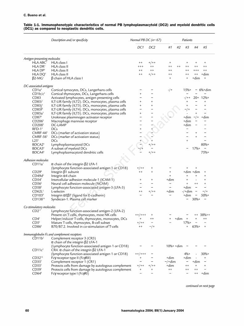
Figure 3. Representative bivariate dot plots showing thedifferent pattern of expression of CD116 (GM-CSF recep-tor) and CD123 (IL-3 receptor alpha chain) in peripheralblood DC1/myeloid dendritic cells (gray dots) andDC2/lymphoplasmacytoid dendritic cells (black events)from a healthy adult subject.
haematologica 2004; 89(1):January 2004 67
CD4+/HLA DRhi dendritic cell malignancies
itivity for CMRF44 and CMFR56 has only been observedin normal DC after overnight activation,33,43 these mark-ers not being expressed on resting freshly obtained nor-mal PB DC. Therefore, the presence of these moleculeson the surface of fresh blast cells from case #4 couldreflect a state of in vivo activation. Such phenotypicfeatures of case #4 are different from those found fornormal BDCA2+/BDCA4+ DC2/lymphoplasmacytoid DC,which are BDCA3−, CD85a−, CMRF44− and CMRF56−, asalso found for the malignant cells in cases #3 and #5.Although this phenotypic profile could support aDC1/myeloid DC origin for case #4, a DC2/lymphoplas-macytoid DC origin cannot be ruled out, since thesecells also expressed CD123hi and CD45RA, and they wereCD11c−, which are considered to be characteristic fea-tures of DC2/lymphoplasmacytoid DC. Differences werefound between case #4 and the other patients asregards expression of the CD32 and CD64 IgG receptors,as well as the CXCR1 chemokine receptor. Interesting-ly, case #4 did not have skin involvement, but bothsplenomegaly and lymphoadenopathy were present,further supporting the notion that different patterns ofexpression of chemokine receptors might be associat-ed with different homing of neoplastic DC precursors.
In the remaining four cases (cases #1, 2, 3 and 5),reactivity for different markers, other than those direct-ly related to the DC lineage, was found. The expressionof lymphoid-associated markers in cases #1, 3 and 5, aswell as co-expression of CD117 and the myeloid anti-gens CD13 and CD33 in case #5 were particularly inter-esting. Despite co-expression of lymphoid and myeloid-associated antigens, case #5 did not score more than 2points for either the myeloid or for the lymphoid line-age, when the EGIL criteria were followed strictly:27
CD117 was found to be present in <20% blast cells, andno cytoplasmic, but surface CD22 expression was found.Moreover, it should also be taken into account thatCD33, CD13 and CD117 are present during the earlieststages of hematopoiesis35 and that MPO, considered tobe the most specific marker for the myeloid lineage,was negative in this case. In turn, normal human PBlymphoplasmacytoid DC have recently been shown toexpress several lymphoid-associated markers, such asCD2, CD4, CD7 and CD22, as well as the myeloid-asso-ciated marker CD33 in the absence of MPO.9 In addition,most blast cells from this case, apart from showing pos-itivity for the referred myeloid- and lymphoid-associ-ated markers, also expressed BDCA2 and BDCA4. Theselatter two markers have been shown to be specificallyand exclusively expressed on lymphoplasmacytoid DC innon-cultured blood and bone marrow,42 further sup-porting our belief that the malignant cells from case #5were of lymphoplasmacytoid DC origin. Although in cas-es #1, 2 and 3 neither BDCA2 nor BDCA4 was tested,
and certainly in cases #1 and 2 few DC-associatedmarkers were analyzed, all these three cases displayedtypical phenotypic patterns similar to those of lympho-plasmacytoid DC for the remaining markers tested,including strong expression of HLA-I, HLA-II and CD123,and positivity for CD4 in the absence of reactivity forlineage-specific markers; molecular data further sup-ported their non-T and non-B lymphoid origin, since inall these cases TCRγand IgH genes displayed a germlineconfiguration. Recently, Trimoreau et al. reported that,as on normal DC2 dendritic cells, co-expression of highlevels of CD123 and CD45RA when CD116 and CD45ROare both absent or only dimly expressed can be consid-ered as a profile of DC2 malignancy.43 Although thesemarkers were not all systematically tested in our series,in line with these findings, we found that this pheno-typic profile was displayed by tumor cells from cases #1(with the exception of CD45RA, which was negative inthis case), case #3 and #5, further supporting theirDC2/lymphoplasmacytoid DC origin.
Although few studies have so far analyzed the clini-cal and biological characteristics of lymphoplasmacy-toid DC malignancies, there is general agreement thanthe disease has an aggressive clinical course.26,44,45
Accordingly, in most cases patients show skin involve-ment in the presence or absence of organomegalies andcytopenias. In our patients, infiltration of PB and/or BMwas typically found already at diagnosis and increasedduring disease evolution. Previous reports indicate thatCD4+/CD56+ lymphoplasmacytoid DC malignancies havean aggressive clinical evolution and a poor outcome,despite morphologic and clinical remission beingachieved in most cases.18,26 In line with these observa-tions, all four cases reported here who were treated withintensive chemotherapy, achieved complete remission.Despite this, three of them relapsed soon after achiev-ing the remission, while the other patient has remainedin continuous complete remission for 54 months. Inter-estingly, while the three patients who relapsed weretreated with chemotherapy alone, the patient with con-tinuing complete remission had received standard AML-oriented chemotherapy followed by an ASCT. Theseresults suggest that, in these patients, consolidationtherapy with ASCT, once CR has been achieved, might bemore effective than the use of chemotherapy alone, inaccordance with the results reported by Feuillard et al.26
In any case, our study seems to confirm the high remis-sion and relapse rates with conventional chemotherapyregimens and supports the suggestion by Feuillard et al.26
that consolidation with an allogeneic or autologoustransplant might be the treatment of choice, particular-ly if applied immediately after remission.
In summary, in the present study we show that DC-derived malignancies can present as a cutaneous malig-

haematologica 2004; 89(1):January 200468
C. Bueno et al.
nancy or as acute leukemia, and that their incidence isextremely low (<1%). Although most of these DC malig-nancies probably correspond to the neoplastic counter-part of DC2/lymphoplasmacytoid DC, neoplasias ofmyeloid DC might also exist. Despite the low number ofcases studied, our results also support the notion thatconventional chemotherapy alone is associated with a
high remission rate but early relapses in these individ-uals.
Contributions. All authors gave substantial contributions to theconception and design of the study, and to acquisition and analysisof data. The final manuscript was written by AO and critically revisedand approved by all the remaining authors. The authors reported noconflict of interest.
Received on December 13, 2002, accepted November 3, 2003.
References
1. Steinman RM. The dendritic cell systemand its role in immunogenicity. Annu RevImmunol 1991;9:271-96.
2. Hart DNJ. Dendritic cells: unique leuko-cyte populations which control the pri-mary immune response. Blood 1997;90:3245-87.
3. Steinman RM. Dendritic cells and immune-based-therapies. Exp Hematol 1996; 24:859-62.
4. Banchereau J, Steinman RM. Dendriticcells and the control of immunity. Nature1998;392:245-52.
5 Hock DD, Starling GC, Daniel PB, Hart DN.Characterization of CMRF-44, a novelmonoclonal antibody to an activationantigen expressed by the allostimulatorycells within peripheral blood, includingdendritic cells. Immunology 1994;83:573-81.
6. Zhou L, Schwarting R, Smith HM, TedderTF. A novel cell-surface molecule express-ed by human interdigitating reticulumcells, Langerhans cells, and activated lym-phocytes is a new member of the Ig super-family. J Immunol 1992;149:735-42.
7. Dzionek A, Fuchs A, Schmidt P, Cremer S,Zysk M, Miltenyi S, et al. BDCA2, BDCA3and BDCA4: three markers for distinctsubsets of dendritic cells in human peri-pheral blood. J Immunol 2000;165:6037-46.
8. Dzionek A, Showa Y, Nagafune J, Cella M,Colonna M, Fachetti F, et al. A novel pla-macytoid dendritic cell-specific type II C-type lectin mediates antigen capture andis a potent inhibitor of interferon α/βinduction. J Exp Med 2001;194:1823-34.
9. Almeida J, Bueno C, Alguero MC, SanchezML, Cañizo MC, Fernandez ME, et al.Extensive characterization of the immu-nophenotype and pattern of cytokine pro-duction by distinct subpopulations ofnormal human peripheral blood MHCII++/lineage- cells. Clin Exp Immunol1999; 118:392-401.
10. Almeida J, Bueno C, Alguero MC, SanchezML, de Santiago M, Escribano L, et al.Comparative analysis of morphological,cytochemical, immunophenotypical andfunctional characteristics of normalhuman peripheral blood lineage-CD16+/HLA-DR+/CD14-/lo cells, CD14+ monocytesand CD16- dendritic cells. Clin Immunol2001;100:325-38.
11. Reid CD, Fryer PR, Kirk A, Tikerpae J,Knight SC. Identification of hematopoi-etic progenitors of macrophages and den-dritic Langerhans cells (DL-CFU) in humanbone marrow and peripheral blood. Blood1990;76:1139-49.
12. Inaba K, Steinman RM, Pack MW, Aya H,
Inaba M, Sudo T, et al. Identification ofproliferating dendritic cell precursors inmouse blood. J Exp Med 1992;175:1157-67.
13. Scheicher C, Mehlig M, Zecher R, Reske K.Dendritic cells from mouse bone marrow:in vitro differentiation using low doses ofrecombinant granulocyte-macrophagecolony-stimulating factor. J ImmunolMethods 1992;154:253-64.
14. Thomas R, Lipsky PE. Human peripheralblood dendritic cell subsets. Isolation andcharacterization of precursor and matureantigen-presentingcells. J Immunol 1994;153:4016-28.
15. Olweus J, BitMansour A, Warnke R, Thom-son PA, Carballido J, Picker LJ, et al. Den-dritic cell ontogeny: a human dendriticcell lineage of myeloid origin. Proc NatlAcad Sci USA 1997;94:12551-6.
16. O’Doherty U, Peng M, Gezelter S, Swig-gard W, Betjes M, Bhardwaj N, et al.Human blood contains two subsets ofdendritic cells, one immunologicallymature and the other immature. Immu-nology 1994;82:487-93.
17. Lucio P, Parreira A, Orfao A. CD123hi den-dritic cell lymphoma: an unusual case ofnon-Hodgkin lymphoma. Ann Intern Med1999;131:549-50.
18. Chaperot L, Bendriss N, Manches O,Gressin R, Maynadie M, Trimoreau F, etal. Identification of a leukemic counter-part of the plasmacytoid dendritic cells.Blood 2001;97:3210-7.
19. Brody JP, Allen S, Schulman P, Sun T, ChanWC, Friedman HD, et al. Acute agranularCD4-positive natural killer cell leukemia:comprehensive clinicopathologic studiesincluding virologic and in vitro culturewith inducing agents. Cancer 1995;75:2474-83.
20. Kameoka J, Ichinochasama R, Tanaka M,Miura I, Tomiya I, Takahashi S, et al. Acutaneous agranular CD2−CD4+CD56+
“lymphoma”: report of two cases andreview of the literature. Am J Clin Pathol1998;110:478-88.
21. Adachi M, Maeda K, Takekawa M, Hino-da Y, Imai K, Sugiyama S, et al. Highexpression of CD56 (N-CAM) in a patientwith cutaneous CD4-positive lymphoma.Am J Hematol 1994;47:278-82.
22. Bagot M, Bouloc A, Charue D, Wechsler J,Bensussan A, Boumsell L. Do primarycutaneous non-T non-B CD4+ CD56+ lym-phomas belong to the myelo-monocyticlineage? J Invest Dermatol 1998; 111:1242-4.
23. Uchiyama N, Ito K, Kawai K, Sakamoto F,Takaki M, Ito M. CD2−, CD4+, CD56+ agran-ular natural killer cell lymphoma, of theskin. Am J Dermatopathol 1998;20: 513-7.
24. Petrella T, Dalac S, Maynadie M, Mu-
gneret F, Thomine E, Courville P, et al.CD4+/CD56+ cutaneous neoplasm: a dis-tinct hematological entity? Groupe Fran-cais d’Etude des Lymphomes Cutanes(GFELC). Am J Surg Pathol 1999;23:137-46.
25. Harris NL, Jaffe ES, Diebol J, Flandrin G,Muller-Hermelink HK, et al. The WorldHealth Organization classification of neo-plastic diseases of the hematopoietic andlymphoid tissues. Report of the ClinicalAdvisory Committee meeting, AirlieHouse, Virginia, November, 1997. AnnOncol 1999;10:1419-32.
26. Feuillard J, Jacob MC, Valensi F, MaynadieM, Gressin R, Chaperot L, et al. Clinicaland biologic features of CD4+ CD56+
malignancies. Blood 2002;99:1556-63.27. Bene MC, Castoldi G, Kwapp N, Ludwi
WD, Matutes E, Orfao A, et al. Proposalsfor the immunological classification ofacute leukemias. European Group for theImmunological Characterization of Leu-kemias (EGIL). Leukemia 1995;10: 1783-6.
28. Bennett JM, Catovsky D, Daniel MT,Fladrin G, Galton DA, Gralnick HR, et al.Proposed revised criteria for the classifi-cation of acute myeloid leukemia: areport of the French-American-BritishCooperative Group. Ann Intern Med 1985;103:620-5.
29. Bennett JM, Catovsky D, Daniel MT, Flan-drin G, Galton DA, Gralnick HR, et al. Pro-posal for recognition of minimally differ-entiated acute myeloid leukemia (AML-M0). Br J Hematol 1991;78:325-9.
30. Lopez-Perez R, Garcia-Sanz R, GonzalezD, Balanzategui A, Chillo MC, Alaejos I, etal. Gene scanning of VDJH-amplified seg-ments is a clinically relevant technique todetect contaminating tumor cells in theapheresis products of multiple myelomapatients undergoing autologous periph-eral blood stem cell transplantation. BoneMarrow Transplant 2001;28:665-72.
31. Delabesse E, Burtin ML, Millien C, Ma-donik A, Arnult B, Beldjord K, et al. Rapid,multifluorescent TCRG V γ and Jγ typing:application to T cell acute lymphoblasticleukemia and to the detection of minorclonal populations. Leukemia 2000;14:1143-52.
32. Tabernero MD, Bortoluci AM, Alaejos I,Lopez-Berges MC, Rasillo A, Garcia-SanzR, et al. Adult precursor B-ALL withBCR/ABL gene rearrangements displays aunique immunophenotype based on thepattern of CD10, CD34, CD13 and CD38expression. Leukemia 2001;15:406-14.
33. MacDonald KPA, Munster DJ, Clark GJ,Dzionek A, Schmitz J, Hart DNJ. Charac-terization of human blood dendritic cellsubsets. Blood 2002;100:4512-20.
34. Petrella T, Comeau MR, Maynadie M,
haematologica 2004; 89(1):January 2004 69
CD4+/HLA DRhi dendritic cell malignancies
Couillault G, De Muret A, Maliszewski CR,et al. Agranular CD4+/CD56+ hemato-dermic neoplasm (blastic NK cell lym-phoma) originates from a population ofCD56+ precursor cells related to plasma-cytoid monocytes. Am J Surg Pathol2002;26:852-62.
35. Macedo A, Orfao A, Vidriales MB, Lopez-Berges MC, Valverde B, Gonzalez M, etal. Characterization of aberrant pheno-types in acute myeloblastic leukemia.Ann Hematol 1995;70:189-94.
36. Garcia-Sanz R, Gonzalez M, Orfao A,Moro MJ, Hernandez JM, Borrego D, et al.Analysis of natural killer-associated anti-gens in peripheral blood and bone mar-row of multiple myeloma and prognosticimplications. Br J Haematol 1996;93:81-8.
37. Lima M, Teixeira A, Fonseca S, GonçalvesC, Guerra M, Queiros ML, et al. Immuno-phenotypic aberrations, DNA content,and cell cycle analysis of plasma cells inpatients with myeloma and monoclonal
gammopathies. Blood Cells Mol Dis 2000;2:634-45.
38. Raspadori D, Damiani D, Lenoci M, Ron-delli D, Testoni N, Nardi G, et al. CD56antigenic expression in acute myeloidleukemia identifies patients with poorclinical prognosis. Leukemia 2001;15:1161-4.
39. Mann KP, DeCastro CM, Liu J, Moore J,Bigner SH, Traweek ST. Neural cell adhe-sion molecule (CD56)-positive acutemyelogenous leukemia and myelodys-plastic and myeloproliferative syndromes.Am J Clin Pathol 1997;107:653-60.
40. Doherty DG, Norris S, Madrigal-Estebas L,McEntee G, Traynor O, Hegarty JE, et al.The human liver contains multiple popu-lations of NK cells, T cells and CD3+ CD56+
natural T cells with distinct cytotoxicactivities and Th1, Th2 and Th0 cytokinesecretion patterns. J Immunol 1999;163:2314-21.
41. Khan WA, Yu L, Eisenbery Crisan D, Alsaa-di A, Daris BH, et al. Hepatosplenic gam-
ma/delta T-cell lymphoma in immuno-compromised patients. Report of twocases and review of literature. Am J ClinPathol 2001;116:41-50.
42. Lopez JA, Bioley G, Turtle CJ, Pinzon-Charry A, Hoc S, Vuckovic S, et al. Singlestep enrichment of blood dendritic cellsby positive immunoselection. J ImmunolMethods 2003;274:47-61.
43. Trimoreau F, Donnaed M, Turture P,Gachard N, Bordessoule D, Feuillard J. TheCD4+, CD56+, CD116−, CD123+, CD45RA+,CD45RO- profile is specific of DC2 malig-nancies. Haematologica 2003;88:ELT10.
44. Thomas DA, Kantarjian HM. Lymphoblas-tic lymphoma. Hematol Oncol Clin NorthAm 2001;15:51-95.
45. Zinzani PL, Bendandi M, Visani G, Gher-linzoni F, Frezza G, Merla E, et al. Adultlymphoblastic lymphoma: clinical fea-tures and prognostic factor in 53patients. Leuk Lymphoma 1996;23:577-82.
Copyright © 2022 FDOKUMEN