LOSS OF CD4 MEMBRANE EXPRESSION AND CD4 rnRNA DURING ACUTE HUMAN IMMUNODEFICIENCY VIRUS REPLICATION
17
LOSS OF CD4 MEMBRANE EXPRESSION AND CD4 rnRNA DURING ACUTE HUMAN IMMUNODEFICIENCY VIRUS REPLICATION BY PATRICK SALMON,* RENE OLIVIER,I YVES RIVIERE,I EDITH BRISSON,* JEAN-CLAUDE GLUCKMAN,* MARIE-PAULE KIENY,§ LUC MONTAGNIER,I AND DAVID KLATZMANN* From *the Laboraioire de Biologie et Genetique des Deficits Immunitaires, Faculte de Medecine Pitie-Salpetriere, 75651 Paris; Ithe Unite dOncologie Virale, Institut Pasteur, 75524 Paris; and $Transgene, 67000 Strasbourg France HIV is the only retrovirus that has as of now an identified receptor, the CD4 mol- ecule (1-3), to which its outer membrane glycoprotein (gp110) binds with high affinity (4, 5) . This glycoprotein is produced from the cleavage of a gp160 precursor gener- ating gp110 and the gp41 transmembrane protein (6) . This receptor/envelope inter- action occurs in three distinct situations. Envelope gp110 expressed on a viral par- ticle binds membrane expressed CD4 on target cells (4), controlling the host range and tissue specificity of virus infection (7-9) ; gp110 expressed on the surface of an infected cell, can bind to CD4 expressed on other(s) cell(s), inducing cell fusion and syncytium formation (10, 11) . Finally, CD4 and gp110 can interact within a single cell, inducing the formation of immunoprecipitable gp110-CD4 complexes that are thought to account for the disappearance of CD4 from the cell surface of HIV infected cells (7, 12) . The presence of such receptor/envelope complexes in infected cells has been postulated for many other retroviruses, causing the so called "interference" phenomenon ;i .e., cells infected with a given strain of avian leukemia virus (ALV)r are resistant to infection with other related but distinct strains of this virus (13) . This is commonly viewed as resulting from the occupancy of the receptor, used by both retroviruses, by endogeneously produced envelope of the first strain . Since the nature of the receptor for these viruses is unknown, it has not yet been possible to verify this hypothesis . However, modification of the receptor synthesis at the tran- scriptional or post-transcriptional levels could also be involved in down-modulation of retrovirus receptor, as it has been proposed for HIV . Hoxie et al . (12) have shown that CD4+ cells infected with HIV presented decreased levels of both CD4 mRNA and protein as compared with the initial uninfected cells after 2 to 3 mo of chronic viral replication . From these experiments, the authors could not discriminate be- tween regulation of CD4 gene transcription or selection of low CD4-expressing sub- clones, more resistant to HIV cytopathic effect . In contrast, Stevenson et al . (14), This work was supported by University Paris VI, the Caisse Nationale dAssurance Maladie des Travaiileurs Salaries, the Programme National de Recherche sur le SIDA, and the Korber Stiftung (Hamburg, FRG). P . Salmon is a fellow of the Association pour la Recherche sur le Cancer, Villejuif, France . ' Abbreviations used in this paper: ALV, avian leukemia virus ; HI-VV HIV vaccine recombinant virus ; nt, nucleotide; PFU, plaque-forming units ; RT, reverse transcriptase ; SB, staining buffer . J . Exp . MEo . © The Rockefeller University Press - 0022-1007/88/12/1953/17 $2 .00 1953 Volume 168 December 1988 1953-1969 on May 20, 2015 jem.rupress.org Downloaded from Published December 1, 1988
-
Upload
independent -
Category
Documents
-
view
0 -
download
0
Transcript of LOSS OF CD4 MEMBRANE EXPRESSION AND CD4 rnRNA DURING ACUTE HUMAN IMMUNODEFICIENCY VIRUS REPLICATION

LOSS OF CD4 MEMBRANE EXPRESSION AND CD4 rnRNADURING ACUTE HUMAN IMMUNODEFICIENCY
VIRUS REPLICATION
BY PATRICK SALMON,* RENE OLIVIER,I YVES RIVIERE,I EDITH BRISSON,*JEAN-CLAUDE GLUCKMAN,* MARIE-PAULE KIENY,§ LUC MONTAGNIER,I
AND DAVID KLATZMANN*From *the Laboraioire de Biologie et Genetique des Deficits Immunitaires, Faculte de Medecine
Pitie-Salpetriere, 75651 Paris; Ithe Unite dOncologie Virale, Institut Pasteur, 75524 Paris; and$Transgene, 67000 Strasbourg France
HIV is the only retrovirus that has as of now an identified receptor, the CD4mol-ecule (1-3), to which its outer membrane glycoprotein (gp110) binds with high affinity(4, 5) . This glycoprotein is produced from the cleavage of a gp160 precursor gener-ating gp110 and the gp41 transmembrane protein (6) . This receptor/envelope inter-action occurs in three distinct situations. Envelope gp110 expressed on a viral par-ticle binds membrane expressed CD4 on target cells (4), controlling the host rangeand tissue specificity of virus infection (7-9); gp110 expressed on the surface of aninfected cell, can bind to CD4 expressed on other(s) cell(s), inducing cell fusion andsyncytium formation (10, 11) . Finally, CD4 and gp110 can interact within a singlecell, inducing the formation of immunoprecipitable gp110-CD4 complexes that arethought to account for the disappearance ofCD4 from the cell surface ofHIVinfectedcells (7, 12). The presence of such receptor/envelope complexes in infected cells hasbeen postulated for many other retroviruses, causing the so called "interference"phenomenon ; i.e., cells infected with a given strain of avian leukemia virus (ALV)rare resistant to infection with other related but distinct strains of this virus (13) .This is commonly viewed as resulting from the occupancy of the receptor, used byboth retroviruses, by endogeneously produced envelope of the first strain . Since thenature of the receptor for these viruses is unknown, it has not yet been possible toverify this hypothesis . However, modification of the receptor synthesis at the tran-scriptional or post-transcriptional levels could also be involved in down-modulationofretrovirus receptor, as it has been proposed for HIV. Hoxie et al . (12) have shownthat CD4+ cells infected with HIV presented decreased levels of both CD4 mRNAand protein as compared with the initial uninfected cells after 2 to 3 mo of chronicviral replication . From these experiments, the authors could not discriminate be-tween regulation ofCD4 gene transcription or selection of low CD4-expressing sub-clones, more resistant to HIV cytopathic effect . In contrast, Stevenson et al . (14),
This work was supportedbyUniversity Paris VI, theCaisse Nationale dAssurance Maladie des TravaiileursSalaries, the Programme National de Recherche sur le SIDA, and the Korber Stiftung(Hamburg, FRG).P. Salmon is a fellow of the Association pour la Recherche sur le Cancer, Villejuif, France .
' Abbreviations used in this paper: ALV, avian leukemia virus ; HI-VV HIV vaccine recombinant virus ;nt, nucleotide; PFU, plaque-forming units ; RT, reverse transcriptase ; SB, staining buffer.
J . Exp . MEo . © The Rockefeller University Press - 0022-1007/88/12/1953/17 $2 .00
1953Volume 168 December 1988 1953-1969
on May 20, 2015
jem.rupress.org
Dow
nloaded from
Published December 1, 1988

1954
CD4 AND HUMAN IMMUNODEFICIENCY VIRUS
using a subclone of the CEM line that was resistant to HIV cytopathic effect, pro-posed that upon HIV replication, a post-transcriptional mechanism affected CD4protein production but also that of other T cell membrane markers . In both cases,these studies were conducted in established cell lines and did not necessarily reflectwhat really occurred in HIVinfected normal lymphocytes .We have studied membrane expression and gene regulation of CD4 during acute
HIV replication in normal CD4 lymphocytes as well as in CEM cells and after in-fection with HIV/vaccinia recombinant viruses (HIV-VV) expressing mutated envgene . We show that both CD4/gp110 complex formation and the rapid disappear-ence of CD4 mRNAs account for the modulation of the HIV receptor.
Materials and MethodsPreparation, Infection, and Culture of Cells .
PBMC were obtained from HIV- normal indi-viduals, using the Ficoll-Hypaque (Pharmacia, Uppsala, Sweden) separation method . Whennecessary, PBMC were depleted ofCD8' T lymphocytes by specific cytotoxicity with mAbIOT8a (Immunotech, Marseille, France) and rabbit complement (Biokar, Pantin, France)before infection . After cytotoxicity, <1% of CD8' cells remain in the CD4'-enriched PBMC.The CEM subclone (CEM 13) was isolated after cloning in semi-solid agar of the originalCEM Tlymphoblastoid cell line .
Infection was performed with supernatant of cultured HIV-1-infected PBMC (LAVBRU iso-late (15), corresponding to the peak of viral production : >5.10 5 cpm/ml; >10 5 IDW (16) .PBMC or CD4'-enriched PBMC were suspended for 1 h at 370C (107 cells/0 .1 ml HIV su-pernatant). Normal and infected lymphocytes were adjusted at 106 cells/ml and cultured at37°C in an atmosphere of 5% C02 in RPMI 1640 supplemented with 10% FCS, 2 mMglutamine, 1% antibiotics (penicillin, streptomycin, neomycin ; Gibco-BRL, Uxbridge, UK)and 4 wg/ml PHA (PHA-M ; Gibco-BRL) . On day 3, cells were pelleted and resuspendedin this complete medium containing IL-2 (20 U/ml) instead of PHA. Under these cultureconditions, monocytes and B lymphocytes do not proliferate and at day 5, when we startedanalysis, <5% of nonT cells were present in the culture . CEM cells were cultured in thesame medium without PHA and IL-2 .
Viral production in the supernatant of HIV-infected cells was determined by particle-associated reverse transcriptase assay (17) and immunocapture ELISA (ELAVIA ; DiagnosticsPasteur, Marnes-la-Coquette, France) using 10-fold dilutions of cell-free supernatants . Ab-sorbance (A) was determined with an ELISA-plaque reader (Diagnostics Pasteur) at 492 nm.
Immunofluorescence Analysis.
Control and infected cells (2-5 x 10 5 cells per staining) wereharvested, rinsed in cold PBS and incubated for 30 min at 4°C with appropriate mAbs dilutedat 1 :100 to 1 :200 in staining buffer (SB) that comprised PBS, 0.5% BSA, and 0.05% sodiumazide . mAbs used were as follows : anti-CD4 : Leu3a PE (Becton Dickinson & Co., MountainView, CA), T4-FITC (Coulter Corp., Hialeah, FL), OKT4 (Ortho Diagnostic Systems, West-wood, MA); anti-T cells : T3-RDI (CD3), T11-RD1(CD2), 4134 RDl (CDw29) (Coulter Corp .),Leu2a (CD8) (BectonDickinson & Co.) ; anti-HIV: 41-1 (anti-gp41), 110-4 (anti-gp110) (GeneticSystems Corp., Seattle, WA). Unlabeled mAbs (OKT4, 41-1, 110-4) were revealed by sequen-tial incubation with biotinylated sheep anti-mouse antibodies (Amersham Corp., Bucking-hamshire, UK) at 1 :25, and phycoerythrin-conjugated streptavidin (Becton Dickinson & Co.)at 1 :25 . When double-color labeling was performed (T4/OKT4, T4/41-1, T4/110-4), controlascites fluid (Dakopatts, Copenhagen, Denmark) at 1 :25 was added to phycoerythrin-streptavidin in order to saturate the remaining binding sites of sheep anti-mouse antibodiesbefore incubation with the second mAb, T4-FITC . After staining, cells were resuspendedin SB containing 1% paraformaldehyde, kept at 4°C, and analyzed together with the FAGSanalyzer (Becton Dickinson & Co.) . As described elsewhere (18), volume versus right-anglelight scatter dot plot was used to gate viable cells and exclude debris as well as dead andgiant autofluorescent cells . In initial experiments, the volume-scatter gate was correlated withpropidium iodide staining and encloses a population with <5% of dead cells . Fluorescence
on May 20, 2015
jem.rupress.org
Dow
nloaded from
Published December 1, 1988

SALMON ET AL .
1955
intensity was then determined within these gated viable cells and plotted in a 3-decade log-scale . Results are presented as histograms (cell number versus fluorescence intensity) for single-color staining experiments, or as autoscaled contour maps of cell number in double-colorstaining experiments, each axis representing fluorescence intensity in one channel (vertical,red ; horizontal, green) .
Cytoplasmic Extracts (Cytodots).
Control and infected cells (2 .5 x 105 viable cells as mea-sured by trypan blue exclusion) were rinsed three times in cold PBS, resuspended in 100 plof Tris (10 mM, pH 7.0), 1 mM EDTA, 2 mM Vanadyl ribonucleoside complex (BethesdaResearch Laboratories, Gaithersburg, MD), 0.5% NP-40 (BDH Chemicals, Poole, England)and vortexed for 5 min on ice . Then, 10 ul of 5 % NP-40 was added, cells were vortexed for5 min on ice, and nuclei were pelleted by centrifugation at 40C for 10 min at 10 000 g, 100wl of supernatant was completed with 100 g1 of 12 x SSC (1 x SSC NaCl 0.15 M, 0.015 Mtrisodium citrate, pH 7.0) 15% formaldehyde, incubated 15 min at 65°C and spotted on anylon membrane (Hybond-N, Amersham Corp.) using the Minifold apparatus (Schleicher& Schuell, Dassel, Federal Republic of Germany) .
Northern Blots .
Total cellular RNA was extracted using the guanidinium thiocyanate/cesiumchloride method (19) . After centrifugation, RNA pellets were solubilized in 200 P1 of 10 mMTris, 1 mM EDTA, 0.1 17o SDS, pH 7.0, and were precipitated with 2.5 vol ethanol in thepresence of 0,3 M ammonium acetate. Precipitates were washed with 70% ethanol, dried,and redissolved in 200 ul diethylpyrocarbonate-treated H2O. RNA (10 wg per lane) was dena-tured for 15 min in 50% formamide, 20 mM Mops, 5 mM sodium acetate, 6% formalde-hyde, pH 7 .0, at 65°C, and was separated by electrophoresis in 1.1% agarose, 20 MM Mops,5 mM sodium acetate gels as previously described (19) . RNAs were transferred to nylon mem-branes (Hybond N, Amersham Corp.) using 20x SSC for 12-16 h .
Hybridization, Rehybridization Procedures.
Nylon membranes (cytoplasmic extracts or Northernblots) were baked for 90 min at 80°C, prehybridized, and hybridized as described elsewhere(19) . Specific probes ofCD4 (pT4B) (3), HIVA (Sac I subclone of %J19) (20), TCRS2 chain(4131) (21) and P-actin (pAL41) (22) were a-32P-radiolabeled using the random primingmethod (Multiprime DNA Labeling Systems; Amersham Corp.) . Their specific activity rangedfrom 5 to 10 x 108 cpm/ug DNA. After hybridization, filters were washed under stringentconditions (three times in 2 x SSC ; 0 .1 % SDS at room temperature, and twice for 45 minin 0 .1 x SSC; 0.1% SDS at 65°C), and exposed to XAR5 films (Eastman Kodak Co., Roch-ester, NY) in Quanta III cassette (DuPont de Nemours, Orsay, France) for 24-48 h . Whennecessary, filters were dehybridized for 2 h in 2 mM EDTA, 5 mM Tris, 1 x Denhardt's (0.02 %Ficoll, 0.02% polyvinyl pyrrolidone, 0.02 % BSA), pH 8.0, at 65°C, washed twice in 2 x SSC,0.1% SDS, and rehybridized as described above .
Construction ofHIV-Vaccinia Recombinant Viruses.
Manipulation of DNA was performed ac-cording to standard protocols and oligo-nucleotide-directed mutagenesis of subclones intoM13 vectors was performed essentially as described (23) . Construction of HIV-VV was real-ized as described (24) . For HIV-VV 1135 the sequence encoding the signal peptide of therabies virus glycoprotein gene (nucleotides [nt] 1-64 in Anilionis et al . [25]) was fused withthe first codons of mature gp110 (nt 5857 in Wain-Hobson et al . [26]) using oligonucleotide-directed mutagenesis (amino acid sequence at the site of fusion : CFGXNK) and the gp41transmembrane domain (from nt 7834) was replaced by the rabies glycoprotein counterpart(nt 1387-1714; junction KIRYVL) . The sequence containing the two Lys-Arg cleavage sitesbetween gp110 and gp41 has been altered from KAKRRVVQREKR to KAQNHVVQNEHQ(nt 7279-7314) . HIV-VV 1138 is equivalent to 1135 except that the sequences correspondingto gp110 and the NH2-terminal hydrophobic domain of gp41 have been removed by oligo-nucleotide-directed deletion (nt 5857-7398 ; junction CFG.QAF).
Infection with HIV-Vaccinia Recombinant Viruses.
PHA-stimulated PBMC or CD4'-enrichedlymphocytes were infected with HIV-VV 1135, or 1138, or the wild-type vaccinia virus for1 h at 37°C at a multiplicity of infection of 50 plaque-forming units (PFU)/cell . Cells wereharvested at various times (see Fig . 3) and investigated for membrane proteins as describedfor HIV-infected cells . The efficiency ofinfection was monitored by immunofluorescence onfixed cells with antivaccinia mouse sera .
on May 20, 2015
jem.rupress.org
Dow
nloaded from
Published December 1, 1988

1956
CD4 AND HUMAN IMMUNODEFICIENCY VIRUS
ResultsInfection of stimulated CD4+ -enriched lymphocytes with HIVcontaining super-
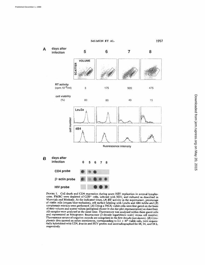
natant induces virus replication that usually becomes detectable after a few daysand peaks between day 6 and 8. The cell viability in the culture, as assessed by vitaldye staining, begins to decrease within a day after initial viral detection . After thepeak of viral replication, all cells ultimately die. To analyze the modification of cellsurface marker expression during HIV replication, at a time when viral proteinsare synthesized, it is therefore necessary to discriminate viable from dead cells . Thiscan be routinely achieved by using a cell analyzer: viable cells can be distinguishedfrom dead cells and debris on the basis of their volume and right-angle light scattervalues (18) . Using these two parameters, a gate can be drawn around the living cellscluster. Unlike dead cells, such gated cells exclude propidium iodide, a fluorescentvital dye, and are not stained by irrelevant antibodies . Thus, even at the time whenmassive death occurred in the infected cell culture, specific fluorescence could stillbe analyzed in the remaining viable gated cells, as shown in Fig. 1. In this experi-ment, till day 6 of the culture, most cells are viable . A day later, after the first detec-tion of reverse transcriptase (RT) in the culture supernatant, the number of viablecells progressively decreased, as shown by both the scatter-volume dot plot and thecount of cells excluding trypan blue . We have analyzed the expression of the CD4molecule within the gated cell population using Leu3a mAb, and as a control weanalyzed the expression of the CDw 29 molecule (27) detected by the mAb 4B4.We often used this mAb because CDw29 is also highly expressed on the CD4+CEM line for which we performed similar experiments . Throughout the culturetime, no gross modification of CDw29 expression could be observed . On the con-trary, gated cells expressing Leu3a gradually decreased in number andwere almostabsent at the end of the experiment . The rapidity of the appearance of theseLeu3a- ,4B4+ viable cells, as well as the absence of detection of CD8+ cells (datanot shown), rules out the possible emergence of a population of CD4- con-taminating cells . Thus, loss of Leu3a staining could not be explained only by thedeath of HIV-replicating CD4+ cells .
Co-expression ofgp110 and CD4 on HIVReplicating CD4+ Cells .
To more preciselystudy modulation ofCD4at the surface ofHIV-replicating cells we performed double-staining of CD4+ enriched cells that were infected with HIV. Cells were labeledeither with anti-gp41 or anti-gp110 mAbs or with different mAbs recognizing theCD4 molecule . Recently, amino acid substitution in the CD4 molecule allowed thelocalization of the epitopes recognized by the anti-CD4 mAbs as well as the HIVbinding site . Leu3a(Becton Dickinson & Co.) andT4 (Coulter Immunology) recog-nize epitopes in the very vicinity or at the HIV binding site (28) . They block HIVbinding to CD4and, reciprocally HIV blocks their binding to CD4 (4, 8, 29). OKT4(Ortho Diagnostic Systems) recognizes an epitope more distant from HIV bindingsite and it does not interfere with the interaction between gp110 and CD4 (4, 8, 29).Using double-color staining procedure with OKT4, anti-gp41, or anti-gp110 mAbson one hand (red, vertical axis) and T4 mAb on the other hand (green, horizontalaxis), and after gating using volume-scatter dot blot as described in Fig. 1, we ob-served the emergence of three distinct populations of cells from the initiallyT4`OKT4'gpll0 - lymphocytes (Fig . 2) : (a) gp110+OKT4+T4 + cells ; (b)gp110+OKT4+T4- cells ; and (c) gp110+ OKT4- T4- cells . Until day 4, infected cells
on May 20, 2015
jem.rupress.org
Dow
nloaded from
Published December 1, 1988
SALMON ET AL,
Fm=E I,
CHI death and CD4 expression during acute HIV replication in normal lympho-cytes. PMBC were depleted of CD8' cells, infected with HIV, and cultured as described inMaterials and Methods. At the indicated times, (A) RT activity in the supernatant, percentageof viable cells (trypan blue exclusion), cell surface labeling with Leu3a and 4114 mAbs andQcytoplasmic extracts were performed. (A) Using a FAGS, viable cellswere first gated on the basisoftheir volume and scatter values (polygonal cluster in the dot-plot representation) as described.All samples were analyzed at the same time. Fluorescence was analyzed within these gated cellsand represented as histograms: fluorescence (3-decade logarithmic scale) versus cell number.Fluorescence means ofnegative controls are comprised in the first decade (not shown). (B) Cyto-plasmic dots spotted on nylon membranes, corresponding to 2.5 x 10 5 viable cells, were sequen-tially hybridized with CD4, P-actin and HIV probes and autoradiographed for48, 24, and 24 h,respectively,
1957
on May 20, 2015
jem.rupress.org
Dow
nloaded from
Published December 1, 1988

1958
CD4 AND HUMAN IMMUNODEFICIENCY VIRUS
FIGURE 2 .
Coexpression ofCD4 and HIVglycoproteins at the surface of HIVinfected lympho-cytes . CD4+ enriched lymphocytes were infected and cultured as described in Fig. 1 . At the in-dicated times, (A) cell surface staining, (8) viral production in the supernatant (immunocaptureELISA), percentage of viable cells ("pan blue exclusion), and (C) cytoplasmic extracts wereperformed as described in Materials and Methods. (A) Infected cells (indicated time)andnonin-fected cells (day 5) were monitored for cell surface markers. Staining with OKT4 (CD4), 41-1(gp41), and 110-4 (gp110) mAbs was analyzed in the red channel (phycoerythrin, vertical axis) .T4 (CD4) was analyzed in the green channel (fluorescein, horizontal axis). Before fluorescenceanalysis, cells were gated on the basis oftheir volume and right-angle light scatter as describedin Fig. I (not shown). Clusters represent auto-scaled contour graphs of cell number, and fluores-cence intensity was plotted in 3-decade logarithmic scales. Quadrants were set usingnoninfectedcells (day 5) stained with control antibodies (left) : biotinylated sheep anti-mouse + phycoerythrin-streptavidin (spe) (red negative, green negative), Leu3a (red positive, green negative), and T4(red negative, green positive). (C) Cytoplasmic dots on nylon membranes corresponding to 2.5x 10⁵ viable cells were hybridized with CD4, HIV and p-actin probes . CD4 and HIV probingwere performed sequentially on the same filter. R-actin probing was performed on an indepen-dent filter. Autoradiography times were 48, 24, and 24 h, respectively.
kept their original gp110-OKT4'T4' phenotype. On day 5, a significant popula-tion ofgp110+OKT4+ T4' cells could be detected, few syncytia were noted and viralantigens detected by ELISA appeared in the supernatant . On day 6, at the timewhen significant loss of viability in the culture was observed, most cells were
on May 20, 2015
jem.rupress.org
Dow
nloaded from
Published December 1, 1988

SALMON ET AL .
1959
gp110+OKT4+T4 - while some were already gp110+OKT4- T4-. At this time gp41appeared at the cell surface. Subsequently, this latter population increased and cellsfinally died. In these experiments, the expression of control antigens CD2 and 4B4remained unchanged at the cell surface of gated cells (data not shown) . The almostcomplete disappearance of T4 staining in the face of the persistent OKT4 expres-sion indicated that CD4 molecule was still present at the lymphocyte membraneat this time of viral replication (day 6), but in a configuration in which the epitoperecognized by T4 mAb was no longer accessible . This, and co-expression of gp110,argued for the existence ofCD4in a complexed form with gp110. Indeed, gp110 bindingto CD4 during adsorption of inactivated virions, or after incubation with purifiedgp110, induces similar modification of OKT4a but not of OKT4 staining (4, 30),andHIV gp/CD4 complexes can be immunoprecipitated by OKT4 but not OKT4amAbs (12) .CD4 Expression after HIV-Vaccinia Recombinant Virus Infection .
It was not possiblefrom these experiments to determine to what extent gp110 was produced by infectedcells and presented to their membrane before the assembly of viral particles, or wasin the form of soluble gp110 passively adsorbed on uninfected CD4+ cells, or per-haps at the surface ofmature virions that were in the process ofinfecting new cells,both cases being responsible for CD4 masking (8, 30, 31).To demonstrate that interaction between CD4 and HIV envelope actually occurred
in each individual cell, we used vaccinia recombinant virus . Infection with such virusesallows the expression of foreign genes in vitro as well as in vivo (32) and is currentlythe best way to express high levels of normal or mutated HIVena gene products inthe infected cells (10, 11, 24, 33, 34). Initial experiments with normal ena gene hadshown an important shedding of gp110 from the cell surface ofHIV-VV-infected cells(24) . We therefore used a construction designed to optimize expression of HIV envelopeat the cell membrane . In this construction (1135), the two cleavage sites that generategp110 and gp41 from the gp160 precursor (Fig . 3 A) were inactivated by amino acidsubstitutions (see Materials and Methods). The signal peptide, the transmembraneand intracytoplasmic regions ofHIV were replaced by those of rabies virus envelopeglycoprotein . As control, we used a similar construction containing only the gp41external region (1138) and also the wild-type vaccinia virus. High multiplicity ofinfection (50 PFU/cell) allowed synchronous, rapid, and massive infection of morethan 90% of the cells, as determined by immunofluorescence with specific mousesera to vaccinia antigens, further leading to cell death within 24-36 h. We first per-formed a time course experiment (Fig . 3 B) in which normal PBMC were infectedby the two recombinants. Stainings using 4B4 and Leu3a mAbs were analyzed asdescribed for HIV infection . While expression of a control molecule like CDw29remained unchanged until 24 h after infection, a progressive decrease of Leu3a ex-pression was observed after infection with HIV-VV 1135 but not with HIV-VV 1138 .This effect, which appeared as early as 6 h after infection, could be clearly seen at16 h and was almost complete at 24 h. Subsequent experiments were performed be-tween 20 and 24 h after infection . We then analyzed expression of CD4 and HIVenvelope after HIV-VV infection as we did after HIV infection (Fig . 3 C) . Infectionwith the wild-type vaccinia or HIV-VV 1138 did not modify membrane expressionof either Leu3a or OKT4 epitopes of the CD4 molecule, nor did they modify theexpression ofcontrol molecules like CDw29 or CD3 (data not shown) . Infection with
on May 20, 2015
jem.rupress.org
Dow
nloaded from
Published December 1, 1988

1960
CD4 AND HUMAN IMMUNODEFICIENCY VIRUS
FIGURE 3.
Expression ofCD4and HIV glycoproteins after infection with vaccinia recombinantviruses expressing HIVenv gene products . (A) HIV eno gene (HIV) and sequences inserted inthe thymidine kinase gene of HIV-VV 1135 (1135) andHIVVV 1138 (1138) are represented . HIVsequences (clear boxes) and rabies virus sequences (shaded boxes) were fused as described in Materialsand Methods. The whole gp41 sequence comprised boxes named gp41' (NHZ-terminal and ex-ternal regions), TM (transmembrane region), and CYT(intracytoplasmic region) . In HIVVV1138, gp41"" represents the external region of gp41 fused with rabies glycoprotein signal peptide(SP, left) and transmembrane (TM) and cytoplasmic (CYT) rabies glycoprotein regions. Arrowsindicate the two cleavage sites described in text and Materials and Methods. (B) At the indicatedtimes, PBMC infected with HIV-VV 1135 or HIV-VV 1138 were labeled with Leu3a and 4B4
on May 20, 2015
jem.rupress.org
Dow
nloaded from
Published December 1, 1988

SALMON ET AL.
1961
HIV-VV 1135 led to the expression of the hybrid envelope onto the cell surface thatcan be detected with anti-gp110 mAb. This was accompanied by the complete disap-pearance of the Leu3a epitope, while the reduction of OKT4 staining was aboutfourfold . In this experiment, a twofold decrease of 4B4 could be observed, aphenomenon that was not, however, consistently observed (Fig. 3 B) . Since HIVglycoprotein produced by HIV-VV 1135, like its analogous construct lacking enve-lope cleavage sites, is not shed in the culture supernatant (35), it was concluded thatin each individual cell the glycoprotein-CD4 interaction was mediated by endogene-ously produced HIV glycoprotein .CD4 Transcripts Are Lost during Acute HIVReplication.
During HIV replication, thedecrease and, finally, the complete disappearance of OKT4 staining strongly sug-gested the disappearance ofexisting CD4 molecules from the cell surface . CD4-gp110complexes could be internalized or alternatively shed from the cell surface, but thisalso suggested a lack of new synthesis of CD4 molecules. One possible mechanismfor the latter assumption could be the impairment of CD4 gene expression due toHIV replication . To investigate such a possibility, we analyzed CD4mRNA duringvirus replication in Tlymphocytes andCEMcells . In the two first experiments (Figs .1 and 2) cytoplasmic extracts of 2.5 x 10⁵ viable cells were prepared and spottedon nylon membranes at the times of immunofluorescence staining . Hybridizationwith a 0-actin probe demonstrated that similar amounts of mRNAs were spotted.CD4 mRNA levels dramatically dropped on day 7 (Fig . 1) or on day 6 (Fig . 2), aday after HIVRNA and viral production could be detected . We next looked at mRNAlevels by Northern blot analysis . Viral production and CD4 membrane expressionwere sequentially monitored in cultures of HIV-infected, or uninfected PBMC con-taining both CD4+ and CD8' cells . Less than 47o of CD4- CD8- cells could bedetected throughout the culture period of control uninfected cells (Fig. 4) . On thecontrary, a significant proportion of CD4-CD8- cells emerged in the infected cul-ture ; as demonstrated in the previous experiment, they arise from the originallyCD4+CD8- cells that had lost membrane CD4 upon viral replication . Total RNAwas extracted each day from both infected and uninfected cells and 10 pg were ana-lyzed by Northern blotting . Compared with 0-actin, CD4 mRNA levels started todecrease in the infected culture on day 6, a time at which the decrease of CD4+cells and increase of CD4-CD8- cells was observed . By day 7, when the propor-tion of CD4-CD8- cells peaked, reaching approximately the percentage ofcells thatwere originally CD4+ and on day 8, CD4 mRNA levels became undetectable .Later, on day 9, CD4 mRNA remained absent, but at that time, all surviving cellswere of the CD8' phenotype . We next investigated whether this dramatic disap-pearance of CD4 mRNA could also be observed in a CD4' Tcell line, using a clonederived from CEM cells . This CD4 highly expressing clone (CEM 13) acutely repli-cates HIV and is highly sensitive to its cytopathic effects (Salmon, P, andD. Klatz-mann, unpublished observations). As shown in Fig. 5, after HIV infection CD4
mAbs and analyzed by FAGS . (C) 20 h after infection with the indicated virus, CD4' -enrichedlymphocytes were labeled with Leu2a (negative control), Leu3a, OKT4, 110-4 (gp110), and 4B4(positive control) mAbs, and were analyzed by FRCS . For each sample, mean of fluorescenceintensity and the relative fluorescence intensity were shown (calculated as follows) : 100 x[(mAb1135,1135 - Leu2a1135-1138)/(mAbwild type - Leu2a~ild type)] .
on May 20, 2015
jem.rupress.org
Dow
nloaded from
Published December 1, 1988

1962
CD4 AND HUMAN IMMUNODEFICIENCY VIRUS
FIGURE 4.
CD4 mRNA levels during acute HIVreplication in normal lymphocytes . At the in-dicated times, (A) cells were labeled with Leu2a (CD8) and Leu3a (CD4), (B) RNAs were ex-tracted and analyzed by Northern blotting, and (C) viral production in the supernatant was de-termined using an immunocapture ELISA. (A) Percentages of noninfected (ni) and infected (i)cells expressing either Leu3a or Leu2a as well as Leu2a- Leu3a cells, are shown. Leu2a- ,Leu3a- cells arose from Leu2a- ,Leu3a* cells that have lost CD4 membrane molecules uponHIV replication . (B) Northern blot was performed as described in Materials and Methods, inboth uninfected (-) and infected (+) cells . The nylon membrane was sequentially hybridizedwith CD4 and 0-actin probes . Autoradiography times were 48 h.
transcripts were almost undetectable, when no more CD4 molecules were detect-able on the cell surface as assessed by Leu3a and OKT4 staining . As controls,0-actin mRNA and a nonstructural protein mRNA coding for the TCR 0 chainwere both present at slightly decreased or comparable amounts, respectively, in in-fected and uninfected cells . This indicated that CD4 transcript accumulation wasspecifically impaired during acute HIV replication in this clone of TlymphoblastoidCEM line as for normal lymphocytes.
DiscussionWhen we initially reported the selective tropism of HIV for CD4+ T lympho-
cytes (2), we already mentioned the striking disappearance of CD4 staining on thecell surface of infected cells . This observation has been reproduced and expandedand it is now well accepted that normal CD4+ T lymphocytes, or Tlymphoblastoidcell lines, and CD4+ monocyte/macrophages or monocytic cell lines, all lose CD4
on May 20, 2015
jem.rupress.org
Dow
nloaded from
Published December 1, 1988

SALMON ET AL.
1963
FIGURE 5 .
CD4mRNA levels during acute HIV replication in aCEM clone. CEM-13 (GEMline-derived clone, see text)cells were infected and cultured as described in Materials andMethods.(A) At day 9, when viral production peaks (data not shown), cells were labeled with Leu3a andOKT4 mAbs. Control lines represent fluorescence histograms of cells treated only with biotin-coupled sheep antimouse antibodies plus phycoerythrin labeled streptavidin. (B) Northern blotanalysis of uninfected (day 0) and infected (day 9) cells . The nylon membrane was sequentiallyhybridized with CD4, TcR 02 chain, and 0-actin probes . RNA from uninfected U937 (a mono-cytic cell line) was used as negative control for TcR N2 chain signal and positive control for CD4and 6-actin signals .
expression upon HIV-1 replication (12, 36, 37), but also upon HIV-2 and SIV repli-cation (37) . However, the mechanism of such a phenomenon is unclear. This couldresult from the HIVs cytopathic effect, which would first affect cells highly expressingCD4. Indeed, different CD4+ cell lines or even individual clones from a given CD4+cell line appear to display heterogeneous sensitivity to HIV cytopathic effect, whichcorrelates with the extent of CD4 expression (38) . Impairment of CD4 expressionat the cell surface could also be due to its interaction with gp110. Such an interactioncould involve endogeneously produced gp110 (12) or alternatively soluble gp110 thatis known to be released in the culture supernatant (24, 39, 40), or even virion ad-sorption to the cell membrane . Impairment of transcriptional (12) and post-transcriptional (14) events in CD4 gene expression have also been proposed as pos-sible mechanisms . To investigate the mechanism ofCD4loss during HIVinfection,it was therefore necessary to simultaneously monitor cell death, membrane expres-sion of both CD4 and gp env, as well as CD4 mRNA amounts.Although cell death rapidly occurred upon HIV replication in normal CD4+ lym-
phocytes, this could not account for the acute loss of CD4 by this cell population .While it may be argued that these cells are already "sick" because of virus replica-tion, we were able to identify a state in which these lymphocytes continued to ex-press normal amounts of some membrane molecules such as 4B4 (which waschosen
on May 20, 2015
jem.rupress.org
Dow
nloaded from
Published December 1, 1988

1964
CD4 AND HUMAN IMMUNODEFICIENCY VIRUS
because it is also highly expressed by CEM cells), or CD3 and CD2 (data not shown),while they had almost completely lost CD4 expression, as detected by Leu3a. Theobservation of CD4 down-modulation at a time when the cell can exclude a vitaldye does not imply, however, that cell death is independent of CD4 impaired expres-sion, which actually precedes cell death. Decreased expression of CD4 after chronicinfection of established CD4' cells could well be due to the selection, among thispopulation, of cells for which the equilibrium between HIV replication and cytopathiceffect allows the emergence of alow C134-expressing cell population (12) . Stevensonet al . (14), using a subclone of the CEM cell line resistant to HIV cytopathic effectand with low CD4 expression, noted that CD41oss was accompanied by decreasedmembrane expression of T3 (CD3), T11 (CD2), T8 (CD8), and T10 (CD38) . Thiscould indicate a different behavior of HIV in CD4' normal lymphocytes and celllines and prompted us to use normal lymphocytes for subsequent experiments.HIV gp env detection at the cell surface of viable infected cells has long been un-
successful, and usually only immunofluorescence of fixed cells using HIV' humanserum has been shown. The only cell surface staining with mAbs directed to gp110has been reported by Lyerly et al . (30) . However, in this report, gp110 staining wasweaker on HIVinfected cells than on normal CD4' cells coated with gp110. We havedeveloped a sensitive assay using well-characterized mAbs (41-1; 110-4) (41) and biotin-streptavidin amplification. This allowed us to observe that gp110 expression at thecell membrane precedes its interaction with CD4. The subsequent loss of T4 epi-topes coexisting with the persistence of OKT4 epitopes is in line with results ob-tained by others in which binding of HIV or gp110, respectively, to the target cellsinduced the disappearance of Leu3a but not that of OKT4 epitopes (4, 30). Thisargues for the presence of CD4/gp complexes at the cell membrane . Hoxie et al .(12) have shown that such complexes can indeed be immunoprecipitated from HIVinfected cell extracts . They proposed that these complexes were intracytoplasmicbecause they could not detect membrane CD4 at the time of immunoprecipitation .Although we also have been able to immunoprecipitate such complexes at a timewhen we could not detect any membrane CD4 molecules (data not shown), our ob-servation of the emergence of a T4-OKT4'gp110' cell population indicates thatthese complexes are actually present at the cell membrane at an early stage of HIVreplication . However, it is impossible from these experiments to conclude if theyhave been formed at the membrane or in the cytoplasm or both . The observationthat gp110 could first be detected before any change of Leu3a staining could suggestthat they are in part formed at the membrane .
gp110 and gp41 are not associated by disulfide bonds (4) and it is known thatsoluble gp110 is released in the culture supernatant of infected cells (24, 39, 40).This and the production ofviral particles could be the cause of the CD4/gp110 com-plex formation. To assess whether these complexes result from the interaction of en-dogeneously produced proteins, we used the advantage of infecting cells with HIVVV, which allows high expression of normal or mutated env gene products at thecell surface . However, this system is limited by the rapid cell death induced by vac-cinia replication . In the LAVHF. isolate both Lys-Arg cleavage sites that can appar-ently be used to produce gp110 and gp41 from the HIV env precursor gp160 (35)were inactivated in the construct we used, and actually almost no soluble env couldbe detected in cell culture supernatant after infection by HIV-VV 1135 . The use of
on May 20, 2015
jem.rupress.org
Dow
nloaded from
Published December 1, 1988
SALMON ET AL.
1965
heterogeneous signal peptide transmembrane and cytoplasmic regions did not affectthe correct expression of the construct at the cell membrane (35), which could stillbe detected by our mAb against gp110, but also which could effectively bind CD4.Similarly, HIV-VV 1138 led to a strong expression of recombinant envelope mole-cules that could be detected by anti-gp41 mAb (data not shown) . Using these ex-perimental infections, we demonstrated that, in the absence of released soluble envelopeor virions, endogeneously produced ena gene product and CD4can associate to formcomplexes that are detected at the cell membrane . Whether they are membrane orcytoplasmic formed cannot be discriminated . Wild-type vaccinia infection, as wellas HIV-VV 1138, induces cell death at a similar rate as that of HIV-VV 1135 . Theabsence of any modification of CD4 expression with these two control infections pro-vides additional evidence that cell "sickness;' which may precede cell death as de-tected by vital dye staining, is not responsible for the observed phenomenon .Our results contrast with those of Stevenson et al . (42) . After transfection with
an expression vector containing the ena gene and a selection gene, the selected clonesdisplay 50% decrease of both OKT4 and OKT4a, compared with the original cells .However, while these authors could demonstrate cytoplasmic staining of acetone-fixed cells with a human serum, they did not demonstrate the presence of cell sur-face glycoprotein and thus, decreased OKT4 expression in their clones may be theresult of selection .In both HIV or HIV-VV infection, decrease of Leu3a by masking of this epitope
by ena gene products was followed by a decrease in OKT4 staining. This suggeststhe actual diminution of the number of CD4 molecules at the cell membrane, whichcan result from the protein interaction allowing either shedding or internalizationof such complexes . However, after HIV-VV infection, OKT4 reduction is only par-tial, varying from 3-10-fold according to the experiments. This may be due to therapid cell death induced by vaccinia or it can suggest that additional mechanismsare responsible forCD4 modulation during HIV replication . For instance, the soleexpression of F (3'orf) gene product can cause the decrease of 30-50% of OKT4staining . Because F protein presents homologies with oncogenic proteins known toactivate protein kinase C, it has been proposed that F protein can induce CD4internal-ization by its phosphorylation (43) . In addition, Hoxie et al . (12) have observed adecrease of CD4 mRNA levels in cells from CD4+ cell lines that survive chronicHIV replication . In contrast, Stevenson et al . (14) did not observe any importantdecrease in CD4 mRNA levels after HIV replication in a CEM clone that was resis-tant to HIV cytopathic effect . In both cases, observations were made on cells resis-tant to HIV cytopathic effect, and thus behaved differently from normal CD4+ lym-phocytes . For these reasons, we looked to CD4 mRNA during acute HIV replicationin normal CD4+ lymphocytes and in a CEM clone. We always observed a dramaticdecrease in the level of these transcripts in both cases . The kinetics of the observeddisappearance ofCD4 mRNAs usually showed that the first significant decrease oc-curred on the day when HIV became detectable either by RNA detection, RT orantigen measurement, and surface expression ofHIV gp110. Theday after, this dis-appearance was almost complete, while at least 50% of the initial cultured cells werestill viable. This very rapid decrease makes it unlikely that a selective process occurs,in which cells containing very high CD4 mRNA levels would first disappear, leavinga cell population expressing extremely low or undetectable levels of these mRNAs.
on May 20, 2015
jem.rupress.org
Dow
nloaded from
Published December 1, 1988

1966
CD4 AND HUMAN IMMUNODEFICIENCY VIRUS
In these experiments, the number of viable cells from which we prepared extractsin the blot experiments was calculated after trypan blue exclusion . Hybridizationwith a P-actin probe also provided an internal control for the amount of mRNAspotted or electrophoresed . In addition, the 1.3-kb TCR 0 chain mRNA, which isproduced after a complex transcriptional and post-transcriptional regulation (44,45), was normally synthesized in these cells . The mechanism of disappearance ofCD4 mRNA has yet to be elucidated . Events preceding cell death could lead to amore rapid degradation of CD4 mRNA than of other mRNAs. Alternatively, viralproteins could directly or indirectly regulate CD4 mRNA synthesis . It has alreadybeen demonstrated that NFKB, a cellular factor involved in the regulation ofimmu-noglobulin synthesis, can activate HIV enhancers (46) . It would be an interestingpossibility that, on the other hand, viral proteins could regulate the CD4 gene, amember of the immunoglobulin supergene family. Experiments are currently in prog-ress to investigate these alternative possibilities .
In any case, at the time viral particles are produced, CD4 molecules are absentfrom the cell surface . The question then arose: Why did HIV regulate the level ofits receptor expression both at cell membrane and mRNA levels? This phenomenonis reminiscent ofthe well-described viral interference, in which cells that are chroni-cally infected by a virus are protected from reinfection by another distinct strainthat uses the same cellular receptor (13) . Similarly, in the myxovirus family, neur-aminidase of influenza virus, previously known as "receptor destroying protein," re-moves sialic acid from the membrane of the host cell (47) that is used as receptorby the virus (48) . Mutants of influenza virus that possess a non-functional neur-aminidase are less infectious probably because of their reuptake by the remainingreceptors (49) . In the case of HIV, virus envelope and receptor are coexpressed bythe infected cell and their cytoplasmic and/or membrane interaction will not allowthe expression of a sufficient amount of free env gene products, possibly decreasingthe production and infectivity of virus particles. A selective advantage could thushave led HIV, and perhaps other retroviruses, to develop this multiple mechanismto downregulate its receptor during replication .Given the nature of HIV receptor, if mRNA loss corresponds to an actual regula-
tion of gene expression, such a complete shut-off of CD4 expression suggests theinteresting possibility that HIVcould use mechanisms similar to those used in normalcellular T cell differentiation during which CD4 and CD8 genes may be sequen-tially turned on and off (50) . Our observations may thus provide new insights intothese mechanisms of regulation .
SummaryUsing mAbs and genomic probe to theCD4 molecule, the HIVreceptor, we demon-
strated that HIV replication induces the disappearance of its functional receptorfrom the cell surface by two distinct mechanisms . First, after being expressed ontothe cell surface, HIVenvelope gp110 will complex CD4, efficiently masking theCD4epitope used by the virus to bind its receptor. This phenomenon occurs on the sur-face of each infected cell and is not due to the release of soluble gp110; infectionwith recombinant HIV/vaccinia viruses expressing amutatedHIVenv gene designedto prevent gp110 release from the cell surface induces a similar gp/CD4 complexesformation. Second, virus replication induces a dramatic and rapid loss ofCD4 mRNA
on May 20, 2015
jem.rupress.org
Dow
nloaded from
Published December 1, 1988

transcripts, preventing new CD4 molecules from being synthesized . These two mech-anisms of receptor modulation could have been developed to avoid reinfection ofcells replicating the virus as well as to produce more infectious particles . These resultssuggest that the classical virus interference documented for other retroviruses mightnot only be due to receptor/envelope interaction, but might also depend on receptorgene expression .
We thank P J . Maddon, M. Alizon, S. Alonso, and J . C . Strominger for generous gift ofCD4, HIV, 0-actin, and TCR 02 chain plasmids, respectively ; M. Yagelo for FAGS anal-ysis, C . Chagas for typing the manuscript, and T. Jouault, P J . Maddon, and I . Lowy forhelpful discussions .
Receivedfor publication 8 August 1988.
SALMON ET AL .
1967
References1 . Dalgleish, A . G., P C . L . Beverley, P. L . Clapham, D. H . Crawford, M. F. Greave, andR. A . Weiss. 1984 . The CD4 (T4) antigen is an essential component of the receptor forthe AIDS retrovirus . Nature (Loud.). 312 :763 .
2 . Klatzmann, D., E . Champagne, S. Chamaret, J . Gruest, D. Guetard, T. Hercend,J . C .Gluckman, and L. Montagnier. 1984 . T-lymphocyte T4 molecule behaves as the receptorfor human retrovirus LAV. Nature (Load.) . 312:767 .
3 . Maddon, P J., D . R . Littman, M. Godfrey, D. E . Maddon, L . Chess, and R. Axel .1985 . The isolation and nucleotide sequence of a cDNA encoding the T cell surface pro-tein T4 : a new member of the immunoglobulin gene family. Cell. 42:93 .
4 . McDouglas, J . S., M. S . Kennedy, J . M . Slight, S. P Cort, A . Mawle, and J . K . A .Nicholson . 1986 . Binding of HTLVIII/LAV to T4' cells by a complex of the 110K viralprotein and the T4 molecule. Science (Wash. DC). 231:382 .
5 . Lasky, L . A ., G . Nakamura, D. H. Smith, C . Fennie, C . Shimasaki, C. Patzer, P Bergman,T Gregory, and D. J . Capon . 1987 . Delineation of a region of the human im-munodeficiency virus type 1 gp120 glycoprotein critical for interaction with the CD4receptor. Cell. 50:975 .
6 . Veronese, F. D., A . L . de Vico, T D. Copeland, S. Oroszlan, R . C . Gallo, and M. G .Sarngadharan . 1985 . Characterization of gp41 as the transmembrane protein coded bythe HTLV III/LAV envelope gene . Science (Wash. DC). 229:1402 .
7 . Klatzmann, D., F. Barre-Sinoussi, M. T. Nugeyre, C . Dauguet, E . Vilmer, C . Griscelli,F. BrunVezinet, C. Rouzioux, J . C . Gluckman, J . C . Chermann, and L . Montagnier.1984 . Selective tropism of lymphadenopathy associated virus (LAV) for helper-inducerT lymphocytes. Science (Wash. DC). 225:59 .
8 . McDougal, J . S., A . Mawle, S . P Cort, J . K . A . Nicholson, G . D. Cross, J . A . Scheppler-Campbell, D. Hicks, andJ . Sligh . 1985 . Cellular tropism ofthe human retrovirus HTLVIII/LAV. 1 . Role ofT cell activation and expression ofthe T4 antigenj Immunol. 135:3151 .
9 . Nicholson, J . K. A., G. D. Cross, C . S . Callaway, and J . S. McDougal. 1986. In vitroinfection of human monocytes with human Tlymphotropic virus type III/lymphadeno-pathy-associated virus (HTLVIII/LAV) . J. Immunol. 137:323 .
10 . Sodroski, J ., W. Chun Goh, C. Rosen, K. Campbell, and W. A . Haseltine. 1986 . Roleof the HTLVIII/LAV envelope in syncytium formation and cytopathicity. Nature (Lond.).322:470 .
11 . Lifson, J . D., M . B. Feinberg, G. R. Reyes, L . Rabin, B. Banapour, S . Chakrabarti,B . Moss, F. Wong-Staal, K. S. Steimer, and E . G . Engleman . 1986 . Induction of CD4-dependent cell fusion by the HTLVIII/LAV envelope glycoprotein . Nature(Lond). 323:725 .
12 . Hoxie, J . A ., J . D. Alpers, J . L . Raackowski, K. Huebner, B . S . Maggarty, A . J . Cedar-
on May 20, 2015
jem.rupress.org
Dow
nloaded from
Published December 1, 1988

1968
CD4 AND HUMAN IMMUNODEFICIENCY VIRUS
baum, and J . C . Reed . 1986. Alterations in T4 (CD4) protein and mRNA synthesis incells infected with HIV. Science (Wash. DC). 234:1123 .
13 . Weiss, R . A . 1984 . Experimental biology and assay of RNA tumor viruses . In RNATumor Viruses . Vol. 1 . 2nd ed. R. . Weiss, N . Teich, H . Vermus, andJ . Coffin, editors .Cold Spring Harbor Laboratory, Cold Spring Harbor, NY. 226-240 .
14 . Stevenson, M., X . Zhang, and D. J . Volsky. 1987 . Down regulation ofcell surface mole-cules during non cytopathic infection of T-cells with human immunodeficiency virus .J. Virol. 61:3741 .
15 . Barre-Sinoussi, F., J . C . Chermann, F. Rey, M . T Nugeyre, S. Chamaret, J . Gruest,C . Dauguet, C . Axler-Blin, F. BrunVezinet, C . Rouzioux, W. Rozenbaum, and L. Mon-tagnier. 1983 . Isolation ofa Tlymphotropic retrovirus from a patient at risk for acquiredimmunodeficiency syndrome. Science (Wash . DC). 220:868.
16 . McDougal, J . S., S . P Cort, M. S . Kennedy, C . D. Cabridilla, P M. Feorino, D. P. Francis,D. Hicks, V. S. Kalyanaraman, and L . S. Martin . 1985 . Immunoassay for the detectionand quantitation ofinfectious human retrovirus, lymphadenopathy associated virus (LAV).,I Immunol. Methods. 76:171 .
17 . Poiesz, B. J ., F. W. Ruscetti, A. F. Gazdar, P. A . Bunn, J . D. Minna, and R. C. Gallo .1980 . Detection and isolation of type C retrovirus particles from fresh and cultured lym-phocytes ofa patient with cutaneous Tcell lymphoma. Proc. Natl. Acad. Sci. USA. 77:7415 .
18 . Saunders, A . M., and C . Chang . 1986 . A new immune monitoring system for the deter-mination of lymphoid cell subsets . Ann. NY Acad. Sci. (Clinical Cytometry) . 468 :128 .
19 . Maniatis, T., E. F. Fritsch, andJ . Sambrook . 1982 . Molecular Cloning : A LaboratoryManual . Cold Springer Harbor Laboratory, Cold Spring Harbor, New York .
20 . Alizon, M., P. Sonigo, F. Barre-Sinoussi, J . C . Chermann, P. Tiollais, L . Montagnier,and S. Wain-Hobson . 1984 . Molecular cloning oflymphadenopathy-associated virus. Nature(Load.). 312:757 .
21 . Jones, N., J . Leiden, D. Dialynas, J . Fraser, M. Clarby, T. Kishimoto, J . L . Strominger,D. Andrews, W. Lane, and J . Woody. 1985 . Partial primary structure of the alpha andbeta chains of human tumor Tcell receptors . Science (Wash . DC) . 227 :311 .
22 . Alonso S., A . Minty, Y Bourlet, andM. Buckingham . 1986. Comparison ofthree actin-coding sequences in the mouse . Evolutionary relationships between the actin genes ofwarm-blooded vertebrates. J Mol. Evol. 23 :11 .
23 . Fritz, H . J . 1985 . DNA Cloning. A Practical Approach . Vol. 1 . IRL Press, Oxford . 151-163 .24 . Kieny, M. P, G. Rautmann, D. Scmitt, K . Dott, S . Wain-Hobson, M. Alizon, M. Girard,
S. Chamaret, A. Laurent, L . Montagnier, and J . P Lecocq. 1986 . AIDS virus env pro-tein expressed from a recombinant vaccinia virus . Biotechnology. 4:790 .
25 . Anilionis, A., W. H . Wunner, and P. J . Curtis . 1981 . Structure of the glycoprotein genein the rabies virus . Nature (Land.). 294:275 .
26 . Wain-Hobson, S ., P. Sonigo, O. Danos, S . Cole, and M. Alizon . 1985 . Nucleotide se-quence of the AIDS virus, LAV. Cell. 40:9 .
27 . Morimoto, C., N . L . Letvin, A . W. Boyd, M. Hagan, H. M. Brown, M. M. Kornacki,and S. F. Schlossman . 1985 . The isolation and characterization ofthe human helper in-ducer T cell subset . J. Immunol. 134:3762 .
28 . Peterson, A., and B. Seed . 1988 . Genetic analysis ofmonoclonal antibody and HIV bindingsites on the human lymphocyte antigen CD4 . Cell. 54:65 .
29 . Sattentau, Q; J ., A . G . Dalgleish, R . A . Weiss, and P. C . L . Beverley. 1986 . Epitope sof the CD4 antigen and HIV infection . Science (Wash . DC). 234:1120 .
30 . Lyerly, H . K ., T. J . Matthews, A . J . Langlois, D. P Bolognesi, and K. J . Weinhold .1987 . Human Tcell lymphotropic virus III glycoprotein (gp120) bound to CD4 deter-minants on normal lymphocytes and expressed by infected cells serves as target for im-mune attack . Proc. Natl. Acad. Sci. USA. 84:4601 .
31 . Mann, D. L ., F. Lasane, M. Popovic, L . O. Arthur, W. Gerry Robey, W. A . Blattner,
on May 20, 2015
jem.rupress.org
Dow
nloaded from
Published December 1, 1988

SALMON ET AL .
1969
and M. J . Newman. 1987 . HTLVIII large envelope protein (gp120) suppresses PHA-induced lymphocyte blastogenesis . J. Immunol. 138:2640 .
32 . Smith, G. L ., M. Mackett, and B . Moss. 1983 . Infectious vaccinia virus recombinantsthat express hepatitis B virus surface antigen . Nature (Lond). 302:490 .
33 . Chakrabarti, S., M. Robert-Guroff, F Wong-Staal, R. C. Gallo, and B . Moss . 1986 .Expression ofthe HTLVIII envelope gene by a recombinant vaccinia virus . Nature (Load.).320:535 .
34 . Hu, S., S . G . Kosowski, andJ . M . Dalrymple . 1986 . Expression ofAIDS virus envelopegene in recombinant vaccinia viruses . Nature (Lond) . 320:537 .
35 . Kieny, M. P., R . Lathe, Y. Riviere, K . Dott, D. Schmitt, M. Girard, L . Montagnier,and J . P Lecocq. 1988 . Improved antigenicity of the HIV env protein by cleavage siteremoval . Protein Engineering. 2 :219 .
36 . Clapham, P. R ., R . A . Weiss, A . G. Dalgleish, M. Exley, D . Whitby, and N. Hogg. 1987 .Human immunodeficiency virus infection ofmonocytic and Tlymphocyytc cells : receptormodulation and differentiation induced by phorbol ester. Virology. 158:44 .
37 . Sattentau, Q J., P. R . Clapham, R. A. Weiss, P C . L . Beverley, L . Montagnier, M. FAl Halabi, J . C . Gluckman, and D. Klatzmann . 1988 . The human and simian im-munodeficiency viruses HIV-1, HIV-2 and SIV interact with similar epitopes on theircellular receptor, the CD4 molecule . AIDS. 2 :101 .
38 . Asjo, B ., I . Ivhed, M. Gidlund, S . Fuerstenberg, E. V. Fenyo, K . Nilsson, and H. Wig-zelle . 1987 . Susceptibility to infection by the human immunodeficiency virus (HIV) corre-lates with T4 expression in a parental monocytic cell line and its subclones. Virology. 157:359.
39 . Montagnier, L., F. Clavel, B. Krust, S. Chamaret, F. Rey, F Barre-Sinoussi, andJ . C .Chermann . 1985 . Identificatio n and antigenicity of the major envelope glycoprotein oflymphadenopathy-associated virus . Virology. 144:283 .
40 . Schneider, J ., O. Kaaden, T. D . Copeland, S . Oroszlan, and G . Hunsmann . 1986. Shed-ding and interspecies type sero-reactivity ofthe envelope glycopeptide gp120 of the humanimmunodeficiency virus . J Gen. Virol 67:2533 .
41 . Thomas, E . K.,J . N. Weber, J . McClure, P R. Clapham, M. C. Singhal, M. K . Shriver,and R. A . Weiss . 1988. Neutralizing monoclonal antibodies to the AIDS virus . AIDS. 2 :25 .
42 . Stevenson, M., C . Meier, A. M. Mann, N . Chapman, and A. Wasiak. 1988 . Envelopeglycoprotein ofHIV induces interference and cytolysis resistance in CD4+ cells : Mech-anism for persistence in AIDS. Cell. 53:483 .
43 . Guy, B., M. P Kieny, Y Riviere, C . Le Peuch, K . Dott, M. Girard, L . Montagnier,andJ . P Lecocq. 1987 . HIV F/3orfencodes a phosphorylated GTP-binding protein resem-bling an oncogene product . Nature (Loud.) . 330:266 .
44 . Raulet, D. H., R . D. Garman, H. Saito, and S. Tonegawa . 1985 . Developmental regula-tion of Tcell receptor gene expression . Nature (Lond). 314:103 .
45 . Toyonaga, B., Y Yoshikai, V. Vadasz, B. Chin, and T. W. Mak. 1985 . Organization andsequences ofthe diversity, joining, and constant region genes of the human Tcell receptor0-chain . Proc. Natl. Acad. Sci. USA. 82:8624 .
46 . Nabel, G., and D. Baltimore. 1987 . An inducible transcription factor activates expres-sion of human immunodeficiency virus in T cells. Nature (Loud.). 326:711 .
47 . Stone, J . D. 1947 . Comparison of the action of v. cholerae enzyme and of viruses onthe red cell surface. Aust. J Exp. Biol. Med. Sci. 25 :137 .
48 . Weis, W., J . H . Brown, S. Cusack, J . C . Paulson, J . J . Skehel, and D. C . Wiley. 1988 .Structure ofthe influenza virus haemagglutinin complexed with its receptor, sialic acid.Nature (Loud.). 333 :426.
49 . Palese, P, K . Tobita, M. Ueda, and R . W. Compans . 1974. Characterization of tempera-ture sensitive influenza virus mutants defective in neuraminidase . Virology. 61:397 .
50 . Littman, D. R. 1987 . The structure ofthe CD4 and CD8 genes . Annu. Rev. Immunol. 5 :561 .
on May 20, 2015
jem.rupress.org
Dow
nloaded from
Published December 1, 1988