Mechanisms of CD4+ T lymphocyte cell death in human immunodeficiency virus infection and AIDS
13
Downloaded from www.microbiologyresearch.org by IP: 54.224.121.223 On: Sun, 29 May 2016 08:59:05 Review Mechanisms of CD4 + T lymphocyte cell death in human immunodeficiency virus infection and AIDS Judie B. Alimonti, T. Blake Ball and Keith R. Fowke Correspondence Judie Alimonti [email protected] Department of Medical Microbiology and Infectious Diseases, University of Manitoba, 539-730 William Avenue, Winnipeg, MB, Canada R3E 0W3 AIDS, caused by the retroviruses human immunodeficiency virus type 1 and type 2 (HIV-1 and HIV-2), has reached pandemic proportions. Therefore, it is critical to understand how HIV causes AIDS so that appropriate therapies can be formulated. Primarily, HIV infects and kills CD4 + T lymphocytes, which function as regulators and amplifiers of the immune response. In the absence of effective anti-retroviral therapy, the hallmark decrease in CD4 + T lymphocytes during AIDS results in a weakened immune system, impairing the body’s ability to fight infections or certain cancers such that death eventually ensues. The major mechanism for CD4 + T cell depletion is programmed cell death (apoptosis), which can be induced by HIV through multiple pathways. Death of HIV-infected cells can result from the propensity of infected lymphocytes to form short-lived syncytia or from an increased susceptibility of the cells to death. However, the apoptotic cells appear to be primarily uninfected bystander cells and are eradicated by two different mechanisms: either a Fas-mediated mechanism during activation-induced cell death (AICD), or as a result of HIV proteins (Tat, gp120, Nef, Vpu) released from infected cells stimulating apoptosis in uninfected bystander cells. There is also evidence that as AIDS progresses cytokine dysregulation occurs, and the overproduction of type-2 cytokines (IL-4, IL-10) increases susceptibility to AICD whereas type-1 cytokines (IL-12, IFN-c) may be protective. Clearly there are multiple causes of CD4 + T lymphocyte apoptosis in AIDS and therapies that block or decrease that death could have significant clinical benefit. INTRODUCTION Human immunodeficiency virus (HIV) is a retrovirus that causes AIDS, and currently infects 42 million individuals worldwide (WHO, 2002). One of the principal cellular targets of HIV infection is the CD4 + T helper lymphocyte (Th). Due to their central role in controlling immune responses, Th lymphocytes have become a focus of study with cytomegalovirus (Zeevi et al., 1999), leishmaniasis (Lehmann & Alber, 1998), cancer (Ohmi et al., 1999), transplantation (Schirren et al., 2000) and allergy (Robinson et al., 1993; Macaubas et al., 1999). The Th immune res- ponse can be grouped according to which arm of the immune system is activated. While not absolute, type-1 helper responses are critical in controlling intracellular infections via cytotoxic T lymphocyte (CTL)-mediated mechanisms whereas type-2 helper responses protect against pathogens neutralized by antibodies (Maloy et al., 2000). CD8 + T cells represent a major arm of the cellular immune response and their differentiation into effector cells usually requires Th cell stimulation. The importance of CTL in clearing virus infections is well established and has been demonstrated for several viruses (Lukacher et al., 1984; Murray et al., 1992; Lehmann-Grube et al., 1993). The role of Th cells in the generation and maintenance of functional virus-specific CTL is not fully understood and some evid- ence suggests that CD4 + and CD8 + T cells can mount specific immune responses independently (Kasaian et al., 1991). However, studies on the impact of Th lymphocytes in CTL responses to a chronic murine lymphocytic chorio- meningitis virus (LCMV) infection demonstrated that, when Th cells were depleted, the LCMV-specific memory CTL responses decreased significantly, resulting in reduced protection and a persistent virus infection (Matloubian et al., 1994; von Herrath et al., 1996). An HIV-1 study demonstrated that a strong p24-specific Th proliferative response correlated with the magnitude of the Gag-specific CTL response, and the control of viraemia (Kalams et al., 1999). In contrast, some in vitro data demonstrated strong CTL responses in people with high HIV virus loads (Koenig et al., 1995). This discrepancy suggests a difference between immune response dynamics in vivo compared to in vitro. In addition to stimulating CTL activity, Th cells may also control HIV infection by producing, along with CD8 + T cells, b-chemokines that competitively inhibit HIV attach- ment and down-regulate the HIV co-receptor chemokine receptor proteins (Kinter et al., 1998; Saha et al., 1998). Published ahead of print on 22 April 2003 as DOI 10.1099/ vir.0.19110-0 Journal of General Virology VIR23335.3d 4/6/03 15:14:51 Rev 7.51n/W The Charlesworth Group, Huddersfield 01484 517077 (gamma) 0001-9110 G 2003 SGM Printed in Great Britain 1649 Journal of General Virology (2003), 84, 1649–1661 DOI 10.1099/vir.0.19110-0
-
Upload
independent -
Category
Documents
-
view
1 -
download
0
Transcript of Mechanisms of CD4+ T lymphocyte cell death in human immunodeficiency virus infection and AIDS

Downloaded from www.microbiologyresearch.org by
IP: 54.224.121.223
On: Sun, 29 May 2016 08:59:05
Review Mechanisms of CD4+ T lymphocyte cell death inhuman immunodeficiency virus infection and AIDS
Judie B. Alimonti, T. Blake Ball and Keith R. Fowke
Correspondence
Judie Alimonti
Department of Medical Microbiology and Infectious Diseases, University of Manitoba, 539-730William Avenue, Winnipeg, MB, Canada R3E 0W3
AIDS, caused by the retroviruses human immunodeficiency virus type 1 and type 2 (HIV-1 and
HIV-2), has reached pandemic proportions. Therefore, it is critical to understand how HIV causes
AIDS so that appropriate therapies can be formulated. Primarily, HIV infects and kills CD4+ T
lymphocytes, which function as regulators and amplifiers of the immune response. In the absence of
effective anti-retroviral therapy, the hallmark decrease in CD4+ T lymphocytes during AIDS
results in a weakened immune system, impairing the body’s ability to fight infections or certain
cancers such that death eventually ensues. The major mechanism for CD4+ T cell depletion is
programmed cell death (apoptosis), which can be induced byHIV throughmultiple pathways. Death
of HIV-infected cells can result from the propensity of infected lymphocytes to form short-lived
syncytia or from an increased susceptibility of the cells to death. However, the apoptotic cells
appear to be primarily uninfected bystander cells and are eradicated by two different mechanisms:
either a Fas-mediated mechanism during activation-induced cell death (AICD), or as a result of HIV
proteins (Tat, gp120, Nef, Vpu) released from infected cells stimulating apoptosis in uninfected
bystander cells. There is also evidence that as AIDS progresses cytokine dysregulation occurs, and
the overproduction of type-2 cytokines (IL-4, IL-10) increases susceptibility to AICD whereas
type-1 cytokines (IL-12, IFN-c) may be protective. Clearly there are multiple causes of CD4+
T lymphocyte apoptosis in AIDS and therapies that block or decrease that death could have
significant clinical benefit.
INTRODUCTION
Human immunodeficiency virus (HIV) is a retrovirus thatcauses AIDS, and currently infects 42 million individualsworldwide (WHO, 2002). One of the principal cellulartargets of HIV infection is the CD4+ T helper lymphocyte(Th). Due to their central role in controlling immuneresponses, Th lymphocytes have become a focus of studywith cytomegalovirus (Zeevi et al., 1999), leishmaniasis(Lehmann & Alber, 1998), cancer (Ohmi et al., 1999),transplantation (Schirren et al., 2000) and allergy (Robinsonet al., 1993; Macaubas et al., 1999). The Th immune res-ponse can be grouped according to which arm of theimmune system is activated. While not absolute, type-1helper responses are critical in controlling intracellularinfections via cytotoxic T lymphocyte (CTL)-mediatedmechanisms whereas type-2 helper responses protect againstpathogens neutralized by antibodies (Maloy et al., 2000).CD8+ T cells represent a major arm of the cellular immuneresponse and their differentiation into effector cells usuallyrequires Th cell stimulation. The importance of CTL inclearing virus infections is well established and has been
demonstrated for several viruses (Lukacher et al., 1984;Murray et al., 1992; Lehmann-Grube et al., 1993). The roleof Th cells in the generation and maintenance of functionalvirus-specific CTL is not fully understood and some evid-ence suggests that CD4+ and CD8+ T cells can mountspecific immune responses independently (Kasaian et al.,1991). However, studies on the impact of Th lymphocytes inCTL responses to a chronic murine lymphocytic chorio-meningitis virus (LCMV) infection demonstrated that,when Th cells were depleted, the LCMV-specific memoryCTL responses decreased significantly, resulting in reducedprotection and a persistent virus infection (Matloubianet al., 1994; von Herrath et al., 1996). An HIV-1 studydemonstrated that a strong p24-specific Th proliferativeresponse correlated with the magnitude of the Gag-specificCTL response, and the control of viraemia (Kalams et al.,1999). In contrast, some in vitro data demonstrated strongCTL responses in people with high HIV virus loads (Koeniget al., 1995). This discrepancy suggests a difference betweenimmune response dynamics in vivo compared to in vitro. Inaddition to stimulating CTL activity, Th cells may alsocontrol HIV infection by producing, along with CD8+
T cells, b-chemokines that competitively inhibit HIV attach-ment and down-regulate the HIV co-receptor chemokinereceptor proteins (Kinter et al., 1998; Saha et al., 1998).
Published ahead of print on 22 April 2003 as DOI 10.1099/vir.0.19110-0Journal
of
General
Virology
VIR23335.3d
4/6/03
15:14:51
Rev
7.51n/W
TheCharlesworth
Group,Huddersfield
01484517077
(gamma)
0001-9110 G 2003 SGM Printed in Great Britain 1649
Journal of General Virology (2003), 84, 1649–1661 DOI 10.1099/vir.0.19110-0
Downloaded from www.microbiologyresearch.org by
IP: 54.224.121.223
On: Sun, 29 May 2016 08:59:05
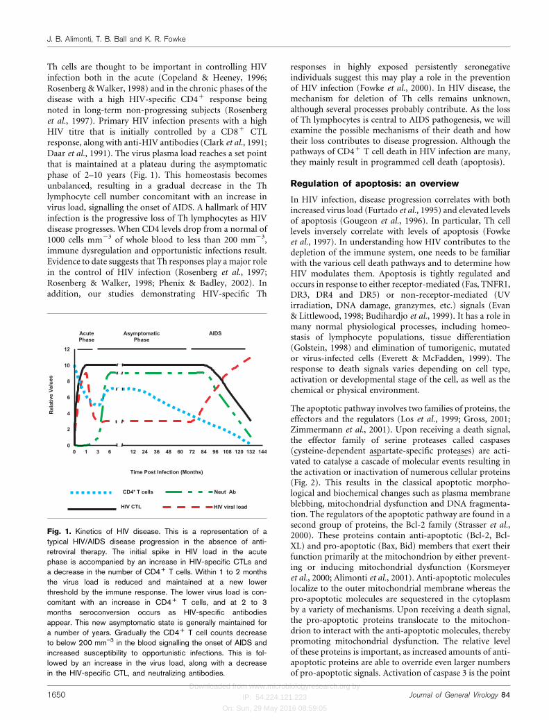
Th cells are thought to be important in controlling HIVinfection both in the acute (Copeland & Heeney, 1996;Rosenberg &Walker, 1998) and in the chronic phases of thedisease with a high HIV-specific CD4+ response beingnoted in long-term non-progressing subjects (Rosenberget al., 1997). Primary HIV infection presents with a highHIV titre that is initially controlled by a CD8+ CTLresponse, along with anti-HIV antibodies (Clark et al., 1991;Daar et al., 1991). The virus plasma load reaches a set pointthat is maintained at a plateau during the asymptomaticphase of 2–10 years (Fig. 1). This homeostasis becomesunbalanced, resulting in a gradual decrease in the Thlymphocyte cell number concomitant with an increase invirus load, signalling the onset of AIDS. A hallmark of HIVinfection is the progressive loss of Th lymphocytes as HIVdisease progresses. When CD4 levels drop from a normal of1000 cells mm23 of whole blood to less than 200 mm23,immune dysregulation and opportunistic infections result.Evidence to date suggests that Th responses play a major rolein the control of HIV infection (Rosenberg et al., 1997;Rosenberg & Walker, 1998; Phenix & Badley, 2002). Inaddition, our studies demonstrating HIV-specific Th
responses in highly exposed persistently seronegativeindividuals suggest this may play a role in the preventionof HIV infection (Fowke et al., 2000). In HIV disease, themechanism for deletion of Th cells remains unknown,although several processes probably contribute. As the lossof Th lymphocytes is central to AIDS pathogenesis, we willexamine the possible mechanisms of their death and howtheir loss contributes to disease progression. Although thepathways of CD4+ T cell death in HIV infection are many,they mainly result in programmed cell death (apoptosis).
Regulation of apoptosis: an overview
In HIV infection, disease progression correlates with bothincreased virus load (Furtado et al., 1995) and elevated levelsof apoptosis (Gougeon et al., 1996). In particular, Th celllevels inversely correlate with levels of apoptosis (Fowkeet al., 1997). In understanding how HIV contributes to thedepletion of the immune system, one needs to be familiarwith the various cell death pathways and to determine howHIV modulates them. Apoptosis is tightly regulated andoccurs in response to either receptor-mediated (Fas, TNFR1,DR3, DR4 and DR5) or non-receptor-mediated (UVirradiation, DNA damage, granzymes, etc.) signals (Evan& Littlewood, 1998; Budihardjo et al., 1999). It has a role inmany normal physiological processes, including homeo-stasis of lymphocyte populations, tissue differentiation(Golstein, 1998) and elimination of tumorigenic, mutatedor virus-infected cells (Everett & McFadden, 1999). Theresponse to death signals varies depending on cell type,activation or developmental stage of the cell, as well as thechemical or physical environment.
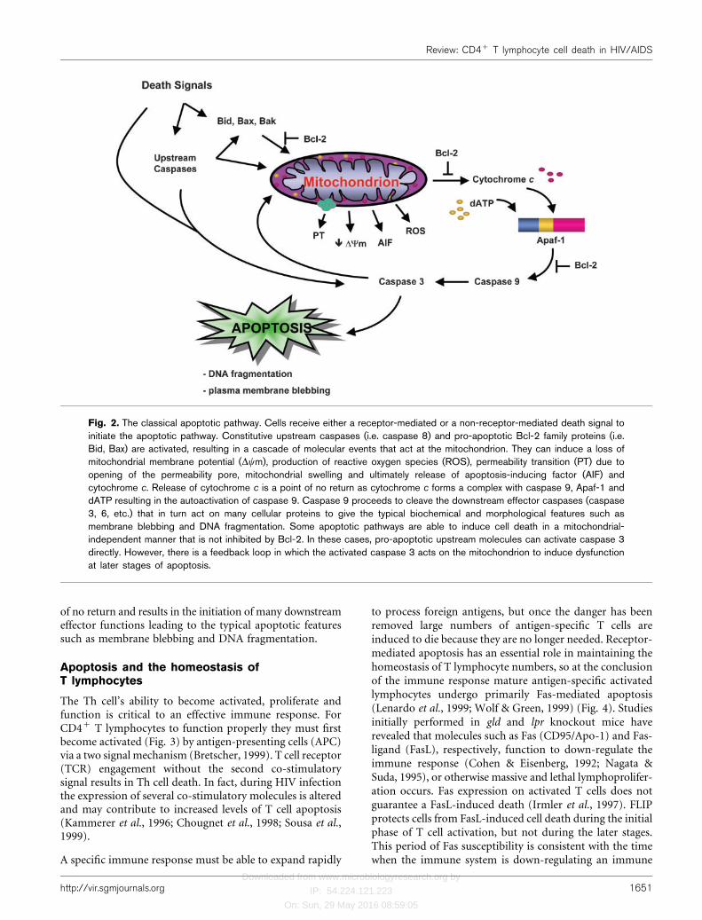
The apoptotic pathway involves two families of proteins, theeffectors and the regulators (Los et al., 1999; Gross, 2001;Zimmermann et al., 2001). Upon receiving a death signal,the effector family of serine proteases called caspases(cysteine-dependent aspartate-specific proteases) are acti-vated to catalyse a cascade of molecular events resulting inthe activation or inactivation of numerous cellular proteins(Fig. 2). This results in the classical apoptotic morpho-logical and biochemical changes such as plasma membraneblebbing, mitochondrial dysfunction and DNA fragmenta-tion. The regulators of the apoptotic pathway are found in asecond group of proteins, the Bcl-2 family (Strasser et al.,2000). These proteins contain anti-apoptotic (Bcl-2, Bcl-XL) and pro-apoptotic (Bax, Bid) members that exert theirfunction primarily at the mitochondrion by either prevent-ing or inducing mitochondrial dysfunction (Korsmeyeret al., 2000; Alimonti et al., 2001). Anti-apoptotic moleculeslocalize to the outer mitochondrial membrane whereas thepro-apoptotic molecules are sequestered in the cytoplasmby a variety of mechanisms. Upon receiving a death signal,the pro-apoptotic proteins translocate to the mitochon-drion to interact with the anti-apoptotic molecules, therebypromoting mitochondrial dysfunction. The relative levelof these proteins is important, as increased amounts of anti-apoptotic proteins are able to override even larger numbersof pro-apoptotic signals. Activation of caspase 3 is the point
CD4+ T cells
HIV CTL
Neut Ab
HIV viral load
0 1 3 6 12 24 36 48 60 72 84 96 108 120 132 1440
2
4
6
8
10
12
Death
Time Post Infection (Months)
Rel
ativ
e Va
lues
AcutePhase
AsymptomaticPhase
AIDS
Fig. 1. Kinetics of HIV disease. This is a representation of atypical HIV/AIDS disease progression in the absence of anti-retroviral therapy. The initial spike in HIV load in the acutephase is accompanied by an increase in HIV-specific CTLs anda decrease in the number of CD4+ T cells. Within 1 to 2 monthsthe virus load is reduced and maintained at a new lowerthreshold by the immune response. The lower virus load is con-comitant with an increase in CD4+ T cells, and at 2 to 3months seroconversion occurs as HIV-specific antibodiesappear. This new asymptomatic state is generally maintained fora number of years. Gradually the CD4+ T cell counts decreaseto below 200 mm”3 in the blood signalling the onset of AIDS andincreased susceptibility to opportunistic infections. This is fol-lowed by an increase in the virus load, along with a decreasein the HIV-specific CTL, and neutralizing antibodies.
1650 Journal of General Virology 84
J. B. Alimonti, T. B. Ball and K. R. Fowke
Downloaded from www.microbiologyresearch.org by
IP: 54.224.121.223
On: Sun, 29 May 2016 08:59:05
of no return and results in the initiation of many downstreameffector functions leading to the typical apoptotic featuressuch as membrane blebbing and DNA fragmentation.
Apoptosis and the homeostasis ofT lymphocytes
The Th cell’s ability to become activated, proliferate andfunction is critical to an effective immune response. ForCD4+ T lymphocytes to function properly they must firstbecome activated (Fig. 3) by antigen-presenting cells (APC)via a two signal mechanism (Bretscher, 1999). T cell receptor(TCR) engagement without the second co-stimulatorysignal results in Th cell death. In fact, during HIV infectionthe expression of several co-stimulatory molecules is alteredand may contribute to increased levels of T cell apoptosis(Kammerer et al., 1996; Chougnet et al., 1998; Sousa et al.,1999).
A specific immune response must be able to expand rapidly
to process foreign antigens, but once the danger has beenremoved large numbers of antigen-specific T cells areinduced to die because they are no longer needed. Receptor-mediated apoptosis has an essential role in maintaining thehomeostasis of T lymphocyte numbers, so at the conclusionof the immune response mature antigen-specific activatedlymphocytes undergo primarily Fas-mediated apoptosis(Lenardo et al., 1999; Wolf & Green, 1999) (Fig. 4). Studiesinitially performed in gld and lpr knockout mice haverevealed that molecules such as Fas (CD95/Apo-1) and Fas-ligand (FasL), respectively, function to down-regulate theimmune response (Cohen & Eisenberg, 1992; Nagata &Suda, 1995), or otherwise massive and lethal lymphoprolifer-ation occurs. Fas expression on activated T cells does notguarantee a FasL-induced death (Irmler et al., 1997). FLIPprotects cells from FasL-induced cell death during the initialphase of T cell activation, but not during the later stages.This period of Fas susceptibility is consistent with the timewhen the immune system is down-regulating an immune
Fig. 2. The classical apoptotic pathway. Cells receive either a receptor-mediated or a non-receptor-mediated death signal toinitiate the apoptotic pathway. Constitutive upstream caspases (i.e. caspase 8) and pro-apoptotic Bcl-2 family proteins (i.e.Bid, Bax) are activated, resulting in a cascade of molecular events that act at the mitochondrion. They can induce a loss ofmitochondrial membrane potential (Dym), production of reactive oxygen species (ROS), permeability transition (PT) due toopening of the permeability pore, mitochondrial swelling and ultimately release of apoptosis-inducing factor (AIF) andcytochrome c. Release of cytochrome c is a point of no return as cytochrome c forms a complex with caspase 9, Apaf-1 anddATP resulting in the autoactivation of caspase 9. Caspase 9 proceeds to cleave the downstream effector caspases (caspase3, 6, etc.) that in turn act on many cellular proteins to give the typical biochemical and morphological features such asmembrane blebbing and DNA fragmentation. Some apoptotic pathways are able to induce cell death in a mitochondrial-independent manner that is not inhibited by Bcl-2. In these cases, pro-apoptotic upstream molecules can activate caspase 3directly. However, there is a feedback loop in which the activated caspase 3 acts on the mitochondrion to induce dysfunctionat later stages of apoptosis.
http://vir.sgmjournals.org 1651
Review: CD4+ T lymphocyte cell death in HIV/AIDS

Downloaded from www.microbiologyresearch.org by
IP: 54.224.121.223
On: Sun, 29 May 2016 08:59:05
response. In HIV infection, T cell apoptosis is a complexprocess that involves both HIV-infected and uninfectedcells.
Cell death in HIV-infected cells
The devastating effect HIV has on the immune system is aresult of infecting and killing the immune-regulating Thlymphocytes. HIV infects and replicates primarily in acti-vated CD4+ T lymphocytes and to a lesser extent in macro-phages and dendritic cells. The cell specificity is due to theHIV envelope (Env) glycoprotein binding CD4, along withchemokine receptors CXCR4 or CCR5, for cell entry (Hogan& Hammer, 2001a, b). Once in the cytoplasm the viral RNAis reverse-transcribed into DNA and randomly integratedinto the host cell genome. Activation of the host cellenhances the production of viral proteins, which assembleupon budding out of the cell, utilizing the plasma mem-brane as the viral envelope. HIV plasma levels during allstages of infection range from 50 to 116106 virions ml21
(Piatak et al., 1993). The half-life of most HIV-infectedT cells in vivo is 12–36 h (Ho et al., 1995; Wei et al., 1995;Perelson et al., 1996), with the mechanisms of cell deathbeing virus- or receptor-mediated apoptosis.
There are several mechanisms by which HIV can induce celldeath directly in the cell it infects. CXCR4-tropic HIVisolates, generally found in the later stages of HIV infection,
preferentially infect T cells and induce membrane fusionbetween cells to form a giant multinucleated cell called asyncytium. Although syncytium formation is not necessaryfor the progression to AIDS, syncytia have a short lifespan,and the emergence of CXCR4-tropic strains correlates withan increased depletion of T cells (Richman & Bozzette, 1994;Sylwester et al., 1997; Kimata et al., 1999). In addition, cellviability can be compromised because the plasma mem-brane becomes disrupted or more permeable due to thecontinuous budding of the virion (Fauci, 1988), or due tospecific HIV proteins such as Vpu that can induce mem-brane permeability (Gonzalez & Carrasco, 2001). Virusreplication in the cell also has terminal consequences ascellular toxicity increases due to a build up of un-integratedlinear viral DNA (Shaw et al., 1984; Levy, 1993). Also,through cleavage, the HIV protease can inactivateanti-apoptotic Bcl-2 while simultaneously activating pro-apoptotic procaspase 8, making the cell more susceptible tomitochondrial dysfunction in response to internal orexternal death signals (Korant et al., 1998; Nie et al., 2002).
Both CD4+ and CD8+ T lymphocytes are more susceptibleto Fas-induced apoptosis in HIV+ individuals, and this isrelated to the regulation of surface levels of CD95 (Fas) andFasL. Peripheral blood mononuclear cells (PBMC) fromHIV+ individuals express higher levels of CD95 (Silvestriset al., 1996), and the proportion of these T lymphocytes
Fig. 3. Activation of T lymphocytes. Naive T cells (CD45RA) require a series of signals in order to become activated(CD45RO). The first signal involves the recognition of a peptide in the pocket of the MHC on the antigen-presenting cell(APC) by the T cell receptor (TcR) on the T lymphocyte. Other signals involve the interaction of CD28 and CD40Lcostimulatory molecules on the T cell with CD80/86 and CD40, respectively, on the APC.
1652 Journal of General Virology 84
J. B. Alimonti, T. B. Ball and K. R. Fowke
Downloaded from www.microbiologyresearch.org by
IP: 54.224.121.223
On: Sun, 29 May 2016 08:59:05
increases with disease progression (Aries et al., 1995;Baumler et al., 1996; Estaquier et al., 1996). The HIV pro-teins Nef (Zauli et al., 1999), Env (Oyaizu et al., 1994;Tateyama et al., 2000) and Tat (Ensoli et al., 1990;Westendorp et al., 1995) have also been implicated inincreasing CD95 and FasL levels, which presumablyenhances susceptibility to Fas-mediated killing. Nef isthought to induce FasL by interacting specifically with thezeta chain of the TCR complex (Xu et al., 1999). In addition,there are pro-apoptotic influences on other aspects of theFas death pathway. Tat upregulates initiator caspase 8,resulting in more caspase 8 activity (Bartz & Emerman,1999). HIV proteins also act on the central regulators of thedeath pathway, the Bcl-2 family members, that can eitherinduce or prevent apoptosis. The cell normally has anappropriate balance of pro- versus anti-apoptotic proteinsto ensure cell survival but HIV can alter the balance,resulting in the disruption of mitochondrial function.Levels of anti-apoptotic Bcl-2 are significantly lower in
HIV-infected individuals (Re et al., 1998). In particular twoHIV proteins, Tat (Sastry et al., 1996) and HIV protease(Strack et al., 1996), have been shown to decrease Bcl-2levels. At the same time, Tat also has the ability to increasepro-apoptotic Bax (Sastry et al., 1996) and Bim (Chen et al.,2002). Other evidence demonstrates that deletion of Vpupartially increases survival in response to CD95 cross-linking in Jurkat cells and PBMCs (Casella et al., 1999). Thiscould be due to the ability of Vpu to suppress NF-kB-dependent expression of anti-apoptotic factors like Bcl-XL(Akari et al., 2001).
CTLs are responsible for the removal of virus-infected cellsfrom the body, and for inducing apoptosis in HIV-infectedCD4+ cells. However, like many other viruses, HIV hasdeveloped mechanisms to prevent, or simply delay, apop-tosis of the cells it infects. The TCR on CTLs is used torecognize infected cells through the recognition of a non-selfviral peptide in conjunction with major histocompatibility
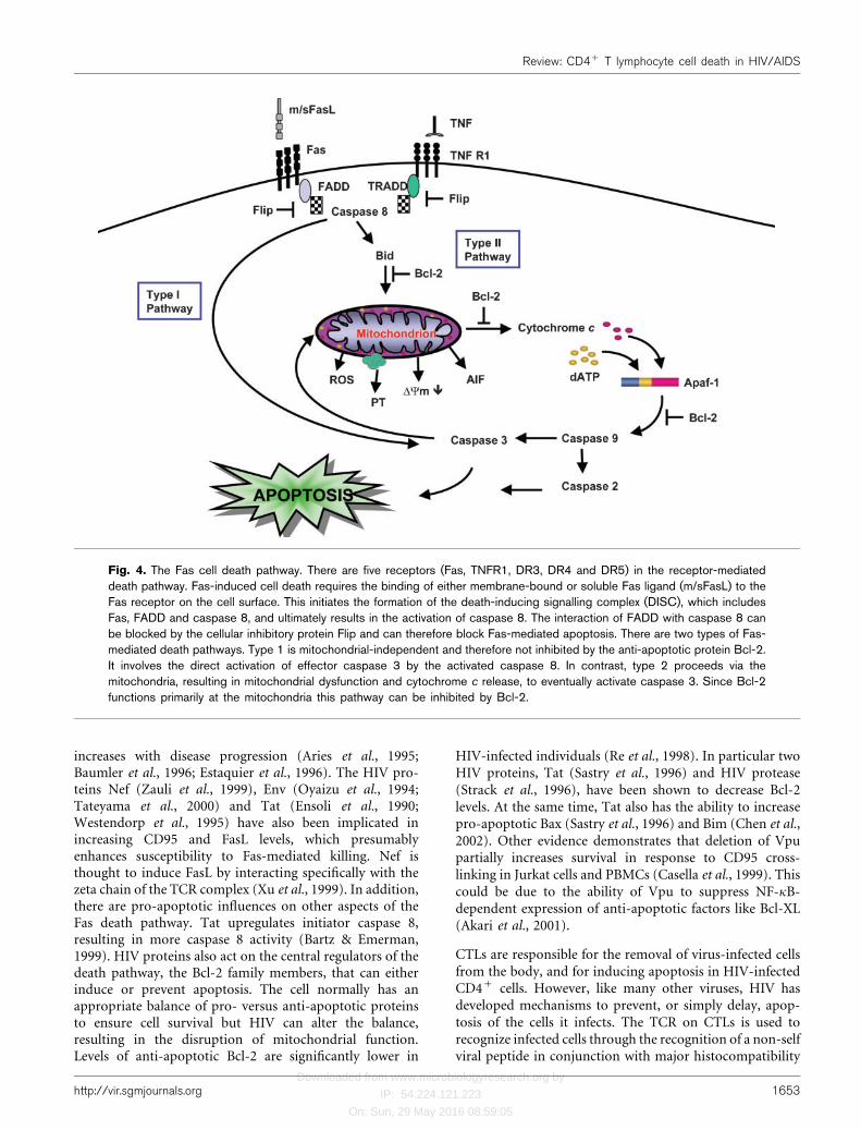
Fig. 4. The Fas cell death pathway. There are five receptors (Fas, TNFR1, DR3, DR4 and DR5) in the receptor-mediateddeath pathway. Fas-induced cell death requires the binding of either membrane-bound or soluble Fas ligand (m/sFasL) to theFas receptor on the cell surface. This initiates the formation of the death-inducing signalling complex (DISC), which includesFas, FADD and caspase 8, and ultimately results in the activation of caspase 8. The interaction of FADD with caspase 8 canbe blocked by the cellular inhibitory protein Flip and can therefore block Fas-mediated apoptosis. There are two types of Fas-mediated death pathways. Type 1 is mitochondrial-independent and therefore not inhibited by the anti-apoptotic protein Bcl-2.It involves the direct activation of effector caspase 3 by the activated caspase 8. In contrast, type 2 proceeds via themitochondria, resulting in mitochondrial dysfunction and cytochrome c release, to eventually activate caspase 3. Since Bcl-2functions primarily at the mitochondria this pathway can be inhibited by Bcl-2.
http://vir.sgmjournals.org 1653
Review: CD4+ T lymphocyte cell death in HIV/AIDS
Downloaded from www.microbiologyresearch.org by
IP: 54.224.121.223
On: Sun, 29 May 2016 08:59:05
complex class I (MHC I). The down-regulation of MHC Isurface expression by Nef (Schwartz et al., 1996) and Tat(Howcroft et al., 1993) avert this recognition and canprevent CTL-mediated killing of the HIV-infected cell. If thecell were to receive a death signal, HIV has a secondarydefence already in place. Vpr protein, which is expressed atlow levels in the cell, is responsible for the increase inanti-apoptotic Bcl-2, while simultaneously decreasingpro-apoptotic Bax (Conti et al., 1998). In contrast, otherevidence indicates that Vpr induces a prolonged G2 cell cycledelay followed by death in M phase (Watanabe et al., 2000),and that apoptosis is mediated through interaction of Vprwith the mitochondrion permeability transition pore, whichopens the pore causing mitochondrial swelling and releaseof cytochrome c (Jacotot et al., 2001; Roumier et al., 2002).The pro-apoptotic functions of Vpr may be secondaryeffects dependent upon induction of cell cycle arrest. Atthe same time, Vpr transactivates the viral promoter, thelong terminal repeat (LTR), to increase virus replication(Gummuluru & Emerman, 1999). The cytoprotective effectsof Vpr may play a role during early infection, allowingproductive virus replication, but ultimately it promotesapoptosis toward the later stages. Finally, Nef, Vpu and Envall decrease CD4 on the surface of infected cells (Crise et al.,1990; Willey et al., 1992; Salghetti et al., 1995). The fact thatthree HIV proteins have this function implies a critical rolein the survival and replication of the virus. Down-regulationof CD4 would prevent a super infection and Env-inducedapoptosis through the CD4 molecule. Overall, HIV hasevolved multiple mechanisms to promote survival for longenough to ensure a productive infection and this may besupported by the fact that infected cells do not undergoapoptosis as readily as uninfected bystander cells (Finkelet al., 1995). However, even infected cells have a shortenedlifespan and, therefore, partially contribute to the overalldecrease in Th cells (Ho et al., 1995; Wei et al., 1995;Perelson et al., 1996).
Cell death in uninfected cells
Apoptosis plays a major role in killing uninfected cells.Although the Th cells infected by HIV can be directly killedby the virus, or by HIV-specific CTL, there are generallymore apoptotic cells than infected cells (Embretson et al.,1993). This is confirmed by direct evidence in lymph nodeswhere apoptosis was seen primarily in the uninfectedbystander cells (Finkel et al., 1995). There are two mecha-nisms by which uninfected cells could be killed: either byHIV proteins released from infected cells acting on neigh-bouring uninfected cells, or by activation-induced cell death(AICD).
Effect of apoptotic HIV proteins on uninfected cells
Inactivated HIV virions (Esser et al., 2001) andHIV proteinsreleased into the extracellular environment can havedramatic effects on uninfected cells. HIV proteins such as
gp120, Tat, Nef and Vpu have been shown to induce celldeath in uninfected cells.
Soluble gp120 induces apoptosis in uninfected Th cellsthrough cross-linking of the CD4 molecule (Banda et al.,1992). The binding initiates part of the T cell activationpathway; however, in the absence of the TCR beingspecifically activated, this signal results in apoptosis orinhibition of antigen-induced cell activation (Marschneret al., 2002). Soluble and membrane-bound gp120 induce,through cell receptors such as CD4, CXCR4 and CCR5, bothFas-dependent (upregulation of Fas/FasL, decreased FLIP)and Fas-independent (increased Bax, decreased Bcl-2)apoptotic pathways (Arthos et al., 2002). Recently, CCR5-tropic viruses were shown to induce Fas and caspase8-dependent apoptosis of uninfected Th cells (Algeciras-Schimnich et al., 2002). Cell surface presentation of gp120can induce a bystander cell death that requires close cell-to-cell contact and gp41 function (Blanco et al., 2003). gp120also has an inhibitory effect when cells at the G0/G1 phase ofthe cell cycle, such as naive T cells, are most sensitive togp120-mediated negative signalling, whereasmemory T cellsare less affected.
The HIV protein Tat, secreted from infected cells (Changet al., 1997), can be endocytosed by neighbouring cells(Ensoli et al., 1990; Mann & Frankel, 1991; Zagury et al.,1998). Tat upregulates caspase 8 (Bartz & Emerman, 1999)and FasL (Li-Weber et al., 2000), and induces apoptosis inneurons (New et al., 1997) and Th cells (Li et al., 1995). AFas-independent Tat-mediated mechanism of bystanderT cell death has also been suggested (Zhang et al., 2001). TatcanupregulateTNF-related apoptosis-inducing ligand (TRAIL)on monocytes which may then interact with uninfectedT cells to induce apoptosis (Zhang et al., 2001). However,Tat also protects T cells from TRAIL-induced apoptosis(Gibellini et al., 2001). Other anti-apoptotic effects ofexogenous Tat include upregulation of Bcl-2, which wasobserved in the Jurkat cell line and PBMCs (Zauli et al.,1995).
HIV Nef protein released into the extracellular matrixinduces death in neuronal cells (Trillo-Pazos et al., 2000)and a wide range of blood cells by a Fas-independentmechanism (Okada et al., 1997, 1998). Much of the toxicityof Nef is likely due to its myristylated N terminus, which caninsert into the plasma membrane and induce cell death inuninfected CD4+ and CD42 T cells (Azad, 2000). It hasbeen suggested that Nef plays a role in allowing HIV-infected cells to evade the immune response by inducingcytotoxic activity in uninfected CD8+ T cells (Silvestriset al., 1999) and down-regulating CD4 expression inneighbouring CD4+ T cells (Pugliese et al., 1999). Also,the extracellular addition of Vpu, or its C terminus, cancause membrane disruption and induce cell death in CD4+
and CD42 cells (Azad, 2000). Clearly a number of extra-cellular HIV products are capable of inducing apoptosis inuninfected cells.
1654 Journal of General Virology 84
J. B. Alimonti, T. B. Ball and K. R. Fowke

Downloaded from www.microbiologyresearch.org by
IP: 54.224.121.223
On: Sun, 29 May 2016 08:59:05
Activation-induced cell death (AICD) in uninfectedcells
One interesting feature of apoptosis is that many of themolecular steps required for apoptosis are shared by cellularactivation pathways. For example, the nuclear condensationthat occurs in programmed cell death is similar to that whichoccurs prior to cell proliferation. Also, caspase activation,long known to be associated with apoptosis, occurs whencells become activated and proliferate (Alam et al., 1999;Kennedy et al., 1999). With these shared similarities it maynot be surprising that activation and cell death are closelylinked. AICD is a normal multi-step regulatory mechanismthat primes a cell for death to limit an activated immuneresponse (Hanabuchi et al., 1994). Priming can be achievedeither by repeated stimulation through CD3/TCR (Kabelitzet al., 1995), sole stimulation through the CD4 receptor(Banda et al., 1992) or activation without co-stimulation(Borthwick et al., 2000). Although not normally expressed athigh levels on resting T cells, Fas and FasL can be induced(Suda et al., 1995). In fact, allo-stimulation induces theexpression of Fas and FasL on the surface of T cells(O’Flaherty et al., 2000). In HIV infection excessive immuneactivation has been suggested to induce apoptosis throughFas/FasL (Badley et al., 1998, 1999; Dockrell et al., 1999).
The binding of Env to the CD4 molecule renders the CD4+
cell more susceptible to Fas-mediated killing (Algeciraset al., 1998) and also causes an increase in effector caspase 3,6 and 8 activity, which can be blocked with soluble CD4(Cicala et al., 1999, 2000; Algeciras-Schimnich et al., 2002).This suggests a CD4-dependent signalling mechanism,which is also known to block antigen-specific activationof primary lymphocytes (Marschner et al., 2002). T cellsacquire an AICD-resistant phenotype when correctly acti-vated by antigen. Engagement of CD4 by Env before TCRsignalling prevents the upregulation of the Fas death path-way regulator FLIP, changing T cells from Fas-resistant toFas-sensitive (Somma et al., 2000).
There are other HIV proteins that do not increase CD95expression but still sensitize the cells to Fas-induced death.The Tat protein has the ability to increase pro-apoptoticproteins caspase 8 and Bax, while simultaneously decreasingthe anti-apoptotic Bcl-2 (Sastry et al., 1996; Bartz & Emerman,1999). Recent data have suggested that elevated levels ofAICD in HIV infection may also be the result of altered FasLlevels. Tat upregulates FasL expression through Egr trans-activation of the FasL promoter resulting in increased AICD(Yang et al., 2002).
Cytokine responses in HIV disease and theireffect on apoptosis
It is not surprising, given the death of Th lymphocytes inHIV infection, that there is a significant dysregulation ofcytokine responses, which likely influences Th cell apoptosissusceptibility. It has been hypothesized that as HIV diseaseprogresses there is a shift in the cytokine response from a
predominantly type-1 cellular immune response (IFN-c,TNF-a, IL-12), to a type-2 humoral response (IL-4, IL-5, IL-10, IL-13) (Clerici & Shearer, 1994). However, additionalstudies of cytokine profiles and HIV disease progression failto completely corroborate these findings (Galli et al., 2001).Type-1 cytokine responses decrease as HIV disease pro-gresses. While there is no clear evidence that type-2 responsesincrease, there is no concurrent decrease in type-2 responses.One possible explanation for the decrease in type-1 res-ponses may be the increased susceptibility of cells expressingIFN-c or TNF-a to AICD, presumably due to decreasedBcl-2 expression in these type-1 Th cells (Ledru et al., 1998).Thus, during HIV disease progression, dysregulation ofcytokine responses results in a diminished ability to mounteffective type-1 cytokine responses.
Resistance to apoptosis in vitro is associated with a pre-dominant type-1 response (Gougeon & Montagnier, 1993).Therefore, deficiencies in type-1 responses could havedrastic effects on apoptotic events regulating normal T cellhomeostasis, and on HIV-induced AICD. IL-12 can protectagainst Fas-mediated apoptosis and AICD in HIV-infectedpatients (Estaquier et al., 1995). In addition, AICD inlymphocytes isolated from HIV-infected patients wasblocked by the addition of recombinant IL-12 and IFN-c,whereas type-2 cytokines IL-4 and IL-10 were shown toincrease susceptibility to AICD (Clerici et al., 1996). Recentgene expression data demonstrated that IFN-a can stronglyinduce a number of pro-apoptotic genes in the TNFsuperfamily (Stylianou et al., 2002), suggesting that sus-ceptibility to apoptosis may not exactly fit the type-1/-2paradigm. Regardless of the assignation of these cytokines toa type-1 or type-2 phenotype, these data strongly suggestthat cytokine dysregulation plays an important role in HIV-induced apoptosis. As HIV disease progresses, not only isapoptosis of Th lymphocytes enhanced due to the dualeffects of pro-apoptotic HIV proteins and increased AICD,but cytokine dysregulation is likely to amplify these effectsby providing a pro-apoptotic environment.
Although some of the strongest pro-apoptotic cytokinesignals are TNF-a and TNF family members, their role inHIV-induced apoptosis is not clear. It is apparent thatregulation of TNF is severely dysregulated in HIV-infectedpatients (Zangerle et al., 1994), as both HIV proteins andHIV infection can induce strong TNF responses in lympho-cytes (Capobianchi, 1996). In fact, HIV infection oflymphocytes, or monocytes, induces TNF that in turnactivates NF-kB, which induces high levels of HIV and TNFtranscription (Han et al., 1996). In addition, serum TNFlevels have been shown to be elevated in HIV-symptomaticbut not asymptomatic individuals (Hober et al., 1996). TNFand TNF family members can initiate apoptosis immedi-ately via membrane-bound receptors, and although it isapparent that TNF levels are indeed dysregulated in HIVinfection, there is limited evidence for TNF-mediatedapoptosis of infected cells directly, or by acting on bystanderlymphocytes. Apoptosis of bystander Th cells can be reduced
http://vir.sgmjournals.org 1655
Review: CD4+ T lymphocyte cell death in HIV/AIDS
Downloaded from www.microbiologyresearch.org by
IP: 54.224.121.223
On: Sun, 29 May 2016 08:59:05
by the addition of soluble TNF receptor decoys (Srivastavaet al., 1999) and can protect against HIV protein-inducedapoptosis (Cicala et al., 2000). However, clinical trials ofanti-TNF therapies have failed to show improvement ineither immunological or clinical outcome (Walker et al.,1996), making the role of TNF in HIV-induced apoptosisunclear. This is not surprising considering the multifactorialrole of TNF in inducing cell activation and death. It is likelythat the role of anti-apoptotic signals that negativelyregulate TNF and TNF family member signalling such ascaspase 8 may play a more important role in determiningwhether or not a particular cell undergoes apoptosis. Theredundancy in cytokine function makes elucidation of a rolein induction of apoptosis difficult.
The role of T cell growth factors IL-2 and IL-15 in HIV-induced apoptosis is also unclear. IL-2 displays both pro-and anti-apoptotic properties depending on the cellularmicroenvironment or activation status of a particular cell.Treatment with recombinant IL-2 or IL-15 can increase ordecrease a cell’s susceptibility to apoptosis. These discre-pancies may be due to the activation status of the cellsundergoing apoptosis. IL-2 and IL-15 were shown toincrease susceptibility to CD95-mediated apoptosis (Naora& Gougeon, 1999) while protecting against spontaneousapoptosis (Adachi et al., 1996). The latter effect may be dueto the ability of IL-2 and IL-15 to upregulate Bcl-2expression (Akbar et al., 1994). Clinically, IL-2 has beenuseful in the treatment of HIV infection under certainconditions. IL-2 can increase Th cell survival independent ofHIV replication, presumably by decreasing the apoptosis ofbystander Th cells. Also, treatment with recombinant IL-2can reduce overall levels of apoptosis in HIV-infected, butnot uninfected, individuals (Adachi et al., 1996), furtherunderscoring the importance of cellular activation, and thecytokine environment in the action of these apoptoticmediators. IL-2 and IL-15 share many similarities in theiraction due to utilization of common receptor elements (Giriet al., 1995). Of considerable interest is the observation thatIL-15 may be a more potent inhibitor of apoptosis, which islikely because IL-15 (unlike IL-2) does not appear to induceHIV replication (Kovacs et al., 2001). Also, the relativelevels of each cytokine in the apoptotic microenviroment arecrucial to their ability to induce, or alternately suppress,apoptosis. A mouse model on senescence suggests that Thcells incapable of producing sufficient levels of IL-2 wereunable to proliferate and were susceptible to apoptosis, butcould be rescued by the addition of exogenous IL-2(Nishimura et al., 2002). Thus, the role of IL-2 and IL-15in apoptosis is likely to be complex due to the immuno-regulatory roles for these cytokines in cellular activation.Determination of whether a cell undergoes apoptosis willdepend not only on the levels of IL-2 and IL-15, but also onthe presence or absence of other appropriate pro- or anti-apoptotic signals such as TNF, other cytokines or Bcl-2expression. These molecules may act directly throughmediation of cellular activation, indirectly through theinduction of other anti-apoptotic mediators such as Bcl-2 or
even through more downstream regulatory events such asthe abilities of these cytokines to induce mediators likeIFN-c that regulate apoptosis in a more direct manner.
It is clear that cytokines play a multifactorial role inapoptosis during HIV disease pathogenesis. The loss ofanti-apoptotic cytokines IFN-c and IL-12 during diseaseprogression and the increase in pro-apoptotic cytokines IL-4and IL-10 are likely to amplify the role of apoptosis in theloss of Th cells. TNF-a and the TNF family of cytokines caninduce apoptosis in a direct manner by inducing theappropriate (or inappropriate) apoptotic signals, while IL-2and IL-15 appear to act in a more indirect manner viamediation of cell activation. Cytokines play an importantrole in apoptosis, and this is likely to be even moreimportant during HIV infection when normal cytokineresponses become dysregulated.
CONCLUSION
HIV causes AIDS and the loss of Th lymphocytes is a centralfactor in the progression of the disease. Due to the vital roleof these cells in regulating and amplifying the immuneresponse, any decline in their number results in deficits inboth humoral and cell-mediated immunity. Understandinghow these cells are eliminated is critical to the developmentof new, effective therapies for AIDS. However, difficultiesarise as there are multiple mechanisms involved in the deathof the Th lymphocytes. HIV-infected Th lymphocytes have ashortened lifespan due to syncytia formation, lysis by CTLand direct cytopathic effects of HIV, but the number ofapoptotic cells in infected individuals greatly exceeds thenumber of HIV-infected cells. This suggests that HIV hasadditional detrimental effects on the uninfected bystanderTh cell population. These bystander cells are attacked by twodiffering mechanisms. Firstly, several HIV proteins, whetherattached to the virion or released by infected cells, can utilizea number of disparate death pathways to initiate apoptosisin uninfected cells. It is unclear in the in vivo situation whatthe relative contribution that gp120, Tat or Nef makes to theoverall level of apoptosis; however, a therapy may bedeveloped that would require the neutralization of all oftheir effects. On the other hand, there may be a point furtheralong at which the HIV-induced apoptotic pathwaysconverge. If we were to identify this point then blockingdeath may require intervention at a single focal point.Clearly more understanding of the death pathways initiatedby these proteins is required. The second mechanism ofbystander killing is AICD. HIV-infected individuals havehigher levels of immune activation, which have been sug-gested to contribute to increased apoptosis. The dominantdeath pathway in AICD is through Fas and, therefore,inhibiting this path would be a logical point for therapeuticintervention. Currently, it is unknown whether the HIVproteins or AICD kill the majority of uninfected cells.Further studies of AICD in models of HIV and chronicimmune stimulation could help determine its significance indisease progression.
1656 Journal of General Virology 84
J. B. Alimonti, T. B. Ball and K. R. Fowke
Downloaded from www.microbiologyresearch.org by
IP: 54.224.121.223
On: Sun, 29 May 2016 08:59:05
Local environmental factors can also influence cell survivaland the effectiveness of the HIV-specific immune response.During HIV infection the switch from the production ofsome type-1 cytokines to other type-2 cytokines could bevery important since, respectively, they are associated witheither protection from or enhancement of AICD.
This review has highlighted the many different modes bywhich HIV induces apoptotic cell death in both infected anduninfected Th cells. One of the ultimate goals of research inthis field is to prevent or minimize death in Th cells. If thiswere achieved it may be possible to convert HIV infectionfrom a progressively immunosuppressive and ultimatelyfatal disease to a chronic manageable infection.
ACKNOWLEDGEMENTS
J. B. A. is supported by a postdoctoral fellowship from the CanadianInstitutes for Health Research (CIHR). The authors would also like tothank the CIHR, theManitoba Health Research Council and CANVACfor their financial support and Dr Jody Berry and John Rutherford for acritical review of this manuscript.
REFERENCES
Adachi, Y., Oyaizu, N., Than, S., McCloskey, T. W. & Pahwa, S.(1996). IL-2 rescues in vitro lymphocyte apoptosis in patients withHIV infection: correlation with its ability to block culture-induceddown-modulation of Bcl-2. J Immunol 157, 4184–4193.
Akari, H., Bour, S., Kao, S., Adachi, A. & Strebel, K. (2001). Thehuman immunodeficiency virus type 1 accessory protein Vpuinduces apoptosis by suppressing the nuclear factor kB-dependentexpression of antiapoptotic factors. J Exp Med 194, 1299–1311.
Akbar, A. N., Salmon, M. & Janossy, G. (1994). Role of bcl-2 andapoptosis in viral infections. Int Arch Allergy Immunol 105, 359–362.
Alam, A., Cohen, L. Y., Aouad, S. & Sekaly, R. P. (1999). Earlyactivation of caspases during T lymphocyte stimulation results inselective substrate cleavage in nonapoptotic cells. J Exp Med 190,1879–1890.
Algeciras, A., Dockrell, D. H., Lynch, D. H. & Paya, C. V. (1998). CD4regulates susceptibility to Fas ligand- and tumor necrosis factor-mediated apoptosis. J Exp Med 187, 711–720.
Algeciras-Schimnich, A., Vlahakis, S. R., Villasis-Keever, A.,Gomez, T., Heppelmann, C. J., Bou, G. & Paya, C. V. (2002).CCR5 mediates Fas- and caspase-8 dependent apoptosis of bothuninfected and HIV infected primary human CD4 T cells. AIDS 16,1467–1478.
Alimonti, J. B., Shi, L., Baijal, P. K. & Greenberg, A. H. (2001).Granzyme B induces BID-mediated cytochrome c release andmitochondrial permeability transition. J Biol Chem 276, 6974–6982.
Aries, S. P., Schaaf, B., Muller, C., Dennin, R. H. & Dalhoff, K.(1995). Fas (CD95) expression on CD4+ T cells from HIV-infectedpatients increases with disease progression. J Mol Med 73, 591–593.
Arthos, J., Cicala, C., Selig, S. M. & 10 other authors (2002). Therole of the CD4 receptor versus HIV coreceptors in envelope-mediated apoptosis in peripheral blood mononuclear cells. Virology292, 98–106.
Azad, A. A. (2000). Could Nef and Vpr proteins contribute to diseaseprogression by promoting depletion of bystander cells and prolonged
survival of HIV-infected cells? Biochem Biophys Res Commun 267,
677–685.
Badley, A. D., Dockrell, D. H., Algeciras, A. & 10 other authors(1998). In vivo analysis of Fas/FasL interactions in HIV-infected
patients. J Clin Invest 102, 79–87.
Badley, A. D., Parato, K., Cameron, D. W. & 7 other authors (1999).Dynamic correlation of apoptosis and immune activation duringtreatment of HIV infection. Cell Death Differ 6, 420–432.
Banda, N. K., Bernier, J., Kurahara, D. K., Kurrle, R., Haigwood, N.,Sekaly, R. P. & Finkel, T. H. (1992). Crosslinking CD4 by humanimmunodeficiency virus gp120 primes T cells for activation-induced
apoptosis. J Exp Med 176, 1099–1106.
Bartz, S. R. & Emerman, M. (1999). Human immunodeficiency virus
type 1 Tat induces apoptosis and increases sensitivity to apoptoticsignals by up-regulating FLICE/caspase-8. J Virol 73, 1956–1963.
Baumler, C. B., Bohler, T., Herr, I., Benner, A., Krammer, P. H. &Debatin, K. M. (1996). Activation of the CD95 (APO-1/Fas) systemin T cells from human immunodeficiency virus type-1-infected
children. Blood 88, 1741–1746.
Blanco, J., Barretina, J., Ferri, K. F. & 7 other authors (2003). Cell-surface-expressed HIV-1 envelope induces the death of CD4T cells during GP41-mediated hemifusion-like events. Virology 305,
318–329.
Borthwick, N. J., Lowdell, M., Salmon, M. & Akbar, A. N. (2000). Lossof CD28 expression on CD8+ T cells is induced by IL-2 receptor
gamma chain signalling cytokines and type I IFN, and increasessusceptibility to activation-induced apoptosis. Int Immunol 12,
1005–1013.
Bretscher, P. A. (1999). A two-step, two-signal model for the
primary activation of precursor helper T cells. Proc Natl Acad Sci U SA 96, 185–190.
Budihardjo, I., Oliver, H., Lutter, M., Luo, X. & Wang, X. (1999).Biochemical pathways of caspase activation during apoptosis. AnnuRev Cell Dev Biol 15, 269–290.
Capobianchi, M. R. (1996). Induction of lymphomonocyte activationby HIV-1 glycoprotein gp120. Possible role in AIDS pathogenesis.
J Biol Regul Homeost Agents 10, 83–91.
Casella, C. R., Rapaport, E. L. & Finkel, T. H. (1999). Vpu increasessusceptibility of human immunodeficiency virus type 1-infected cells
to fas killing. J Virol 73, 92–100.
Chang, H. C., Samaniego, F., Nair, B. C., Buonaguro, L. & Ensoli, B.(1997). HIV-1 Tat protein exits from cells via a leaderless secretorypathway and binds to extracellular matrix-associated heparan sulfate
proteoglycans through its basic region. AIDS 11, 1421–1431.
Chen, D., Wang, M., Zhou, S. & Zhou, Q. (2002). HIV-1 Tat targetsmicrotubules to induce apoptosis, a process promoted by the pro-
apoptotic Bcl-2 relative Bim. EMBO J 21, 6801–6810.
Chougnet, C., Thomas, E., Landay, A. L., Kessler, H. A., Buchbinder,S., Scheer, S. & Shearer, G. M. (1998). CD40 ligand and IFN-csynergistically restore IL-12 production in HIV-infected patients. Eur
J Immunol 28, 646–656.
Cicala, C., Arthos, J., Ruiz, M., Vaccarezza, M., Rubbert, A., Riva, A.,Wildt, K., Cohen, O. & Fauci, A. S. (1999). Induction of
phosphorylation and intracellular association of CC chemokinereceptor 5 and focal adhesion kinase in primary human CD4+ T cells
by macrophage-tropic HIV envelope. J Immunol 163, 420–426.
Cicala, C., Arthos, J., Rubbert, A., Selig, S., Wildt, K., Cohen, O. J. &Fauci, A. S. (2000). HIV-1 envelope induces activation of caspase-3
and cleavage of focal adhesion kinase in primary human CD4+
T cells. Proc Natl Acad Sci U S A 97, 1178–1183.
Clark, S. J., Saag, M. S., Decker, W. D., Campbell-Hill, S.,Roberson, J. L., Veldkamp, P. J., Kappes, J. C., Hahn, B. H. &
http://vir.sgmjournals.org 1657
Review: CD4+ T lymphocyte cell death in HIV/AIDS

Downloaded from www.microbiologyresearch.org by
IP: 54.224.121.223
On: Sun, 29 May 2016 08:59:05
Shaw, G. M. (1991). High titers of cytopathic virus in plasma of
patients with symptomatic primary HIV-1 infection. New Engl J Med
324, 954–960.
Clerici, M. & Shearer, G. M. (1994). The Th1-Th2 hypothesis of HIV
infection: new insights. Immunol Today 15, 575–581.
Clerici, M., Sarin, A., Berzofsky, J. A. & 10 other authors (1996).
Antigen-stimulated apoptotic T-cell death in HIV infection is
selective for CD4+ T cells, modulated by cytokines and effected
by lymphotoxin. AIDS 10, 603–611.
Cohen, P. L. & Eisenberg, R. A. (1992). The lpr and gld genes in
systemic autoimmunity: life and death in the Fas lane. Immunol
Today 13, 427–428.
Conti, L., Rainaldi, G., Matarrese, P. & 7 other authors (1998). The
HIV-1 vpr protein acts as a negative regulator of apoptosis in a
human lymphoblastoid T cell line: possible implications for the
pathogenesis of AIDS. J Exp Med 187, 403–413.
Copeland, K. F. & Heeney, J. L. (1996). T helper cell activation and
human retroviral pathogenesis. Microbiol Rev 60, 722–742.
Crise, B., Buonocore, L. & Rose, J. K. (1990). CD4 is retained in the
endoplasmic reticulum by the human immunodeficiency virus type 1
glycoprotein precursor. J Virol 64, 5585–5593.
Daar, E. S., Moudgil, T., Meyer, R. D. & Ho, D. D. (1991). Transient
high levels of viremia in patients with primary human immuno-
deficiency virus type 1 infection. New Engl J Med 324, 961–964.
Dockrell, D. H., Badley, A. D., Algeciras-Schimnich, A., Simpson,
M., Schut, R., Lynch, D. H. & Paya, C. V. (1999). Activation-induced
CD4+ T cell death in HIV-positive individuals correlates with Fas
susceptibility, CD4+ T cell count, and HIV plasma viral copy
number. AIDS Res Hum Retroviruses 15, 1509–1518.
Embretson, J., Zupancic, M., Ribas, J. L., Burke, A., Racz, P., Tenner-
Racz, K. & Haase, A. T. (1993). Massive covert infection of helper T
lymphocytes and macrophages by HIV during the incubation period
of AIDS. Nature 362, 359–362.
Ensoli, B., Barillari, G., Salahuddin, S. Z., Gallo, R. C. & Wong-Staal,
F. (1990). Tat protein of HIV-1 stimulates growth of cells derived
from Kaposi’s sarcoma lesions of AIDS patients. Nature 345, 84–86.
Esser, M. T., Bess, J. W., Jr, Suryanarayana, K., Chertova, E.,
Marti, D., Carrington, M., Arthur, L. O. & Lifson, J. D. (2001). Partial
activation and induction of apoptosis in CD4+ and CD8+ T
lymphocytes by conformationally authentic noninfectious human
immunodeficiency virus type 1. J Virol 75, 1152–1164.
Estaquier, J., Idziorek, T., Zou, W., Emilie, D., Farber, C. M., Bourez,
J. M. & Ameisen, J. C. (1995). T helper type 1/T helper type 2
cytokines and T cell death: preventive effect of interleukin 12 on
activation-induced and CD95 (FAS/APO-1)-mediated apoptosis of
CD4+ T cells from human immunodeficiency virus-infected persons.
J Exp Med 182, 1759–1767.
Estaquier, J., Tanaka, M., Suda, T., Nagata, S., Golstein, P. &
Ameisen, J. C. (1996). Fas-mediated apoptosis of CD4+ and CD8+
T cells from human immunodeficiency virus-infected persons:
differential in vitro preventive effect of cytokines and protease
antagonists. Blood 87, 4959–4966.
Evan, G. & Littlewood, T. (1998). A matter of life and cell death.
Science 281, 1317–1322.
Everett, H. & McFadden, G. (1999). Apoptosis: an innate immune
response to virus infection. Trends Microbiol 7, 160–165.
Fauci, A. S. (1988). The human immunodeficiency virus: infectivity
and mechanisms of pathogenesis. Science 239, 617–622.
Finkel, T. H., Tudor-Williams, G., Banda, N. K., Cotton, M. F., Curiel,
T., Monks, C., Baba, T. W., Ruprecht, R. M. & Kupfer, A. (1995).
Apoptosis occurs predominantly in bystander cells and not in
productively infected cells of HIV- and SIV-infected lymph nodes.
Nat Med 1, 129–134.
Fowke, K. R., D’Amico, R., Chernoff, D. N., Pottage, J. C., Jr, Benson,
C. A., Sha, B. E., Kessler, H. A., Landay, A. L. & Shearer, G. M.
(1997). Immunologic and virologic evaluation after influenza
vaccination of HIV- 1-infected patients. AIDS 11, 1013–1021.
Fowke, K. R., Kaul, R., Rosenthal, K. L. & 9 other authors (2000).
HIV-1-specific cellular immune responses among HIV-1-resistant
sex workers. Immunol Cell Biol 78, 586–595.
Furtado, M. R., Kingsley, L. A. & Wolinsky, S. M. (1995). Changes in
the viral mRNA expression pattern correlate with a rapid rate of
CD4+ T-cell number decline in human immunodeficiency virus type
1-infected individuals. J Virol 69, 2092–2100.
Galli, G., Annunziato, F., Cosmi, L., Manetti, R., Maggi, E. &
Romagnani, S. (2001). Th1 and th2 responses, HIV-1 coreceptors
and HIV-1 infection. J Biol Regul Homeost Agents 15, 308–313.
Gibellini, D., Re, M. C., Ponti, C., Maldini, C., Celeghini, C.,
Cappellini, A., La Placa, M. & Zauli, G. (2001). HIV-1 Tat protects
CD4+ Jurkat T lymphoblastoid cells from apoptosis mediated by
TNF-related apoptosis-inducing ligand. Cell Immunol 207, 89–99.
Giri, J. G., Anderson, D. M., Kumaki, S., Park, L. S., Grabstein, K. H.
& Cosman, D. (1995). IL-15, a novel T cell growth factor that shares
activities and receptor components with IL-2. J Leukoc Biol 57,
763–766.
Golstein, P. (1998). Cell death in us and others. Science 281, 1283.
Gonzalez, M. E. & Carrasco, L. (2001). Human immunodeficiency
virus type 1 VPU protein affects Sindbis virus glycoprotein pro-
cessing and enhances membrane permeabilization. Virology 279,
201–209.
Gougeon, M. L. & Montagnier, L. (1993). Apoptosis in AIDS. Science
260, 1269–1270.
Gougeon, M. L., Lecoeur, H., Dulioust, A., Enouf, M. G., Crouvoiser,
M., Goujard, C., Debord, T. & Montagnier, L. (1996). Programmed
cell death in peripheral lymphocytes from HIV-infected persons:
increased susceptibility to apoptosis of CD4 and CD8 T cells
correlates with lymphocyte activation and with disease progression.
J Immunol 156, 3509–3520.
Gross, A. (2001). BCL-2 proteins: regulators of the mitochondrial
apoptotic program. IUBMB Life 52, 231–236.
Gummuluru, S. & Emerman, M. (1999). Cell cycle- and Vpr-
mediated regulation of human immunodeficiency virus type 1
expression in primary and transformed T-cell lines. J Virol 73, 5422–
5430.
Han, X., Becker, K., Degen, H. J., Jablonowski, H. & Strohmeyer, G.
(1996). Synergistic stimulatory effects of tumour necrosis factor a
and interferon c on replication of human immunodeficiency virus
type 1 and on apoptosis of HIV-1-infected host cells. Eur J Clin
Invest 26, 286–292.
Hanabuchi, S., Koyanagi, M., Kawasaki, A. & 7 other authors
(1994). Fas and its ligand in a general mechanism of T-cell-mediated
cytotoxicity. Proc Natl Acad Sci U S A 91, 4930–4934.
Ho, D. D., Neumann, A. U., Perelson, A. S., Chen, W., Leonard, J. M.
& Markowitz, M. (1995). Rapid turnover of plasma virions and CD4
lymphocytes in HIV-1 infection. Nature 373, 123–126.
Hober, D., Benyoucef, S., Delannoy, A. S., de Groote, D., Ajana, F.,
Mouton, Y. & Wattre, P. (1996). High plasma level of soluble tumor
necrosis factor receptor type II (sTNFRII) in asymptomatic HIV-1-
infected patients. Infection 24, 213–217.
Hogan, C. M. & Hammer, S. M. (2001a). Host determinants in HIV
infection and disease. Part 1: cellular and humoral immune
responses. Ann Intern Med 134, 761–776.
1658 Journal of General Virology 84
J. B. Alimonti, T. B. Ball and K. R. Fowke

Downloaded from www.microbiologyresearch.org by
IP: 54.224.121.223
On: Sun, 29 May 2016 08:59:05
Hogan, C. M. & Hammer, S. M. (2001b). Host determinants in HIV
infection and disease. Part 2: genetic factors and implications forantiretroviral therapeutics. Ann Intern Med 134, 978–996.
Howcroft, T. K., Strebel, K., Martin, M. A. & Singer, D. S. (1993).Repression of MHC class I gene promoter activity by two-exon Tatof HIV. Science 260, 1320–1322.
Irmler, M., Thome, M., Hahne, M. & 10 other authors (1997).Inhibition of death receptor signals by cellular FLIP. Nature 388,
190–195.
Jacotot, E., Ferri, K. F., El Hamel, C. & 17 other authors (2001).Control of mitochondrial membrane permeabilization by adenine
nucleotide translocator interacting with HIV-1 viral protein rR andBcl-2. J Exp Med 193, 509–519.
Kabelitz, D., Pohl, T. & Pechhold, K. (1995). T cell apoptosistriggered via the CD3/T cell receptor complex and alternative
activation pathways. Curr Top Microbiol Immunol 200, 1–14.
Kalams, S. A., Buchbinder, S. P., Rosenberg, E. S., Billingsley, J. M.,Colbert, D. S., Jones, N. G., Shea, A. K., Trocha, A. K. & Walker, B. D.(1999). Association between virus-specific cytotoxic T-lymphocyteand helper responses in human immunodeficiency virus type 1
infection. J Virol 73, 6715–6720.
Kammerer, R., Iten, A., Frei, P. C. & Burgisser, P. (1996). Expansionof T cells negative for CD28 expression in HIV infection. Relation toactivation markers and cell adhesion molecules, and correlation with
prognostic markers. Med Microbiol Immunol (Berl) 185, 19–25.
Kasaian, M. T., Leite-Morris, K. A. & Biron, C. A. (1991). The role ofCD4+ cells in sustaining lymphocyte proliferation during lympho-
cytic choriomeningitis virus infection. J Immunol 146, 1955–1963.
Kennedy, N. J., Kataoka, T., Tschopp, J. & Budd, R. C. (1999).Caspase activation is required for T cell proliferation. J Exp Med 190,1891–1896.
Kimata, J. T., Kuller, L., Anderson, D. B., Dailey, P. & Overbaugh, J.(1999). Emerging cytopathic and antigenic simian immunodeficiencyvirus variants influence AIDS progression. Nat Med 5, 535–541.
Kinter, A., Catanzaro, A., Monaco, J. & 12 other authors (1998). CC-chemokines enhance the replication of T-tropic strains of HIV-1 in
CD4+ T cells: role of signal transduction. Proc Natl Acad Sci U S A95, 11880–11885.
Koenig, S., Conley, A. J., Brewah, Y. A. & other authors (1995).Transfer of HIV-1-specific cytotoxic T lymphocytes to an AIDSpatient leads to selection for mutant HIV variants and subsequent
disease progression. Nat Med 1, 330–336.
Korant, B. D., Strack, P., Frey, M. W. & Rizzo, C. J. (1998). A cellular
anti-apoptosis protein is cleaved by the HIV-1 protease. Adv ExpMed Biol 436, 27–29.
Korsmeyer, S. J., Wei, M. C., Saito, M., Weiler, S., Oh, K. J. &Schlesinger, P. H. (2000). Pro-apoptotic cascade activates BID,which oligomerizes BAK or BAX into pores that result in the release
of cytochrome c. Cell Death Differ 7, 1166–1173.
Kovacs, J. A., Vogel, S., Metcalf, J. A. & 14 other authors (2001).Interleukin-2 induced immune effects in human immunodeficiencyvirus-infected patients receiving intermittent interleukin-2 immuno-
therapy. Eur J Immunol 31, 1351–1360.
Ledru, E., Lecoeur, H., Garcia, S., Debord, T. & Gougeon, M. L.(1998). Differential susceptibility to activation-induced apoptosis
among peripheral Th1 subsets: correlation with Bcl-2 expression andconsequences for AIDS pathogenesis. J Immunol 160, 3194–3206.
Lehmann, J. & Alber, G. (1998). Murine leishmaniosis: a paradigm
for the importance of T helper 1 and T helper 2 cells. Rev Sci Tech17, 176–187.
Lehmann-Grube, F., Lohler, J., Utermohlen, O. & Gegin, C. (1993).Antiviral immune responses of lymphocytic choriomeningitis
virus-infected mice lacking CD8+ T lymphocytes because of
disruption of the beta 2-microglobulin gene. J Virol 67, 332–339.
Lenardo, M., Chan, K. M., Hornung, F., McFarland, H., Siegel, R.,Wang, J. & Zheng, L. (1999). Mature T lymphocyte apoptosis –
immune regulation in a dynamic and unpredictable antigenicenvironment. Annu Rev Immunol 17, 221–253.
Levy, J. A. (1993). Pathogenesis of human immunodeficiency virusinfection. Microbiol Rev 57, 183–289.
Li, C. J., Friedman, D. J., Wang, C., Metelev, V. & Pardee, A. B.(1995). Induction of apoptosis in uninfected lymphocytes by HIV-1Tat protein. Science 268, 429–431.
Li-Weber, M., Laur, O., Dern, K. & Krammer, P. H. (2000). T cellactivation-induced and HIV tat-enhanced CD95(APO-1/Fas) ligand
transcription involves NF-kB. Eur J Immunol 30, 661–670.
Los, M., Wesselborg, S. & Schulze-Osthoff, K. (1999). The role ofcaspases in development, immunity and apoptotic signal transduc-
tion: lessons from knockout mice. Immunity 10, 629–639.
Lukacher, A. E., Braciale, V. L. & Braciale, T. J. (1984). In vivo
effector function of influenza virus-specific cytotoxic T lymphocyteclones is highly specific. J Exp Med 160, 814–826.
Macaubas, C., Sly, P. D., Burton, P. & 7 other authors (1999).Regulation of T-helper cell responses to inhalant allergen duringearly childhood. Clin Exp Allergy 29, 1223–1231.
Maloy, K. J., Burkhart, C., Junt, T. M., Odermatt, B., Oxenius, A.,Piali, L., Zinkernagel, R. M. & Hengartner, H. (2000). CD4+ T cell
subsets during virus infection. Protective capacity depends oneffector cytokine secretion and on migratory capability. J Exp Med
191, 2159–2170.
Mann, D. A. & Frankel, A. D. (1991). Endocytosis and targeting ofexogenous HIV-1 Tat protein. EMBO J 10, 1733–1739.
Marschner, S., Hunig, T., Cambier, J. C. & Finkel, T. H. (2002).Ligation of human CD4 interferes with antigen-induced activation of
primary T cells. Immunol Lett 82, 131–139.
Matloubian, M., Concepcion, R. J. & Ahmed, R. (1994). CD4+ T cellsare required to sustain CD8+ cytotoxic T-cell responses during
chronic viral infection. J Virol 68, 8056–8063.
Murray, R. J., Kurilla, M. G., Brooks, J. M., Thomas, W. A., Rowe, M.,Kieff, E. & Rickinson, A. B. (1992). Identification of target antigensfor the human cytotoxic T cell response to Epstein–Barr virus (EBV):
implications for the immune control of EBV-positive malignancies.J Exp Med 176, 157–168.
Nagata, S. & Suda, T. (1995). Fas and Fas ligand: lpr and gld muta-
tions. Immunol Today 16, 39–43.
Naora, H. & Gougeon, M. L. (1999). Interleukin-15 is a potent
survival factor in the prevention of spontaneous but not CD95-induced apoptosis in CD4 and CD8 T lymphocytes of HIV-infected
individuals. Correlation with its ability to increase BCL-2 expression.Cell Death Differ 6, 1002–1011.
New, D. R., Ma, M., Epstein, L. G., Nath, A. & Gelbard, H. A. (1997).Human immunodeficiency virus type 1 Tat protein induces deathby apoptosis in primary human neuron cultures. J Neurovirol 3,
168–173.
Nie, Z., Phenix, B. N., Lum, J. J., Alam, A., Lynch, D. H., Beckett, B.,Krammer, P. H., Sekaly, R. P. & Badley, A. D. (2002). HIV-1 proteaseprocesses procaspase 8 to cause mitochondrial release of cytochrome
c, caspase cleavage and nuclear fragmentation. Cell Death Differ 9,1172–1184.
Nishimura, Y., Hosokawa, T., Hosono, M., Baba, M. & Hosokawa, M.(2002). Insufficient interleukin-2 production from splenic CD4+
T cells causes impaired cell proliferation and early apoptosis in
SAMP1, a strain of senescence-accelerated mouse. Immunology 107,190–198.
http://vir.sgmjournals.org 1659
Review: CD4+ T lymphocyte cell death in HIV/AIDS

Downloaded from www.microbiologyresearch.org by
IP: 54.224.121.223
On: Sun, 29 May 2016 08:59:05
O’Flaherty, E., Wong, W. K., Pettit, S. J., Seymour, K., Ali, S. & Kirby,J. A. (2000). Regulation of T-cell apoptosis: a mixed lymphocyte
reaction model. Immunology 100, 289–299.
Ohmi, Y., Shiku, H. & Nishimura, T. (1999). Tumor-specific targeting
of T helper type 1 (Th1) cells by anti-CD3 x anti-c-ErbB-2 bispecific
antibody. Cancer Immunol Immunother 48, 456–462.
Okada, H., Takei, R. & Tashiro, M. (1997). HIV-1 Nef protein-
induced apoptotic cytolysis of a broad spectrum of uninfected
human blood cells independently of CD95(Fas). FEBS Lett 414,
603–606.
Okada, H., Morikawa, S. & Tashiro, M. (1998). HIV-1 Nef binding
protein expressed on the surface of murine blood cells. Med
Microbiol Immunol (Berl) 186, 201–207.
Oyaizu, N., McCloskey, T. W., Than, S., Hu, R., Kalyanaraman, V. S.& Pahwa, S. (1994). Cross-linking of CD4 molecules upregulates Fas
antigen expression in lymphocytes by inducing interferon-gamma
and tumor necrosis factor-a secretion. Blood 84, 2622–2631.
Perelson, A. S., Neumann, A. U., Markowitz, M., Leonard, J. M. & Ho,D. D. (1996). HIV-1 dynamics in vivo: virion clearance rate, infected
cell life-span, and viral generation time. Science 271, 1582–1586.
Phenix, B. N. & Badley, A. D. (2002). Influence of mitochondrial
control of apoptosis on the pathogenesis, complications and
treatment of HIV infection. Biochimie 84, 251–264.
Piatak, M., Jr, Saag, M. S., Yang, L. C., Clark, S. J., Kappes, J. C.,Luk, K. C., Hahn, B. H., Shaw, G. M. & Lifson, J. D. (1993). High
levels of HIV-1 in plasma during all stages of infection determined
by competitive PCR. Science 259, 1749–1754.
Pugliese, A., Cantamessa, C., Saini, A., Piragino, A., Gennero, L.,Martini, C. & Torre, D. (1999). Effects of the exogenous Nef protein
on HIV-1 target cells. Cell Biochem Funct 17, 183–192.
Re, M., Gibellini, D., Aschbacher, R., Vignoli, M., Furlini, G.,Ramazzotti, E., Bertolaso, L. & La Placa, M. (1998). High levels
of HIV-1 replication show a clear correlation with downmodulation
of Bcl-2 protein in peripheral blood lymphocytes of HIV-1-
seropositive subjects. J Med Virol 56, 66–73.
Richman, D. D. & Bozzette, S. A. (1994). The impact of the
syncytium-inducing phenotype of human immunodeficiency virus
on disease progression. J Infect Dis 169, 968–974.
Robinson, D. S., Hamid, Q., Jacobson, M., Ying, S., Kay, A. B. &Durham, S. R. (1993). Evidence for Th2-type T helper cell control of
allergic disease in vivo. Springer Semin Immunopathol 15, 17–27.
Rosenberg, E. S. & Walker, B. D. (1998). HIV type 1-specific helper
T cells: a critical host defense. AIDS Res Hum Retroviruses 14
(Suppl. 2), S143–147.
Rosenberg, E. S., Billingsley, J. M., Caliendo, A. M., Boswell, S. L.,Sax, P. E., Kalams, S. A. & Walker, B. D. (1997). Vigorous HIV-1-
specific CD4+ T cell responses associated with control of viremia.
Science 278, 1447–1450.
Roumier, T., Vieira, H. L., Castedo, M. & 8 other authors (2002).The C-terminal moiety of HIV-1 Vpr induces cell death via a
caspase-independent mitochondrial pathway. Cell Death Differ 9,
1212–1219.
Saha, K., Bentsman, G., Chess, L. & Volsky, D. J. (1998). Endo-genous production of b-chemokines by CD4+, but not CD8+, T-cell
clones correlates with the clinical state of human immunodeficiency
virus type 1 (HIV-1)-infected individuals and may be responsible for
blocking infection with non-syncytium-inducing HIV-1 in vitro.
J Virol 72, 876–881.
Salghetti, S., Mariani, R. & Skowronski, J. (1995). Human
immunodeficiency virus type 1 Nef and p56lck protein-tyrosine
kinase interact with a common element in CD4 cytoplasmic tail. Proc
Natl Acad Sci U S A 92, 349–353.
Sastry, K. J., Marin, M. C., Nehete, P. N., McConnell, K., el-Naggar,A. K. & McDonnell, T. J. (1996). Expression of human immuno-deficiency virus type I tat results in down-regulation of bcl-2 andinduction of apoptosis in hematopoietic cells. Oncogene 13, 487–493.
Schirren, C. A., Jung, M., Worzfeld, T. & 7 other authors (2000).Cytokine profile of liver- and blood-derived nonspecific T cells afterliver transplantation: T helper cells type 1/0 lymphokines dominatein recurrent hepatitis C virus infection and rejection. Liver Transpl 6,222–228.
Schwartz, O., Marechal, V., Le Gall, S., Lemonnier, F. & Heard, J. M.(1996). Endocytosis of major histocompatibility complex class Imolecules is induced by the HIV-1 Nef protein. Nat Med 2, 338–342.
Shaw, G. M., Hahn, B. H., Arya, S. K., Groopman, J. E., Gallo, R. C. &Wong-Staal, F. (1984). Molecular characterization of human T-cellleukemia (lymphotropic) virus type III in the acquired immunedeficiency syndrome. Science 226, 1165–1171.
Silvestris, F., Cafforio, P., Frassanito, M. A., Tucci, M., Romito, A.,Nagata, S. & Dammacco, F. (1996). Overexpression of Fas antigenon T cells in advanced HIV-1 infection: differential ligation
constantly induces apoptosis. AIDS 10, 131–141.
Silvestris, F., Camarda, G., Del Prete, A., Tucci, M. & Dammacco, F.(1999). Nef protein induces differential effects in CD8+ cells from
HIV-1- infected patients. Eur J Clin Invest 29, 980–991.
Somma, F., Tuosto, L., Montani, M. S., Di Somma, M. M., Cundari,E. & Piccolella, E. (2000). Engagement of CD4 before TCR triggeringregulates both Bax- and Fas (CD95)-mediated apoptosis. J Immunol164, 5078–5087.
Sousa, A. E., Chaves, A. F., Doroana, M., Antunes, F. & Victorino,R. M. (1999). Early reduction of the over-expression of CD40L, OX40and Fas on T cells in HIV-1 infection during triple anti-retroviraltherapy: possible implications for lymphocyte traffic and functionalrecovery. Clin Exp Immunol 116, 307–315.
Srivastava, R. K., Sasaki, C. Y., Hardwick, J. M. & Longo, D. L.(1999). Bcl-2-mediated drug resistance: inhibition of apoptosis byblocking nuclear factor of activated T lymphocytes (NFAT)-induced
Fas ligand transcription. J Exp Med 190, 253–265.
Strack, P. R., Frey, M. W., Rizzo, C. J. & 7 other authors (1996).Apoptosis mediated by HIV protease is preceded by cleavage of
Bcl-2. Proc Natl Acad Sci U S A 93, 9571–9576.
Strasser, A., O’Connor, L. & Dixit, V. M. (2000). Apoptosis signaling.Annu Rev Biochem 69, 217–245.
Stylianou, E., Yndestad, A., Sikkeland, L. I., Bjerkeli, V., Damas, J. K.,Haug, T., Eiken, H. G., Aukrust, P. & Froland, S. S. (2002). Effects ofinterferon-a on gene expression of chemokines and members of the
tumour necrosis factor superfamily in HIV-infected patients. ClinExp Immunol 130, 279–285.
Suda, T., Okazaki, T., Naito, Y., Yokota, T., Arai, N., Ozaki, S., Nakao,K. & Nagata, S. (1995). Expression of the Fas ligand in cells of T celllineage. J Immunol 154, 3806–3813.
Sylwester, A., Murphy, S., Shutt, D. & Soll, D. R. (1997). HIV-induced T cell syncytia are self-perpetuating and the primary cause
of T cell death in culture. J Immunol 158, 3996–4007.
Tateyama, M., Oyaizu, N., McCloskey, T. W., Than, S. & Pahwa, S.(2000). CD4 T lymphocytes are primed to express Fas ligand by CD4cross-linking and to contribute to CD8 T-cell apoptosis via Fas/FasLdeath signaling pathway. Blood 96, 195–202.
Trillo-Pazos, G., McFarlane-Abdulla, E., Campbell, I. C., Pilkington,G. J. & Everall, I. P. (2000). Recombinant nef HIV-IIIB protein istoxic to human neurons in culture. Brain Res 864, 315–326.
von Herrath, M. G., Yokoyama, M., Dockter, J., Oldstone, M. B. &Whitton, J. L. (1996). CD4-deficient mice have reduced levels ofmemory cytotoxic T lymphocytes after immunization and show
1660 Journal of General Virology 84
J. B. Alimonti, T. B. Ball and K. R. Fowke
Downloaded from www.microbiologyresearch.org by
IP: 54.224.121.223
On: Sun, 29 May 2016 08:59:05
diminished resistance to subsequent virus challenge. J Virol 70,
1072–1079.
Walker, R. E., Spooner, K. M., Kelly, G. & 10 other authors (1996).
Inhibition of immunoreactive tumor necrosis factor-a by a chimeric
antibody in patients infected with human immunodeficiency virus
type 1. J Infect Dis 174, 63–68.
Watanabe, N., Yamaguchi, T., Akimoto, Y., Rattner, J. B., Hirano,
H. & Nakauchi, H. (2000). Induction of M-phase arrest and apop-
tosis after HIV-1 Vpr expression through uncoupling of nuclear and
centrosomal cycle in HeLa cells. Exp Cell Res 258, 261–269.
Wei, X., Ghosh, S. K., Taylor, M. E. & other authors (1995). Viral
dynamics in human immunodeficiency virus type 1 infection. Nature
373, 117–122.
Westendorp, M. O., Frank, R., Ochsenbauer, C., Stricker, K., Dhein,
J., Walczak, H., Debatin, K. M. & Krammer, P. H. (1995). Sensi-
tization of T cells to CD95-mediated apoptosis by HIV-1 Tat and
gp120. Nature 375, 497–500.
WHO. (2002). AIDS epidemic update 2002. UNAIDS.
Willey, R. L., Maldarelli, F., Martin, M. A. & Strebel, K. (1992). Human
immunodeficiency virus type 1 Vpu protein induces rapid degrada-
tion of CD4. J Virol 66, 7193–7200.
Wolf, B. B. & Green, D. R. (1999). Suicidal tendencies: apoptotic
cell death by caspase family proteinases. J Biol Chem 274, 20049–
20052.
Xu, X. N., Laffert, B., Screaton, G. R., Kraft, M., Wolf, D., Kolanus, W.,
Mongkolsapay, J., McMichael, A. J. & Baur, A. S. (1999). Induction
of Fas ligand expression by HIV involves the interaction of Nef with
the T cell receptor zeta chain. J Exp Med 189, 1489–1496.
Yang, Y., Dong, B., Mittelstadt, P. R., Xiao, H. & Ashwell, J. D.
(2002). HIV Tat binds Egr proteins and enhances Egr-dependent
transactivation of the Fas ligand promoter. J Biol Chem 277, 19482–19487.
Zagury, D., Lachgar, A., Chams, V. & 10 other authors (1998).Interferon a and Tat involvement in the immunosuppression ofuninfected T cells and C-C chemokine decline in AIDS. Proc NatlAcad Sci U S A 95, 3851–3856.
Zangerle, R., Gallati, H., Sarcletti, M., Wachter, H. & Fuchs, D.(1994). Tumor necrosis factor a and soluble tumor necrosis factorreceptors in individuals with human immunodeficiency virusinfection. Immunol Lett 41, 229–234.
Zauli, G., Gibellini, D., Caputo, A., Bassini, A., Negrini, M., Monne, M.,Mazzoni, M. & Capitani, S. (1995). The human immunodeficiencyvirus type-1 Tat protein upregulates Bcl-2 gene expression in JurkatT-cell lines and primary peripheral blood mononuclear cells. Blood86, 3823–3834.
Zauli, G., Gibellini, D., Secchiero, P., Dutartre, H., Olive, D., Capitani,S. & Collette, Y. (1999). Human immunodeficiency virus type 1 Nefprotein sensitizes CD4+ T lymphoid cells to apoptosis via functionalupregulation of the CD95/CD95 ligand pathway. Blood 93, 1000–1010.
Zeevi, A., Spichty, K., Banas, R. & 8 other authors (1999). Clinicalsignificance of cytomegalovirus-specific T helper responses andcytokine production in lung transplant recipients. Intervirology 42,291–300.
Zhang, M., Li, X., Pang, X., Ding, L., Wood, O., Clouse, K., Hewlett,I. & Dayton, A. I. (2001). Identification of a potential HIV-inducedsource of bystander-mediated apoptosis in T cells: upregulation oftrail in primary human macrophages by HIV-1 tat. J Biomed Sci 8,290–296.
Zimmermann, K. C., Bonzon, C. & Green, D. R. (2001). Themachinery of programmed cell death. Pharmacol Ther 92, 57–70.
http://vir.sgmjournals.org 1661
Review: CD4+ T lymphocyte cell death in HIV/AIDS