Regulation of B lymphocyte replication and maturation
18
Journal of Cellular Biochemistry 19:315-332 (1982) Tumor Viruses and Differentiation 87-104 Regulation of B Lymphocyte Replication and Maturation Fritz Melchers, Jan Anderson, Catherine Corbel, Maria Leptin, Waldemar Lernhardt, Walter Gerhard, and Jesper Zeuthen Basel Institute for Immunology, Grenzacherstrasse 487, CH-4056 Basel, Switzerland (F. M., C.C., M.L., W.L., J.Z.); Biomedicum, University of Uppsala, Uppsala, Sweden (J.A.); and The Wistar Institute of Anatomy and Biology, Philadelphia, Pennsylvania 19104 (W. G.) The majority of B lymphocytes in the immune system are resting cells. When antigen enters the system it stimulates to replication and maturation to antibody (immunoglobulin, Is) secretion those B cells that possess antigen-specific Ig mole- cules on their surface membrane. One B cell produces only one type of Ig molecule in many copies. The wide variety of different Ig molecules with different antigen recognition capacities is therefore a consequence of a wide variety of different B cells producing them [ 1, 21. Although the selection of B cells is initiated by the binding of antigen to surface- bound Ig molecules, this binding appears insufficient for stimulation to replication and maturation-ie, to an immune response of B cells [3, 41. B cells must cooperate with other cells, such as T cells and macrophages [5-71 or with mitogens often produced by bacteria [&lo] to be stimulated. Functionally synonymous with the term “macrophage” the following terms are often used and will be used in this paper: accessory cell, antigen-presenting cell (APC), adherent cell, irradiated spleen cell, and peritoneal exudate cell. Depending on whether T cells are needed for stimulation or not, B cell stimulation is called T cell-dependent or T cell-independent. T cell- dependent B cell stimulation was found to be restricted in its functioning by products of genes encoded in the I region of the H-2 complex [ 111. It is now clear that such H- 2 restriction operates at two levels of cellular cooperation. At the first level T cells (called ‘helpers’) recognize antigen in the context of Ia determinants on macrophages [ 121. B cells and B-lymphomas expressing Ia-determinants have also recently been found to be functioning at this level of cooperation [ 131. The cooperation leads to Waldemar Lernhardt is now at the Molecular Biology Institute, University of California, Los Angeles, CA 90024. Jesper Zeuthen is now at the Institute for Human Genetics, Aarhus University, Aarhus, Denmark Received May 25, 1982; accepted July 1, 1982. 0730-2312/82/1904-0315$05.00 0 1982 Alan R. Liss, Inc.
-
Upload
independent -
Category
Documents
-
view
1 -
download
0
Transcript of Regulation of B lymphocyte replication and maturation
Journal of Cellular Biochemistry 19:315-332 (1982) Tumor Viruses and Differentiation 87-104
Regulation of B Lymphocyte Replication and Maturation Fritz Melchers, Jan Anderson, Catherine Corbel, Maria Leptin, Waldemar Lernhardt, Walter Gerhard, and Jesper Zeuthen
Basel Institute for Immunology, Grenzacherstrasse 487, CH-4056 Basel, Switzerland (F. M., C.C., M.L., W.L., J.Z.); Biomedicum, University of Uppsala, Uppsala, Sweden (J.A.); and The Wistar Institute of Anatomy and Biology, Philadelphia, Pennsylvania 19104 (W. G.)
The majority of B lymphocytes in the immune system are resting cells. When antigen enters the system it stimulates to replication and maturation to antibody (immunoglobulin, Is) secretion those B cells that possess antigen-specific Ig mole- cules on their surface membrane. One B cell produces only one type of Ig molecule in many copies. The wide variety of different Ig molecules with different antigen recognition capacities is therefore a consequence of a wide variety of different B cells producing them [ 1, 21.
Although the selection of B cells is initiated by the binding of antigen to surface- bound Ig molecules, this binding appears insufficient for stimulation to replication and maturation-ie, to an immune response of B cells [3, 41. B cells must cooperate with other cells, such as T cells and macrophages [5-71 or with mitogens often produced by bacteria [&lo] to be stimulated. Functionally synonymous with the term “macrophage” the following terms are often used and will be used in this paper: accessory cell, antigen-presenting cell (APC), adherent cell, irradiated spleen cell, and peritoneal exudate cell. Depending on whether T cells are needed for stimulation or not, B cell stimulation is called T cell-dependent or T cell-independent. T cell- dependent B cell stimulation was found to be restricted in its functioning by products of genes encoded in the I region of the H-2 complex [ 111. It is now clear that such H- 2 restriction operates at two levels of cellular cooperation. At the first level T cells (called ‘helpers’) recognize antigen in the context of Ia determinants on macrophages [ 121. B cells and B-lymphomas expressing Ia-determinants have also recently been found to be functioning at this level of cooperation [ 131. The cooperation leads to
Waldemar Lernhardt is now at the Molecular Biology Institute, University of California, Los Angeles, CA 90024.
Jesper Zeuthen is now at the Institute for Human Genetics, Aarhus University, Aarhus, Denmark
Received May 25, 1982; accepted July 1, 1982.
0730-2312/82/1904-0315$05.00 0 1982 Alan R. Liss, Inc.
316:JCB Melchers et a1
activation of the helper T cells and to the production of helper factors by either the T cells or the macrophages. At the second level the activated helper T cells, the antigen, and the factors produced in the first stage of interaction act on B cells. For this second interaction B cells have to express 1) Ig, to recognize antigen; 2) Ia, to be recognized by helper T cells; and 3) receptors R, for factors produced in the first stage (Fig. 1).
This review summarizes our experiments with murine cells that have contributed to this picture of B cell activation and to our current views how T cell-independent and T cell-dependent antigens function in the immune system.
HELPER T CELLS Establishment of Influenza-Virus-Specific T Cell Clones
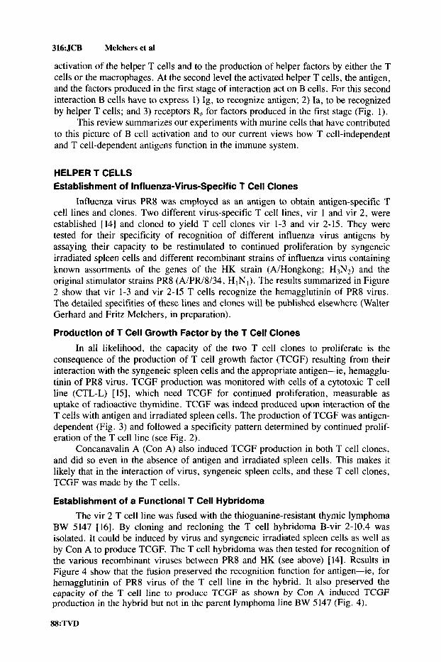
Influenza virus PR8 was employed as an antigen to obtain antigen-specific T cell lines and clones. Two different virus-specific T cell lines, vir 1 and vir 2, were established [14] and cloned to yield T cell clones vir 1-3 and vir 2-15. They were tested for their specificity of recognition of different influenza virus antigens by assaying their capacity to be restimulated to continued proliferation by syngeneic irradiated spleen cells and different recombinant strains of influenza virus containing known assortments of the genes of the HK strain (A/Hongkong; H3N2) and the original stimulator strains PR8 (A/PR/8/34, H INI ) . The results summarized in Figure 2 show that vir 1-3 and vir 2-15 T cells recognize the hemagglutinin of PR8 virus. The detailed specifities of these lines and clones will be published elsewhere (Walter Gerhard and Fritz Melchers, in preparation).
Production of T Cell Growth Factor by the T Cell Clones
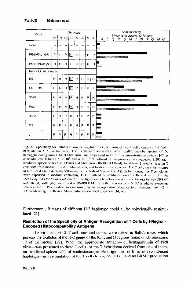
In all likelihood, the capacity of the two T cell clones to proliferate is the consequence of the production of T cell growth factor (TCGF) resulting from their interaction with the syngeneic spleen cells and the appropriate antigen-ie, hemagglu- tinin of PR8 virus. TCGF production was monitored with cells of a cytotoxic T cell line (CTL-L) [ 151, which need TCGF for continued proliferation, measurable as uptake of radioactive thymidine. TCGF was indeed produced upon interaction of the T cells with antigen and irradiated spleen cells. The production of TCGF was antigen- dependent (Fig. 3) and followed a specificity pattern determined by continued prolif- eration of the T cell line (see Fig. 2).
Concanavalin A (Con A) also induced TCGF production in both T cell clones, and did so even in the absence of antigen and irradiated spleen cells. This makes it likely that in the interaction of virus, syngeneic spleen cells, and these T cell clones, TCGF was made by the T cells.
Establishment of a Functional T Cell Hybridoma The vir 2 T cell line was fused with the thioguanine-resistant thymic lymphoma
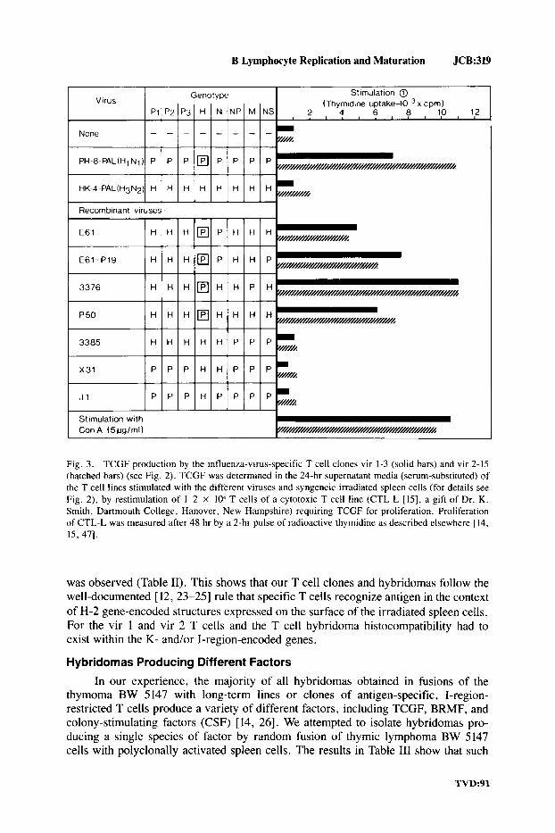
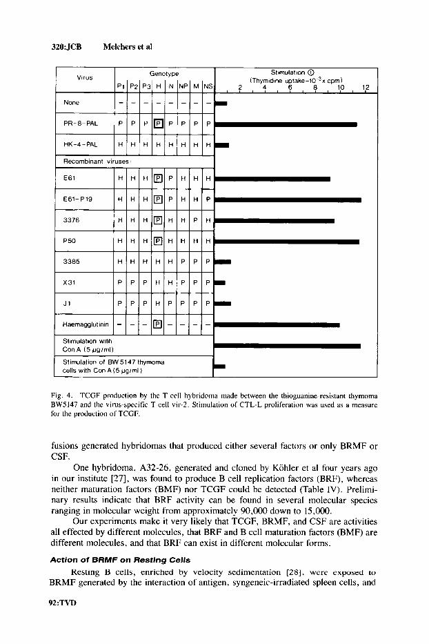
BW 5147 [16]. By cloning and recloning the T cell hybridoma B-vir 2-10.4 was isolated. It could be induced by virus and syngeneic irradiated spleen cells as well as by Con A to produce TCGF. The T cell hybridoma was then tested for recognition of the various recombinant viruses between PR8 and HK (see above) [14]. Results in Figure 4 show that the fusion preserved the recognition function for antigen-ie, for hemagglutinin of PR8 virus of the T cell line in the hybrid. It also preserved the capacity of the T cell line to produce TCGF as shown by Con A-induced TCGF production in the hybrid but not in the parent lymphoma line BW 5147 (Fig. 4).
88:TVD

B Lymphocyte Replication and Maturation JCB:317
Antigen
( IgM-specific antibodies) I B cell resgonse
Replication J+ Maturation to Ig secretion I
Rep I ication and Maturation Factors
Fig. 1. maturation of B cells.
Three essential structures on a resting B cell involved in the regulation of replication and
FACTORS ACTING ON B CELLS Production of B Cell Replication and Maturation Factors by the T Cell Clones and the T Cell Hybridoma
Single clones of antigen-specific T cells have previously been shown to produce a variety of replication and differentiation factors active on different cellular targets [ 17-20]. Among them are B cell replication and maturation factors (BRMF), which can be monitored by the continued replication of mitogen-activated B blasts and by their continued maturation to Ig-secreting celis, detected as plaque-forming cells [2 11.
Results in Table I show that BRMF were produced by the two T tell clones and the T cell hybridoma when stimulated either by antigen and syngeneic spleen cells or by Con A. Replication and maturation of the B blasts by BRMF was polyclonal as shown by the expansion of B blasts with various specificities-ie, for sheep or horse erythrocytes or for trinitrophenylated sheep erythrocytes and their frequencies relative to all IgM-secreting cells.
These findings are in agreement with earlier results obtained with T cell clones of other antigen specificities which showed that polyclonal restimulation of mitogen- activated B blasts involved a majority and in some cases all activated cells 1211.
TVD:89
318:JCB Melchers et a1
J1 P E P P P H P P P
Fig. 2. Specificity for influenza virus hemagglutinin of PR8 virus of two T cell clones, vir 1-3 (solid bars) and vir 2-15 (hatched bars). The T cells were activated in v ivo in Balb/c mice by injection of 100 hemagglutinating units (HAU) PR8 virus, and propagated in vitro in serum-substituted cultures 147) at concentrations between 5 X lo4 and 4 X 10' T cells/rnl in the presence of syngeneic. 2,200 rad- irradiated spleen cells (2 X 10⁶/ml) and PR8 virus (10-100 HAU/ml) for at least 2 months, feeding T cells with fresh medium, fresh irradiated cells, and fresh virus every week. The T cells were then cloned in semi-solid agar essentially following the methods of Sredni et al 1481. Before testing, the T cell clones were expanded in medium containing TCGF instead of irradiated spleen cells and virus. For the specificity tests the viruses indicated in the figure (which included seven recombinants betwen PR8 (P) and HK (H) virus [49]) were used at 10-100 HAUlml in the presence of 2 X 1oh irradiated syngeneic spleen cells/ml. Proliferation was measured by the incorporation of radioactive thymidine into 1-2 x 10⁴ proliferating T cells in a 2-hour pulse as described elsewhere [ 14, 471.
Furthermore, B blasts of different H-2 haplotype could all be polyclonally restimu- lated [21].
Restriction of the Specificity of Antigen Recognition of T Cells by I-Region- Encoded Histocompatibility Antigens
The vir 1 and vir 2 T cell lines and clones were raised in Balbk mice, which possess the d-alleles of the H-2 genes of the K, I, and D regions found on chromosome 17 of the mouse [22]. When the appropriate antigen-ie, hemagglutinin of PR8 virus-was presented to these T cells, or the T hybridoma derived from one of them, on irradiated spleen cells of nonhistocompatible origin-ie, of b- or of recombinant haplotype-no restimulation of the T cell clones, no TCGF, and no BRMF production
90:TVD
B Lymphocyte Replication and Maturation JCB:319
Fig. 3. TCGF production by the influenza-virus-specific T cell clones vir 1-3 (solid bars) and vir 2-15 (hatched bars) (see Fig. 2 ) . TCGF was determined in the 24-hr supernatant media (serum-substituted) of the T cell lines stimulated with the different viruses and syngeneic irradiated spleen cells (for details see Fig. 2). by restimulation of 1-2 x I@ T cells of a cytotoxic T cell line (CTL-L [15], a gift of Dr. K. Smith, Dartmouth College, Hanover, New Hampshire) requiring TCGF for proliferation. Proliferation of CTL-L was measured after 48 hr by a 2-hr pulse of radioactive thymidine as described elsewhere [ 14, 15, 471.
was observed (Table 11). This shows that our T cell clones and hybridomas follow the well-documented [ 12, 23-25] rule that specific T cells recognize antigen in the context of H-2 gene-encoded structures expressed on the surface of the irradiated spleen cells. For the vir 1 and vir 2 T cells and the T cell hybridoma histocompatibility had to exist within the K- and/or I-region-encoded genes.
Hybridomas Producing Different Factors In our experience, the majority of all hybridomas obtained in fusions of the
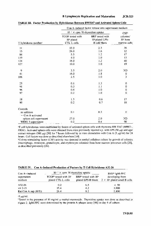
thymoma BW 5147 with long-term lines or clones of antigen-specific, I-region- restricted T cells produce a variety of different factors, including TCGF, BRMF, and colony-stimulating factors (CSF) [ 14, 261. We attempted to isolate hybridomas pro- ducing a single species of factor by random fusion of thymic lymphoma BW 5147 cells with polyclonally activated spleen cells. The results in Table 111 show that such
TVD:91
320:JCB Melchers et a1
Virus
None
PR-8-PAL
HK-4-PAL
Genotype Stimulation @ (Thymidine ~ p t a k e - 1 0 . ~ ~ cpm)
p1 P2 P3 H N NP M NS 2 4 6 8 10 12
- - - - - - - - -----__
P P P P P P P
H H H H H t i H H
Stimulation with Con A (5 vglml) I Stimulation of BW 5147 thymoma cells with Con A (5 pg/ml)
Fig. 4. TCGF production by the T cell hybridoma made between the thioguanine-resistant thymoma BW5147 and the virus-specific T cell vir-2. Stimulation of CTL-L proliferation was used as a measure for the production of TCGF.
fusions generated hybridomas that produced either several factors or only BRMF or CSF.
One hybridoma, A32-26, generated and cloned by Kohler et a1 four years ago in our institute [27], was found to produce B cell replication factors (BRF), whereas neither maturation factors (BMF) nor TCGF could be detected (Table IV). Prelimi- nary results indicate that BRF activity can be found in several molecular species ranging in molecular weight from approximately 90,000 down to 15,000.
Our experiments make it very likely that TCGF, BRMF, and CSF are activities all effected by different molecules, that BRF and B cell maturation factors (BMF) are different molecules, and that BRF can exist in different molecular forms.
Action of BRMF on Resting Cells
Resting B cells, enriched by velocity sedimentation [ Z S ] , were exposed to BRMF generated by the interaction of antigen, syngeneic-irradiated spleen cells, and
92:TVD

B Lymphocyte Replication and Maturation JCB:321
TABLE I. BRMF Production by the Virus-Specific T Cell Hybridoma
BRMF productionh tested with 102 plated
LPS-activated B blasts
Factor-producing 10 x cpm 10 x IgM celP Stimulationh 'H-thymidine uptake PFC
T cell clone vir 1-3
+ antigen + k? spleen cells + Con A + O
4.8 3.0
6.2 0. I
8. I 0. I
T cell clone vir 2-15
+ antigen + 9 spleen cells + Con A + O
10.2 14.8
14.2 0.3
22.4 0.1
T cell hybridoma B vir 2-10.4
+ antigen + Y spleen cells + Con A + O
12.0 16.2
11.4 0.2
14.4 0.1
Thymoma BW 5147
+ antigen + k? spleen cells + Con A + O
I .o 0.4
0.2 0.3
0. I 0.1
None + antigen + k? spleen cells + Con A + O
0.8 0.5
0.2 0.2
0.1 0.1
Rat Con A sup 21.0 55.0
'For the description of the cell lines see Figures 2, 3, and 4 and the text (141. "or details of the stimulation conditions and the assays see legends to Figures 2, 3, and 4 [21].
antigen-specific T cell clones. Two T cell clones (with different antigen specificities) were chosen to show the difference in action (Table V) observed with many more T cell clones with various specificities for antigen (not shown). None of the BRMF preparations produced by the different T cell clones induced proliferation of the resting B cells, whereas activated B blasts were stimulated to continued proliferation (see above). Some, but not all, preparations of BRMF (in Table V, the BRMF preparation induced by the HRC-specific T cell clone) induced polyclonal maturation to IgM-secreting cells of a large fraction (ie, more than 30%) of all resting B cells. The kinetics of this induction to Ig secretion and its polyclonality have been docu- mented previously [ 19, 291. It is interesting to note that the induction to maturation abolished the capacity of the B cells to be induced to replication. Replication and maturation may well be antagonistic processes in a B cell. Other BRMF preparations (in Table V, those induced by the virus-specific T cell lines) induced less or not detectable maturation of resting B cells to Ig secretion. Whenever maturation to secretion was observed it could occur in resting B cells of histocompatible as well as nonhistocompatible origin.
TVD:93

322:JCB Melchers et al
TABLE 11. Restriction of T Cell Recognition Capacity for Antigen by H-2 as Measured by Their CaDacitv to Produce Factors
10 ‘ X cpm ’H-thymidine uptake TCGF tested with I@ BRF tested with I@
Cells’ Stimulationh CTL-L cells LPS-activated B blasts
T cell clone antigen + ’Z ddd 11.2 3.8 vir 1-3 antigen + ’Z bbb 0.6 0.4
antigen + ’Z bbd 0.5 0.2
T cell clone antigen + 9 ddd 18.5
antigen + ’Z bbd 0.4 vir 2-15 antigen + 9 bbb 0.2
13.2 0.2 0.3
T cell hybridorna antigen + ’Z ddd 16.4 0.2 B-vir 2-10.4 antigen + 9 bbb 0.2 0.2
antigen + ’Z bbd 0.2 0.2
Cultures at 1-2 x 10⁴cells/ml. hAntigen was PR8 virus at 10’ HAU/ml. Y ddd are 2,200 r irradiated spleen cells of Balb/c (KdIaJDd), ’Z bbd those of BIO.A(SR) (KhIahDJ) mice, and ’Z bbb those of C57BL/6J (KhIahDh), all at 2 x lo6 cells/ ml. Supernatant media of 24 hours’ incubation were tested for TCGF and BRF as described in the legend to Figure 3.
Therefore, resting B cells are susceptible to the action of BMF, provided the interaction of T cell clones with irradiated spleen cells and antigen has produced sufficient quantities of it. They are, however, not susceptible to BRF, but must be rendered susceptible by specific interactions described below.
Step 1 of T Cell-Dependent 6 Cell Activation
Taken together, these results are interpreted as by others [ 2 3 , 30-321 (Fig. 5) to mean that the specific interaction of antigen, K- or I-region-histocompatible irradiated spleen cells and cloned T cells or a T cell hybridoma, can result in the production of T cell and B cell replication and maturation factors. Whenever either the antigen or the K- or I-region-encoded Ia antigen expressed on the irradiated spleen cells does not fit the specificity of the cloned T cell or its hybridoma, there is no production of factors.
HELPER T CELL-6 CELL COOPERATION Action of T Cell Help on Resting 6 Cells
Instead of exposing resting B cells to the soluble products of the specific interaction of T cells, antigen, and irradiated spleen cells, they were now added to this specific interaction we have termed “T cell help” [19, 301. This was done in limiting dilutions by adding graded numbers of resting B cells of histocompatible or nonhistocompatible origin to a fixed, optimal mixture of cloned T cells responding to either the antigen for which the cloned T cells were specific, or to a “bystander” antigen also added to the cultures, which shows no detectable cross reaction with the other antigen. The results are summarized in Table VI. It is evident that antigen- specific resting B cells are stimulated to develop into clones of IgM-secreting cells in frequencies as high as those observed upon polyclonal stimulation of the same resting
94:TVD
B Lymphocyte Replication and Maturation JCB:323
TABLE 111. Factor Production by Hybridomas Between BW5147 and Activated Spleen Cells
Con A-induced factor release into SuDernatant medium
lo-' x cpm 'H-thymidine uptake BRF tested with
lo" plated lo-' plated LPS- TCGF tested with
T hybridoma numberd CTL-L cells B-cell blasts
I I 15.0 2.3 15 26.0 1.6 84 5.5 1.2
1 I9 4.0 I .o 124 16.0 1.2 127 10.0 1 .o
9 41
106
25 91 96 97
18 90
3.5 16.0 2.5
0.1 0.3 0.4 0.2
I .5 0.2
2.0 1.8 1 .o
1.3 1 . 1 1 .o I .o
0.6 0.7
CSFh colonies/ 10' bone
marrow cells
36 12 17 13 40 19
ND 0 3
17 18
Controls no addition 0. I 0.3 0 + Con A-activated
spleen cell supernatant 27.0 2.0 ND WEHI-3 supernatant 0.2 0.4 90
&T cell hybridomas were established by fusion of activated spleen cells with thymoma BW 5 147 (HPRT-, OUA). Activated spleen cells were obtained from mice previously injected i.p. with LPS (50 pg) and agar extract mitogen (300 pg) [50] for 7 hours followed by in vitro stimulation with Con A (5 pg/ml) for 24 hours. Cell fusion was done as described elsewhere [ 14). hColony-stimulating factor (CSF) activity was detected in methyl cellulose culture by growth of colonies (macrophage, monocyte, granulocyte, and erythrocyte colonies) from bone marrow precursor cells [20], as described previously [26].
TABLE IV. Con A-Induced Production of Factors by T Cell Hybridoma A32-26
Con k-induced lo-' x cpm 'H thymidine uptake BMFh IgM-PFC supernatant TCGFh tested with 1oJ BRFh tested with lo-' developing from medium plated CTL-L cells plated LPS-B blasts 2 X lo-' plated small B cells
A32-26 0.2 6.5 < 50 vir 2-15 15.4 4.2 1 ,ooo Rat Con A sup (10%) 26.4 8.2 2,800
.'5 pg/ml. hTested in the presence of 10 mgiml a-methyl-mannoside. Thymidine uptake was done as described in Figure 2. IgM-PFC were determined by the protein A-plaque assay [46] on day 4 of culture.
TVD:95
324 JCB Melchers et a1
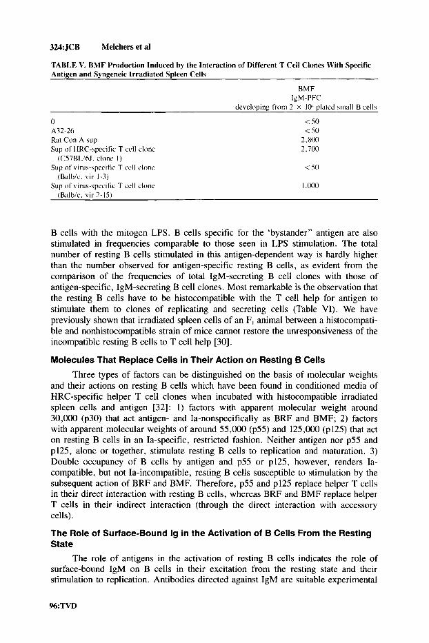
TABLE V. BMF Production Induced by the Interaction of Different T Cell Clones With Specific Antigen and Syngeneic Irradiated Spleen Cells
BMF
developing from 2 X 10’ plated small B cells IgM-PFC
0 < 50 A32-26 < 50 Rat Con A sup 2.800 Sup of HRC-specific T cell clonc 2.700
Sup of virus-specific T cell clonc <so
Sup of virus-specific T cell clonc I ,O(m
(C57BLi6J. clone I )
(Balbic. vir 1-3)
(Balbic. vir 2-15)
B cells with the mitogen LPS. B cells specific for the ‘bystander” antigen are also stimulated in frequencies comparable to those seen in LPS stimulation. The total number of resting B cells stimulated in this antigen-dependent way is hardly higher than the number observed for antigen-specific resting B cells, as evident from the comparison of the frequencies of total IgM-secreting B cell clones with those of antigen-specific, IgM-secreting B cell clones. Most remarkable is the observation that the resting B cells have to be histocompatible with the T cell help for antigen to stimulate them to clones of replicating and secreting cells (Table VI). We have previously shown that irradiated spleen cells of an F , animal between a histocompati- ble and nonhistocompatible strain of mice cannot restore the unresponsiveness of the incompatible resting B cells to T cell help [30].
Molecules That Replace Cells in Their Action on Resting B Cells
Three types of factors can be distinguished on the basis of molecular weights and their actions on resting B cells which have been found in conditioned media of HRC-specific helper T cell clones when incubated with histocompatible irradiated spleen cells and antigen [32]: 1) factors with apparent molecular weight around 30,000 (p30) that act antigen- and Ia-nonspecifically as BRF and BMF; 2) factors with apparent molecular weights of around 55,000 (p55) and 125,000 (p125) that act on resting B cells in an Ia-specific, restricted fashion. Neither antigen nor p55 and p125, alone or together, stimulate resting B cells to replication and maturation. 3) Double occupancy of B cells by antigen and p55 or p125, however, renders Ia- compatible, but not Ia-incompatible, resting B cells susceptible to stimulation by the subsequent action of BRF and BMF. Therefore, p55 and p125 replace helper T cells in their direct interaction with resting B cells, whereas BRF and BMF replace helper T cells in their indirect interaction (through the direct interaction with accessory cells).
The Role of Surface-Bound Ig in the Activation of B Cells From the Resting State
The role of antigens in the activation of resting B cells indicates the role of surface-bound IgM on B cells in their excitation from the resting state and their stimulation to replication. Antibodies directed against IgM are suitable experimental
96:TVD

B Lymphocyte Replication and Maturation JCB:325
Helper Tcell Adherent cell 7
TCGF
BRMF [BRF 1 BMF CSF and others
Fig. 5 . The first step in the T cell-dependent activation of B cells.
tools to investigate further the role of surface IgM in B cell stimulation, since their action must necessarily be polyclonal and therefore easy to detect.
A series of monoclonal rat antibodies specific for murine p-H-chains were generated by fusing the non-Ig-producing, azaguanine-resistant hybridoma Sp 2/0 [33] with spleen cells of rats immunized with mouse myeloma or hybridoma IgM molecules in their 19s secreted form. Antibodies against all four domains of the constant region of p-H-chains ( C p , , Cp2, Cp3, and Cp4) were found [34]. These antibodies, when immobilized on Sepharose 4B [35], without any further additions, stimulated resting B cells to low levels of proliferation (Table VII). This small proliferation was reduced to background values when the B cells were depleted of adherent cells (Leptin, Corbel, and Melchers, in preparation). Their presence in insoluble form did not impair the action of LPS on resting B cells, which induced polyclonal proliferation and maturation to Ig-secreting cells. Addition of factors containing TCGF and BRMF obtained by incubation of rat spleen cells with Con A as purified from conditioned media by ammonium sulfate precipitation and gel filtration of Sephadex GlOO [ 17, 181 resulted in polyclonal proliferation and matura- tion to Ig secretion of resting B cells when treated with some but not all the monoclonal p-specific antbodies, insolubilized on Sepharose 4B (Table VII).
These results show that occupance of Ig alone is insufficient to initiate poly- clonal proliferation of resting B cells. For such proliferation to occur, factors from conditioned media from either Con A-stimulated rat spleen cells or antigen-stimulated
TVD:97
326: JCB Melchers et a1
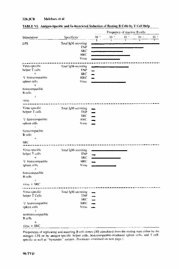
TABLE VI. Antigen-Specific and Ia-Restricted Induction of Resting B Cells by T Cell Help
Freauencv of reactive B cells Stimulation. Specificityh 10 10 10 ' 10 10 '
I I I 1 I LPS Total IgM-secreting
TNP SRC HRC Virus
Virus-specitic Total IgM-secreting helper T cells TNP I
+ SRC I
-----------------------------------------------------------,
t? histocompatible HRC - spleen cells Virus
histocompatible B cells
virus
Virus-specific Total IgM-secreting .I helpter T cells TNP I
+ SRC I t? histocompatible HRC - spleen cells Virus I
histocompatible B cells
SRC
Virus-specific Total IgM-secreting helper T cells TNP I
+ SRC t? histocompatible HRC .I spleen cells Virus
histocompatible B cells
virus + SRC
Virus-specific Total 1gM-secreting - helper T Cells TNP -
+ SRC - t? histocompatible HRC - spleen cells Virus -
+ nonhistocompatible B cells
+ virus + SRC
Frequencies of replicating and maturing B cell clones 130) stimulated from the resting state either by the mitogen LPS or by antigen-specific helper cells, histocompatible-irradiated spleen cells, and T cell- specific as well as "bystander" antigen. (Footnotes continued on next page.)
+
+ . . . . . . . . . . . . . . . . . . . . . . . . . . . . . . . . . . . . . . . . . . . . . . . . . . . . . . . . . . .
+
+ ...........................................................
+
+ ............................................................
98:TVD
B Lymphocyte Replication and Maturation JCB:327
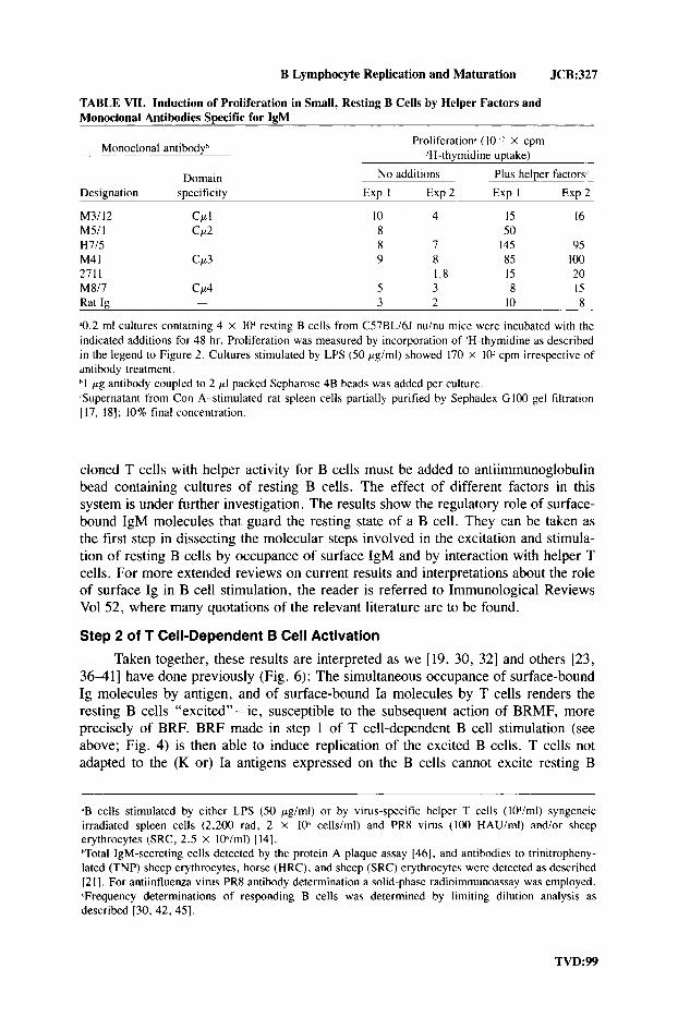
TABLE VII. Induction of Proliferation in Small, Resting B Cells by Helper Factors and Monoclonal Antibodies Swcific for IeM
Monoclonal antibodyh Proliferation* (I0 x cpm ’H-thymidine uptake)
Domain No additions Plus helper factors Designation specificity Exp I E x p 2 Exp I Exp 2
M3/12 Ccc I 10 4 15 16 M5/ I CP2 8 50 H7/5 8 7 145 95 M4 I CLt3 9 8 85 100 2711 1.8 IS 20 M817 CP4 S 3 8 15
3 2 10 8 Rat Ig -
Q.2 ml cultures containing 4 X 10’ resting B cells from CS7BLi6J nuhu mice were incubated with the indicated additions for 48 hr. Proliferation was measured by incorporation of ‘H-thymidine as described in the legend to Figure 2. Cultures stimulated by LPS (50 pgiml) showed 170 X 10’ cpm irrespective of antibody treatment. hl pg antibody coupled to 2 pI packed Sepharose 4B beads was added per culture. .Supernatant from Con A-stimulated rat spleen cells partially purified by Sephadex GI00 gel filtration [ 17, 181; 10% final concentration.
cloned T cells with helper activity for B cells must be added to antiimmunoglobulin bead containing cultures of resting B cells. The effect of different factors in this system is under further investigation. The results show the regulatory role of surface- bound IgM molecules that guard the resting state of a B cell. They can be taken as the first step in dissecting the molecular steps involved in the excitation and stimula- tion of resting B cells by occupance of surface IgM and by interaction with helper T cells. For more extended reviews on current results and interpretations about the role of surface Ig in B cell stimulation, the reader is referred to Immunological Reviews Vol 52, where many quotations of the relevant literature are to be found.
Step 2 of T Cell-Dependent B Cell Activation
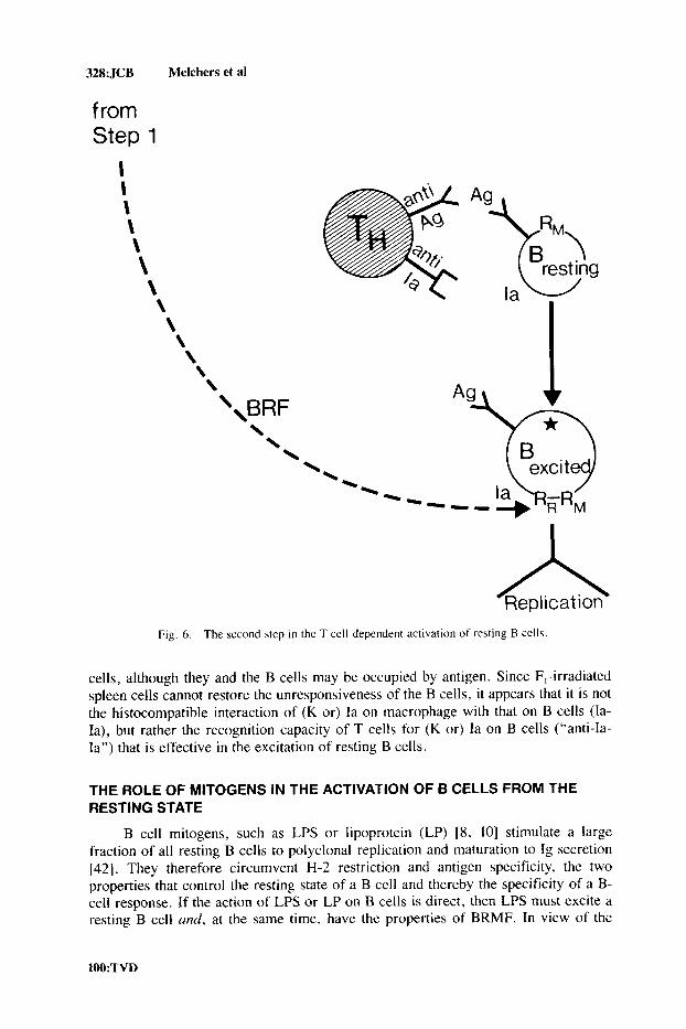
Taken together, these results are interpreted as we [19, 30, 321 and others [23, 36-41] have done previously (Fig. 6): The simultaneous occupance of surface-bound Ig molecules by antigen, and of surface-bound Ia molecules by T cells renders the resting B cells “excited”-ie, susceptible to the subsequent action of BRMF, more precisely of BRF. BRF made in step 1 of T cell-dependent B cell stimulation (see above; Fig. 4) is then able to induce replication of the excited B cells. T cells not adapted to the (K or) Ia antigens expressed on the B cells cannot excite resting B
’B cells stimulated by either LPS (50 pgiml) or by virus-specific helper T cells (10Jiml) syngeneic irradiated spleen cells (2,200 rad, 2 X loh cells/ml) and PR8 virus ( I 0 0 HAU/ml) and/or sheep erythrocytes (SRC, 2.5 X 10hlml) [ 141. ”Total IgM-secreting cells detected by the protein A plaque assay [46], and antibodies to trinitropheny- lated (TNP) sheep erythrocytes, horse (HRC), and sheep (SRC) erythrocytes were detected as described [2 I]. For antiinfluenza virus PR8 antibody determination a solid-phase radioimmunoassay was employed. ?Frequency determinations of responding B cells was determined by limiting dilution analysis as described [30, 42, 451.
328: JCB Melchers et al
from Step 1
I 1 \ \ \ \ \ \ \ \ \ \ \
\,BRF \ \ \
resting
I z
Fig. 6 . The second \tep i n the T cell-dependent activation of resting B cells.
cells, although they and the B cells may be occupied by antigen. Since F,-irradiated spleen cells cannot restore the unresponsiveness of the B cells, it appears that it is not the histocompatible interaction of (K or) Ia on macrophage with that on B cells (Ia- Ia), but rather the recognition capacity of T cells for (K or) Ia on B cells (“anti-Ia- Ia”) that is effective in the excitation of resting B cells.
THE ROLE OF MITOGENS IN THE ACTIVATION OF 6 CELLS FROM THE RESTING STATE
B cell mitogens, such as LPS or lipoprotein (LP) [8, 101 stimulate a large fraction of all resting B cells to polyclonal replication and maturation to Ig secretion [42]. They therefore circumvent H-2 restriction and antigen specificity, the two properties that control the resting state of a B cell and thereby the specificity of a B- cell response. If the action of LPS or LP on B cells is direct, then LPS must excite a resting B cell and, at the same time, have the properties of BRMF. In view of the
100:TVD

B Lymphocyte Replication and Maturation JCB:329
role of helper T cells and accessory cells (irradiated spleen cells, macrophages, adherent cells) as potential producers of BRMF, it is, however, possible that LPS activates accessory cells to BRF production while it excites resting cells to a state of susceptibility to accessory cell-derived BRMF.
We have tested these two alternatives (Corbel and Melchers, in preparation). Resting splenic B cells were further depleted of accessory cells by adherence to plastic followed by passage through G 10-Sephadex [43]. This removal of accessory cells abolished the capacity of LPS and LP to induce polyclonal replication and maturation to Ig secretion (Fig. 7). Responsiveness could be restored when accessory cells such as peritoneal cells were added back to the accessory cell-depleted B cells. It could also be restored by soluble factors from activated macrophages and a macrophage line (called P388DJ [51].
CELL CYCLE CONTROL OF B CELL REPLICATION
Studies on the kinetics of activation of single resting B cells by mitogens such as LPS to replicating clones of IgM-secreting cells have shown that this activation is asynchronous [44]. All B cells are activated within the first 18-24 hours after addition of the mitogen. Single clones of replicating B cells divide regularly every 18 hours for at least 5-7 divisions. Every cell in the clone divides, and every dividing cell secretes IgM [45].
Activated B blasts can be synchronized by selection of a given size of cells separable by velocity sedimentation [28, 441, which represents a given stage of the cell cycle-the smallest cells being early in the G1 phase of the cell cycle, the largest late in G2. Synchronized cells in positions early in G1 to late in G2, when recultured in the absence of mitogen, will complete their cell cycle and divide. After mitosis they presumably remain in early GI, at least do not divide any more. If recultured in the presence of mitogen, however, these cells show synchronous divisions every 18 hours for at least 2-3 more cell cycles [44].
These experiments have been interpreted to indicate that the growth-promoting mitogen has to be present for at least a short fraction of the cell cycle time early in GI for the B blasts to proceed through the next cycle and through the next mitosis [44]. In view of the findings reported above, it remains to be investigated whether the synchronized B blast populations contain BRMF as long as LPS is present, and thus stimulate them. BRMF would then be used up by the B blasts at a given stage of the cell cycle. Experiments with synchronized B blasts using purified BRF should further clarify this issue.
The Emerging Picture of T Cell-Dependent and T Cell-Independent B Cell Stimulation
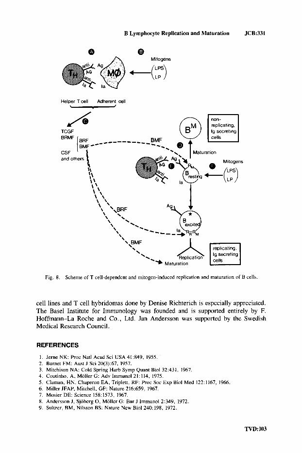
In conclusion, two steps are discernible in the activation of resting B cells to replication and to maturation to Ig secretion (Fig. 8). In step 1, helper T cells recognize specific antigen and Ia antigen on accessory cells (A, Fig. 8). This leads to activation of the helper T cells and to production of soluble factors that stimulate replication (BRF) and maturation (BMF) (C, Fig. 8). These factors are probably separate entities. Other factors, including TCGF and CSF, are often also made. Often the factors are produced by the T cells, but there may be cases in which the accessory cells make the factors.
TVD:lOl

330:JCB Melchers et a1
Fig. 7. Mitogenic stimulation of small, resting B cells is adherent cell-dependent and can be restored by supernatant factors from peritoneal macrophages or a macrophage cell line. Serum-free cultures 1471 containing 5 X lo4 small, resting spleen cells from C3HiTif mice were stimulated for 48 hr by lipopolysaccharide (LPS; 50 pg/ml) or lipoprotein (LP: 10 pg/rnl). Depletion of adherent cells was done by first incubating the spleen cells on plastic for 2 h r at 37°C and subsequently passaging the nonadherent cells through a Sephadex G I 0 column according to Ly and Mishell (431. U N o additions. W Supernatant from peritoneal cells 5 x lo4 cellsiml cultured with agar extract (20 pgiml) [50]. E Peritoneal cells; lo4 cells/culture. E Supernatant of the macrophage cell line P388 DI [ S l ] after 24 hours’ treatment of lo5 cellsiml with agar extract (20 pgiml).
Mitogens, such as LPS or LP, may activate accessory cells directly to factor production (B, Fig. 8), thereby circumventing the need for helper T cells in step 1.
BRF acts on activated B blasts, and is probably required early in G1 phase of each cell cycle to stimulate the B blasts polyclonally through the next cell cycle and through mitosis. BMF induces further maturation to Ig secretion of replicating B blasts already secreting lower levels of Ig. BRF does not act on resting B cells. BMF induces polyclonal maturation to Ig secretion without replication. Its action is antag- onistic to replication (D, Fig. 8).
In order for resting B cells to become susceptible to the action of BRF-ie, to enter replication-they have to be “excited.” In the T cell-dependent way of excita- tion, Ig molecules on the surface of B cells have to be occupied by antigen or anti-Ig antibodies, and Ia molecules on the surface have to be recognized by helper T cells (E, Fig. 8). This double occupancy induces susceptibility to BRF. The requirements for antigen-specific (Ig) and H-2-restricted (Ia) excitation can be circumvented by mitogens, such as LPS or LP (F, Fig. S), which polyclonally and H-2 unrestrictedly induce susceptibility of resting B cells to BRF.
ACKNOWLEDGMENTS
The able technical assistance of Denise Richterich, Lotta Haraldson, Annick Peter, and Bente Jensen is gratefully acknowledged. The continuous propagation of T
102:TVD
B Lymphocyte Replication and Maturation JCB:331
A Mitogens
Helper Tcell Adherent cell
TCGF
and others
replicating , lg secreting
Fig. 8. Scheme of T cell-dependent and mitogen-induced replication and maturation of B cells.
cell lines and T cell hybridomas done by Denise Richterich is especially appreciated. The Base1 Institute for Immunology was founded and is supported entirely by F. Hoffmann-La Roche and Co., Ltd. Jan Anderson was supported by the Swedish Medical Research Council.
REFERENCES
1. Jerne NK: Proc Natl Acad Sci USA 41:849, 1955. 2. Burnet FM: Aust J Sci 20(3):67, 1957. 3. Mitchison NA: Cold Spring Harb Symp Quant Biol 32:431, 1967. 4. Coutinho, A, Moller G: Adv Immunol 21:114, 1975. 5. Claman, HN, Chaperon EA, Triplett, RF: Proc SOC Exp Biol Med 122:1167, 1966. 6. Miller JFAP, Mitchell, GF: Nature 216:659, 1967. 7. Mosier DE: Science 158:1573, 1967. 8. Anderson J, Sjoberg 0, Moller G: Eur J Immunol 2:349, 1972. 9. Sultzer, BM, Nilsson BS: Nature New Biol 240: 198, 1972.
TVD: 103

332:JCB Melchers et a1
10. Melchers F, Braun V, Galanos C: J Exp Med 142:473, 1975. 11. Katz, DH, Hamaoka T, Dorf ME, Benacerraf B: Proc Natl Acad Sci USA 70:2624, 1973. 12. Rosenthal AS, Shevach EM: J Exp Med 138:1194, 1973. 13. McKean DJ, Infante AJ, Nilson A, Kimoto K, Fathman CG, Walker E, Warner N: J Exp Med
154:1419, 1981. 14. Melchers F, Zeuthen J, Gerhard W: Curr Top Microbiol Immunol 100: 153, 1982. 15. Gillis S, Ferm MM, Ou W, Smith KA: J Immunol 120:2027, 1978. 16. Trowbridge IS, Hyman R, Mazauskas, C: Cell 14:21, 1978. 17. Schreier MH, Iscove NM, Tees R, Aarden L. Von Boehmer H: Immunol Rev 51:315, 1980. 18. Gronvik K-0, Andersson J: Immunol Rev 51:315, 1980. 19. Melchers F, Andersson J, Lernhardt W, Schreier MH: Immunol Rev 52:89, 1980. 20. Schreier MH, Iscove NN: Nature 287:228, 1980. 21. Schreier MH, Andersson 3, Lernhardt W, Melchers F: J Exp Med 151: 194, 1980. 22. Klein J: “Biology of the Mouse Histocompatibility-2 Complex.” New York: Springer-Verlag, 1975. 23. Sprent J: Immunol Rev 42: 108, 1978. 24. Schwartz RH, David CS, Sachs DH, Paul WE: J Immunol 117531, 1976. 25. Corradin G, Etlinger HM, Chiller JM: J Immunol 119: 1048, 1977. 26. Corbel C: Curr Top Microbiol Immunol 100:231, 1982. 27. Kohler G, Lefkovits I, Elliott B, Coutinho A: Eur J Immunol 7:758, 1977. 28. Miller RG, Phillips RA: J Cell Physiol 73: 191, 1969. 29. Melchers F, Andersson J , Lernhardt W, Schreier MH: Eur J Immunol 10:679, 1980. 30. Andersson J, Schreier MH, Melchers F: Proc Natl Acad Sci USA 77: 1612. 1980. 31. Kappler JW, Skidmore B. White J . Marrack P: J Exp Med 153: 1198. 1981 32. Andersson J, Melchers F: Proc Nail Acad Sci USA 78:2497, 1981. 33. Shulman M, Wilde CD, Kohler G: Nature 276:269. 1978. 34. Leptin M: Thesis, University of Heidelberg, 1983. 35. Axen R, Porath J. Ernbacks S: Nature 214: 1302, 1967. 36. Keller DM, Swierkosz JE, Marrack P, Kappler JW: J Immunol 124: 1350. 1980. 37. Hoffman MK: J Immunol 125:2076. 1980. 38. Delovitch TL, Watson J. Battistella R , Harris JF, Shaw J, Paetkau V: J Exp Med 153:107, 1981. 39. Martinez AC, Coutinho A: Nature (Lond) 290:60, 1981. 40. Jones B, Janeway CA Jr: Nature 292:547. 1981. 41. Julius MH, Von Boehmer H, Sidman CL: Proc Natl Acad Sci USA 79: 1989, 1982. 42. Andersson J, Coutinho A , Melchers F: J Exp Med 145:1511. 1977. 43. Ly IA, Mischell RI: J Immunol Meth 5:239. 1974. 44. Lernhardt W: Exp Cell Res, 1982 (submitted). 45. Andersson J, Coutinho A , Lernhardt W. Melchers F: Cell 10:27, 1977. 46. Gronowicz E, Coutinho A, Melchers F: Eur J Immunol 6:588. 1976. 47. Iscove NN, Melchers F: J Exp Med 147:923. 1978. 48. Sredni B, Schwartz RH: Nature 287:855, 1980. 49. Palese P: Cell 10: I , 1977. 50. Kincade PW, Ralph P, Moore MAS: J Exp Med 143: 1265, 1976. 51. Lachman LB, Hacker MP, Blyden GT. Handschumacher RE: Cell Immunol 34:416, 1977.
104:TVD