Concurrent Replication and Methylation at Mammalian Origins of Replication
Upload
khangminh22Category
view
1download
0
F I N E S T R U C T U R E AND R E P L I C A T I O N OF
T H E K I N E T O P L A S T OF T R Y P A N O S O M A L E W I S I
P A U L R. B U R T O N and D O N A L D G. D U S A N I C
From the Departments of Zoology and Microbiology, University of Kansas, Lawrence, Kansas 66044
A B S T R A C T
The kinetoplastic DNA of Trypanosoma lewisi is described as a filamentous body lying within a mitochondrion, with the filaments oriented parallel to the long axis of the cell. The manner of fixation, the replicative state, and perhaps the physiological state of the cell, may result in slight morphological differences among such bodies. The kinetoplastic DNA replicates to form "left" and "right" rather than "upper" and "lower" members, and both the kinetoplast and nucleus incorporate radiothymidine as shown by radioautography. Radioautographic analyses suggest a random incorporation of radiothymidine by kineto- plasts. Silver grains were occasionally observed over centriolar elements. Finally, the obser- vations are discussed with respect to the sequential replication of the aforementioned or- ganelles by T. lewisi.
I N T R O D U C T I O N
Trager (1964, 1965) has reviewed the literature dealing with the DNA-containing kinetoplast of certain flagellates. In the family Trypanoso- matidae, the kinetoplast appears in the light microscope as a small body just behind the basal body of the flagellum which: (1) stains with nu- clear stains, as shown by many workers, (2) is Feulgen positive (Bresslau and Scremin, 1924, and others), and (3) incorporates thymidine in its replication cycle (Steinert et al., 1958; Steinert and Steinert, 1962).
Extranuclear DNA has recently been isolated from blood flagellates (DuBuy et al., 1965, 1966; Riou et al., 1966, 1967). When one considers the large size of the kinetoplast in relation to the rest of the cell, it is probable that much of the isolated cytoplasmic DNA is of kinetoplastic origin. Even though the workers who reported isolation and characterization of kinetoplast DNA did not make a clear distinction between DNA of the kinetoplast proper and that of the mitochondria remote from the kinetoplast region, it should be noted that Riou and Paoletti (1967) obtained cytoplasmic DNA
with two different buoyant densities from Try- panosoma cruzi. Cytoplasmic DNA isolated from blood flagellates differs from nuclear DNA in buoyant density, base ratio, and degree of rena- turation. Also, the cytoplasmic DNA appears to be more homogeneous than the nuclear DNA of the cell (DuBuy et al., 1966), and electron micro- scopic studies indicate that at least a portion of the cytoplasmic DNA may assume a circular or ring configuration (Riou and Paoletti, 1967).
A number of workers have examined the kineto- plast region with the electron microscope, and Mayer et al. (1958) first noted its separateness from the nearby basal body. Steinert (1960) made the observation that the dense, Feulgen-positive material of the "kinetonucleus" of Trypanosoma mega actually lies within the matrix of a mito- chondrion, which was later confirmed by Pitelka (1961) in her studies of Bodo saltans, a free-living flagellate possessing a kinetoplast. Filamentous elements of the kinetoplast often have electron- lucent centers when seen in transverse section, as if they were tubular (Schulz and MacClure, 1961).
318

These fibrils of the disclike kinetoplast, as dia- g rammed by Schulz and MacClu re (1961), have long axes tha t are or iented parallel to the long axis of the cell, bu t perpendicular to the f lat tened plane of the kinetoplast proper. According to these authors, the kinetoplast may conta in cont inuous filaments tha t are looped back and forth upon themselves.
Litt le is known of kinetoplast repl icat ion except tha t one kinetoplast gives rise to two and thymidine is incorporated in the process (Steinert et al., 1958). Meyer et al. (1958) noted tha t dur ing division the "kinetonucleus '~ increases in volume and " . . . (bends) down in the midd le . " Mfihlp- fordt (1963) noted tha t division occurs by simple " s t r angu la t ion . " T h e complexity of the process was po in t edou t by Mi lder and Deane (1967), who suggested tha t a new kinetoplast mass may arise parallel to the paren t disc, and that , after separa- t ion, one of the fi lamentous masses migrates " . . . to a position where its long axis becomes approxi- mately contiguous to the long axis of the o t h e r . . . before invaginat ion of the (mitochondrial ) mem- b rane completes the division."
T h e present study represents an a t t empt to elucidate the s t ructure and replicat ion of the kinetoplast of cul ture forms of Trypanosoma lewisi by the use of electron microscopy and radioauto- graphy. For a more general t r ea tment of the fine s tructure of T. lewisi, the reader is directed to articles by J u d g e and Anderson (1964) and Anderson and Ellis (1965). T h e kinetoplast is usually defined as a complex including bo th the compact mass of DNA-conta in ing filaments and the l imit ing mi tochondr ia l membranes immedi- ately sur rounding the mass.
M A T E R I A L S A N D M E T H O D S
Culture forms of Trypanosoma lewisi were grown in a diphasie (blood agar--Locke's solution) medium (Dusanie, 1968). The organisms were harvested from the liquid overlay and washed in cold phosphate- buffered glucose-saline (pH 7.2) by centrifugation at 1,200 g at 4°C. Cultures inoculated with 1.0 X 10 e trypanosomes/ml reached 2.0 X 101 to 7.0 X 10⁷ trypanosomes/ml after 5-6 days of incubation at 27°C. Blood-stream forms were maintained by syringe passage in female Holtzman rats. Blood was collected from infected animals by cardiac bleeding into 4% sodium citrate, and the organisms were separated from the blood cells by differential centrifugation in cold phosphate-buffered glucose- saline.
The supernatant fluid was poured from the pellets
of washed blood-stream or culture form flagellates, fixative was added to the tube, and the cells were resuspended. Fixation was accomplished in 6% glutaraldehyde (Sabatini et al., 1963) buffered with s-collidine at pH 7.4-7.5. Both sucrose (Caulfield, 1957) and calcium chloride were added to the fixative, the latter at a concentration of 10 -a M. Fixation was carried out for 1-2 hr in the cold (0-4°C), followed by four washes over a 1-2-hr period in cold buffer. The cells were centrifuged after each change, and the pellet was broken into small pieces with a steel probe. After washing, the small pieces were kept intact during further process- ing. Postfixation was carried out for 1 hr in l~o OsO4 buffered as above and kept at room tempera- ture. Some cells were fixed directly in 1% OsO4 buffered with s-eollidine at pH 7.4-7.5. Sucrose and CaCI2 were added as indicated above, and fixations were performed both in the cold and at room temperature. After fixation, cells were dehy- drated in a graded alcohol series to propylene oxide, and embedded in Epon 819. Silver or gray sections were obtained on a Porter-Blum microtome with glass or diamond knives and picked up on grids coated with Parlodion and a thin film of amorphous carbon. Sections were stained with lead citrate (Reynolds, 1963), saturated (aqueous) uranyl acetate, or, more often, with both.
For radioautography, trypanosomes were cul- tured in medium containing H3-1abeled thymidine (sp. activity 6.7 c/mmole) at a concentration of 10 /ze/ml. Following inoculation, crithidial cells were collected after 4, 7, 24, 48, and 72 hr. It should be noted that, although cultured blood flagellates divide asynchronously, mean generation times of 18.9 hr have been obtained for Trypanosoma mega (Steinert and Steinert, 1962) and 19.4 hr for T. ¢ruzi (Chang, 1947). The mean generation time for T. lewisi during the first 4 days of growth is ap- proximately 21.8 hr. After fixation, sections of labeled cells, and sections of cells grown in the absence of tritiated thymidine to serve as controls, were radio- autographed with the use of Ilford L-4 liquid emul- sion. The methods used are modifications of those described by Caro and van Tubergen (1962) and Revel (1964, personal communication). A wire loop about 12 mm in diameter was dipped in the emul- sion, and the excess emulsion was quickly drained from the edge of the loop onto a tissue as the loop was withdrawn. The film of gelled emulsion was then low- ered onto the section-covered specimen screen which was resting, sections up, on a square Lucite post about 20 mm long and 2 ~ mm wide. Although the film would rupture as it was draped over the specimen screen and post, a thin film of emulsion remained attached to the sections. Specimen screens without sections were similarly prepared, some of which were immediately processed (developed) without exposure
P. R. BURTON AND D. G. D~sAmc Kinetoplast of T. lewisi 319

to light other than that of the safelight, while others were exposed to the light of a bulb or to sunlight and then processed. These preparations indicated the sort of background one could expect, which increases with age and exposure to cosmic radiation. Also, such preparations provided information on the distribu- tion of grains within the film as well as its thickness. The fresh L-4 emulsion used in this study showed very few grains with no exposure to radiation, and the loop method was found to be satisfactory for applying a thin layer of emulsion to sections. After films were placed on sections, the specimen screens were captured by application to the adhesive edges of 3-ram holes punched in Johnson and Johnson "Air-Vent" band-aid tape. Strips of tape bearing specimen screens were then affixed to glass slides, with the sections up and exposed through the holes in the tape.
The exposure period was 4 months in a light-tight box kept in a desiccator at 5°C. A (302 atmosphere was maintained in the desiccator. After exposure, specimen screens covered with emulsion were de- veloped for 11/~ rain in full-strength Mierodol-X followed by a 5-sec rinse in glass-distilled water. The emulsion was then cleared in Kodak acid- fixer for 11/~ rain, and washed in two changes of glass-distilled water for 1 min each. After the speci- men screens were air-dried, sections were stained for 1 min in lead citrate and then for 15-30 man in uranyl acetate.
An RCA EMU-3H electron microscope, equipped with a 35-40-/z aperture and operating at 50 kv, was used for examining and obtaining negatives of the material. 1V[icrographs were made on Kodak latern slide plates at initial magnifications of up to about 20,000 diameters and subsequently were enlarged photographically with the use of Schneider Companon lenses. For purposes of critical measure- ments, the microscope was calibrated by the use of two different carbon grating replicas.
O B S E R V A T I O N S
T h e general spatial relationships between the kinctoplast and its ne ighbor ing organellcs are d iagrammat ica l ly shown in Fig. I. In a nondivid- ing cell, a centriole lies at a r ight angle to another centriole serving as a basal body. Dur ing division, however, the free centriole comes to lie parallel to the basal body as it part icipates in the forma- t ion of a second flagellum. Al though the centrioles and kinetoplast are near one another , connections have not been observed between these elements.
I n longi tudinal sections of the cell, the kineto- plast usually appears elongate or rodlike (Figs. 7-10), while in transverse sections it appears circular or oval in outl ine (Fig. 14). Studies of
.... FmUI~E 1 Diagram showing relationships between organelles under consideration. This represents the erithidial fol-m of T. lewisi, where the flagellmn origi- nates about midway between the two ends of the elongate cell. The flagellum (FL] originates from a basal body (BB); a free eentriole (CE) is also present, and procentrioles (PC) arise in association with both centriolar elements, although not until the cell begins to divide. Vacuoles (V) are commonly seen in this region, and mierotubules (MT) lie beneath the surface of the plasma membrane (PM). The kinetoplast (K) is associated with a mitochondrion (M) between the centriolar dements and the nucleus (N), in which a proufinent nucleolus (NU) is seen.
serial sections indicate tha t the most c o m m o n shape of a kinetoplast is tha t of a disc or an elon- gate plate. The morphology of the kinetoplast, however, is not static, and r ing or doughnu t con- figurations are sometimes seen in transverse sec- tions of cells (Fig. 11). Whi le the dense mater ia l of a kinetoplast is most often loosely fi lamentous (Figs. 2, 3), it may also appear compac t and granular (Figs. 8, 10, 12, 13, 15). Kinetoplasts fixed only in OsO4 appear more fi lamentous and less compact t han those fixed in g lu tara ldehyde (see Figs. 2, 11, 14 with 3, 4-10). Obviously, the kinetoplast is somewhat polymorphic, wi th its s tructure reflecting its replicative state, the m a n n e r of fixation, the plane of sectioning, and perhaps the funct ional state of the mi tochondr ion or cell in which it is contained. In a replicating kineto- plast, one fi lamentous m e m b e r may be twisted in respect to the other such tha t their appearance is somewhat dissimilar in a given section (Figs. 4-6).
In OsO4-fixed material , the mat r ix of the mito- chondr ion is less dense than in glutaraldehyde-
320 THE JOU-I:tNAL OF CELL BIOLOGY • VOLUME 39, 1968

Abbreviations used in the electron micrographs: b, basal body; c, centriole; er, endoplasmic reticulum; g, Golgi elements; k, kinetoplast; m, mitochondrion; mr, microtubule; n, nucleus; po, procentriole.
FIGURE ~ Kinetoplast region of blood-stream form isolated from rat after 81/~ days of infection. OsOa- collidine. Lead citrate (LC) stain only. X 6~,400.
321

FIGURE $ Kinetoplast (longitudinal profile) of erithidial form after 6 days of culture. Arrow indicates "tubular" filaments in longitudinal view. Note that some filaments appear to terminate in dense median zone while others appear to extend from one side to the other. Glut.-coUidine. Uranyl acetate (UA) stain only. X ~0,000.
322

FIGURES 4--6 Serial mierographs of apparently dividing kinetoplast of crithidial form after 6 days of culture; one member is twisted with respect to another. Plane of section is slightly oblique to long axis of cell. Arrow in Fig. 6 indicates a "tubular" filament seen in cross-section. Jus t below arrow are pro- fries of smaller, "nontubular" f laments measuring about 86 A in diameter. Glut.-collidine. UA only. X 88,000.
323

fixed cells. Figs. 2 and 11 clearly show the distribu- tion of the kinetoplast mass within a structurally typical mitochondrion. Although that part of the mitochondrion in which the filamentous mass lies sometimes appears to have fewer cristae than other parts of the mitochondrion, the membranes of this region are generally no different from those of the remainder of the mitochondrion. Filaments in the kinetoplast of OsO4-fixed cells are 30-50 A in width, and they may be loosely organized (Fig. 2), more compact and well oriented (Fig. 11), or even organized into granular-appearing masses (Fig. 14).
In glutaraldehyde-fixed cells, the kinetoplastic DNA appears more dense than in material fixed in only OsO4, and the masses appear very compact and granular or, more often, filamentous, In both cases, the granules and/or filaments are oriented parallel to the long axis of the cell. The matrix of mitochondria in glutaraldehyde-fixed cells is granular and the two mitochondrial membranes are not so easily seen as in OsO4-fixed cells. In
longitudinal sections of cells, kinetoplasts are cut perpendicular to their flattened plane such that their masses of DNA appear as rods. Hereafter, a kinetoplast cut in this manner will be described as appearing in longitudinal profile. Conversely, in transverse sections of cells the kinetoplasts are cut parallel to their flattened surface and will be described as showing a transverse profile. Masses of kinetoplastic DNA in longitudinal profile fre- quently appear ladder-like and bilateral (Figs. 3, 4-6, 7). Regions of increased density extend along the two long margins and the median axis, and they appear to be connected by irregularly-spaced filaments such that the over-all appearance is similar to two ladders lying side-by-side. When seen in cross-section, the filaments often have electron-lucent centers, as if they were tubular (Figs. 3, 5, 6). Such filaments are 100-160 A in diameter with "walls" 40-60 A thick. Filaments that do not appear tubular are also present (Fig. 6), and they measure about 80 A in diameter. In some instances filaments appear to extend the
FIGImE 7 Longitudinal profile of replicating kinetoplasts of crithidial form after 6 days of culture. Glut.-collidine. LC-UA. X 68#00.
FIGURE 8 Longitudinal profile of replicating kinetoplasts of erithidial cell after $1/~ days of culture. Glut.-collidine. LC-UA. X 78,500.
324 THE JOURNAL OF CELL BIOLOGY • VOLUME 39, 1968

FIOUaE 9 Longitudinal profile of replicating kinetoplasts of crithidial form after 7 hr of culture. Glut.- collidine. LC-UA. )< 41,600.
FIGURE 10 Longitudinal profile of replicating kinetoplasts of erithidial form after 8 ~ days of culture. Glut.-collidine. LC-UA. )< 73,500.
entire distance from one margin of the mass to the other, while in other cases they appear to extend only half the distance and merge with the dense median region (Figs. 3, 4-6).
Masses of kinetoplastic D N A appear to replicate longitudinally rather than transversely (Figs. 4-6, 7-9). An unusual condition is shown in Fig. 10, where three masses can be seen within a common mitochondrial matrix, as if replication might have resulted in three daughter masses. Another explanation, although considered unlikely, is that one disc of a pair is bent back upon itself. In any event, the three-membered condition is rarely seen. Whether a kinetoplast mass appears more granular than filamentous or ladder-like may not necessarily depend on its replicative state, for both conditions have been observed among kineto- plasts in a comparable division state (cf. Fig. 7 with Figs. 8, 9). The two members of a pair, how- ever, were always observed to have the same appearance, either compact and granular or
obviously filamentous. I t should be noted that cells fixed in the same pellet usually had kineto- plasts of similar appearance, and it is suspected that whether a mass of kinetoplastic D N A appears compact and granular or less compact and fila- mentous may depend on subtle differences in fixation. Such morphological differences are noted even when efforts are made to fiX different batches in the same manner. Also, it is interesting that in kinetoplast masses having a compact, granular appearance, there was never indication of the ladder-like configuration described above.
Although cultured trypanosomes do not divide in a synchronous manner, radioautography was employed in order to obtain information on the replication of kinetoplasts. Examination of silver grains over sections showed that tritium-labeled thymidine was well localized in nuclei and kineto- plasts, and background grains were relatively few in number. No consistent differences were noted in the distribution of silver grains over sections
P. R. BURTON AND D. G. DCSANIC Kinetoplast of T. lewisi 325

from cells harvested after 4, 7, 24, 48, and 72 hr in the trit ium-containing medium. While labeled kinetoplasts and nuclei were seen in most cells in each of the five culture groups, some cells, even after 72 hr of incubation, appeared to be un- labeled.
The distribution of silver grains most frequently observed in longitudinal sections of cells is shown in Fig. 12, in which the periphery of the disc is obviously labeled. Sometimes, however, only one member of an apparent daughter pair of D N A masses was labeled (Fig. 13). Also, in a kinetoplast seen in transverse profile, exactly half of its diam- eter showed incorporation of the labeled thymidine (Fig. 14). In transverse sections of cells, kineto- plasts often appear horseshoe- or doughnut-shaped, and in such cases the grains are usually distributed in a somewhat random manner, as in Fig. 11. I t is possible that the circular or doughnut-shaped profiles represent the " r i m " of a saucer-shaped kinetoplast, in which case the distribution of silver grains might suggest random incorporation around the periphery of the saucer. Generally, the periphery of nuclei are more heavily labeled than the central region, perhaps corresponding to peripheral masses of dense material (Figs. 1, 11, 15). Also, as shown in Fig. 1 I, centrioles or basal bodies were sometimes labeled. In no case were both centriolar elements labeled in a given cell, but our sample was not great since both elements were not often seen in a given section.
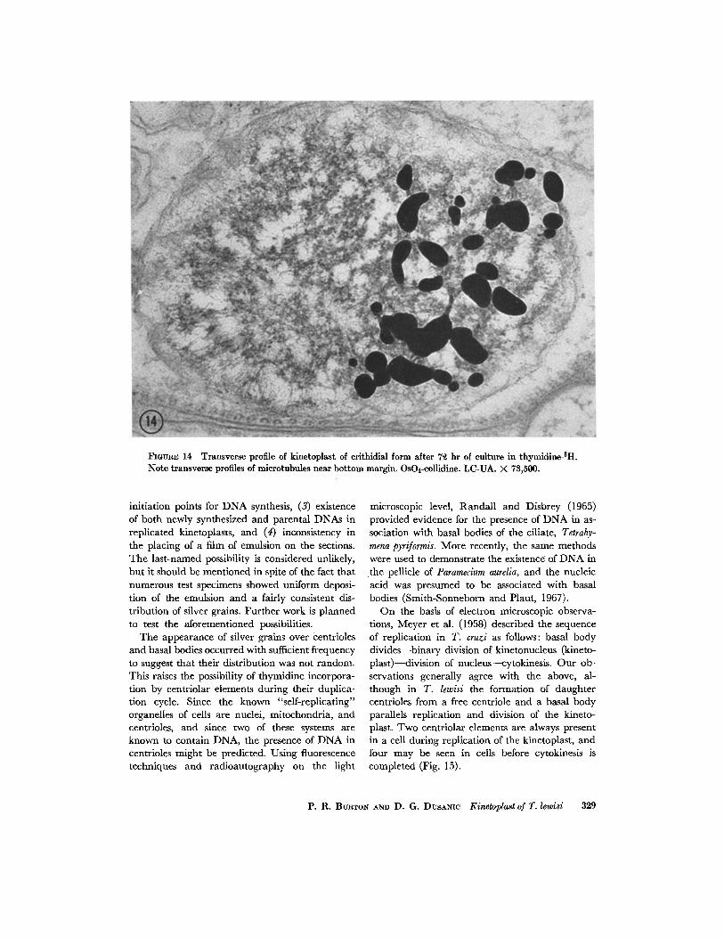
Fig. 15 shows a case of incomplete cytokinesis in which the two daughter cells are still attached along a portion of their surface (note the "shared" microtubules in the lower right corner). The kine- toplasts in the daughter cells are labeled, but in a different manner. One kinetoplast appears to be rather uniformly labeled, while in the other, which appears to be the longer of the two, the label is restricted to the ends of the profile, or the periphery of the kinetoplastic mass. I t is possible that in the latter case the cell was preparing to divide again, for the free centriole is oriented some- what parallel to the long axis of the cell, an orientation which precedes formation of a second flagellum. It is further possible that the D N A mass in the kinetoplast of this daughter cell, which was perhaps unlabeled at the onset of cytokinesis, had begun to replicate itself by incorporating labeled thymidine at its periphery. The replicative con- dition of the other daughter cell cannot be deter- mined.
Although it is not the main concern of this paper, some mention should be made of the repli- cative sequence as regards centrioles, kinetoplast, nucleus, and cytokinesis. I t appears that an initial event may be a change in orientation of the free centriole such that its long axis comes to lie parallel to that of the basal body and it is favorably oriented to participate in the formation of a second flagellum. During or shortly after this change in centriolar orientation, procentriolar masses can be seen near the two centriolar elements. Also, during or shortly after shifting of the free centriole, the kinetoplast begins replication. It appears that nuclear division follows kinetoplast division, and that the nucleus constricts into two without the appearance of chromosomes. Nuclear division is then followed by cytokinesis.
D I S C U S S I O N
In spite of thc sometimes granular appearance of the kinetoplastic D N A of T. lewisi, it is likely that this D N A mass and other such masses arc basically filamentous structures. I t is thought that these filaments are primarily DNA, and thus their disposition and replication are of considerable interest. The function of the kinetoplast, with its unusually large mass of intramitochondrial DNA, is unknown. It is intriguing to consider the possibility that kinctoplastic D N A may diffcr from the rcmainder of the mitochondrial DNA, for Riou and Paolctti (1967) obtained two classes of extranuclear D N A from Trypanosoma cruzi.
According to Schulz and MacClure (1961), the mass of D N A in the kinetoplast of T. cruzi is made up of filaments having a diameter of 125 A which are looped back and forth upon themselves in a direction paralleling the long axis of the parent cell. The filaments were shown to have a lucent center when seen in cross-section, with the lucent region measuring about 35 A in diameter and the dense periphery, or wall, measuring about 45 A in width. These values are somewhat lower than those recorded in the present work for similar filaments, which may be due to differences in fixation, and it was impossible for us to determine with certainty whether the filaments are looped as described for those of T. cruzi (Schulz and MacClure, 1961). A ladder-like arrangement of the filaments has not been reported elsewhere. The presence of a median dense region, which may be viewed as dividing the kinetoplast disc into upper and lower halves, may result from a branching or
326 ThE $OVRNAL OF CELL BIOT,OGV • VoLtrME 39, 1968

F m v ~ 11 Kinetoplast region of crithidial form after 72 hr of culture in thymidine-3H. Section is oblique through the cell, but more transverse than longitudinal. One of two centrioles is probably serving as basal body. OsO4-collidine. LC-UA. X 62,400.
P. R. BURTON AND D. G. DUSANIC Kinetoplast of T. lewisl 327

FIGURE 1~ Longitudinal profile of kinetoplast of crithidial form after 7 hr of culture in thymidine-aH. Glut.-eollidine. LC-UA. >~ 78,500.
FIGURE 13 Longitudinal profile of replicating kinetoplast of erithidial form after 48 hr of culture in thymidine-aH. Only one of two members appears labeled. Glut.-collidine; LC-UA. X 78,500.
anastomosing of some of the filaments in this region. There are apparent differences between kinetoplasts fixed in glutaraldehyde and those fixed in only OsO4. Some of the substance of the kinetoplast may be lost in OsO4-fixed cells, or perhaps the filaments undergo rearrangement under certain conditions of fixation.
Replication of kinetoplastic DNA appears to result in separation of the disc into "left" and "right" rather than "upper" and "lower" mem- bers. This is puzzling, since the filaments of the disc are oriented parallel to the plane of division. If a duplicate of each filament is made during replication, and if each of the two daughter kinetoplasts receives one of the duplicates, then what mechanisms are involved in such a dis- tribution? In other words, since the plane of separation parallels the axis of the filaments, how do the two members end up with qualitatively identical filaments?
I t is difficult to interpret the radioautographic results, although they do support the observation that kinetoplasts replicate to form "left" and "right" rather than "upper" and "lower" mem- bers. Incorporation of tritiated thymidine ap- parently occurred more often along the periphery of the disc than elsewhere. However, there seemed to be no predictable pattern of labeling, as if in- corporation of thymidine occurred in a somewhat random manner, even when planes of sectioning were taken into account. Although half of the filamentous disc in the kinetoplast in Fig. 14 is labeled, in similar views of other kinetoplasts silver grains appeared over the entire structure, or in another ease grains appeared over only a quarter of the circumference. The somewhat random distribution of silver grains over the kinetoplasts might reflect one or more of the following: (i) asynchronous growth of the organ- isms during the incubation period, (2) multiple
328 THE JOURNAL OF CELL BIOLOGY • VOLUME 89, 1968
FmUR]~ 14 Transverse profile of kinetoplast of crithidial form after 72 hr of culture in thymidine-3H. Note transverse profiles of microtubules near bottom margin. OsO4-collidine. LC-UA. X 73,500.
initiation points for DNA synthesis, (3) existence of both newly synthesized and parental DNAs in replicated kinetoplasts, and (4) inconsistency in the placing of a film of emulsion on the sections. The last-named possibility is considered unlikely, but it should be mentioned in spite of the fact that numerous test specimens showed uniform deposi- tion of the emulsion and a fairly consistent dis- tribution of silver grains. Further work is planned to test the aforementioned possibilities.
The appearance of silver grains over centrioles and basal bodies occurred with sufficient frequency to suggest that their distribution was not random. This raises the possibility of thymidine incorpora- tion by centriolar elements during their duplica- tion cycle. Since the known "self-replicating" organelles of cells are nuclei, mitochondria, and centrioles, and since two of these systems are known to contain DNA, the presence of DNA in centrioles might be predicted. Using fluorescence techniques and radioautography on the light
microscopic level, Randall and Disbrey (1965) provided evidence for the presence of DNA in as- sociation with basal bodies of the ciliate, Tetrahy- mena pyriformis. More recently, the same methods were used to demonstrate the existence of DNA in the pellicle of Paramecium aurelia, and the nucleic acid was presumed to be associated with basal bodies (Smith-Sonneborn and Plaut, 1967).
On the basis of electron microscopic observa- tions, Meyer ct al. (1958) dcscribcd the sequence of replication in T. cruzi as :follows: basal body divides--binary division of kinetonucleus (kineto- plast)--division of nucleus---cytokinesis. Our ob- servations generally agree with the above, al- though in T. lewisi the formation of daughter ccntrioles from a free centriole and a basal body parallels replication and division of the kineto- plast. Two ccntriolar elements arc always present in a cell during replication of the kinctoplast, and four may bc seen in cells before cytokinesis is completed (Fig. 15).
P. R. BC:aTON AND D. G. DrJsxNm Kinetoplast of T. lewisi 329
~GU~E 15 Kinetoplast regions of daughter crithidial ceils after 7 hr of culture in thymidine-3H. Cyto- kinesis is not complete, and shared mierotubules can be seen at lower right. Arrow indicates distinct striated pattern in microtubule. Glut.-collidine. LC-UA. X 7~,800.
330

This research was supported, in part, by United States Public Health Service Grants AI-06448 (to Dr. Burton), AI-07243 (to Dr. Dusanic), Public Health Service Career Development Award 1-K3-GM- 8620-01 from the Institute of General Medical Sciences (Dr. Burton), and National Science Founda-
R E F E R E N C E S
ANDERSON, W. A., and R. A. ELLIS. 1965. J . Protozool. 12:483.
BRESSLAU, E., and L. SCREmN. 1924. Arch. Protistenk. 48:509.
(2ARO, L. G., and R. P. VAN TUBERGEN. 1962. J. Cell Biol. 15:173.
(2AULnELD, J. B. 1957. J. Biophys. Biochem. Cytol. 3:827.
CHANG, S. L. 1947. or. Infect. DIS. 80:164. Du BuY, H. G., (2. F. T. MA~TERN, and F. L. RILEY.
1965. Science. 147:754. Du BuY, H. G., (2. F. T. MATaXRN, and F. L. RILEY.
1966. Biochim, Biophys. Acta. 123:298. DUSANIC, D. G. 1968. J. Protozool. 15:328. JUDGE, D. M., and M. S. AI~DERSON. 1964. J. Para-
sitol. 50:757. MEYER, H., O. MUSACCHIO, and I. MENDONCA.
1958. Parasitology. 48:1. MILDER, M., and M. P. DEANE. 1967. J. Protozool.
14:65. MiJHLPFORDT, H. 1963. Z. Tropenmed. Parasitol.
14:357. PITELKA, D. t(. 1961. Exptl. Cell Res. 25:87.
tion Grant GB-4167 (Dr. Dusanic). A portion of this paper was written while Dr. Burton was a guest in the Department of Zoology at the University of North Carolina.
Received for publication 22 April 1968, and in revised form 18 June 1968.
t(ANDALL~ J., and C. DISBI~Y. 1965. Proc. Roy. Soc. (London) Ser. B. 162:473.
REYNOLDS, E. S. 1963. J. Cell Biol. 17:208. Rlou, G., R. PAUTRIZEL, and (2. PAOLETTr. 1966.
Compt. Rend., Paris. 262:2376. RIou, G., and (2. PAOLETTI. 1967. J. Mol. Biol.
28:377. SABATINI, D. D., K. BENSCH, and R. J . BAR.RNETT.
1963. J. Cell Biol. 17:19. S c ~ L z , H., and E. MACCLURE. 1961. Z. Zellforsch.
Mikr. Anat. 55:389. SMITH-SONNEBORN, J., and W. PLAUT. 1967. J. Cell
ScL 2:225. STEINERT, M. 1960. J. Biophys. Biovhem. Cytol. 8:542 STEINERT, G., H. FmKET, and M. ST~XNERT. 1958.
Exptl. Cell Res. 15:632. STEINERT, M., and G. STEINERT. 1962. J. Protozool.
9:203. TRAGER, W. 1964. In The Cell. J. Braehet and A. E.
MmsKY, editors. Academic Press Inc., New York. 6:81.
TRAGER, W. 1965. Am. Naturalist. 99: 255.
P. R. BURTON AND D. G. DUSAmC Kinetoplast of T. lewisi 331
Copyright © 2022 FDOKUMEN