Concurrent Replication and Methylation at Mammalian Origins of Replication
10
1998, 18(6):3475. Mol. Cell. Biol. Maria Zannis-Hadjopoulos Felipe D. Araujo, J. David Knox, Moshe Szyf, Gerald B. Price and Mammalian Origins of Replication Concurrent Replication and Methylation at http://mcb.asm.org/content/18/6/3475 Updated information and services can be found at: These include: REFERENCES http://mcb.asm.org/content/18/6/3475#ref-list-1 This article cites 41 articles, 21 of which can be accessed free at: CONTENT ALERTS more» cite this article), Receive: RSS Feeds, eTOCs, free email alerts (when new articles CORRECTIONS here page, please click An erratum has been published regarding this article. To view this http://journals.asm.org/site/misc/reprints.xhtml Information about commercial reprint orders: http://journals.asm.org/site/subscriptions/ To subscribe to to another ASM Journal go to: on April 23, 2014 by guest http://mcb.asm.org/ Downloaded from on April 23, 2014 by guest http://mcb.asm.org/ Downloaded from on April 23, 2014 by guest http://mcb.asm.org/ Downloaded from
-
Upload
independent -
Category
Documents
-
view
3 -
download
0
Transcript of Concurrent Replication and Methylation at Mammalian Origins of Replication

1998, 18(6):3475. Mol. Cell. Biol.
Maria Zannis-HadjopoulosFelipe D. Araujo, J. David Knox, Moshe Szyf, Gerald B. Price and Mammalian Origins of ReplicationConcurrent Replication and Methylation at
http://mcb.asm.org/content/18/6/3475Updated information and services can be found at:
These include:
REFERENCEShttp://mcb.asm.org/content/18/6/3475#ref-list-1This article cites 41 articles, 21 of which can be accessed free at:
CONTENT ALERTS more»cite this article),
Receive: RSS Feeds, eTOCs, free email alerts (when new articles
CORRECTIONS herepage, please click
An erratum has been published regarding this article. To view this
http://journals.asm.org/site/misc/reprints.xhtmlInformation about commercial reprint orders: http://journals.asm.org/site/subscriptions/To subscribe to to another ASM Journal go to:
on April 23, 2014 by guest
http://mcb.asm
.org/D
ownloaded from
on A
pril 23, 2014 by guesthttp://m
cb.asm.org/
Dow
nloaded from
on April 23, 2014 by guest
http://mcb.asm
.org/D
ownloaded from

MOLECULAR AND CELLULAR BIOLOGY,0270-7306/98/$04.0010
June 1998, p. 3475–3482 Vol. 18, No. 6
Copyright © 1998, American Society for Microbiology
Concurrent Replication and Methylation at MammalianOrigins of Replication
FELIPE D. ARAUJO,1,2 J. DAVID KNOX,3 MOSHE SZYF,3* GERALD B. PRICE,1
AND MARIA ZANNIS-HADJOPOULOS1,2
McGill Cancer Centre,1 Department of Biochemistry,2 and Department of Pharmacologyand Therapeutics,3 McGill University, Montreal, Quebec, Canada H3G 1Y6
Received 30 June 1997/Returned for modification 16 December 1997/Accepted 12 March 1998
Observations made with Escherichia coli have suggested that a lag between replication and methylation reg-ulates initiation of replication. To address the question of whether a similar mechanism operates in mamma-lian cells, we have determined the temporal relationship between initiation of replication and methylation inmammalian cells both at a comprehensive level and at specific sites. First, newly synthesized DNA containingorigins of replication was isolated from primate-transformed and primary cell lines (HeLa cells, primary hu-man fibroblasts, African green monkey kidney fibroblasts [CV-1], and primary African green monkey kidneycells) by the nascent-strand extrusion method followed by sucrose gradient sedimentation. By a modified near-est-neighbor analysis, the levels of cytosine methylation residing in all four possible dinucleotide sequences ofboth nascent and genomic DNAs were determined. The levels of cytosine methylation observed in the nascentand genomic DNAs were equivalent, suggesting that DNA replication and methylation are concomitant events.Okazaki fragments were also demonstrated to be methylated, suggesting that the rapid kinetics of methylationis a feature of both the leading and the lagging strands of nascent DNA. However, in contrast to previous ob-servations, neither nascent nor genomic DNA contained detectable levels of methylated cytosines at dinucle-otide contexts other than CpG (i.e., CpA, CpC, and CpT are not methylated). The nearest-neighbor analysisalso shows that cancer cell lines are hypermethylated in both nascent and genomic DNAs relative to theprimary cell lines. The extent of methylation in nascent and genomic DNAs at specific sites was determined aswell by bisulfite mapping of CpG sites at the lamin B2, c-myc, and b-globin origins of replication. The meth-ylation patterns of genomic and nascent clones are the same, confirming the hypothesis that methylationoccurs concurrently with replication. Interestingly, the c-myc origin was found to be unmethylated in all clonestested. These results show that, like genes, different origins of replication exhibit different patterns of meth-ylation. In summary, our results demonstrate tight coordination of DNA methylation and replication, which isconsistent with recent observations showing that DNA methyltransferase is associated with proliferating cellnuclear antigen in the replication fork.
DNA methylation at cytosine residues at the CpG dinucle-otide sequence is now recognized as an important mechanismof epigenetic regulation of genomic function (25–27, 36). Al-though methylated cytosines, in contrast to other forms ofepigenetic control, are part of the covalent structure of the ge-nome, they are inherited by a postreplicative enzymatic trans-fer of methyl groups from S-adenosyl methionine, which is cat-alyzed by DNA methyltransferase (MeTase) (1). Unresolvedquestions involve how the replication of epigenetic and geneticinformation is coordinated and whether DNA methylation playsa regulatory role in mammalian DNA replication. An interest-ing biological example of a role for DNA methylation in reg-ulating DNA replication occurs at the origin of replication ofEscherichia coli, oriC (5, 6, 29). Methylation of oriC by the DamMeTase lags 8 min behind its replication, maintaining it at ahemimethylated state throughout replication. The origin is se-questered by the bacterial plasma membrane (6), making it in-accessible to the limiting levels of Dam MeTase available inthe cell (31). This hemimethylated state inhibits reinitiationfrom the origin before a full round of replication is com-pleted. The challenge of preventing reinitiation from multipleorigins of replication in eukaryotic cells is far greater than the
one facing E. coli. It is possible that eukaryotic cells have de-veloped a similar function for DNA methylation (30). An ad-ditional control mechanism in E. coli that is dependent on a lagbetween replication and methylation is methyl-directed mis-match repair, whereby strand discrimination is based on thedifference in methylation between the nascent and parentalstrands (21).
A recent report has identified cell cycle-dependent denselymethylated islands at two chromosomal origins of replication:ori-b, which is located ;17 kb downstream of the dihydrofo-late reductase (DHFR) locus in Chinese hamster ovary cells;and ori-RPS14, which is located at the 59 region of the ribo-somal protein 14 (RPS14) locus (35). These densely methyl-ated islands were reported to contain cytosine residues whichwere fully methylated at all four possible dinucleotide contexts(CpA, CpC, CpG, and CpT). Methylation of cytosines locatedin dinucleotide sequences other than CpG has been reportedbefore (39), but the specific concentration of these methylatedsequences in chromosomal origins of replication has raised theinteresting possibility that they might be involved in regulatingspecific functions during replication, such as attachment to thenuclear matrix, licensing of specific origins, and inhibiting re-activation of origins (35). However, a more recent report hasdemonstrated the presence of a high-density cluster of cellcycle-independent, methylated CpG dinucleotides on the 59region of both ori-b and ori-RPS14 (28), but methylation of Cin other dinucleotide sequences was not observed. It was pos-
* Corresponding author. Mailing address: Department of Phar-macology, McGill University, 3655 Drummond St., Montreal, PQH3G1Y6 Canada. Phone: (514) 398-7107. Fax: (514) 398-6690. E-mail:[email protected].
3475
on April 23, 2014 by guest
http://mcb.asm
.org/D
ownloaded from

tulated that methylated CpG clusters mark specific originsfor replication through changes in chromatin structure (28).Other origins of replication have not been examined. It is notclear, however, whether heavy methylation is a basic structuralcharacteristic of any origin, as has been suggested elsewhere(28), or whether different origins are differentially methylated,which is consistent with a regulatory role for DNA methylationin origin function. The methylation pattern of one origin mightreflect the idiosyncrasy of this origin rather than a general rule.
The kinetics of methylation after replication of mamma-lian DNA has been previously examined (10, 40), but the re-sults appear to be conflicting. One report has suggested a lagbetween replication and methylation of as much as 6 h in someparts of the chromosome (40), while others have suggested amuch shorter lag of approximately 1 min (10). The identifica-tion of fork targeting sequences in the DNA MeTase directingit to sites of replication (17) is consistent with the model thatreplication and methylation can occur concurrently. Anotherrecent finding (8) showed that the DNA methyltransferase isable to associate with the proliferating cell nuclear antigen(PCNA) and to compete with p21 for its binding site, furthersupporting the hypothesis that methylation patterns are inher-ited as replication proceeds. However, the targeting of DNAMeTase to the replication fork and its ability to associate withPCNA do not exclude the possibility that origin sequences areprotected from methylation during replication, as they are inE. coli.
To elucidate the role of DNA methylation in replication andto understand how the pattern of methylation is inherited, onehas first to determine the kinetics of methylation of newly syn-thesized DNA containing origins of DNA replication and itsdinucleotide sequence specificity. To address this issue, we iso-lated newly synthesized DNA containing origins of replicationfrom primary and transformed cell lines by extrusion of nas-cent DNA using a previously established protocol (42), and weused it as a substrate for both nearest-neighbor analyses andbisulfite mapping. This enabled us to look at the rate of meth-ylation in the growing replication fork within a few hundredbase pairs from the points of initiation. Since nonsynchronizedgrowing cells were used, these origin-enriched DNA samplesinclude an accurate representation of all active origins in thesecells. Using these approaches, we directly measured the meth-ylation status of active origins of replication and Okazaki frag-ments at the dinucleotide level in primary and transformed celllines. This study establishes the temporal relationship betweeninitiation of replication and propagation of genetic and epige-netic information encoded by the genome.
MATERIALS AND METHODS
Cell culture. HeLa and CV-1 cells were purchased from the American TypeCulture Collection; primary African green monkey kidney cells and humannormal skin fibroblasts were purchased from BioWhittaker. All cells were main-tained as monolayers and were grown to approximately 30% confluence in alphamodified Eagle medium supplemented with 10% fetal bovine serum (Flow Lab.,McLean, Va.).
Extrusion of nascent DNA. To isolate sequences of DNA located in closeproximity to points of initiation of replication, nascent strands were extruded bybranch migration by using previously described methods (12, 34, 42), with slightmodifications. The harvested (mid-log-phase) cells were washed three times in 10ml of ice-cold phosphate-buffered saline and lysed in 4 ml of Hirt lysis buffer (11)with gentle shaking. The lysates were decanted, 0.1 mg of proteinase K per mlwas added, and the samples were incubated at 37°C overnight. Following phenol-chloroform extraction and ethanol precipitation, the DNA was dissolved in TEbuffer (10 mM Tris, 1 mM EDTA [pH 8]). The nascent DNA strands wereextruded by incubation at 50°C for 16 to 18 h. To isolate sequences located atdifferent distances from replication initiation points, the nascent DNA was sizefractionated on a 5 to 30% sucrose gradient (0.2 M NaCl, 10 mM TE [pH 8],0.02% sodium azide) by centrifugation at 24,000 rpm in an SW27 rotor at 9°C for16 to 18 h and then precipitated in ethanol, dissolved in TE, and analyzed by
electrophoresis on a 1% agarose gel (see Fig. 1A). For both the bisulfite mappingand the competitive PCR amplification reactions, the nascent DNA was furthersize selected (0.4 to 1.2 kb) by gel electrophoresis purification with a SephaglasBandPrep kit (Pharmacia Biotech).
Competitive PCR analysis. To verify that our method isolates highly enrichedorigin DNA, we determined whether our nascent DNA fractions contained non-origin-derived DNA by the highly sensitive competitive PCR analysis as previ-ously described (34). Both nascent ('20 to 260 ng) and genomic HeLa DNAs('100 ng) were used as a template for PCR amplification reactions. Two primersets from human c-myc (GenBank accession no. J00120) were used. The first set(59-TGCCGTGGAATAACACAAAA-39 [sense, starting at bp 761], 59-CTTTCCAGGTCCTCTTTCCC-39 [antisense, starting at 1134], and 59-TAACACAAAAGATCATTTCAGGGAGCAAAC-39 [primer used to design competitor at thec-myc ori]) amplifies the region of the c-myc origin of replication (34, 37), and thesecond set (59-GGTTCTAAGATGCTTCCTGG-39 [sense, starting at 7848], 59-ATGGGTCCAGATTGCTGCTT-39 [antisense, starting at 8299], and 59-TGCTTCCTGGGAGAAGGTGAGAGGTAGGCA-39 [primer used to design compet-itor downstream of the c-myc ori]) amplifies a region located 6,711 bp down-stream from the first set of primers. PCR conditions were as follows: 94°C for 1min, 55°C for 1 min, and 72°C for 1 min (30 cycles).
Nearest-neighbor analysis. Nearest-neighbor analysis was performed as pre-viously described (26, 33). A 1-mg amount of nascent DNA from each of thesamples was incubated at 37°C for 15 min with 0.1 U of DNase I. Then, 1 ml ofeither a-32P-labeled (10 mCi/ml; Amersham) dATP, dCTP, dGTP, or dTTP wasadded, together with 1 U of Kornberg DNA polymerase, and the mixture wasincubated for 15 min at 30°C. A 30-ml volume of water was added to the reactionmixture, and the unincorporated nucleotides were removed by spinning througha Microspin S-300 HR column (Pharmacia Biotech, Inc.). The labeled DNA wasdigested with 70 mg of micrococcal nuclease (Pharmacia) in the manufacturer’srecommended buffer for 10 h at 37°C. The samples were loaded on thin-layerchromatography (TLC) phosphocellulose plates (13255 cellulose; Eastman-Kodak),and the 39-mononucleotides were separated in the first dimension (isobutyricacid-H2O-NH4OH [66:33:1]) and in the second dimension [(NH4)2SO4-isopro-panol-Na acetate [80:2:18]). Two labeled controls were used to indicate therelative migrations of [32P]methyl-dCMP and [32P]dCMP. Fully methylatedmethyl-dC-dG or nonmethylated dC-dG double-stranded oligomers were la-beled with [a-32P]dGTP and digested to 39-methyl-dCMP or 39-dCMP as previ-ously described (33). The chromatograms were exposed to Fuji phosphoimagingplates and scanned in a BAS 2000 PhosphorImager, and percentages of corre-sponding cytosines and 5-methylcytosines were calculated after respective quan-tifications. In general, the standard deviation of the assay was in the range of 1to 3%.
Okazaki fragment identification. Fractions 1 to 4 from the sucrose gradientcontained DNA with the size of the Okazaki fragment (see Fig. 1A). The 59 endsof the Okazaki DNA fraction (100 to 250 bp), which contained RNA primers,were labeled with polynucleotide kinase (PNK) for 1 h with 5 ml of [g-32P]ATP(10 mCi/ml; Amersham). The sample was separated into two; the first half wastreated with 0.4 M NaOH, and the other half was left untreated. The two halveswere fractionated by 5% polyacrylamide alkaline gel electrophoresis and wereanalyzed by autoradiography (Fig. 1B).
Bisulfite mapping. Bisulfite mapping was performed as described previously,with small modifications (7). A 3.6 M solution of sodium bisulfite (ACS grade,pH 5; Sigma) was prepared fresh each time, and a 20 mM stock of solution ofhydroquinone was prepared and stored at 220°C. A 5-mg amount of DNA (di-gested with EcoRI) was incubated for 15 min at 37°C with 60 ml of 0.3 N NaOH.Following this incubation, 431 ml of a 3.6 M Nabisulfite–1 mM hydroquinonesolution was added. A 100-ml volume of mineral oil was added to overlay thesolution, and the tube was heated at 55°C for 12 h. The bisulfite reaction wasrecovered from beneath the mineral oil and desalted by using the PromegaWizard Prep (following the manufacturer’s protocol). A 6-ml volume of 3 NNaOH was added to the desalted solution, and the tube was incubated for 15 minat 37°C. Following ethanol precipitation (in the presence of 0.3 M NH4OAc), theDNA was resuspended in 100 ml in double-distilled H2O. Approximately 50 ngof DNA was used in each of the PCR amplifications. PCR products were used astemplates for subsequent PCRs utilizing nested primers. The PCR products ofthe second reaction were then subcloned with the Invitrogen TA cloning kit (wefollowed the manufacturer’s protocol), and the clones were sequenced with theT7 sequencing kit (we followed the manufacturer’s protocol [procedure C]). Theprimers used for the c-myc origin (GenBank accession no. J00120) were MYC(IN)1 (59-CCTTTCCCAAATCCTCTTTCC-39 [positions 1135 to 1116]), MYC(in)2 (59-GTGAGGGATTAAGGATGAGA-39 [positions 721 to 730]), MYC(OUT)1 (59-AACCATTAACTCTTTCCTCC-39 [positions 1178 to 1159]),and MYC(out)2 (59-TTAAAATGTTTTTGGGTGAGG-39 [positions 706 to726]). The primers used for the human lamin B2 origin (GenBank accession no.M94363) were HL.B2.IN.A (59-AAAAAAAAACCCTAACTTAACC-39 [posi-tions 4372 to 4349]), HL.B2.OUT.A (59-AAAAACTACAACTCCCACAC-39[positions 4502 to 4483]), HL.B2.OUT.S (59-TTTTTAAGAAGATGTATGTTTAG-39 [positions 3871 to 3893]), and HL.B2.IN.S (59-TTAATGATTTGTAATATATATTTTAT-39 [positions 3852 to 3876]). The primers used for the hu-man b-globin origin of replication (GenBank accession no. HUMHH 73008)were HBG.IN1 (59-TTTTTTGGGGATTTGTTTATTTTT-39 [positions 62449to 62473]), HBG.OUT1 (59-TTAGGTTGTTGGTGGTTTATTT-39 [positions
3476 ARAUJO ET AL. MOL. CELL. BIOL.
on April 23, 2014 by guest
http://mcb.asm
.org/D
ownloaded from

62404 to 62426]), HBG.IN2 (59-AAAATATTTCCTTTTATTATACACA-39[positions 62910 to 62885]), and HBG.OUT2 (59-TCCAAATAATAATATACTAAACAAA-39 [positions 62974 to 62950]).
RESULTS
Methylation of cytosines in CpG dinucleotides at origins ofreplication occurs concurrently with their replication. Howfast are origins of replication methylated relative to initiationof replication? To answer this question while avoiding biasesarising from particular methylation patterns associated withspecific origins, such as, for example, ori-b and ori-RPS14, wechose first to examine a population of origins represented inorigin-enriched DNA (12, 34, 42). Nascent DNAs, which canbe extruded from bulk DNA due to the unique physical prop-erties of the replication bubble (34, 42), were prepared fromhuman and monkey cell lines in order to establish whethermethylation at origins of replication represents a species-spe-cific property. Logarithmically growing CV-1 and HeLa cellswere used in order to obtain a representative sample of thepopulation of functional origins of replication in these cells.Extrusion of nascent DNA (12, 34, 42) was followed by sizefractionation of the DNA in a neutral sucrose gradient. Recentreports have demonstrated that additional steps, such as label-ing the nascent DNA with bromodeoxyuridine (BrdU) fol-lowed by anti-BrdU immunoprecipitation, are unnecessary (15,34). Fractions 8 to 10, comprising newly synthesized DNA of500 to 1,000 bp, were used in the nearest-neighbor analyses(Fig. 1A).
The nascent DNA fraction containing 100 to 250 bp shouldcontain mostly Okazaki fragments. In order to examine thissupposition, we alkali treated the corresponding DNA frac-tions (100 to 250 bp) in order to degrade the 59 RNA primersfrom the Okazaki fragments. The results from Okazaki iden-tification (Fig. 1B) show that nearly all of the labeled 100- to250-bp fraction is sensitive to alkali treatment, suggesting that.90% of this fraction is indeed composed of Okazaki DNA.
We verified by competitive PCR that our isolation protocolresults in fractions that are highly enriched for nascent DNAlocated within ;500 bp from the points of initiation of DNA
replication and demonstrated that the enriched nascent DNAdoes not contain non-origin-related genomic DNA. As shownin Fig. 2, sequences bearing the point of initiation of the c-mycorigin of replication (34, 37) are amplified from the nascentDNA fractions (Fig. 2A), whereas a sequence located approx-imately 7 kb downstream of the c-myc origin is not amplifiedfrom the same fractions (Fig. 2B). Competitive PCR amplifi-cations were performed to exclude nonspecific inhibition of theamplification reaction as an explanation for the absence of theamplified product and to normalize the reaction product to aknown standard as described in Materials and Methods. Thefact that no genomic DNA was detected by competitive PCR inour nascent fractions indicates that our nascent DNA was notcontaminated with broken fragments of genomic DNA.
The state of cytosine methylation in CpG dinucleotides inthe different DNA fractions was determined as described inMaterials and Methods (Fig. 3A). The results (Fig. 3B) dem-onstrate that newly synthesized DNA located approximately250 to 500 bp on either side of origins of replication is nearlyfully methylated (79% for HeLa cells and 72% for CV-1 cells[Fig. 3B, gray bars]) compared to the state of methylation ofgenomic DNA (80% for both HeLa and CV-1 cells [Fig. 3B,black bars]). These data suggest that unlike in E. coli, methyl-ation of origin sequences in mammals is initiated before thesynthesis of ;250 bp is completed. This rapid rate of methyl-ation of vertebrate DNA differs from previous published values(10, 40) and might be due to the fact that previous studies didnot specifically look at origin-enriched DNA.
Origins of replication are rapidly methylated irrespective oftheir states of transformation. Previous reports have suggestedthat CpG-rich sequences are hypermethylated in cancer cells(2, 20, 23) and that this hypermethylation reflects an increasein the activity of the DNA methylation machinery (9, 13). It hasalso been previously suggested that changes in the kinetics ofDNA methylation of origins of replication might be involved incellular transformation (30).
We first determined whether the kinetics of DNA methyl-ation of origins of replication is different in transformed hu-man (HeLa) cells from that in primary normal human skinfibroblasts. Then, we compared the cytosine methylation statesof CpG dinucleotides in monkey-transformed (CV-1) cells and
FIG. 1. (A) Fractionation of nascent DNA from human normal skin fibro-blasts. DNA was prepared and extruded by branch migration as described inMaterials and Methods from human normal skin fibroblasts and size fractionatedon a sucrose gradient. The fractions were electrophoresed on a 1% agarose geland ethidium bromide stained. Okazaki fragments (fractions 1 to 4) as well ashigher-molecular-weight nascent DNA (fractions 8 to 10) were used as substratesfor nearest-neighbor analysis Lane 1 (M), a 100-bp ladder marker; lanes 2 to 14,nascent DNA fractions with increasing molecular weights. (B) Fractions 1 to 4containing mostly Okazaki fragments. DNA from fractions 1 to 4 was 59 labeledwith [g-32P]ATP with PNK and subjected to alkaline treatment with 0.4 MNaOH. The alkaline-treated and untreated samples were electrophoresedthrough a 5% polyacrylamide alkaline gel. An autoradiogram of the dried gel isshown. The lability of the 59 label in NaOH suggests that most of the DNAs inthese fractions bear RNA nucleotides as expected from Okazaki fragments.
FIG. 2. Nascent DNA is highly enriched for sequences corresponding to sitesof initiation of replication. (A) HeLa cell nascent DNA (0 to 13 ml as indicated['20 ng/ml]) and 200 molecules of competitor were used as a template forcompetitive PCR reactions with primers targeted to the c-myc origin of replica-tion. (B) HeLa cell nascent DNA (0 to 13 ml as indicated ['20 ng/ml]) and 200molecules of competitor were used as a template for competitive PCRs, but withprimers and competitor targeted to a region ;7 kb downstream of the c-mycorigin.
VOL. 18, 1998 CONCURRENT REPLICATION AND METHYLATION 3477
on April 23, 2014 by guest
http://mcb.asm
.org/D
ownloaded from

African green monkey primary kidney cells. The states ofmethylation of sequences located ;500 bp from the point ofinitiation of replication were compared with the average stateof methylation of genomic DNA. The results (Fig. 4A) showthat nascent DNA from primary human cells is 68% methyl-ated (gray bar) at CpG sequences, compared to 72% of ge-nomic DNA (black bar). African green monkey primary kidneycells have 52% of their CpGs methylated at the nascent DNA(gray bar), compared to 53% in genomic DNA (black bar). Thesmall difference observed in primary human cells between cy-tosine methylation of CpG sequences in nascent DNA (68%)and genomic DNA (72%) is close to the standard error of ourassay. However, this small difference might alternatively sug-gest that few CpG sites remain nonmethylated in primary nas-cent DNA. These results imply that similarly to transformedcells (HeLa and CV-1 cells), primary human and monkey cellsinitiate methylation immediately after replication. Methylationof cytosine residues residing in the dinucleotide CpG se-quences is initiated before ;500 bp are synthesized followinginitiation of replication. The fact that the level of methylationof origin DNA is similar to the level of total genomic DNAindicates that, on average, origins are not more methylatedthan the rest of the genome, as has been previously proposed(35). Whereas the kinetics of methylation after replicationseem to be similar in transformed and primary cells, the overalllevel of methylation of the primary cells in the present study is
lower. This result is consistent with the general hyperactivationof DNA MeTase activity observed in cancer cells (3). Alterna-tively, in the case of HeLa cells versus primary human fibro-blasts, the lower level of methylation might reflect the specificcell type of the untransformed cells.
Okazaki fragments are methylated. To determine whetherrapid methylation is characteristic of sequences near or at thepoint of initiation of replication or whether all nascent DNA ismethylated at similar rates, we determined the state of meth-ylation of Okazaki fragments. Okazaki fragments are replica-tion intermediates synthesized on the lagging strand of DNAboth near and distal to the point of initiation of DNA replica-tion. To determine that the Okazaki fractions isolated by ourprocedure are indeed Okazaki fragments rather than shearedgenomic DNA, we took advantage of the fact that Okazakifragments contain RNA primers at their 59 ends which aresensitive to NaOH treatment (Fig. 1B), whereas genomic DNAis not. Fractions 1 to 4 (Fig. 1A and B), containing DNA withthe size of the Okazaki fragment (100 to 250 bp), were col-lected following sucrose gradient fractionation, and the per-centage of CpG methylation was determined as described inMaterials and Methods. The results (Fig. 4B) show that Oka-zaki fragments are partially methylated, suggesting that meth-
FIG. 3. HeLa and CV-1 cell nascent DNA is rapidly methylated. (A) NascentDNA fractions 8 to 10 (500 bp to 1 kb) and genomic DNA prepared from HeLaand CV-1 cells as well as a poly(methyl-dCdG) control (which served as a controlfor migration of 39-methyl-dCMP) were used as substrates for nearest-neighboranalysis with [a-32P]dGTP as described in Materials and Methods. Two differentreactions, each loaded twice (four lanes per sample), were performed for eachcell type. The labeled DNA was digested to 39 mononucleotides, which were thenseparated by TLC. The positions of migration of cold mononucleotide standardsare indicated. (B) Quantification of a triplicate assay similar to the one presentedin panel A. The percentages of methylated cytosines relative to total cytosineswere determined by PhosphorImager quantification of the signals obtained for59-methyl-dCMP and dCMP. The results are averages of three independentdeterminations 6 standard deviations. Filled boxes, nascent DNA; shaded boxes,genomic DNA; 5 mC, 59-methylcytosine.
FIG. 4. (A) Nascent DNA is rapidly methylated in transformed and untrans-formed cells. Nascent DNA (shaded boxes) and genomic DNA (filled boxes)prepared from CV-1, African green monkey (AGM), HeLa, and human skinfibroblast cells were subjected to nearest-neighbor analysis of DNA methylationat CpG dinucleotides as described in Materials and Methods. The results pre-sented are averages of three determinations 6 standard deviations. (B) Meth-ylation of Okazaki fragments. Okazaki fragment containing DNA was preparedas described in Materials and Methods (fractions 1 to 4) and subjected tonearest-neighbor analysis of DNA methylation at CpG dinucleotides. The resultsare averages of three independent determinations 6 standard deviations.
3478 ARAUJO ET AL. MOL. CELL. BIOL.
on April 23, 2014 by guest
http://mcb.asm
.org/D
ownloaded from

ylation of DNA is initiated before Okazaki fragments are li-gated to form-longer DNAs. Since the majority of Okazakifragments are derived from the growing points of replicationforks and not from origins of replication, this suggests thatrapid kinetics of methylation is a feature of all nascent DNA.One interesting observation is that different cell lines showdifferent levels of methylation of Okazaki fragments. Humancells (HeLa cells and human skin fibroblasts) exhibit lowerpercentages of CpG methylation (30 and 35%, respectively)than African green monkey (CV-1 and primary kidney) cells(65 and 47%, respectively) (Fig. 4B). These differences do notcorrelate with the state of transformation of the cells. Onepossible explanation for the partial methylation of Okazakifragments compared to origin-derived DNA might be a differ-ential association of DNA MeTase with the leading- and lag-ging-strand replication machinery.
CpG is the only methylated dinucleotide sequence in originsof replication. A previous report has indicated that two Chi-nese hamster origins of replication, ori-b and ori-RPS14, beara high concentration of methylated cytosines that do not residein the consensus CpG dinucleotide sequence (35), but thisobservation has not been confirmed. Using the nearest-neigh-bor assay, which allows determination of the state of cytosinemethylation at each of the four possible CpX dinucleotidesequences, we addressed the question of whether origins ofreplication, in general, have cytosines methylated in dinucle-otide sequences other than CpG. Since this assay could detectfewer than 1 methylated cytosine in 100 cytosines (26, 33), acluster of methylated cytosines per origin would be easily de-tected. Nascent and genomic DNAs prepared from HeLa cellswere labeled with either a-32P-labeled dCTP, dATP, dGTP, ordTTP, digested to 39 mononucleotides, and separated by TLCin one or two dimensions. The results show the absence ofmethylation of CpC or CpT (Fig. 5A) and CpA (Fig. 5B). Thecontrol experiment shows where the 59-methylcytosine mi-grates in a two-dimensional TLC relative to the unmethylatedcytosine (Fig. 5C). These results support the conclusion thatmethylation of cytosines at the CpG dinucleotide sequence isthe main modification of DNA located at origins of replicationand is probably carried out by the same enzymatic machineryresponsible for methylation of the rest of the genome.
Specific CpG sites are rapidly methylated. To determine ifspecific sites were being methylated concurrently with replica-tion, we decided to perform bisulfite mapping of the lamin B2,the c-myc, and the b-globin origins of replication (Fig. 6 and 7)and compare the methylation patterns of genomic DNA withthose of nascent DNA. This technique allowed visualization ofmethylated cytosines at a single base resolution (7). The laminB2 origin was found to be partially methylated (Fig. 6A and Band 7) in all clones tested (five nascent and five genomic cloneswere tested). Interestingly, one specific CpG was found meth-ylated in all five genomic clones (Fig. 6A) and in all five nas-cent clones (Fig. 6B), supporting our hypothesis that replica-tion and methylation occur concomitantly. The c-myc originwas found unmethylated in all clones tested (five nascent andfive genomic clones), both genomic (Fig. 6C) and nascent (Fig.6D). As a control, we also sequenced this region using non-bisulfited DNA (Fig. 6E). Four of five CpG sites included inthe b-globin origin of replication (4, 14, 22) were found to bemethylated in all nascent and genomic clones tested (Fig. 6Fand G). This result suggests that not all active origins have tobe associated with a high-density cluster of methylated CpGdinucleotides as previously suggested (28), but rather that or-igins are differentially methylated. Some origins, such as theDHFR ori-b and ori-RPS14 and human b-globin, are heavilymethylated (28); other origins, such as the lamin B2 origin, are
partially methylated, and the c-myc origin is not methylated.This is consistent with the observation that the general level ofmethylation of CpG dinucleotides residing near origins of DNAreplication is not different from that for general genomic DNA.
DISCUSSION
Whereas DNA methylation is now accepted as an importantepigenetic mechanism for regulation of genome function, the
FIG. 5. CpG is the only dinucleotide sequence methylated in nascent DNA.(A) HeLa cell nascent DNA prepared as described in Materials and Methods(500 to 1,000 bp, fractions 8 to 10) was subjected to nearest-neighbor analysis forCpC (labeled with [a-32P]dCTP), CpT (labeled with [a-32P]dTTP), and CpG(labeled with [a-32P]dGTP) methylation. (B) To study methylation at dCpAsequences, the 39 mononucleotides were separated in two dimensions as de-scribed in Materials and Methods. Two dimensions were used, since dADP (adegradation product of the labeled dATP) comigrates with 59-methyl-dCMP.Open circle, migration of 59-methylcytosine. (C) Two-dimensional analysis of dGneighbors showing 80% methylation at CpG dinucleotide sequences. Open cir-cle, migration of the unmethylated cytosine. 59 mC, 59-methylcytosine; con.,control methylated CpG oligonucleotide.
VOL. 18, 1998 CONCURRENT REPLICATION AND METHYLATION 3479
on April 23, 2014 by guest
http://mcb.asm
.org/D
ownloaded from

mechanism by which replication of the genome and its meth-ylation are coordinated has been unknown. Different modelshave been proposed for the possible function that DNA meth-ylation plays in replication (5, 6, 28, 35), but there have been nodata to support or nullify these hypotheses. The study pre-sented here defines some of the basic rules that govern meth-ylation patterns at the earliest events in replication, that is,methylation of origins of replication. We first showed thatmethylation occurs immediately after initiation of replication,where the replication fork has advanced less than 500 bp afterthe point of initiation of replication. Second, tight coordinationof initiation of replication and methylation is a characteristic ofmammalian cells regardless of their state of transformation.Third, methylation occurs rapidly also at segments of the ge-nome that are located distal to origins of replication, sincemethylation occurs before Okazaki fragments are ligated toform longer nascent strands. The level of methylation observedin the lagging strand is less than that occurring in the leadingstrand. This might reflect a difference between the kinetics ofmethylation at the origin of replication, which is fully methyl-ated, and at sites distal to it. Either these sites may be lesstightly coordinated with replication, or the lagging strandmight be under a differential action of the MeTase. Methyl-ation was complete before 3,000 bp of DNA were synthesized(data not shown). Assuming that forks move at 3 kb/min, thiswould allow a window of only 30 s for the methyl-directedmismatch machinery to work on the hemimethylated template.These results are inconsistent with methylation being a factorin strand discrimination during mismatch repair, in contrast tothe mechanism proposed for bacterial cells (21). Fourth, ori-gins of replication are methylated only at the CpG dinucleotidesequence, as is the rest of the genome. This is consistent withthe hypothesis that methylation of origin DNA occurs by the
same enzymatic machinery that methylates the rest of the ge-nome.
The data indicate a tight coordination between replicationand methylation. This coordination is probably maintained toensure that the pattern of methylation is appropriately inher-ited. Upregulation (41) and downregulation (16, 19, 24) of
FIG. 6. Specific methylated CpG sites are rapidly methylated, but not all active origins are methylated. (A) HeLa cell genomic DNA was used as a template forbisulfite mapping of sites within the lamin B2 origin of replication (positions 3910 to 4100). Lollipops, methylated CpGs; thin lines, unmethylated CpGs. (B) HeLa cellnascent DNA was used as a template for the same assay mapping the same positions of the lamin B2 origin. (C) HeLa cell genomic DNA was used as a template forbisulfite mapping of sites within the c-myc origin of replication (positions 850 to 980). (D) HeLa cell nascent DNA was used as a template for the same assay mappingthe same positions of the c-myc origin. (E) Non-bisulfite-treated HeLa cell genomic DNA was used as a template for the same assay mapping the same positions ofthe c-myc origin, as a control for panels C and D. (F) HeLa cell genomic DNA was used as a template for bisulfite mapping of sites within the b-globin origin (positions62449 to 62935). (G) HeLa cell nascent DNA was used as a template for the same assay mapping the same positions of the b-globin origin.
FIG. 7. Methylation patterns at different sites. Lollipops, CpG dinucleotidesanalyzed in this study (filled lollipops, CpG dinucleotides methylated in all clonestested; shaded lollipops, CpG dinucleotides methylated in 50% of the clonestested); thin vertical lines, unmethylated CpG dinucleotides.
3480 ARAUJO ET AL. MOL. CELL. BIOL.
on April 23, 2014 by guest
http://mcb.asm
.org/D
ownloaded from

DNA MeTase activity have been previously shown to alter cel-lular phenotype. Furthermore, embryonic deficiency in DNAMeTase expression is lethal (18). Several mechanisms areprobably involved in ascertaining that these processes are co-ordinated. The expression of DNA MeTase is regulated at theposttranscriptional level with DNA synthesis (31), and theDNA MeTase bears a replication fork targeting signal (17).The simplest explanation for the data in this study is that theDNA MeTase is part of the DNA replication fork complex.This hypothesis is strengthened by the observation that theDNA MeTase associates with PCNA (8). The fact that onlyCpG sequences are methylated is consistent with the conclu-sion that the known CpG-specific DNA MeTase is the onlyDNA MeTase included in the replication fork complex. Inter-esting questions include whether DNA MeTase is limited tothe replication fork and, if so, how repair patches are meth-ylated. It was previously shown that methylation of repairpatches is an inefficient process (38), probably because of thelocalization of DNA MeTase to replication forks. Alternative-ly, another population of DNA MeTase that is not targeted tothe replication fork might exist.
Does DNA methylation play a role in regulating the activityof the replication fork? The data here suggest that differentialmethylation of active origins during the cell cycle does not playa role in regulating origin function, as has been previouslyproposed (30) based on the E. coli model (5, 6). However,recent observations suggest that clusters of methylated CpGsmight be necessary to mark functional origins (28). We showhere that, as with genes, different origins are differently meth-ylated. Some origins, such as lamin B2, are partially methyl-ated, some, such as c-myc, are not methylated, and some, suchas the previously reported ori-b and ori-RPS14 and the humanb-globin, are heavily methylated. Future experiments will de-termine whether differential methylation of origins plays a rolein the regulation of origins of replication. Thus, the numberand the time of replication of origins in a cell might be regu-lated by methylation. It has been previously observed thatectopic expression of DNA MeTase can lead to cellular trans-formation (41), while inhibition of DNA MeTase can reversetransformation (16, 19, 24). The state of methylation of originsmight be one mechanism through which hypermethylationplays a role in carcinogenesis (30).
In summary, the data presented here demonstrate that ini-tiation of replication and methylation are tightly coordinatedand that different origins exhibit different patterns of methyl-ation. Whether methylation of specific sites plays a role inregulating replication activity remains an open question.
ACKNOWLEDGMENTS
This work was supported by grants from the Medical ResearchCouncil of Canada to M.S. and M.Z.-H. and by the Cancer ResearchSociety, Inc., to G.B.P.
REFERENCES
1. Adams, R. L., E. L. McKay, L. M. Craig, and R. H. Burdon. 1979. MouseDNA methylase: methylation of native DNA. Biochim. Biophys. Acta 561:345–357.
2. Baylin, S. B., M. Makos, J. J. Wu, R. W. Yen, A. de Bustros, P. Vertino, andB. D. Nelkin. 1991. Abnormal patterns of DNA methylation in human neo-plasia: potential consequences for tumor progression. Cancer Cells 3:383–390.
3. Belinsky, S. A., K. J. Nikula, S. B. Baylin, and J. P. J. Issa. 1996. Increasedcytosine DNA-methyltransferase activity is target-cell-specific and an earlyevent in lung cancer. Proc. Natl. Acad. Sci. USA 93:4045–4050.
4. Bergamn, A. D., and M. Johnson. 1992. The HeLa Pur factor binds single-stranded DNA and a specific element conserved in gene flanking regions andorigins of DNA replication. Mol. Cell. Biol. 12:1257–1265.
5. Boye, E., and A. Lobner-Olesen. 1990. The role of dam methyltransferase in
the control of DNA replication in E. coli. Cell 62:981–989.6. Campbell, J. L., and N. Kleckner. 1990. E. coli oriC and the dnaA gene
promoter are sequestered from the dam methyltransferase following passageof the chromosomal replication fork. Cell 62:967–979.
7. Clark, S. J., J. Harrison, C. L. Paul, and M. Frommer. 1994. High sensitivitymapping of methylated cytosines. Nucleic Acids Res. 22:2990–2997.
8. Chuang, L. S.-H., H.-I. Ian, T.-W. Koh, H.-H. Ng, G. Xu, and B. F. L. Li.1997. Human DNA-(cytosine-5) methyltransferase-PCNA complex as a tar-get for p21waf1. Science 277:1996–2000.
9. el-Deiry, W. S., B. D. Nelkin, P. Celano, R. W. Yen, J. P. Falco, S. R.Hamilton, and S. B. Baylin. 1991. High expression of the DNA methyltrans-ferase gene characterizes human neoplastic cells and progression stages ofcolon cancer. Proc. Natl. Acad. Sci. USA 88:3470–3474.
10. Gruenbaum, Y., M. Szyf, H. Cedar, and A. Razin. 1983. Methylation ofreplicating and post-replicated mouse L-cell DNA. Proc. Natl. Acad. Sci.USA 80:4919–4921.
11. Hirt, B. 1967. Selective extraction of polyoma DNA from infected mouse cellcultures. J. Mol. Biol. 26:365–369.
12. Kaufmann, G., M. Zannis-Hadjopoulos, and R. G. Martin. 1985. Cloning ofnascent monkey DNA synthesized early in cell cycle. Mol. Cell. Biol. 5:721–727.
13. Kautiainen, T. L., and P. A. Jones. 1986. DNA methyltransferase levels intumorigenic and nontumorigenic cells in culture. J. Biol. Chem. 261:1594–1598.
14. Kitsberg, D., S. Selig, I. Keshet, and H. Cedar. 1993. Replication structure ofthe human b-globin gene domain. Nature 366:588–590.
15. Kumar, S., M. Giacca, P. Nori, G. Biamonti, S. Riva, and A. Falaschi. 1996.Utilization of the same DNA replication origin by human cells of differentderivation. Nucleic Acids Res. 24:3289–3294.
16. Laird, P. W., L. Jacksongrusby, A. Fazeli, S. L. Dickinson, W. E. Jung, E. Li,R. A. Weinberg, and R. Jaenisch. 1995. Suppression of intestinal neoplasia byDNA hypomethylation. Cell 81:197–205.
17. Leonhardt, H., A. W. Page, H.-U. Weier, and T. Bestor. 1992. A targetingsequence directs DNA methyltransferase to sites of DNA replication inmammalian nuclei. Cell 71:865–873.
18. Li, E., T. H. Bestor, and R. Jaenisch. 1992. Targeted mutation of the DNAmethyltransferase gene results in embryonic lethality. Cell 69:915–926.
19. MacLeod A. R., and M. Szyf. 1995. Expression of an antisense to the DNAmethyltransferase mRNA induces DNA demethylation and inhibits tumor-igenesis. J. Biol. Chem. 270:8037–8043.
20. Merlo, A., J. G. Herman, L. Mao, D. J. Lee, E. Gabrielson, P. Burger, S. B.Baylin, and D. Sidransky. 1995. 59 CpG islands methylation is associatedwith transcription silencing of the tumor suppressor p16/CDKN2/MTS1 inhuman cancers. Nature Med. 1:686–692.
21. Modrich, P. 1987. DNA mismatch correction. Annu. Rev. Biochem. 56:435–466.
22. Moschones, N., E. Boer, and R. A. Flavell. 1982. The DNA sequence of the59 flanking region of the human b-globin gene: evolutionary conservationand polymorphic differences. Nucleic Acids Res. 10:2109–2128.
23. Nelkin, B. D., D. Przepiorka, P. J. Burke, E. D. Thomas, and S. B. Baylin.1991. Abnormal methylation of the calcitonin gene marks progression ofchronic myelogenous leukemia. Blood 77:2431–2434.
24. Ramchandani, S., R. A. MacLeod, M. Pinard, E. von Hofe, and M. Szyf.1997. Inhibition of tumorigenesis by a cytosine-DNA, methyltransferase,antisense oligodeoxynucleotide. Proc. Natl. Acad. Sci. USA 94:684–689.
25. Razin, A., and H. Cedar. 1991. DNA methylation and gene expression.Microbiol. Rev. 55:451–458.
26. Razin, A., E. Feldmesser, T. Kafri, and M. Szyf. 1985. Cell specific DNAmethylation and a nucleosome locking model for their function. Prog. Clin.Biol. Res. 198:239–253.
27. Razin, A., and A. D. Riggs. 1980. DNA methylation and gene function.Science 210:604–610.
28. Rein, T., H. Zorbas, and M. DePamphilis. 1997. Active mammalian replica-tion origins are associated with a high-density cluster of mCpG dinucleo-tides. Mol. Cell. Biol. 17:416–426.
29. Roberts, D., B. C. Hoopes, W. R. McClure, and N. Kleckner. 1985. IS10transposition is regulated by DNA adenine methylation. Cell 43:117–130.
30. Szyf, M. 1996. The DNA methylation machinery as a target for anticancertherapy. Pharmacol. Ther. 70:1–37.
31. Szyf, M., K. Avraham-Haetzni, A. Reifman, J. Shlomai, F. Kaplan, A. Op-penheim, and A. Razin. 1984. DNA methylation pattern is determined by theintracellular level of the methylase. Proc. Natl. Acad. Sci. USA 81:3278–3282.
32. Szyf, M., V. Bozovic, and G. Tanigawa. 1991. Growth regulation of mouseDNA methyltransferase gene expression. J. Biol. Chem. 266:10027–10030.
33. Szyf, M., J. Theberge, and V. Bozovic. 1995. Ras induces a general DNAdemethylation activity in mouse embryonal P19 cells. J. Biol. Chem. 270:12690–12696.
34. Tao, L., T. Nielsen, P. Friedlander, M. Zannis-Hadjopoulos, and G. Price.1997. Differential DNA replication origin activities in human normal skinfibroblast and HeLa cell lines. J. Mol. Biol. 273:509–518.
35. Tasheva, E. S., and D. J. Roufa. 1994. Densely methylated DNA islands in
VOL. 18, 1998 CONCURRENT REPLICATION AND METHYLATION 3481
on April 23, 2014 by guest
http://mcb.asm
.org/D
ownloaded from

mammalian chromosomal replication origins. Mol. Cell. Biol 14:5636–5644.36. Tate, P. H., and A. P. Bird. 1993. Effects of DNA methylation on DNA-
binding proteins and gene expression. Curr. Opin. Gen. Dev. 3:226–231.37. Vassilev, L. T., and E. M. Johnson. 1990. An initiation zone of chromosomal
DNA replication located upstream of the c-myc gene in proliferating HeLacells. Mol. Cell. Biol. 10:4685–4689.
38. Wilson, V. L., and P. A. Jones. 1983. Inhibition of DNA methylation bychemical carcinogens in vitro. Cell 32:239–246.
39. Woodcock, D. M., P. J. Crowther, and W. P. Diver. 1987. The majority ofmethylated deoxycytidines in human DNA are not in the CpG dinucleotide.Biochem. Biophys. Res. Commun. 145:888–894.
40. Woodcock, D. M., D. L. Simmons, P. J. Crowther, L. A. Cooper, K. J.Trainor, and A. A. Morley. 1986. Delayed DNA methylation is an integralfeature of DNA replication in mammalian cells. Exp. Cell. Res. 166:102–112.
41. Wu, J., J. P. Issa, J. Herman, D. E. Bassett, Jr., B. D. Nelkin, and S. B.Baylin. 1993. Expression of an exogenous eukaryotic DNA methyltrans-ferase gene induces transformation of NIH3T3 cells. Proc. Natl. Acad. Sci.USA 90:8891–8895.
42. Zannis-Hadjopoulos, M., M. Perisco, and R. G. Martin. 1981. The remark-able instability of replication loops provides a general method for isolation oforigins of replication. Cell 27:155–163.
3482 ARAUJO ET AL. MOL. CELL. BIOL.
on April 23, 2014 by guest
http://mcb.asm
.org/D
ownloaded from
AUTHOR’S CORRECTION
Concurrent Replication and Methylation at MammalianOrigins of Replication
FELIPE D. ARAUJO, J. DAVID KNOX, MOSHE SZYF, GERALD B. PRICE,AND MARIA ZANNIS-HADJOPOULOS
McGill Cancer Centre, Department of Biochemistry, and Department of Pharmacologyand Therapeutics, McGill University, Montreal, Quebec, Canada H3G 1Y6
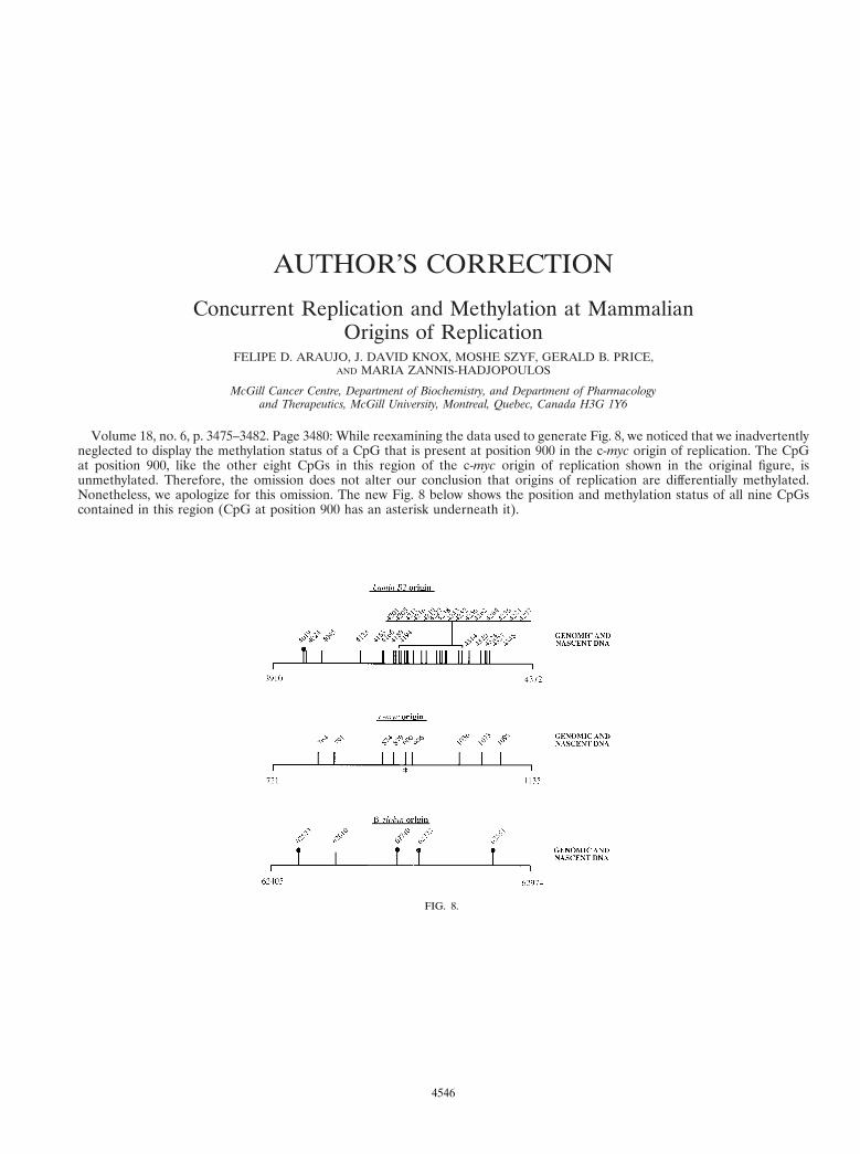
Volume 18, no. 6, p. 3475–3482. Page 3480: While reexamining the data used to generate Fig. 8, we noticed that we inadvertentlyneglected to display the methylation status of a CpG that is present at position 900 in the c-myc origin of replication. The CpGat position 900, like the other eight CpGs in this region of the c-myc origin of replication shown in the original figure, isunmethylated. Therefore, the omission does not alter our conclusion that origins of replication are differentially methylated.Nonetheless, we apologize for this omission. The new Fig. 8 below shows the position and methylation status of all nine CpGscontained in this region (CpG at position 900 has an asterisk underneath it).
FIG. 8.
4546