A Versatile Experimentation Model for Study of Structures ...
Upload
khangminh22Category
view
0download
0
The ISME Journalhttps://doi.org/10.1038/s41396-020-00889-4
ARTICLE
Mercury methylation by metabolically versatile and cosmopolitanmarine bacteria
Heyu Lin 1● David B. Ascher2,3 ● Yoochan Myung2,3
● Carl H. Lamborg4● Steven J. Hallam 5,6
●
Caitlin M. Gionfriddo7,10● Kathryn E. Holt 8,9
● John W. Moreau 1,11
Received: 3 June 2020 / Accepted: 17 December 2020© The Author(s) 2021. This article is published with open access
AbstractMicrobes transform aqueous mercury (Hg) into methylmercury (MeHg), a potent neurotoxin that accumulates in terrestrialand marine food webs, with potential impacts on human health. This process requires the gene pair hgcAB, which encodesfor proteins that actuate Hg methylation, and has been well described for anoxic environments. However, recent studiesreport potential MeHg formation in suboxic seawater, although the microorganisms involved remain poorly understood. Inthis study, we conducted large-scale multi-omic analyses to search for putative microbial Hg methylators along definedredox gradients in Saanich Inlet, British Columbia, a model natural ecosystem with previously measured Hg and MeHgconcentration profiles. Analysis of gene expression profiles along the redoxcline identified several putative Hg methylatingmicrobial groups, including Calditrichaeota, SAR324 and Marinimicrobia, with the last the most active based on hgctranscription levels. Marinimicrobia hgc genes were identified from multiple publicly available marine metagenomes,consistent with a potential key role in marine Hg methylation. Computational homology modelling predicts thatMarinimicrobia HgcAB proteins contain the highly conserved amino acid sites and folding structures required for functionalHg methylation. Furthermore, a number of terminal oxidases from aerobic respiratory chains were associated with severalputative novel Hg methylators. Our findings thus reveal potential novel marine Hg-methylating microorganisms with agreater oxygen tolerance and broader habitat range than previously recognized.
Introduction
Mercury (Hg), a highly toxic metal, is widespread in theenvironment from primarily anthropogenic sources, leadingto increased public concern over the past few decades.[e.g., 1–3]. Methylmercury (MeHg) is recognized as a
* John W. [email protected]
1 School of Earth Sciences, The University of Melbourne,Parkville, VIC 3010, Australia
2 Structural Biology and Bioinformatics, Department ofBiochemistry and Molecular Biology, Bio21 Molecular Scienceand Biotechnology Institute, The University of Melbourne,Parkville, VIC 3010, Australia
3 Computational Biology and Clinical Informatics, Baker Heart andDiabetes Institute, PO Box 6492, Melbourne, VIC 3004, Australia
4 Department of Ocean Sciences, University of California,Santa Cruz, CA 95064, USA
5 Department of Microbiology and Immunology, University ofBritish Columbia, Vancouver, BC V6T 1Z1, Canada
6 Genome Science and Technology Program, University of BritishColumbia, Vancouver, BC V6T 1Z4, Canada
7 Biosciences Division, Oak Ridge National Laboratory, PO Box2008, Oak Ridge, TN 37831, USA
8 Department of Infectious Diseases, Central Clinical School,Monash University, Monash, VIC 3800, Australia
9 Department of Infection Biology, London School of Hygiene &Tropical Medicine, London WC1E 7HT, UK
10 Present address: Smithsonian Environmental Research Center,Edgewater, MD 21037, USA
11 Present address: Currently at School of Geographical & EarthSciences, University of Glasgow, Glasgow G12 8QQ, UK
Supplementary information The online version of this article (https://doi.org/10.1038/s41396-020-00889-4) contains supplementarymaterial, which is available to authorized users.
1234
5678
90();,:
1234567890();,:

potent neurotoxin that bioaccumulates through both marineand terrestrial food webs [2, 4], with potential deleteriousimpacts on human health. With implementation of theMinamata Convention on Mercury [3], a better under-standing is expected of potential MeHg sources, in thecontext of global biogeochemical cycles and factors influ-encing Hg speciation. For example, oxygen gradients inseawater have been observed to expand to both a widerdepth range and larger geographical area in response toclimate change [5], and the resulting impacts on biogeo-chemical cycles, e.g., carbon, nitrogen, and sulfur, havebeen studied [6]. Effects on Hg cycling, however, are rarelyconsidered.
The environmental transformation of Hg(II) to MeHg is amicrobially-mediated process, for which the proteins areencoded by the two-gene cluster hgcA and hgcB [7]. Pos-session of the hgcAB gene pair is a predictor for Hgmethylation capability [8], and the discovery of hgc geneshas stimulated a search for potential Hg methylatingmicrobes in diverse environments [9]. To date, all experi-mentally confirmed Hg methylators are anaerobes [10] fromthree Deltaproteobacteria clades (sulfate-reducing bacteria,Fe-reducing bacteria and syntrophic bacteria), one cladebelonging to fermentative Firmicutes, and an archaeal clade(Methanomicrobia). These microorganisms are ubiquitousin soils, sediments, seawater, freshwater, and extremeenvironments, as well as the digestive tracts of some ani-mals [9]. A few other hgc carriers, not only from anaerobicbut also microaerobic habitats, were discovered usingculture-independent approaches, including Chloroflexi,Chrysiogenetes, Spirochaetes [9], Nitrospina [11, 12], andVerrucomicrobia [13]. Two more recent studies haveexpanded the Hg-methylating community [14, 15] andrevealed the role played by Hg methylators in the context ofglobal biogeochemical cycling.
Apart from Hg methylation, MeHg demethylation alsoplays a key role in global Hg cycling. This process is mainlycatalyzed by alkylmercury lyase encoded by the merB gene,which has been largely found in aerobes (e.g., Bacilluscereus, Staphylococcus aureus, and Escherichia coli). Yetsome obligate and facultative anaerobes (e.g., Geobacterbemidjiensis and Geobacter sulfurreducens) also possessmerB and can contribute to demethylation [16] and there-fore total environmental MeHg levels.
A global Hg survey [17] found a prevalence for MeHgin suboxic waters, where dissolved O2 and H2S con-centrations are typically very low (<50 μm and <10 μm,respectively), especially in regionally widespread oxygengradients at upper and intermediate ocean depths. Oxygenconcentrations can be reduced by a combination of phy-sical and biological forcing effects [6, 18]. These gra-dients can exist as permanent features in the watercolumn, impinge on coastal margins, or manifest more
transiently (e.g., induced by phytoplankton blooms). Asoxygen levels decrease, metabolic energy gets increas-ingly diverted to alternative electron acceptors, resultingin coupling of other biogeochemical cycles, e.g., C, N, S,Fe, and Mn [6, 19, 20]. Recent findings of microaerophilicmicrobial Hg methylation potential in sea ice and seawater[e.g., 11, 21–23] raise the possibility that this processcontributes significantly to ocean MeHg biomagnification.
In this study, we measured the concentrations of Hg(total) and MeHg in the waters of Saanich Inlet, a sea-sonally anoxic fjord on the coast of Vancouver Island(British Columbia, Canada), and performed targetedmetagenomic and metatranscriptomic analyses of sea-water sampled from varying depths. Saanich Inlet, as amodel natural ecosystem for studying microbial activityalong defined oxygen gradients [24], provides an ideal siteto search for novel putative Hg methylators and studymicrobially-mediated Hg cycling in low-oxygen envir-onments. Physico-chemical and biogeochemical para-meters, including nutrient and dissolved gasconcentrations, have been published from data acquiredfrom 2006 to 2014 [25]. This study clearly illustrates thedynamic character of the oxygen gradient in Saanich Inletwaters. Computational homology modelling was per-formed to predict the functionality of HgcAB proteinsencoded for by putative novel Hg-methylators. Finally,we scanned global metagenomic datasets for recognizablehgcA genes, to reassess the environmental distribution ofmicrobial mercury methylation potential.
Results and discussion
Hg and MeHg concentrations along redox gradientsin Saanich Inlet
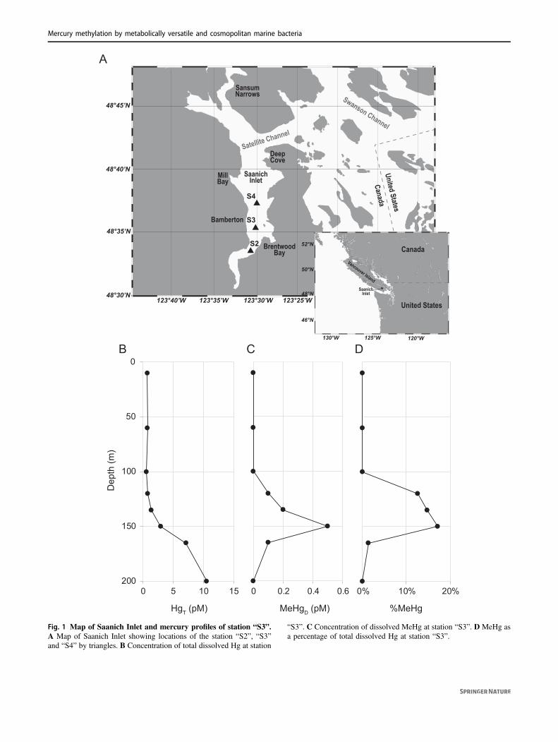
Concentrations of total dissolved Hg (HgT) and mono-methylmercury (MeHg) from filtered Saanich Inlet S3 station(see Fig. 1A for a map) seawater samples, obtained andanalyzed in March–April of 2010, were vertically profiled ateight different depths from surface (10m) to bottom (200m)waters. The concentration of HgT at sea surface was ~0.70 pMand remained nearly constant in seawater above 120m depth,increasing to 1.35 pM and ~10.56 pM at 135m and 200mdepths, respectively (Fig. 1B). MeHg was below detectionlimit (<0.1 pM) for seawater above 100m depth, butincreased to 0.50 pM (17.2% of total Hg) at 150m depth.However, MeHg then decreased to 0.1 pM at 165m depth,becoming undetectable at 200m (Fig. 1C and 1D). SimilarMeHg peaks under suboxic conditions have been observed inother seawater depth profiles: the Pacific Ocean [26], ArcticOcean [27], Southern Ocean [28], Arabian Sea [29], andMediterranean Sea [30].
H. Lin et al.
48°30’N
48°35’N
48°40’N
48°45’N
123°40’W 123°35’W 123°30’W 123°25’W 123.20°W 123.15°W 123.10°W
S4
S2
S3
46°N
48°N
50°N
52°N
125°W130°W 120°W
SaanichInlet
SaanichInlet
MillBay
BrentwoodBay
United States
Canada
DeepCove
Bamberton
SansumNarrows
Vancouver Island
Satellite Channel
Swanson Channel
United States
Canada
A
0% 10% 20%
%MeHg
Dep
th (m
)
B DC
0 0.2 0.4 0.6
MeHgD (pM)
0
50
100
150
2000 5 10 15
HgT (pM)
Fig. 1 Map of Saanich Inlet and mercury profiles of station “S3”.A Map of Saanich Inlet showing locations of the station “S2”, “S3”and “S4” by triangles. B Concentration of total dissolved Hg at station
“S3”. C Concentration of dissolved MeHg at station “S3”. D MeHg asa percentage of total dissolved Hg at station “S3”.
Mercury methylation by metabolically versatile and cosmopolitan marine bacteria
MAG (metagenome-assembled genome)reconstruction and putative Hg methylatoridentification
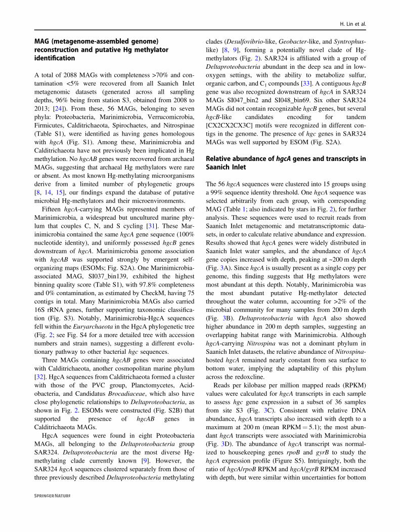
A total of 2088 MAGs with completeness >70% and con-tamination <5% were recovered from all Saanich Inletmetagenomic datasets (generated across all samplingdepths, 96% being from station S3, obtained from 2008 to2013; [24]). From these, 56 MAGs, belonging to sevenphyla: Proteobacteria, Marinimicrobia, Verrucomicrobia,Firmicutes, Calditrichaeota, Spirochaetes, and Nitrospinae(Table S1), were identified as having genes homologouswith hgcA (Fig. S1). Among these, Marinimicrobia andCalditrichaeota have not previously been implicated in Hgmethylation. No hgcAB genes were recovered from archaealMAGs, suggesting that archaeal Hg methylators were rareor absent. As most known Hg-methylating microorganismsderive from a limited number of phylogenetic groups[8, 14, 15], our findings expand the database of putativemicrobial Hg-methylators and their microenvironments.
Fifteen hgcA-carrying MAGs represented members ofMarinimicrobia, a widespread but uncultured marine phy-lum that couples C, N, and S cycling [31]. These Mar-inimicrobia contained the same hgcA gene sequence (100%nucleotide identity), and uniformly possessed hgcB genesdownstream of hgcA. Marinimicrobia genome associationwith hgcAB was supported strongly by emergent self-organizing maps (ESOMs; Fig. S2A). One Marinimicrobia-associated MAG, SI037_bin139, exhibited the highestbinning quality score (Table S1), with 97.8% completenessand 0% contamination, as estimated by CheckM, having 75contigs in total. Many Marinimicrobia MAGs also carried16S rRNA genes, further supporting taxonomic classifica-tion (Fig. S3). Notably, Marinimicrobia-HgcA sequencesfell within the Euryarchaeota in the HgcA phylogenetic tree(Fig. 2; see Fig. S4 for a more detailed tree with accessionnumbers and strain names), suggesting a different evolu-tionary pathway to other bacterial hgc sequences.
Three MAGs containing hgcAB genes were associatedwith Calditrichaeota, another cosmopolitan marine phylum[32]. HgcA sequences from Calditrichaeota formed a clusterwith those of the PVC group, Planctomycetes, Acid-obacteria, and Candidatus Brocadiaceae, which also haveclose phylogenetic relationships to Deltaproteobacteria, asshown in Fig. 2. ESOMs were constructed (Fig. S2B) thatsupported the presence of hgcAB genes inCalditrichaeota MAGs.
HgcA sequences were found in eight ProteobacteriaMAGs, all belonging to the Deltaproteobacteria groupSAR324. Deltaproteobacteria are the most diverse Hg-methylating clade currently known [9]. However, theSAR324 hgcA sequences clustered separately from those ofthree previously described Deltaproteobacteria methylating
clades (Desulfovibrio-like, Geobacter-like, and Syntrophus-like) [8, 9], forming a potentially novel clade of Hg-methylators (Fig. 2). SAR324 is affiliated with a group ofDeltaproteobacteria abundant in the deep sea and in low-oxygen settings, with the ability to metabolize sulfur,organic carbon, and C1 compounds [33]. A contiguous hgcBgene was also recognized downstream of hgcA in SAR324MAGs SI047_bin2 and SI048_bin69. Six other SAR324MAGs did not contain recognizable hgcB genes, but severalhgcB-like candidates encoding for tandem[CX2CX2CX3C] motifs were recognized in different con-tigs in the genome. The presence of hgc genes in SAR324MAGs was well supported by ESOM (Fig. S2A).
Relative abundance of hgcA genes and transcripts inSaanich Inlet
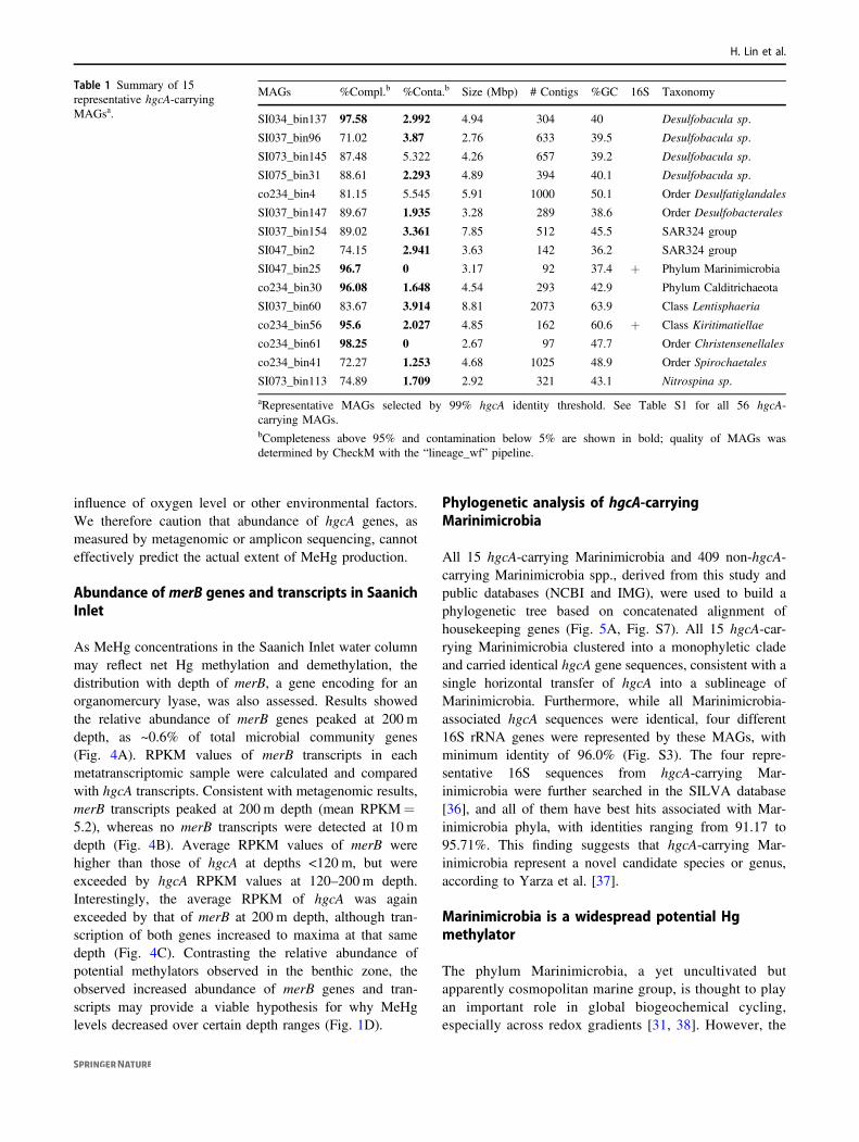
The 56 hgcA sequences were clustered into 15 groups usinga 99% sequence identity threshold. One hgcA sequence wasselected arbitrarily from each group, with correspondingMAG (Table 1; also indicated by stars in Fig. 2), for furtheranalysis. These sequences were used to recruit reads fromSaanich Inlet metagenomic and metatranscriptomic data-sets, in order to calculate relative abundance and expression.Results showed that hgcA genes were widely distributed inSaanich Inlet water samples, and the abundance of hgcAgene copies increased with depth, peaking at ~200 m depth(Fig. 3A). Since hgcA is usually present as a single copy pergenome, this finding suggests that Hg methylators weremost abundant at this depth. Notably, Marinimicrobia wasthe most abundant putative Hg-methylator detectedthroughout the water column, accounting for >2% of themicrobial community for many samples from 200 m depth(Fig. 3B). Deltaproteobacteria with hgcA also showedhigher abundance in 200 m depth samples, suggesting anoverlapping habitat range with Marinimicrobia. AlthoughhgcA-carrying Nitrospina was not a dominant phylum inSaanich Inlet datasets, the relative abundance of Nitrospina-hosted hgcA remained nearly constant from sea surface tobottom water, implying the adaptability of this phylumacross the redoxcline.
Reads per kilobase per million mapped reads (RPKM)values were calculated for hgcA transcripts in each sampleto assess hgc gene expression in a subset of 36 samplesfrom site S3 (Fig. 3C). Consistent with relative DNAabundance, hgcA transcripts also increased with depth to amaximum at 200 m (mean RPKM= 5.1); the most abun-dant hgcA transcripts were associated with Marinimicrobia(Fig. 3D). The abundance of hgcA transcript was normal-ized to housekeeping genes rpoB and gyrB to study thehgcA expression profile (Figure S5). Intriguingly, both theratio of hgcA/rpoB RPKM and hgcA/gyrB RPKM increasedwith depth, but were similar within uncertainties for bottom
H. Lin et al.
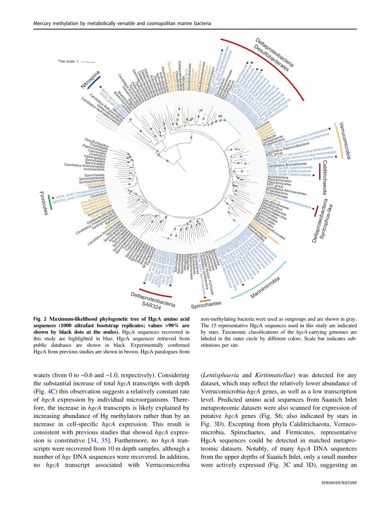
waters (from 0 to ~0.6 and ~1.0, respectively). Consideringthe substantial increase of total hgcA transcripts with depth(Fig. 4C) this observation suggests a relatively constant rateof hgcA expression by individual microorganisms. There-fore, the increase in hgcA transcripts is likely explained byincreasing abundance of Hg methylators rather than by anincrease in cell-specific hgcA expression. This result isconsistent with previous studies that showed hgcA expres-sion is constitutive [34, 35]. Furthermore, no hgcA tran-scripts were recovered from 10 m depth samples, although anumber of hgc DNA sequences were recovered. In addition,no hgcA transcript associated with Verrucomicrobia
(Lentisphaeria and Kiritimatiellae) was detected for anydataset, which may reflect the relatively lower abundance ofVerrucomicrobia-hgcA genes, as well as a low transcriptionlevel. Predicted amino acid sequences from Saanich Inletmetaproteomic datasets were also scanned for expression ofputative hgcA genes (Fig. S6; also indicated by stars inFig. 3D). Excepting from phyla Calditrichaeota, Verruco-microbia, Spirochaetes, and Firmicutes, representativeHgcA sequences could be detected in matched metapro-teomic datasets. Notably, of many hgcA DNA sequencesfrom the upper depths of Saanich Inlet, only a small numberwere actively expressed (Fig. 3C and 3D), suggesting an
Methanomassiliicoccales
Igna
vibac
teria
e
SI073_bin33 Desulfo
bacula
SI07
5_bin
31 D
esulf
obac
ula
Deltaproteobacteria
Lentzea
Desulfobulbaceae
Spiroch
aetaceae
Geobacter
Desu
lfotig
num
Firmicutes
Candidate division FC
PU426
SI03
4_bin
137
Desulf
obac
ula
SI053_bin85 Nitrospina
SI034_bin40 Marinimicrobia
Candidatus Aminicenantes
Spirochaetes
Desulfosporosinus
Spiro
chae
tia
Anaerolineales
SI037S2_bin78 Marinim
icrobia
Palu
diba
cter
Deltaproteobacteria
Acetonema
Fibrobacteres
Spirochaetales
Candidatus Bathyarchaeota
SI03
7_bi
n96
Desu
lfoba
cula
SI039_bin117 Desulfobacula
SI074_bin48 Marinimicrobia
co234_bin40 Marinim
icrobia
Candidatus G
oldbacteria
Actino
bacte
ria
Des
ulfo
cocc
us
SI03
7S2_
bin5
8 SA
R32
4
Candidatus Firestonebacteria
Deltaproteobacteria
Proteobacteria
SI03
7S2_
bin4
5 D
esul
foba
cter
ales
Syntrophus
Gam
maproteobacteria
Spiro
chae
tales
Bdellovibrionales
SI037_bin165 Calditrichaeota
Spiro
chae
tales
SI037_
bin17
4 Des
ulfob
acula
co23
4_bin
15 D
esulf
obac
ula
co234_bin41 Spirochaetales
Candidatus Aminicenans
SI053_bin92 Marinimicrobia
Elusimicrobia
Elusimicrobia
Des
ulfo
bulb
acea
e
Elusimicrobia
Methanosarcina
Candidatus Aminicenantes
Myxococcales
Spiro
chae
tales
SI05
3_bi
n102
SAR
324
Archaeoglobus
Bdellovibrionales
Desulfosporosinus
SI03
7_bi
n154
SAR
324
SI07
3_bi
n72
SAR
324
co234_bin4 DeltaproteobacteriaD
esul
fom
icro
bium
SI073_bin38 Marinimicrobia
Des
ulfo
vibr
io
Lentisphaerae
Methanomethylovorans
Trep
onem
a
Planctomycetes
SI037S2_bin88 Desulfobacula
Clostridiales
Acidobacteria
Deltaproteobacteria
Pseu
dode
sulfo
vibr
io
Spirochaetes
Methanolobus
SI048_bin84 Marinim
icrobia
co234_bin30 Calditrichaeota
Methanoregula
Spiro
chae
tae
Desulfitobacterium
Desu
lfam
plus
co23
4_bi
n52
Des
ulfo
cocc
acea
e
Candidatus Raymondbacteria
co234_bin53 Verrucomicrobia Lentisphaeria
SI04
8_bi
n69
SAR
324
Deferribacteres
SI03
6_bin
13 D
esulf
obac
ula
arips
orti
N
EthanoligenensSI037S2_bin60 Christensenellaceae
Bact
eroi
dete
s
Haloimpatiens
SI054_bin54 Marinimicrobia
Spirochaetes
SI048_
bin88
Des
ulfob
acula
Syntrophomonas
co234_bin56 Verrucomicrobia Kiritimatiellae
Peptoclostridium
SI034_bin97 Calditrichaeota
Geotherm
obacter
SI037_bin139 Marinim
icrobia
Spirochaetes
SI037_bin111 Verrucomicrobia Kiritimatiellae
Fibrobacteres
Geobacter
SI060_bin23 Marinim
icrobia
SI07
3_bi
n145
Des
ulfo
bacu
la
Des
ulfo
mic
robi
um
Des
ulfo
vibr
io
PVC group
eari
psort
iN
DesulfuribacillusSI
034_
bin8
8 SA
R32
4
SI04
7_bi
n125
SAR
324
Desulfobulbus
Spirochaetales
Geobacter
SI037_bin60 Verrucomicrobia Lentisphaeria
Desu
lfoba
cter
acea
e
Dehalococcoidia
earipsortiN
Geobacter
SI072_bin5 Marinimicrobia
Spiroc
haeta
ceae
Stappia
Desulfovibrio
Desulfovibrio
SI075_bin21 Marinim
icrobia
SI047_bin66 Desulfobacula
SI039_bin42 Marinim
icrobia
Desulfobulbaceae
Solib
acte
rale
s
Spirochaetaceae
Des
ulfo
bact
erac
eae
Spiroc
haete
s
DethiobacterS
piro
chae
tae
Clostridium
Trep
onem
a
Bradyrhizobium
Balneolia
SI037S2_bin34 Verrucomicrobia Kiritimatiellae
Deltaproteobacteria
Desulfovibrio
Actinobacteria
Caloramator
SI054_
bin7 D
esulf
obac
ula
Deltaproteobacteria
Dethiosulfatarculus
Desulfitobacterium
Syntrophorhabdus
Clostridiumsublubofluse
D
Desulfovibrio
Proteobacteria
SI073_bin113 Nitrospina
Candidatus Heimdallarchaeota
SI03
7_bi
n147
Des
ulfo
bacu
la
SI04
7_bi
n2 S
AR32
4
Clostridium
PVC group
co234_bin61 Christensenellaceae
Desulfonatronospira
Syntrophaceae
Acidobacteria
Methanosarcina
Bact
eroi
dale
s
Nitrospina
co23
4_bin
87 D
esulf
obac
ula
Planctomycetes
Myxococcales
Defluviimonas
Des
ulfo
mic
robi
um
Spirochaetes
Syntrophomonas
Bacteroidia
Spiroc
haeta
ceae
Candidatus Aminice
nantes
SI036_bin53 Marinim
icrobia
Spiroc
haeta
lesSI053_bin3 Desulfobacula
SI047_bin25 Marinim
icrobia
Actinobacteria
Candidatus Brocadiaceae
Actinobacteria
SI060_
bin46
Des
ulfob
acula
Geobacter
Tree scale: 1
DeltaproteobacteriaSAR324
Firmicutes
Nitrosp
ina
Verrucomicrobia
atoe
ahcir
tidl
aC
Deltaproteobacteria
Desulfobacterales
Del
tapr
oteo
bact
eria
Sy
ntro
phus
-like
Marinimicrobia
Spirochaetes
Fig. 2 Maximum-likelihood phylogenetic tree of HgcA amino acidsequences (1000 ultrafast bootstrap replicates; values >90% areshown by black dots at the nodes). HgcA sequences recovered inthis study are highlighted in blue. HgcA sequences retrieved frompublic databases are shown in black. Experimentally confirmedHgcA from previous studies are shown in brown. HgcA paralogues from
non-methylating bacteria were used as outgroups and are shown in gray.The 15 representative HgcA sequences used in this study are indicatedby stars. Taxonomic classifications of the hgcA-carrying genomes arelabeled in the outer circle by different colors. Scale bar indicates sub-stitutions per site.
Mercury methylation by metabolically versatile and cosmopolitan marine bacteria
influence of oxygen level or other environmental factors.We therefore caution that abundance of hgcA genes, asmeasured by metagenomic or amplicon sequencing, cannoteffectively predict the actual extent of MeHg production.
Abundance of merB genes and transcripts in SaanichInlet
As MeHg concentrations in the Saanich Inlet water columnmay reflect net Hg methylation and demethylation, thedistribution with depth of merB, a gene encoding for anorganomercury lyase, was also assessed. Results showedthe relative abundance of merB genes peaked at 200 mdepth, as ~0.6% of total microbial community genes(Fig. 4A). RPKM values of merB transcripts in eachmetatranscriptomic sample were calculated and comparedwith hgcA transcripts. Consistent with metagenomic results,merB transcripts peaked at 200 m depth (mean RPKM=5.2), whereas no merB transcripts were detected at 10 mdepth (Fig. 4B). Average RPKM values of merB werehigher than those of hgcA at depths <120 m, but wereexceeded by hgcA RPKM values at 120–200 m depth.Interestingly, the average RPKM of hgcA was againexceeded by that of merB at 200 m depth, although tran-scription of both genes increased to maxima at that samedepth (Fig. 4C). Contrasting the relative abundance ofpotential methylators observed in the benthic zone, theobserved increased abundance of merB genes and tran-scripts may provide a viable hypothesis for why MeHglevels decreased over certain depth ranges (Fig. 1D).
Phylogenetic analysis of hgcA-carryingMarinimicrobia
All 15 hgcA-carrying Marinimicrobia and 409 non-hgcA-carrying Marinimicrobia spp., derived from this study andpublic databases (NCBI and IMG), were used to build aphylogenetic tree based on concatenated alignment ofhousekeeping genes (Fig. 5A, Fig. S7). All 15 hgcA-car-rying Marinimicrobia clustered into a monophyletic cladeand carried identical hgcA gene sequences, consistent with asingle horizontal transfer of hgcA into a sublineage ofMarinimicrobia. Furthermore, while all Marinimicrobia-associated hgcA sequences were identical, four different16S rRNA genes were represented by these MAGs, withminimum identity of 96.0% (Fig. S3). The four repre-sentative 16S sequences from hgcA-carrying Mar-inimicrobia were further searched in the SILVA database[36], and all of them have best hits associated with Mar-inimicrobia phyla, with identities ranging from 91.17 to95.71%. This finding suggests that hgcA-carrying Mar-inimicrobia represent a novel candidate species or genus,according to Yarza et al. [37].
Marinimicrobia is a widespread potential Hgmethylator
The phylum Marinimicrobia, a yet uncultivated butapparently cosmopolitan marine group, is thought to playan important role in global biogeochemical cycling,especially across redox gradients [31, 38]. However, the
Table 1 Summary of 15representative hgcA-carryingMAGsa.
MAGs %Compl.b %Conta.b Size (Mbp) # Contigs %GC 16S Taxonomy
SI034_bin137 97.58 2.992 4.94 304 40 Desulfobacula sp.
SI037_bin96 71.02 3.87 2.76 633 39.5 Desulfobacula sp.
SI073_bin145 87.48 5.322 4.26 657 39.2 Desulfobacula sp.
SI075_bin31 88.61 2.293 4.89 394 40.1 Desulfobacula sp.
co234_bin4 81.15 5.545 5.91 1000 50.1 Order Desulfatiglandales
SI037_bin147 89.67 1.935 3.28 289 38.6 Order Desulfobacterales
SI037_bin154 89.02 3.361 7.85 512 45.5 SAR324 group
SI047_bin2 74.15 2.941 3.63 142 36.2 SAR324 group
SI047_bin25 96.7 0 3.17 92 37.4 + Phylum Marinimicrobia
co234_bin30 96.08 1.648 4.54 293 42.9 Phylum Calditrichaeota
SI037_bin60 83.67 3.914 8.81 2073 63.9 Class Lentisphaeria
co234_bin56 95.6 2.027 4.85 162 60.6 + Class Kiritimatiellae
co234_bin61 98.25 0 2.67 97 47.7 Order Christensenellales
co234_bin41 72.27 1.253 4.68 1025 48.9 Order Spirochaetales
SI073_bin113 74.89 1.709 2.92 321 43.1 Nitrospina sp.
aRepresentative MAGs selected by 99% hgcA identity threshold. See Table S1 for all 56 hgcA-carrying MAGs.bCompleteness above 95% and contamination below 5% are shown in bold; quality of MAGs wasdetermined by CheckM with the “lineage_wf” pipeline.
H. Lin et al.
Hg-methylation potential of Marinimicrobia has not pre-viously been recognized. A few HgcA sequences fromSaanich Inlet were discovered by Podar et al. [9] frommetagenomic analyses; these previously unidentifiedsequences are shown here to co-locate phylogeneticallywith the Marinimicrobia-HgcA identified in this study atwhole genome resolution. In order to explore the globaldistribution of hgcA-carrying Marinimicrobia, a widersearch was performed against NCBI SRA metagenomicdatasets. In total, 58 BioProjects from a range of
environments, including seawater, soil, sediment, fresh-water, industrial wastewater, and plant rhizomes, con-tained Marinimicrobia-hgcA associated reads (Fig. 5B,Table S2), suggesting a wide ecological distribution.Interestingly, our findings of hgcA-carrying Mar-inimicrobia from metagenomic datasets from Gulf ofMexico (PRJNA288120) and Canadian Arctic(PRJNA266338) waters contaminated by oil spills[39, 40] may provide one explanation for observed in situMeHg production [41].
Representative MAGs
Met
agen
omic
sam
ples
Abundance (%)1.0
1.5
2.0
SI053S3SI034S3SI048S3SI072S3SI073S3SI037S3SI074S3SI075S3SI039S3SI053S3SI054S3SI034S3SI047S3SI036S3SI048S3SI060S3SI072S3SI073S3SI037S2SI037S3SI037S4SI074S3SI075S3SI039S3SI037S3SI053S3SI054S3SI034S3SI047S3SI036S3SI048S3SI072S3SI073S3SI037S3SI074S3SI075S3SI039S3SI037S3SI037S3SI037S4SI053S3SI054S3SI034S3SI047S3SI036S3SI048S3SI072S3SI073S3SI074S3SI075S3SI039S3SI053S3SI054S3SI034S3SI047S3SI036S3SI048S3SI060S3SI072S3SI073S3SI075S3SI037S2SI037S3SI037S4SI074S3SI039S3SI072S3SI073S3SI074S3SI075S3SI053S3SI054S3SI034S3SI047S3SI036S3SI048S3SI060S3SI072S3SI073S3SI037S2SI037S3SI074S3SI075S3SI039S3
Depth10 m
100 m
110~135 m
150~165 m
200 m
Desulfatiglanales co234_bin4
Desulfobacula sp. SI034_bin137
Desulfobacterales SI037_bin147
Desulfobacula sp. SI073_bin145
Desulfobacula sp. SI075_bin31
Desulfobacula sp. SI037_bin96
SAR324 SI037_bin154
SAR324 SI047_bin2
Marinimicrobia SI047_bin25
Calditrichaeota co234_bin30
Lentisphaeria SI037_bin60
Kiritimatiellae co234_bin56
Spirochaetales co234_bin41
Christensenellales co234_bin61
Nitrospina SI073_bin113Representative MAGs
Met
atra
nscr
ipto
mic
sam
ples
RPKM
Desulfatiglanales co234_bin4
Desulfobacula sp. SI034_bin137
Desulfobacterales SI037_bin147
Desulfobacula sp. SI073_bin145
Desulfobacula sp. SI075_bin31
Desulfobacula sp. SI037_bin96
SAR324 SI037_bin154
SAR324 SI047_bin2
Marinimicrobia SI047_bin25
Calditrichaeota co234_bin30
Lentisphaeria SI037_bin60
Kiritimatiellae co234_bin56
Spirochaetales co234_bin41
Christensenellales co234_bin61
Nitrospina SI073_bin113
SI048S3SI072S3SI073S3SI074S3SI047S3SI048S3SI054S3SI072S3SI074S3SI075S3SI047S3SI048S3SI054S3SI074S3SI075S3SI047S3SI048S3SI054S3SI072S3SI074S3SI075S3SI047S3SI048S3SI054S3SI072S3SI074S3SI075S3SI072S3SI073S3SI074S3SI047S3SI048S3SI054S3SI072S3SI074S3SI075S3
Depth10 m
100 m
110~135 m
150~165 m
200 m
B D
A C
10 100 110~135 150~165 200 10 100 120~135 150~165 200Depth (m) Depth (m)
hgcA
abu
ndan
cein
met
agen
omic
dat
a (%
)
hgcA
RP
KM
in m
etat
rans
crip
tom
ic d
ata
5.0
4.0
3.0
2.0
1.0
0
12.5
10.0
7.5
5.0
2.5
0
2.0
4.0
6.0
Fig. 3 Relative abundance of representative hgcA genes andtranscripts in different samples from Saanich Inlet. Gene abun-dance is normalized by gene length and genome equivalent for eachsample, and transcript abundances are represented as RPKM values.A Relative abundance of the sum of representative hgcA genes fordifferent depths. B Relative abundance of different representative hgcAgenes from different metagenomic datasets. Larger circles indicate a
higher percentage of the whole microbial community. Different shadesof blue indicate different depths from which the samples were taken.C RPKM values of the sum of representative hgcA transcripts indifferent depths. D RPKM values of different representative hgcAtranscripts in different metatranscriptomic samples. HgcA sequencesfrom metaproteomic samples are indicated with stars.
Mercury methylation by metabolically versatile and cosmopolitan marine bacteria

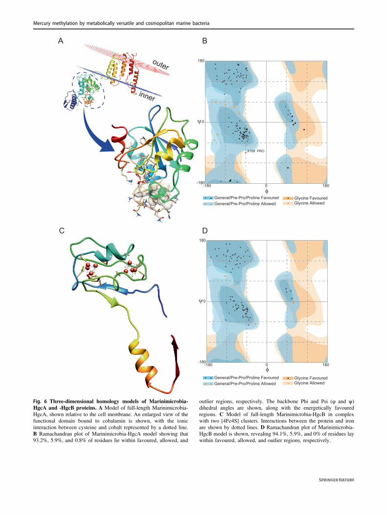
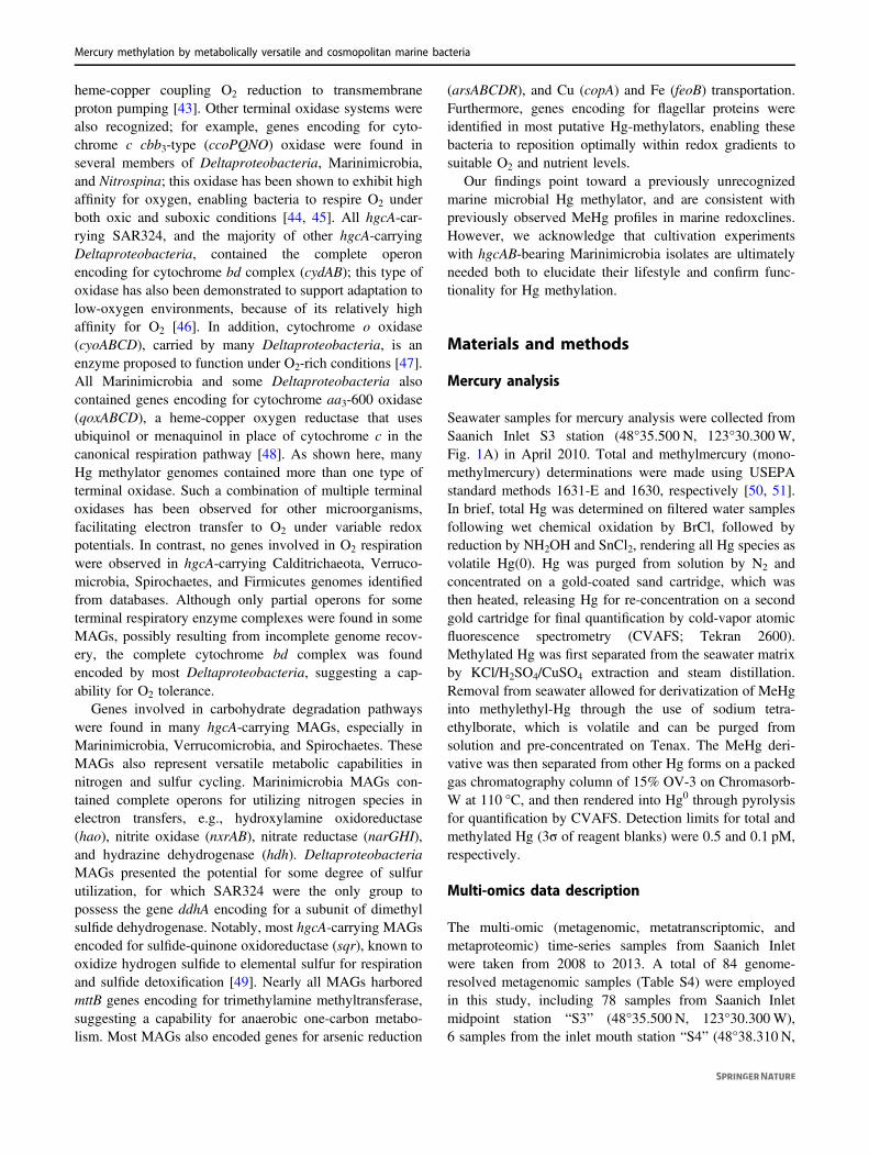
Homology models of Marinimicrobia-HgcA and-HgcB proteins
As no cultivated representative of Marinimicrobia is cur-rently available to assay for functional Hg methylation, weconstructed homology models to test the hypothesis thatHgcA and HgcB proteins encoded for by Marinimicrobia
possess the three-dimensional structural functionalityrequired for methylating Hg. Analysis of the putativeMarinimicrobia-HgcA sequence revealed a comparabledomain structure (Fig. 6A) to the previously characterizedHgcA from the functionally validated Hg-methylating strainDesulfovibrio desulfuricans ND132 [7], including the pre-sence of a globular domain at the N-terminus and five
50° S0°
50° N
100° W 0° 100° E 180° E
Human-associated (10)Host-associated (10)Soil (9)Seawater (8)Bioreactor (8)Freshwater (7)Sediment (4)Wastewater (3)Plant-associated (1)
Environment (# project)
A B
SI054_bin54
Deferribacter desulfuricans SSM1Flexistipes sinusarabici DSM 4947
SI036_bin53
SI074_bin48
co234_bin40
SI037_bin139
SI039_bin42
SI073_bin38
SI034_bin40SI048_bin84
SI060_bin23SI075_bin21
Denitrovibrio acetiphilus DSM 12809Caldithrix abyssi DSM 13497
Geovibrio thiophilus DSM 11263
SI053_bin92
SI037S2_bin78SI072_bin5
SI047_bin25
93
94
96
90
99
100
100
93
95
100
100
100
95
96
94
95
100
100
100
100
100
100
100
Tree scale: 0.1
Outgroup
Marinimicrobia
hgcA-carryingMarinimicrobia
46 taxa
40 taxa
2 taxa
13 taxa
105 taxa
74 taxa
83 taxa
5 taxa
11 taxa
30 taxa
Fig. 5 Phylogeny and global distribution of phylum Mar-inimicrobia. A Maximum-likelihood phylogenetic tree of phylumMarinimicrobia based on concatenated housekeeping genes (1000ultrafast bootstrap replicates; values >90% are shown at the nodes).Scale bar indicates substitutions per site. A total of 424 Mar-inimicrobia genomes were used for the tree; Marinimicrobia lineages
without hgcA genes were collapsed to simplify presentation (seeFig. S6 for a more detailed tree). B Distribution of hgcA-carryingMarinimicrobia in various environments globally, shown as differentcolors; total numbers of BioProjects from each environment are shownin brackets.
0
50
100
150
200
Dep
th (m
)
0.1 0.3 0.40.2 0.5 0.6 0.7 0.8merB abundance
in metagenomic data (%)merB RPKM
in metatranscriptomic dataAverage RPKM
in metatranscriptomic data
0 4 62 8 10 0 2 31 4 5
A B0
50
100
150
200D
epth
(m)
C0
50
100
150
200
Dep
th (m
)
hgcAmerB
Gene
Fig. 4 DNA and RNA abundance of merB in Saanich Inlet datasetschanging with depth. A merB gene abundance in Saanich Inletmetagenomic datasets. Percentage abundance was normalized by genelength and genome equivalent for each sample. Line plot depicts theaverage abundance of merB with depth; shaded area depicts 95%confidence interval. B merB transcript abundance in Saanich Inlet
metatranscriptomic datasets, as represented by RPKM values. Lineplot depicts mean RPKM value of merB with depth; shaded areadepicts 95% confidence interval. C Comparison between RPKMvalues for hgcA (orange) and merB (green) in Saanich Inlet meta-transcriptomic datasets.
H. Lin et al.
A B
C D
outer
inner
-180
0
180
-180 0 180φ
ψ
General/Pre-Pro/Proline FavouredGeneral/Pre-Pro/Proline Allowed
Glycine FavouredGlycine Allowed
-180
0
180
-180 0 180φ
ψ
General/Pre-Pro/Proline FavouredGeneral/Pre-Pro/Proline Allowed
Glycine FavouredGlycine Allowed
A108 PRO
Fig. 6 Three-dimensional homology models of Marinimicrobia-HgcA and -HgcB proteins. A Model of full-length Marinimicrobia-HgcA, shown relative to the cell membrane. An enlarged view of thefunctional domain bound to cobalamin is shown, with the ionicinteraction between cysteine and cobalt represented by a dotted line.B Ramachandran plot of Marinimicrobia-HgcA model showing that93.2%, 5.9%, and 0.8% of residues lie within favoured, allowed, and
outlier regions, respectively. The backbone Phi and Psi (φ and ψ)dihedral angles are shown, along with the energetically favouredregions. C Model of full-length Marinimicrobia-HgcB in complexwith two [4Fe4S] clusters. Interactions between the protein and ironare shown by dotted lines. D Ramachandran plot of Marinimicrobia-HgcB model is shown, revealing 94.1%, 5.9%, and 0% of residues laywithin favoured, allowed, and outlier regions, respectively.
Mercury methylation by metabolically versatile and cosmopolitan marine bacteria
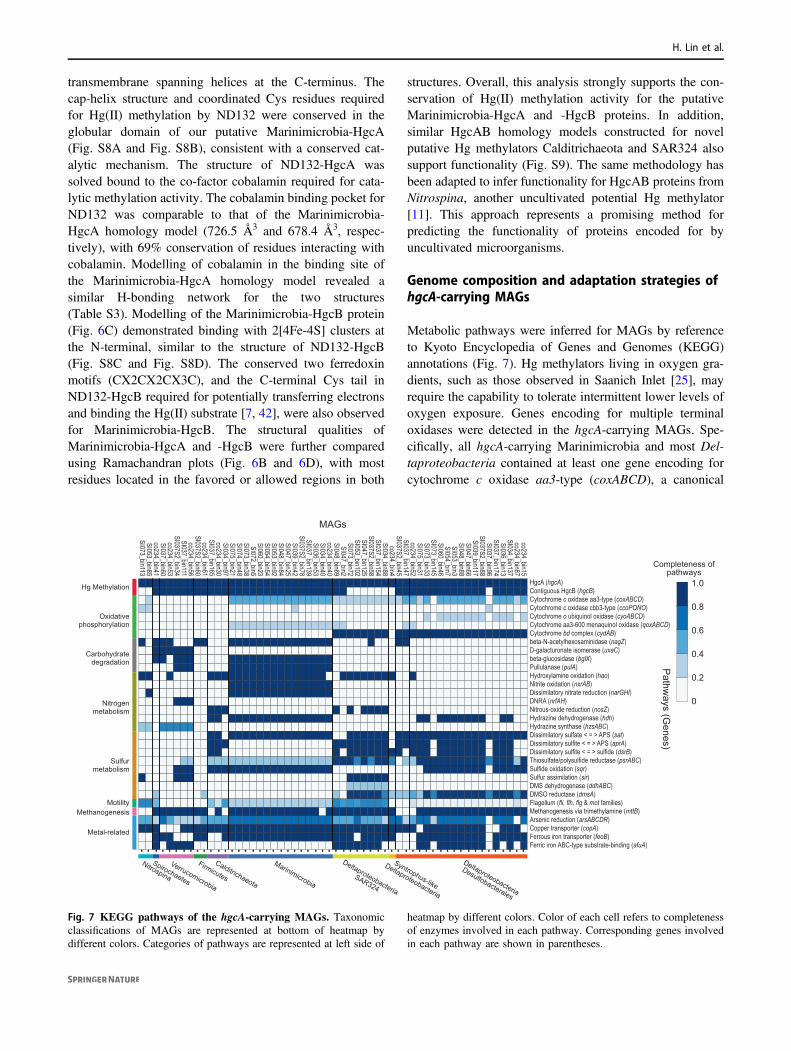
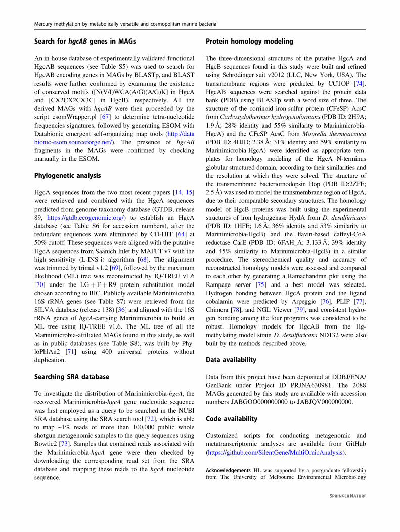
transmembrane spanning helices at the C-terminus. Thecap-helix structure and coordinated Cys residues requiredfor Hg(II) methylation by ND132 were conserved in theglobular domain of our putative Marinimicrobia-HgcA(Fig. S8A and Fig. S8B), consistent with a conserved cat-alytic mechanism. The structure of ND132-HgcA wassolved bound to the co-factor cobalamin required for cata-lytic methylation activity. The cobalamin binding pocket forND132 was comparable to that of the Marinimicrobia-HgcA homology model (726.5 Å3 and 678.4 Å3, respec-tively), with 69% conservation of residues interacting withcobalamin. Modelling of cobalamin in the binding site ofthe Marinimicrobia-HgcA homology model revealed asimilar H-bonding network for the two structures(Table S3). Modelling of the Marinimicrobia-HgcB protein(Fig. 6C) demonstrated binding with 2[4Fe-4S] clusters atthe N-terminal, similar to the structure of ND132-HgcB(Fig. S8C and Fig. S8D). The conserved two ferredoxinmotifs (CX2CX2CX3C), and the C-terminal Cys tail inND132-HgcB required for potentially transferring electronsand binding the Hg(II) substrate [7, 42], were also observedfor Marinimicrobia-HgcB. The structural qualities ofMarinimicrobia-HgcA and -HgcB were further comparedusing Ramachandran plots (Fig. 6B and 6D), with mostresidues located in the favored or allowed regions in both
structures. Overall, this analysis strongly supports the con-servation of Hg(II) methylation activity for the putativeMarinimicrobia-HgcA and -HgcB proteins. In addition,similar HgcAB homology models constructed for novelputative Hg methylators Calditrichaeota and SAR324 alsosupport functionality (Fig. S9). The same methodology hasbeen adapted to infer functionality for HgcAB proteins fromNitrospina, another uncultivated potential Hg methylator[11]. This approach represents a promising method forpredicting the functionality of proteins encoded for byuncultivated microorganisms.
Genome composition and adaptation strategies ofhgcA-carrying MAGs
Metabolic pathways were inferred for MAGs by referenceto Kyoto Encyclopedia of Genes and Genomes (KEGG)annotations (Fig. 7). Hg methylators living in oxygen gra-dients, such as those observed in Saanich Inlet [25], mayrequire the capability to tolerate intermittent lower levels ofoxygen exposure. Genes encoding for multiple terminaloxidases were detected in the hgcA-carrying MAGs. Spe-cifically, all hgcA-carrying Marinimicrobia and most Del-taproteobacteria contained at least one gene encoding forcytochrome c oxidase aa3-type (coxABCD), a canonical
HgcA (hgcA)Contiguous HgcB (hgcB)Cytochrome c oxidase aa3-type (coxABCD)Cytochrome c oxidase cbb3-type (ccoPQNO)Cytochrome o ubiquinol oxidase (cyoABCD)Cytochrome aa3-600 menaquinol oxidase (qoxABCD)Cytochrome bd complex (cydAB)beta-N-acetylhexosaminidase (nagZ)D-galacturonate isomerase (uxaC)beta-glucosidase (bglX)Pullulanase (pulA)Hydroxylamine oxidation (hao)Nitrite oxidation (nxrAB)Dissimilatory nitrate reduction (narGHI)DNRA (nrfAH)Nitrous-oxide reduction (nosZ)Hydrazine dehydrogenase (hdh)Hydrazine synthase (hzsABC)Dissimilatory sulfate < = > APS (sat)Dissimilatory sulfite < = > APS (aprA)Dissimilatory sulfite < = > sulfide (dsrB)Thiosulfate/polysulfide reductase (psrABC)Sulfide oxidation (sqr)Sulfur assimilation (sir)DMS dehydrogenase (ddhABC)DMSO reductase (dmsA)Flagellum (fli, flh, flg & mot families)Methanogenesis via trimethylamine (mttB)Arsenic reduction (arsABCDR)Copper transporter (copA)Ferrous iron transporter (feoB)Ferric iron ABC-type substrate-binding (afuA)
1.0
0.8
0.6
0.4
0.2
0
Hg Methylation
Oxidativephosphorylation
Carbohydratedegradation
Nitrogenmetabolism
Sulfurmetabolism
MotilityMethanogenesis
Metal-related
co234_bin15co234_bin87
SI034_bin137SI036_bin13
SI037_bin174SI037_bin96
SI037S2_bin88SI039_bin117SI047_bin66SI048_bin88SI053_bin3SI054_bin7
SI060_bin46SI073_bin145SI073_bin33SI075_bin31co234_bin52
SI037_bin147SI037S2_bin45
co234_bin4SI034_bin88
SI037_bin154SI037S2_bin58
SI047_bin125SI053_bin102
SI073_bin72SI047_bin2
SI048_bin69co234_bin40SI034_bin40SI036_bin53
SI037_bin139SI037S2_bin78
SI039_bin42SI047_bin25SI048_bin84SI053_bin92SI054_bin54SI060_bin23SI072_bin5
SI073_bin38SI074_bin48SI075_bin21SI034_bin97co234_bin30
SI037_bin165co234_bin61
SI037S2_bin60co234_bin56
SI037_bin111SI037S2_bin34
co234_bin53SI037_bin60co234_bin41SI053_bin85
SI073_bin113
Syntrophus-like
Deltaproteobacteria
Deltaproteobacteria
Desulfobacterales
Deltaproteobacteria
SAR324
Marinimicrobia
Calditrichaeota
Firmicutes
Verrucomicrobia
Spirochaetes
Nitrospina
MAGs
Pathw
ays (Genes)
Completeness ofpathways
Fig. 7 KEGG pathways of the hgcA-carrying MAGs. Taxonomicclassifications of MAGs are represented at bottom of heatmap bydifferent colors. Categories of pathways are represented at left side of
heatmap by different colors. Color of each cell refers to completenessof enzymes involved in each pathway. Corresponding genes involvedin each pathway are shown in parentheses.
H. Lin et al.
heme-copper coupling O2 reduction to transmembraneproton pumping [43]. Other terminal oxidase systems werealso recognized; for example, genes encoding for cyto-chrome c cbb3-type (ccoPQNO) oxidase were found inseveral members of Deltaproteobacteria, Marinimicrobia,and Nitrospina; this oxidase has been shown to exhibit highaffinity for oxygen, enabling bacteria to respire O2 underboth oxic and suboxic conditions [44, 45]. All hgcA-car-rying SAR324, and the majority of other hgcA-carryingDeltaproteobacteria, contained the complete operonencoding for cytochrome bd complex (cydAB); this type ofoxidase has also been demonstrated to support adaptation tolow-oxygen environments, because of its relatively highaffinity for O2 [46]. In addition, cytochrome o oxidase(cyoABCD), carried by many Deltaproteobacteria, is anenzyme proposed to function under O2-rich conditions [47].All Marinimicrobia and some Deltaproteobacteria alsocontained genes encoding for cytochrome aa3-600 oxidase(qoxABCD), a heme-copper oxygen reductase that usesubiquinol or menaquinol in place of cytochrome c in thecanonical respiration pathway [48]. As shown here, manyHg methylator genomes contained more than one type ofterminal oxidase. Such a combination of multiple terminaloxidases has been observed for other microorganisms,facilitating electron transfer to O2 under variable redoxpotentials. In contrast, no genes involved in O2 respirationwere observed in hgcA-carrying Calditrichaeota, Verruco-microbia, Spirochaetes, and Firmicutes genomes identifiedfrom databases. Although only partial operons for someterminal respiratory enzyme complexes were found in someMAGs, possibly resulting from incomplete genome recov-ery, the complete cytochrome bd complex was foundencoded by most Deltaproteobacteria, suggesting a cap-ability for O2 tolerance.
Genes involved in carbohydrate degradation pathwayswere found in many hgcA-carrying MAGs, especially inMarinimicrobia, Verrucomicrobia, and Spirochaetes. TheseMAGs also represent versatile metabolic capabilities innitrogen and sulfur cycling. Marinimicrobia MAGs con-tained complete operons for utilizing nitrogen species inelectron transfers, e.g., hydroxylamine oxidoreductase(hao), nitrite oxidase (nxrAB), nitrate reductase (narGHI),and hydrazine dehydrogenase (hdh). DeltaproteobacteriaMAGs presented the potential for some degree of sulfurutilization, for which SAR324 were the only group topossess the gene ddhA encoding for a subunit of dimethylsulfide dehydrogenase. Notably, most hgcA-carrying MAGsencoded for sulfide-quinone oxidoreductase (sqr), known tooxidize hydrogen sulfide to elemental sulfur for respirationand sulfide detoxification [49]. Nearly all MAGs harboredmttB genes encoding for trimethylamine methyltransferase,suggesting a capability for anaerobic one-carbon metabo-lism. Most MAGs also encoded genes for arsenic reduction
(arsABCDR), and Cu (copA) and Fe (feoB) transportation.Furthermore, genes encoding for flagellar proteins wereidentified in most putative Hg-methylators, enabling thesebacteria to reposition optimally within redox gradients tosuitable O2 and nutrient levels.
Our findings point toward a previously unrecognizedmarine microbial Hg methylator, and are consistent withpreviously observed MeHg profiles in marine redoxclines.However, we acknowledge that cultivation experimentswith hgcAB-bearing Marinimicrobia isolates are ultimatelyneeded both to elucidate their lifestyle and confirm func-tionality for Hg methylation.
Materials and methods
Mercury analysis
Seawater samples for mercury analysis were collected fromSaanich Inlet S3 station (48°35.500 N, 123°30.300W,Fig. 1A) in April 2010. Total and methylmercury (mono-methylmercury) determinations were made using USEPAstandard methods 1631-E and 1630, respectively [50, 51].In brief, total Hg was determined on filtered water samplesfollowing wet chemical oxidation by BrCl, followed byreduction by NH2OH and SnCl2, rendering all Hg species asvolatile Hg(0). Hg was purged from solution by N2 andconcentrated on a gold-coated sand cartridge, which wasthen heated, releasing Hg for re-concentration on a secondgold cartridge for final quantification by cold-vapor atomicfluorescence spectrometry (CVAFS; Tekran 2600).Methylated Hg was first separated from the seawater matrixby KCl/H2SO4/CuSO4 extraction and steam distillation.Removal from seawater allowed for derivatization of MeHginto methylethyl-Hg through the use of sodium tetra-ethylborate, which is volatile and can be purged fromsolution and pre-concentrated on Tenax. The MeHg deri-vative was then separated from other Hg forms on a packedgas chromatography column of 15% OV-3 on Chromasorb-W at 110 °C, and then rendered into Hg0 through pyrolysisfor quantification by CVAFS. Detection limits for total andmethylated Hg (3σ of reagent blanks) were 0.5 and 0.1 pM,respectively.
Multi-omics data description
The multi-omic (metagenomic, metatranscriptomic, andmetaproteomic) time-series samples from Saanich Inletwere taken from 2008 to 2013. A total of 84 genome-resolved metagenomic samples (Table S4) were employedin this study, including 78 samples from Saanich Inletmidpoint station “S3” (48°35.500 N, 123°30.300W),6 samples from the inlet mouth station “S4” (48°38.310 N,
Mercury methylation by metabolically versatile and cosmopolitan marine bacteria

123°30.007W) and the inlet end station “S2” (48°33.148 N,123°31.969W; Fig. 1A). A set of shotgun metatran-scriptomic raw data corresponding to 36 metagenomicsamples (Table S4) was used to investigate the transcrip-tional activity of genes of interest. Protein sequences pre-dicted from the Saanich Inlet metaproteomic dataset [52]were also used to confirm the expression of target proteinsin the environment. Sample collection, DNA/ RNA/ proteinextraction, and sequencing methods of the datasets weredescribed previously [24]. Briefly, seawater samples span-ning six major depths (10, 100, 120, 135, 150, and 200 m)were collected and filtered onto 0.22 μm Sterivex (Milli-pore) filters. 1.8 ml of RNAlater (Ambion) was added tometatranscriptomic sample filters, and 1.8 ml of sucroselysis buffer was added to metaproteomic sample filters.Filters were stored at −80 °C until processing. For meta-genomic samples, Sterivex filters were thawed on ice andincubated at 37 °C for 1 h with lysozyme (Sigma). Protei-nase K (Sigma) and 20% SDS were added subsequently andincubated at 55 °C for 2 h with rotation. Filters were rinsedwith sucrose lysis buffer after lysate was removed. Com-bined lysate was extracted with phenol-chloroform followedby chloroform. The aqueous layer was washed with TEbuffer (pH 8.0) for three times and concentrated to150–400 μl. The metagenomic samples were sequenced atthe DOE Joint Genome Institute (JGI) and sequenced on theIllumina HiSeq 2000 platform. For metatranscriptomicsamples, total RNA was extracted using the mirVanamiRNA Isolation kit (Ambion) modified for Sterivex filters.RNAlater was removed from thawed filters by extrusionand rinsed with Ringer’s solution (Sigma). After incubationat 25 °C for 20 min with rotation, Ringer’s solutionwas removed by extrusion. Lysozyme was added, followingby incubating at 37 °C for 30 min with rotation. Lysate wasremoved into 15 ml tube and subjected to organic extrac-tion. TURBO DNA-free kit (Thermofisher) was used toremove DNA and the RNeasy MinElute Cleanup kit (Qia-gen) was used for purification. Metatranscriptomic shotgunlibraries were generated at the JGI on the HiSeq 2000platform. For metaproteomic samples, Bugbuster (Nova-gen) was added to thawed filters and incubated at 25 °C for20–30 min with rotation. Filters were rinsed with 1 ml lysisbuffer after removing lysate. Combined lysate was thensubject to buffer exchange using Amicon Ultra 10K (Mil-lipore) with 100 mM NH4HCO3 for three times. Urea wasadded to a final concentration of 8M and dithiothreitoladded to a final concentration of 5 mM. Samples wereincubated at 60 °C for 30 min and diluted ten times with100 mM NH4HCO3, following by digesting at 37 °C for 6 hwith trypsin. C18 solid phase extraction and strongcation exchange were carried out subsequently. Tandemmass spectrometry (MS/MS) was used to sequence the
protein samples, and MS-GFDB [53] was used to identifypeptides based on the matched Saanich Inlet metagenomicsequences.
Metagenomic assembly and binning
The raw Illumina reads from metagenomic samples (2 × 150bp paired-end, median 13.75Gbp per sample) were first fil-tered and trimmed by Trimmomatic v3.6 [54]. Samples fromthe same station and time point were co-assembled withMEGAHIT v1.2.8. MAGs were recovered using MetaBATv2.14 [55] and MaxBin v2.2.7 [56], which were then mergedand refined with MetaWRAP v1.2 [57]. In order to furtherimprove the quality of the MAGs, metagenomic reads wereremapped to each MAG and then reassembled by SPAdes[58] in careful mode. The qualities of derived MAGs wereexamined using CheckM [59]. 16S rRNA genes were pre-dicted by RNAmmer [60] and then used to assign taxonomicclassifications of the MAGs. For those MAGs that lacked 16SrRNA genes, GTDB-Tk v0.3.2 [61] was used to predicttaxonomies according to genome contents. Genome annota-tion was conducted with Prokka v1.14 [62]. KEGG pathwaysof MAGs of interest were predicted with BlastKOALA(http://www.kegg.jp/blastkoala/), and completeness of thepathways was estimated by KEGG-Decoder [63] according tothe KEGG Pathway Database (https://www.genome.jp/kegg/pathway.html).
Relative abundance calculation
CD-HIT v4.6 [64] was used to choose representative hgcAsequences from all the MAGs under a 99% sequenceidentity threshold. Paired-end reads were remapped to thesehgcA sequences with BWA-MEM algorithm [65]. BBMap(http://sourceforge.net/projects/bbmap/) was used to calcu-late the average coverage. MicrobeCensus [66] was used toestimate the genome equivalent of every sample. The rela-tive abundance of every representative hgcA in everysample was calculated by the formula “hgcA coverage /genome equivalent”; relative abundance of hgcA genes wasassumed to represent that of corresponding MAGs, since allreported methylators only carry one copy of hgcA gene inthe genome. To evaluate and compare expression levels ofgenes of interest, RPKM values were calculated for eachgene, normalized for both gene length and metatran-scriptomic sequencing depth. RPKM values for house-keeping genes rpoB and gyrB were also calculated tonormalize the expression of hgcA, with the ratio of hgcA/rpoB RPKM and hgcA/gyrB RPKM representing theexpression of hgcA in individual microorganism regardlessof the abundance variation of the total hgcA-carryingmicroorganisms.
H. Lin et al.
Search for hgcAB genes in MAGs
An in-house database of experimentally validated functionalHgcAB sequences (see Table S5) was used to search forHgcAB encoding genes in MAGs by BLASTp, and BLASTresults were further confirmed by examining the existenceof conserved motifs ([N(V/I)WCA(A/G)(A/G)K] in HgcAand [CX2CX2CX3C] in HgcB), respectively. All thederived MAGs with hgcAB were then proceeded by thescript esomWrapper.pl [67] to determine tetra-nucleotidefrequencies signatures, followed by generating ESOM withDatabionic emergent self-organizing map tools (http://databionic-esom.sourceforge.net/). The presence of hgcABfragments in the MAGs were confirmed by checkingmanually in the ESOM.
Phylogenetic analysis
HgcA sequences from the two most recent papers [14, 15]were retrieved and combined with the HgcA sequencespredicted from genome taxonomy database (GTDB, release89, https://gtdb.ecogenomic.org/) to establish an HgcAdatabase (see Table S6 for accession numbers), after theredundant sequences were eliminated by CD-HIT [64] at50% cutoff. These sequences were aligned with the putativeHgcA sequences from Saanich Inlet by MAFFT v7 with thehigh-sensitivity (L-INS-i) algorithm [68]. The alignmentwas trimmed by trimal v1.2 [69], followed by the maximumlikelihood (ML) tree was reconstructed by IQ-TREE v1.6[70] under the LG+ F+ R9 protein substitution modelchosen according to BIC. Publicly available Marinimicrobia16S rRNA genes (see Table S7) were retrieved from theSILVA database (release 138) [36] and aligned with the 16SrRNA genes of hgcA-carrying Marinimicrobia to build anML tree using IQ-TREE v1.6. The ML tree of all theMarinimicrobia-affiliated MAGs found in this study, as wellas in public databases (see Table S8), was built by Phy-loPhlAn2 [71] using 400 universal proteins withoutduplication.
Searching SRA database
To investigate the distribution of Marinimicrobia-hgcA, therecovered Marinimicrobia-hgcA gene nucleotide sequencewas first employed as a query to be searched in the NCBISRA database using the SRA search tool [72], which is ableto map ~1% reads of more than 100,000 public wholeshotgun metagenomic samples to the query sequences usingBowtie2 [73]. Samples that contained reads associated withthe Marinimicrobia-hgcA gene were then checked bydownloading the corresponding read set from the SRAdatabase and mapping these reads to the hgcA nucleotidesequence.
Protein homology modeling
The three-dimensional structures of the putative HgcA andHgcB sequences found in this study were built and refinedusing Schrödinger suit v2012 (LLC, New York, USA). Thetransmembrane regions were predicted by CCTOP [74].HgcAB sequences were searched against the protein databank (PDB) using BLASTp with a word size of three. Thestructure of the corrinoid iron-sulfur protein (CFeSP) AcsCfrom Carboxydothermus hydrogenoformans (PDB ID: 2H9A;1.9Å; 28% identity and 55% similarity to Marinimicrobia-HgcA) and the CFeSP AcsC from Moorella thermoacetica(PDB ID: 4DJD; 2.38 Å; 31% identity and 59% similarity toMarinimicrobia-HgcA) were identified as appropriate tem-plates for homology modeling of the HgcA N-terminusglobular structured domain, according to their similarities andthe resolution at which they were solved. The structure ofthe transmembrane bacteriorhodopsin Bop (PDB ID:2ZFE;2.5 Å) was used to model the transmembrane region of HgcA,due to their comparable secondary structures. The homologymodel of HgcB proteins was built using the experimentalstructures of iron hydrogenase HydA from D. desulfuricans(PDB ID: 1HFE; 1.6 Å; 36% identity and 53% similarity toMarinimicrobia-HgcB) and the flavin-based caffeyl-CoAreductase CarE (PDB ID: 6FAH_A; 3.133 Å; 39% identityand 45% similarity to Marinimicrobia-HgcB) in a similarprocedure. The stereochemical quality and accuracy ofreconstructed homology models were assessed and comparedto each other by generating a Ramachandran plot using theRampage server [75] and a best model was selected.Hydrogen bonding between HgcA protein and the ligandcobalamin were predicted by Arpeggio [76], PLIP [77],Chimera [78], and NGL Viewer [79], and consistent hydro-gen bonding among the four programs was considered to berobust. Homology models for HgcAB from the Hg-methylating model strain D. desulfuricans ND132 were alsobuilt by the methods described above.
Data availability
Data from this project have been deposited at DDBJ/ENA/GenBank under Project ID PRJNA630981. The 2088MAGs generated by this study are available with accessionnumbers JABGOO000000000 to JABJQV000000000.
Code availability
Customized scripts for conducting metagenomic andmetatranscriptomic analyses are available from GitHub(https://github.com/SilentGene/MultiOmicAnalysis).
Acknowledgements HL was supported by a postgraduate fellowshipfrom The University of Melbourne Environmental Microbiology
Mercury methylation by metabolically versatile and cosmopolitan marine bacteria
Research Initiative. HL and JWM designed the project and wrote thepaper in consultation with all co-authors. DBA supervised the proteinhomology modelling, supported by an Investigator Grant from theNational Health and Medical Research Council (NHMRC) of Australia[GNT1174405], and by the Victorian Government OIS Program. CHLthanks Woods Hole Oceanographic Institution for support and TracyMincer for help and inspiration. KEH co-advised meta-omics andphylogenetic analyses, supported by a Senior Medical Research Fel-lowship from the Viertel Foundation of Australia. HL, DBH, YM,RW, KEH and JWM gratefully acknowledge the use of data generatedunder the auspices of the US Department of Energy (DOE) JointGenome Institute and Office of Science User Facility, supported by theOffice of Science of the U.S. Department of Energy under ContractDE-AC02- 05CH11231, the G. Unger Vetlesen and Ambrose MonellFoundations, and the Natural Sciences and Engineering ResearchCouncil of Canada through grants awarded to SJH.
Compliance with ethical standards
Conflict of interest The authors declare that they have no conflict ofinterest.
Publisher’s note Springer Nature remains neutral with regard tojurisdictional claims in published maps and institutional affiliations.
Open Access This article is licensed under a Creative CommonsAttribution 4.0 International License, which permits use, sharing,adaptation, distribution and reproduction in any medium or format, aslong as you give appropriate credit to the original author(s) and thesource, provide a link to the Creative Commons license, and indicate ifchanges were made. The images or other third party material in thisarticle are included in the article’s Creative Commons license, unlessindicated otherwise in a credit line to the material. If material is notincluded in the article’s Creative Commons license and your intendeduse is not permitted by statutory regulation or exceeds the permitteduse, you will need to obtain permission directly from the copyrightholder. To view a copy of this license, visit http://creativecommons.org/licenses/by/4.0/.
References
1. Fitzgerald WF, Clarkson TW. Mercury and monomethylmercury:present and future concerns. Environ Health Perspect.1991;96:159–66.
2. Selin NE. Global biogeochemical cycling of mercury: a review.Annu Rev Environ Resour. 2009;34:43–63.
3. Hsu-Kim H, Eckley CS, Selin NE. Modern science of a legacyproblem: mercury biogeochemical research after the MinamataConvention. Environ Sci-Process Impacts. 2018;20:582–3.
4. Lee C-S, Fisher NS. Bioaccumulation of methylmercury in amarine copepod. Environ Toxicol Chem. 2017;36:1287–93.
5. Stramma L, Johnson GC, Sprintall J, Mohrholz V. Expandingoxygen-minimum zones in the tropical oceans. Science.2008;320:655–8.
6. Wright JJ, Konwar KM, Hallam SJ. Microbial ecology ofexpanding oxygen minimum zones. Nat Rev Microbiol.2012;10:381–94.
7. Parks JM, Johs A, Podar M, Bridou R, Hurt RA Jr., Smith SD,et al. The genetic basis for bacterial mercury methylation. Science.2013;339:1332–5.
8. Gilmour CC, Podar M, Bullock AL, Graham AM, Brown SD,Somenahally AC, et al. Mercury methylation by novel micro-organisms from new environments. Environ Sci Technol.2013;47:11810–20.
9. Podar M, Gilmour CC, Brandt CC, Soren A, Brown SD, CrableBR, et al. Global prevalence and distribution of genes andmicroorganisms involved in mercury methylation. Sci Adv.2015;1:e1500675.
10. Grégoire DS, Poulain AJ, Ivanova EP. Shining light on recentadvances in microbial mercury cycling. Facets. 2018;3:858–79.
11. Gionfriddo CM, Tate MT, Wick RR, Schultz MB, Zemla A,Thelen MP, et al. Microbial mercury methylation in Antarctic seaice. Nat Microbiol. 2016;1:16127.
12. Tada, Y, Marumoto, K, Takeuchi, A. Nitrospina-like bacteria arepotential mercury methylators in the mesopelagic zone in the EastChina Sea. Front Microbiol. 2020;11:1369.
13. Jones, DS, Walker, GM, Johnson, NW, Mitchell, CPJ, ColemanWasik, JK, Bailey, JV. Molecular evidence for novel mercurymethylating microorganisms in sulfate-impacted lakes. ISME J.2019;13:1659–75.
14. Gionfriddo, CM, Wymore, AM, Jones, DS, Wilpiszeski, RL,Lynes, MM, Christensen, GA et al. An improved hgcAB primerset and direct high-throughput sequencing expand Hg-methylatordiversity in nature. bioRxiv. 2020:2020.2003.2010.983866.
15. McDaniel, EA, Peterson, BD, Stevens, SLR, Tran, PQ, Ana-ntharaman, K, McMahon, KD. Expanded phylogenetic diversityand metabolic flexibility of mercury-methylating microorganisms.mSystems. 2020;5:20.
16. Lu X, Liu Y, Johs A, Zhao L, Wang T, Yang Z, et al. Anaerobicmercury methylation and demethylation by Geobacter bemid-jiensis Bem. Environ Sci Technol. 2016;50:4366–73.
17. Lamborg CH, Hammerschmidt CR, Bowman KL, Swarr GJ,Munson KM, Ohnemus DC, et al. A global ocean inventory ofanthropogenic mercury based on water column measurements.Nature. 2014;512:65–68.
18. Paulmier A, Ruiz-Pino D. Oxygen minimum zones (OMZs) in themodern ocean. Prog Oceanogr. 2009;80:113–28.
19. Moore JK, Doney SC. Iron availability limits the ocean nitrogeninventory stabilizing feedbacks between marine denitrification andnitrogen fixation. Glob Biogeochem Cycle. 2007;21:2.
20. Bertagnolli, AD, Stewart, FJ. Microbial niches in marine oxygenminimum zones. Nat Rev Microbiol. 2018;16:723–9.
21. Villar, E, Cabrol, L, Heimburger-Boavida, LE. Widespreadmicrobial mercury methylation genes in the global ocean. EnvironMicrobiol Rep. 2020;12:277–87.
22. Capo, E, Bravo, AG, Soerensen, AL, Bertilsson, S, Pinhassi, J, Feng,C et al. Marine snow as a habitat for microbial mercury methylators inthe Baltic Sea. bioRxiv. 2020:2020.2003.2004.975987.
23. Bowman KL, Collins RE, Agather AM, Lamborg CH, Ham-merschmidt CR, Kaul D, et al. Distribution of mercury-cycling genesin the Arctic and equatorial Pacific Oceans and their relationship tomercury speciation. Limnol Oceanogr. 2020;65:S310–S320.
24. Hawley AK, Torres-Beltran M, Zaikova E, Walsh DA, Mueller A,Scofield M, et al. A compendium of multi-omic sequence infor-mation from the Saanich Inlet water column. Sci Data.2017;4:170160.
25. Torres-Beltrán M, Hawley AK, Capelle D, Zaikova E, Walsh DA,Mueller A, et al. A compendium of geochemical information fromthe Saanich Inlet water column. Sci Data. 2017;4:170159.
26. Hammerschmidt CR, Bowman KL. Vertical methylmercury dis-tribution in the subtropical North Pacific Ocean. Mar Chem.2012;132-133:77–82.
27. Wang F, Macdonald RW, Armstrong DA, Stern GA. Total andmethylated mercury in the Beaufort Sea: the role of local and recentorganic remineralization. Environ Sci Technol. 2012;46:11821–8.
28. Cossa D, Heimbürger L-E, Lannuzel D, Rintoul SR, Butler ECV,Bowie AR, et al. Mercury in the Southern Ocean. GeochimCosmochim Acta. 2011;75:4037–52.
29. Chakraborty P, Mason RP, Jayachandran S, Vudamala K,Armoury K, Sarkar A, et al. Effects of bottom water oxygen
H. Lin et al.
concentrations on mercury distribution and speciation in sedi-ments below the oxygen minimum zone of the Arabian Sea. MarChem. 2016;186:24–32.
30. Cossa D, Averty B, Pirrone N. The origin of methylmercury inopen Mediterranean waters. Limnol Oceanogr. 2009;54:837–44.
31. Hawley AK, Nobu MK, Wright JJ, Durno WE, Morgan-Lang C,Sage B, et al. Diverse Marinimicrobia bacteria may mediatecoupled biogeochemical cycles along eco-thermodynamic gra-dients. Nat Commun. 2017;8:1507.
32. Marshall IPG, Starnawski P, Cupit C, Fernandez Caceres E,Ettema TJG, Schramm A. et al. The novel bacterial phylumCalditrichaeota is diverse, widespread and abundant in marinesediments and has the capacity to degrade detrital proteins.Environ Microbiol Rep.2017;9:397–403.
33. Sheik CS, Jain S, Dick GJ. Metabolic flexibility of enigmaticSAR324 revealed through metagenomics and metatran-scriptomics. Environ Microbiol. 2014;16:304–17.
34. Goni-Urriza M, Corsellis Y, Lanceleur L, Tessier E, Gury J,Monperrus M, et al. Relationships between bacterial energeticmetabolism, mercury methylation potential, and hgcA/hgcB geneexpression in Desulfovibrio dechloroacetivorans BerOc1. EnvironSci Pollut Res Int. 2015;22:13764–71.
35. Gilmour CC, Elias DA, Kucken AM, Brown SD, Palumbo AV,Schadt CW, et al. The sulfate-reducing bacterium Desulfovibriodesulfuricans ND132 as a model for understanding bacterialmercury methylation. Appl Environ Microbiol. 2011;77:3938–51.
36. Quast C, Pruesse E, Yilmaz P, Gerken J, Schweer T, Yarza P,et al. The SILVA ribosomal RNA gene database project:improved data processing and web-based tools. Nucleic AcidsRes. 2012;41:D590–D596.
37. Yarza P, Yilmaz P, Pruesse E, Glockner FO, Ludwig W, SchleiferKH, et al. Uniting the classification of cultured and unculturedbacteria and archaea using 16S rRNA gene sequences. Nat RevMicrobiol. 2014;12:635–45.
38. Bertagnolli AD, Padilla CC, Glass JB, Thamdrup B, Stewart FJ.Metabolic potential and in situ activity of marine Marinimicrobiabacteria in an anoxic water column. Environ Microbiol.2017;19:4392–416.
39. Yergeau E, Maynard C, Sanschagrin S, Champagne J, Juck D, LeeK, et al. Microbial community composition, functions, andactivities in the Gulf of Mexico 1 year after the deepwater horizonaccident. Appl Environ Microbiol. 2015;81:5855–66.
40. Yergeau E, Michel C, Tremblay J, Niemi A, King TL, WyglinskiJ, et al. Metagenomic survey of the taxonomic and functionalmicrobial communities of seawater and sea ice from the CanadianArctic. Sci Rep. 2017;7:42242.
41. Liu B, Schaider LA, Mason RP, Shine JP, Rabalais NN, Senn DB.Controls on methylmercury accumulation in northern Gulf ofMexico sediments. Estuar Coast Shelf S. 2015;159:50–59.
42. Rush, KW. Expression and characterization of HgcA and HgcB,two proteins involved in methylmercury biosynthesis. Ph.D. the-sis. Ann Arbor, MI: University of Michigan;2018.
43. Sousa FL, Alves RJ, Ribeiro MA, Pereira-Leal JB, Teixeira M,Pereira MM. The superfamily of heme-copper oxygen reductases:types and evolutionary considerations. Biochim Biophys Acta.2012;1817:629–37.
44. Visser JM, Jong GAH, Vries S, Robertson LA, Kuenen JG. cbb3-type cytochrome oxidase in the obligately chemolithoautotrophicThiobacillus sp. W5. FEMS Microbiol Lett. 2006;147:127–32.
45. Colburn-Clifford J, Allen C. A cbb(3)-type cytochrome C oxidasecontributes to Ralstonia solanacearum R3bv2 growth in micro-aerobic environments and to bacterial wilt disease development intomato. Mol Plant Microbe Interact. 2010;23:1042–52.
46. Borisov VB, Gennis RB, Hemp J, Verkhovsky MI. The cyto-chrome bd respiratory oxygen reductases. Biochim Biophys Acta.2011;1807:1398–413.
47. Cotter PA, Chepuri V, Gennis RB, Gunsalus RP. Cytochrome o(cyoABCDE) and d (cydAB) oxidase gene expression inEscherichia coli is regulated by oxygen, pH, and the fnr geneproduct. J Bacteriol. 1990;172:6333–8.
48. Xu J, Ding Z, Liu B, Yi SM, Li J, Zhang Z, et al. Structure of thecytochrome aa3-600 heme-copper menaquinol oxidase bound toinhibitor HQNO shows TM0 is part of the quinol binding site.Proc Natl Acad Sci USA 2020;117:872–6.
49. Marcia M, Ermler U, Peng G, Michel H. The structure of Aquifexaeolicus sulfide:quinone oxidoreductase, a basis to understandsulfide detoxification and respiration. Proc Natl Acad Sci USA2009;106:9625–30.
50. Telliard WA. Method 1630: Methyl mercury in water by dis-tillation, aqueous ethylation, purge and trap, and cold vaporatomic fluorescence spectrometry. US Environmental ProtectionAgency-Office of Water. 1998.
51. Telliard, WA, Gomez-Taylor M. Method 1631, Revision E: Mercuryin Water by Oxidation, Purge and Trap, and Cold Vapor AtomicFluorescence Spectrometry. United States Environmental ProtectionAgency, Office of Water, 4303. EPA-821-R-02-019, 2002.
52. Hawley AK, Brewer HM, Norbeck AD, Pasa-Tolic L, Hallam SJ.Metaproteomics reveals differential modes of metabolic couplingamong ubiquitous oxygen minimum zone microbes. Proc NatlAcad Sci USA 2014;111:11395–11400.
53. Kim S, Mischerikow N, Bandeira N, Navarro JD, Wich L,Mohammed S, et al. The generating function of CID, ETD, andCID/ETD pairs of tandem mass spectra: applications to databasesearch. Mol Cell Proteom. 2010;9:2840–52.
54. Bolger AM, Usadel B, Lohse M. Trimmomatic: a flexible trimmerfor Illumina sequence data. Bioinformatics. 2014;30:2114–20.
55. Kang DD, Froula J, Egan R, Wang Z. MetaBAT, an efficient toolfor accurately reconstructing single genomes from complexmicrobial communities. PeerJ. 2015;3:e1165.
56. Wu Y-W, Tang Y-H, Tringe SG, Simmons BA, Singer SW.MaxBin: an automated binning method to recover individualgenomes from metagenomes using an expectation-maximizationalgorithm. Microbiome. 2014;2:26.
57. Uritskiy GV, DiRuggiero J, Taylor J. MetaWRAP—a flexiblepipeline for genome-resolved metagenomic data analysis. Micro-biome. 2018;6:158.
58. Bankevich A, Nurk S, Antipov D, Gurevich AA, Dvorkin M,Kulikov AS, et al. SPAdes: a new genome assembly algorithm andits applications to single-cell sequencing. J Comput Biol.2012;19:455–77.
59. Parks DH, Imelfort M, Skennerton CT, Hugenholtz P, Tyson GW.CheckM: assessing the quality of microbial genomes recoveredfrom isolates, single cells, and metagenomes. Genome Res.2015;25:1043–55.
60. Lagesen K, Hallin P, Rødland EA, Staerfeldt H-H, Rognes T,Ussery DW. RNAmmer: consistent and rapid annotation of ribo-somal RNA genes. Nucleic Acids Res. 2007;35:3100–8.
61. Parks DH, Chuvochina M, Waite DW, Rinke C, Skarshewski A,Chaumeil P-A, et al. A standardized bacterial taxonomy based ongenome phylogeny substantially revises the tree of life. Nat Bio-technol. 2018;36:996.
62. Seemann T. Prokka: rapid prokaryotic genome annotation.Bioinformatics. 2014;30:2068–9.
63. Graham ED, Heidelberg JF, Tully BJ. Potential for primary pro-ductivity in a globally-distributed bacterial phototroph. ISME J.2018;12:1861–6.
64. Godzik A, Li W. Cd-hit: a fast program for clustering and com-paring large sets of protein or nucleotide sequences. Bioinfor-matics. 2006;22:1658–9.
65. Li, H. Aligning sequence reads, clone sequences and assemblycontigs with BWA-MEM. arXiv preprint arXiv:1303.3997.2013.
Mercury methylation by metabolically versatile and cosmopolitan marine bacteria

66. Nayfach S, Pollard KS. Average genome size estimation improvescomparative metagenomics and sheds light on the functionalecology of the human microbiome. Genome Biol. 2015;16:51.
67. Dick GJ, Andersson AF, Baker BJ, Simmons SL, Thomas BC,Yelton AP, et al. Community-wide analysis of microbial genomesequence signatures. Genome Biol. 2009;10:R85.
68. Standley DM, Katoh K. MAFFT multiple sequence alignmentsoftware version 7: improvements in performance and usability.Mol Biol Evol. 2013;30:772–80.
69. Silla-Martínez JM, Capella-Gutiérrez S, Gabaldón T. trimAl: atool for automated alignment trimming in large-scale phylogeneticanalyses. Bioinformatics. 2009;25:1972–3.
70. Schmidt HA, Minh BQ, von Haeseler A, Nguyen L-T. IQ-TREE:a fast and effective stochastic algorithm for estimating maximum-likelihood phylogenies. Mol Biol Evol. 2014;32:268–74.
71. Segata N, Börnigen D, Morgan XC, Huttenhower C. PhyloPhlAnis a new method for improved phylogenetic and taxonomic pla-cement of microbes. Nat Commun. 2013;4:2304–2304.
72. Levi, K, Rynge, M, Abeysinghe, E, Edwards, RA. Searching thesequence read archive using Jetstream and Wrangler. Proc PractExp Adv Res Comput. 2018:1–7.
73. Langmead B, Salzberg SL. Fast gapped-read alignment withBowtie 2. Nat Methods. 2012;9:357–9.
74. Dobson L, Reményi I, Tusnády GE. CCTOP: a Consensus Con-strained TOPology prediction web server. Nucleic Acids Res.2015;43:W408–W412.
75. Lovell SC, Davis IW, Arendall WB III, de Bakker PIW, WordJM, Prisant MG, et al. Structure validation by Cα geometry: ϕ,ψand Cβ deviation. Proteins. 2003;50:437–50.
76. Jubb HC, Higueruelo AP, Ochoa-Montaño B, Pitt WR, AscherDB, Blundell TL. Arpeggio: a web server for calculating andvisualising interatomic interactions in protein structures. J MolBiol. 2017;429:365–71.
77. Salentin S, Schreiber S, Haupt VJ, Adasme MF, Schroeder M.PLIP: fully automated protein–ligand interaction profiler. NucleicAcids Res. 2015;43:W443–W447.
78. Pettersen EF, Goddard TD, Huang CC, Couch GS, GreenblattDM, Meng EC, et al. UCSF Chimera—A visualization system forexploratory research and analysis. J Comput Chem.2004;25:1605–12.
79. Rose AS, Hildebrand PW. NGL Viewer: a web application formolecular visualization. Nucleic Acids Res. 2015;43:W576–W579.
H. Lin et al.
Minerva Access is the Institutional Repository of The University of Melbourne
Author/s:Lin, H;Ascher, DB;Myung, Y;Lamborg, CH;Hallam, SJ;Gionfriddo, CM;Holt, KE;Moreau, JW
Title:Mercury methylation by metabolically versatile and cosmopolitan marine bacteria
Date:2021-01-27
Citation:Lin, H., Ascher, D. B., Myung, Y., Lamborg, C. H., Hallam, S. J., Gionfriddo, C. M., Holt, K.E. & Moreau, J. W. (2021). Mercury methylation by metabolically versatile and cosmopolitanmarine bacteria. ISME JOURNAL, 15 (6), pp.1810-1825. https://doi.org/10.1038/s41396-020-00889-4.
Persistent Link:http://hdl.handle.net/11343/274241
License:CC BY
Copyright © 2022 FDOKUMEN