DNMT3B Variants Regulate DNA Methylation in a Promoter-Specific Manner
Upload
khangminh22Category
view
0download
0
International Journal of Research & Review (www.ijrrjournal.com) 275 Vol.6; Issue: 11; November 2019
International Journal of Research and Review www.ijrrjournal.com E-ISSN: 2349-9788; P-ISSN: 2454-2237
Original Research Article
Histone Methylation Befuddledness in the
Infralimbic Prefrontal Cortex and Their Association
with Extinction Memory
Rohit Kanojia1, Dinesh Raj Modi
2, Suman Mishra
1, Kamal Jaiswal
1
1Department of Zoology, Babasaheb Bhimrao Ambedkar University, Lucknow-226025, India. 2Department of Biotechnology, Babasaheb Bhimrao Ambedkar University, Lucknow-226025, India.
Corresponding Author: Kamal Jaiswal
ABSTRACT
The limited neurological understanding especially the molecular mechanism involved in fear
extinction has been attributed to the need for improved animal models for the treatment of anxiety disorders. We presently hypothesized that the mechanism, how timing of fear extinction for a specific
fear affect the histone methylation and their effect in retracement. Fear memory acquisition followed
by extension training was given to rats for 10 minutes which deficits in the retention of extinction memory when compared to the other which went for 24 hours of extinction after fear acquisition. The
first one is immediate extinction (IE) and the second is delayed extinction (DE). We analyzed that the
activity of infralimbic prefrontal cortex (IL) to prelimbic cortex (PL) was decreased in IE when
compared to DE and confounded with the activity and expression of c-fos in mPFC. As a confirmation we further analyzed the acetylation of histone H3/H4 and levels of CREB binding
protein (CBP) which is a histone acetyltransferase (HAT) and found that this was associated with the
activation of neuron and is significantly decreased in IL of IE as compared to the DE. We finally conclude that the deficits in IE is mainly due to the sustained activation of IL because of it is
associated with the changes involved in histone methylation.
Key words: Fear memory, histone acetyltransferase, histone methylation, CREB binding protein, prelimbic cortex
INTRODUCTION
Post traumatic stress disorder
(PTSD) a major fear related anxiety
disorders is developed mainly due to the
failure to extinguish traumatic memories in
many persons (VanElzakker et al., 2014).
These persons are mainly under the
treatment therapy of exposure usually based
on extinction learning followed by retention
(Craske et al., 2008). Pavlovian translational
model is the bench mark for researchers
worked on fear related anxiety disorders
(Maren, 2005, Pare et al., 2004). The model
is well described the effects of conditioned
stimulus (CS) and unconditioned stimulus
(US) (Myers and Davis, 2007, Bouton et al.,
2006, Pavlov, 1927). Now this is been taken
up as a challenge by many researchers
around the globe and worked to design new
therapies for the effective treatment for such
disorders (Muigg et al., 2008, Wessa and
Flor, 2007, Rosen and Schulkin, 1998).
Published reports suggests that the fear
learning followed by extinction timing had a
varied effect on extinction strength (Golkar
et al., 2012, Huff et al., 2009, Maren and
Chang, 2006, Myers et al., 2006, Norrholm
et al., 2008). It was reported that in fear
learning followed by extinction training
results either “erasure” (Norrholm et al.,
Rohit Kanojia et.al. Histone Methylation Befuddledness in the Infralimbic Prefrontal Cortex and Their
Association with Extinction Memory
International Journal of Research & Review (www.ijrrjournal.com) 276 Vol.6; Issue: 11; November 2019
2008) or may reduce the fear (Chang and
Maren, 2009). Moreover, other reports
published controversial results on fear
extinction and suggest that the IE was not as
effective as in the case of DE in inhibiting
the return of fear this in turn to known as
„„immediate extinction deficit (IED)”
(Maren, 2014, Stafford et al., 2013, Long
and Fanselow, 2012, Archbold et al., 2010,
Kim et al., 2010, Woods and Bouton, 2008).
Apart from this, Chang and Maren, 2009,
found that the reduction in fear observed
after IE is for short time which may be via.
short term habituation and not a long term
extinction. From all published reports it was
suggested that the altered neural activity
basically in the region of amygdala and IL
subregion of mPFC plays a major role in the
regulation of fear (Greenberg et al., 2013) as
well as memory extinction (Sotres-Bayon et
al., 2006, Quirk et al., 2000). This was
supported by other studies where lesions of
mPFC which results the impairment to
recall the memory extinction (Milad and
Quirk, 2002). During fear and extinction
learning the infralimbic prefrontal cortex
(IL) and prelimbic prefrontal cortex (PL)
subregions of the mPFC play an important
role (Quirk and Mueller, 2008) and the
activity of IL positively correlates to recall
the memory extinction (Milad and Quirk,
2002) as well as the PL activity to the
expression of fear response (Burgos-Robles
et al., 2009, Likhtik et al., 2005). Histone
acetyl transferases (HATs), like CREB-
binding protein (CBP/p300) are involved in
the acetylation of histone at Lysine residues
which is significantly associated with the
consolidation of memory following fear and
extinction learning (Alarcon et al., 2004,
Levenson et al., 2004, Sintoni et al., 2013,
Stefanko et al., 2009, Levenson et al.,
2004). Increased histone methylation (H4)
in neurons of IL-PFC is a well documented
fact in the role to the storage of fear
extinction memories (Ferreira et al., 2015).
Histone methylation (H3) in CA1 (field
CA1 of the hippocampus) is important for
contextual fear learning (Miller et al., 2008,
Lubin and Sweatt, 2007). To hypothesize
this we have focused to find the effect of
neuronal activity in IL with association of
retention of extinction memory and changed
neuronal activity in the IL following IE and
may lead to the deficits in retention of
memory extinction.
MATERIALS AND METHODS
SUBJECTS
Rats aged 2-3 months; weight 150-
200 grams were used in the study. The
experiments were done as per CPCSEA
guidelines (853/AC/04/CPCSEA), Govt. of
India, New Delhi. All subjects were on
light/dark cycle, ambient temperature and
proper food and water resources and they
have individual cases. Sample size was 20-
22 rats animals in each group and all
experiments were performed in triplets.
Behavior Apparatus
Two plexiglass identical observation
chambers kept in sound proof cabinets were
used for all training such as fear and
extinction. The chambers were made as per
standard protocols and sophistication. A
speaker was mounted outside the chamber
to capture the acoustic CS. The chambers
are fully ventilated and have fan. First
chamber is used as conditioning (Context A)
and other is for extinction (Contest B).
Conditioning
The conditioning is for 7 days for at
least 5 Minutes each day. Firstly the rat
directly plased into the first chamber which
is dark to expose the fear learning to adapt
themselves for the environment in context A
for at least 3 minutes to record the freezing
baseline. This was then followed by 5
consecutive session and document the
freezing when no movement is recorded.
The 10 second exposure of CS (tone) having
the intensity of 80 dB coterminous with US
(shock) of 1 second having intensity of 0.70
mA. The difference between these two trials
was 1 minute and the data were recorded
both by software and manually.
Extinction
Context B was used for extinction
training at two point scale i.e. 10 mins and
24 hours after fear learning. Before hand the
Rohit Kanojia et.al. Histone Methylation Befuddledness in the Infralimbic Prefrontal Cortex and Their
Association with Extinction Memory
International Journal of Research & Review (www.ijrrjournal.com) 277 Vol.6; Issue: 11; November 2019
baseline freezing was recorded for all the
studied groups followed by exposure to the
3 minutes extinction context by keeping the
period free from tone or shock. 30 CS tone
of 80 dB for 10 sec with interval of 10 sec
in monitoring the freezing behavior and the
data were recorded. Total 5 blocks having
30 trials i.e. 6 repetitive trials were taken for
analysis with two controls one is immediate
no extinction and delayed no extinction to
find the correlation.
Retention test
A group containing 20 animals of
these 10 was for the retention test having 24
hours of extinction training and the other 10
were sacrificed for immunohistochemistry
of brain after 2 hours of extinction. In this
test, the subjects were taken in 5
consecutive trials of 80 dB for 10 sec as
extinction context to record the freezing
score to analyze the retention of extinction
learning.
Study of Brain Sub-Regions
Brains of sacrificed subjects were
taken and the antibodies against each
portion were used and the data were
recorded as mean of positive neurons from
each subject.
Tissue Sectioning
Subjects were anesthetized in both
the trial blocks and perfused transcardially
using normal saline followed by chilled 4.0
% paraformaldehyde (PFA). The resulted
brains were then stored in 4.0 % PFA for at
least 24 hours. Serial sucrose solution of 10,
20 and 30 % were used in next day to settle
the brains followed by isopentane freezing
at -30 0
C for 30 minutes. The samples then
stored in -80 0
C deep freezer for
immunohistochemistry.
Immunohistochemistry
Cryostat sectioning of 20 µm
thickness containing mPFC coronal brain
sections were collected. These sections were
blocked with 1% NHS (NHS Vecta-stain
Elite ABC kit, Vector Laboratories,
Burlingame, CA, USA) and 0.25 % tween
20 and followed by overnight incubation
with c-fos primary antibody, acetyl H3K9,
acetyl H4K5 and CBP. After this the
sections were incubated with the
biotinylated secondary antibody for 2 hours
followed by ABC complex for 2 hours at 25 0
C. Finally, DAB substrate was added and
the immunostained sections were then fixed
mounted on clean slides. Reading of PL and
IL sub regions were recorded (All reading in
triplicate).
Statistical Analysis
Behavioral data has been shown as
means ± standard error and the data was
analyzed using three-way repeated measures
ANOVA. Post hoc corrections were made
by Tukey's post hoc analysis. Two-way
ANOVA was used to analyze other data
using online software.
RESULTS
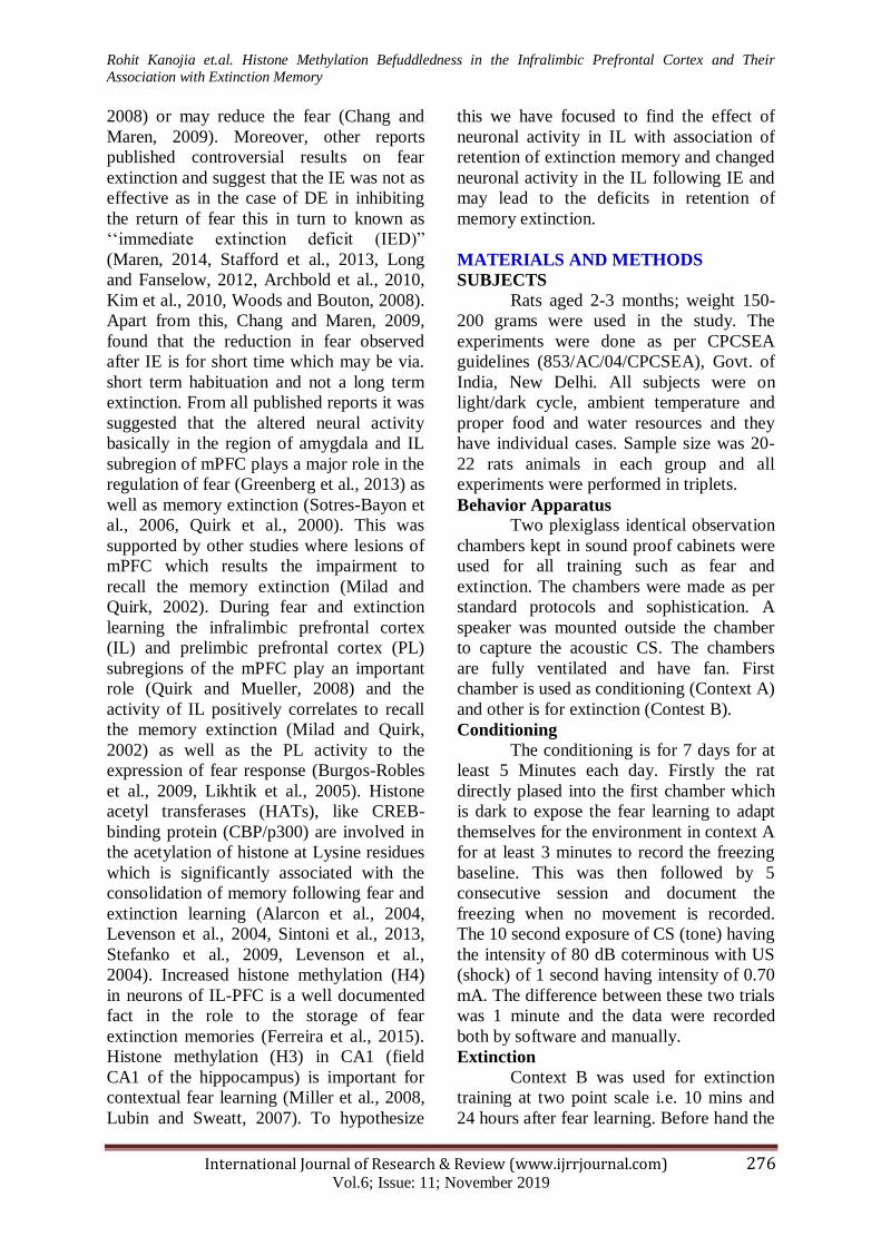
PFC and P-CREB expression following
immediate and delayed extinction
The expression of P-CREB was
found to be higher in extinction group as
compared to their control in the PL region
however; the changes were non-significant
between IE and DE group. Two-way
ANOVA analysis for P-CREB expression in
PL region revealed a significant main effect
of extinction condition (extinction vs. no
Extinction) [F (3, 26) =163.3, P < 0.0001],
However in IL, the expression of P-CREB
was significantly higher in DE group as
compared with the IE group and delayed no
extinction and this was confirmed by two
way ANOVA analysis [F (3, 26) =139.0, P
< 0.0001]. Extinction time (IE vs. DE) [F (3,
26) = 5.820, P < 0.0001] as well as
extinction condition and extinction time
interaction [F (3, 26) = 176.0, P < 0.0001].
Or we may say that the two sub regions of
Amygdala responded differentially to
respective two extinction conditions. This
differential expression of P-CREB
exemplifying the activity in the PL and LA
in the IE and DE group which may be
responsible for the deficit in the retention of
Extinction memory as observed after IE
(Figure 1).
Rohit Kanojia et.al. Histone Methylation Befuddledness in the Infralimbic Prefrontal Cortex and Their
Association with Extinction Memory
International Journal of Research & Review (www.ijrrjournal.com) 278 Vol.6; Issue: 11; November 2019
Figure 1. The expression of P-CREB in PFC following IE and DE learning: The expression of p-CREB was increased in the PL and IL of
both IE and DE when compared to their respective controls. While, the expression of p-CREB was significantly increased in PL of DE as
compared IE.
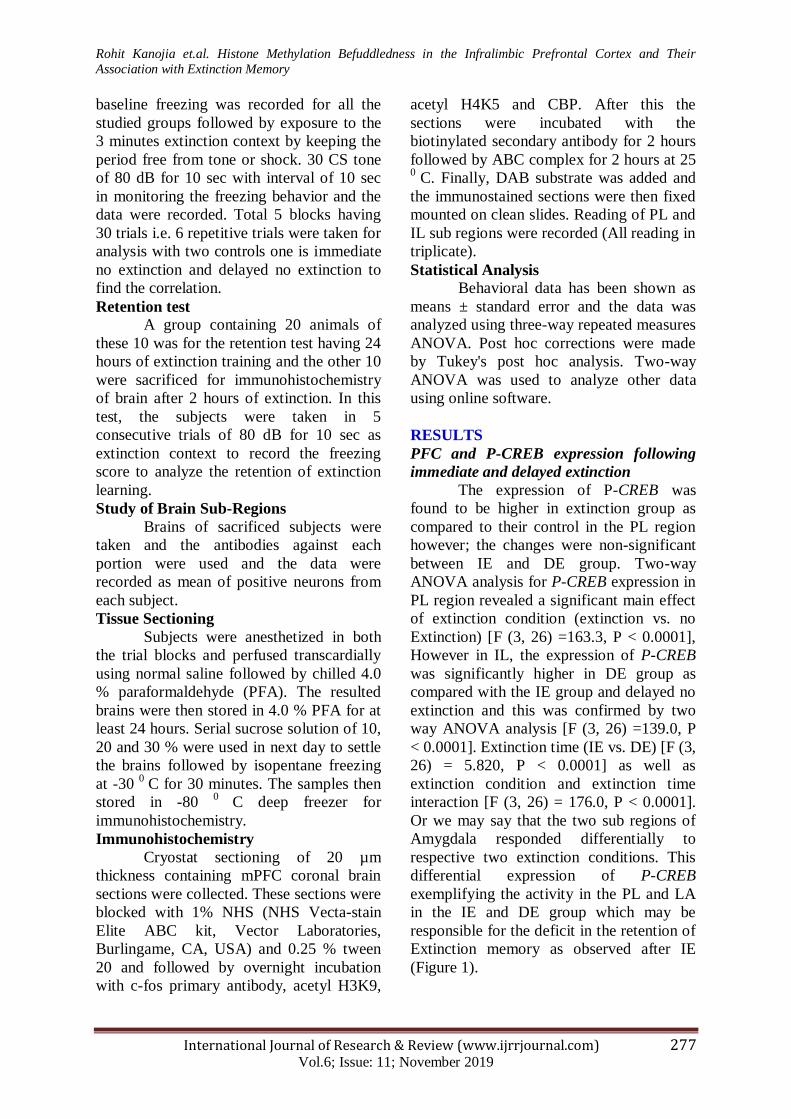
CREB expression following immediate
and delayed extinction
The CREB expression is quite
similar to P-CREB and higher in extinction
groups when compared to their control
however, no significant changes were
observed between IE and DE groups in PL
region. Two-way ANOVA analysis for
CREB expression in PL region showed
significant association [F (3, 26) =83.19, P <
0.0001] but no effect of extinction time in
IL of IE vs. DE [F (3, 26) =191.3, P <
0.0001]. Moreover in IL, significant
increment in CREB expression was
observed in DE. The data further supported
by using two way ANOVA analysis that
explains the same significant association [F
(3, 26) =5.565, P < 0.0001] extinction time
[F (3, 26) =9.50, P < 0.0001]. Expression of
CREB also seems to be highly associated
with neuronal activity in the IL and PL
following IE and DE. We further analyzed
that whether the increased CREB levels in
these subregions culminated in methylation
of H3 and H4 which are being studies and
suggest that the methylation of Histone at
various lysine residues to be associated with
increased gene expression required for
synaptic activity and memory consolidation
(Figure 2).
Figure 2. The expression of CREB in PFC following IE and DE learning: The expression of CREB was increased in the PL and IL of both
IE and DE when compared to their respective controls. While, the expression of CREB was significantly increased in PL of DE as compared
IE.
IMM EXT
IMM NO EXT
DELAYED EXT
DELAYED NO EXT 0
50
100
150
***
***
******
p-CREB Representation in PL
no.o
f C
RE
B p
ositiv
e n
euro
ns
IMM EXT
IMM NO EXT
DELAYED EXT
DELAYED NO EXT 0
20
40
60
80
100
*****
****
p-CREB Representation in IL
no.o
f C
RE
B p
ositi
ve n
euro
ns
IMM EXT
IMM NO EXT
DELAYED EXT
DELAYED NO EXT 0
50
100
150
***
***
******
CREB Representation in PL
no.o
f C
RE
B p
ositiv
e n
euro
ns
IMM EXT
IMM NO EXT
DELAYED EXT
DELAYED NO EXT 0
20
40
60
80
100
*****
****
CREB Representation in IL
no.o
f C
RE
B p
ositi
ve n
euro
ns
Rohit Kanojia et.al. Histone Methylation Befuddledness in the Infralimbic Prefrontal Cortex and Their
Association with Extinction Memory
International Journal of Research & Review (www.ijrrjournal.com) 279 Vol.6; Issue: 11; November 2019
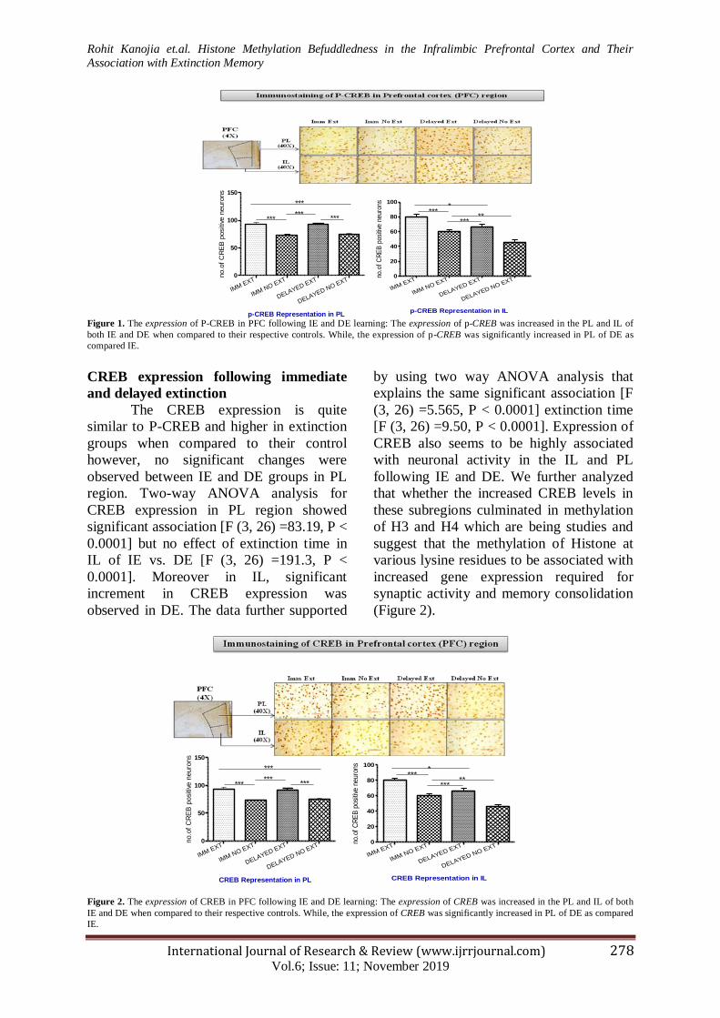
ARC expression following immediate and
delayed extinction
The same were observed in this i.e.
the expression was higher in extinction
groups however no significant association
were observed between IE and DE in PL
region. The same two-way ANOVA
analysis for CREB expression in PL region
revealed significant association with
extinction condition [F (3, 26) =131.7, P <
0.0001] but no effect of extinction time (IE
vs. DE) and condition with time interaction.
However in IL, there was a significant
increment in gene expression of CREB
when observed in DE as compared with IE
and delayed no extinction group as
supported by two way ANOVA analysis [F
(3, 26) =310.6, P < 0.0001]. Expression of
ARC seems to be highly associated with
neuronal activity in the PL and IL following
IE and DE. We further want to clear
whether the increased ARC levels in these
regions were related to the methylation of
H3 and H4, suggesting methylation of
Histone at various lysine residues which
may be associated with increased gene
expression (Figure 3).
Figure 3. The expression of ARC in PFC following IE and DE learning: The number of ARC + ve neurons was higher in PL and IL of both
IE ad DE. While, the numbers of ARC + ve neurons was significantly increased in IL of the IE as compared IE in PL.
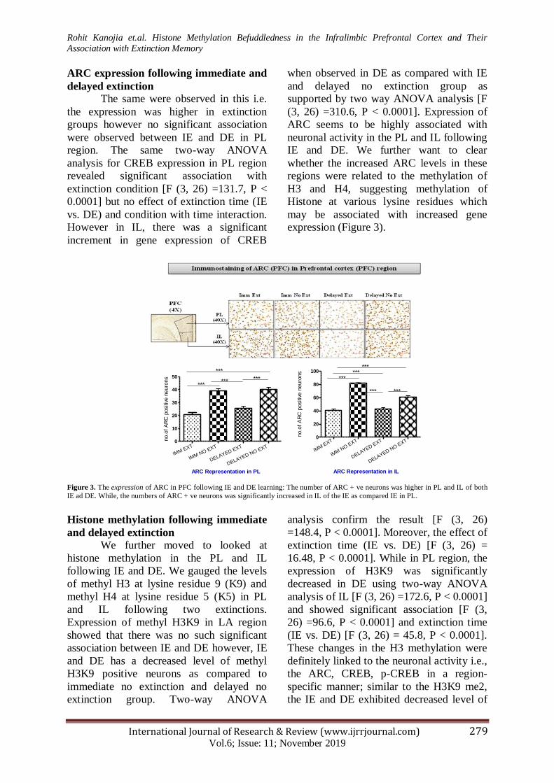
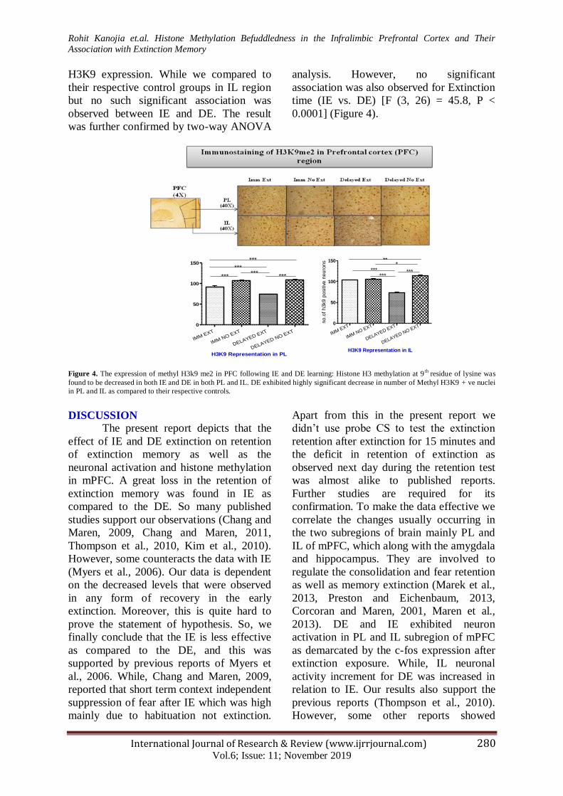
Histone methylation following immediate
and delayed extinction
We further moved to looked at
histone methylation in the PL and IL
following IE and DE. We gauged the levels
of methyl H3 at lysine residue 9 (K9) and
methyl H4 at lysine residue 5 (K5) in PL
and IL following two extinctions.
Expression of methyl H3K9 in LA region
showed that there was no such significant
association between IE and DE however, IE
and DE has a decreased level of methyl
H3K9 positive neurons as compared to
immediate no extinction and delayed no
extinction group. Two-way ANOVA
analysis confirm the result [F (3, 26)
=148.4, P < 0.0001]. Moreover, the effect of
extinction time (IE vs. DE) [F (3, 26) =
16.48, P < 0.0001]. While in PL region, the
expression of H3K9 was significantly
decreased in DE using two-way ANOVA
analysis of IL [F (3, 26) =172.6, P < 0.0001]
and showed significant association [F (3,
26) =96.6, P < 0.0001] and extinction time
(IE vs. DE) [F (3, 26) = 45.8, P < 0.0001].
These changes in the H3 methylation were
definitely linked to the neuronal activity i.e.,
the ARC, CREB, p-CREB in a region-
specific manner; similar to the H3K9 me2,
the IE and DE exhibited decreased level of
IMM EXT
IMM NO EXT
DELAYED EXT
DELAYED NO EXT 0
10
20
30
40
50
***
***
*** ***
ARC Representation in PL
no.o
f A
RC
posi
tive n
euro
ns
IMM E
XT
IMM N
O EXT
DELAYED EXT
DELAYED NO E
XT 0
20
40
60
80
100
******
***
*** ***
ARC Representation in IL
no.o
f A
RC
positiv
e n
euro
ns
Rohit Kanojia et.al. Histone Methylation Befuddledness in the Infralimbic Prefrontal Cortex and Their
Association with Extinction Memory
International Journal of Research & Review (www.ijrrjournal.com) 280 Vol.6; Issue: 11; November 2019
H3K9 expression. While we compared to
their respective control groups in IL region
but no such significant association was
observed between IE and DE. The result
was further confirmed by two-way ANOVA
analysis. However, no significant
association was also observed for Extinction
time (IE vs. DE) [F (3, 26) = 45.8, P <
0.0001] (Figure 4).
Figure 4. The expression of methyl H3k9 me2 in PFC following IE and DE learning: Histone H3 methylation at 9
th residue of lysine was
found to be decreased in both IE and DE in both PL and IL. DE exhibited highly significant decrease in number of Methyl H3K9 + ve nuclei
in PL and IL as compared to their respective controls.
DISCUSSION
The present report depicts that the
effect of IE and DE extinction on retention
of extinction memory as well as the
neuronal activation and histone methylation
in mPFC. A great loss in the retention of
extinction memory was found in IE as
compared to the DE. So many published
studies support our observations (Chang and
Maren, 2009, Chang and Maren, 2011,
Thompson et al., 2010, Kim et al., 2010).
However, some counteracts the data with IE
(Myers et al., 2006). Our data is dependent
on the decreased levels that were observed
in any form of recovery in the early
extinction. Moreover, this is quite hard to
prove the statement of hypothesis. So, we
finally conclude that the IE is less effective
as compared to the DE, and this was
supported by previous reports of Myers et
al., 2006. While, Chang and Maren, 2009,
reported that short term context independent
suppression of fear after IE which was high
mainly due to habituation not extinction.
Apart from this in the present report we
didn‟t use probe CS to test the extinction
retention after extinction for 15 minutes and
the deficit in retention of extinction as
observed next day during the retention test
was almost alike to published reports.
Further studies are required for its
confirmation. To make the data effective we
correlate the changes usually occurring in
the two subregions of brain mainly PL and
IL of mPFC, which along with the amygdala
and hippocampus. They are involved to
regulate the consolidation and fear retention
as well as memory extinction (Marek et al.,
2013, Preston and Eichenbaum, 2013,
Corcoran and Maren, 2001, Maren et al.,
2013). DE and IE exhibited neuron
activation in PL and IL subregion of mPFC
as demarcated by the c-fos expression after
extinction exposure. While, IL neuronal
activity increment for DE was increased in
relation to IE. Our results also support the
previous reports (Thompson et al., 2010).
However, some other reports showed
IMM EXT
IMM NO EXT
DELAYED EXT
DELAYED NO EXT
0
50
100
150
***
***
******
***
H3K9 Representation in PL
IMM EXT
IMM NO EXT
DELAYED EXT
DELAYED NO EXT 0
50
100
150
***
**
***
*
***
H3K9 Representation in IL
no.o
f h3k9 p
ositiv
e n
euro
ns
Rohit Kanojia et.al. Histone Methylation Befuddledness in the Infralimbic Prefrontal Cortex and Their
Association with Extinction Memory
International Journal of Research & Review (www.ijrrjournal.com) 281 Vol.6; Issue: 11; November 2019
different for fear behavior regulated by PL
in relation IL (Sotres-Bayon et al., 2006,
Milad et al., 2004, Courtin et al., 2013)
through up/down regulated neuron
activation in these regions (Sierra-Mercado
et al., 2011, Sotres-Bayon and Quirk, 2010)
as suggested in our present report. The loss
in retention of memory extinction as
observed in the IE in our report and
previously published other reports (Chang
and Maren, 2009, Chang and Maren, 2011,
Thompson et al., 2010, Kim et al., 2010).
This is mainly due to the result of
suppressed activation of IL neurons as well
as electrical stimulation of mPFC which
results in elimination of this loss (Kim et al.,
2010). While expression of c-fos, we had
looked at expression of CBP which was
found to be increased in IL than that of IE
which positively correlates with the
expression of c-fos expression. As a known
fact that CBP is associated with histone
acetyltransferase and its increased activity
results in the acetylation of histones H3/H4
which may modulate the expression of gene
in the formation of memory (Peixoto and
Abel, 2013). Our result suggested that there
is decreased activity of neurons in IL of rats
which fail to relate the fear as compared to
extinguish fear normally. This usually
associated with the lysine residues which
are present in the core of histone proteins
(Roth et al., 2001). This report predict that
the neuronal activity in the IL and PL is
associated with histone methylation. The
methylation of H3/H4 at residues K9/K5
were increased in IL and PL regions
following IE and DE and is usually
associated with the massive change in CBP
levels. Our major findings suggest long-
term extinction is minimal when extinction
is conducted for a short time after fear
learning in rats and this loss is usually by
the decreased neuronal activation in IL of
rats which are in IE. This loss in longterm
extinction is shown that it may be related to
the level of histone methylation in the IL of
the IE. The data from the present report may
be useful in traumatic planning and
execution of psychological and
pharmacological interventions.
ACKNOWLEDGEMENTS The work was supported by institutional research grant from Department of Zoology,
School of Biosciences and Biotechnology, BBA
University, Lucknow.
Conflict of Interest
Authors declare no conflict of interest.
REFERENCES
Alarcón, J.M., Malleret, G., Touzani, K.,
Vronskaya, S., Ishii, S., Kandel, E.R.,
Barco, A., 2004. Chromatin acetylation, memory, and LTP are impaired in CBP+/-
mice: A model for the cognitive deficit in
Rubinstein-Taybi syndrome and its
amelioration. Neuron 42, 947–959. https://doi.org/10.1016/j.neuron.2004.05.02
1
Archbold, G.E.B., Bouton, M.E., Nader, K.,
2010. Evidence for the persistence of contextual fear memories following
immediate extinction. Eur. J. Neurosci. 31,
1303–1311. https://doi.org/10.1111/j.1460-9568.2010.07161.x
Bouton, M.E., Westbrook, R.F., Corcoran,
K.A., Maren, S., 2006. Contextual and
Temporal Modulation of Extinction:
Behavioral and Biological Mechanisms. Biol. Psychiatry.
https://doi.org/10.1016/j.biopsych.2005.12.0
15
Burgos-Robles, A., Vidal-Gonzalez, I.,
Quirk, G.J., 2009. Sustained Conditioned
Responses in Prelimbic Prefrontal Neurons
Are Correlated with Fear Expression and
Extinction Failure. J. Neurosci. 29, 8474–8482.
https://doi.org/10.1523/JNEUROSCI.0378-
09.2009
Chang, C. -h., Maren, S., 2009. Early
extinction after fear conditioning yields a
context- independent and short-term
suppression of conditional freezing in rats. Learn. Mem. 16, 62–68.
https://doi.org/10.1101/lm.1085009
Chang, C., Maren, S., 2011. Medial
prefrontal cortex activation facilitates re-
extinction of fear in rats. Learn. Mem. 18, 221–5. https://doi.org/10.1101/lm.2070111
Rohit Kanojia et.al. Histone Methylation Befuddledness in the Infralimbic Prefrontal Cortex and Their
Association with Extinction Memory
International Journal of Research & Review (www.ijrrjournal.com) 282 Vol.6; Issue: 11; November 2019
Corcoran, K.A., Maren, S., 2001.
Hippocampal inactivation disrupts
contextual retrieval of fear memory after
extinction. J. Neurosci. 21, 1720–6.
Courtin, J., Chaudun, F., Rozeske, R.R.,
Karalis, N., Gonzalez-Campo, C., Wurtz,
H., Abdi, A., Baufreton, J., Bienvenu,
T.C.M., Herry, C., 2014. Prefrontal
parvalbumin interneurons shape neuronal activity to drive fear expression. Nature 505,
92–96. https://doi.org/10.1038/nature12755
Craske, M.G., Kircanski, K., Zelikowsky,
M., Mystkowski, J., Chowdhury, N., Baker, A., 2008. Optimizing inhibitory learning
during exposure therapy. Behav. Res. Ther.
46, 5– 27. https://doi.org/10.1016/j.brat.2007.10.003
Ferreira, A.N., Yousuf, H., Dalton, S.,
Sheets, P.L., 2015. Highly differentiated
cellular and circuit properties of infralimbic
pyramidal neurons projecting to the periaqueductal gray and amygdala. Front.
Cell. Neurosci. 9.
https://doi.org/10.3389/fncel.2015.00161
Golkar, A., Bellander, M., Olsson, A.,
Öhman, A., 2012. Are fear memories
erasable?– reconsolidation of learned fear
with fear-relevant and fear-irrelevant stimuli. Front. Behav. Neurosci. 6.
https://doi.org/10.3389/fnbeh.2012.00080
Greenberg, T., Carlson, J.M., Cha, J.,
Hajcak, G., Mujica-Parodi, L.R., 2013.
Ventromedial prefrontal cortex reactivity is altered in generalized anxiety disorder
during fear generalization. Depress. Anxiety
30, 242–250. https://doi.org/10.1002/da.22016
Huff, N.C., Hernandez, J.A., Blanding,
N.Q., LaBar, K.S., 2009. Delayed
Extinction Attenuates Conditioned Fear
Renewal and Spontaneous Recovery in Humans. Behav. Neurosci. 123, 834–843.
https://doi.org/10.1037/a0016511
Kim, S.C., Jo, Y.S., Kim, I.H., Kim, H.,
Choi, J.-S., 2010. Lack of Medial Prefrontal Cortex Activation Underlies the Immediate
Extinction Deficit. J. Neurosci. 30, 832–
837. https://doi.org/10.1523/JNEUROSCI.4145-
09.2010
Levenson, J.M., O‟Riordan, K.J., Brown,
K.D., Trinh, M.A., Molfese, D.L., Sweatt,
J.D., 2004. Regulation of histone acetylation during memory formation in the
hippocampus. J. Biol. Chem. 279, 40545–
59. https://doi.org/10.1074/jbc.M402229200
Likhtik, E., 2005. Prefrontal Control of the
Amygdala. J. Neurosci. 25, 7429–7437. https://doi.org/10.1523/JNEUROSCI.2314-
05.2005
Long, V.A., Fanselow, M.S., 2012. Stress-
enhanced fear learning in rats is resistant to
the effects of immediate massed extinction. Stress 15, 627–636.
https://doi.org/10.3109/10253890.2011.650
251
Lubin, F.D., Sweatt, J.D., 2007. The
IkappaB kinase regulates chromatin
structure during reconsolidation of
conditioned fear memories. Neuron 55, 942–57.
https://doi.org/10.1016/j.neuron.2007.07.03
9
Marek, R., Strobel, C., Bredy, T.W., Sah, P.,
2013. The amygdala and medial prefrontal cortex: partners in the fear circuit. J.
Physiol. 591, 2381–2391.
https://doi.org/10.1113/jphysiol.2012.248575
Maren, S., 2005. Building and burying fear
memories in the brain. Neuroscientist.
https://doi.org/10.1177/1073858404269232
Maren, S., 2014. Nature and causes of the
immediate extinction deficit: A brief review.
Neurobiol. Learn. Mem.
https://doi.org/10.1016/j.nlm.2013.10.012
Maren, S., Chang, C. -h., 2006. Recent fear
is resistant to extinction. Proc. Natl. Acad. Sci. 103, 18020–18025.
https://doi.org/10.1073/pnas.0608398103.
Milad, M.R., Vidal-Gonzalez, I., Quirk,
G.J., 2004. Electrical Stimulation of Medial Prefrontal Cortex Reduces Conditioned Fear
in a Temporally Specific Manner. Behav.
Neurosci. 118, 389–394. https://doi.org/10.1037/0735-
7044.118.2.389
Milad, M.R.R., Quirk, G.J.J., 2002. Neurons
in medial prefrontal cortex signal memory
for fear extinction. Nature 420, 70–74. https://doi.org/10.1038/nature01144.1.
Miller, C.A., Campbell, S.L., Sweatt, J.D.,
2008. DNA methylation and histone
acetylation work in concert to regulate memory formation and synaptic plasticity.
Neurobiol. Learn. Mem. 89, 599–603.
https://doi.org/10.1016/j.nlm.2007.07.016
Rohit Kanojia et.al. Histone Methylation Befuddledness in the Infralimbic Prefrontal Cortex and Their
Association with Extinction Memory
International Journal of Research & Review (www.ijrrjournal.com) 283 Vol.6; Issue: 11; November 2019
Muigg, P., Hetzenauer, A., Hauer, G.,
Hauschild, M., Gaburro, S., Frank, E.,
Landgraf, R., Singewald, N., 2008. Impaired
extinction of learned fear in rats selectively bred for high anxiety - Evidence of altered
neuronal processing in prefrontal-amygdala
pathways. Eur. J. Neurosci. 28, 2299–2309.
https://doi.org/10.1111/j.1460-9568.2008.06511.x
Myers, K.M., 2006. Different mechanisms
of fear extinction dependent on length of
time since fear acquisition. Learn. Mem. 13, 216–223. https://doi.org/10.1101/lm.119806
Myers, K.M., Davis, M., 2007. Mechanisms
of fear extinction. Mol. Psychiatry.
https://doi.org/10.1038/sj.mp.4001939
Norrholm, S.D., Vervliet, B., Jovanovic, T.,
Boshoven, W., Myers, K.M., Davis, M.,
Rothbaum, B., Duncan, E.J., 2008. Timing
of Extinction Relative to Acquisition: A
Parametric Analysis of Fear Extinction in Humans. Behav. Neurosci. 122, 1016–1030.
https://doi.org/10.1037/a0012604
Pare, D., 2004. New Vistas on Amygdala
Networks in Conditioned Fear. J. Neurophysiol. 92, 1–9.
https://doi.org/10.1152/jn.00153.2004
Pavlov, 1927. Classics in the History of
Psychology Lecture XVIII. http://psychclassics.yorku.ca/Pavlov/lecture
18.htm
Peixoto, L., Abel, T., 2013. The Role of
Histone Acetylation in Memory Formation
and Cognitive Impairments. Neuropsychopharmacology 38, 62–76.
https://doi.org/10.1038/npp.2012.86
Preston, A.R., Eichenbaum, H., 2013.
Interplay of hippocampus and prefrontal cortex in memory. Curr. Biol. 23, R764-73.
https://doi.org/10.1016/j.cub.2013.05.041
Quirk, G.J., Mueller, D., 2008. Neural
mechanisms of extinction learning and retrieval. Neuropsychopharmacology.
https://doi.org/10.1038/sj.npp.1301555
Quirk, G.J., Russo, G.K., Barron, J.L.,
Lebron, K., 2000. The role of ventromedial
prefrontal cortex in the recovery of extinguished fear. J. Neurosci. 20, 6225–
6231. https://doi.org/20/16/6225 [pii]
Rosen, J.B., Schulkin, J., 1998. From
Normal Fear to Pathological Anxiety. Psychol. Rev. https://doi.org/10.1037/0033-
295X.105.2.325
Roth, S.Y., Denu, J.M., Allis, C.D., 2001.
Histone Acetyltransferases. Annu. Rev.
Biochem. 70, 81–120.
https://doi.org/10.1146/annurev.biochem.70.1.81
Sierra-Mercado, D., Padilla-Coreano, N.,
Quirk, G.J., 2011. Dissociable roles of
prelimbic and infralimbic cortices, ventral
hippocampus, and basolateral amygdala in the expression and extinction of conditioned
fear. Neuropsychopharmacology 36, 529–
38. https://doi.org/10.1038/npp.2010.184
Sintoni, S., Kurtys, E., Scandaglia, M.,
Contestabile, A., Monti, B., 2013. Chronic
valproic acid administration impairs
contextual memory and dysregulates hippocampal GSK-3β in rats. Pharmacol.
Biochem. Behav. 106, 8–15.
https://doi.org/10.1016/j.pbb.2013.02.013
Sotres-Bayon, F., Cain, C.K., LeDoux, J.E.,
2006. Brain Mechanisms of Fear Extinction: Historical Perspectives on the Contribution
of Prefrontal Cortex. Biol. Psychiatry.
https://doi.org/10.1016/j.biopsych.2005.10.012
Sotres-Bayon, F., Quirk, G.J., 2010.
Prefrontal control of fear: more than just
extinction. Curr. Opin. Neurobiol. 20, 231–235.
https://doi.org/10.1016/j.conb.2010.02.005
Stafford, J.M., Maughan, D.K., Ilioi, E.C.,
Lattal, K.M., 2013. Exposure to a fearful
context during periods of memory plasticity impairs extinction via hyperactivation of
frontal- amygdalar circuits. Learn. Mem. 20,
156–163. https://doi.org/10.1101/lm.029801.112
Stefanko, D.P., Barrett, R.M., Ly, A.R.,
Reolon, G.K., Wood, M.A., 2009.
Modulation of long-term memory for object
recognition via HDAC inhibition. Proc. Natl. Acad. Sci. 106, 9447–9452.
https://doi.org/10.1073/pnas.0903964106
Thompson, B.M., Baratta, M. V,
Biedenkapp, J.C., Rudy, J.W., Watkins, L.R., Maier, S.F., 2010. Activation of the
infralimbic cortex in a fear context enhances
extinction learning. Learn. Mem. 17, 591–9. https://doi.org/10.1101/lm.1920810
VanElzakker, M.B., Kathryn Dahlgren, M.,
Caroline Davis, F., Dubois, S., Shin, L.M.,
2014. From Pavlov to PTSD: The extinction
of conditioned fear in rodents, humans, and
Rohit Kanojia et.al. Histone Methylation Befuddledness in the Infralimbic Prefrontal Cortex and Their
Association with Extinction Memory
International Journal of Research & Review (www.ijrrjournal.com) 284 Vol.6; Issue: 11; November 2019
anxiety disorders. Neurobiol. Learn. Mem.
https://doi.org/10.1016/j.nlm.2013.11.014
Wessa, M., Flor, H., 2007. Failure of
extinction of fear responses in posttraumatic stress disorder: Evidence from second-order
conditioning. Am. J. Psychiatry 164, 1684–
1692.
https://doi.org/10.1176/appi.ajp.2007.07030
525
Woods, A.M., Bouton, M.E., 2008.
Immediate extinction causes a less durable loss of performance than delayed extinction
following either fear or appetitive
conditioning. Learn. Mem. 15, 909–920.
https://doi.org/10.1101/lm.1078508
******
How to cite this article: Kanojia R, Modi
DR, Mishra S et.al. Histone methylation befuddledness
in the infralimbic prefrontal cortex and their association with extinction memory. International
Journal of Research and Review. 2019; 6(11):275-284.
Copyright © 2022 FDOKUMEN