Mismatch repair-dependent processing of methylation ...
15
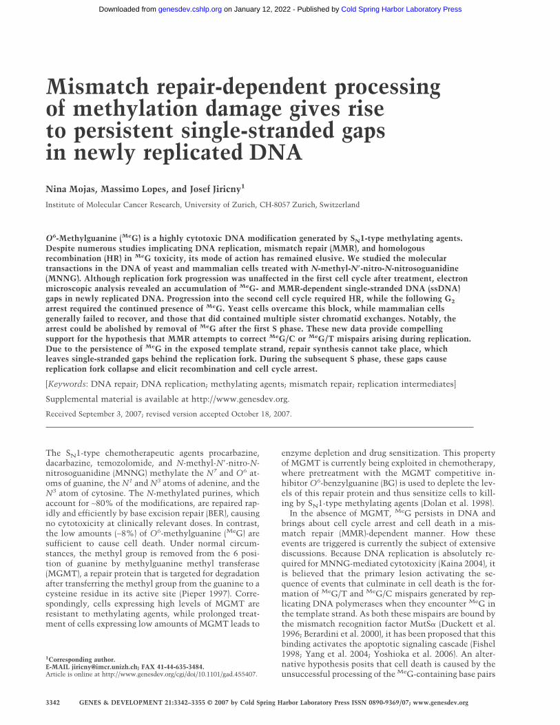
Mismatch repair-dependent processing of methylation damage gives rise to persistent single-stranded gaps in newly replicated DNA Nina Mojas, Massimo Lopes, and Josef Jiricny 1 Institute of Molecular Cancer Research, University of Zurich, CH-8057 Zurich, Switzerland O 6 -Methylguanine ( Me G) is a highly cytotoxic DNA modification generated by S N 1-type methylating agents. Despite numerous studies implicating DNA replication, mismatch repair (MMR), and homologous recombination (HR) in Me G toxicity, its mode of action has remained elusive. We studied the molecular transactions in the DNA of yeast and mammalian cells treated with N-methyl-N-nitro-N-nitrosoguanidine (MNNG). Although replication fork progression was unaffected in the first cell cycle after treatment, electron microscopic analysis revealed an accumulation of Me G- and MMR-dependent single-stranded DNA (ssDNA) gaps in newly replicated DNA. Progression into the second cell cycle required HR, while the following G 2 arrest required the continued presence of Me G. Yeast cells overcame this block, while mammalian cells generally failed to recover, and those that did contained multiple sister chromatid exchanges. Notably, the arrest could be abolished by removal of Me G after the first S phase. These new data provide compelling support for the hypothesis that MMR attempts to correct Me G/C or Me G/T mispairs arising during replication. Due to the persistence of Me G in the exposed template strand, repair synthesis cannot take place, which leaves single-stranded gaps behind the replication fork. During the subsequent S phase, these gaps cause replication fork collapse and elicit recombination and cell cycle arrest. [Keywords: DNA repair; DNA replication; methylating agents; mismatch repair; replication intermediates] Supplemental material is available at http://www.genesdev.org. Received September 3, 2007; revised version accepted October 18, 2007. The S N 1-type chemotherapeutic agents procarbazine, dacarbazine, temozolomide, and N-methyl-N-nitro-N- nitrosoguanidine (MNNG) methylate the N 7 and O 6 at- oms of guanine, the N 1 and N 3 atoms of adenine, and the N 3 atom of cytosine. The N-methylated purines, which account for ∼80% of the modifications, are repaired rap- idly and efficiently by base excision repair (BER), causing no cytotoxicity at clinically relevant doses. In contrast, the low amounts (∼8%) of O 6 -methylguanine ( Me G) are sufficient to cause cell death. Under normal circum- stances, the methyl group is removed from the 6 posi- tion of guanine by methylguanine methyl transferase (MGMT), a repair protein that is targeted for degradation after transferring the methyl group from the guanine to a cysteine residue in its active site (Pieper 1997). Corre- spondingly, cells expressing high levels of MGMT are resistant to methylating agents, while prolonged treat- ment of cells expressing low amounts of MGMT leads to enzyme depletion and drug sensitization. This property of MGMT is currently being exploited in chemotherapy, where pretreatment with the MGMT competitive in- hibitor O 6 -benzylguanine (BG) is used to deplete the lev- els of this repair protein and thus sensitize cells to kill- ing by S N 1-type methylating agents (Dolan et al. 1998). In the absence of MGMT, Me G persists in DNA and brings about cell cycle arrest and cell death in a mis- match repair (MMR)-dependent manner. How these events are triggered is currently the subject of extensive discussions. Because DNA replication is absolutely re- quired for MNNG-mediated cytotoxicity (Kaina 2004), it is believed that the primary lesion activating the se- quence of events that culminate in cell death is the for- mation of Me G/T and Me G/C mispairs generated by rep- licating DNA polymerases when they encounter Me G in the template strand. As both these mispairs are bound by the mismatch recognition factor MutS (Duckett et al. 1996; Berardini et al. 2000), it has been proposed that this binding activates the apoptotic signaling cascade (Fishel 1998; Yang et al. 2004; Yoshioka et al. 2006). An alter- native hypothesis posits that cell death is caused by the unsuccessful processing of the Me G-containing base pairs 1 Corresponding author. E-MAIL [email protected]; FAX 41-44-635-3484. Article is online at http://www.genesdev.org/cgi/doi/10.1101/gad.455407. 3342 GENES & DEVELOPMENT 21:3342–3355 © 2007 by Cold Spring Harbor Laboratory Press ISSN 0890-9369/07; www.genesdev.org Cold Spring Harbor Laboratory Press on January 12, 2022 - Published by genesdev.cshlp.org Downloaded from
-
Upload
khangminh22 -
Category
Documents
-
view
6 -
download
0
Transcript of Mismatch repair-dependent processing of methylation ...
Mismatch repair-dependent processingof methylation damage gives riseto persistent single-stranded gapsin newly replicated DNANina Mojas, Massimo Lopes, and Josef Jiricny1
Institute of Molecular Cancer Research, University of Zurich, CH-8057 Zurich, Switzerland
O6-Methylguanine (MeG) is a highly cytotoxic DNA modification generated by SN1-type methylating agents.Despite numerous studies implicating DNA replication, mismatch repair (MMR), and homologousrecombination (HR) in MeG toxicity, its mode of action has remained elusive. We studied the moleculartransactions in the DNA of yeast and mammalian cells treated with N-methyl-N�-nitro-N-nitrosoguanidine(MNNG). Although replication fork progression was unaffected in the first cell cycle after treatment, electronmicroscopic analysis revealed an accumulation of MeG- and MMR-dependent single-stranded DNA (ssDNA)gaps in newly replicated DNA. Progression into the second cell cycle required HR, while the following G2
arrest required the continued presence of MeG. Yeast cells overcame this block, while mammalian cellsgenerally failed to recover, and those that did contained multiple sister chromatid exchanges. Notably, thearrest could be abolished by removal of MeG after the first S phase. These new data provide compellingsupport for the hypothesis that MMR attempts to correct MeG/C or MeG/T mispairs arising during replication.Due to the persistence of MeG in the exposed template strand, repair synthesis cannot take place, whichleaves single-stranded gaps behind the replication fork. During the subsequent S phase, these gaps causereplication fork collapse and elicit recombination and cell cycle arrest.
[Keywords: DNA repair; DNA replication; methylating agents; mismatch repair; replication intermediates]
Supplemental material is available at http://www.genesdev.org.
Received September 3, 2007; revised version accepted October 18, 2007.
The SN1-type chemotherapeutic agents procarbazine,dacarbazine, temozolomide, and N-methyl-N�-nitro-N-nitrosoguanidine (MNNG) methylate the N7 and O6 at-oms of guanine, the N1 and N3 atoms of adenine, and theN3 atom of cytosine. The N-methylated purines, whichaccount for ∼80% of the modifications, are repaired rap-idly and efficiently by base excision repair (BER), causingno cytotoxicity at clinically relevant doses. In contrast,the low amounts (∼8%) of O6-methylguanine (MeG) aresufficient to cause cell death. Under normal circum-stances, the methyl group is removed from the 6 posi-tion of guanine by methylguanine methyl transferase(MGMT), a repair protein that is targeted for degradationafter transferring the methyl group from the guanine to acysteine residue in its active site (Pieper 1997). Corre-spondingly, cells expressing high levels of MGMT areresistant to methylating agents, while prolonged treat-ment of cells expressing low amounts of MGMT leads to
enzyme depletion and drug sensitization. This propertyof MGMT is currently being exploited in chemotherapy,where pretreatment with the MGMT competitive in-hibitor O6-benzylguanine (BG) is used to deplete the lev-els of this repair protein and thus sensitize cells to kill-ing by SN1-type methylating agents (Dolan et al. 1998).
In the absence of MGMT, MeG persists in DNA andbrings about cell cycle arrest and cell death in a mis-match repair (MMR)-dependent manner. How theseevents are triggered is currently the subject of extensivediscussions. Because DNA replication is absolutely re-quired for MNNG-mediated cytotoxicity (Kaina 2004), itis believed that the primary lesion activating the se-quence of events that culminate in cell death is the for-mation of MeG/T and MeG/C mispairs generated by rep-licating DNA polymerases when they encounter MeG inthe template strand. As both these mispairs are bound bythe mismatch recognition factor MutS� (Duckett et al.1996; Berardini et al. 2000), it has been proposed that thisbinding activates the apoptotic signaling cascade (Fishel1998; Yang et al. 2004; Yoshioka et al. 2006). An alter-native hypothesis posits that cell death is caused by theunsuccessful processing of the MeG-containing base pairs
1Corresponding author.E-MAIL [email protected]; FAX 41-44-635-3484.Article is online at http://www.genesdev.org/cgi/doi/10.1101/gad.455407.
3342 GENES & DEVELOPMENT 21:3342–3355 © 2007 by Cold Spring Harbor Laboratory Press ISSN 0890-9369/07; www.genesdev.org
Cold Spring Harbor Laboratory Press on January 12, 2022 - Published by genesdev.cshlp.orgDownloaded from

by the MMR system, which activates a cell cycle arrest(Stojic et al. 2004) from which most cells fail to recover.This latter hypothesis is supported by the finding thatthe arrest is activated only in the second S phase afterMNNG treatment (Zhukovskaya et al. 1994; Stojic et al.2004), whereas the direct signaling hypothesis wouldpredict that the cells should arrest after the first S phase,given that MeG-containing mispairs are formed alreadyduring the first replication.
In eukaryotic cells, MMR is tightly linked with repli-cation through PCNA, which interacts directly withMutS� (Clark et al. 2000; Kleczkowska et al. 2001) andthus provides MMR with ready access to mismatchesgenerated by the polymerases. It might therefore be an-ticipated that when MMR attempts to process a MeG-containing mispair, the replicating polymerase might beretarded, because the modified guanine in the templatestrand does not have a perfect Watson-Crick partner, andthus that the MMR process cannot be completed. Shouldthe presence of MeG in the template DNA fail to cause asignificant DNA synthesis delay, it would imply that theprocessing of this lesion is decoupled from replication.The first goal of this study was to address this issue.
That loss of homologous recombination (HR) sensi-tizes MMR-proficient yeast and mammalian cells to kill-ing by MNNG (Cejka et al. 2005; Tsaryk et al. 2006)provided further support for the processing hypothesis,suggesting that intermediates arising during the futileattempts of the MMR machinery to correct MeG-con-taining mispairs are channeled into HR. However, thesestudies failed to reveal the possible structure(s) of the HRintermediates. The second goal of this study was there-fore to gain novel insights into the MMR-dependenttransactions in the DNA of MNNG-treated mammalianand yeast cells. We also wanted to identify the processesthat allow the treated cells to bypass MeG during the firstS phase and to undergo cell division with damaged DNA.
Results
MNNG-induced arrest in the second cell cycleis dependent solely on MeG
Exposure of mammalian cells to MNNG brings about aMMR-dependent cell cycle arrest in the second G2 phaseafter treatment (Stojic et al. 2004). However, the MeG/Tand MeG/C mispairs, which are believed to activateMMR, arise already in the first S phase, so why do theyfail to trigger the cell cycle arrest then? How does theDNA of newly treated cells differ from that of cells thathave undergone one division? As mentioned above, MeGis only a minor product of MNNG treatment. Thischemical generates predominantly N7-methyguanineand N3-methyladenine, which are not substrates forMMR, but which are rapidly and efficiently repaired byBER (Wyatt and Pittman 2006). The latter modificationswill thus have been removed from DNA prior to theonset of the second S phase, and only MeG will persist.We therefore wanted to test whether it was the process-ing of the N-methylated purines during the first cell
cycle that allowed the cells to progress to the next one.In this scenario, a second treatment with MNNG afterthe cell division should carry the cells also across thesecond mitotic boundary, and the activation of the cellcycle arrest should be delayed until the third cycle. Inorder to test this possibility, we used 293T L� cells,which are MGMT- and MLH1-deficient, but carry a sta-bly integrated hMLH1 cDNA minigene controlled by theTetOffTM expression system (Cejka et al. 2003). In theabsence of doxycycline (Dox), these cells, referred to as293T L�+, express hMLH1 and are MMR-proficient andMNNG-sensitive. In contrast, when the same cells aregrown in the presence of 50 ng/mL Dox (293T L�− cells),they shut off hMLH1 expression and become MMR-de-ficient and resistant to MNNG. We synchronized the293T L� cells with 2 mM hydroxyurea (HU) for 16 h (Fig.1A) and released them into fresh medium without (Con-trol) or with 0.2 µM MNNG. The cells were then treatedwith 0.2 µM MNNG a second time, 22 h after release,when the majority were in the G1 phase of the secondcell cycle (Double MNNG). As the cells treated at thepoint of release from the HU block and those treatedwith MNNG twice arrested at the same time point, weconcluded that processing of lesions other than MeG doesnot promote cell cycle progression after MNNG treat-ment.
To further confirm the above hypothesis, we treatedthe cells with 6-thioguanine (6TG), which is incorpo-rated into DNA and converted to MeTG, an analog ofMeG (Karran 2006). The DNA of cells treated with TGthus does not contain modified nucleotides other thanMeTG. As shown in Figure 1B, treatment of synchronized293T L� cells with 3 µM 6TG for 21 h brought about aMMR-dependent arrest two cell cycles after treatment(52 h after release), similar to that induced by MNNG(Stojic et al. 2004). Taken together, the above data showthat the MNNG-induced second cell cycle arrest is trig-gered by the processing of a single lesion, namely, MeG.
MMR-dependent processing of MeG does not affectreplication fork progression, but leads to a reducedreplication rate in the second cell cycle
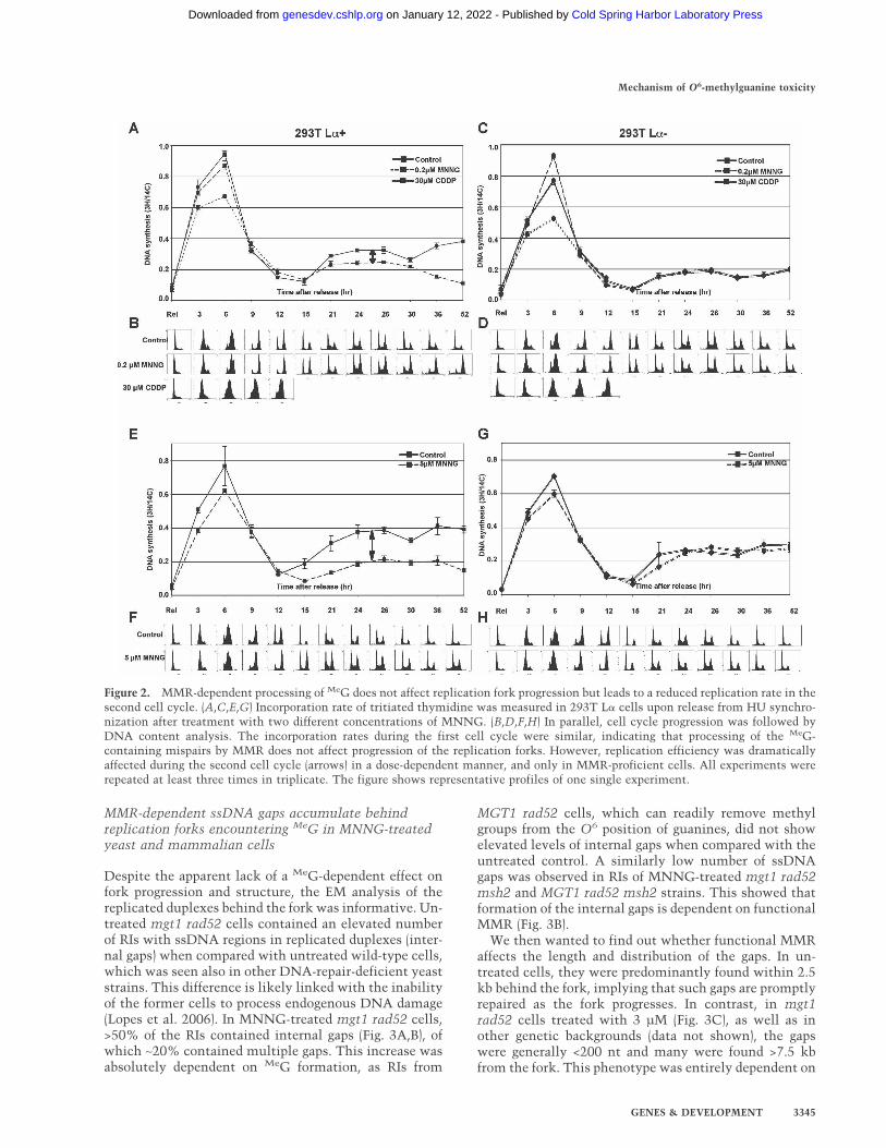
Our previous experiments (Stojic et al. 2004) suggestedthat progression of the cells through the first S phase waslargely unaffected by MNNG treatment, at least asjudged by flow cytometric analysis (see also Fig. 7A, be-low). However, we wanted to study the replication ratesof treated and untreated cells in greater detail. Using293T L� cells, we measured the effect of two differentconcentrations of MNNG on the incorporation of radio-active nucleotides into genomic DNA. As a positive con-trol, we used cisplatin (CDDP), a cross-linking agentknown to interfere with replication fork progression(Henry-Mowatt et al. 2003). As shown in Figure2A,C,E,G, DNA synthesis levels were highest during thefirst S phase and were similar in both 0.2 µM MNNG-treated and control samples, while the replication ratesof cisplatin-treated cells were substantially lower in both
Mechanism of O6-methylguanine toxicity
GENES & DEVELOPMENT 3343
Cold Spring Harbor Laboratory Press on January 12, 2022 - Published by genesdev.cshlp.orgDownloaded from
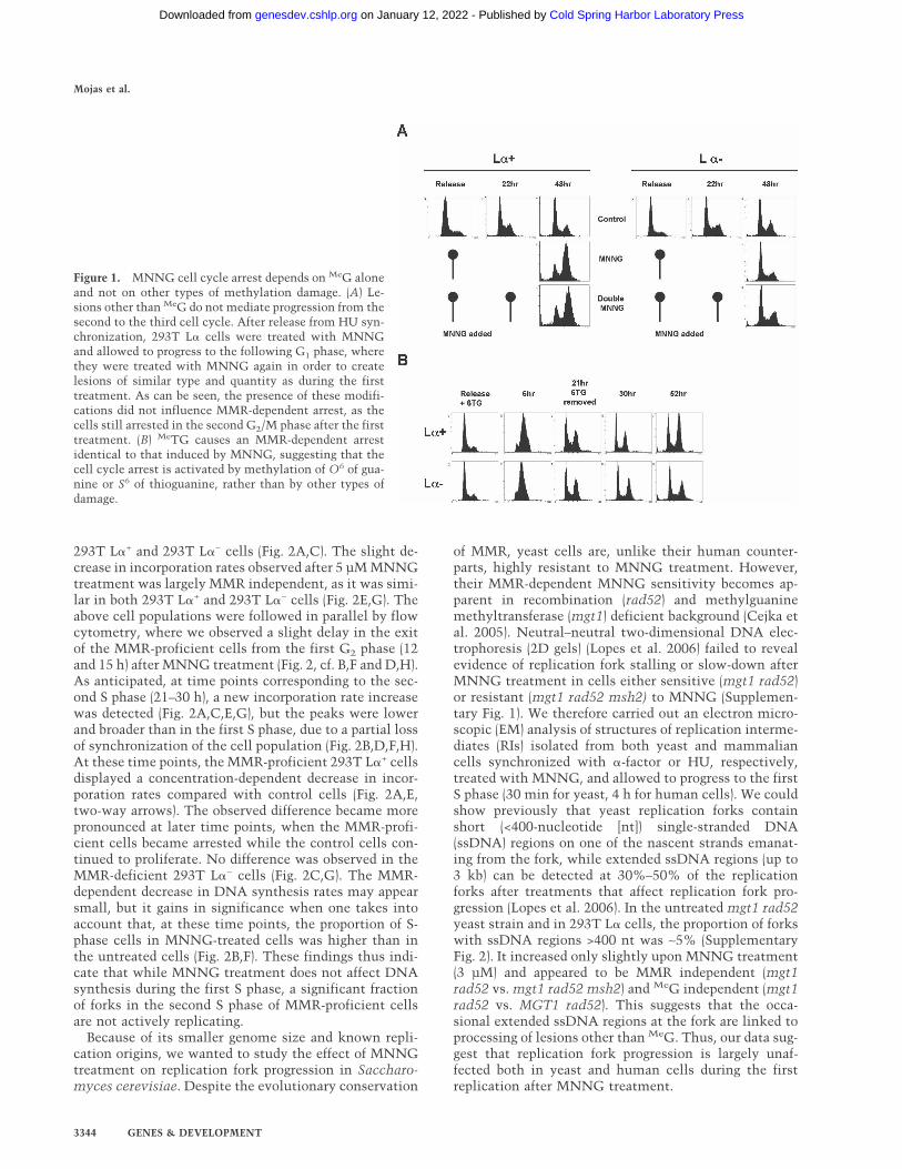
293T L�+ and 293T L�− cells (Fig. 2A,C). The slight de-crease in incorporation rates observed after 5 µM MNNGtreatment was largely MMR independent, as it was simi-lar in both 293T L�+ and 293T L�− cells (Fig. 2E,G). Theabove cell populations were followed in parallel by flowcytometry, where we observed a slight delay in the exitof the MMR-proficient cells from the first G2 phase (12and 15 h) after MNNG treatment (Fig. 2, cf. B,F and D,H).As anticipated, at time points corresponding to the sec-ond S phase (21–30 h), a new incorporation rate increasewas detected (Fig. 2A,C,E,G), but the peaks were lowerand broader than in the first S phase, due to a partial lossof synchronization of the cell population (Fig. 2B,D,F,H).At these time points, the MMR-proficient 293T L�+ cellsdisplayed a concentration-dependent decrease in incor-poration rates compared with control cells (Fig. 2A,E,two-way arrows). The observed difference became morepronounced at later time points, when the MMR-profi-cient cells became arrested while the control cells con-tinued to proliferate. No difference was observed in theMMR-deficient 293T L�− cells (Fig. 2C,G). The MMR-dependent decrease in DNA synthesis rates may appearsmall, but it gains in significance when one takes intoaccount that, at these time points, the proportion of S-phase cells in MNNG-treated cells was higher than inthe untreated cells (Fig. 2B,F). These findings thus indi-cate that while MNNG treatment does not affect DNAsynthesis during the first S phase, a significant fractionof forks in the second S phase of MMR-proficient cellsare not actively replicating.
Because of its smaller genome size and known repli-cation origins, we wanted to study the effect of MNNGtreatment on replication fork progression in Saccharo-myces cerevisiae. Despite the evolutionary conservation
of MMR, yeast cells are, unlike their human counter-parts, highly resistant to MNNG treatment. However,their MMR-dependent MNNG sensitivity becomes ap-parent in recombination (rad52) and methylguaninemethyltransferase (mgt1) deficient background (Cejka etal. 2005). Neutral–neutral two-dimensional DNA elec-trophoresis (2D gels) (Lopes et al. 2006) failed to revealevidence of replication fork stalling or slow-down afterMNNG treatment in cells either sensitive (mgt1 rad52)or resistant (mgt1 rad52 msh2) to MNNG (Supplemen-tary Fig. 1). We therefore carried out an electron micro-scopic (EM) analysis of structures of replication interme-diates (RIs) isolated from both yeast and mammaliancells synchronized with �-factor or HU, respectively,treated with MNNG, and allowed to progress to the firstS phase (30 min for yeast, 4 h for human cells). We couldshow previously that yeast replication forks containshort (<400-nucleotide [nt]) single-stranded DNA(ssDNA) regions on one of the nascent strands emanat-ing from the fork, while extended ssDNA regions (up to3 kb) can be detected at 30%–50% of the replicationforks after treatments that affect replication fork pro-gression (Lopes et al. 2006). In the untreated mgt1 rad52yeast strain and in 293T L� cells, the proportion of forkswith ssDNA regions >400 nt was ∼5% (SupplementaryFig. 2). It increased only slightly upon MNNG treatment(3 µM) and appeared to be MMR independent (mgt1rad52 vs. mgt1 rad52 msh2) and MeG independent (mgt1rad52 vs. MGT1 rad52). This suggests that the occa-sional extended ssDNA regions at the fork are linked toprocessing of lesions other than MeG. Thus, our data sug-gest that replication fork progression is largely unaf-fected both in yeast and human cells during the firstreplication after MNNG treatment.
Figure 1. MNNG cell cycle arrest depends on MeG aloneand not on other types of methylation damage. (A) Le-sions other than MeG do not mediate progression from thesecond to the third cell cycle. After release from HU syn-chronization, 293T L� cells were treated with MNNGand allowed to progress to the following G1 phase, wherethey were treated with MNNG again in order to createlesions of similar type and quantity as during the firsttreatment. As can be seen, the presence of these modifi-cations did not influence MMR-dependent arrest, as thecells still arrested in the second G2/M phase after the firsttreatment. (B) MeTG causes an MMR-dependent arrestidentical to that induced by MNNG, suggesting that thecell cycle arrest is activated by methylation of O6 of gua-nine or S6 of thioguanine, rather than by other types ofdamage.
Mojas et al.
3344 GENES & DEVELOPMENT
Cold Spring Harbor Laboratory Press on January 12, 2022 - Published by genesdev.cshlp.orgDownloaded from
MMR-dependent ssDNA gaps accumulate behindreplication forks encountering MeG in MNNG-treatedyeast and mammalian cells
Despite the apparent lack of a MeG-dependent effect onfork progression and structure, the EM analysis of thereplicated duplexes behind the fork was informative. Un-treated mgt1 rad52 cells contained an elevated numberof RIs with ssDNA regions in replicated duplexes (inter-nal gaps) when compared with untreated wild-type cells,which was seen also in other DNA-repair-deficient yeaststrains. This difference is likely linked with the inabilityof the former cells to process endogenous DNA damage(Lopes et al. 2006). In MNNG-treated mgt1 rad52 cells,>50% of the RIs contained internal gaps (Fig. 3A,B), ofwhich ∼20% contained multiple gaps. This increase wasabsolutely dependent on MeG formation, as RIs from
MGT1 rad52 cells, which can readily remove methylgroups from the O6 position of guanines, did not showelevated levels of internal gaps when compared with theuntreated control. A similarly low number of ssDNAgaps was observed in RIs of MNNG-treated mgt1 rad52msh2 and MGT1 rad52 msh2 strains. This showed thatformation of the internal gaps is dependent on functionalMMR (Fig. 3B).
We then wanted to find out whether functional MMRaffects the length and distribution of the gaps. In un-treated cells, they were predominantly found within 2.5kb behind the fork, implying that such gaps are promptlyrepaired as the fork progresses. In contrast, in mgt1rad52 cells treated with 3 µM (Fig. 3C), as well as inother genetic backgrounds (data not shown), the gapswere generally <200 nt and many were found >7.5 kbfrom the fork. This phenotype was entirely dependent on
Figure 2. MMR-dependent processing of MeG does not affect replication fork progression but leads to a reduced replication rate in thesecond cell cycle. (A,C,E,G) Incorporation rate of tritiated thymidine was measured in 293T L� cells upon release from HU synchro-nization after treatment with two different concentrations of MNNG. (B,D,F,H) In parallel, cell cycle progression was followed byDNA content analysis. The incorporation rates during the first cell cycle were similar, indicating that processing of the MeG-containing mispairs by MMR does not affect progression of the replication forks. However, replication efficiency was dramaticallyaffected during the second cell cycle (arrows) in a dose-dependent manner, and only in MMR-proficient cells. All experiments wererepeated at least three times in triplicate. The figure shows representative profiles of one single experiment.
Mechanism of O6-methylguanine toxicity
GENES & DEVELOPMENT 3345
Cold Spring Harbor Laboratory Press on January 12, 2022 - Published by genesdev.cshlp.orgDownloaded from
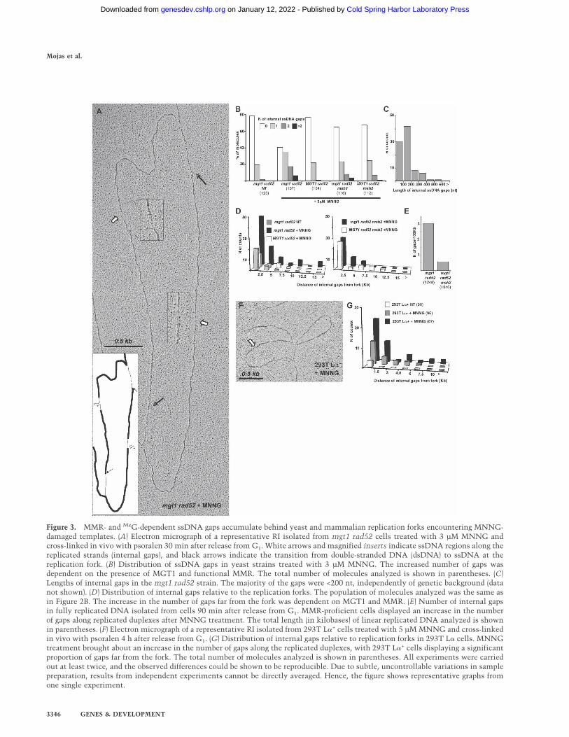
Figure 3. MMR- and MeG-dependent ssDNA gaps accumulate behind yeast and mammalian replication forks encountering MNNG-damaged templates. (A) Electron micrograph of a representative RI isolated from mgt1 rad52 cells treated with 3 µM MNNG andcross-linked in vivo with psoralen 30 min after release from G1. White arrows and magnified inserts indicate ssDNA regions along thereplicated strands (internal gaps), and black arrows indicate the transition from double-stranded DNA (dsDNA) to ssDNA at thereplication fork. (B) Distribution of ssDNA gaps in yeast strains treated with 3 µM MNNG. The increased number of gaps wasdependent on the presence of MGT1 and functional MMR. The total number of molecules analyzed is shown in parentheses. (C)Lengths of internal gaps in the mgt1 rad52 strain. The majority of the gaps were <200 nt, independently of genetic background (datanot shown). (D) Distribution of internal gaps relative to the replication forks. The population of molecules analyzed was the same asin Figure 2B. The increase in the number of gaps far from the fork was dependent on MGT1 and MMR. (E) Number of internal gapsin fully replicated DNA isolated from cells 90 min after release from G1. MMR-proficient cells displayed an increase in the numberof gaps along replicated duplexes after MNNG treatment. The total length (in kilobases) of linear replicated DNA analyzed is shownin parentheses. (F) Electron micrograph of a representative RI isolated from 293T L�+ cells treated with 5 µM MNNG and cross-linkedin vivo with psoralen 4 h after release from G1. (G) Distribution of internal gaps relative to replication forks in 293T L� cells. MNNGtreatment brought about an increase in the number of gaps along the replicated duplexes, with 293T L�+ cells displaying a significantproportion of gaps far from the fork. The total number of molecules analyzed is shown in parentheses. All experiments were carriedout at least twice, and the observed differences could be shown to be reproducible. Due to subtle, uncontrollable variations in samplepreparation, results from independent experiments cannot be directly averaged. Hence, the figure shows representative graphs fromone single experiment.
Mojas et al.
3346 GENES & DEVELOPMENT
Cold Spring Harbor Laboratory Press on January 12, 2022 - Published by genesdev.cshlp.orgDownloaded from
MeG, as the distant gaps were absent from RIs of MNNG-treated MGT1 rad52 cells. Similarly, in RIs of MMR-deficient mgt1 rad52 msh2 cells, the proportion of gapsclose to the fork was comparable with that seen in un-treated cells, but the distant gaps were largely absent. Asanticipated, the effect of MGT1 in the absence of MMRwas substantially lower than in MMR-proficient cells.
The larger number of gaps in close proximity to thefork in MNNG-treated mgt1 rad52 cells suggests thatthe gaps were generated during replication, but that theydid not affect fork progression (Supplementary Figs. 1, 2).To assess the persistence of the gaps, we analyzed DNAof cells allowed to progress for 90 min after release from�-factor and MNNG treatment. At this time point, themajority of cells were in G2 (Fig. 5D, below; data notshown) and their DNA was fully replicated. As shown inFigure 3E, DNA from mgt1 rad52 cells contained ap-proximately five times more ssDNA gaps than that iso-lated from mgt1 rad52 msh2 cells, which showed thatthe persistence of the gaps in replicated duplexes islinked with functional MMR.
This analysis was extended to RIs isolated from hu-man 293T L� cells 4 h after release from HU arrest, whenthe cells were in S phase. The contribution of MMR tothe number and distribution of the ssDNA gaps observedin yeast cells was seen also in human cells, even thoughit was less prominent (Fig. 3F). Upon treatment with 5µM MNNG, the number of gaps in nascent DNA in-
creased significantly in both MMR-proficient (293T L�+)and -deficient (293T L�−) cells, but their number andpersistence farther away from the fork was substantiallyhigher in the former (Fig. 3G).
Taken together, these data indicate that MMR-inde-pendent gaps, which arise most likely through process-ing of lesions other than MeG, are rapidly repaired behindthe progressing replication fork. In contrast, gaps arisingthrough MMR-dependent processing of MeG remain inthe DNA even after the completion of S phase.
MNNG treatment brings about retention of PCNAin chromatin
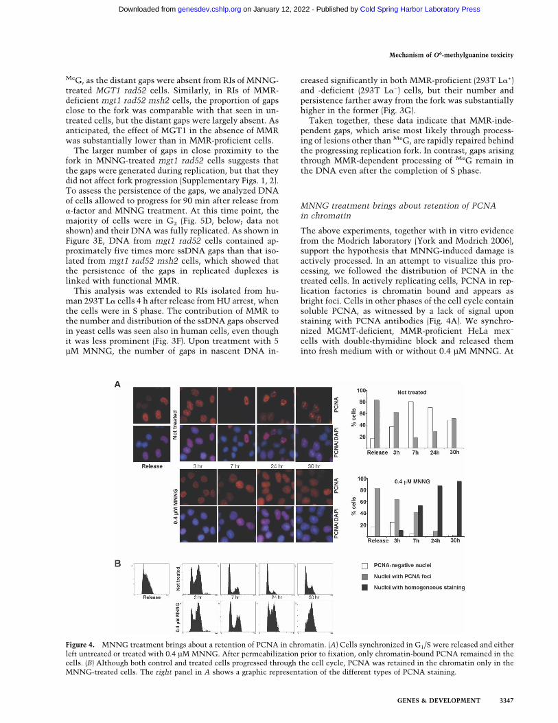
The above experiments, together with in vitro evidencefrom the Modrich laboratory (York and Modrich 2006),support the hypothesis that MNNG-induced damage isactively processed. In an attempt to visualize this pro-cessing, we followed the distribution of PCNA in thetreated cells. In actively replicating cells, PCNA in rep-lication factories is chromatin bound and appears asbright foci. Cells in other phases of the cell cycle containsoluble PCNA, as witnessed by a lack of signal uponstaining with PCNA antibodies (Fig. 4A). We synchro-nized MGMT-deficient, MMR-proficient HeLa mex−
cells with double-thymidine block and released theminto fresh medium with or without 0.4 µM MNNG. At
Figure 4. MNNG treatment brings about a retention of PCNA in chromatin. (A) Cells synchronized in G1/S were released and eitherleft untreated or treated with 0.4 µM MNNG. After permeabilization prior to fixation, only chromatin-bound PCNA remained in thecells. (B) Although both control and treated cells progressed through the cell cycle, PCNA was retained in the chromatin only in theMNNG-treated cells. The right panel in A shows a graphic representation of the different types of PCNA staining.
Mechanism of O6-methylguanine toxicity
GENES & DEVELOPMENT 3347
Cold Spring Harbor Laboratory Press on January 12, 2022 - Published by genesdev.cshlp.orgDownloaded from
the indicated time points, the cells were incubated inpermeabilization buffer (see Materials and Methods)prior to fixation in ice-cold methanol. This procedure,developed for the study of MRE11 (Mirzoeva and Petrini2001), permits the extraction of soluble proteins that arenot bound to chromatin.
The cells were MNNG-treated at the point of releasefrom the double-thymidine block, when the majoritywere in G1/early S phase, and their progress through thecell cycle was monitored by flow cytometry. Three hourslater, both control and MNNG-treated cells were in lateS phase, where 60%–70% of the cells displayed promi-nent PCNA staining, indicative of chromatin-bound pro-tein engaged in DNA replication. Seven hours after re-lease, the majority of both control and treated cells werein the G1 phase of the second cell cycle. In the untreatedsample, only a minor fraction of the cells was PCNA-positive. In contrast, ∼90% of the cells treated withMNNG displayed strong PCNA staining, even thoughcell cycle analysis indicated that the majority was in G1.In most cells, the pattern of PCNA staining was alsosomewhat different from that seen in the unperturbed Sphase. The MNNG-dependent staining was not as in-tense and lacked the focal pattern characteristic of rep-licating cells; rather, the staining was relatively homo-geneous. At the later time points (24 and 30 h after re-lease), the untreated cells reverted to asynchronousprofiles, and the PCNA staining revealed a mixed popu-lation of cells with and without foci, indicative of cellsin different cell cycle phases. In contrast, MNNG-treatedcells were accumulating in the second S phase, suggest-ing that their DNA replication had been impaired. ThePCNA staining of the treated cells was also substantiallydifferent from that of the untreated cells. Specifically, weobserved a fairly intense homogeneous staining in >90%of the cells, and although the cells at these time points(24 and 30 h) were in the phase of the cell cycle thatresembled the point of release as judged by cell cycleanalysis, the staining pattern of chromatin-bound PCNAwas remarkably different. Particularly striking was thelack of foci, characteristic of S phase in untreated cells.Thus, although progression through the first cell cyclewas not significantly affected by MNNG treatment (Fig.4B, Release to 7 h), the persistence of chromatin-boundPCNA is suggestive of DNA processing that is un-coupled from replication. This processing continued intothe second cell cycle, as witnessed by the retention ofdiffuse PCNA staining in chromatin.
Transition of MNNG-treated cells from the firstinto the second cell cycle requires recombination
The persistence of chromatin-bound PCNA staining inMNNG-treated cells suggested that replication-indepen-dent DNA processing was occurring already during thefirst cell cycle after treatment. That MNNG treatmentbrings about activation of HR in bacteria (Nowosielskaet al. 2006), yeast (Cejka et al. 2005), and mammaliancells (Zhang et al. 2000; Tsaryk et al. 2006) has been welldocumented. We wanted to test whether recombination
proficiency affects the sensitivity of cells to this agent.To this end, we studied the MNNG response of twomatched pairs of cell lines: the XRCC2-deficient Irs1cells and their derivative in which the HR defect wascomplemented with XRCC2 cDNA, and the XRCC3-de-ficient Irs1SF cells and their parental, HR-proficient lineAA8. Irs1 cells fail to form RAD51 foci in response toDNA damage and are hypersensitive to a variety of DNAdamaging agents such as the cross-linking agent Mito-mycin C (Jones et al. 1987). Expression of human XRCC2cDNA in these cells restores DNA damage resistanceand RAD51 focus formation (O’Regan et al. 2001). Irs1SFcells are similarly hypersensitive to DNA damagingagents.
As shown in Figure 5A, cell survival after MNNGtreatment was highly dependent on functional HR, asshown recently also for HR-deficient mouse embryonicfibroblasts (MEFs) (Tsaryk et al. 2006). The Irs1 andIrs1SF lines were substantially more sensitive to MNNGthan their recombination-proficient counterparts. Wewanted to learn how HR deficiency affected treatment-dependent cell cycle distribution and arrest. As shown inSupplementary Figure 3, the HR-proficient Irs1/XRCC2and AA8 cells displayed a typical profile of MNNG-treated MMR-proficient cells. They required two DNAreplication cycles to activate the checkpoint and arrestedin G2/M. In contrast, the HR-deficient Irs1 and Irs1SFlines accumulated in G2/M phase already 16 h afterMNNG treatment, a time period during which the cellsreplicated their DNA only once.
To follow the MMR-dependent cell cycle arrest in re-combination-proficient and -deficient cells in more de-tail, we synchronized the Irs1 and Irs1/XRCC2 cells inG1/S and released them into medium with or withoutMNNG (Fig. 5B). Control synchronizations showed thatboth HR-proficient and -deficient cells readily recoveredfrom the HU-induced arrest. Progression of the Irs1/XRCC2 cells followed an already known pattern ofMMR-proficient cells, where they clearly entered thesecond cell cycle after treatment as indicated at 12 and16 h after release by an increase in the G1 fraction. Incontrast, the Irs1 cells were still in G2 at these timepoints, with only a very small proportion entering thesecond cycle. The difference was also apparent at latertime points, where many more Irs1 cells were detectedin the sub-G1 region of the histogram, which is indica-tive of dead cells.
To assess the contribution of HR to cell cycle progres-sion of yeast cells, the recombination-proficient mgt1RAD52 and recombination-impaired mgt1 rad52 strainswere synchronized by �-factor and released, and the cellswere counted for the following 360 min, which corre-sponded to approximately three cell cycles. The recom-bination-proficient mgt1 RAD52 cells were not affectedby MNNG treatment and continued to double theirnumber similarly to the untreated control. In contrast,the recombination-deficient mgt1 rad52 cells doubledonly once in the presence of MNNG, even when thesubstantially slower growth of these cells, linked mostlikely with their reduced ability to repair endogenous
Mojas et al.
3348 GENES & DEVELOPMENT
Cold Spring Harbor Laboratory Press on January 12, 2022 - Published by genesdev.cshlp.orgDownloaded from
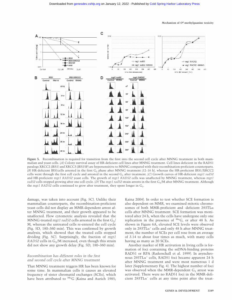
damage, was taken into account (Fig. 5C). Unlike theirmammalian counterparts, the recombination-proficientyeast cells did not display an MMR-dependent arrest af-ter MNNG treatment, and their growth appeared to beunaffected. Flow cytometric analysis revealed that theMNNG-treated mgt1 rad52 cells arrested in the first G2/M, whereas the untreated cells re-entered the cell cycle(Fig. 5D, 180–360 min). This was confirmed by growthanalysis, which showed that the treated cells stoppeddividing (Fig. 5C). Surprisingly, the fraction of mgt1RAD52 cells in G2/M increased, even though this straindid not show any growth delay (Fig. 5D, 180–360 min).
Recombination has different roles in the firstand second cell cycle after MNNG treatment
That MNNG treatment triggers HR has been known forsome time. In mammalian cells it causes an elevatedfrequency of sister chromatid exchanges (SCEs), whichhave been attributed to MeG (Kaina and Aurich 1985;
Kaina 2004). In order to test whether SCE formation isalso dependent on MMR, we examined mitotic chromo-somes of both MMR-proficient and -deficient 293TL�cells after MNNG treatment. SCE formation was moni-tored after 24 h, when the cells have undergone only onereplication in the presence of MeG, or after 48 h. Asshown in Figure 6A, elevated SCE levels were observedonly in 293TL�+ cells and only 48 h after MNNG treat-ment; the number of SCEs per cell rose from an averageof 3.14 to about four times as much, with many cellshaving as many as 20 SCEs.
Another marker of HR activation in living cells is for-mation of foci containing the ssDNA-binding proteinsRAD51 or RPA (Raderschall et al. 1999). In asynchro-nous 293TL�+ cells, RAD51 foci became apparent 24 hafter MNNG treatment and were most numerous 1 dlater (Supplementary Fig. 4). The highest number of fociwas observed when the MMR-dependent G2 arrest wasactivated. There were no RAD51 foci in the MMR-defi-cient 293TL�− cells at any time point after the treat-
Figure 5. Recombination is required for transition from the first into the second cell cycle after MNNG treatment in both mam-malian and yeast cells. (A) Colony survival assay of HR-deficient cell lines after MNNG treatment. Cell lines deficient in the RAD51paralogs XRCC2 (IRS1) and XRCC3 (IRS1SF) are hypersensitive to MNNG compared with their recombination-proficient counterparts.(B) HR-deficient IRS1cells arrested in the first G2 phase after MNNG treatment (12–16 h), whereas the HR-proficient IRS1/XRCC2cells went through the first cell cycle and arrested in the second G2 after treatment. (C) Growth curves of HR-deficient mgt1 rad52and HR-proficient mgt1 RAD52 yeast cells. The growth of mgt1 RAD52 cells was unaffected by MNNG treatment, whereas mgt1rad52 cells stopped growing after one cell cycle. (D) The mgt1 rad52 strain arrests in the first G2/M after MNNG treatment. Althoughthe mgt1 RAD52 cells continued to grow after treatment, they spent longer in G2.
Mechanism of O6-methylguanine toxicity
GENES & DEVELOPMENT 3349
Cold Spring Harbor Laboratory Press on January 12, 2022 - Published by genesdev.cshlp.orgDownloaded from
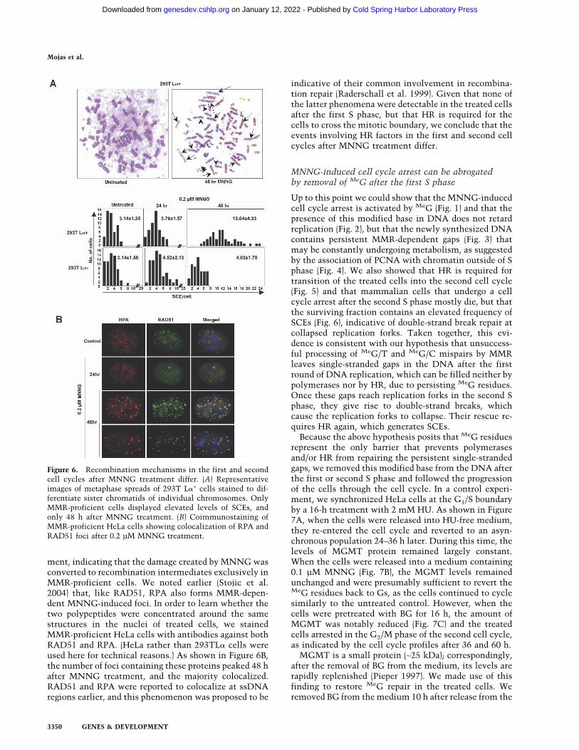
ment, indicating that the damage created by MNNG wasconverted to recombination intermediates exclusively inMMR-proficient cells. We noted earlier (Stojic et al.2004) that, like RAD51, RPA also forms MMR-depen-dent MNNG-induced foci. In order to learn whether thetwo polypeptides were concentrated around the samestructures in the nuclei of treated cells, we stainedMMR-proficient HeLa cells with antibodies against bothRAD51 and RPA. (HeLa rather than 293TL� cells wereused here for technical reasons.) As shown in Figure 6B,the number of foci containing these proteins peaked 48 hafter MNNG treatment, and the majority colocalized.RAD51 and RPA were reported to colocalize at ssDNAregions earlier, and this phenomenon was proposed to be
indicative of their common involvement in recombina-tion repair (Raderschall et al. 1999). Given that none ofthe latter phenomena were detectable in the treated cellsafter the first S phase, but that HR is required for thecells to cross the mitotic boundary, we conclude that theevents involving HR factors in the first and second cellcycles after MNNG treatment differ.
MNNG-induced cell cycle arrest can be abrogatedby removal of MeG after the first S phase
Up to this point we could show that the MNNG-inducedcell cycle arrest is activated by MeG (Fig. 1) and that thepresence of this modified base in DNA does not retardreplication (Fig. 2), but that the newly synthesized DNAcontains persistent MMR-dependent gaps (Fig. 3) thatmay be constantly undergoing metabolism, as suggestedby the association of PCNA with chromatin outside of Sphase (Fig. 4). We also showed that HR is required fortransition of the treated cells into the second cell cycle(Fig. 5) and that mammalian cells that undergo a cellcycle arrest after the second S phase mostly die, but thatthe surviving fraction contains an elevated frequency ofSCEs (Fig. 6), indicative of double-strand break repair atcollapsed replication forks. Taken together, this evi-dence is consistent with our hypothesis that unsuccess-ful processing of MeG/T and MeG/C mispairs by MMRleaves single-stranded gaps in the DNA after the firstround of DNA replication, which can be filled neither bypolymerases nor by HR, due to persisting MeG residues.Once these gaps reach replication forks in the second Sphase, they give rise to double-strand breaks, whichcause the replication forks to collapse. Their rescue re-quires HR again, which generates SCEs.
Because the above hypothesis posits that MeG residuesrepresent the only barrier that prevents polymerasesand/or HR from repairing the persistent single-strandedgaps, we removed this modified base from the DNA afterthe first or second S phase and followed the progressionof the cells through the cell cycle. In a control experi-ment, we synchronized HeLa cells at the G1/S boundaryby a 16-h treatment with 2 mM HU. As shown in Figure7A, when the cells were released into HU-free medium,they re-entered the cell cycle and reverted to an asyn-chronous population 24–36 h later. During this time, thelevels of MGMT protein remained largely constant.When the cells were released into a medium containing0.1 µM MNNG (Fig. 7B), the MGMT levels remainedunchanged and were presumably sufficient to revert theMeG residues back to Gs, as the cells continued to cyclesimilarly to the untreated control. However, when thecells were pretreated with BG for 16 h, the amount ofMGMT was notably reduced (Fig. 7C) and the treatedcells arrested in the G2/M phase of the second cell cycle,as indicated by the cell cycle profiles after 36 and 60 h.
MGMT is a small protein (∼25 kDa); correspondingly,after the removal of BG from the medium, its levels arerapidly replenished (Pieper 1997). We made use of thisfinding to restore MeG repair in the treated cells. Weremoved BG from the medium 10 h after release from the
Figure 6. Recombination mechanisms in the first and secondcell cycles after MNNG treatment differ. (A) Representativeimages of metaphase spreads of 293T L�+ cells stained to dif-ferentiate sister chromatids of individual chromosomes. OnlyMMR-proficient cells displayed elevated levels of SCEs, andonly 48 h after MNNG treatment. (B) Coimmunostaining ofMMR-proficient HeLa cells showing colocalization of RPA andRAD51 foci after 0.2 µM MNNG treatment.
Mojas et al.
3350 GENES & DEVELOPMENT
Cold Spring Harbor Laboratory Press on January 12, 2022 - Published by genesdev.cshlp.orgDownloaded from

HU block, when the cells passed the first S phase, buthave not yet entered the second cell cycle. As shown inFigure 7A, the period between BG removal (10 h) and thetime point when the arrest normally became effective(36 h) allowed for the recovery of normal MGMT levelsand, presumably, MeG repair. Importantly, the cells re-turned to a normal cycling profile (see 60-h time point).When BG was removed from the medium 36 h after re-lease from HU—i.e., when the checkpoint had alreadybeen activated (Fig. 7A)—the cells remained arrested.This experiment shows that MeG is the only lesion in theDNA of MNNG-treated cells that must persist in order
for the checkpoint to be activated. But the experimentalso shows that MeG does not signal to the checkpointmachinery directly; the checkpoint would be activatedsooner if it did. Most importantly, the data show that thelesion(s) triggering the checkpoint can be repaired rap-idly and efficiently once MeG is removed from DNA.This would argue more in favor of single-stranded gapsthan complex recombination intermediates.
Discussion
The cytotoxicity of SN1-type methylating agents hasbeen unambiguously linked with the modification ofguanine on the O6 position. However, how MeG bringsabout cell death has been subject to discussion. The so-called “signaling hypothesis” suggested that MeG-con-taining mispairs arising during the first DNA replicationare bound by MutS� and that this is sufficient to activatecell cycle arrest (Fishel 1998; Yoshioka et al. 2006). The“processing hypothesis” posits that it is the futile pro-cessing of these mispairs by MMR that gives rise to DNAintermediates, which then signal to the checkpoint ma-chinery (Karran and Bignami 1996). In the present study,we show that the mere presence of MeG in the genomicDNA of MMR-proficient cells does not bring about cellkilling, even if the cells are allowed to undergo oneround of replication, during which MeG/C and MeG/Tpairs can form; to activate the checkpoint, these mis-pairs must be processed.
The finding that MSH3 and MSH6 carry PCNA-bind-ing motifs and interact with this polymerase processiv-ity factor in vitro and in vivo implied that MMR is inti-mately linked with the replication machinery (Clark etal. 2000; Flores-Rozas et al. 2000; Kleczkowska et al.2001). The ability of MMR factors to halt the progress ofthe replicating polymerase upon mismatch detectionmight liberate the 3� end of the newly synthesized strandand thus provide the excision machinery with an entrypoint, where degradation of the error-containing strandcould commence. In this scenario, iterative attempts ofthe replicative polymerase to find a perfect match forMeG would be expected to stall, or at least slow down,the progression of the replication fork. DNA synthesisacross MeG has indeed been shown to retard replicativepolymerases in vitro (Haracska et al. 2000; York andModrich 2006); however, neither the early experiments(Plant and Roberts 1971) nor our measurements of grossreplication rates in mammalian cells (Fig. 2), nor exami-nation of the progression of replication forks from a de-fined origin in yeast cells (Supplementary Fig. 1), de-tected a MMR-dependent reduction of replication ratesin the first S phase after MNNG treatment. This sug-gests that MMR acting on MeG is a post-replicative pro-cess, as shown recently for translesion synthesis (Lopeset al. 2006).
Futile attempts of DNA polymerase(s) to incorporatethe correct nucleotide opposite MeG were proposed toresult in the generation of ssDNA gaps, the formation ofwhich has recently been inferred in biochemical experi-ments (York and Modrich 2006). Our EM analysis of RIs
Figure 7. MNNG-activated cell cycle arrest requires the per-sistence of MeG in DNA through two cell cycles. Cells weresynchronized with 2 mM HU and released into fresh medium(A) or medium containing 0.1 µM MNNG (B–E). (C) Cells incu-bated with BG arrested at G2/M two cell cycles after releasefrom HU. (D) When BG was removed 10 h after release (whencells passed the first S phase), the cells continued to proliferatewithout arresting. (E) In contrast, if BG was removed at thepoint when the checkpoint was already activated, the cells re-mained arrested.
Mechanism of O6-methylguanine toxicity
GENES & DEVELOPMENT 3351
Cold Spring Harbor Laboratory Press on January 12, 2022 - Published by genesdev.cshlp.orgDownloaded from
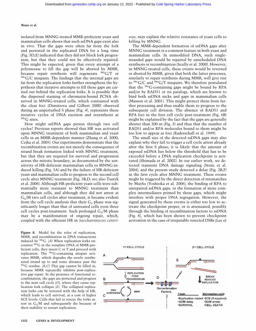
isolated from MNNG-treated MMR-proficient yeast andmammalian cells shows that such ssDNA gaps exist alsoin vivo. That the gaps were often far from the forkand persisted in the replicated DNA for a long time(Fig. 3D,E) indicated that they did not block fork progres-sion, but that they could not be effectively repaired.This might be expected, given that every attempt of apolymerase to fill the gap will be aborted by MMR,because repair synthesis will regenerate MeG/T orMeG/C mispairs. The findings that the internal gaps arefar from the replication forks further strengthens the hy-pothesis that iterative attempts to fill these gaps are car-ried out behind the replication forks. It is possible thatthe dispersed staining of chromatin-bound PCNA ob-served in MNNG-treated cells, which contrasted withthe clear foci (Dimitrova and Gilbert 2000) observedduring an unperturbed S phase (Fig. 4), represents theseiterative cycles of DNA excision and resynthesis atMeG sites.
How might ssDNA gaps persist through two cellcycles? Previous reports showed that HR was activatedupon MNNG treatment of both mammalian and yeastcells in an MMR-dependent manner (Zhang et al. 2000;Cejka et al. 2005). Our experiments demonstrate that therecombination events are not merely the consequence ofstrand break formation linked with MNNG treatment,but that they are required for survival and progressionacross the mitotic boundary, as documented by the sen-sitivity of HR-deficient mammalian cells to MNNG-in-duced killing (Fig. 5A) and by the failure of HR-deficientyeast and mammalian cells to progress to the second cellcycle after MNNG treatment (Fig. 5B,D; see also Tsaryket al. 2006). Although HR-proficient yeast cells were sub-stantially more resistant to MNNG treatment thanmammalian cells, and although they did not arrest atG2/M two cell cycles after treatment, it became evidentfrom the cell cycle analysis that their G2 phase was sig-nificantly longer than that of untreated cells even threecell cycles post-treatment. Such prolonged G2/M phasemay be a manifestation of ongoing repair, which,coupled with the efficient HR in Saccharomyces cerevi-
siae, may explain the relative resistance of yeast cells tokilling by MNNG.
The MMR-dependent formation of ssDNA gaps afterMNNG treatment is a common feature in both yeast andmammalian cells. In unmodified DNA, such single-stranded gaps would be repaired by unscheduled DNAsynthesis or recombination (Scully et al. 2000). However,in MNNG-treated cells, these events would be reversedor aborted by MMR, given that both the latter processes,similarly to repair synthesis during MMR, will give riseto MeG/C and MeG/T mispairs. We therefore postulatedthat the MeG-containing gaps might be bound by RPAand/or by RAD51 or its paralogs, which are known tobind both ssDNA nicks and gaps in mammalian cells(Masson et al. 2001). This might protect them from fur-ther processing and thus enable them to progress to thesubsequent cell division. The absence of RAD51 andRPA foci in the first cell cycle post-treatment (Fig. 6B)might be explained by the fact that the gaps are generallyshorter than 200 nt (Fig. 3) and thus that the number ofRAD51 and/or RPA molecules bound to them might betoo low to appear as foci (Raderschall et al. 1999).
The small size of the detected ssDNA gaps may alsoexplain why they fail to trigger a cell cycle arrest alreadyafter the first S phase; it is likely that the amount ofexposed ssDNA lies below the threshold that has to beexceeded before a DNA replication checkpoint is acti-vated (Shimada et al. 2002). In our earlier work, we de-tected transient DNA damage signaling (Stojic et al.2004), and the present study detected a delay (Fig. 2B,F)in the first cycle after MNNG treatment. These eventsmight be triggered by the direct detection of mismatchesby MutS� (Yoshioka et al. 2006), the binding of RPA tounrepaired ssDNA gaps, or the formation of more com-plex intermediates primed by these gaps, which mightinterfere with proper DNA segregation. However, thesignal generated by these events is either too low to ac-tivate the checkpoint proper, or is attenuated, possiblythrough the binding of recombination factors to ssDNA(Fig. 8), which has been shown to prevent checkpointactivation in the case of irreparable resected DSBs (Lee et
Figure 8. Model for the roles of replication,MMR, and recombination in DNA transactionsinduced by MeG. (A) When replication forks en-counter MeG in the template DNA of MMR-pro-ficient cells, they insert C or T and proceed withreplication. The MeG-containing mispair acti-vates MMR, which degrades the newly synthe-sized strand up to and some distance past theMeG residue. (B,C) This gap cannot be filled in,because MMR repeatedly inhibits post-replica-tive gap repair. In the presence of functional re-combination, the gaps are protected and progressto the next cell cycle (D), where they cause rep-lication fork collapse (E). The collapsed replica-tion forks can be restored with the help of HR,which leads to cell survival, at a cost of higherSCE levels. Cells that fail to rescue the forks ar-rest in G2/M and subsequently die because oftheir inability to restart replication.
Mojas et al.
3352 GENES & DEVELOPMENT
Cold Spring Harbor Laboratory Press on January 12, 2022 - Published by genesdev.cshlp.orgDownloaded from
al. 2003). Only during the second replication would theseprimary lesions (ssDNA gaps and/or structures derivedfrom them) give rise to full checkpoint activation, cellcycle arrest, and cell death.
We cannot formally demonstrate that these gaps per-sist through mitosis. However, their persistence untilthe second round of replication is supported by threelines of evidence. First, gaps in template DNA shouldbring about replication fork collapse, which is consistentwith our earlier findings showing that MNNG inducesan ATR-dependent G2 arrest after the second S phase(Stojic et al. 2004). Second, when replication forks col-lapse as a result of a double-strand break, the ends of thechromatid are resected to give rise to long ssDNA fila-ments, which are bound by many RAD51 and/or RPAmolecules. This agrees with the appearance of large fociof these two proteins after the second S phase (Fig. 6B).Third, collapsed replication forks give rise to two linearDNA molecules (Fig. 8E), which would not be isolated asRIs by our experimental procedure. Indeed, analysis ofRIs isolated from the second cell cycle of MNNG-treatedhuman cells failed to reveal the presence of abnormalDNA structures (data not shown). Taken together, thisevidence lends support to the hypothesis that the MMRand MNNG dose-dependent reduction of DNA synthesisrates in the second cell cycle (Fig. 2) is indeed linked toreplication fork collapse caused by persistent gaps intemplate DNA.
In wild-type yeast cells, the high efficiency of HR over-comes the cytotoxic effects of MNNG treatment, andthe only consequence of the damage is a delay in G2/M.In contrast, mammalian cells cannot cope with the highnumber of collapsed replication forks (Figs. 2, 8), and thesmall proportion of cells that recover from the G2/Marrest in the second cell cycle display elevated levels ofSCEs (Kaina et al. 1997), which are strictly dependent onMMR (Fig. 6B). While SCEs are considered to arisethrough HR-mediated processing of stalled or collapsedreplication forks (Richardson et al. 1998; Johnson andJasin 2000), recombination-dependent processing ofssDNA gaps, proposed to result in the formation ofdouble Holliday junctions, does not give rise to SCEs(Helleday 2003). Given that SCEs were observed onlyafter the second S phase in the 293T La+ cells providesfurther support for the hypothesis that MNNG treat-ment of MGMT-deficient cells leads to the MMR-depen-dent generation of persistent single-stranded gaps innewly replicated DNA.
In cells expressing low amounts of MGMT, such astumors in which the gene is epigenetically silenced (Ja-cinto and Esteller 2007), the cytotoxicity of SN1-typemethylating agents was linked with the persistence ofMeG until DNA replication, where unsuccessful at-tempts of the MMR system to correct MeG/C and MeG/Tmispairs lead to cell death (Karran 2001). In our previousstudy, we postulated that unsuccessful MMR leaves ir-reparable ssDNA gaps in the DNA after the first S phase,which give rise to double-strand breaks during the sub-sequent replication (Stojic et al. 2004). In the presentstudy, we substantiate this hypothesis experimentally
by demonstrating the existence of these gaps. Moreover,we show that HR permits the cells with incompletelyrepaired DNA to enter the subsequent cell cycle, wherethese discontinuities cause replication fork collapse andcell cycle arrest. Although most mammalian cells neverrecover from this block, some are rescued by HR-medi-ated repair of the collapsed replication forks, which givesrise to SCEs. Finally, the ready abrogation of the cellcycle arrest brought about by re-expression of MGMTafter the first S phase argues against the existence ofcomplex irreparable recombination intermediates aris-ing after DNA replication; rather, it shows that MeG rep-resents the only hurdle to the repair or resolution ofthese structures. In an attempt to provide experimentalsupport for this hypothesis, we allowed the MNNG-treated cells to re-express MGMT before the second Sphase and thus remove these modifications. As shown inFigure 7D, this abolished the G2/M arrest, and the cellscontinued to cycle. The most likely explanation for thisfinding is that removal of MeG from the gaps or recom-bination intermediates permits their immediate repair.
SN1-type methylating agents are in widespread use incancer chemotherapy. By elucidating their mode of ac-tion, and in particular by demonstrating the requirementfor HR in the processing of DNA damage induced bythese agents, our work implies that inhibition of HRwould substantially increase the efficacy of this impor-tant class of chemotherapeutics.
Materials and methods
Cell lines and chemicals
The 293T L� cell line was established in our laboratory and waspropagated as described (Cejka et al. 2003). The HeLa and He-LaMR (a kind gift of Margherita Bignami) cells were maintainedin DMEM (OmniLab) supplemented with 10% fetal calf serum(FCS; Life Technologies), penicillin (100 U/mL), and streptomy-cin (100 µg/mL). The HR-deficient Chinese hamster cell lineIRS1 was mutated in XRCC2; the defect was corrected in theIRS1/XRCC2 line by a stable transfection with human XRCC2cDNA. Both lines were kindly provided by John Thacker. TheXRCC3-deficient cells and their HR-proficient parental cell lineAA8 were kindly provided by Orlando Schärer. All four lineswere maintained in DMEM:F10 (1:1; OmniLab), supplementedwith 10% FCS. To inhibit MGMT activity, the cells were pre-treated with 10 µM BG. MNNG, BG, HU, and 6TG were pur-chased from Sigma, and Dox was purchased from Clontech.
The yeast strains used in this study were described previously(Cejka et al. 2005). They were grown in YPD at 25°C.
Cell synchronization and cell cycle analysis
The yeast cells were synchronized in G1 phase by adding 2 µg/mL �-factor and, after centrifugation, were released into a me-dium with or without 3 µM MNNG.
The mammalian cells were synchronized with 2 mM HU for16 h. They were washed twice with PBS and incubated in freshmedium with or without MNNG and BG. At the indicated timepoints, cells were trypsinized and processed for cell cycle analy-ses as described previously (Stojic et al. 2004). Cell cycle analy-ses were performed using a Beckman Coulter FC 500 cytometer.
Mechanism of O6-methylguanine toxicity
GENES & DEVELOPMENT 3353
Cold Spring Harbor Laboratory Press on January 12, 2022 - Published by genesdev.cshlp.orgDownloaded from
Antibodies and immunoblotting
Immunoblotting and total protein extractions were performedas described previously (Cejka et al. 2003). The anti-�-tubulin(D-10) antibody was from Santa Cruz Biotechnology, and theanti-MGMT antibody (Clone MT 23.2) was from GeneTex.
Replication rates
The cells were plated and allowed to attach for 24 h. [14C] Thy-midine [20 nCi/mL] was added, and the cells were allowed togrow for an additional 24 h. After washing with PBS, the cellswere incubated in growth medium for 10 h, and 2 mM HU wasadded for an additional 16 h. At this time point (Release), thecells were washed with PBS and incubated with normal growthmedium without (Control) or with MNNG or cisplatin. Fifteenminutes before the indicated time points, 2.5 µCi/mL [3H] thy-midine was added. One-fifth of each sample was processed forcell cycle analysis, and the rest was processed for scintillationcounting. The cells were collected by centrifugation, the pelletswere resuspended in 280 µL of ice-cold PBS, and 720 µL of ice-cold methanol were added dropwise. The cells were pelleted andresuspended in 200 µL of PBS, and 5 mL of scintillation liquid(Optiphase “Hisafe” 2; PerkinElmer) were added. The sampleswere incubated overnight at +4°C before scintillation counting.DNA synthesis was estimated as the ratio of [3H]/[14C].
EM analysis of total genomic DNA
In vivo psoralen cross-linking, isolation of total genomic DNA,and enrichment of the RIs from yeast cells were performed asdescribed previously (Lopes et al. 2006). For EM analysis of themammalian RIs, 293T L� cells were seeded and synchronizedwith 2 mM HU as described above. The cells were released intofresh medium without (Not Treated, NT) or with 5 µM MNNGand were collected by trypsinization after 4 h. At this timepoint, the majority of the cells were in mid-S phase, as assessedby cell cycle analysis (Fig. 2; data not shown). Approximately107 cells were resuspended in 10 mL of ice-cold PBS and trans-ferred to a 6-cm dish to which 10 µg/mL trimethylpsoralen(TMP) were added for 5 min in the dark. The cells were irradi-ated with 366-nm monochromatic light for 5 min, at a distanceof ∼1 cm from the lamps (Stratalinker; Stratagene). The TMPaddition in the dark and irradiation were repeated three moretimes. Subsequently, the cells were spun down, and genomicDNA was isolated using the QIAamp DNA blood minikit (Qia-gen), according to the manufacturer’s instructions. The DNAwas digested with PvuII, and RIs were enriched using the sameprocedure as for the yeast DNA.
Immunofluorescence staining
For PCNA staining with permabilization before fixation, cellsgrown on glass coverslips were synchronized with double-thy-midine block as described (Stojic et al. 2004), treated or mock-treated with MNNG, and incubated for the indicated times. Theslides were immersed for 5 min. in ice-cold permeabilizationbuffer (10 mM Tris at pH 7.6, 2.5 mM MgCl2, 0.5% NP-40, 0.2mM PMSF) before fixation in ice-cold methanol. This treatmentremoves all soluble proteins from the nucleoplasm, while thechromatin-bound fraction is retained. The slides were subse-quently stained with an anti-PCNA antibody (PC-10; SantaCruz Biotechnology). Cells stained with RAD51 (rabbit anti-body; Pharmingen) and RPA (mouse anti-p32; Oncogene) anti-bodies were fixed in 3.7% formaldehyde and permeabilized with0.2% Triton-X, and the incubation with antibodies was carried
out as for PCNA. After washing, the cells were incubated at37°C with TR-conjugated anti-mouse antibody (Abcam) andanti-rabbit FITC (Sigma). Cell nuclei were counterstained withDAPI and mounted in anti-fade (Molecular Probes). Images wereacquired on Olympus IX81 microscope, equipped with appro-priate filter sets and using CellR imaging software.
SCE assay
To differentially label sister chromatids, the cells were incu-bated for 48 h with 10 µM BrdU (two cell cycles). Assessing theeffect of MNNG was accomplished by the addition of 0.2 µMMNNG 24 or 48 h before harvesting. Preparation of the mitoticspreads and subsequent staining were carried out as described(Kaina and Aurich 1985). The number of SCE events shown inFigure 6A was counted in 50 metaphases.
Clonogenic survival assay
The cells were treated with the indicated concentrations ofMNNG for 2 h, trypsinized, and counted. Serial dilutions ofcontrol and treated cells were plated and left for 14–20 d, afterwhich the colonies were stained with 10% Giemsa and counted.
Acknowledgments
We thank Lovorka Stojic and Petr Cejka for helpful discussions,and Margherita Bignami, Orlando Schärer, and John Thacker forthe kind gift of the cell lines. We are grateful to the ElectronMicroscopy Center of the ETH Zürich for technical support. Wealso gratefully acknowledge the financial support of the Bonizzi-Theler Stiftung, the European Community, and the Swiss Na-tional Science Foundation.
References
Berardini, M., Mazurek, A., and Fishel, R. 2000. The effect ofO6-methylguanine DNA adducts on the adenosine nucleo-tide switch functions of hMSH2–hMSH6 and hMSH2–hMSH3. J. Biol. Chem. 275: 27851–27857.
Cejka, P., Stojic, L., Mojas, N., Russell, A.M., Heinimann, K.,Cannavo, E., di Pietro, M., Marra, G., and Jiricny, J. 2003.Methylation-induced G(2)/M arrest requires a full comple-ment of the mismatch repair protein hMLH1. EMBO J. 22:2245–2254.
Cejka, P., Mojas, N., Gillet, L., Schar, P., and Jiricny, J. 2005.Homologous recombination rescues mismatch-repair-de-pendent cytotoxicity of S(N)1-type methylating agents in S.cerevisiae. Curr. Biol. 15: 1395–1400.
Clark, A.B., Valle, F., Drotschmann, K., Gary, R.K., and Kunkel,T.A. 2000. Functional interaction of proliferating cellnuclear antigen with MSH2–MSH6 and MSH2–MSH3 com-plexes. J. Biol. Chem. 275: 36498–36501.
Dimitrova, D.S. and Gilbert, D.M. 2000. Temporally coordi-nated assembly and disassembly of replication factories inthe absence of DNA synthesis. Nat. Cell Biol. 2: 686–694.
Dolan, M.E., Roy, S.K., Garbiras, B.J., Helft, P., Paras, P., Chae,M.-Y., Moschel, R.C., and Pegg, A.E. 1998. O6-Alkylguanine-DNA alkyltransferase inactivation by ester prodrugs of O6-benzylguanine derivatives and their rate of hydrolysis by cel-lular esterases. Biochem. Pharmacol. 55: 1701–1709.
Duckett, D.R., Drummond, J.T., Murchie, A.I.H., Reardon, J.T.,Sancar, A., Lilley, D.M.J., and Modrich, P. 1996. HumanMutS� recognizes damaged DNA base pairs containing O6-methylguanine, O4-methylthymine, or the cisplatin-d(GpG)
Mojas et al.
3354 GENES & DEVELOPMENT
Cold Spring Harbor Laboratory Press on January 12, 2022 - Published by genesdev.cshlp.orgDownloaded from
adduct. Proc. Natl. Acad. Sci. 93: 6443–6447.Fishel, R. 1998. Mismatch repair, molecular switches, and sig-
nal transduction. Genes & Dev. 12: 2096–2101.Flores-Rozas, H., Clark, D., and Kolodner, R.D. 2000. Prolifer-
ating cell nuclear antigen and Msh2p–Msh6p interact toform an active mispair recognition complex. Nat. Genet. 26:375–378.
Haracska, L., Prakash, S., and Prakash, L. 2000. Replication pastO6-methylguanine by yeast and human DNA polymerase �.Mol. Cell. Biol. 20: 8001–8007.
Helleday, T. 2003. Pathways for mitotic homologous recombi-nation in mammalian cells. Mutat. Res. 532: 103–115.
Henry-Mowatt, J., Jackson, D., Masson, J.Y., Johnson, P.A.,Clements, P.M., Benson, F.E., Thompson, L.H., Takeda, S.,West, S.C., and Caldecott, K.W. 2003. XRCC3 and Rad51modulate replication fork progression on damaged verte-brate chromosomes. Mol. Cell 11: 1109–1117.
Jacinto, F.V. and Esteller, M. 2007. MGMT hypermethylation:A prognostic foe, a predictive friend. DNA Repair (Amst.) 6:1155–1160.
Johnson, R.D. and Jasin, M. 2000. Sister chromatid gene con-version is a prominent double-strand break repair pathway inmammalian cells. EMBO J. 19: 3398–3407.
Jones, N.J., Cox, R., and Thacker, J. 1987. Isolation and cross-sensitivity of X-ray-sensitive mutants of V79-4 hamstercells. Mutat. Res./DNA Repair Reports 183: 279–286.
Kaina, B. 2004. Mechanisms and consequences of methylatingagent-induced SCEs and chromosomal aberrations: A longroad traveled and still a far way to go. Cytogenet. GenomeRes. 104: 77–86.
Kaina, B. and Aurich, O. 1985. Dependency of the yield of sister-chromatid exchanges induced by alkylating agents on fixa-tion time: Possible involvement of secondary lesions insister-chromatid exchange induction. Mutat. Res. 149:451–461.
Kaina, B., Ziouta, A., Ochs, K., and Coquerelle, T. 1997. Chro-mosomal instability, reproductive cell death and apoptosisinduced by O6-methylguanine in Mex−, Mex+ and methyl-ation-tolerant mismatch repair compromised cells: Factsand models. Mutat. Res. 381: 227–241.
Karran, P. 2001. Mechanisms of tolerance to DNA damagingtherapeutic drugs. Carcinogenesis 22: 1931–1937.
Karran, P. 2006. Thiopurines, DNA damage, DNA repair andtherapy-related cancer. Br. Med. Bull. 79-80: 153–170.
Karran, P. and Bignami, M. 1996. Drug-related killings: A case ofmistaken identity. Chem. Biol. 3: 875–879.
Kleczkowska, H.E., Marra, G., Lettieri, T., and Jiricny, J. 2001.hMSH3 and hMSH6 interact with PCNA and colocalizewith it to replication foci. Genes & Dev. 15: 724–736.
Lee, S.E., Pellicioli, A., Vaze, M.B., Sugawara, N., Malkova, A.,Foiani, M., and Haber, J.E. 2003. Yeast Rad52 and Rad51recombination proteins define a second pathway of DNAdamage assessment in response to a single double-strandbreak. Mol. Cell. Biol. 23: 8913–8923.
Lopes, M., Foiani, M., and Sogo, J.M. 2006. Multiple mecha-nisms control chromosome integrity after replication forkuncoupling and restart at irreparable UV lesions. Mol. Cell21: 15–27.
Masson, J.Y., Tarsounas, M.C., Stasiak, A.Z., Stasiak, A., Shah,R., McIlwraith, M.J., Benson, F.E., and West, S.C. 2001. Iden-tification and purification of two distinct complexes con-taining the five RAD51 paralogs. Genes & Dev. 15: 3296–3307.
Mirzoeva, O.K. and Petrini, J.H.J. 2001. DNA damage-depen-dent nuclear dynamics of the Mre11 complex. Mol. Cell.Biol. 21: 281–288.
Nowosielska, A., Smith, S.A., Engelward, B.P., and Marinus,M.G. 2006. Homologous recombination prevents methyl-ation-induced toxicity in Escherichia coli. Nucleic AcidsRes. 34: 2258–2268.
O’Regan, P., Wilson, C., Townsend, S., and Thacker, J. 2001.XRCC2 is a nuclear RAD51-like protein required for dam-age-dependent RAD51 focus formation without the need forATP binding. J. Biol. Chem. 276: 22148–22153.
Pieper, R.O. 1997. Understanding and manipulating O6-meth-ylguanine-DNA methyltransferase expression. Pharmacol.Ther. 74: 285–297.
Plant, J.E. and Roberts, J.J. 1971. A novel mechanism for theinhibition of DNA synthesis following methylation: The ef-fect of N-methyl-N-nitrosourea on HeLa cells. Chem. Biol.Interact. 3: 337–342.
Raderschall, E., Golub, E.I., and Haaf, T. 1999. Nuclear foci ofmammalian recombination proteins are located at single-stranded DNA regions formed after DNA damage. Proc.Natl. Acad. Sci. 96: 1921–1926.
Richardson, C., Moynahan, M.E., and Jasin, M. 1998. Double-strand break repair by interchromosomal recombination:Suppression of chromosomal translocations. Genes & Dev.12: 3831–3842.
Scully, R., Puget, N., and Vlasakova, K. 2000. DNA polymerasestalling, sister chromatid recombination and the BRCAgenes. Oncogene 19: 6176–6183.
Shimada, K., Pasero, P., and Gasser, S.M. 2002. ORC and theintra-S-phase checkpoint: A threshold regulates Rad53p ac-tivation in S phase. Genes & Dev. 16: 3236–3252.
Stojic, L., Mojas, N., Cejka, P., Di Pietro, M., Ferrari, S., Marra,G., and Jiricny, J. 2004. Mismatch repair-dependent G2checkpoint induced by low doses of SN1 type methylatingagents requires the ATR kinase. Genes & Dev. 18: 1331–1344.
Tsaryk, R., Fabian, K., Thacker, J., and Kaina, B. 2006. Xrcc2deficiency sensitizes cells to apoptosis by MNNG and thealkylating anticancer drugs temozolomide, fotemustine andmafosfamide. Cancer Lett. 239: 305–313.
Wyatt, M.D. and Pittman, D.L. 2006. Methylating agents andDNA repair responses: Methylated bases and sources ofstrand breaks. Chem. Res. Toxicol. 19: 1580–1594.
Yang, G., Scherer, S.J., Shell, S.S., Yang, K., Kim, M., Lipkin, M.,Kucherlapati, R., Kolodner, R.D., and Edelmann, W. 2004.Dominant effects of an Msh6 missense mutation on DNArepair and cancer susceptibility. Cancer Cell 6: 139–150.
York, S.J. and Modrich, P. 2006. Mismatch repair-dependentiterative excision at irreparable O6-methylguanine lesions inhuman nuclear extracts. J. Biol. Chem. 281: 22674–22683.
Yoshioka, K.-I., Yoshioka, Y., and Hsieh, P. 2006. ATR kinaseactivation mediated by MutS� and MutL� in response tocytotoxic O6-methylguanine adducts. Mol. Cell 22: 501–510.
Zhang, H., Marra, G., Jiricny, J., Maher, V.M., and McCormick,J.J. 2000. Mismatch repair is required for O6-methylguanine-induced homologous recombination in human fibroblasts.Carcinogenesis 21: 1639–1646.
Zhukovskaya, N., Branch, P., Aquilina, G., and Karran, P. 1994.DNA replication arrest and tolerance to DNA methylationdamage. Carcinogenesis 15: 2189–2194.
Mechanism of O6-methylguanine toxicity
GENES & DEVELOPMENT 3355
Cold Spring Harbor Laboratory Press on January 12, 2022 - Published by genesdev.cshlp.orgDownloaded from

10.1101/gad.455407Access the most recent version at doi: 21:2007, Genes Dev.
Nina Mojas, Massimo Lopes and Josef Jiricny rise to persistent single-stranded gaps in newly replicated DNAMismatch repair-dependent processing of methylation damage gives
Material
Supplemental
http://genesdev.cshlp.org/content/suppl/2007/11/28/21.24.3342.DC1
References
http://genesdev.cshlp.org/content/21/24/3342.full.html#ref-list-1
This article cites 42 articles, 16 of which can be accessed free at:
License
ServiceEmail Alerting
click here.right corner of the article or
Receive free email alerts when new articles cite this article - sign up in the box at the top
Copyright © 2007, Cold Spring Harbor Laboratory Press
Cold Spring Harbor Laboratory Press on January 12, 2022 - Published by genesdev.cshlp.orgDownloaded from