A critical re-assessment of DNA repair gene promoter methylation in non-small cell lung carcinoma
8
A critical re-assessment of DNA repair gene promoter methylation in non-small cell lung carcinoma Hongdo Do 1,2,6 , Nicholas C. Wong 2,6 , Carmel Murone 2 , Thomas John 3 , Benjamin Solomon 4,5 , Paul L. Mitchell 3 & Alexander Dobrovic 1,2,4,5,6 1 Molecular Pathology Research and Development Laboratory, Department of Pathology, Peter MacCallum Cancer Centre, Melbourne, Victoria, 8006, Australia, 2 Translational Genomics and Epigenomics Laboratory, Ludwig Institute for Medical Research, Olivia Newton-John Cancer and Wellness Centre, Austin Hospital, Heidelberg, Victoria, 3084, Australia, 3 Joint Austin-Ludwig Medical Oncology Department, Olivia Newton-John Cancer and Wellness Centre, Austin Hospital, Heidelberg, Victoria, 3084, Australia, 4 Sir Peter MacCallum Department of Oncology, University of Melbourne, Parkville, Victoria, 3010, Australia, 5 Division of Cancer Medicine, Peter MacCallum Cancer Centre, Melbourne, Victoria, 8006, Australia, 6 Department of Pathology, University of Melbourne, Parkville, Victoria, 3010, Australia. DNA repair genes that have been inactivated by promoter methylation offer potential therapeutic targets either by targeting the specific repair deficiency, or by synthetic lethal approaches. This study evaluated promoter methylation status for eight selected DNA repair genes (ATM, BRCA1, ERCC1, MGMT, MLH1, NEIL1, RAD23B and XPC) in 56 non-small cell lung cancer (NSCLC) tumours and 11 lung cell lines using the methylation-sensitive high resolution melting (MS-HRM) methodology. Frequent methylation in NEIL1 (42%) and infrequent methylation in ERCC1 (2%) and RAD23B (2%) are reported for the first time in NSCLC. MGMT methylation was detected in 13% of the NSCLCs. Contrary to previous studies, methylation was not detected in ATM, BRCA1, MLH1 and XPC. Data from The Cancer Genome Atlas (TCGA) was consistent with these findings. The study emphasises the importance of using appropriate methodology for accurate assessment of promoter methylation. T here is accumulating evidence that tumour response to DNA-damaging agents is associated with the expression levels of DNA repair genes. BRCA1 mRNA expression has shown to be associated with cisplatin resistance 1–3 and docetaxel sensitivity 4,5 in breast, ovarian, lung and gastric cancers. Tailored chemotherapy based on BRCA1 mRNA levels has also shown to improve patient survival in lung 2,6 , bladder 7 and ovarian cancers 8 . Similarly, high ERCC1 and MGMT mRNA expression has shown to confer resistance to platinum compounds 9 and sensitivity to alkylating agents 10,11 . Expression profiling of selected DNA repair genes thus has potential for the better stratification of cancer patients who are likely to respond to DNA-damaging agents 12 . Formalin-fixed paraffin-embedded (FFPE) tissues are commonly the only available clinical material for dia- gnostic analysis and for cancer biomarker studies. When FFPE tissue is used, expression profiling of DNA repair genes is often limited by low quantity and degraded RNA due to the detrimental effects of formalin fixation on nucleic acids. Moreover, normal cell contamination that is almost inevitably occurring during RNA extraction, even after macrodissection of tumour-enriched regions, confounds the interpretation of gene expression data. Promoter methylation is an epigenetic mechanism leading to transcriptional silencing of gene expression and thus has been used for indirect assessment of gene expression. The use of relatively stable DNA for testing of molecular biomarkers provides an important advantage over RNA-based testing with respect to extraction, handling and storage conditions 13 . In human cancers, a number of DNA repair genes have been shown to undergo transcriptional silencing by DNA methylation 14 . Previous studies have reported the occurrence of promoter methylation in several DNA repair genes in non- small cell lung cancer (NSCLC). However, the reported frequency of promoter methylation in NSCLC varies markedly between studies; ATM (0–47%), BRCA1 (4–30%), MGMT (8–50%) and MLH1 (0–68%) (Table 1). This high variation reflects the different methodologies used and emphasises that the DNA methylation status of clinically relevant genes needs to be validated using reliable and reproducible methodology before testing for their methylation has any clinical validity. OPEN SUBJECT AREAS: DNA METHYLATION BIOMARKER RESEARCH PREDICTIVE MARKERS NON-SMALL-CELL LUNG CANCER Received 17 July 2013 Accepted 13 January 2014 Published 26 February 2014 Correspondence and requests for materials should be addressed to A.D. (alex.dobrovic@ ludwig.edu.au) SCIENTIFIC REPORTS | 4 : 4186 | DOI: 10.1038/srep04186 1
-
Upload
independent -
Category
Documents
-
view
3 -
download
0
Transcript of A critical re-assessment of DNA repair gene promoter methylation in non-small cell lung carcinoma
A critical re-assessment of DNA repairgene promoter methylation in non-smallcell lung carcinomaHongdo Do1,2,6, Nicholas C. Wong2,6, Carmel Murone2, Thomas John3, Benjamin Solomon4,5,Paul L. Mitchell3 & Alexander Dobrovic1,2,4,5,6
1Molecular Pathology Research and Development Laboratory, Department of Pathology, Peter MacCallum Cancer Centre,Melbourne, Victoria, 8006, Australia, 2Translational Genomics and Epigenomics Laboratory, Ludwig Institute for Medical Research,Olivia Newton-John Cancer and Wellness Centre, Austin Hospital, Heidelberg, Victoria, 3084, Australia, 3Joint Austin-LudwigMedical Oncology Department, Olivia Newton-John Cancer and Wellness Centre, Austin Hospital, Heidelberg, Victoria, 3084,Australia, 4Sir Peter MacCallum Department of Oncology, University of Melbourne, Parkville, Victoria, 3010, Australia, 5Division ofCancer Medicine, Peter MacCallum Cancer Centre, Melbourne, Victoria, 8006, Australia, 6Department of Pathology, University ofMelbourne, Parkville, Victoria, 3010, Australia.
DNA repair genes that have been inactivated by promoter methylation offer potential therapeutic targetseither by targeting the specific repair deficiency, or by synthetic lethal approaches. This study evaluatedpromoter methylation status for eight selected DNA repair genes (ATM, BRCA1, ERCC1, MGMT, MLH1,NEIL1, RAD23B and XPC) in 56 non-small cell lung cancer (NSCLC) tumours and 11 lung cell lines usingthe methylation-sensitive high resolution melting (MS-HRM) methodology. Frequent methylation inNEIL1 (42%) and infrequent methylation in ERCC1 (2%) and RAD23B (2%) are reported for the first timein NSCLC. MGMT methylation was detected in 13% of the NSCLCs. Contrary to previous studies,methylation was not detected in ATM, BRCA1, MLH1 and XPC. Data from The Cancer Genome Atlas(TCGA) was consistent with these findings. The study emphasises the importance of using appropriatemethodology for accurate assessment of promoter methylation.
There is accumulating evidence that tumour response to DNA-damaging agents is associated with theexpression levels of DNA repair genes. BRCA1 mRNA expression has shown to be associated with cisplatinresistance1–3 and docetaxel sensitivity4,5 in breast, ovarian, lung and gastric cancers. Tailored chemotherapy
based on BRCA1 mRNA levels has also shown to improve patient survival in lung2,6, bladder7 and ovariancancers8. Similarly, high ERCC1 and MGMT mRNA expression has shown to confer resistance to platinumcompounds9 and sensitivity to alkylating agents10,11. Expression profiling of selected DNA repair genes thus haspotential for the better stratification of cancer patients who are likely to respond to DNA-damaging agents12.
Formalin-fixed paraffin-embedded (FFPE) tissues are commonly the only available clinical material for dia-gnostic analysis and for cancer biomarker studies. When FFPE tissue is used, expression profiling of DNA repairgenes is often limited by low quantity and degraded RNA due to the detrimental effects of formalin fixation onnucleic acids. Moreover, normal cell contamination that is almost inevitably occurring during RNA extraction,even after macrodissection of tumour-enriched regions, confounds the interpretation of gene expression data.
Promoter methylation is an epigenetic mechanism leading to transcriptional silencing of gene expression andthus has been used for indirect assessment of gene expression. The use of relatively stable DNA for testing ofmolecular biomarkers provides an important advantage over RNA-based testing with respect to extraction,handling and storage conditions13. In human cancers, a number of DNA repair genes have been shown to undergotranscriptional silencing by DNA methylation14.
Previous studies have reported the occurrence of promoter methylation in several DNA repair genes in non-small cell lung cancer (NSCLC). However, the reported frequency of promoter methylation in NSCLC variesmarkedly between studies; ATM (0–47%), BRCA1 (4–30%), MGMT (8–50%) and MLH1 (0–68%) (Table 1). Thishigh variation reflects the different methodologies used and emphasises that the DNA methylation status ofclinically relevant genes needs to be validated using reliable and reproducible methodology before testing for theirmethylation has any clinical validity.
OPEN
SUBJECT AREAS:DNA METHYLATION
BIOMARKER RESEARCH
PREDICTIVE MARKERS
NON-SMALL-CELL LUNG CANCER
Received17 July 2013
Accepted13 January 2014
Published26 February 2014
Correspondence andrequests for materials
should be addressed toA.D. (alex.dobrovic@
ludwig.edu.au)
SCIENTIFIC REPORTS | 4 : 4186 | DOI: 10.1038/srep04186 1

It is important to note that most of the previous studies usedmethylation-specific PCR (MSP), a method that has multiple limita-tions15,16. Detection of methylation status by MSP is based on end-point analysis of PCR amplification by gel electrophoresis, whichonly provides qualitative results. As a consequence, the level ofmethylation and the pattern of methylation (homogeneous or het-erogeneous) cannot be assessed. Furthermore, false positive resultscan be generated depending on primer design and the stringency ofthe assay conditions17.
It is thus important to critically re-evaluate previous methylationreports using methodologies with a low risk of false positives. In thisstudy, we used methylation sensitive–high resolution melting (MS-HRM) methodology for detection of promoter methylation. MS-HRM is a sensitive and closed-tube methodology developed fordetection of methylation in a locus-specific manner, utilizing thedifferent melting temperatures (Tm) of methylated and unmethy-lated DNA after bisulfite modification18. Due to the high Tm differ-ence between methylated and unmethylated DNA, methylatedsamples are readily identifiable by analysing melting profiles. MS-HRM allows semi-quantitative assessment of methylation levelswhen all the examined CpG sites are methylated, but like othermethods cannot quantify when the methylation is heterogeneous16.
The aim of this study was to assess the promoter region methyla-tion status of DNA repair genes of potential clinical importance.One set of genes had been previously investigated in NSCLC (ATM,BRCA1, MLH1, MGMT and XPC). We also assessed two DNArepair genes that had been reported as methylated in other tumourtypes (NEIL1 and RAD23B), and a gene (ERCC1) whose geneexpression levels had been reported as having predictive significancein NSCLC19.
ResultsRe-assessing the methylation status of DNA repair genes reportedto be methylated in NSCLC. DNA methylation of the promoterregion CpG islands in the ATM, BRCA1, MGMT, MLH1 and XPCgenes has been previously reported in lung cancer. We assessed themethylation status of these CpG islands in 11 lung cancer cell linesand 56 NSCLC tumours using methylation sensitive–high resolutionmelting (MS- HRM). The MS-HRM assay conditions for ATM,BRCA1, MGMT and MLH1 were robust and published in ourprevious studies18,20–22. We designed our ERCC1 and XPC MS-
HRM assay to screen the region where methylation was reportedin a previous study23. A series of dilutions of methylated DNA inunmethylated DNA (100%, 50%, 25%, 10%, 5%, 1%, 0%) were usedin each MS-HRM run as controls.
We did not find promoter methylation in four of these five DNArepair genes (ATM, BRCA1, MLH1 and XPC) in any of the 11 lungcancer cell lines (Table 2) or in the 56 NSCLC tumours (Figure 1).Although XPC methylation was previously reported in the H1355and Calu-1 cell lines, we did not find any XPC methylation in thesecell lines (Figure 1). All methylated controls tested in each MS-HRMrun were readily interpreted as methylated as their melting profilesdiffered significantly from that of unmethylated controls.
MGMT methylation was found in three lung cancer cell lines(H69, H1666, and H1755) (Table 2) and seven of the 56 NSCLCtumours (13%) (Figure 2. Panel A). The melting patterns of themethylated lung cancer cell lines and NSCLC tumours were indi-cative of heterogeneous methylation i.e. the methylation status ofindividual CpGs varied across the amplicon. The level of heterogen-eous methylation cannot be readily estimated by visual examinationof the melting curves as the methylation dilution controls are onlyuseful for estimation of methylated alleles of homogeneously methy-lated samples. However, the melting profiles of the lung cancer celllines were indicative of low (H69, H1975) and moderate (H1666)numbers of methylated cytosines in the interrogated region. Simi-larly, only low or moderate levels of heterogeneous MGMT methyla-tion were detected in seven NSCLC tumours.
Assessing the methylation status of DNA repair genes not pre-viously tested in NSCLC. We also assessed the promoter methy-lation of NEIL1, ERCC1 and RAD23B as their methylation has beenreported in other cancers, but has not previously been examined inNSCLC24–26.
Frequent NEIL1 methylation was found in lung cancer cell linesand in NSCLC tumours. In the lung cancer cell lines, high levels ofNEIL1 methylation were detected in H460 (100%) and H1355(nearly 100%). The other cell lines showed heterogeneous methyla-tion either at moderate levels (H1650, H2087 and H1975) or at lowlevels (H1395, H1755, H1666, H69, H2228, and Calu-1). BisulfiteSanger sequencing of selected lung cancer cell lines confirmed thepresence of NEIL1 methylation. NEIL1 methylation was also de-tected in 23 of the 56 NSCLC tumours (42%). All methylated
Table 1 | Methylation of DNA repair genes in NSCLC reported in the literature
Gene Sample Frequency (%) Method Reference
ATM 105 tumours 47 MSP 50
37 tumours 0 MSP 17
180 tumours 19 MSP 51
BRCA1 28 tumours 18 3-D microarray with linker-PCR 52
98 tumours 30 MSP 39
158 tumours 4 MSP 38
MGMT 220 tumours 50 MSP 53
49 tumours 8 MethyLight 54
122 tumours 30 MSP 55
72 tumours 17 MSP 56
105 tumours 10 MSP 50
MLH1 31 tumours 0 MSP 57
105 tumours 59 MSP 50
239 tumours 35 MSP 58
77 tumours 56 COBRA 59
116 tumours 68 COBRA 60
49 tumours 2 MethyLight 54
XPC 158 tumours 33 MSP 23
ERCC1 Not reported - - -NEIL1 Not reported - - -RAD23B Not reported - - -
www.nature.com/scientificreports
SCIENTIFIC REPORTS | 4 : 4186 | DOI: 10.1038/srep04186 2
NSCLC tumours showed heterogeneous methylation pattern eitherat moderate levels (13 samples) or at low levels (10 samples)(Figure 2. Panel B).
Methylation in ERCC1 and RAD23B was rarely detected. ERCC1methylation was found in H2087 and one of the NSCLC tumours, theformer showing a low-level heterogeneous methylation pattern andthe latter showing a moderate-level heterogeneous methylation pat-tern. RAD23B methylation was detected in one NSCLC tumour andwas not found in any of the lung cancer cell lines. Bisulfite Sangersequencing of the tumour samples confirmed the methylation status(Figure 2. Panels C and D).
Methylation status of two control genes using MS-HRM. Given thelow frequency of methylation of our target DNA repair gene set, we
also assessed the promoter methylation status of the APC andCDKN2A genes in the cell lines and tumours to confirm thatneither the bisulfite modification protocol nor the MS-HRM ana-lysis precluded the detection of promoter methylation. The APC andCDKN2A genes were chosen as a relatively high methylation frequ-ency for each has been previously found by multiple laboratories27–29.A high proportion of samples were methylated for either one or bothof the genes, indicating that methylation, where present, could bereadily identified by our MS-HRM assays.
APC methylation was found in five lung cancer cell lines (H460,H1355, H1650, H2087, and H2228) (Figure 3. Panel A). The meltingpatterns of all methylated lung cancer cell lines were suggestive ofhomogeneous methylation. By comparing with methylation dilutioncontrols, the level of APC methylation was estimated to be 100% in
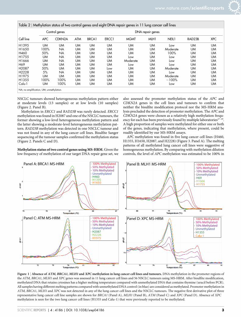
Table 2 | Methylation status of two control genes and eight DNA repair genes in 11 lung cancer cell lines
Cell line
Control genes DNA repair genes
APC CDKN2A ATM BRCA1 ERCC1 MGMT MLH1 NEIL1 RAD23B XPC
H1395 UM UM UM UM UM UM UM Low UM UMH1650 100% NA UM UM UM UM UM Moderate UM UMH460 50% NA UM UM UM UM UM 100% UM UMH1755 UM NA UM UM UM Low UM Low UM UMH1666 UM NA UM UM UM Moderate UM Low UM UMH69 UM UM UM UM UM Low UM Low UM UMH2087 50% UM UM UM Low UM UM Moderate UM UMH2228 5% NA UM UM UM UM UM Low UM UMH1975 UM UM UM UM UM UM UM Moderate UM UMH1355 100% 100% UM UM UM UM UM ,100% UM UMCalu-1 UM 100% UM UM UM UM UM Low UM UM
NA; no amplification, UM; unmethylation.
Figure 1 | Absence of ATM, BRCA1, MLH1 and XPC methylation in lung cancer cell lines and tumours. DNA methylation in the promoter regions of
the ATM, BRCA1, MLH1 and XPC genes was assessed in 11 lung cancer cell lines and 56 NSCLC tumours using MS-HRM. After bisulfite modification,
methylated DNA that retains cytosines has a higher melting temperature compared with unmethylated DNA that contains thymine (uracil before PCR).
All samples having different melting patterns compared with unmethylated DNA control (in blue) are considered as methylated. Promoter methylation in
ATM, BRCA1, MLH1 and XPC was not detected in any of the lung cancer cell lines and the NSCLC tumours. The negative first derivative plot of three
representative lung cancer cell line samples are shown for BRCA1 (Panel A), MLH1 (Panel B), ATM (Panel C) and XPC (Panel D). Absence of XPC
methylation is seen for the two lung cancer cell lines (H1355 and Calu-1) that were previously reported to be methylated.
www.nature.com/scientificreports
SCIENTIFIC REPORTS | 4 : 4186 | DOI: 10.1038/srep04186 3
H1650 and H1355, and 50% in H460 and H2987. Interestingly, APCmethylation was estimated to be about 5–10% in H2228, suggestiveof differential APC methylation status within the H2228 cells.
Homogenous CDKN2A methylation was found in two lung cancercell lines (H1355 and Calu-1) and was estimated to be 100%(Figure 3. Panel B). Five of the lung cancer cell lines (H460,H1650, H1666, H1755, and H2228), that were previously found tohave a homozygous CDKN2A deletion (CONAN database), were notamplified by the CDKN2A MS-HRM assay, confirming the absenceof CDKN2A template. Four cell lines (H1395, H69, H2987 andH1975) were negative for CDKN2A methylation.
We also tested the 56 NSCLC tumours for methylation in the APCand CDKN2A promoter regions. APC and CDKN2A methylationwas detected in 14 NSCLC tumours each (25%) (Figure 3. Panels Cand D). A total of 23 NSCLC tumours (41%) were methylated for atleast one of the genes and methylation in both genes was detected in 5NSCLC tumours.
External validation using The Cancer Genome Atlas (TCGA)database. As we observed a considerable discrepancy in the methy-lation frequency of tested DNA repair genes between our MS-HRMresults and the literature, we sought to validate our results using asecond dataset generated by another methodology with a low falsepositive rate. We analysed the methylation data from the TCGANSCLC database that provides genome-wide methylation statusassessed by the HumanMethylation 450 k beadchip (Illumina).
Methylation data were available from 568 NSCLC tumours, com-prising of 341 adenocarcinomas and 227 squamous cell carcinomas.
We searched for HM450k probes overlapping the CpG sites withinour eight MS- HRM amplicons. No overlapping CpG sites weredetected for ATM, ERCC1 and MGMT. Eight probes were identifiedfor the remaining genes; two probes for BRCA1, one for MLH1, onefor NEIL1, three for RAD23B and one for XPC (Figure 4).
Methylation was completely absent in the three CpG sites ofRAD23B and the CpG site of XPC in the 568 NSCLC samples.Very low frequency methylation was seen for MLH1 (3/567, 0.5%).Three primary squamous cell carcinomas of lung showed methyla-tion at both CpG sites of BRCA1 (3/568, 0.5%) although one of thesites in the third sample was just below the 20% cut-off. These resultswere consistent with our MS-HRM results finding either absent orvery rare methylation in the BRCA1, MLH1, RAD23B and XPCpromoters.
The CpG site in the NEIL1 promoter was frequently methylated(478/568, 84%), although the level of methylation varied among themethylated samples. This TCGA methylation data again confirmedour MS-HRM results demonstrating high frequency NEIL1 methy-lation in NSCLC.
DiscussionEpigenetic alterations in cancer are a potential source of therapeutictargets for personalised cancer treatment. MGMT methylation inglioma is the best known example where it predicts a durable res-
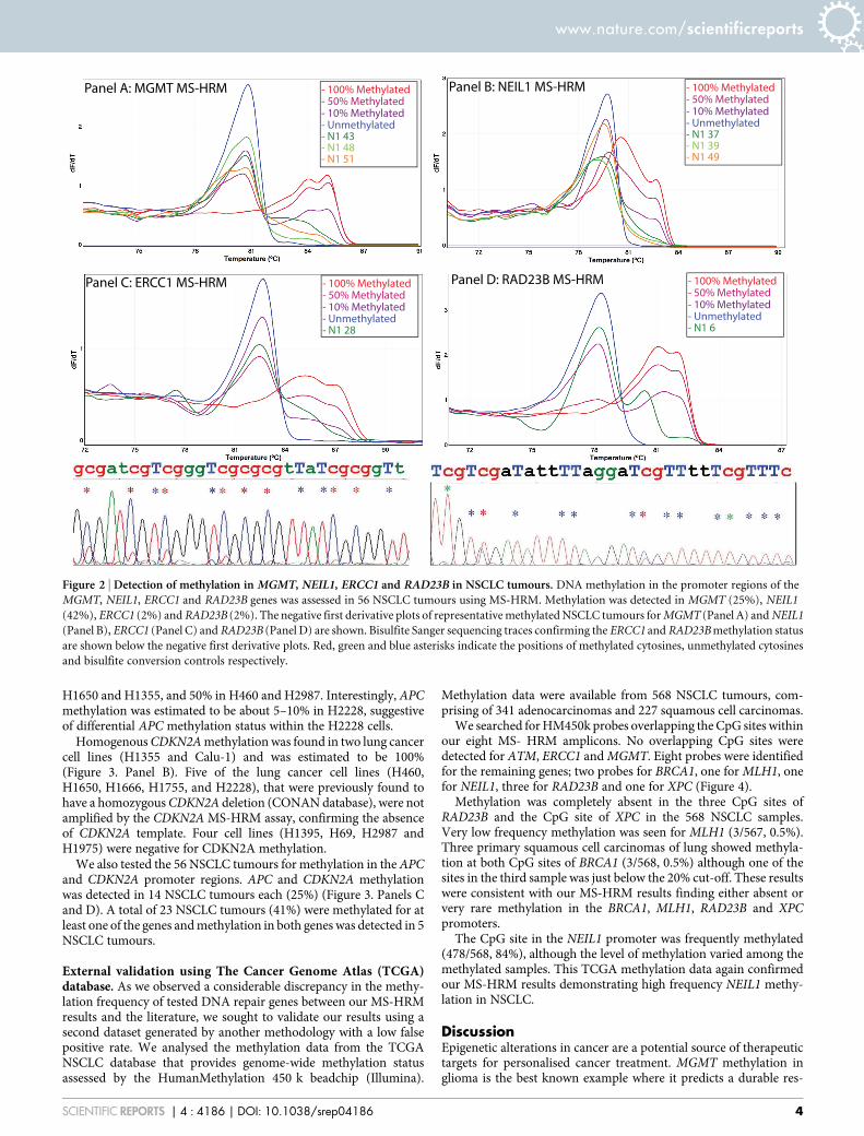
Figure 2 | Detection of methylation in MGMT, NEIL1, ERCC1 and RAD23B in NSCLC tumours. DNA methylation in the promoter regions of the
MGMT, NEIL1, ERCC1 and RAD23B genes was assessed in 56 NSCLC tumours using MS-HRM. Methylation was detected in MGMT (25%), NEIL1
(42%), ERCC1 (2%) and RAD23B (2%). The negative first derivative plots of representative methylated NSCLC tumours for MGMT (Panel A) and NEIL1
(Panel B), ERCC1 (Panel C) and RAD23B (Panel D) are shown. Bisulfite Sanger sequencing traces confirming the ERCC1 and RAD23B methylation status
are shown below the negative first derivative plots. Red, green and blue asterisks indicate the positions of methylated cytosines, unmethylated cytosines
and bisulfite conversion controls respectively.
www.nature.com/scientificreports
SCIENTIFIC REPORTS | 4 : 4186 | DOI: 10.1038/srep04186 4

ponse to treatment with alkylating agents30. Methylation of otherDNA repair genes also has been considered for the selection ofoptimal chemotherapeutic agents for the treatment of cancer,although these have not been clinically implemented up to now31.Before clinical implementation, individual methylation markersneed to be rigorously validated, ideally by using different methodo-logies13. In this study, we sought to validate a range of previouslyreported and to examine novel methylated DNA repair markers thatcould potentially be therapeutically exploited.
In the literature, highly variable estimates of the frequency ofmethylation for the ATM (0–47%), BRCA1 (4–30%), MLH1 (0–68%), MGMT (8–50%), and XPC (33%) genes have been reportedin NSCLC tumours (Table 1). As most of the previous studies usedthe MSP method to determine the methylation status of candidategenes, the previous findings needed to be validated using other meth-odologies that are less prone to give false results. When we assessedDNA methylation using MS-HRM, we did not find methylation inany of these four DNA repair genes (ATM, BRCA1, MLH1, and XPC)in our 11 lung cancer cell lines and 56 NSCLC samples. This wasconsistent with the TCGA data which showed either absent or verylow frequency of methylation for these promoters.
There are several possible explanations for the discrepant results,including differences in ethnicity or clinicopathological features ofsamples tested in each study. Several studies have reported varyingmethylation frequencies in cancer between different ethnic groups,including in the promoters of the IGFBP332, TMS133 and GSTP1genes34. However, the real reasons for the discrepant results are likelyto be technical such as scoring of low-level methylation, false posi-tives due to the use of inadequately designed primers or amplificationof methylated pseudogene sequences.
Low levels of methylation, especially present at #1%, can causediscrepant results due to the different analytic sensitivity of detectionmethods. The lower limit of MSP can be close to 0.1%35, allowingsamples with low level methylation to be interpreted as methylated.
Figure 3 | Assessment of methylation status for APC and CDKN2A in lung cancer cell lines and tumours using MS-HRM. DNA methylation
in the promoter regions of the APC and CDKN2A genes was assessed in 11 lung cancer cell lines and 56 NSCLC tumours using MS-HRM. APC was found
in 5 lung cancer cell lines and 14 NSCLC tumours. CDKN2A methylation was detected in 2 lung cancer cell lines and 14 NSCLC tumours. The negative
first derivative plots of three representative methylated lung cancer cell lines for APC (Panel A) and CDKN2A (Panel B) and three representative
methylated NSCLC tumours for APC (Panel C) and CDKN2A (Panel D) are shown.
Figure 4 | The TCGA methylation data of five DNA repair genes. TCGA
methylation data from 568 non-small cell lung cancers for the eight
overlapping CpG sites with our MS-HRM amplicons is presented as
boxplots. Two overlapping CpG sites for BRCA1, one for MLH1, one for
NEIL1, three for RAD23B and one for XPC were analysed. A b-value of
greater than 0.2 was used to define the presence of DNA methylation as
shown by the horizontal line. Consistent with our MS-HRM results, absent
or very rare methylation was found for BRCA1, MLH1, RAD23B and XPC,
and highly frequent methylation was found for NEIL1.
www.nature.com/scientificreports
SCIENTIFIC REPORTS | 4 : 4186 | DOI: 10.1038/srep04186 5
A high frequency of false positive ATM methylation calls derivingfrom the use of inadequate MSP conditions has been previouslydemonstrated in NSCLC17. ATM methylation was not detected inNSCLC when strict guidelines for performance of MSP are used17.We did not find ATM methylation in our 11 lung cancer cell lines and56 lung tumours, confirming the absence of ATM methylation.
The BRCA1 pseudogene (BRCA1P1), a duplicated region ofBRCA1 exons 1A, 1B, and 2, has a strong sequence homology tothe BRCA1 gene. As methylation of BRCA1P1 has been previouslyreported in cancers36,37, there is a risk of false positives due to amp-lification of the methylated BRCA1P1 sequence. Two previous stud-ies have assessed the BRCA1 methylation status in NSCLC tumoursusing MSP as a detection method38,39. The MSP primers designed byLee et al. have 19 (19/21 in the forward) and 17 (17/20 bases in thereverse) matched bases, including the nine consecutive bases fromthe 39 end of both primers, to the BRCA1 pseudogene39. The fre-quency of BRCA1 methylation (30%) reported by Lee et al. was 7-foldhigher than that of being reported by Marsit et al. (4%) where morestringent MSP primers were used to avoid the amplification of themethylated BRCA1P1 sequence.
XPC methylation was initially reported in NSCLC tumours andlung cancer cell lines23. Wu et al. reported that XPC methylation wasdetectable in 34% of NSCLC tumours by HpaII-based PCR and infour lung cancer cell lines harboring TP53 mutations. Surprisingly,XPC methylation was not detected in this cohort of 56 NSCLCtumours and in two of the lung cancer cell lines (H1355 and Calu-1) previously reported as methylated23. The absence of XPC methyla-tion was confirmed in the TCGA data. None of the 568 NSCLCtumours of the TCGA study had XPC methylation at the overlappingCpG site with our MS-HRM amplicon. As our XPC MS-HRM assaywas designed to amplify those methylated CpG sites in the Wu et al.study, the discordant results was thus not likely to be caused by theexamination of different CpG sites. To assess the XPC methylationstatus in NSCLC tumours, Wu et al. used the HpaII restriction endo-nuclease for selective cleavage of unmethylated DNA before PCRamplification23. However, there is a risk of incomplete enzymaticdigestion of unmethylated DNA by HpaII, potentially resulting infalse positives40,41.
This study is the first report showing methylation of ERCC1,RAD23B and NEIL1 in NSCLC. The excision repair cross-comple-menting group 1 (ERCC1) is a rate-limiting protein involved in therecognition and excision of DNA adducts. Recently, Chen et al.reported that ERCC1 methylation was significantly associatedwith chemosensitivity to cisplatin in glioma cell lines and gliomatumours25. In NSCLC, low levels of ERCC1 expression were corre-lated with favorable clinical outcomes of prolonged survival andsensitivity to platinum-based chemotherapies19. Therefore, ERCC1methylation may serve as a predictive biomarker for identification ofNSCLC patients who are highly sensitive to platinum-based che-motherapies. These patients potentially would have a more durableresponse than patients with high levels of ERCC1 expression thatwere not methylated.
The RAD23B protein forms a DNA damage recognition complexwith the XPC and centrin 2 proteins. The RAD23B/XPC/centrin 2complex recognises and interacts with the damaged bases or thesugar-phosphate backbone of DNA in the NER pathway42. HighRAD23B expression has been suggested as a promising biomarkerassociated with response to histone deacetylase (HDAC) inhibitorsin cutaneous T-cell lymphoma patients43. As anti-tumour activitiesof HDAC inhibitors have been demonstrated in NSCLC44, it can bespeculated that silencing of RAD23B expression through promotermethylation makes tumour cells more resistant to HDAC inhibitors.On the other hand, RAD23B methylation may make tumour cellsmore sensitive to DNA-damaging agents.
Nei-like 1 (NEIL1), an ortholog of E.coli Nei (endonuclease VIII),is a bifunctional DNA glycosylase that repairs oxidative DNAdamage of 8-hydroxyguanine and thymine glycol45 and protects cellsfrom radiation-mediated cell death46. Recently, epigenetic silencingof NEIL1 through promoter methylation was reported in head andneck squamous cell carcinomas24. In this study, we found that theNEIL1 promoter is methylated in NSCLC at a high frequency (42%).This may identify patients that are sensitive to radiotherapy whichdeserves further study.
In conclusion, this study showed that methylation frequency ofATM, BRCA1, MLH1 and XPC in NSCLC is likely to be over-estimated in the literature, emphasising the importance of rigorous
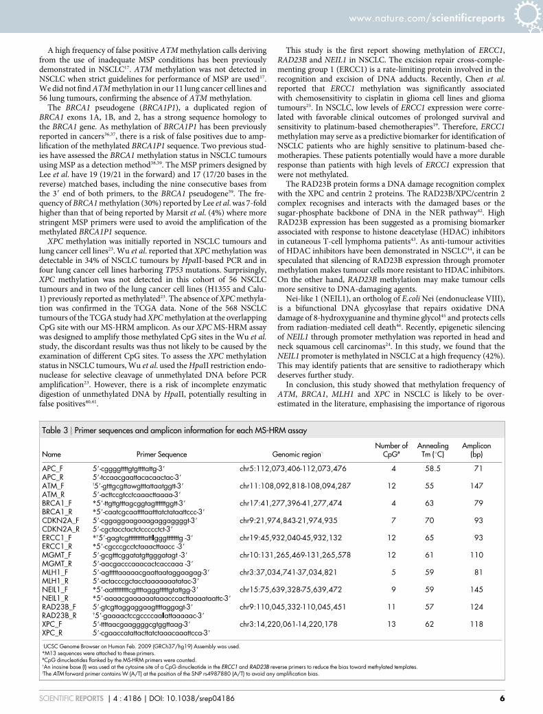
Table 3 | Primer sequences and amplicon information for each MS-HRM assay
Name Primer Sequence Genomic region‘
Number ofCpG#
AnnealingTm (uC)
Amplicon(bp)
APC_F 59-cggggttttgtgttttattg-39 chr5:112,073,406-112,073,476 4 58.5 71APC_R 59-tccaacgaattacacaactac-39
ATM_F !59-gtttgcgttawgtttattaatggtt-39 chr11:108,092,818-108,094,287 12 55 147ATM_R 59-acttccgtcctcaaacttaaaa-39
BRCA1_F *59-ttgttgtttagcggtagttttttggtt-39 chr17:41,277,396-41,277,474 4 63 79BRCA1_R *59-caatcgcaattttaatttatctataattccc-39
CDKN2A_F 59-cggaggaagaaagaggaggggt-39 chr9:21,974,843-21,974,935 7 70 93CDKN2A_R 59-cgctacctactctccccctct-39
ERCC1_F *{59-gagtcgtttttttttattIgggtttttttg -39 chr19:45,932,040-45,932,132 12 65 93ERCC1_R *59-cgcccgcctctaaacttaacc -39
MGMT_F 59-gcgtttcggatatgttgggatagt -39 chr10:131,265,469-131,265,578 12 61 110MGMT_R 59-aacgacccaaacactcaccaaa -39
MLH1_F 59-agtttttaaaaacgaattaataggaagag-39 chr3:37,034,741-37,034,821 5 59 81MLH1_R 59-actacccgctacctaaaaaaatatac-39
NEIL1_F *59-aatttttttttcgttttagggtttttgtattgg-39 chr15:75,639,328-75,639,472 9 59 145NEIL1_R *59-aaaacgaaaaaataaacccacttaaaataattc-39
RAD23B_F 59-gtcgttaggaggaagttttaggagt-39 chr9:110,045,332-110,045,451 11 57 124RAD23B_R {59-gaaaactccgccccaaIattaaaaac-39
XPC_F 59-ttttaacgaaggggcgtggttaag-39 chr3:14,220,061-14,220,178 13 62 118XPC_R 59-cgaaccatattacttatctaaacaaattcca-39
‘UCSC Genome Browser on Human Feb. 2009 (GRCh37/hg19) Assembly was used.*M13 sequences were attached to these primers.#CpG dinucleotides flanked by the MS-HRM primers were counted.{An inosine base (I) was used at the cytosine site of a CpG dinucleotide in the ERCC1 and RAD23B reverse primers to reduce the bias toward methylated templates.!The ATM forward primer contains W (A/T) at the position of the SNP rs4987880 (A/T) to avoid any amplification bias.
www.nature.com/scientificreports
SCIENTIFIC REPORTS | 4 : 4186 | DOI: 10.1038/srep04186 6
validation of previous data on DNA methylation. In particular, theuse of adequate methodology is critical to avoid false positive results.DNA methylation in the ERCC1, RAD23B and especially the NEIL1DNA repair genes may serve as useful biomarkers for the determina-tion of molecularly tailored therapies in a subset of NSCLC patients.
MethodsSamples. Fifty-six N1 stage NSCLC tumours were collected at the Austin Hospital,Melbourne, Australia with the approval of the Austin Human Research EthicsCommittee (project title and approval number ‘‘Biomarkers in the Australian NonSmall Cell Lung Cancer Population’’ – H2006-02394). The methylation study wasapproved by the Ethics of Human Research Committee at the Peter MacCallumCancer Centre, Melbourne, Australia (project title and approval number ‘‘MolecularPathology of Cancer: Methylation, Mutation & Expression’’ - 02/26).
DNA extraction from lung cancer cell lines. Lung cancer cell lines were cultured inRPMI 1640 medium with 25 mM HEPES supplemented with 10% fetal bovine serum,and 0.1 units/mL of penicillin and 0.1 mg/mL of streptomycin. Cells were maintainedat 37uC in a humidified chamber containing 5% CO2. Cultured cells were harvestedand washed twice with Dulbecco’s phosphate-buffered saline buffer, followed bycentrifugation at the speed of 10,000 rpm for 10 minutes. Cell pellets were thensuspended in 200 mL of Dulbecco’s phosphate-buffered saline buffer. After additionof 20 mL of proteinase K (20 mg/mL) and 200 mL of buffer AL, the suspended cellswere incubating at 56uC for overnight. Genomic DNA was extracted from culturedlung cancer cell lines using the QIAamp DNA Blood Mini Kit (Qiagen, Hilden,Germany).
DNA extraction from NSCLC tumours. The tumour purity of individual NSCLCcases was assessed by pathologists at Peter MacCallum Cancer Centre and wasestimated to be 40–95% with a median of 67%. Two to five 5 mm formalin-fixed tissuesections were washed with 1 mL of xylene to remove paraffin and were incubated at40uC for 10 minutes. Supernatant was removed after a centrifugation at 13,000 rpmfor 10 minutes and tissue pellets were sequentially washed with 100% and 70%ethanol. The tissue pallets were then resuspended with 100 mL of ATL buffer of theDNeasy Tissue kit (Qiagen) and incubated at 97uC for 15 mins, followed byproteinase K digestion for 3 days at 56uC. Genomic DNA was then extracted using theDNeasy Tissue kit according to the manufacturer’s instructions.
Bisulfite modification. One microgram of genomic DNA was bisulfite modifiedusing the MethylEasy Xceed kit (Human Genetic Signatures, North Ryde, Australia)according to the manufacturer’s instructions. Bisulfite modified DNA was elutedtwice with 50 mL of elution buffer in an estimated concentration of 10 ng/mL.
Preparation of methylation standards. Commercially available methylated DNA(Millipore, Billerica, MA) was used as a fully methylated control. To prepareunmethylated DNA, peripheral blood DNA obtained from a healthy individualunderwent two rounds of whole genome amplification (WGA) using the IllustraGenomiPhi V2 DNA Amplification Kit (GE Healthcare, Giles, UK) according to themanufacturer’s instructions. The first round of WGA was performed with 1 ng ofperipheral blood DNA. One microliter of a 10-fold dilution of the first round WGAproduct was used for the second round of WGA. After bisulfite modification, a qPCRassay that amplified a region lacking CpG dinucleotides within the COL2A1 gene wasperformed to quantify the amount of template47. A series of methylation dilutionstandards of 100%, 50%, 20%, 10%, 5% and 1% were prepared by mixing of the fullymethylated DNA with the WGA DNA.
Methylation sensitive-high resolution melting assays. MS-HRM assays weredesigned either to target the promoter region or to overlap the regions used inpreviously reported assays. To better work with fragmented FFPE DNA, MS-HRMassays were designed to amplify a short amplicon size of less than 150 bp. Primersequences, amplicon sizes, MgCl2 concentration, annealing temperatures, and thenumber of CpG dinucleotides analysed are summarised in Table 3. PCR amplificationand MS-HRM were performed on the RotorGene Q (Qiagen). The PCR mixture wasprepared in a final volume of 20 mL and contained: 10 ng of bisulfite modifiedtemplate, 13 PCR buffer, 2.5–4 mM MgCl2, 200–400 nM each primer, 200 mMdNTPs, 5 mM SYTO9 and 0.5 U HotStar Taq (Qiagen). PCR cycling and meltingconditions were as follows; one cycle of 95uC for 15 minutes; 50–55 cycles of 95uC for10 seconds, annealing temperature of each assay for 20 seconds, 72uC for 25 seconds;one cycle of 97uC for one minute and a melt from 70uC to 95uC rising 0.2uC persecond. The melting profiles of amplicons were analysed using RotorGene Q Software(v1.7). All samples were tested in duplicate.
Bisulfite Sanger sequencing. To confirm the MS-HRM results for ERCC1, NEIL1and RAD23B methylation, MS- HRM products were sequenced using the Big DyeTerminator v3.1 chemistry (Applied Biosystems). Sequencing products were purifiedwith the Agencourt CleanSEQ reagent (Beckman Coulter) and analysed by capillaryelectrophoresis on an ABI3730 sequencer (Applied Biosystems). The sequencing datawas analysed using Sequencher 4.6 (Gene Codes Corporation).
Analysis of DNA methylation from publically available data. Marmal-aid (http://www.marmal-aid.org) is a data repository of publically available genome-scale DNAmethylation analysis using the Illumina HumanMethylation 450 K (HM450K)beadchip. At the time of download, 8,654 data sets were available from marmal-aidand also contained data from The Cancer Genome Atlas (TCGA) study48. At the timeof download, a total of 568 non-small cell lung cancers (341 adenocarcinomas and 227squamous carcinomas) were available from marmal-aid from TCGA. We identified11 HM450K probes interrogating CpG sites within our MS-HRM amplicons. Rawbeta values were extracted using marmal-aid in R. Data were extracted according totheir annotations in marmal-aid. Samples annotated with ‘‘lung’’ and ‘‘cancer’’ andour probes of interest were used for downstream analysis and visualization in R(http://www.cran.org). Using guidelines previously described49, a b-value of greaterthan 0.2 was used to define the presence of DNA methylation.
1. Taron, M. et al. BRCA1 mRNA expression levels as an indicator ofchemoresistance in lung cancer. Hum Mol Genet 13, 2443–9 (2004).
2. Rosell, R. et al. Customized treatment in non-small-cell lung cancer based onEGFR mutations and BRCA1 mRNA expression. PLoS One 4, e5133 (2009).
3. Papadaki, C. et al. ERCC1 and BRAC1 mRNA expression levels in the primarytumor could predict the effectiveness of the second-line cisplatin-basedchemotherapy in pretreated patients with metastatic non-small cell lung cancer.J Thorac Oncol 7, 663–71 (2012).
4. Papadaki, C. et al. Correlation of BRCA1, TXR1 and TSP1 mRNA expression withtreatment outcome to docetaxel-based first-line chemotherapy in patients withadvanced/metastatic non-small-cell lung cancer. Br J Cancer 104, 316–23 (2011).
5. Boukovinas, I. et al. Tumor BRCA1, RRM1 and RRM2 mRNA expression levelsand clinical response to first-line gemcitabine plus docetaxel in non- small-celllung cancer patients. PLoS One 3, e3695 (2008).
6. Rosell, R. et al. BRCA1: a novel prognostic factor in resected non-small-cell lungcancer. PLoS One 2, e1129 (2007).
7. Font, A. et al. BRCA1 mRNA expression and outcome to neoadjuvant cisplatin-based chemotherapy in bladder cancer. Ann Oncol 22, 139–44 (2011).
8. Weberpals, J. et al. The DNA repair proteins BRCA1 and ERCC1 as predictivemarkers in sporadic ovarian cancer. Int J Cancer 124, 806–15 (2009).
9. Dabholkar, M. et al. ERCC1 and ERCC2 expression in malignant tissues fromovarian cancer patients. J Natl Cancer Inst 84, 1512–7 (1992).
10. Middleton, M. R. & Margison, G. P. Improvement of chemotherapy efficacy byinactivation of a DNA-repair pathway. Lancet Oncol 4, 37–44 (2003).
11. Esteller, M. et al. Inactivation of the DNA-repair gene MGMT and the clinicalresponse of gliomas to alkylating agents. N Engl J Med 343, 1350–4 (2000).
12. Helleday, T., Petermann, E., Lundin, C., Hodgson, B. & Sharma, R. A. DNA repairpathways as targets for cancer therapy. Nat Rev Cancer 8, 193–204 (2008).
13. Mikeska, T., Bock, C., Do, H. & Dobrovic, A. DNA methylation biomarkers incancer: progress towards clinical implementation. Expert Rev Mol Diagn 12,473–87 (2012).
14. Curtin, N. J. DNA repair dysregulation from cancer driver to therapeutic target.Nat Rev Cancer 12, 801–17 (2012).
15. Mikeska, T., Candiloro, I. & Dobrovic, A. The implications of heterogeneous DNAmethylation for the accurate quantification of methylation. Epigenomics 2,561–573 (2010).
16. Cottrell, S. E. & Laird, P. W. Sensitive detection of DNA methylation. Ann N YAcad Sci 983, 120–30 (2003).
17. Brandes, J. C., Carraway, H. & Herman, J. G. Optimal primer design using thenovel primer design program: MSPprimer provides accurate methylation analysisof the ATM promoter. Oncogene 26, 6229–37 (2007).
18. Wojdacz, T. K. & Dobrovic, A. Methylation-sensitive high resolution melting(MS-HRM): a new approach for sensitive and high-throughput assessment ofmethylation. Nucleic Acids Res 35, e41 (2007).
19. Lord, R. V. et al. Low ERCC1 expression correlates with prolonged survival aftercisplatin plus gemcitabine chemotherapy in non-small cell lung cancer. ClinCancer Res 8, 2286–91 (2002).
20. Huang, K. T. et al. DNA methylation profiling of phyllodes and fibroadenomatumours of the breast. Breast Cancer Res Treat 124, 555–65 (2010).
21. Wong, E. M. et al. Constitutional methylation of the BRCA1 promoter isspecifically associated with BRCA1 mutation-associated pathology in early- onsetbreast cancer. Cancer Prev Res (Phila) 4, 23–33 (2011).
22. Mikeska, T., Carney, D. A., Seymour, J. F. & Dobrovic, A. No evidence for DNAmethylation of the ATM promoter CpG island in chronic lymphocytic leukemia.Leuk Lymphoma 53, 1420–2 (2012).
23. Wu, Y. H. et al. Xeroderma pigmentosum group C gene expression ispredominantly regulated by promoter hypermethylation and contributes to p53mutation in lung cancers. Oncogene 26, 4761–73 (2007).
24. Chaisaingmongkol, J. et al. Epigenetic screen of human DNA repair genesidentifies aberrant promoter methylation of NEIL1 in head and neck squamouscell carcinoma. Oncogene 31, 5108–16 (2012).
25. Chen, H. Y., Shao, C. J., Chen, F. R., Kwan, A. L. & Chen, Z. P. Role of ERCC1promoter hypermethylation in drug resistance to cisplatin in human gliomas. Int JCancer 126, 1944–54 (2010).
26. Peng, B. et al. Epigenetic silencing of the human nucleotide excision repair gene,hHR23B, in interleukin-6-responsive multiple myeloma KAS-6/1 cells. J BiolChem 280, 4182–7 (2005).
www.nature.com/scientificreports
SCIENTIFIC REPORTS | 4 : 4186 | DOI: 10.1038/srep04186 7
27. Virmani, A. K. et al. Aberrant methylation of the adenomatous polyposis coli(APC) gene promoter 1A in breast and lung carcinomas. Clin Cancer Res 7,1998–2004 (2001).
28. Brock, M. V. et al. DNA methylation markers and early recurrence in stage I lungcancer. N Engl J Med 358, 1118–28 (2008).
29. Vaissiere, T. et al. Quantitative analysis of DNA methylation profiles in lungcancer identifies aberrant DNA methylation of specific genes and its associationwith gender and cancer risk factors. Cancer Res 69, 243–52 (2009).
30. Hegi, M. E. et al. MGMT gene silencing and benefit from temozolomide inglioblastoma. N Engl J Med 352, 997–1003 (2005).
31. Hegi, M. E., Sciuscio, D., Murat, A., Levivier, M. & Stupp, R. Epigeneticderegulation of DNA repair and its potential for therapy. Clin Cancer Res 15,5026–31 (2009).
32. Tomii, K. et al. Aberrant promoter methylation of insulin-like growth factorbinding protein-3 gene in human cancers. Int J Cancer 120, 566–73 (2007).
33. Das, P. M. et al. Methylation mediated silencing of TMS1/ASC gene in prostatecancer. Mol Cancer 5, 28 (2006).
34. Enokida, H. et al. Ethnic group-related differences in CpG hypermethylation ofthe GSTP1 gene promoter among African-American, Caucasian and Asianpatients with prostate cancer. Int J Cancer 116, 174–81 (2005).
35. Herman, J. G., Graff, J. R., Myohanen, S., Nelkin, B. D. & Baylin, S. B. Methylation-specific PCR: a novel PCR assay for methylation status of CpG islands. Proc NatlAcad Sci U S A 93, 9821–6 (1996).
36. Chiang, J. W., Karlan, B. Y., Cass, L. & Baldwin, R. L. BRCA1 promotermethylation predicts adverse ovarian cancer prognosis. Gynecol Oncol 101,403–10 (2006).
37. Dobrovic, A. & Simpfendorfer, D. Methylation of the BRCA1 gene in sporadicbreast cancer. Cancer Res 57, 3347–50 (1997).
38. Marsit, C. J. et al. Inactivation of the Fanconi anemia/BRCA pathway in lung andoral cancers: implications for treatment and survival. Oncogene 23, 1000–4 (2004).
39. Lee, M. N. et al. Epigenetic inactivation of the chromosomal stability control genesBRCA1, BRCA2, and XRCC5 in non-small cell lung cancer. Clin Cancer Res 13,832–8 (2007).
40. van Dijk, J. P. et al. A novel, essential control for clonality analysis with humanandrogen receptor gene polymerase chain reaction. Am J Pathol 161, 807–12(2002).
41. Kupper, D., Reuter, M., Meisel, A. & Kruger, D. H. Reliable detection of DNA CpGmethylation profiles by the isoschizomers MspI/HpaII using oligonucleotidestimulators. Biotechniques 23, 843–7 (1997).
42. Nouspikel, T. DNA repair in mammalian cells: Nucleotide excision repair:variations on versatility. Cell Mol Life Sci 66, 994–1009 (2009).
43. Khan, O. et al. HR23B is a biomarker for tumor sensitivity to HDAC inhibitor-based therapy. Proc Natl Acad Sci U S A 107, 6532–7 (2010).
44. Miyanaga, A. et al. Antitumor activity of histone deacetylase inhibitors in non-small cell lung cancer cells: development of a molecular predictive model. MolCancer Ther 7, 1923–30 (2008).
45. Bandaru, V., Sunkara, S., Wallace, S. S. & Bond, J. P. A novel human DNAglycosylase that removes oxidative DNA damage and is homologous toEscherichia coli endonuclease VIII. DNA Repair (Amst) 1, 517–29 (2002).
46. Rosenquist, T. A. et al. The novel DNA glycosylase, NEIL1, protects mammaliancells from radiation-mediated cell death. DNA Repair (Amst) 2, 581–91 (2003).
47. Virmani, A. K. et al. Hierarchical clustering of lung cancer cell lines using DNAmethylation markers. Cancer Epidemiol Biomarkers Prev 11, 291–7 (2002).
48. Cancer Genome Atlas Research, N. Comprehensive genomic characterization ofsquamous cell lung cancers. Nature 489, 519–25 (2012).
49. Bibikova, M. et al. Genome-wide DNA methylation profiling using Infinium(R)assay. Epigenomics 1, 177–200 (2009).
50. Safar, A. M. et al. Methylation profiling of archived non-small cell lung cancer: apromising prognostic system. Clin Cancer Res 11, 4400–5 (2005).
51. Safar, A. M. et al. Promoter hypermethylation for molecular nodal staging in non-small cell lung cancer. Arch Pathol Lab Med 131, 936–41 (2007).
52. Wang, Y. et al. Multiple gene methylation of nonsmall cell lung cancers evaluatedwith 3-dimensional microarray. Cancer 112, 1325–36 (2008).
53. Wu, J. Y. et al. Association of O6-methylguanine-DNA methyltransferase(MGMT) promoter methylation with p53 mutation occurrence in non-small celllung cancer with different histology, gender, and smoking status. Ann Surg Oncol15, 3272–7 (2008).
54. Feng, Q. et al. DNA methylation in tumor and matched normal tissues from non-small cell lung cancer patients. Cancer Epidemiol Biomarkers Prev 17, 645–54(2008).
55. Liu, Y., Lan, Q., Siegfried, J. M., Luketich, J. D. & Keohavong, P. Aberrantpromoter methylation of p16 and MGMT genes in lung tumors from smokingand never-smoking lung cancer patients. Neoplasia 8, 46–51 (2006).
56. Kim, Y. T. et al. Prognostic implication of aberrant promoter hypermethylation ofCpG islands in adenocarcinoma of the lung. J Thorac Cardiovasc Surg 130, 1378(2005).
57. Tang, M., Torres-Lanzas, J., Lopez-Rios, F., Esteller, M. & Sanchez-Cespedes, M.Wnt signaling promoter hypermethylation distinguishes lung primaryadenocarcinomas from colorectal metastasis to the lung. Int J Cancer 119, 2603–6(2006).
58. Seng, T. J. et al. DLEC1 and MLH1 promoter methylation are associated with poorprognosis in non-small cell lung carcinoma. Br J Cancer 99, 375–82 (2008).
59. Wang, Y. C. et al. Inactivation of hMLH1 and hMSH2 by promoter methylation inprimary non-small cell lung tumors and matched sputum samples. J Clin Invest111, 887–95 (2003).
60. Geng, X., Wang, F., Zhang, L. & Zhang, W. M. Loss of heterozygosity combinedwith promoter hypermethylation, the main mechanism of human MutL Homolog(hMLH1) gene inactivation in non-small cell lung cancer in a Chinese population.Tumori 95, 488–94 (2009).
AcknowledgmentsWe would like to thank Thomas Mikeska for critical discussion. This project was supportedby funding from Cancer Australia to A.D., the Cancer Council of Victoria to A.D., and theNational Health and Medical Research Council of Australia to P.M. and A.D. H.D. is arecipient of Postdoctoral Fellowship from the Cancer Council of Victoria.
Author contributionsSuitable samples were identified by P.M., T.J., B.S. and C.M. The experimental work wascarried out by H.D. and the TCGA data was analysed by N.W., H.D. and N.W. preparedfigures. H.D. and A.D. co-wrote the manuscript. All authors approved the final manuscript.
Additional informationCompeting financial interests: The authors declare no competing financial interests.
How to cite this article: Do, H. et al. A critical re-assessment of DNA repair gene promotermethylation in non-small cell lung carcinoma. Sci. Rep. 4, 4186; DOI:10.1038/srep04186(2014).
This work is licensed under a Creative Commons Attribution-NonCommercial-ShareAlike 3.0 Unported license. To view a copy of this license,
visit http://creativecommons.org/licenses/by-nc-sa/3.0
www.nature.com/scientificreports
SCIENTIFIC REPORTS | 4 : 4186 | DOI: 10.1038/srep04186 8