DSB, DSF: Pressure monitors and pressure switches - sauter ...
ARTICLE
Received 14 Oct 2013 | Accepted 25 Apr 2014 | Published 9 Jun 2014
An RNA polymerase II-coupled function for histoneH3K36 methylation in checkpoint activation andDSB repairDeepak Kumar Jha1 & Brian D. Strahl1,2
Histone modifications are major determinants of DNA double-strand break (DSB) response
and repair. Here we elucidate a DSB repair function for transcription-coupled Set2 methy-
lation at H3 lysine 36 (H3K36me). Cells devoid of Set2/H3K36me are hypersensitive to
DNA-damaging agents and site-specific DSBs, fail to properly activate the DNA-damage
checkpoint, and show genetic interactions with DSB-sensing and repair machinery. Set2/
H3K36me3 is enriched at DSBs, and loss of Set2 results in altered chromatin architecture and
inappropriate resection during G1 near break sites. Surprisingly, Set2 and RNA polymerase II
are programmed for destruction after DSBs in a temporal manner—resulting in H3K36me3 to
H3K36me2 transition that may be linked to DSB repair. Finally, we show a requirement of Set2
in DSB repair in transcription units—thus underscoring the importance of transcription-
dependent H3K36me in DSB repair.
DOI: 10.1038/ncomms4965
1 Department of Biochemistry and Biophysics, School of Medicine, UNC-Chapel Hill, North Carolina, USA. 2 Lineberger Comprehensive Cancer Center,School of Medicine, UNC-Chapel Hill, North Carolina 27599, USA. Correspondence and requests for materials should be addressed to B.D.S.(email: [email protected]).
NATURE COMMUNICATIONS | 5:3965 | DOI: 10.1038/ncomms4965 | www.nature.com/naturecommunications 1
& 2014 Macmillan Publishers Limited. All rights reserved.
Eukaryotic genomes are constantly subjected to exogenousand endogenous forms of DNA damage1. Double strandbreaks (DSBs) can lead to temporary loss of genetic
information, and failure to repair DSBs can lead to severegenome instability and cell death. DSBs can be programmed (forexample, meiosis and immunoglobulin rearrangements) orcaused by exogenous agents (for example, ionizing radiation(IR), ultra-violet (UV) and anti-cancer drugs)1,2.
In eukaryotes, chromatin plays a fundamental role inregulating the cellular response to DNA damage2. Among thefactors that contribute to the DNA damage response (DDR) arehistones and their post-translational modifications (for example,phosphorylated H2A serine 129; g-H2A.X in metazoans), ATP-dependent chromatin remodellers (for example, RSC, INO80,TIP60), and histone variants (for example, Htz1)2. Transcription-associated histone modifications, including H3 lysine 4methylation (H3K4me) and H3 lysine 79 methylation(H3K79me) also contribute to DDR and repair2. Interestingly, amajority of stalled replication sites tend to occur in transcribedgenes3, increasing the propensity for the collision of transcriptionand replication bubbles eventually resulting in DSBs4. Yet, howthe transcription apparatus influences DSB repair is not wellunderstood.
One major regulator of chromatin structure during transcrip-tion is H3 lysine 36 methylation (H3K36me). This mark isdeposited by the Set2 methyltransferase and is highly conservedfrom yeast to humans5–10. Set2 is recruited, at least in part,through binding to hyper-phosphorylated RNA Polymerase II(RNAPII) after RNAPII enters the productive elongation phase5–10.Set2 recognizes the phosphorylated C-terminal domain (CTD) ofRNAPII through the presence of a domain called the SRI domain(for Set2-RNAPII interaction domain)7,8. In yeast, Set2/H3K36meregulates chromatin structure in the bodies of genes by activatingthe histone deacetylase complex Rpd3S5,11–13 and by regulatinghistone exchange through regulating the activity of Asf1, Chd1 andISW1b complexes14,15. Outside of a role for Set2 in transcriptionelongation, much less is known regarding how this enzymecontributes to other DNA-templated functions.
Here we describe a role for Set2/H3K36me in DNA repair,specifically repair of DSBs. We show that in budding yeast Set2/H3K36 is required for appropriate activation of the response toDSB as well as subsequent repair, a finding that is restricted to itsfunction in transcription. Interestingly, we find that Set2 andRNAPII are programmed for destruction after DSBs, which forH3K36me, results in a temporal patterning of H3K36me2 andH3K36me3 around DSBs that we propose underlies theregulation of chromatin structure at break sites. Significantly,we find that Set2 loss leads to altered chromatin structure andinappropriate resection in G1 at break sites, which ultimatelyimpacts pathway repair choice.
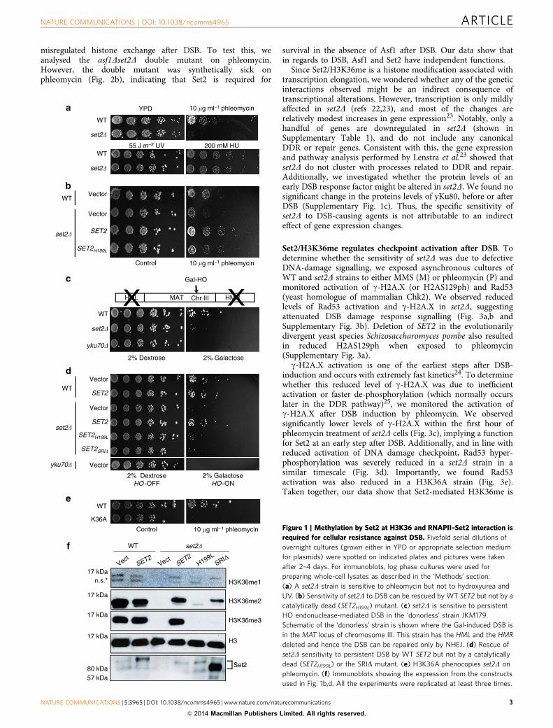
ResultsSet2/H3K36me is required for resistance to DSB. To testwhether Set2 and H3K36me function in DNA damage repair, weassessed the sensitivity of yeast cells lacking SET2 to variousgenotoxic agents. Deletion of SET2 caused sensitivity to the DSB-causing agent phleomycin (an IR-mimic), mild sensitivity to thealkylating agent methyl methane sulphonate (MMS), but nosensitivity to hydroxyurea (replication stress) or UV (nucleotidedamage) (Fig. 1a). The phleomycin sensitivity was rescued bytransforming the set2D strain with full-length SET2 (Fig. 1b). Incontrast, sensitivity of the set2D strain could not be suppressed bytransformation with a catalytically inactive SET2 mutant(SET2H199L); thus, methylation activity is required for survivalafter DSB (Fig. 1b,f). To further confirm the importance of Set2 in
the DSB response, we utilized a strain (JKM179) in which agalactose-inducible, site-specific DSB in chromosome III can berepaired only by non-homologous end joining (NHEJ)16 (Fig. 1c).Consistent with the phleomycin sensitivity found with deletion ofSET2, set2D was also sensitive to this galactose-induced DSB andwas rescued by expressing wild-type (WT), but not catalyticallyinactive, Set2 (Fig. 1c,d,f). We ruled out the MAT-specific effectby using a strain with the homothallic (HO) endonuclease cut sitein ADH4 gene and found that set2D was sensitive to a reparableGal-inducible DSB as well (Supplementary Fig. 1a). Recently, itwas suggested that Set2 functions in DNA damage in a catalysis-independent manner17; however examination of the levels ofH3K36me in the presumed C201A catalytic mutant used byWinsor et al.17 revealed that it only abolished the tri-methyl formof H3K36 (Supplementary Fig. 1b). Importantly, and consistentwith a role of Set2 methylation at H3K36 in this phenotype andDSB repair, we found that mutation of H3K36 (H3K36A) conferssensitivity to phleomycin (Fig. 1e).
Set2 functions through its association with the elongating(phosphorylated) form of RNAPII7. We therefore investigatedwhether the DNA damage-induced phenotype we observed in theabsence of Set2 and H3K36me is connected to its RNAPIIfunction. We previously characterized a domain in the Cterminus of Set2, termed the SRI domain, which is essential forcoupling Set2 with the transcribing polymerase7. Mutation of thisdomain uncouples Set2 from RNAPII and results in selective lossof H3K36me3 (Fig. 1f and ref. 18). Significantly, mutation of theSRI domain of SET2 (SRID) could not rescue the sensitivity ofset2D to persistent gal-induced DSB (Fig. 1d). Taken together,these data suggest that Set2 functions in DSB repair through itsinteraction with elongating RNAPII. They further suggest thepossibility that transcribing polymerase itself may play animportant role in DSB repair—a finding that would beconsistent with studies showing RNAPII is present at DSBs19.
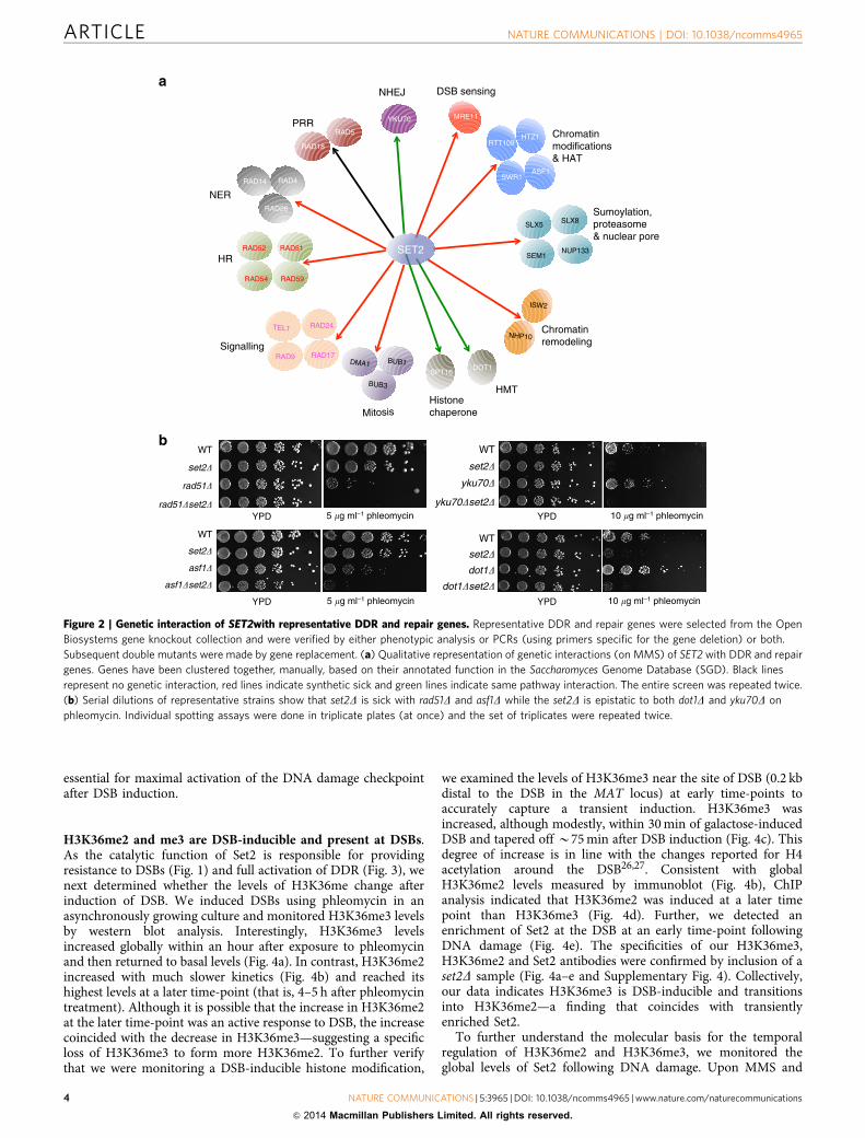
SET2 is functionally connected to DSB response and repair. Tofurther establish that Set2 functions in cellular survival againstgenotoxic insults, we selected 30 genes representative of thepathways involved in DDR and repair and analysed doublemutant sensitivity to MMS. We performed our genetic interactionscreen using MMS because of its ability to activate several DNArepair pathways as compared with phleomycin, thus allowing usto initially capture more possible genetic interactions betweenSET2 and DDR genes. Our screen revealed that SET2 geneticallyinteracts (both positively and negatively) with a subset of DDRand repair genes, albeit to different extents (Fig. 2a andSupplementary Fig. 2a). In addition to recapitulating some priorgenetic interactions (for example, between SET2 and SLX5)reported by the Idekker group20, we discovered several novelgenetic interactions upon MMS treatment (for example, RAD9and RAD51) (Fig. 2a). We focused on DSB-specific genes andrepeated the genetic interactions using phleomycin (examplesshown in Fig. 2b and Supplementary Fig. 2a,b). Interestingly, theset2Drad9D double mutant was more sensitive to MMS andphleomycin than either single mutant alone, suggesting that Set2functions in parallel to Rad9, to provide resistance to DSB(Fig. 2a, and Supplementary Fig. 2c). Like the set2D singlemutants, the set2Drad9D synthetic sickness was rescued by WTSET2, but not by a catalytically dead version of SET2 (SET2H199L)(Supplementary Fig. 2c). These results agree with similar findingsrecently reported in a high-throughput genetic interaction mapusing multiple genotoxic agents21.
As Set2 antagonizes the histone chaperone Asf1 to regulatehistone exchange during transcription elongation15, we alsoinvestigated whether the sensitivity of set2D might be due to
ARTICLE NATURE COMMUNICATIONS | DOI: 10.1038/ncomms4965
2 NATURE COMMUNICATIONS | 5:3965 | DOI: 10.1038/ncomms4965 | www.nature.com/naturecommunications
& 2014 Macmillan Publishers Limited. All rights reserved.
misregulated histone exchange after DSB. To test this, weanalysed the asf1Dset2D double mutant on phleomycin.However, the double mutant was synthetically sick onphleomycin (Fig. 2b), indicating that Set2 is required for
survival in the absence of Asf1 after DSB. Our data show thatin regards to DSB, Asf1 and Set2 have independent functions.
Since Set2/H3K36me is a histone modification associated withtranscription elongation, we wondered whether any of the geneticinteractions observed might be an indirect consequence oftranscriptional alterations. However, transcription is only mildlyaffected in set2D (refs 22,23), and most of the changes arerelatively modest increases in gene expression23. Notably, only ahandful of genes are downregulated in set2D (shown inSupplementary Table 1), and do not include any canonicalDDR or repair genes. Consistent with this, the gene expressionand pathway analysis performed by Lenstra et al.23 showed thatset2D do not cluster with processes related to DDR and repair.Additionally, we investigated whether the protein levels of anearly DSB response factor might be altered in set2D. We found nosignificant change in the proteins levels of yKu80, before or afterDSB (Supplementary Fig. 1c). Thus, the specific sensitivity ofset2D to DSB-causing agents is not attributable to an indirecteffect of gene expression changes.
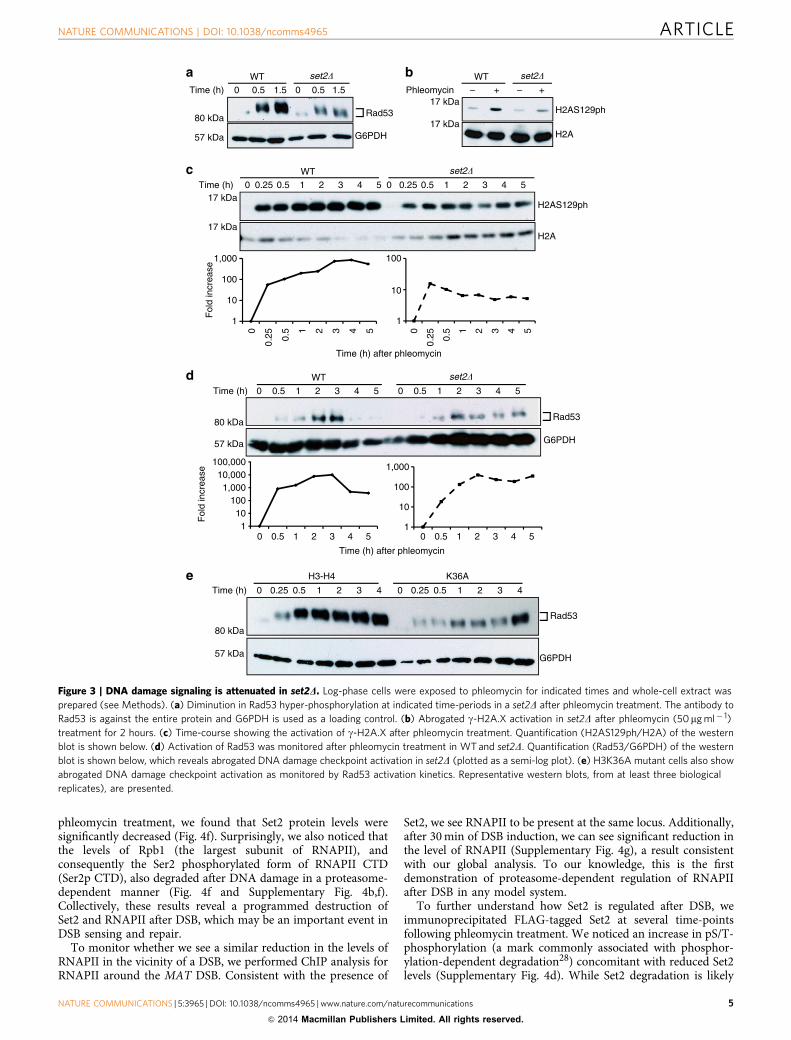
Set2/H3K36me regulates checkpoint activation after DSB. Todetermine whether the sensitivity of set2D was due to defectiveDNA-damage signalling, we exposed asynchronous cultures ofWT and set2D strains to either MMS (M) or phleomycin (P) andmonitored activation of g-H2A.X (or H2AS129ph) and Rad53(yeast homologue of mammalian Chk2). We observed reducedlevels of Rad53 activation and g-H2A.X in set2D, suggestingattenuated DSB damage response signalling (Fig. 3a,b andSupplementary Fig. 3b). Deletion of SET2 in the evolutionarilydivergent yeast species Schizosaccharomyces pombe also resultedin reduced H2AS129ph when exposed to phleomycin(Supplementary Fig. 3a).
g-H2A.X activation is one of the earliest steps after DSB-induction and occurs with extremely fast kinetics24. To determinewhether this reduced level of g-H2A.X was due to inefficientactivation or faster de-phosphorylation (which normally occurslater in the DDR pathway)25, we monitored the activation ofg-H2A.X after DSB induction by phleomycin. We observedsignificantly lower levels of g-H2A.X within the first hour ofphleomycin treatment of set2D cells (Fig. 3c), implying a functionfor Set2 at an early step after DSB. Additionally, and in line withreduced activation of DNA damage checkpoint, Rad53 hyper-phosphorylation was severely reduced in a set2D strain in asimilar timescale (Fig. 3d). Importantly, we found Rad53activation was also reduced in a H3K36A strain (Fig. 3e).Taken together, our data show that Set2-mediated H3K36me is
2% Dextrose
Control 10 �g ml–1 phleomycin
WT
K36A
Control 10 �g ml–1 phleomycin
2% DextroseHO -OFF
2% GalactoseHO -ON
Vector
Vector
yku70� Vector
WT
SET2H199L
SET2SRI�
SET2
SET2
WT
VectSE
T2Vect
SET2
H199LSRIΔ
n.s.*
Set2
H3K36me3
H3
H3K36me2
H3K36me1
WT
set2�
set2�
yku70�
57 kDa80 kDa
17 kDa
17 kDa
17 kDa
17 kDa
Vector
SET2
SET2H199L
Vector
set2�
WT
200 mM HU55 J m–2 UV
YPD
WT
set2�
WT
set2�
HMRHML MAT
Gal-HO
Chr III
10 �g ml–1 phleomycin
2% Galactose
set2�
Figure 1 | Methylation by Set2 at H3K36 and RNAPII–Set2 interaction is
required for cellular resistance against DSB. Fivefold serial dilutions of
overnight cultures (grown either in YPD or appropriate selection medium
for plasmids) were spotted on indicated plates and pictures were taken
after 2–4 days. For immunoblots, log phase cultures were used for
preparing whole-cell lysates as described in the ‘Methods’ section.
(a) A set2D strain is sensitive to phleomycin but not to hydroxyurea and
UV. (b) Sensitivity of set2D to DSB can be rescued by WT SET2 but not by a
catalytically dead (SET2H199L) mutant. (c) set2D is sensitive to persistent
HO endonuclease-mediated DSB in the ‘donorless’ strain JKM179.
Schematic of the ‘donorless’ strain is shown where the Gal-induced DSB is
in the MAT locus of chromosome III. This strain has the HML and the HMR
deleted and hence the DSB can be repaired only by NHEJ. (d) Rescue of
set2D sensitivity to persistent DSB by WT SET2 but not by a catalytically
dead (SET2H199L) or the SRID mutant. (e) H3K36A phenocopies set2D on
phleomycin. (f) Immunoblots showing the expression from the constructs
used in Fig. 1b,d. All the experiments were replicated at least three times.
NATURE COMMUNICATIONS | DOI: 10.1038/ncomms4965 ARTICLE
NATURE COMMUNICATIONS | 5:3965 | DOI: 10.1038/ncomms4965 | www.nature.com/naturecommunications 3
& 2014 Macmillan Publishers Limited. All rights reserved.
essential for maximal activation of the DNA damage checkpointafter DSB induction.
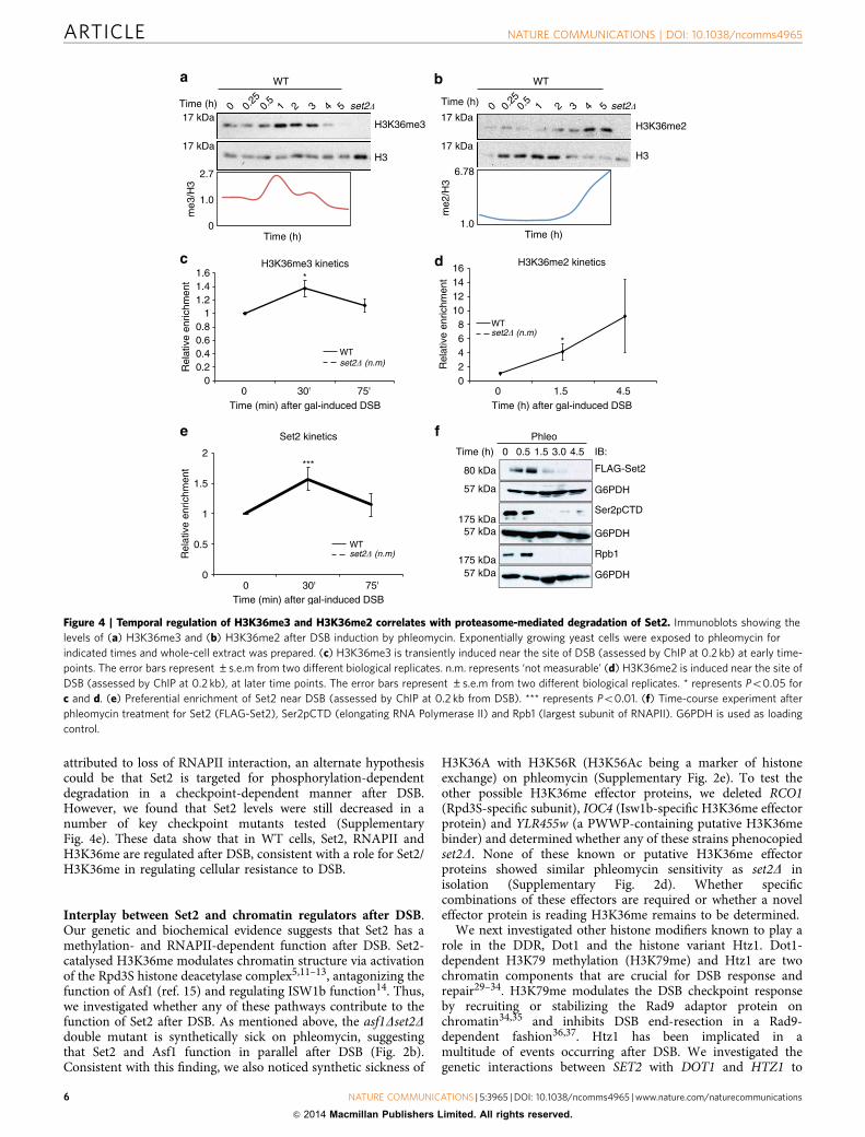
H3K36me2 and me3 are DSB-inducible and present at DSBs.As the catalytic function of Set2 is responsible for providingresistance to DSBs (Fig. 1) and full activation of DDR (Fig. 3), wenext determined whether the levels of H3K36me change afterinduction of DSB. We induced DSBs using phleomycin in anasynchronously growing culture and monitored H3K36me3 levelsby western blot analysis. Interestingly, H3K36me3 levelsincreased globally within an hour after exposure to phleomycinand then returned to basal levels (Fig. 4a). In contrast, H3K36me2increased with much slower kinetics (Fig. 4b) and reached itshighest levels at a later time-point (that is, 4–5 h after phleomycintreatment). Although it is possible that the increase in H3K36me2at the later time-point was an active response to DSB, the increasecoincided with the decrease in H3K36me3—suggesting a specificloss of H3K36me3 to form more H3K36me2. To further verifythat we were monitoring a DSB-inducible histone modification,
we examined the levels of H3K36me3 near the site of DSB (0.2 kbdistal to the DSB in the MAT locus) at early time-points toaccurately capture a transient induction. H3K36me3 wasincreased, although modestly, within 30 min of galactose-inducedDSB and tapered off B75 min after DSB induction (Fig. 4c). Thisdegree of increase is in line with the changes reported for H4acetylation around the DSB26,27. Consistent with globalH3K36me2 levels measured by immunoblot (Fig. 4b), ChIPanalysis indicated that H3K36me2 was induced at a later timepoint than H3K36me3 (Fig. 4d). Further, we detected anenrichment of Set2 at the DSB at an early time-point followingDNA damage (Fig. 4e). The specificities of our H3K36me3,H3K36me2 and Set2 antibodies were confirmed by inclusion of aset2D sample (Fig. 4a–e and Supplementary Fig. 4). Collectively,our data indicates H3K36me3 is DSB-inducible and transitionsinto H3K36me2—a finding that coincides with transientlyenriched Set2.
To further understand the molecular basis for the temporalregulation of H3K36me2 and H3K36me3, we monitored theglobal levels of Set2 following DNA damage. Upon MMS and
RAD5
RAD18
RAD51
RAD54
RAD52
RAD59
RAD24
RAD9
TEL1
RAD17
RAD4
RAD26
RAD14
NHP10
ISW2
SPT16DOT1
SWR1
RTT109HTZ1
ASF1
MRE11YKU70
SLX5 SLX8
SEM1NUP133SET2
NER
HR
Signalling
WT
YPD 5 �g ml–1 phleomycin
WT
set2�
WT
set2�
yku70�
5 �g ml–1 phleomycinYPD 10 �g ml–1 phleomycinYPD
10 �g ml–1 phleomycinYPD
DMA1 BUB1
BUB3
Mitosis
HMT
DSB sensingNHEJa
b
Chromatinmodifications& HAT
Sumoylation,proteasome& nuclear pore
Chromatinremodeling
PRR
Histonechaperone
set2�
rad51�
rad51�set2�
WT
set2�
asf1�
asf1�set2�
yku70�set2�
dot1�set2�
dot1�
Figure 2 | Genetic interaction of SET2with representative DDR and repair genes. Representative DDR and repair genes were selected from the Open
Biosystems gene knockout collection and were verified by either phenotypic analysis or PCRs (using primers specific for the gene deletion) or both.
Subsequent double mutants were made by gene replacement. (a) Qualitative representation of genetic interactions (on MMS) of SET2 with DDR and repair
genes. Genes have been clustered together, manually, based on their annotated function in the Saccharomyces Genome Database (SGD). Black lines
represent no genetic interaction, red lines indicate synthetic sick and green lines indicate same pathway interaction. The entire screen was repeated twice.
(b) Serial dilutions of representative strains show that set2D is sick with rad51D and asf1D while the set2D is epistatic to both dot1D and yku70D on
phleomycin. Individual spotting assays were done in triplicate plates (at once) and the set of triplicates were repeated twice.
ARTICLE NATURE COMMUNICATIONS | DOI: 10.1038/ncomms4965
4 NATURE COMMUNICATIONS | 5:3965 | DOI: 10.1038/ncomms4965 | www.nature.com/naturecommunications
& 2014 Macmillan Publishers Limited. All rights reserved.
phleomycin treatment, we found that Set2 protein levels weresignificantly decreased (Fig. 4f). Surprisingly, we also noticed thatthe levels of Rpb1 (the largest subunit of RNAPII), andconsequently the Ser2 phosphorylated form of RNAPII CTD(Ser2p CTD), also degraded after DNA damage in a proteasome-dependent manner (Fig. 4f and Supplementary Fig. 4b,f).Collectively, these results reveal a programmed destruction ofSet2 and RNAPII after DSB, which may be an important event inDSB sensing and repair.
To monitor whether we see a similar reduction in the levels ofRNAPII in the vicinity of a DSB, we performed ChIP analysis forRNAPII around the MAT DSB. Consistent with the presence of
Set2, we see RNAPII to be present at the same locus. Additionally,after 30 min of DSB induction, we can see significant reduction inthe level of RNAPII (Supplementary Fig. 4g), a result consistentwith our global analysis. To our knowledge, this is the firstdemonstration of proteasome-dependent regulation of RNAPIIafter DSB in any model system.
To further understand how Set2 is regulated after DSB, weimmunoprecipitated FLAG-tagged Set2 at several time-pointsfollowing phleomycin treatment. We noticed an increase in pS/T-phosphorylation (a mark commonly associated with phosphor-ylation-dependent degradation28) concomitant with reduced Set2levels (Supplementary Fig. 4d). While Set2 degradation is likely
1
10
100
1,000
110
1001,000
10,000100,000
–WT set2�
+ – +
H2AS129ph
H2A
PhleomycinWT set2�
Time (h)
G6PDH
Time (h)WT set2�
H2A
H2AS129phF
old
incr
ease
57 kDa
80 kDa
WT set2�
Time (h)
Rad53
G6PDH57 kDa
80 kDa
17 kDa
17 kDa
17 kDa
17 kDa
Rad53
G6PDH
H3-H4
Time (h)
K36A
Fol
d in
crea
se
Time (h) after phleomycin
1
10
100
0
0.25 0.
5 1 2 3 4 5
1
10
100
1,0000
0.25 0.5 1 2 3 4 5
57 kDa
80 kDa
Time (h) after phleomycin
Rad53
1.50.501.50.50
543210.50.250543210.50.250
543210.50
543210.50 543210.50
543210.50
43210.50.250 43210.50.250
Figure 3 | DNA damage signaling is attenuated in set2D. Log-phase cells were exposed to phleomycin for indicated times and whole-cell extract was
prepared (see Methods). (a) Diminution in Rad53 hyper-phosphorylation at indicated time-periods in a set2D after phleomycin treatment. The antibody to
Rad53 is against the entire protein and G6PDH is used as a loading control. (b) Abrogated g-H2A.X activation in set2D after phleomycin (50mg ml� 1)
treatment for 2 hours. (c) Time-course showing the activation of g-H2A.X after phleomycin treatment. Quantification (H2AS129ph/H2A) of the western
blot is shown below. (d) Activation of Rad53 was monitored after phleomycin treatment in WT and set2D. Quantification (Rad53/G6PDH) of the western
blot is shown below, which reveals abrogated DNA damage checkpoint activation in set2D (plotted as a semi-log plot). (e) H3K36A mutant cells also show
abrogated DNA damage checkpoint activation as monitored by Rad53 activation kinetics. Representative western blots, from at least three biological
replicates), are presented.
NATURE COMMUNICATIONS | DOI: 10.1038/ncomms4965 ARTICLE
NATURE COMMUNICATIONS | 5:3965 | DOI: 10.1038/ncomms4965 | www.nature.com/naturecommunications 5
& 2014 Macmillan Publishers Limited. All rights reserved.
attributed to loss of RNAPII interaction, an alternate hypothesiscould be that Set2 is targeted for phosphorylation-dependentdegradation in a checkpoint-dependent manner after DSB.However, we found that Set2 levels were still decreased in anumber of key checkpoint mutants tested (SupplementaryFig. 4e). These data show that in WT cells, Set2, RNAPII andH3K36me are regulated after DSB, consistent with a role for Set2/H3K36me in regulating cellular resistance to DSB.
Interplay between Set2 and chromatin regulators after DSB.Our genetic and biochemical evidence suggests that Set2 has amethylation- and RNAPII-dependent function after DSB. Set2-catalysed H3K36me modulates chromatin structure via activationof the Rpd3S histone deacetylase complex5,11–13, antagonizing thefunction of Asf1 (ref. 15) and regulating ISW1b function14. Thus,we investigated whether any of these pathways contribute to thefunction of Set2 after DSB. As mentioned above, the asf1Dset2Ddouble mutant is synthetically sick on phleomycin, suggestingthat Set2 and Asf1 function in parallel after DSB (Fig. 2b).Consistent with this finding, we also noticed synthetic sickness of
H3K36A with H3K56R (H3K56Ac being a marker of histoneexchange) on phleomycin (Supplementary Fig. 2e). To test theother possible H3K36me effector proteins, we deleted RCO1(Rpd3S-specific subunit), IOC4 (Isw1b-specific H3K36me effectorprotein) and YLR455w (a PWWP-containing putative H3K36mebinder) and determined whether any of these strains phenocopiedset2D. None of these known or putative H3K36me effectorproteins showed similar phleomycin sensitivity as set2D inisolation (Supplementary Fig. 2d). Whether specificcombinations of these effectors are required or whether a noveleffector protein is reading H3K36me remains to be determined.
We next investigated other histone modifiers known to play arole in the DDR, Dot1 and the histone variant Htz1. Dot1-dependent H3K79 methylation (H3K79me) and Htz1 are twochromatin components that are crucial for DSB response andrepair29–34. H3K79me modulates the DSB checkpoint responseby recruiting or stabilizing the Rad9 adaptor protein onchromatin34,35 and inhibits DSB end-resection in a Rad9-dependent fashion36,37. Htz1 has been implicated in amultitude of events occurring after DSB. We investigated thegenetic interactions between SET2 with DOT1 and HTZ1 to
00.20.40.60.8
11.21.41.6
H3K36me3 kinetics*
WTset2� (n.m)
Time (min) after gal-induced DSB
Rel
ativ
e en
richm
ent
02468
10121416
0 30' 75' 0 1.5 4.5
*
WTset2� (n.m)
H3K36me2 kinetics
Time (h) after gal-induced DSB
me3
/H3
Time (h)
2.7
1.0
0
WT
H3K36me3
H3
Time (h) 0 0.25
0.5
1 2 3 4 5 set2�17 kDa
17 kDa
Time (h)
me2
/H3
6.78
1.0
WT
H3K36me2
H3
Time (h) set2�17 kDa
17 kDa
Rel
ativ
e en
richm
ent
0
0.5
1
1.5
2
0 30' 75'
Set2 kinetics
Time (min) after gal-induced DSB
WTset2� (n.m)
*** FLAG-Set2
Phleo
0 0.5 1.5 3.0 4.5
G6PDH
IB:
Ser2pCTD
G6PDH
G6PDH
Rpb1
Time (h)
57 kDa
80 kDa
57 kDa175 kDa
57 kDa175 kDa
Rel
ativ
e en
richm
ent
0 0.25
0.5
1 2 3 4 5
Figure 4 | Temporal regulation of H3K36me3 and H3K36me2 correlates with proteasome-mediated degradation of Set2. Immunoblots showing the
levels of (a) H3K36me3 and (b) H3K36me2 after DSB induction by phleomycin. Exponentially growing yeast cells were exposed to phleomycin for
indicated times and whole-cell extract was prepared. (c) H3K36me3 is transiently induced near the site of DSB (assessed by ChIP at 0.2 kb) at early time-
points. The error bars represent ±s.e.m from two different biological replicates. n.m. represents ‘not measurable’ (d) H3K36me2 is induced near the site of
DSB (assessed by ChIP at 0.2 kb), at later time points. The error bars represent ±s.e.m from two different biological replicates. * represents Po0.05 for
c and d. (e) Preferential enrichment of Set2 near DSB (assessed by ChIP at 0.2 kb from DSB). *** represents Po0.01. (f) Time-course experiment after
phleomycin treatment for Set2 (FLAG-Set2), Ser2pCTD (elongating RNA Polymerase II) and Rpb1 (largest subunit of RNAPII). G6PDH is used as loading
control.
ARTICLE NATURE COMMUNICATIONS | DOI: 10.1038/ncomms4965
6 NATURE COMMUNICATIONS | 5:3965 | DOI: 10.1038/ncomms4965 | www.nature.com/naturecommunications
& 2014 Macmillan Publishers Limited. All rights reserved.
determine whether Set2 is part of any of the known pathways (orprocesses) in which these proteins function after DSB. Weobserved that set2D was synthetically sick with htz1D and swr1D(Supplementary Fig. 2e), but set2D suppressed the mild resistanceof dot1D to phleomycin (Fig. 2b). These data reveal that Set2/H3K36me is required for cell survival in the absence of Swr1/Htz1 function. They also suggest that Set2 and Dot1 mightfunction in antagonistic manner to regulate cellular responseto DSB.
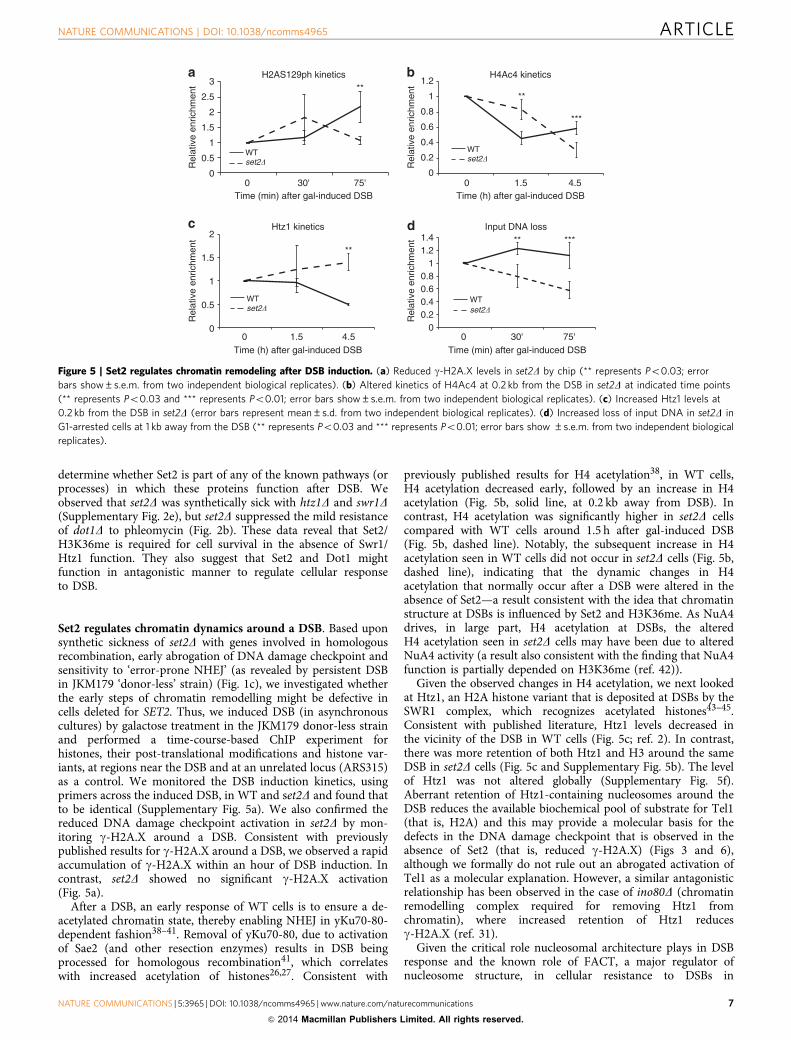
Set2 regulates chromatin dynamics around a DSB. Based uponsynthetic sickness of set2D with genes involved in homologousrecombination, early abrogation of DNA damage checkpoint andsensitivity to ‘error-prone NHEJ’ (as revealed by persistent DSBin JKM179 ‘donor-less’ strain) (Fig. 1c), we investigated whetherthe early steps of chromatin remodelling might be defective incells deleted for SET2. Thus, we induced DSB (in asynchronouscultures) by galactose treatment in the JKM179 donor-less strainand performed a time-course-based ChIP experiment forhistones, their post-translational modifications and histone var-iants, at regions near the DSB and at an unrelated locus (ARS315)as a control. We monitored the DSB induction kinetics, usingprimers across the induced DSB, in WT and set2D and found thatto be identical (Supplementary Fig. 5a). We also confirmed thereduced DNA damage checkpoint activation in set2D by mon-itoring g-H2A.X around a DSB. Consistent with previouslypublished results for g-H2A.X around a DSB, we observed a rapidaccumulation of g-H2A.X within an hour of DSB induction. Incontrast, set2D showed no significant g-H2A.X activation(Fig. 5a).
After a DSB, an early response of WT cells is to ensure a de-acetylated chromatin state, thereby enabling NHEJ in yKu70-80-dependent fashion38–41. Removal of yKu70-80, due to activationof Sae2 (and other resection enzymes) results in DSB beingprocessed for homologous recombination41, which correlateswith increased acetylation of histones26,27. Consistent with
previously published results for H4 acetylation38, in WT cells,H4 acetylation decreased early, followed by an increase in H4acetylation (Fig. 5b, solid line, at 0.2 kb away from DSB). Incontrast, H4 acetylation was significantly higher in set2D cellscompared with WT cells around 1.5 h after gal-induced DSB(Fig. 5b, dashed line). Notably, the subsequent increase in H4acetylation seen in WT cells did not occur in set2D cells (Fig. 5b,dashed line), indicating that the dynamic changes in H4acetylation that normally occur after a DSB were altered in theabsence of Set2—a result consistent with the idea that chromatinstructure at DSBs is influenced by Set2 and H3K36me. As NuA4drives, in large part, H4 acetylation at DSBs, the alteredH4 acetylation seen in set2D cells may have been due to alteredNuA4 activity (a result also consistent with the finding that NuA4function is partially depended on H3K36me (ref. 42)).
Given the observed changes in H4 acetylation, we next lookedat Htz1, an H2A histone variant that is deposited at DSBs by theSWR1 complex, which recognizes acetylated histones43–45.Consistent with published literature, Htz1 levels decreased inthe vicinity of the DSB in WT cells (Fig. 5c; ref. 2). In contrast,there was more retention of both Htz1 and H3 around the sameDSB in set2D cells (Fig. 5c and Supplementary Fig. 5b). The levelof Htz1 was not altered globally (Supplementary Fig. 5f).Aberrant retention of Htz1-containing nucleosomes around theDSB reduces the available biochemical pool of substrate for Tel1(that is, H2A) and this may provide a molecular basis for thedefects in the DNA damage checkpoint that is observed in theabsence of Set2 (that is, reduced g-H2A.X) (Figs 3 and 6),although we formally do not rule out an abrogated activation ofTel1 as a molecular explanation. However, a similar antagonisticrelationship has been observed in the case of ino80D (chromatinremodelling complex required for removing Htz1 fromchromatin), where increased retention of Htz1 reducesg-H2A.X (ref. 31).
Given the critical role nucleosomal architecture plays in DSBresponse and the known role of FACT, a major regulator ofnucleosome structure, in cellular resistance to DSBs in
0
0.2
0.4
0.6
0.8
1
1.2
0 1.5 4.5
***
H4Ac4 kinetics
Rel
ativ
e en
richm
ent
Time (h) after gal-induced DSB
WTset2�
00.20.40.60.8
11.21.4
0 30' 75'
***
Rel
ativ
e en
richm
ent
Time (min) after gal-induced DSB
Input DNA loss
WTset2�
**
0
0.5
1
1.5
2
0 1.5 4.5
Rel
ativ
e en
richm
ent
Time (h) after gal-induced DSB
Htz1 kinetics
WTset2�
**
Rel
ativ
e en
richm
ent
0
0.5
1
1.5
2
2.5
3
0 30' 75'
H2AS129ph kinetics
Time (min) after gal-induced DSB
WTset2�
****
Figure 5 | Set2 regulates chromatin remodeling after DSB induction. (a) Reduced g-H2A.X levels in set2D by chip (** represents Po0.03; error
bars show±s.e.m. from two independent biological replicates). (b) Altered kinetics of H4Ac4 at 0.2 kb from the DSB in set2D at indicated time points
(** represents Po0.03 and *** represents Po0.01; error bars show±s.e.m. from two independent biological replicates). (c) Increased Htz1 levels at
0.2 kb from the DSB in set2D (error bars represent mean±s.d. from two independent biological replicates). (d) Increased loss of input DNA in set2D in
G1-arrested cells at 1 kb away from the DSB (** represents Po0.03 and *** represents Po0.01; error bars show ±s.e.m. from two independent biological
replicates).
NATURE COMMUNICATIONS | DOI: 10.1038/ncomms4965 ARTICLE
NATURE COMMUNICATIONS | 5:3965 | DOI: 10.1038/ncomms4965 | www.nature.com/naturecommunications 7
& 2014 Macmillan Publishers Limited. All rights reserved.
mammalian cells46, we next investigated whether SET2functionally interacts with the FACT complex. Interestingly, theSpt16 subunit of FACT regulates H2A.X exchange at DSBs inhuman cells, indicating that FACT regulates nucleosomedynamics at DSBs. In yeast, mutants of Spt16 (for example,spt16-11) are sensitive to replication stress and high temperatureand significantly, are suppressed by deletion of SET2 (refs 47,48).We therefore reasoned that FACT mutants may be sensitive togenotoxic agents and this genotoxic sensitivity would besuppressed by set2D. As shown in Supplementary Fig. 5c, wefound that the spt16-11 allele was extremely sensitive tophleomycin at permissive temperature. Consistent with the ideathat Set2 opposes the action of FACT function48, deletion of SET2suppressed, albeit partially, the DSB sensitivity of the spt16-11strain. These data suggest the intriguing possibility that Set2 andFACT have opposing effects on nucleosomal dynamics at DSBs.
Set2 regulates DSB repair in the context of transcription. Wenext investigated what the functional consequence of losing Set2and H3K36me would be on DNA repair and repair pathway
choice. Our data show that loss of Set2/H3K36me results inaberrant retention of Htz1-containing nucleosomes at DSBs,suggesting an unstable nucleosome architecture around theseregions since Htz1-containing nucleosomes result in less stablenucleosomes49. Importantly, a recent study showed that Htz1-containing nucleosomes aid in Exo1-dependent end-resection50.We therefore used the input DNA from our cycling cells andmonitored the loss of input DNA (as a surrogate readout of endresection). We did not detect any difference in the rate of inputDNA loss compared with that of set2D cells (SupplementaryFig. 5d). However, end-resection is heavily influenced by the cellcycle (G1 versus G2/M), and smaller differences in resectionkinetics may not become apparent in cycling cells. Therefore, wearrested cells in G1 by a-factor and monitored the loss of inputDNA. In WT cells, we did not see any significant loss in inputDNA, but, in set2D cells, the input DNA was rapidly lost,suggesting that set2D cells show faster end-processing inG1-arrested cells—a time when resection is normallysuppressed (Fig. 5d). We confirmed this by monitoring theenrichment of single-strand DNA binding protein, RPA, inG1-arrested cells. We observed that even within 30 min after DSB
00.5
11.5
22.5
33.5
4
0
0.2
0.4
0.6
0.8
1
1.21.4
WT
yku70�
set2�
Rel
igat
ion
effic
ienc
y(li
near
/circ
ular
)
SmaI
URA3
MCS
URA3
M CS
URA3
la cZ
URA3
lacZ
**
WT
rad51�
set2�
rad51Δset2�
Rel
igat
ion
effic
ienc
y(li
near
/circ
ular
)
SmaI
WT
DSB
Repressive chromatin
HR
G1
Genome maintenance
Me
MeK36
Set2
RNAPIIH4 H4
Me
set2�
DSB
Open chromatin
G1
Genome instability
H4 H4RNAPII
AcAc
AcAcAc
Ac
Inefficient DNA damage checkpointactivation and DSB repair
HR
Htz1
**
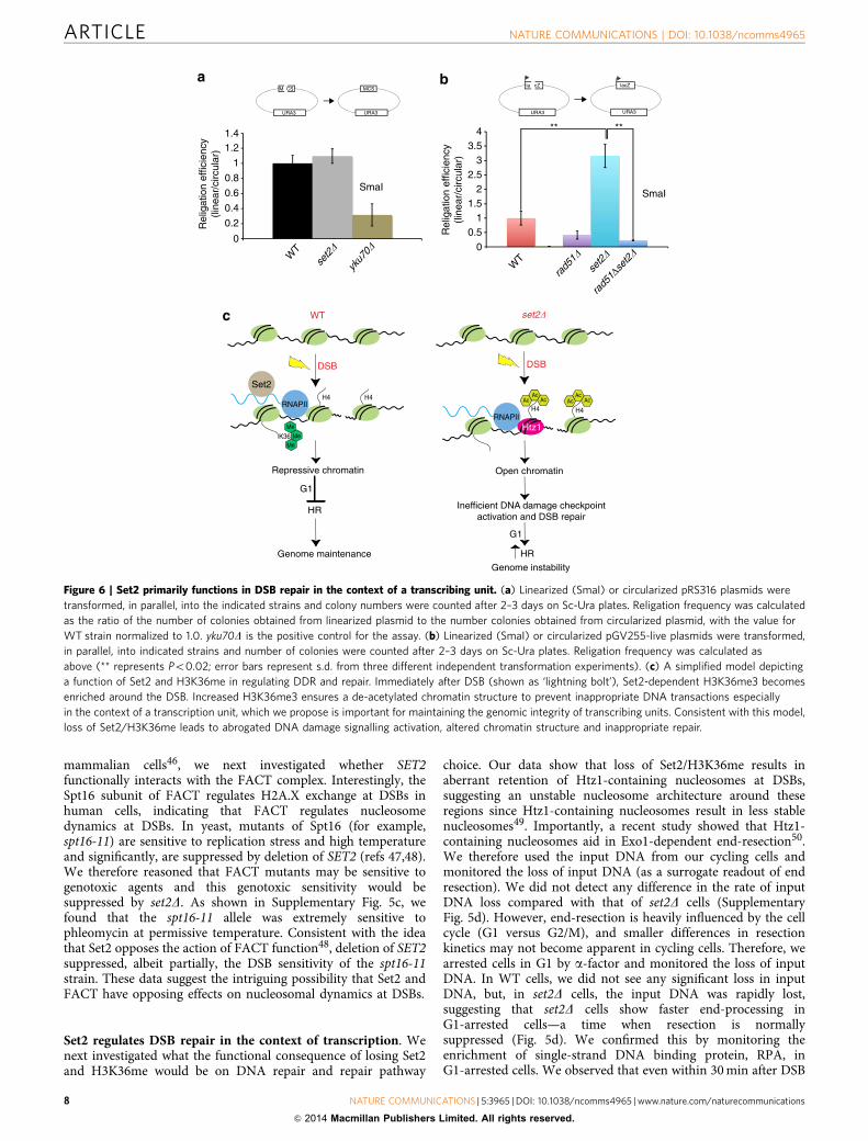
Figure 6 | Set2 primarily functions in DSB repair in the context of a transcribing unit. (a) Linearized (SmaI) or circularized pRS316 plasmids were
transformed, in parallel, into the indicated strains and colony numbers were counted after 2–3 days on Sc-Ura plates. Religation frequency was calculated
as the ratio of the number of colonies obtained from linearized plasmid to the number colonies obtained from circularized plasmid, with the value for
WT strain normalized to 1.0. yku70D is the positive control for the assay. (b) Linearized (SmaI) or circularized pGV255-live plasmids were transformed,
in parallel, into indicated strains and number of colonies were counted after 2–3 days on Sc-Ura plates. Religation frequency was calculated as
above (** represents Po0.02; error bars represent s.d. from three different independent transformation experiments). (c) A simplified model depicting
a function of Set2 and H3K36me in regulating DDR and repair. Immediately after DSB (shown as ‘lightning bolt’), Set2-dependent H3K36me3 becomes
enriched around the DSB. Increased H3K36me3 ensures a de-acetylated chromatin structure to prevent inappropriate DNA transactions especially
in the context of a transcription unit, which we propose is important for maintaining the genomic integrity of transcribing units. Consistent with this model,
loss of Set2/H3K36me leads to abrogated DNA damage signalling activation, altered chromatin structure and inappropriate repair.
ARTICLE NATURE COMMUNICATIONS | DOI: 10.1038/ncomms4965
8 NATURE COMMUNICATIONS | 5:3965 | DOI: 10.1038/ncomms4965 | www.nature.com/naturecommunications
& 2014 Macmillan Publishers Limited. All rights reserved.
induction, there is more RPA around a DSB in a set2D ascompared with WT (Supplementary Fig. 6a,b). This effect is likelya direct consequence of having more Htz1-containingnucleosomes in a set2D as well as reduced g-H2A.X, which isknown to be inhibitory to resection37.
One prediction from this observation would be that Set2should regulate NHEJ, and in the absence of Set2, one should seelower end-joining efficiency. A canonical assay used to monitorthe efficiency of end-joining is the calculation of the ratio ofcolony numbers obtained after transformation of linearizedplasmids to that obtained from transforming a circular plasmid(that is, re-ligation efficiency). Using a plasmid where wegenerated a blunt-end cut in the plasmid multiple cloning site,we observed that there was no difference in the re-ligationefficiency between WT and set2D strains (Fig. 6a). However, sinceSet2 functions in the context of transcription units, wehypothesized that the function of Set2 in DNA repair would bemore evident if our cut site was in the context of a gene body. Wetherefore carried out the plasmid re-ligation assay again, but inthis case, with the cut site within the LacZ gene body. Strikingly,we observed a significant increase in plasmid re-ligation efficiencyin set2D strains (Fig. 6b). We take these results to mean thatalthough Set2 regulates pathway choice with a likely preferencetowards promoting NHEJ, the loss of Set2 does not completelyablate this function—hence, some plasmid would be re-ligatedthat would then serve as a template for ensuing HR as the balancehas been tilted to this pathway. Importantly, HR is a canonicalmechanism of repair in transcription units51,52, which wouldfurther exacerbate the set2D phenotype, resulting in morere-ligation.
We next investigated whether this increased re-ligationefficiency is indeed a consequence of more recombination events,presumably due to transcription51. To answer that question, wedeleted RAD51 in set2D and monitored the re-ligation efficiency.Consistent with the idea proposed above, we observed that theincrease in plasmid re-ligation efficiency in a set2D wassuppressed by deletion of RAD51 (Fig. 6b), indicating thatset2D cells exhibit increased HR. To investigate whether this effectwas specific for the type of DSB induced, we used anotherrestriction enzyme (SacI) that creates staggered ends, again in thecontext of transcription. Even in this context, we observed higherplasmid re-ligation efficiency in set2D strains (SupplementaryFig. 6c). This phenotype of set2D is reminiscent of rsc and sth1mutants, where linearization of the plasmid by digestion in atranscription unit resulted in higher re-ligation efficiency thanWT cells53. Although the precise mechanism of increasedre-ligation efficiency is not yet understood, these resultsreinforce the idea that Set2/H3K36me plays an important rolein DSB repair, especially in the context of transcription.
DiscussionIn this article, we provide genetic and biochemical evidence thatSet2, and its methylation at H3K36, functions at an early stepafter DSB and plays a role in DSB repair. Specifically, we findSet2/H3K36me is critical for proper DSB checkpoint activationand impinges on pathway choice—results that can be linked to itsfunction with elongating RNAPII. The role of Set2/H3K36me inDDR and repair is likely to maintain appropriate chromatinstructure at break sites, which impinges on the molecular eventsthat occur during DNA repair.
Our data provide a temporal picture of how Set2 andH3K36me might modulate chromatin structure after a DSB.Using time-course immunoblotting and ChIP, we found that lossof Set2 leads to reduced g-H2A.X, increased acetylation of H4 andaberrant retention of H3 and Htz1 at break sites. We propose that
this altered chromatin architecture underlies the increasedend-resection observed in G1-arrested cells, suggesting that HRis activated sooner in set2D at G1. Additionally, we found thatloss of Set2 leads to increased HR, when a DSB is in the genebody. Future experiments will be required to determine whetherH3K36me3 or H3K36me2 plays an important role in recruitingany DSB repair factors to control repair pathway choice, in a cell-cycle and/or transcription-dependent manner. AlthoughH3K36me2 affects Ku70-80 recruitment to DSB in human cells54,such recruitment appears not to occur in cycling yeast cells as wedid not see any significant alterations in Ku80 recruitment inset2D (Supplementary Fig. 5e).
We found that Set2 and RNAPII are programmed fordestruction after DSBs in a proteasome-dependent manner,which correlates with a transition of H3K36me3 to H3K36me2(Fig. 4 and Supplementary Fig. 4). Although the function of thistransition in DSB repair is not yet known, we hypothesize thatthis methyl-state transition after DSB functions to ensure achromatin structure that is temporally ‘tunable’ for DSB repair.We speculate that the fine-tuning of chromatin structure viaH3K36me states would come from the ability of these differentmethyl states to activate/regulate functions of HATs andHDACs55.
With regards to Htz1, the dynamic level of Htz1 around a DSBis critical for multiple steps in the response to DSB. Intriguingly,both in vivo and in vitro, H3K36me3 and Htz1 are mutuallyexclusive56,57; hence, one function of an early increase inH3K36me3 at sites of DSBs could be to prevent Htz1deposition, thereby allowing more H2A (H2A and Htz1 beingmutually exclusive in chromatin) to be phosphorylated to formg-H2A.X domains. Consistent with this idea, we observedincreases in Htz1 and lower g-H2A.X in a set2D (Figs 3 and 5).
Our study found that Set2’s function in DDR and repair isdependent on its association with elongating RNAPII (Fig. 1).Consistent with this, we observed that when a DSB is in thecontext of transcription, deletion of SET2 leads to an increase inHR events (Fig. 6). This result might be explained by the fact thatSet2 loss tips the balance away from NHEJ to HR, which can be apreferred mechanism for break site repair in transcription units.We speculate that Set2-dependent chromatin compaction in G1aids in preventing HR events, which would further preventinappropriate recombination events that would arise in genes atthis point in the cell cycle. It is also interesting to note that Set2/H3K36me is correlated with transcription rates and genelengths15, which might fine-tune the appropriate type of repairthat would occur in genes undergoing distinct rates oftranscription.
The dependence of Set2/H3K36me for proper repair in genebodies provides further evidence for an RNAPII-dependent DNAdamage-sensing mechanism. Lindsey-Boltz and Sancar58
postulated the existence of such a DNA damage ‘sensor’ due tothe extremely slow off-rate of RNAPII on DNA. Consistent withthis idea, studies (including these herein) have found RNAPII atsites of DSB in yeast (and mammals)19 and NuA4 and otherchromatin modifiers that associate and function at DSBs are alsoassociated with RNAPII2. In mammalian cells, DSB induces localchromatin silencing if the DSB is in a gene body. We surmise thatthe transcription machinery can be ‘co-opted’ by the DNA repairand signalling machinery to sense DNA damage and then use thealready available chromatin modifiers (ATP-dependentremodellers/histone variants) to access and repair the damage,followed by restoration of the chromatin structure. This scenariowould be especially useful during gene transcription inducedR-loop formation, which can result in single or double-strandbreaks59. Interestingly, both transcription and DSBs result infairly identical mitotic recombination events52, making the
NATURE COMMUNICATIONS | DOI: 10.1038/ncomms4965 ARTICLE
NATURE COMMUNICATIONS | 5:3965 | DOI: 10.1038/ncomms4965 | www.nature.com/naturecommunications 9
& 2014 Macmillan Publishers Limited. All rights reserved.
possibility of RNAPII as a ‘constitutive DSB sensor’ even moreplausible58. Given the stochastic nature of transcription60, usingthe transcription apparatus as a sensor for DNA damage can be auseful strategy for cells to rapidly respond to exogenous orendogenous genomic insults. Consistent with this model, a recentpaper by the Legube group61 shows that transcription unitspreferentially shunt the DSB to HR-dependent pathways. Thisphenomenon also was dependent upon the presence of themammalian Set2 homologue, SETD2, and the presence ofH3K36me3. We speculate that the DSB repair machinery canutilize the context-dependent chromatin environment to accessand repair the DSB and maintain genome integrity.
Finally, the conservation of H3K36me across different specieslends credibility to the idea that H3K36me is also important forDSB repair and damage response in other organisms. Indeed, TimHumphrey’s group62 has discovered that the S. pombe Set2 alsoregulates pathway choice, because deletion of SET2 alters thebalance between NHEJ and HR. Interestingly, they find that Gcn5acetylates H3K36 (ref. 63) when Set2 is absent, and this activityleads to increased HR by presumably making the chromatin moreaccessible to the HR pathway. Additionally, Fnu et al.54 showedthat H3K36me2 catalysed by SETMAR/Metnase, which is not acanonical H3K36 methyltransferase, is critical for NHEJ inhuman cells. Taken together, we propose Set2 as a key player inthe DNA repair pathway that likely impinges on genome stability.As human SETD264 is mutated in a variety of cancers65, wespeculate that SETD2 has a conserved role in DNA repair thatwould explain its connection to human disease.
MethodsYeast strains and plasmids. All strains, unless otherwise stated, were in theBY4741 background. The strain for GAL-inducible HO endonuclease (JKM179)was obtained from James Haber, Brandeis University and subsequent genedeletions (SET2 and YKU70) were performed by gene replacement using the PCRtool kit. The strains are listed in Supplementary Table 2.
Plasmids expressing SET2 from its own promoter were obtained from ScottBriggs (Purdue University) and the H199L mutation was made in full-length SET2by site-directed mutagenesis (Quikchange, Stratagene).
DNA damage sensitivity assays. Cultures grown overnight were diluted to anOD600 of 0.25, fivefold serially diluted and spotted on plates with or withoutrelevant drugs. For UV experiments, cells were spotted on control plates andexposed to a range of UV dosage (from 20–60 J m� 2 in Stratagene crosslinkerP1800, rad4D served as a positive control for sensitivity to UV). For GAL-inducibleDSB, overnight grown cells were serially diluted (starting ODs of between 2 and 5)as indicated above and spotted on Sc-Ura plates containing either 2% dextrose or2% galactose. Pictures were taken after 2–4 days. For liquid culture experiments,log phase cultures were exposed to phleomycin (250 mg ml� 1 except for Fig. 3b,wherein 50mg ml� 1 was used to test whether the phenotype was due to a highconcentration of phleomycin) for indicated times, and then extracts were preparedby SUMEB method. About 5 O.D equivalent of cells were taken and lysed by beadbeating using the lysis buffer containing (1% SDS, 8 M urea, 10 mM MOPS, pH 6.8,10 mM EDTA, 0.01% bromophenol blue). In all, 200 ml of buffer with 200 mlequivalent of glass beads were used to lyse the cells. Bead beating was done for6–8 min, intermittently, and the extracts were centrifuged, to clarify, and boiled at95 �C for 5 min before loading the SDS–PAGE gel.
Whole-cell protein extract and immunoblots. Asynchronously grown mid-log(0.6–0.8 OD) phase cultures were lysed by SUMEB using glass beads, as mentionedabove. For histones, 15% SDS–PAGE gels were run using Laemmli buffer. For Set2,RNA Polymerase II and Rad53 blots, 8% SDS–PAGE gels were run. Gels weretransferred using a semi-dry Hoeffer apparatus at 45 mA per gel (constant currentsetting), 50 V for 1.5 h. For RNA Polymerase II, the Hoeffer setting was 55 mA pergel (constant current setting), 50 V for 1.5 h. Primary antibodies were incubated in5% milk overnight and secondary antibodies were incubated in 5% milk for anhour. The immunoblots were developed using ECL-Prime from Amersham. Anuncropped version of major immunoblots can be found in the SupplementaryFig. 7. Antibodies: H3K36me1 (ab9048; 1:1,000); H3K36me2 (active motif 39255;1:1,000 and 4 ml for ChIP), H3K36me3 (ab9050; 1:10,000 and 3 ml for ChIP),C-terminal H3 (EpiCypher 13-0001; 1:2,500 and 2 ml for ChIP), Set2 (raised in lab;1:5,000 or 1:10,000 and 5 ml for ChIP), Rad53 (obtained from Daniel Durocher,Canada; 1:2,000 (for Fig. 3a; ref. 66) and Abcam 104232; 1:2,000 (for all otherRad53 blots), H2AS129ph (Active Motif 39271; 1:1,000 and 2 ml for ChIP),
H2A (Active Motif 39235; 1:25,000 and 2 ml for ChIP), G6PDH (Sigma; 1:100,000),Ser2pCTD (1:100, gift from Dirk Eick, LMU, Munich, Germany), Rpb1(Santa Cruz, yN-18, 1:1,000), anti-Myc (9E10, 1:2,500), 4H8 against RNAPolymerase II (Active Motif; 1:10,000 and 4 ml for ChIP). Rabbit (Amersham,Donkey anti-Rabbit), goat and rat (both Jackson ImmunoResearch Laboratories)secondary antibodies was used at 1:10,000. RPA antibody (2 ml for B1 mg ofchromatin) from Valerie Borde (Insitut Pasteur, Paris, France) was used for ChIPin Supplementary Fig. 6.
ChIP. JKM179 and derivatives thereof were grown overnight in YPD and thesaturated culture was used to start a fresh culture in YEP lactate, pH 5.5 (SC-Uraplus 2% Sucrose for Fig. 5d) at an OD600 of 0.1 and grown at 30�C till the OD600
reached 0.8–1.0. Galactose was added to a final concentration of 2% to induce theexpression of HO endonuclease (at t¼ 0 h). Samples were collected at requiredtime-points and fixed with 1% (final concentration) formaldehyde. Cells were lysedusing 300 mM FA lysis buffer (containing protease inhibitor cocktail). The lysedcells were sonicated (30% output, 6 pulses 6 times; 90% duty cycle) and clarifiedby centrifuging at full speed for 15 min. Immuno-precipitations were set up,overnight, with desired amount of proteins and antibodies (antibody amounts havebeen listed above). Protein G-sepharose beads (100 ml 1:10 diluted beads per 400mlimmunoprecipitation reaction) were added to the immuno-precipitation reactionsand the reactions were allowed to incubate for 1 hour. Subsequent washes weredone in 1.4 ml of 300 mM FA Lysis buffer, 500 mM FA-lysis buffer and LiClsolution (250 mM LiCl, 10 mM Tris, 0.5% each of NP-40 and sodium deoxycholateand 1 mM EDTA). The immunoprecipitates were resuspended in TE (pH 8.0) andtreated with RNase for 30 min. The immunoprecipitates were then washed with TE(pH 8.0) and elution buffer was used to elute the DNA (15 min incubation inelution buffer followed by centrifugation at 3,000 r.p.m. for 2 minutes). The eluateswere kept at 65�C overnight to carry out the de-crosslinking step and PCRpurification kit (Qiagen) was used to extract the DNA. Details of PCR conditions,primer locations and analysis methodologies can be found in the SupplementaryMethods.
Immunoprecipitation. Immunoprecipitation for Set2-FLAG was performed inaccordance with the manufacturer’s protocol (SIGMA, catalogue no. A2220), usingthe 300 mM FA-lysis buffer used for ChIP experiments.
MG-132 experiment. Cells were grown in media containing proline as a nitrogensource overnight. The following morning, a fresh culture was started at OD 0.5with 0.003% SDS and grown for 3 h before treatment with MG-132 (75 mM) foranother 30 min. Subsequently, phleomycin (250 mg ml� 1) was added and sampleswere obtained at indicated times, and western blotting was performed as describedabove.
Plasmid re-ligation assay. About 3–5 mg of circular plasmids were digested byindicated restriction enzymes. Running the digested plasmids on gels and per-forming routine gel extraction confirmed their linearization. Subsequently, 100 ngof linearized or circularized plasmids were transformed into indicated strains(LiAc/PEG method), in parallel. The colonies were counted on Sc-Ura plates after2–3 days. Three independent transformation reactions were carried out.
Statistical analysis. For arriving at the P values in all the relevant experiments(ChIP assays and the plasmid re-ligation assay), we carried out unpaired Student’st-test in Microsoft Excel. The explanations for the error bars are mentioned in thecorresponding figure legends with relevant n values. P o0.05 was consideredstatistically significant.
References1. Hoeijmakers, J. H. DNA damage, aging, and cancer. N. Engl. J. Med. 361,
1475–1485 (2009).2. Papamichos-Chronakis, M. & Peterson, C. L. Chromatin and the genome
integrity network. Nat. Rev. Genet. 14, 62–75 (2013).3. Azvolinsky, A., Giresi, P. G., Lieb, J. D. & Zakian, V. A. Highly transcribed
RNA polymerase II genes are impediments to replication fork progression inSaccharomyces cerevisiae. Mol. Cell 34, 722–734 (2009).
4. Bermejo, R., Lai, M. S. & Foiani, M. Preventing replication stress to maintaingenome stability: resolving conflicts between replication and transcription. Mol.Cell 45, 710–718 (2012).
5. Keogh, M. C. et al. Cotranscriptional set2 methylation of histone H3 lysine 36recruits a repressive Rpd3 complex. Cell 123, 593–605 (2005).
6. Strahl, B. D. et al. Set2 is a nucleosomal histone H3-selective methyltransferasethat mediates transcriptional repression. Mol. Cell Biol. 22, 1298–1306 (2002).
7. Kizer, K. O. et al. A novel domain in Set2 mediates RNA polymerase IIinteraction and couples histone H3 K36 methylation with transcript elongation.Mol. Cell Biol. 25, 3305–3316 (2005).
8. Xiao, T. et al. Phosphorylation of RNA polymerase II CTD regulates H3methylation in yeast. Genes Dev. 17, 654–663 (2003).
ARTICLE NATURE COMMUNICATIONS | DOI: 10.1038/ncomms4965
10 NATURE COMMUNICATIONS | 5:3965 | DOI: 10.1038/ncomms4965 | www.nature.com/naturecommunications
& 2014 Macmillan Publishers Limited. All rights reserved.
9. Wagner, E. J. & Carpenter, P. B. Understanding the language of Lys36methylation at histone H3. Nat. Rev. Mol. Cell Biol. 13, 115–126 (2012).
10. Li, B., Howe, L., Anderson, S., Yates, 3rd J. R. & Workman, J. L. The Set2histone methyltransferase functions through the phosphorylated carboxyl-terminal domain of RNA polymerase II. J. Biol. Chem. 278, 8897–8903 (2003).
11. Carrozza, M. J. et al. Histone H3 methylation by Set2 directs deacetylation ofcoding regions by Rpd3S to suppress spurious intragenic transcription. Cell123, 581–592 (2005).
12. Joshi, A. A. & Struhl, K. Eaf3 chromodomain interaction with methylatedH3-K36 links histone deacetylation to Pol II elongation. Mol. Cell 20, 971–978(2005).
13. Govind, C. K. et al. Phosphorylated Pol II CTD recruits multiple HDACs,including Rpd3C(S), for methylation-dependent deacetylation of ORFnucleosomes. Mol. Cell 39, 234–246 (2010).
14. Smolle, M. et al. Chromatin remodelers Isw1 and Chd1 maintain chromatinstructure during transcription by preventing histone exchange. Nat. Struct. Mol.Biol. 19, 884–892 (2012).
15. Venkatesh, S. et al. Set2 methylation of histone H3 lysine 36 suppresses histoneexchange on transcribed genes. Nature 489, 452–455 (2012).
16. Moore, J. K. & Haber, J. E. Cell cycle and genetic requirements of two pathwaysof nonhomologous end-joining repair of double-strand breaks inSaccharomyces cerevisiae. Mol. Cell Biol. 16, 2164–2173 (1996).
17. Winsor, T. S., Bartkowiak, B., Bennett, C. B. & Greenleaf, A. L. A. DNA damageresponse system associated with the phosphoCTD of elongating RNApolymerase II. PLoS One 8, e60909 (2013).
18. Youdell, M. L. et al. Roles for Ctk1 and Spt6 in regulating the differentmethylation states of histone H3 lysine 36. Mol. Cell Biol. 28, 4915–4926(2008).
19. Tsukuda, T., Fleming, A. B., Nickoloff, J. A. & Osley, M. A. Chromatinremodelling at a DNA double-strand break site in Saccharomyces cerevisiae.Nature 438, 379–383 (2005).
20. Bandyopadhyay, S. et al. Rewiring of genetic networks in response to DNAdamage. Science 330, 1385–1389 (2010).
21. Guenole, A. et al. Dissection of DNA damage responses using multiconditionalgenetic interaction maps. Mol. Cell 49, 346–358 (2013).
22. Tompa, R. & Madhani, H. D. Histone H3 lysine 36 methylation antagonizessilencing in Saccharomyces cerevisiae independently of the Rpd3S histonedeacetylase complex. Genetics 175, 585–593 (2007).
23. Lenstra, T. L. et al. The specificity and topology of chromatin interactionpathways in yeast. Mol. Cell 42, 536–549 (2011).
24. Shroff, R. et al. Distribution and dynamics of chromatin modificationinduced by a defined DNA double-strand break. Curr. Biol. 14, 1703–1711(2004).
25. Keogh, M. C. et al. A phosphatase complex that dephosphorylatesgammaH2AX regulates DNA damage checkpoint recovery. Nature 439,497–501 (2006).
26. Tamburini, B. A. & Tyler, J. K. Localized histone acetylation and deacetylationtriggered by the homologous recombination pathway of double-strand DNArepair. Mol. Cell Biol. 25, 4903–4913 (2005).
27. Lin, Y. Y. et al. A comprehensive synthetic genetic interaction networkgoverning yeast histone acetylation and deacetylation. Genes Dev. 22,2062–2074 (2008).
28. Warmerdam, D. O. & Kanaar, R. Dealing with DNA damage: relationshipsbetween checkpoint and repair pathways. Mutat. Res. 704, 2–11 (2010).
29. Kalocsay, M., Hiller, N. J. & Jentsch, S. Chromosome-wide Rad51 spreadingand SUMO-H2A.Z-dependent chromosome fixation in response to a persistentDNA double-strand break. Mol. Cell 33, 335–343 (2009).
30. Lazzaro, F. et al. Histone methyltransferase Dot1 and Rad9 inhibit single-stranded DNA accumulation at DSBs and uncapped telomeres. EMBO J. 27,1502–1512 (2008).
31. Papamichos-Chronakis, M., Krebs, J. E. & Peterson, C. L. Interplay betweenIno80 and Swr1 chromatin remodeling enzymes regulates cell cyclecheckpoint adaptation in response to DNA damage. Genes Dev. 20, 2437–2449(2006).
32. van Attikum, H., Fritsch, O. & Gasser, S. M. Distinct roles for SWR1 andINO80 chromatin remodeling complexes at chromosomal double-strandbreaks. EMBO J. 26, 4113–4125 (2007).
33. Wysocki, R. et al. Role of Dot1-dependent histone H3 methylation in G1 and Sphase DNA damage checkpoint functions of Rad9. Mol. Cell Biol. 25,8430–8443 (2005).
34. Grenon, M. et al. Docking onto chromatin via the Saccharomyces cerevisiaeRad9 Tudor domain. Yeast 24, 105–119 (2007).
35. Huyen, Y. et al. Methylated lysine 79 of histone H3 targets 53BP1 to DNAdouble-strand breaks. Nature 432, 406–411 (2004).
36. Costelloe, T. et al. The yeast Fun30 and human SMARCAD1 chromatinremodellers promote DNA end resection. Nature 489, 581–584 (2012).
37. Chen, X. et al. The Fun30 nucleosome remodeller promotes resection of DNAdouble-strand break ends. Nature 489, 576–580 (2012).
38. Jazayeri, A., McAinsh, A. D. & Jackson, S. P. Saccharomyces cerevisiae Sin3pfacilitates DNA double-strand break repair. Proc. Natl Acad. Sci. USA 101,1644–1649 (2004).
39. Bird, A. W. et al. Acetylation of histone H4 by Esa1 is required for DNAdouble-strand break repair. Nature 419, 411–415 (2002).
40. Miller, K. M. et al. Human HDAC1 and HDAC2 function in the DNA-damageresponse to promote DNA nonhomologous end-joining. Nat. Struct. Mol. Biol.17, 1144–1151 (2010).
41. Symington, L. S. & Gautier, J. Double-strand break end resection and repairpathway choice. Annu. Rev. Genet. 45, 247–271 (2011).
42. Ginsburg, D. S., Govind, C. K. & Hinnebusch, A. G. NuA4 lysineacetyltransferase Esa1 is targeted to coding regions and stimulates transcriptionelongation with Gcn5. Mol. Cell Biol. 29, 6473–6487 (2009).
43. Altaf, M. et al. NuA4-dependent acetylation of nucleosomal histones H4 andH2A directly stimulates incorporation of H2A.Z by the SWR1 complex. J. Biol.Chem. 285, 15966–15977 (2010).
44. Raisner, R. M. et al. Histone variant H2A.Z marks the 5’ ends of both active andinactive genes in euchromatin. Cell 123, 233–248 (2005).
45. Zhang, H., Roberts, D. N. & Cairns, B. R. Genome-wide dynamics of Htz1, ahistone H2A variant that poises repressed/basal promoters for activationthrough histone loss. Cell 123, 219–231 (2005).
46. Heo, K. et al. FACT-mediated exchange of histone variant H2AX regulated byphosphorylation of H2AX and ADP-ribosylation of Spt16. Mol. Cell 30, 86–97(2008).
47. Formosa, T. et al. Defects in SPT16 or POB3 (yFACT) in Saccharomycescerevisiae cause dependence on the Hir/Hpc pathway: polymerase passage maydegrade chromatin structure. Genetics 162, 1557–1571 (2002).
48. Biswas, D. et al. A role for Chd1 and Set2 in negatively regulating DNAreplication in Saccharomyces cerevisiae. Genetics 178, 649–659 (2008).
49. Jin, C. & Felsenfeld, G. Nucleosome stability mediated by histone variants H3.3and H2A.Z. Genes Dev. 21, 1519–1529 (2007).
50. Adkins, N. L., Niu, H., Sung, P. & Peterson, C. L. Nucleosome dynamicsregulates DNA processing. Nat. Struct. Mol. Biol. 20, 836–842 (2013).
51. Aguilera, A. The connection between transcription and genomic instability.EMBO J. 21, 195–201 (2002).
52. Gonzalez-Barrera, S., Garcia-Rubio, M. & Aguilera, A. Transcription anddouble-strand breaks induce similar mitotic recombination events inSaccharomyces cerevisiae. Genetics 162, 603–614 (2002).
53. Chai, B., Huang, J., Cairns, B. R. & Laurent, B. C. Distinct roles for the RSC andSwi/Snf ATP-dependent chromatin remodelers in DNA double-strand breakrepair. Genes Dev. 19, 1656–1661 (2005).
54. Fnu, S. et al. Methylation of histone H3 lysine 36 enhances DNA repair bynonhomologous end-joining. Proc. Natl Acad. Sci. USA 108, 540–545 (2011).
55. Zentner, G. E. & Henikoff, S. Regulation of nucleosome dynamics by histonemodifications. Nat. Struct. Mol. Biol. 20, 259–266 (2013).
56. Chen, J., Miller, A., Kirchmaier, A. L. & Irudayaraj, J. M. Single-molecule toolselucidate H2A.Z nucleosome composition. J. Cell Sci. 125, 2954–2964 (2012).
57. Li, B. et al. Preferential occupancy of histone variant H2AZ at inactivepromoters influences local histone modifications and chromatin remodelling.Proc. Natl Acad. Sci. USA 102, 18385–18390 (2005).
58. Lindsey-Boltz, L. A. & Sancar, A. RNA polymerase: the most specific damagerecognition protein in cellular responses to DNA damage? Proc. Natl Acad. Sci.USA 104, 13213–13214 (2007).
59. Bermejo, R. et al. The replication checkpoint protects fork stability by releasingtranscribed genes from nuclear pores. Cell 146, 233–246 (2011).
60. Raj, A. & van Oudenaarden, A. Nature, nurture, or chance: stochastic geneexpression and its consequences. Cell 135, 216–226 (2008).
61. Aymard, F. et al. Transcriptionally active chromatin recruits homologousrecombination at DNA double-strand breaks. Nat. Struct. Mol. Biol. 21,366–374 (2014).
62. Pai et al. A histone H3K36 chromatin switch coordinates DNA double-strandbreak repair pathway choice. Nat. Commun. 5, 4091 (2014).
63. Morris, S. A. et al. Identification of histone H3 lysine 36 acetylation as a highlyconserved histone modification. J. Biol. Chem. 282, 7632–7640 (2007).
64. Luco, R. F. et al. Regulation of alternative splicing by histone modifications.Science 327, 996–1000 (2010).
65. Lawrence, M. S. et al. Discovery and saturation analysis of cancer genes across21 tumour types. Nature 505, 495–501 (2014).
66. Sweeney, F. D. et al. Saccharomyces cerevisiae Rad9 acts as a Mec1 adaptor toallow Rad53 activation. Curr. Biol. 15, 1364–1375 (2005).
AcknowledgementsWe thank James Haber (Brandeis University) for JKM179 strain and valuable sugges-tions, Daniel Durocher (University of Toronto) for Rad53 antibody used in parts ofFig. 3, Marcus Smolka (Cornell University) for suggesting the Abcam Rad53 antibody(used in parts of Fig. 3), Ugur Yavuzer (Bilkent University, Turkey; for the plasmids usedin Fig. 6b and Supplementary Fig. 6c), Katsunori Sugimoto (University of Medicine andDentistry New Jersey; for the ADH4-HO strain used in Supplementary Fig. 1a), Valerie
NATURE COMMUNICATIONS | DOI: 10.1038/ncomms4965 ARTICLE
NATURE COMMUNICATIONS | 5:3965 | DOI: 10.1038/ncomms4965 | www.nature.com/naturecommunications 11
& 2014 Macmillan Publishers Limited. All rights reserved.

Borde (Institut Curie, Paris) for the RPA antibody and suggestions for RPA chip andScott Briggs for the full-length and the SRID SET2 plasmid. We thank TimothyHumphrey (Oxford University) for communicating results before publication. We arethankful to Jean Cook, Lindsay Faircloth Rizzardi, Zu-Wen Sun, Howard Fried andmembers of the Strahl lab, especially Raghuvar Dronamraju (for helpful suggestions) andStephen McDaniel (for some of the unpublished strains). This work was funded by a NIHgrant to B.D.S. (GM068088).
Author contributionsD.K.J. designed, performed and analysed all the experiments, with valuable inputs fromB.D.S. D.K.J. and B.D.S. wrote the manuscript.
Additional informationSupplementary Information accompanies this paper at http://www.nature.com/naturecommunications
Competing financial interests: The authors declare no competing financial interests.
Reprints and permission information is available online at http://npg.nature.com/reprintsandpermissions/
How to cite this article: Jha, D. K. et al. An RNA Polymerase II-coupled function forhistone H3K36 methylation in checkpoint activation and DSB repair. Nat. Commun.5:3965 doi: 10.1038/ncomms4965 (2014).
ARTICLE NATURE COMMUNICATIONS | DOI: 10.1038/ncomms4965
12 NATURE COMMUNICATIONS | 5:3965 | DOI: 10.1038/ncomms4965 | www.nature.com/naturecommunications
& 2014 Macmillan Publishers Limited. All rights reserved.
Copyright © 2022 FDOKUMEN