Phosphoramidate Dinucleosides as Hepatitis C Virus Polymerase Inhibitors
13
Phosphoramidate Dinucleosides as Hepatitis C Virus Polymerase Inhibitors Ivan Zlatev, †,⊥ He ´le `ne Dutartre, ‡,⊥ Ivan Barvik, § Johan Neyts, | Bruno Canard, ‡ Jean-Jacques Vasseur, † Karine Alvarez,* ,‡ and Franc ¸ois Morvan* ,† Institut des Biomole ´cules Max Mousseron (IBMM), UMR 5247 CNRSsUniVersite ´ Montpellier 1sUniVersite ´ Montpellier 2, Place Euge `ne Bataillon, CC1704, 34095 Montpellier Cedex 5, France, Department of Structural Virology, AFMB UMR 6098, CNRS UniVersite ´ de la Me ´diterrane ´e, Case 925, 163 AVenue de Luminy, 13288 Marseille, France, Institute of Physics, Charles UniVersity, Ke KarloVu 5, Prague 12116 2, Czech Republic, Rega Institute for Medical Research, Katholieke UniVersiteit LeuVen, Minderbroedersstraat 10, B-3000 LeuVen, Belgium ReceiVed May 23, 2008 GC dinucleosides exhibiting a phosphoramidate internucleosidic linkage with neutral, amphiphile, positively or negatively charged side chains were synthesized. Their potential inhibitory effect on the hepatitis C virus (HCV) NS5B polymerase was evaluated in vitro and in HCV replicon containing cells. Whereas the amphiphile and the positively charged analogues were found to be inactive, the neutral (1) and the negatively charged (4) ones inhibited enzyme activity when tested as a diastereoisomeric mixture. The most potent inhibitor proved to be the Sp isomer of the 5′-thiophosphorylated dinucleotide bearing the carboxylic side chain (8) (IC 50 of 25 µM in vitro and an EC 50 of 9 µM in HCV subgenomic replicon). Molecular modeling suggests that the phosphoramidate dinucleoside (8) is stabilized in the active site by interactions with magnesium ions and lysine and arginine residues of the polymerase. Introduction The hepatitis C virus (HCV a ) represents a global health problem; about 3% of the world’s population is chronically infected and at risk of developing liver cirrhosis and/or hepatocellular carcinoma. HCV infection is the leading reason for liver transplantation in the West. 1 Current therapy consists of a combination of interferon-R with the nucleoside analogue ribavirin and results in 40-70% of sustained virological responses depending on the genotype. 2 However, this therapy is associated with significant side effects, 3 and there is no approved therapeutic alternative for nonresponders. There is thus an urgent need for new therapeutic molecules active against the HCV infection. HCV is an enveloped, positive-strand RNA virus; the 9.6 kb genome encodes a single large polyprotein, which is cleaved by cellular and viral proteases into four structural (Core, E1, E2, and p7) and six nonstructural proteins (NS2, NS3, NS4A, NS4B, NS5A, and NS5B). The nonstructural proteins assemble into a replication complex that is responsible for the viral RNA genome synthesis (for a review see ref 4). Because they bear essential enzymatic activities for viral replication, the NS3 protease-helicase and the NS5B polymerase are targets of choice for antiviral intervention. Moreover, the NS5B polymerase is the key component of the replicative complex. It is a endoplas- mic reticulum membrane-associated RNA-dependent-RNA- polymerase (RdRp) able to replicate RNA without a primer in a so-called de novo synthesis mechanism. 5 In vitro, however, the C-terminal truncated recombinant polymerase can use a wide range of hetero- or homopolymeric templates in a primer- dependent or primer-independent fashion. 6 During de novo RNA synthesis, the RdRp has to synthesize first its RNA primer and then copy the entire RNA template. RNA synthesis can thus be separated into two steps 7,8 that are structurally and biochemically distinct: the initiation step leading to the RNA primer synthesis and the elongation step of the neosynthezised RNA up to the end of the template. Intensive drug screening campaigns have led to the discovery of many NS5B inhibitors, some of them being evaluated in clinical settings (reviewed in refs 9, 10). Two main classes of NS5B RdRp inhibitors have been reported: non-nucleoside inhibitors (NNI) and nucleoside inhibitors (NI). NNIs are allosteric, noncompetitive inhibitors that target the alloenzyme, free of RNA substrates (reviewed in ref 11). For most of them, the mode of action requires the binding to the apoenzyme before engagement of the enzyme into an elongative complex. For NIs, the corresponding triphosphates (TPs) are competitors of the natural nucleoside triphosphates and can be used as substrates by the polymerase. After their incorporation into the nascent RNA, they act as nonobligate chain terminators. 12 Alternatively, compounds that target the active site before the enzyme has entered into the elongation steps of RNA synthesis may be potent inhibitors. For example, it has been reported that dinucleotides can be used more efficiently as primer molecules than mononucleotides in in vitro RNA replication assay. 6 On the basis of these results modified dinucleotides, lacking the 3′-hydroxyl necessary for the polymerization reaction, were synthesized and were shown able to decrease the RNA synthesis measured in vitro, although with modest IC 50 values. 13 We took advantage of this novel approach to synthesize new dinucleotides bearing modifications on the internucleosidic linkage in order to increase both their binding to the active site of the polymerase and their affinity to the RNA template and hence their inhibition properties. Our target compounds were * To whom correspondence should be addressed. For F.M.: phone, +33 467 144 961; fax, +33 467 042 029; E-mail: [email protected]. For K.A.: Phone: +33 491 825 571; Fax: +33 491 266 720; E-mail: [email protected]. † Universite ´ Montpellier 2. ‡ Universite ´ de la Me ´diterrane ´e. § Charles University. | Katholieke Universiteit Leuven. ⊥ These authors contributed equally to the work. a Abbreviations: HCV, hepatitis C virus; NS3, non structural protein 3; NS5B, non structural protein 5B; RdRp, RNA-dependent-RNA-polymerase; NNI, non nucleoside inhibitor; NI, nucleoside inhibitor; MP, monophos- phate; TP, triphosphate; GC, guanosin-3′-yl-cytidin-5′-yl dinucleotide; GMP, guanosine monophosphate; GTP, guanosine triphosphate; NTP, nucleoside triphosphate. J. Med. Chem. 2008, 51, 5745–5757 5745 10.1021/jm800617c CCC: $40.75 2008 American Chemical Society Published on Web 08/23/2008
-
Upload
independent -
Category
Documents
-
view
0 -
download
0
Transcript of Phosphoramidate Dinucleosides as Hepatitis C Virus Polymerase Inhibitors

Phosphoramidate Dinucleosides as Hepatitis C Virus Polymerase Inhibitors
Ivan Zlatev,†,⊥ Helene Dutartre,‡,⊥ Ivan Barvik,§ Johan Neyts,| Bruno Canard,‡ Jean-Jacques Vasseur,† Karine Alvarez,*,‡ andFrancois Morvan*,†
Institut des Biomolecules Max Mousseron (IBMM), UMR 5247 CNRSsUniVersite Montpellier 1sUniVersite Montpellier 2,Place Eugene Bataillon, CC1704, 34095 Montpellier Cedex 5, France, Department of Structural Virology, AFMB UMR 6098,CNRS UniVersite de la Mediterranee, Case 925, 163 AVenue de Luminy, 13288 Marseille, France, Institute of Physics, Charles UniVersity, KeKarloVu 5, Prague 12116 2, Czech Republic, Rega Institute for Medical Research, Katholieke UniVersiteit LeuVen, Minderbroedersstraat 10,B-3000 LeuVen, Belgium
ReceiVed May 23, 2008
GC dinucleosides exhibiting a phosphoramidate internucleosidic linkage with neutral, amphiphile, positivelyor negatively charged side chains were synthesized. Their potential inhibitory effect on the hepatitis C virus(HCV) NS5B polymerase was evaluated in vitro and in HCV replicon containing cells. Whereas theamphiphile and the positively charged analogues were found to be inactive, the neutral (1) and the negativelycharged (4) ones inhibited enzyme activity when tested as a diastereoisomeric mixture. The most potentinhibitor proved to be the Sp isomer of the 5′-thiophosphorylated dinucleotide bearing the carboxylic sidechain (8) (IC50 of 25 µM in vitro and an EC50 of 9 µM in HCV subgenomic replicon). Molecular modelingsuggests that the phosphoramidate dinucleoside (8) is stabilized in the active site by interactions withmagnesium ions and lysine and arginine residues of the polymerase.
Introduction
The hepatitis C virus (HCVa) represents a global healthproblem; about 3% of the world’s population is chronicallyinfected and at risk of developing liver cirrhosis and/orhepatocellular carcinoma. HCV infection is the leading reasonfor liver transplantation in the West.1 Current therapy consistsof a combination of interferon-R with the nucleoside analogueribavirin and results in 40-70% of sustained virologicalresponses depending on the genotype.2 However, this therapyis associated with significant side effects,3 and there is noapproved therapeutic alternative for nonresponders. There is thusan urgent need for new therapeutic molecules active againstthe HCV infection.
HCV is an enveloped, positive-strand RNA virus; the 9.6 kbgenome encodes a single large polyprotein, which is cleavedby cellular and viral proteases into four structural (Core, E1,E2, and p7) and six nonstructural proteins (NS2, NS3, NS4A,NS4B, NS5A, and NS5B). The nonstructural proteins assembleinto a replication complex that is responsible for the viral RNAgenome synthesis (for a review see ref 4). Because they bearessential enzymatic activities for viral replication, the NS3protease-helicase and the NS5B polymerase are targets of choicefor antiviral intervention. Moreover, the NS5B polymerase isthe key component of the replicative complex. It is a endoplas-mic reticulum membrane-associated RNA-dependent-RNA-
polymerase (RdRp) able to replicate RNA without a primer ina so-called de novo synthesis mechanism.5 In vitro, however,the C-terminal truncated recombinant polymerase can use a widerange of hetero- or homopolymeric templates in a primer-dependent or primer-independent fashion.6 During de novo RNAsynthesis, the RdRp has to synthesize first its RNA primer andthen copy the entire RNA template. RNA synthesis can thus beseparated into two steps7,8 that are structurally and biochemicallydistinct: the initiation step leading to the RNA primer synthesisand the elongation step of the neosynthezised RNA up to theend of the template.
Intensive drug screening campaigns have led to the discoveryof many NS5B inhibitors, some of them being evaluated inclinical settings (reviewed in refs 9, 10). Two main classes ofNS5B RdRp inhibitors have been reported: non-nucleosideinhibitors (NNI) and nucleoside inhibitors (NI). NNIs areallosteric, noncompetitive inhibitors that target the alloenzyme,free of RNA substrates (reviewed in ref 11). For most of them,the mode of action requires the binding to the apoenzyme beforeengagement of the enzyme into an elongative complex. For NIs,the corresponding triphosphates (TPs) are competitors of thenatural nucleoside triphosphates and can be used as substratesby the polymerase. After their incorporation into the nascentRNA, they act as nonobligate chain terminators.12 Alternatively,compounds that target the active site before the enzyme hasentered into the elongation steps of RNA synthesis may bepotent inhibitors. For example, it has been reported thatdinucleotides can be used more efficiently as primer moleculesthan mononucleotides in in vitro RNA replication assay.6 Onthe basis of these results modified dinucleotides, lacking the3′-hydroxyl necessary for the polymerization reaction, weresynthesized and were shown able to decrease the RNA synthesismeasured in vitro, although with modest IC50 values.13
We took advantage of this novel approach to synthesize newdinucleotides bearing modifications on the internucleosidiclinkage in order to increase both their binding to the active siteof the polymerase and their affinity to the RNA template andhence their inhibition properties. Our target compounds were
* To whom correspondence should be addressed. For F.M.: phone, +33467 144 961; fax, +33 467 042 029; E-mail: [email protected]. ForK.A.: Phone: +33 491 825 571; Fax: +33 491 266 720; E-mail:[email protected].
† Universite Montpellier 2.‡ Universite de la Mediterranee.§ Charles University.| Katholieke Universiteit Leuven.⊥ These authors contributed equally to the work.a Abbreviations: HCV, hepatitis C virus; NS3, non structural protein 3;
NS5B, non structural protein 5B; RdRp, RNA-dependent-RNA-polymerase;NNI, non nucleoside inhibitor; NI, nucleoside inhibitor; MP, monophos-phate; TP, triphosphate; GC, guanosin-3′-yl-cytidin-5′-yl dinucleotide; GMP,guanosine monophosphate; GTP, guanosine triphosphate; NTP, nucleosidetriphosphate.
J. Med. Chem. 2008, 51, 5745–5757 5745
10.1021/jm800617c CCC: $40.75 2008 American Chemical SocietyPublished on Web 08/23/2008
designed based on several previously published structural dataof the NS5B enzyme in its putative RNA synthesis initiationconformation.14,15 As it was also previously demonstrated thatthe 5′-phosphate group of the dinucleotide was required forinhibition,13 probably because it allowed hydrogen bonding andelectrostatic interaction with residues present in the active site,we decided to study this modification thoroughly. Moreover,such 5′-phosphorylated dinucleotide structures would act asdirect substrate to the enzyme, without the need of anyintracellular 5′-phosphorylation, as required for NIs. As amodification for the internucleosidic linkage, we chose aphosphoramidate diester linkage. The latter is a chemicalmodification used either in a prodrug approach for nucleosideanalogues16-18 or in modified oligonucleotides19,20 and allowsthe introduction of a large variety of side chain functionalities.
Because the 3′-end of the viral RNA negative strand templateof HCV is GC3′, the complementary sequence of the dimer is5′GC. A 2′-O-methyl guanosine was chosen instead of naturalguanosine to gain higher enzymatic and chemical stability ofthe final dinucleotide. The phosphoramidate linkage is also moreresistant to nucleases,21 hence the resulting dinucleotides wouldhave higher stability in biological media. Thus, we first designedand synthesized 5′-phosphorylated dinucleotides made of a 2′-O-methyl guanosine and a cytidine, linked together with aphosphoramidate function bearing different side chains.20 Toestablish a structure-activity relationship (SAR), we used sidechains with neutral, cationic, amphiphile, and anionic groupsexhibiting different properties. Then, to study the influence ofthe 5′-environment, phosphoramidate dinucleotide analogueswith a 5′-hydroxyl group or bearing a 5′-phosphorothioatemonoester instead of the 5′-phosphate monoester were prepared.Then, we evaluated their ability to inhibit the NS5B polymeraseactivity in vitro. A molecular model of the NS5B polymeraseand the most potent dinucleotide analogue was constructed toexplain the mechanism of inhibition. Finally, the potential
inhibitory effect on HCV replicon replication in cell cultureswas evaluated.
Results and Discussion
Chemistry. The preparation of these 2′-O-Me-GC phospho-ramidate dinucleosides proceeded by a strategy using convergentand divergent syntheses. First, a convergent synthesis using 3′-OH-2′-O-Me guanosine derivatives (9-11) and the 5′-H-phosphonate monoester of N4,2′,3′-O,O-tribenzoyl cytidine (14)was applied, affording common intermediates 15-17 with aH-phosphonate diester linkage between the 2′-O-Me guanosineand the cytidine moieties. Second, the divergent strategyconcerned the amidative oxidation,22 allowing, from the com-mon intermediates 15-17, the preparation of the different dimersI with various phosphoramidate side chains (Scheme 1).
The building block 9 is commercially available. The buildingblocks 10 and 11 were synthesized from N2-isobutyryl-2′-O-methyl guanosine23 by a selective 5′-phosphitylation using solid-supported reagents as previously reported.24,25
The N4,2′,3′-O,O-tribenzoyl-5′-H-phosphonate monoester ofcytidine (14) was prepared according to a five-step procedurewith a 63% overall yield (Scheme 2). Cytidine was firstselectively benzoylated on the N4 position by means of benzoicanhydride in DMF.26 After replacement of DMF by pyridine,the crude was directly dimethoxytritylated on the 5′-hydroxyl(90%) by addition of dimethoxytrityl chloride (Scheme 2). Afterpurification, the nucleoside 12 was benzoylated on both 2′ and3′ hydroxyls by means of benzoyl chloride, and after workup,the crude was directly treated with benzene sulfonic acid,affording after purification the N4,2′,3′-O,O-tribenzoyl cytidine13 (88%). Finally, 13 was treated with diphenylphosphitefollowed by aqueous hydrolysis,27 affording the expected 5′-H-phosphonate monoester of N4,2′,3′-O,O-tribenzoyl cytidine14 (80%).
Scheme 1. Retrosynthetic Pathway for the Synthesis of Phosphoramidate Dinucleosides I
Scheme 2. Synthetic Pathway for the Synthesis of 14
5746 Journal of Medicinal Chemistry, 2008, Vol. 51, No. 18 ZlateV et al.

The coupling of the two building blocks 14 and 9-11 to formH-phosphonate diesters 15-17 was carried out using a solid-supported acyl chloride activator28 (Scheme 3). This couplingproceeded in two steps, first, the H-phosphonate monoesterfunction of 14 reacted with the solid-supported acyl chlorideand, second, the formed mixed anhydride was displaced by the3′-hydroxyl of 9-11 to yield 15-17 in solution. By this way,the excess of 14 was trapped on the solid support and onlydimers 15-17 and pyridinium salt remained in solution. Asimple filtration, an aqueous extraction, and a precipitationin diethyl ether afforded the dimers 15-17 with >85% HPLCpurity (Figure S1 in Supporting Information). Because thiscoupling reaction is nonstereoselective, the H-phosphonatediesters were all a ∼1:1 mixture of Rp and Sp diastereoiso-mers on the chiral phosphorus center and their presence wasconfirmed by 31P NMR as two peaks (δP-H between 7.3 and9.8 ppm).
The crude H-phosphonate diesters 15-17 were dissolved indry pyridine, and a mixture of carbon tetrachloride and amine
was added. The amidative oxidation occurred to form the twodiastereoisomers (Sp and Rp) of the phosphoramidate I withsome minor formation of phosphodiester linkage correspondingto a side-oxidation due to trace of water. First, the oxidation of15 was performed in the presence of four different amines:2-methoxyethylamine, 3-(dimethylamino)-1-propylamine, his-tamine, or 6-aminocaproic acid methyl ester to afford, afterdeprotection, the corresponding dimers bearing neutral (1),cationic (2), amphiphile (3), or anionic (4) side chains,respectively (Scheme 4). Pure target compounds were obtainedafter two chromatographic steps (ion exchange and reversephase). Finally, sodium ion-exchange was performed, yieldingthe desired compounds as their sodium salt.
To further establish the role of the 5′-terminal function groupin a SAR, dimers 16 and 17 were also oxidized in presence of2-methoxyethylamine and 6-aminocaproic acid methyl ester,affording, after deprotection, phosphoramidate dimers with aneutral or an anionic chain, 5-6 and 7-8, respectively, bearing
Scheme 3. Coupling of 9-11 and 14 Affording H-Phosphonate Diester Dimers 15-17
Scheme 4. Amidative Oxidations of 15 and Deprotection Affording 1, 2, 3, and 4
Phosphoramidate Dinucleosides as HCV Polymerase Inhibitors Journal of Medicinal Chemistry, 2008, Vol. 51, No. 18 5747

a 5′-hydroxyl function for 5 and 7 and a 5′-phosphorothioatemonoester function for 6 and 8 (Figure 1).
For each target compound, a mixture of two diastereoisomersexhibiting the Sp and the Rp absolute configurations on theasymmetric phosphorus atom of the phosphoramidate linkagewas obtained and this was confirmed by NMR and sometimesvisible on the HPLC spectra (as two peaks). The mixture wasdirectly tested for inhibitory evaluation. Henceforth, the morepotent compounds tested as isomeric mixture were furtherseparated by HPLC on a preparative reverse phase C18 column,and each diastereoisomer was isolated pure (see SupportingInformation for HPLC and NMR examples). For all isolateddiastereoisomers, a clear correlation was established betweenthe retention times on RP-HPLC and the 31P NMR chemicalshift of the phosphoramidate bond, as previously noticed29 (seeTable S1 in Supporting Information). This correlation was alsoobserved in the inhibition assays. In analogy to the establishedbehavior of dinucleoside phosphoramidate diastereoisomers,29
the isomer with the shorter retention time on RP-HPLC (named“fast” or F) and with the more upfield 31P chemical shift of theP-N bond (named “uf”) was assigned as the diastereoisomerwith the Rp configuration, while the other (named “slow” or Sand “df”) was assigned to the one with the Sp configuration.The validity of this assignment is supported by the results ofthe molecular dynamics (MD) experiments (see below).
In Vitro Determination of IC50. In a previous work using apolyC template, we studied the RNA polymerization catalyzedby NS5B and identified three kinetically distinct steps.7 First,the very first phosphodiester bond formation from G1 to G2
occurs rapidly, and G2 products accumulate over time. Second,products from G2 up to G6 are formed. Subsequently, a thirdphase occurs in which G6 products are elongated in a processivefashion into template-length, runoff products. The formation ofG2, of G3 to G6, and of >G6 products has been referred to asinitiation, transition, and elongation of RNA synthesis, respec-tively.7 Bearing GC nucleobases complementary to the 3′-endof a synthetic oligonucleotide template corresponding to the 3′-end of the negative strand of the HCV genome, the phospho-ramidate dinucleosides were designed to target the initiation stepof RNA synthesis. This initiation phase is defined as theformation of the first phosphodiester bond between two nucle-otides, leading to a ribodinucleotide primer.
We measured the production of the pppGC product and theability of phosphoramidate dinucleosides to inhibit this reaction(Figure 2A). As reported by others for such a template,30 theHCV polymerase is able to initiate RNA synthesis at two sites,giving rise to the desired product pppGC and a minor pppCCbyproduct, with no effect on the calculations because the CTPconcentration was kept constant throughout all the experiments.When a phosphoramidate dinucleoside is added in the reaction(up to 500 µM), the accumulation of pppGC product isdecreased, reflecting the inhibition of RNA synthesis (Figure2A). The concentration of the pppGC product can be quantified,
and increasing the concentration of the phosphoramidate di-nucleosides allowed determining the IC50 values of the differentphosphoramidate dinucleosides (Figure 2B-C). The phospho-ramidate dinucleosides harboring either positively charged (2)or amphiphile, with a more hindered imidazolylethyl (3), sidechains have no significant effect on pppGC formation. However,when the phosphoramidate linkage is either neutral (1) ornegatively charged (4), a significant and reproducible inhibitionwas observed, with the best effect measured with the negativelycharged phosphoramidate dinucleoside 4 (IC50 ) 69 µM).Therefore, only compounds 1 and 4 were selected for furtherstudy.
We then studied the influence of the chemical nature of the5′-moieties on the inhibitory effect of the inhibitors. In the firstseries, the phosphoramidate dinucleosides were synthesized witha 5′-phosphate monoester. Indeed, it was shown that guanosinemonophosphate (GMP) was able to correctly initiate the RNAsynthesis with a better efficiency than the natural guanosinetriphosphate (GTP),31 suggesting a higher affinity of mono-phosphate compounds for the active site. For SAR studies, the5′-nonphosphorylated and the 5′-thiophosphate analogues werealso synthesized and evaluated for their inhibition in vitro. Asexpected,13 the removal of the phosphate monoester group atthe 5′-position of 1 or 4 resulted in a loss of activity, with anincrease of IC50 to >200 µM and 160 µM for compounds 5and 7, respectively (not shown). However, the 5′-thiophosphatesubstitution increased the inhibition potency of the phosphora-midate dinucleosides (Figure 3) regardless of the nature of thephosphoramidate side chain; for both neutral (6) and negativelycharged (8) phosphoramidate dinucleosides, the IC50 value isdecreased up to 2-fold (Figure 3A: 52 µM for 8 and 47 µM for6) compared to the 5′-monophosphate phosphoramidate di-nucleotides (Figure 3A: 69 µM for 4 and 106 µM for 1).
For phosphoramidate dinucleosides 1, 4, 6, and 8, the Sp andRp diastereoisomeric mixture was separated and distinct isomerswere tested. The IC50 values show that the best inhibitionpotency is always observed for the slowly eluting isomers (i.e.,with longer retention time in RP-HPLC) (Figure 3B, noted Sfor “slow” in the figure). The “slow” dimer is always 2- to 3-foldmore potent than the “fast” one (Figure 3B, for example 54µM for 8F compared to 25 µM for 8S). Moreover, this tendencyis observed irrespective to the nature of the phosphoramidatelinkage. Finally, the separation of the diastereoisomers did notchange the SAR trend obtained with the mixtures: series 1 isalways the less potent, followed by the negatively charged 4,the 5′-thiophosphorylated 6 and 8 series being the most potent.
MolecularDynamicsStudyofthePolymerase-Phosphora-midate Dinucleoside Complex. Structural studies of the HCVpolymerase have indicated that the active site is encircled by apeculiar secondary structure, the flap, on one side, and the fingerson the other.14,15 Therefore, the orientation of the phosphora-midate modification may influence the accommodation of thedinucleotide in this extremely closed and constrained active site.
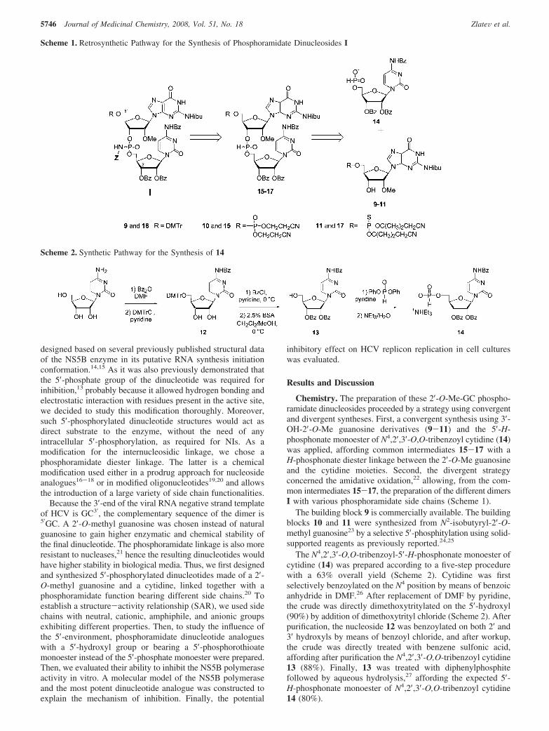
Indeed, only the Sp isomer was straightforwardly dockedwithin the active site of the HCV RdRp (Figure 4). As expected,important hydrogen bond and/or electrostatic interactions be-tween the 5′-phosphate/thiophosphate moiety and three arginineresidues (ARG386, ARG394, ARG158) can be observed in ourmodel. In the Sp absolute configuration, the nonbridging oxygenatom of the internucleosidic linkage is situated in a pseudo-equatorial position, directed toward the two magnesium ionspresent in the polymerization site, thus allowing complexationbetween them. Moreover, the only way to accommodatesmoothly the long phosphoramidate side chain into the HCV
Figure 1. Schematic Structure of Phosphoramidate Dimers 5-8.
5748 Journal of Medicinal Chemistry, 2008, Vol. 51, No. 18 ZlateV et al.

RdRp active site is to direct it toward the nucleotide entrychannel, again in the Sp absolute configuration. Thus, thenegatively charged side chain interacts with arginine and lysineresidues (ARG158, ARG48, LYS155), situated on the incomingpathway for nucleoside triphosphates (NTPs) during polymer-ization. In a steric clash way, this configuration may block theaccess of NTPs to the polymerization site, suggesting anexplanation to the observed inhibitory effect. In the otherorientation, the Rp absolute configuration, the phosphoramidatelinkage would be in conflict with Mg2+ ions in the active site.Possibly, Rp oriented phosphoramidate linkage could evict Mg2+
ions and create direct hydrogen bonds with ASP residues inthe active site. Such stabilizing interaction would be probablyless efficient than the strong Coulombic attraction originatingfrom sandwiching of Mg2+ ions between ASP residues and theoxygen from the phosphoramidate group.
The phosphoramidate dinucleoside mimics the presence ofthe initiation GC dinucleotide product in the active site of theHCV RdRp. Transition from the initiation to the later phase isinitiated by repositioning of the GC product and enables theaccommodation of another NTP into the active site. It seems,however, to be disabled by the cooperative action of three potentanchors: the 5′-phosphate/thiophosphate moiety, the carboxylicside chain and the nonbridging oxygen atom from the phos-phoramidate linkage both positioned in the Sp absolute con-figuration. Moreover, the intrusion of some NTPs into the activesite seems to be obstructed by the long carboxylate side chainsof the phosphoramidate, creating a net of hydrogen bonds withpositively charged ARG/LYS residues, which normally pavedthe way for incoming NTPs into the active site. In that way,ARG/LYS residues interacting with the phosphoramidate sidechains are crippled in serving as clips for negatively charged
Figure 2. Phosphoramidate dinucleosides decrease the formation of initiation products (pppGC and pppCC) synthesized by wild-type NS5B. (A)Time course of the formation of initiation products by the NS5B polymerase using an RNA template corresponding to the 3′ end of the HCV strand(-) genome in the presence of phosphoramidate dinucleoside 4 at the concentration of 50 µM. Initiation products were separated by electrophoresison denaturated sequencing gel. (B) pppGC products obtained for various concentrations of compound 4 were quantified and values reported as afunction of concentration. Curves from 2 independent experiments are shown, and IC50 values calculated from these curves are indicated under thearrow. (C) pppGC products obtained for various concentrations of compounds 1-4 were quantified as described in (B). IC50 values were calculatedand are represented as a bar for each phosphoramidates dinucleosides. All data represents average values for at least three separated experiments.
Figure 3. 5′-Thiophosphate modification and separation of the diastereoisomers increase the inhibition potency of phosphoramidate dinucleosides.(A) IC50 values of negatively charged (4 and 8) or neutral (1 and 6) phosphoramidate dinucleosides. (B) In vitro evaluation of separated diastereoisomersof negatively charged (4 and 8) or neutral (1 and 6) phosphoramidate dinucleosides. “F” stands for “fast”, meaning the fast eluting isomer onRP-HPLC, and “S” stands for “slow”, meaning the slowly eluting isomer.
Phosphoramidate Dinucleosides as HCV Polymerase Inhibitors Journal of Medicinal Chemistry, 2008, Vol. 51, No. 18 5749
triphosphate moieties of incoming NTPs. Interestingly, it wasreported recently that the 5′-phosphoramidate of deoxyadenosinebearing an L-aspartic amino acid could act as a substrate forHIV-1 reverse transcriptase.32,33 In this case, aspartic acidmimics the pyrophosphate of NTP, showing that such acarboxylic function could interact with amino acids of thepolymerase in the active site.
As an addition to the in vitro SAR developed, the model ofthe polymerase-phosphoramidate dinucleoside complex refinedby molecular dynamics simulations settles the better inhibitoryactivity observed for 5′-phosphorylated/thiophosphorylated, ne-gatively charged phosphoramidates in the Sp absolute config-uration (slow eluting diastereoisomers) as the whole of thesemodifications revealed to be important in our model by crucialinteractions with amino acid residues within the binding site.
Activity of Phosphoramidate Dinucleosides on HCV sub-genome Replicon replication. The more potent dinucleotidesactive against the NS5B polymerase were evaluated in Huh-5-2 cells, a hepatocyte cell line stably carrying the HCV repliconI389luc-ubi-neo/NS3-3′/5.1. This replicon contains a firefly lucif-erase-ubiquitin-neomycin phosphotransferase fusion protein usedas a reporter gene, and its expression is strictly dependent onthe level of replicon replication. The expression of the non-structural NS3-5B HCV polyprotein is driven by the EMCV-IRES. The activities of compounds 1, 4, and their 5′-thio-phosphate analogues 6, 8 were evaluated in this model (Figure5). All compounds proved relatively noncytotoxic (CC50 >60
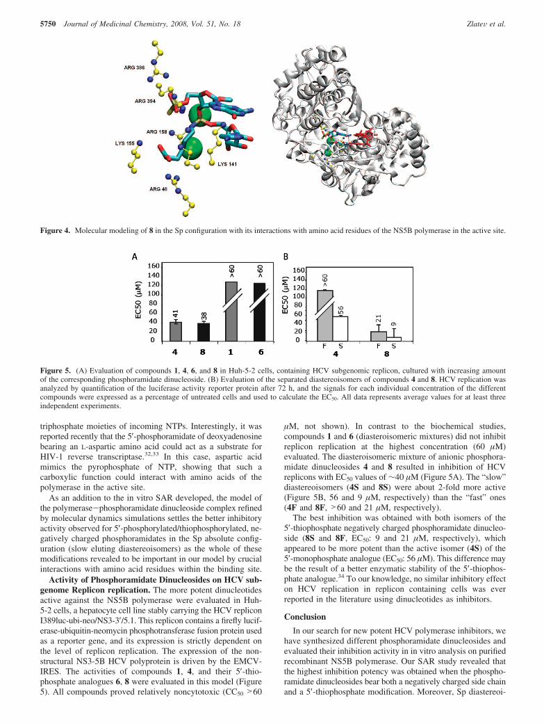
µM, not shown). In contrast to the biochemical studies,compounds 1 and 6 (diasteroisomeric mixtures) did not inhibitreplicon replication at the highest concentration (60 µM)evaluated. The diasteroisomeric mixture of anionic phosphora-midate dinucleosides 4 and 8 resulted in inhibition of HCVreplicons with EC50 values of ∼40 µM (Figure 5A). The “slow”diastereoisomers (4S and 8S) were about 2-fold more active(Figure 5B, 56 and 9 µM, respectively) than the “fast” ones(4F and 8F, >60 and 21 µM, respectively).
The best inhibition was obtained with both isomers of the5′-thiophosphate negatively charged phosphoramidate dinucleo-side (8S and 8F, EC50: 9 and 21 µM, respectively), whichappeared to be more potent than the active isomer (4S) of the5′-monophosphate analogue (EC50: 56 µM). This difference maybe the result of a better enzymatic stability of the 5′-thiophos-phate analogue.34 To our knowledge, no similar inhibitory effecton HCV replication in replicon containing cells was everreported in the literature using dinucleotides as inhibitors.
Conclusion
In our search for new potent HCV polymerase inhibitors, wehave synthesized different phosphoramidate dinucleosides andevaluated their inhibition activity in in vitro analysis on purifiedrecombinant NS5B polymerase. Our SAR study revealed thatthe highest inhibition potency was obtained when the phospho-ramidate dinucleosides bear both a negatively charged side chainand a 5′-thiophosphate modification. Moreover, Sp diastereoi-
Figure 4. Molecular modeling of 8 in the Sp configuration with its interactions with amino acid residues of the NS5B polymerase in the active site.
Figure 5. (A) Evaluation of compounds 1, 4, 6, and 8 in Huh-5-2 cells, containing HCV subgenomic replicon, cultured with increasing amountof the corresponding phosphoramidate dinucleoside. (B) Evaluation of the separated diastereoisomers of compounds 4 and 8. HCV replication wasanalyzed by quantification of the luciferase activity reporter protein after 72 h, and the signals for each individual concentration of the differentcompounds were expressed as a percentage of untreated cells and used to calculate the EC50. All data represents average values for at least threeindependent experiments.
5750 Journal of Medicinal Chemistry, 2008, Vol. 51, No. 18 ZlateV et al.

somers were always found to be more active than Rp diaste-reoisomers, irrespective to the nature of the phosphoramidatelinkage. This was tentatively explained by molecular modelingof the more potent phosphoramidate dinucleoside, 8S, in theHCV polymerase active site. Finally, this compound proved alsomore potent to inhibit the HCV replication as measured in Huh-5-2 replicon containing cells. Thus, phosphoramidate dinucleo-sides bearing a 5-carboxypentyl side chain on the internucleo-sidic linkage show promising inhibiting activities, on both NS5Bpolymerase and HCV subgenomic replication, which makesthem new potential candidates for anti-HCV therapy.
Experimental Section
HCV 1b Polymerase Plasmid Constructs, Enzyme Prepara-tion, and Reagents. NS5B-∆55 gene was tagged with six C-terminalhistidines and expressed from the pDest 14 vector (Invitrogen) inEscherichia coli BL21(DE3) cells (Novagen). NS5B protein waspurified as described previously.7 RNA oligonucleotides wereobtained from MWG-Biotech. R-32P-labeled cytosine 5′-triphos-phate (3000 Ci/mmole) was purchased from Amersham.
Determination of Inhibitory Concentration 50% (IC50). An RNAoligonucleotide corresponding to the 3′ end of the negative strandof the HCV genome (RNA H (-) 5′-UCGGGGGCUGGC-3′) wasused to analyze the synthesis of the first phosphodiester bond andits inhibition induced by the modified phosphoramidate dinucle-otides. The modified dinucleotides concentration, leading to 50%inhibition of NS5B-mediated RNA synthesis, was determined inRdRp buffer (50 mM HEPES pH 8.0, 10 mM KCl, 2 mM MnCl2,1 mM MgCl2, 10 mM DTT, 0.5% Igepal CA630) containing 10µM of RNA template, 100 µM R-32P-CTP (1 µCi), 50 µM GTP,and various concentration of dinucleotides (0, 5, 10, 20, 50, 100,200, 500 µM). Reactions were initiated by the addition of 1 µMNS5B, incubated at 30 °C for 15 min, and quenched with EDTA/formamide. Products were separated using sequencing gel electro-phoresis and quantified using photostimulated plates and a FujiImager (Fuji). IC50 was determined using the equation:
% of active enzyme) 100 ⁄ (1+ (I)2 ⁄ IC50) (1)
where I is the concentration of the inhibitor. IC50 was determinedfrom curve-fitting using Kaleidagraph (Synergy Software).
Evaluation of Antiviral Activity and Cytostatic Activities ofSelected Compounds in HCV Genotype 1b Subgenomic Repli-con Carrying Huh-5-2 Cells. Huh-5-2 cells were cultured in RPMImedium (GIBCO) supplemented with 10% fetal calf serum, 2 mML-glutamine (Life Technologies), 1× nonessential amino acids (LifeTechnologies), 100 IU/mL penicillin and 100 µg/mL streptomycin,and 250 µg/mL G418 (Geneticin, Life Technologies). Cells wereseeded at a density of 7000 cells per well in a 96-well View Plate(Packard) in medium containing the same components as describedabove, except for G418. Cells were allowed to adhere and proliferatefor 24 h. At that time, culture medium was removed and five serialdilutions (5-fold dilutions starting at 50 µg/mL or around 60 µM)of the test compounds were added in culture medium lacking G418.Interferon R-2a (500 IU) was included as a positive control in eachexperiment for internal validation. Plates were further incubated at37 °C and 5% CO2 for 72 h. Replication of the HCV replicon inHuh-5-2 cells resulted in luciferase activity in the cells. Luciferaseactivity was measured by adding 50 µL of 1× Glo-lysis buffer(Promega) for 15 min followed by 50 µL of the Steady-Gloluciferase assay reagent (Promega). Luciferase activity was mea-sured with a luminometer and the signal in each individual well isexpressed as a percentage of the untreated cultures. The 50%effective concentrations (EC50) were calculated from these data sets.Parallel cultures of Huh-5-2 cells, seeded at a density of 7000 cells/well of classical 96-well cell culture plates (Becton-Dickinson) weretreated in a similar fashion except that no Glo-lysis buffer or Steady-Glo luciferase reagent was added. The effect of the compounds onthe proliferation of the cells was measured 3 days after addition ofthe various compounds by means of The CellTiter 96 AQueous
nonradioactive cell proliferation assay (MTS, Promega). In thisassay, 3-(4,5-dimethylthiazol-2-yl)-5-(3-carboxymethoxy-phenyl)-2-(4-sulfophenyl)-2H-tetrazolium (MTS) was bioreduced by cellsinto a formazan that is soluble in tissue. The number of cellscorrelates directly with the production of the formazan. The MTSstained cultures were quantified in a plate reader.
General Procedures. TLC was performed on Merck silica coatedplates 60F254 (art. 5554). Compounds were revealed on UV light(254 nm) and after spraying with 10% sulfuric acid ethanol solutionand heating. Phosphorus containing compounds were revealed afterspraying with the Hanes molybdate reagent35 and heating. Columnchromatography was performed on silica gel Merck 60 (art. 9385).Flash chromatography was performed using a Biotage SP1 ap-paratus and Biotage silica gel columns. Ion exchange purificationswere carried using Pharmacia Sephadex DEAE A-25 resin (art.170), ISCO TRIS Pump and UA-6 detector (254 nm). Triethylam-monium bicarbonate (TEAB) 1 M, pH 7.5, was prepared fromtriethylamine, milli Q water (18.2 MΩ · cm) and CO2 gas. Ionexchange reactions were performed using DOWEX 50WX8-200H+ resin (Aldrich), different ion forms were obtained after washingwith corresponding 2 M aqueous solutions and then water untilneutrality. Polystryrene acyl chloride resin was prepared accordingto previously published procedure28 and vaccuum-dried over P2O5.Analytical HPLC were performed using a Nucleosil C18 column,(150 mm × 4.6 mm, 5 µm) equipped with a prefilter and aprecolumn (Nucleosil, C18, 5 µm) on a Waters apparatus using aWaters-616 pump and a Waters 966 photodiode array detector.Compounds were eluted using a linear gradient 0-80% of aceto-nitrile in a 50 mM triethylammonium acetate buffer (pH 7) with a1 mL/min flow rate. Preparative HPLC purifications were performedwith a Waters DeltaPak C18 preparative column (7.8 mm × 300mm, 15 µm) using the analytical HPLC conditions and a 2 mL/min flow rate. All moist-sensitive reactions were carried underanhydrous conditions using dry glassware, anhydrous solvents andArgon atmosphere. Acetonitrile was distilled from CaH2 and storedover activated 3 Å molecular sieves. Pyridine and triethylaminewere distilled from CaH2. Dichloromethane and carbon tetrachloridewere distilled from P2O5. All commercially available reagents wereused as received. Three Å molecular sieves were dried in a 300 °Coven during 3 h prior to use. FAB-MS and FAB-HRMS spectrawere recorded on a JEOL SX102 mass spectrometer using a mixtureof glycerol/thioglycerol [50/50, v/v, (G-T)] as a matrix. MALDI/Tof-MS spectra were recorded on a Perspective BiosystemsVoyager DE spectrometer using a mixture of 2,4,6-trihydroxyac-etophenone/ammonium citrate [10/1, m/m, saturated solution inwater/acetonitrile, 1/1, v/v, (THAP/cit)] as a matrix. ESI-HRMSspectra were recorded on a Q-Tof Micromass spectrometer. 1H and13C NMR spectra were recorded at room temperature on a Brukerspectrometer at 200, 400, or 300 MHz (1H) and 100 or 75 MHz(13C). Chemical shifts are given in ppm referenced to the solventresidual peak (CDCl3 -7.27 and 77.0 ppm; DMSO-d6 -2.49 and39.5 ppm; D2O -4.79 ppm + drop of CH3OH at 49.5 ppm asinternal reference36). Coupling constants are given in hertz. Signalsplitting patterns are described as singlet (s), doublet (d), triplet(t), quartet (q), broad signal (br), doublet of doublet (dd), doubletof triplet (dt), doublet of quartet (dq), pseudo (p), and multiplet(m). Signal assignments were based on 2D homo-1H/1H, NOE orheteronuclear-1H/13C correlations and D2O exchange. 31P NMRspectra were recorded at 101 and 121 MHz on proton-decoupledmode and on proton-coupled mode for H-phosphonates. Chemicalshifts are given in ppm referenced to external H3PO4-80%; couplingconstants are given in hertz. UV spectra were recorded on a VarianCary 300 Bio UV/vis spectrometer.
For convenience, general procedures have been given for thecoupling, oxidation, and deprotection reactions. Variations fromthese procedures and individual purification methods are given inthe main text for each compound. Preparative and spectroscopicdata for citydine monomer compounds 12-14 is given as Sup-porting Information only.
General Procedure for H-Phosphonate Coupling. In a dry flaskand under argon, PS acyl chloride resin (6 mol equiv) was
Phosphoramidate Dinucleosides as HCV Polymerase Inhibitors Journal of Medicinal Chemistry, 2008, Vol. 51, No. 18 5751

suspended in 10 mL/mmol of anhydrous pyridine/dichloromethane1:1 (v/v) solution. In another dry flask, a mixture of the appropriateguanosine 3′-hydroxy monomer 9-11 (1 mol equiv) and thecytidine 5′-H-phosphonate monomer 14 (1.2 mol equiv) weredissolved in 10 mL/mmol of anhydrous pyridine/dichloromethanesolution after drying by azeotropic distillation with anhydrouspyridine. The nucleoside containing solution was then introducedto the resin containing flask, and the mixture was shaken at roomtemperature for 2 h. The reaction mixture was then diluted withdichloromethane; the resin was filtered off and well rinsed. Filtrateswere evaporated to half-volume and then washed twice with water.Organic layers were dried over Na2SO4, filtered, and evaporatedto dryness. The residue was coevaporated three times withacetonitrile, providing the crude dimer as a white foam (HPLCpurity >85%) in a 1:1 diastereomeric mixture. Compounds wereused in oxidation steps without further purification.
Oxidation Method A. In a dry flask, appropriate H-Phosphonatediester (1 mol equiv) was dried by azeotropic distillation withanhydrous pyridine and dried under vacuum with P2O5 for 30 min.It was then dissolved in anhydrous pyridine (5 mL/mmol). To thissolution were simultaneously added: anhydrous CCl4 (25 molequiv, 2.5 mL/mmol), corresponding amine (10 mol equiv), or10 M solution of histamine (50 mol equiv) in anhydrous pyridine.Reaction color turned to bright yellow and the mixture wasstirred at room temperature for 1 h. The solvents were removedunder reduced pressure and the residue was coevaporated twicewith acetonitrile. Compounds were directly involved in depro-tection steps.
Preparation of 6-Aminocaproic Acid Methyl Ester Hydrochlo-ride.37 First, 400 mg of 6-aminocaproic acid (3 mmol, 1 mol equiv)were suspended in 40 mL of 2,2-dimethoxypropane (10-15 mL/mmol). To the mixture was added 3 mL of 37% HCl and thesolution was stirred at room temperature for 18 h. After removalof the solvents under reduced pressure, the residue was precipitatedin acetonitrile and recrystallized from acetonitrile/ether. Afterfiltration, the white crystals were dried under vacuum with P2O5 atrefluxing ethanol for 2 days (490 mg, 90%). 1H NMR (D2O, 300MHz) δ 3.60 (3H, s, CO2CH3), 2.90 (2H, t, CH2-6, J ) 7.4 Hz),2.33 (2H, t, CH2-2, J ) 7.3 Hz), 1.64 (4H, m, CH2-3, CH2-5),1.32 (2H, q, CH2-4, J ) 7.3 Hz). 13C NMR (D2O, 75 MHz) δ177.3 (CO2CH3), 52.1 (CO2CH3), 39.3 (CH2-6), 33.3 (CH2-2),26.3 (CH2-5), 25.0 (CH2-3), 23.6 (CH2-4).
Oxidation Method B. In a dry flask, appropriate H-phosphonatediester (1 mol equiv) was dried by azeotropic distillation withanhydrous pyridine and dried under vacuum with P2O5 for 30 min.It was then dissolved in anhydrous pyridine (2.5 mL/mmol). Tothis solution were simultaneously added: anhydrous CCl4 (25 molequiv, 2.5 mL/mmol), 4 M solution of 6-aminocaproic acid methylester hydrochloride (10 mol equiv) in anhydrous pyridine, andnhydrous triethylamine (10 mol equiv). Reaction color turned tobright yellow, and the mixture was stirred at room temperature for1 h. The solvents were removed under reduced pressure, and theresidue was coevaporated twice with acetonitrile. Compounds weredirectly involved in deprotection steps.
Deprotection Method A. Appropriate compound was dissolvedin THF/H2O 1:1 (v/v) (14 mL/mmol), and 28% aqueous ammoniasolution was added (70 mL/mmol). The flask was hermeticallyclosed, and the mixture was stirred 7 h at 50 °C. The solvents wereremoved under reduced pressure to give the corresponding com-pound as a brown oil, which was further purified by chromatography.
Deprotection Method B. Appropriate compound was dissolvedin methanol (12 mL/mmol), and 80% acetic acid solution (62 mL/mmol) was added; then the mixture was stirred 2 h at roomtemperature. It was finally diluted with 10 mL of water and washedwith dichloromethane (5 times) and ether (3 times). Aqueous layerwas evaporated to dryness, and then the residue was purified bychromatography.
HPLC Method for Purification and/or Separation of Diastere-oisomers. Waters DeltaPak C18 preparative column was used on aWaters apparatus with a 2 mL/min flow rate (see above). Gradientconditions, run times, and maximal loading of the column are given
for each compound. After purification, the TEAAc buffer wasremoved after several lyophilizations from water. Collected pureisomers were analyzed by analytical HPLC and retention times foreach isomer are given after a coinjection of an equimolar mixture.According to retention times on RP-HPLC, the names “fast eluting”and “slow eluting” were attributed to each isomer.
Synthesis of O-(N4,2′,3′-O,O-Tribenzoylcytidin-5′-yl)-O-N2-isobutyryl-2′-O-methyl-5′-O-[bis-(2-cyanoethyl)phosphoryl]guanosin-3′-yl Hydrogenophosphonate Diester (15). Prepared according togeneral procedure of coupling using monomers 14 (0.35 g, 0.48mmol, 1.2 equiv) and 10 (0.25 g, 0.4 mmol, 1 equiv), dry PS-acylchloride resin (1.2 g, 2.5 mmol, 6 equiv), and 10 mL of anhydrousdichloromethane/pyridine 1:1 (v/v). The residue was finally pre-cipitated in ether to give crude 15 as white foam (HPLC purity>85%) (0.30 g, 60%). tR-HPLC: 16.6 min, 10-80% of acetonitrilein 20 min. 1H NMR (CDCl3, 200 MHz, peaks of both diastereomersare described) δ 12.24 (2H, s, br, NH-G1), 10.52 (2H, s, br, NHibu),10.38 (1H, s, NHBz), 9.14, 9.04 (2H, 2s, H-G8), 8.68 (1H, d,H-C6), 7.96-7.87 (12H, m, Hortho), 7.65-7.33 (19H, m, Hpara,Hmeta, H-C5), 7.27 (1H, d, H-P, 1JH-P ) 744.6 Hz), 7.19 (1H,d, H-P, 1JH-P ) 737.3 Hz), 6.37, 6.26 (2H, 2d, H-C1′, J ) 4.4Hz), 5.99-5.83 (6H, m, H-G1′, H-C2′, H-C3′), 5.04, 4.99 (2H,m, H-G3′), 4.65-4.19 (20H, m, H-C4′, H-G4′, H-C5′,5′′ ,H-G5′,5′′ , HRCNE, HR′CNE), 3.40, 3.37 (6H, 2s, G2′-O-CH3), 2.89(4H, t, HCNE, J ) 5.7 Hz), 2.71 (4H, t, H′CNE, J ) 5.9 Hz),1.26-1.21 (12H, 2d, CH3ibu, J ) 6.9 Hz). 31P NMR (CDCl3, 101MHz) δ 9.4 (1/4, dq, 1JH-P ) 743.8 Hz, 3JH-P ) 7.5 Hz), 8.4 (1/4,dq, 1JH-P ) 746.5 Hz, 3JH-P ) 8.2 Hz), -2.8 (1/4, m, 3JH-P ) 7.0Hz), -3.1 (1/4, m). MALDI+ (THAP/cit) m/z 1155 (M + H)+.
Synthesis of O-(N4,2′,3′-O,O-Tribenzoylcytidin-5′-yl)-O-[N2-isobutyryl-2′-O-methyl-5′-O-(4,4′-dimethoxytrityl)guanosin-3′-yl] Hy-drogenophosphonate Diester (16). Prepared according to generalprocedure of coupling from monomer 14 (0.35 g, 0.48 mmol, 1.2equiv), N2-isobutyryl-2′-O-methyl-5′-O-DMTr guanosine 9 (0.27g, 0.4 mmol, 1 equiv), dry PS-acyl chloride resin (1.2 g, 2.5 mmol,6 equiv), and 10 mL of anhydrous dichloromethane/pyridine 1:1(v/v). Crude 16 was a white foam (HPLC purity >90%) (0.39 g,77%). tR-HPLC: 14.1 min, 14.3 min, 10-80% of acetonitrile in 10min. 1H NMR (CDCl3, 200 MHz, peaks of both diastereomers aredescribed) δ 12.07, 12.02 (2H, 2s, br, NH-G1), 9.45 (1H, s, br,NHBz), 8.99 (2H, 2s, H-G8), 8.64 (1H, d, H-C6), 7.95-6.76 (60H,m, Harom), 7.34 (1H, d, H-P, 1JH-P ) 740.0 Hz), 7.02 (1H, d,H-P, 1JH-P ) 729.8 Hz), 6.42, 6.28 (2H, 2d, H-C1′, J ) 4.2Hz), 5.91-5.83 (6H, m, H-G1′, H-C2′, H-C3′), 4.96, 4.82 (2H,m, H-G3′), 4.86-4.38 (12H, m, H-C4′, H-G4′, H-C5′,5′′ ,H-G5′,5′′ ), 3.78-3.76 (12H, 4s, O-CH3DMTr), 3.52, 3.50 (6H, 2s,G2′-O-CH3), 1.26 (12H, s, CH3ibu). 31P NMR (CDCl3, 101 MHz)δ 9.4 (1/2, dq, 1JH-P ) 740.4 Hz, 3JH-P ) 7.6 Hz), 7.3 (1/2, dq,1JH-P ) 739.1 Hz, 3JH-P ) 8.0 Hz). MALDI+ (THAP/cit) m/z 1272(M + H)+, 969 (M + H-DMTr)+.
Synthesis of O-(N4,2′,3′-O,O-Tribenzoylcytidin-5′-yl)-O-N2-isobutyryl-2′-O-methyl-5′-O-[bis-(2-cyano-1,1-dimethylethyl)thiono-phosphoryl]guanosin-3′-yl Hydrogenophosphonate Diester (17).Prepared using general procedure for coupling from monomers 14(0.25 g, 0.33 mmol, 1.2 equiv), 11 (0.17 g, 0.28 mmol, 1 equiv),dry PS-acyl chloride resin (0.7 g, 1.7 mmol, 6 equiv), and 7 mL ofanhydrous dichloromethane/pyridine 1:1 (v/v). Crude 17 was awhite foam (HPLC purity >93%) (0.23 g, 70%). tR-HPLC: 15.9min, 10-80% of acetonitrile in 15 min. 1H NMR (CDCl3, 200MHz, peaks of both diastereomers are described) δ 12.18-12.16(2H, 2s, br, NH-G1), 9.59, 9.84 (2H, 2s, br, NHibu), 8.67, 8.65(2H, 2s, H-G8), 8.15, 8.08 (2H, 2d, H-C6, J ) 7.6 Hz), 7.97-7.87(12H, m, Hortho), 7.66-7.34 (19H, m, Hpara, Hmeta, H-C5), 7.35(1H, d, H-P, 1JH-P ) 744.2 Hz), 7.22 (1H, d, H-P, 1JH-P ) 736.6Hz), 6.47, 6.31 (2H, 2d, H-C1′, J ) 4.3 Hz), 5.97-5.83 (6H, m,H-G1′, H-C2′, H-C3′), 5.61, 5.48 (2H, m, H-G3′), 4.87-4.34(12H, m, H-C4′, H-G4′, H-C5′,5′′ , H-G5′,5′′ ), 3.46, 3.43 (6H,2s, G2′-O-CH3), 3.11-2.71 (8H, m, CH2CN), 2.68 (2H, m, CHibu),1.73-1.66 (24H, m, C(CH3)2), 1.26-1.21 (12H, 2d, CH3ibu, J )6.8 Hz). 31P NMR (CDCl3, 101 MHz) δ 48.9, 48.7 (1/2, 2t, 3JH-P
) 7.3 Hz), 9.8 (1/4, dq, 1JH-P ) 744.7 Hz, 3JH-P ) 7.6 Hz), 8.0
5752 Journal of Medicinal Chemistry, 2008, Vol. 51, No. 18 ZlateV et al.

(1/4, dq, 1JH-P ) 737.2 Hz, 3JH-P ) 8.1 Hz). MALDI+ (THAP/cit) m/z 1227 (M + H)+.
Synthesis of O-(Cytidin-5′-yl)-N-(2-methoxyethyl)-O-(2′-O-meth-yl-5′-O-phosphorylguanosin-3′-yl) Phosphoramidate, Sodium Salt(1). Prepared according to oxidation method A from H-phosphonatediester 15 (0.07 mmol, 1 equiv), 0.5 mL of anhydrous CCl4, 0.5mL of anhydrous pyridine, and 2-methoxyethylamine (60 µL, 0.7mmol, 10 equiv) and according to deprotection method A using 1mL of THF/H2O 1:1 (v/v) and 5 mL of 28% aqueous ammonia.Residue was dissolved in water and washed twice with ether.Aqueous layer was evaporated to dryness and purified on a DEAE-A25 Sephadex column (linear gradient: TEAB 10-3 to 0.3 M). Highpurity final compound was obtained after purification on preparativeRP-HPLC (gradient: acetonitrile 5-40% in 20 min; maximalloading of the column: 10 mg of crude product per run). Targetcompound was obtained as a sodium salt after elution on aDOWEX-Na+ column with water. After lyophilization from water,1 was a white spongy solid (28 mg, 47%) epimeric mixture (ratio∼1:1). tR-HPLC: 8.2 min; 8.5 min, 0-40% acetonitrile in 15 min.UV (H2O) λmax ) 255 nm (ε ) 15100). 1H NMR (D2O, 300 MHz,peaks of both diastereomers are described) δ 8.19 (2H, s, H-G8),7.71, 7.64 (2H, 2d, H-C6, J ) 7.5 Hz), 5.94 (1H, d, H-G1′, J )6.4 Hz), 5.90 (4H, pd, H-C1′, H-G1′, H-C5), 5.73 (1H, d, H-C5,J ) 7.5 Hz), 5.16 (2H, m, H-G3′), 4.66 (2H, m, H-G2′), 4.52 (2H,m, H-G4′), 4.42-4.18 (10H, m, H-C2′, H-C3′, H-C4′, H-C5′,5′′),4.02 (4H, m, H-G5′,5′′ ), 3.52 (3H, s, G2′-O-CH3), 3.49 (4H, pt,CH2OMe, J ) 5.1 Hz), 3.47 (3H, s, G2′-O-CH3), 3.36, 3.33 (6H,2s, CH2-OCH3), 3.18, 3.16 (4H, m, PNH-CH2, J ) 5.1 Hz). 13CNMR (D2O, 75 MHz) δ 168.2 (C-C4), 161.1 (C-G6), 157.7(C-C2), 152.5 (C-G4), 141.6, 141.4 (C-C6), 138.0 (C-G8), 118.1(C-G5), 96.7, 96.3 (C-C5), 91.1, 90.8 (C-C1′), 85.2, 84.7 (C-G1′), 83.9 (C-G4′), 82.2 (C-C4′), 81.9, 81.8 (C-G2′), 75.1 (C-C3′),74.8 (C-G3′), 72.8 (CH2OMe), 66.9, 66.3 (C-C5′), 63.7 (C-G5′),58.9, 58.6 (G2′-OCH3, CH2-OCH3), 40.8 (PNH-CH2). 31P NMR(D2O, 121 MHz) δ 11.4 (1/4, P-N), 11.0 (1/4, P-N), 3.3 (1/4,P-O), 3.2 (1/4, P-O). FAB+ (GT) m/z 784 (M + H)+; 762 (M-Na + H)+; FAB- (GT) m/z 760 (M-Na)-; 738 (M-2Na + H)-.HRMS: FAB- (GT) m/z calcd for (C23H34N9O15P2)-, 738.1802;obsd, 738.1778.
The two diastereoisomers of the obtained mixture were separatedby preparative RP-HPLC using the general conditions describedabove. Gradient: acetonitrile 7-10% in 30 min; maximal loadingof the column: 1 mg of mixture per run.
Fast Eluting Isomer of 1. tR - HPLC: 8.2 min - 0 to 40%acetonitrile in 15 min. 1H NMR (D2O, 300 MHz) δ 8.19 (1H, s,H-G8), 7.67 (1H, d, H-C6, J ) 7.5 Hz), 5.91 (1H, d, H-G1′, J )6.9 Hz), 5.89 (1H, d, H-C1′, J ) 2.7 Hz), 5.77 (1H, d, H-C5, J) 7.8 Hz), 5.20-5.15 (1H, m, H-G3′), 4.66 (1H, pt, H-G2′, J )5.4 Hz), 4.53 (1H, m, H-G4′), 4.42-4.19 (5H, m, H-C2′, H-C3′,H-C4′, H-C5′,5′′ ), 4.04 (2H, m, H-G5′,5′′ ), 3.55-3.51 (5H, m,G2′-O-CH3, CH2OMe), 3.37 (3H, s, CH2-OCH3), 3.17 (2H, dt,PNH-CH2, 3JH-H ) 5.1 Hz, 3JH-P ) 12.0 Hz). 31P NMR (D2O,121 MHz) δ 10.5 (1/2, P-N), 2.0 (1/2, P-O).
Slow Eluting Isomer of 1. tR-HPLC: 8.4 min, 0-40% acetonitrilein 15 min. 1H NMR (D2O, 300 MHz) δ 8.22 (1H, s, H-G8), 7.75(1H, d, H-C6, J ) 7.5 Hz), 5.98-5.90 (3H, m, H-G1′, H-C1′,H-C5), 5.21-5.16 (1H, m, H-G3′), 4.68 (1H, pt, H-G2′), 4.55 (1H,m, H-G4′), 4.47-4.27 (5H, m, H-C2′, H-C3′, H-C4′, H-C5′,5′′ ),4.03 (2H, m, H-G5′,5′′ ), 3.52 (2H, t, CH2OMe, J ) 5.4 Hz), 3.48(3H, s, G2′-O-CH3,), 3.35 (3H, s, CH2-OCH3), 3.17 (2H, dt, PNH-CH2, 3JH-H ) 5.4 Hz, 3JH-P ) 11.7 Hz). 31P NMR (D2O, 121MHz) δ 10.8 (1/2, P-N), 3.1 (1/2, P-O).
Synthesis of O-(Cytidin-5′-yl)-O-(2′-O-methyl-5′-O-phospho-rylguanosin-3′-yl)-N-[3-(N,N-dimethylamino)propyl] Phosphora-midate, Hydrochloride, Sodium Salt (2). Prepared according tooxidation method A from H-phosphonate diester 15 (0.07 mmol, 1equiv), 0.5 mL of anhydrous CCl4, 0.5 mL of anhydrous pyridine,and 3-(N,N-dimethylamino)propylamine (100 µL, 0.7 mmol, 10equiv) and according to deprotection method A using 1 mL of THF/H2O 1:1 (v/v) and 5 mL of 28% aqueous ammonia. Solvents wereevaporated to dryness; the residue was dissolved in water and
washed twice with ether. Aqueous layer was evaporated to drynessand purified on a DEAE-A25 Sephadex column (linear gradient:TEAB 10-3 to 0.3 M). High purity final compound was obtainedafter purification on preparative RP-HPLC (gradient: acetonitrile7-16% in 20 min; maximal loading of the column: 6.4 mg of crudeproduct per run). Residue was then dissolved in 3 mL of 1 M NaClsolution and eluted on a small RP-18 column (gradient: H2O 100%to 25% of acetonitrile) After lyophilization from water, the suitablesalt form of 2 was a white spongy solid (18 mg, 30%) epimericmixture (ratio ∼1:1). tR-HPLC: 7.6 min, 0-40% acetonitrile in 15min. UV: (H2O) λmax ) 255 nm (ε ) 14800). 1H NMR (D2O, 400MHz, peaks of both diastereomers are described) δ 8.20 (2H, s,br, H-G8), 7.58 (2H, d, H-C6, J ) 7.3 Hz), 5.93 (1H, pd, H-G1′,J ) 5.0 Hz), 5.83-5.66 (5H, m, H-C1′,H-G1′, H-C5), 5.12, 5.01(2H, m, H-G3′), 4.56 (2H, m, H-G2′), 4.45-4.06 (12H, m, H-G4′,H-C2′, H-C3′, H-C4′, H-C5′,5′′ ), 3.89 (4H, m, H-G5′,5′′ ), 3.48,3.47 (6H, 2s, G2′-O-CH3), 3.12-2.81 (8H, m, CH2-PNH, CH2-N(CH3)2), 2.76 (12H, s, N(CH3)2), 1.89 (4H, m, -CH2-). 13C NMR(D2O, 75 MHz) δ 166.9 (C-C4), 156.5 (C-C2), 148.0 (C-G4),141.9 (C-C6), 118.1 (C-G5), 96.5 (C-C5), 91.7, 90.6 (C-C1′,C-G1′), 81.9 (C-4′), 81.9, 81.7 (C-2′, C-4′), 75.1 (C-C3′), 74.6(C-G3′), 69.8, 69.4 (C-C5′, C-G5′), 59.2, 59.1 (G2′-OCH3), 55.8(CH2), 43.4 (N(CH3)2), 38.8 (CH2), 27.4 (CH2). 31P NMR (D2O,101 MHz) δ 10.98, 10.87 (1/2, P-N), 3.7 (1/2, P-O). FAB+ (GT)m/z 811 (M-Cl)+; 789 (M-NaCl + H)+; 767 (M-NaCl-Na + 2H)+;FAB- (GT) m/z 787 (M-NaCl-H)-; 765 (M-2Na-Cl)-. HRMS:FAB- (GT) m/z calcd for (C25H39N10O14P2)- 765.2122; obsd,765.2129.
Synthesis of O-(Cytidin-5′-yl)-N-[2-(imidazol-4-yl)ethyl]-O-(2′-O-methyl-5′-O-phosphorylguanosin-3′-yl) Phosphoramidate, Tri-ethylammonium Salt (3). Prepared according to oxidation methodA from H-phosphonate diester 15 (0.07 mmol, 1 equiv), 0.15 mLof anhydrous CCl4, 0.15 mL of anhydrous pyridine, and dryhistamine base (0.33 g, 3 mmol, 50 equiv) in solution in 0.3 mL ofanhydrous pyridine and according to deprotection method A using1 mL of THF/H2O 1:1 (v/v) and 5 mL of 28% aqueous ammonia.Solvents were evaporated to dryness; the residue was dissolved inwater and washed twice with ether. Aqueous layer was evaporatedto dryness and purified on a DEAE-A25 Sephadex column (lineargradient: TEAB 10-3 to 0.3 M). High purity final compound (atthis stage, the compound was contaminated with 45% of C4-transaminated by histamine, cytosine analogue) was obtained afterpurification on preparative RP-HPLC (gradient: acetonitrile 6-11%in 30 min; maximal loading of the column: 1 mg of crude productper run). Compound 3 was obtained as a white spongy solid bis-triethylammonium salt (15 mg, 22%) epimeric mixture (ratio ∼1:1). Final compound for enzyme assays was obtained as sodiumsalt after elution on a DOWEX-Na+ column with water andlyophilization. tR-HPLC: 7.8 min, 0-40% acetonitrile in 15 min.UV: (H2O) λmax ) 255 nm (ε ) 12300). 1H NMR (D2O, 300 MHz,peaks of both diastereomers are described) δ 8.45, 8.43 (2H, 2s,H-Im2), 8.04, 8.01 (2H, 2s, H-G8), 7.52, 7.49 (2H, 2d, H-C6, J) 7.5 Hz), 7.18 (2H, s, H-Im5), 5.82 (1H, d, H-G1′, J ) 5.6 Hz),5.75 (3H, m, H-C1′,H-G1′), 5.68, 5.57 (2H, 2d, H-C5, J ) 7.4Hz), 4.93, 4.84 (2H, 2m, H-G3′), 4.49 (2H, m, H-G2′, J ) 5.2Hz), 4.38, 4.31 (2H, 2m, H-G4′), 4.21-3.98 (10H, m, H-C2′,H-C3′, H-C4′, H-C5′,5′′ ), 3.90 (4H, m, H-G5′,5′′ ), 3.41, 3.35(6H, 2s, G2′-O-CH3), 3.18 (4H, m, PNHCH2), 3.09 (4H, q, CH2CH3,J ) 7.1 Hz), 2.82 (4H, t, Im-CH2, J ) 6.4 Hz), 1.16 (12H, t,CH2CH3, 3J ) 7.3 Hz). 13C NMR (D2O, 100 MHz) δ 165.8(C-C4), 158.9 (C-G6), 157.1 (C-C2), 153.9 (C-G4), 140.9, 140.8(C-C6), 139.0 (C-G8), 133.2 (C-Im4), 133.1 (C-Im2), 116.5, 116.2(C-Im5), 95.7, 95.5 (C-C5), 90.7 (C-C1′), 84.8, 84.1 (C-G1′),81.5, 81.4 (C-2′, C-4′), 74.0 (C-C3′), 73.9 (C-G3′), 68.8, 68.6(C-C5′, C-G5′), 58.2, 58.2 (G2′-O-CH3), 46.6 (CH2CH3), 39.8,39.7 (PNHCH2), 26.4 (CH2-Im), 8.2 (CH2CH3). 31P NMR (D2O,101 MHz) δ 10.4, 10.1 (1/2, P-N), 2.2, 2.1 (1/2, P-O). FAB+
(GT) m/z 776 (M-2TEAH + 3H)+; FAB- (GT) m/z 774 (M-2TEAH + H)-. HRMS: FAB+ (GT) m/z calcd for(C25H36N11O14P2)+, 776.1918; obsd, 776.1915.
Phosphoramidate Dinucleosides as HCV Polymerase Inhibitors Journal of Medicinal Chemistry, 2008, Vol. 51, No. 18 5753
Synthesis of O-(Cytidin-5′-yl)-O-(2′-O-methyl-5′-O-phospho-rylguanosin-3′-yl)-N-(5-carboxypentyl) Phosphoramidate, SodiumSalt (4). Prepared according to oxidation method B from H-phosphonate diester 15 (0.18 mmol, 1 equiv), 0.5 mL of anhydrousCCl4, 0.4 mL of anhydrous pyridine, dry 6-aminocaproic acidmethyl ester hydrochloride (350 mg, 1.8 mmol, 10 equiv) in 0.5mL of anhydrous pyridine, and anhydrous triethylamine (0.5 mL,10 equiv). Reaction mixture was treated with 0.3 M NaOH for 2 hat room temperature and then quenched with DOWEX-pyridiniumresin. After filtration of the resin, filtrates were evaporated to drynessand treated with saturated ammonia (23 mL) according to depro-tection method A. Crude mixture was then purified on a DEAE-A25 Sephadex column (linear gradient: TEAB 10-3 to 0.3 M). Thetwo diastereoisomers of the obtained crude mixture (135 mg) wereseparated by preparative RP-HPLC using the general conditionsdescribed above. Gradient: acetonitrile 6-8% in 30 min; maximalloading of the column: 7 mg of crude mixture per run. Each pureisomer was then obtained as sodium salt (white spongy solid) afterelution on a DOWEX-Na+ column with water and lyophilization.
Fast Eluting Isomer of 4. Yield 45 mg, 58%. tR-HPLC: 8.3 min,0-40% acetonitrile in 15 min. UV: (H2O) λmax ) 255 nm (ε )14800). 1H NMR (D2O, 300 MHz) δ 8.15 (1H, s, H-G8), 7.69 (1H,d, H-C6, J ) 7.5 Hz), 5.95 (1H, d, H-G1′, J ) 6.3 Hz), 5.89 (1H,d, H-C1′, J ) 2.4 Hz), 5.83 (1H, d, H-C5, J ) 7.5 Hz), 5.17-5.12(1H, m, H-G3′), 4.58 (1H, pt, H-G2′, J ) 5.0 Hz, J ) 5.5 Hz),4.54 (1H, m, H-G4′), 4.44-4.25 (5H, m, H-C2′, H-C3′, H-C4′,H-C5′,5′′ ), 4.09 (2H, m, H-G5′,5′′ ), 3.52 (3H, s, G2′-OCH3),3.02-2.93 (2H, dt, PNH-CH2,
3JH-H ) 7.5 Hz, 3JH-P ) 11.4 Hz),2.19 (2H, t, CH2-COO, J ) 7.9 Hz), 1.60-1.27 (6H, m, 3×-CH2-). 13C NMR (D2O, 100 MHz) δ 183.8 (COO), 165.9(C-C4), 158.9 (C-G6), 157.3 (C-C2), 153.9 (C-G2), 151.8 (C-G4), 140.8 (C-C6), 137.3 (C-G8), 116.1 (C-G5) 95.8 (C-C5),90.4 (C-C1′), 84.2 (C-G1′), 83.3 (pt, C-G4′), 81.7 (d, C-C4′, 3JC-P
) 8.5 Hz), 81.2 (d, C-G2′, 3JC-P ) 3.7 Hz), 74.2 (C-C2′), 74.1(C-G3′), 68.9 (C-C3′), 65.8 (d, C-C5′, 2JC-P ) 4.8 Hz), 63.1 (d,C-G5′, 2JC-P ) 3.6 Hz), 58.3 (G2′-O-CH3), 40.8 (CH2-PNH), 37.5(CH2-COO), 30.8 (d, CH2-CH2PNH, 3JC-P ) 5.0 Hz), 25.9, 25.5(2× -CH2-). 31P NMR (D2O, 101 MHz) δ 10.8 (1/2, P-N), 0.9(1/2. P-O). FAB+ (GT) m/z 796 (M-3Na + 4H)+; FAB- (GT)m/z 794 (M-3Na + 2H)-. HRMS: ESI- m/z calcd for(C26H38N9O16P2)-:, 794.1912; obsd, 794.2012.
Slow Eluting Isomer of 4. Yield 26 mg, 34%. tR-HPLC: 8.5 min,0-40% acetonitrile in 15 min. UV: (H2O) λmax ) 255 nm (ε )14500). 1H NMR (D2O, 300 MHz) δ 8.21 (1H, s, H-G8), 7.75 (1H,d, H-C6, J ) 7.5 Hz), 5.98 - 5.93 (3H, m, H-G1′, H-C1′, H-C5),5.19-5.15 (1H, m, H-G3′), 4.61 (1H, pt, H-G2′), 4.55 (1H, m,H-G4′), 4.46-4.25 (5H, m, H-C2′, H-C3′, H-C4′, H-C5′,5′′ ),4.04 (2H, m, H-G5′,5′′ ), 3.48 (3H, s, G2′-OCH3), 3.01-2.93 (2H,dt, PNH-CH2,
3JH-H ) 7.2 Hz, 3JH-P ) 11.4 Hz), 2.15 (2H, t, CH2-COO, J ) 7.5 Hz), 1.58-1.25 (6H, m, 3× -CH2-). 13C NMR(D2O, 100 MHz) δ 183.8 (COO), 166.0 (C-C4), 159.0 (C-G6),157.5 (C-C2), 153.9 (C-G2), 151.8 (C-G4), 140.9 (C-C6), 137.6(C-G8), 116.1 (C-G5), 96.2 (C-C5), 89.9 (C-C1′), 84.4 (C-G1′),83.7 (pt, C-G4′), 81.8 (d, C-C4′, 3JC-P ) 8.1 Hz), 81.2 (d, C-G2′,3JC-P ) 4.6 Hz), 74.5 (C-G3′), 74.1 (C-C2′), 69.0 (C-C3′), 65.4(d, C-C5′, 2JC-P ) 5.5 Hz), 63.0 (d, C-G5′, 2JC-P ) 1.5 Hz), 58.3(G2′-O-CH3), 40.9 (CH2-PNH), 37.5 (CH2-COO), 30.9 (d,CH2-CH2PNH, 3JC-P ) 5.0 Hz), 25.9, 25.5 (2× -CH2-). 31PNMR (D2O, 101 MHz) δ 11.3 (1/2, P-N), 2.6 (1/2. P-O). FAB+
(GT) m/z 796 (M-3Na + 4H)+; FAB- (GT) m/z 794 (M-3Na +2H)-. HRMS: ESI- m/z calcd for (C26H38N9O16P2)-, 794.1912;obsd, 794.2003.
Synthesis of O-(Cytidin-5′-yl)-N-(2-methoxyethyl)-O-(2′-O-meth-yl-guanosin-3′-yl) Phosphoramidate (5). Prepared according tooxidation method A from H-phosphonate diester 16 (0.08 mmol, 1equiv), 0.2 mL of anhydrous CCl4, 0.5 mL of anhydrous pyridine,and 2-methoxyethylamine (70 µL, 0.8 mmol, 10 equiv) andaccording to deprotection method A using 1.5 mL of THF/MeOH,1:1 (v/v) and 5 mL of 28% aqueous ammonia, then according todeprotection method B using 5 mL of 80% acetic acid solution.The residue was purified by preparative RP-HPLC using the general
conditions described above. Gradient: acetonitrile 10-16% in 30min; maximal loading of the column: 6.5 mg of crude product perrun. After lyophilization from water, pure 5 was a white spongysolid (32 mg, 60%) epimeric mixture (ratio 1:1). tR-HPLC: 9.5 min,0-40% acetonitrile in 15 min. UV: (H2O) λmax ) 255 nm (ε )16600). 1H NMR (D2O, 300 MHz, peaks of both diastereomersare described) δ 7.92, 7.90 (2H, 2s, H-G8), 7.59, 7.52 (2H, 2d,H-C6, J ) 7.5 Hz), 5.83-5.75 (5H, m, H-C1′,H-G1′, H-C5),5.63 (1H, d, H-C5, J ) 7.4 Hz), 4.98 (2H, m, H-G3′), 4.50 (2H,m, H-G2′), 4.34 - 4.06 (12H, m, H-G4′, H-C2′, H-C3′, H-C4′,H-C5′,5′′ ), 3.79-3.69 (4H, m, H-G5′,5′′ ), 3.43-3.36 (10H, m,G2′-O-CH3, CH2OMe), 3.27, 3.25 (6H, 2s, CH2-OCH3), 3.05 (4H,m, PNH-CH2). 13C NMR (D2O, 75 MHz) δ 165.9 (C-C4), 158.9(C-G6), 157.4, 157.2 (C-C2), 153.9 (C-G2), 151.4 (C-G4), 141.3,140.8 (C-C6), 137.6, 137.2 (C-G8), 116.4 (C-G5), 96.0, 95.5(C-C5), 90.7, 90.6 (C-C1′), 85.3, 84.8 (C-G1′), 84.3 (C-G4′), 83.9,83.8 (C-C4′), 81.5, 81.4 (C-G2′), 80.9 (C-C2′), 74.0, 73.7(C-C3′), 73.4 (C-G3′), 72.1 (CH2OMe), 69.0, 68.9 (C-C5′), 60.7,60.5 (C-G5′), 58.3-57.9 (G2′-O-CH3, CH2-O-CH3), 40.1 (PNH-CH2). 31P NMR (D2O, 121 MHz) δ 11.0 (1/2), 10.6 (1/2). FAB+
(GT) m/z 782 (M + Na)+; 760 (M + H)+; FAB- (GT) m/z 658(M-H)-. HRMS: FAB+ (GT) m/z calcd for (C23H35N9O12P)+,660.2143; obsd, 660.2120.
Synthesis of O-(Cytidin-5′-yl)-O-(2′-O-methyl-guanosin-3′-yl)-N-(5-carboxypentyl) Phosphoramidate, Sodium Salt (7). Preparedaccording to oxidation method B from H-phosphonate diester 16(0.08 mmol, 1 equiv), 0.2 mL of anhydrous CCl4, 0.2 mL ofanhydrous pyridine, dry 6-aminocaproic acid methyl ester hydro-chloride (150 mg, 0.8 mmol, 10 equiv) in 0.3 mL of anhydrouspyridine, and anhydrous triethylamine (0.1 mL, 10 equiv). Reactionmixture was treated with 0.3 M NaOH for 2 h at room temperatureand then quenched with DOWEX-pyridinium resin. After filtrationof the resin, filtrates were evaporated to dryness and treated withsaturated ammonia (5 mL) according to deprotection method A,then according to deprotection method B using 5 mL of 80% aceticacid solution. The residue was purified by preparative RP-HPLCusing the general conditions described above. Gradient: acetonitrile10-16% in 30 min; maximal loading of the column: 3 mg of crudeproduct per run. After lyophilization from water, pure 7 was a whitespongy solid (15 mg, 25%) epimeric mixture (ratio 1.5:1). tR-HPLC:8.8 min, 8.9 min, 0-40% acetonitrile in 15 min. UV: (H2O) λmax
) 254 nm (ε ) 22000). 1H NMR (D2O, 300 MHz, peaks of bothdiastereomers are described) δ 7.93, 7.92 (2H, 2s, H-G8), 7.61,7.57 (2H, 2d, H-C6, J ) 7.5 Hz), 5.85-5.77 (5H, m, H-C1′,H-G1′, H-C5), 5.70 (1H, d, H-C5, J ) 7.2 Hz), 4.98 (2H, m,H-G3′), 4.50 (2H, m, H-G2′), 4.34-4.03 (12H, m, H-G4′, H-C2′,H-C3′, H-C4′, H-C5′,5′′ ), 3.76-3.69 (4H, m, H-G5′,5′′ ), 3.42,3.37 (6H, 2s, G2′-O-CH3), 2.87 (4H, m, PNH-CH2), 2.07, 2.05 (4H,2t, CH2-COO, J ) 7.2 Hz), 1.45-1.20 (12H, m, 3× -CH2-). 31PNMR (D2O, 121 MHz) δ 11.4 (1/2), 10.9 (1/2). FAB+ (GT) m/z738 (M + H)+; 716 (M-Na + 2H)+; FAB- (GT) m/z 736 (M-H)-; 714 (M-Na)-. HRMS: FAB+ (GT) m/z calcd for(C26H39N9O13P)+, 716.2405; obsd, 716.2419.
Synthesis of O-(Cytidin-5′-yl)-N-(2-methoxyethyl)-O-(2′-O-meth-yl-5′-O-thiophosphoryl Guanosin-3′-yl) Phosphoramidate, SodiumSalt (6). Prepared according to oxidation method A from H-phosphonate diester 17 (0.06 mmol, 1 equiv), 0.15 mL of anhydrousCCl4, 0.4 mL of anhydrous pyridine. and 2-methoxyethylamine (55µL, 0.6 mmol, 10 equiv) and according to deprotection method Ausing 2 mL of THF/H2O 1:1 (v/v) and 5 mL of 28% aqueousammonia. At this stage, 50% of the second dimethylcyanoethylprotection was still present, so solvents were removed under reducedpressure and 6 mL of ammonia solution were added and the mixturewas stirred at 50 °C for 8 more hours. Solvents were evaporated todryness; the residue was dissolved in water and washed with ether.High purity final compound was obtained after purification onpreparative RP-HPLC using the general conditions described above.Gradient: acetonitrile 6-11% in 30 min; maximal loading of thecolumn: 4.7 mg of crude product per run. Final compound wasobtained as a sodium salt after elution on a DOWEX-Na+ columnwith water. After lyophilization from water, pure 6 was a white
5754 Journal of Medicinal Chemistry, 2008, Vol. 51, No. 18 ZlateV et al.
spongy solid (28 mg, 58%) epimeric mixture (ratio 1:1). tR-HPLC:8.7 min, 8.5 min, 0-40% acetonitrile in 15 min. UV: (H2O) λmax
) 255 nm (ε ) 17600). 1H NMR (D2O, 300 MHz, peaks of bothdiastereomers are described) δ 7.92, 7.90 (2H, 2s, H-G8), 7.59,7.52 (2H, 2d, H-C6, J ) 7.5 Hz), 5.83-5.75 (5H, m, H-C1′,H-G1′, H-C5), 5.63 (1H, d, H-C5, J ) 7.4 Hz), 4.98 (2H, m,H-G3′), 4.50 (2H, m, H-G2′), 4.34-4.06 (12H, m, H-G4′, H-C2′,H-C3′, H-C4′, H-C5′,5′′ ), 3.79-3.69 (4H, m, H-G5′,5′′ ),3.43-3.36 (10H, m, G2′-O-CH3, CH2OMe), 3.27, 3.25 (6H, 2s,CH2-OCH3), 3.12-3.04 (4H, m, PNH-CH2). 13C NMR (D2O, 75MHz) δ 165.7 (C-C4), 158.9 (C-G6), 157.2 (C-C2), 153.9 (C-G2), 140.9 (C-C6), 138.1 (C-G8), 96.0, 95.8 (C-C5), 90.3, 90.1(C-C1′), 84.7 (C-G1′), 84.1 (C-G4′), 83.4 (C-C4′), 81.6 (C-G2′),74.1 (C-C3′), 72.2 (CH2OMe), 68.9, 68.8 (C-C5′, C-G5′),58.3-57.9 (G2′-O-CH3, CH2-O-CH3), 40.1 (PNH-CH2). 31P NMR(D2O, 121 MHz) δ 44.6 (1/2), 10.7, 10.4 (1/2). FAB+ (GT) m/z822 (M + Na)+; 800 (M + H)+; 778 (M-Na + 2H)+; FAB- (GT)m/z 798 (M-H)-; 776 (M-Na)-; 754 (M-2Na + H)-. HRMS: FAB-
(GT) m/z calcd for (C23H34N9O14P2S)-, 754.1421; obsd, 754.1377.The two diastereoisomers of the obtained mixture were separated
by preparative RP-HPLC using the general conditions describedabove. Gradient: acetonitrile 8-11% in 30 min; maximal loadingof the column: 1 mg of mixture per run. Up to 15% of an unknownbyproduct was observed after the separation of the diastereoisomerson the preparative RP-HPLC. Unfortunately, it could not beremoved from the target compound.
Fast Eluting Isomer of 6. tR-HPLC: 8.1 min, 0-40% acetonitrilein 15 min. 1H NMR (D2O, 300 MHz) δ 8.30 (1H, s, H-G8), 7.71(1H, d, H-C6, J ) 7.5 Hz), 5.92-5.90 (2H, m, H-G1′, H-C1′),5.82 (1H, d, H-C5, J ) 7.8 Hz), 5.30-5.26 (1H, m, H-G3′), 4.74(1H, m, H-G2′), 4.56 (1H, m, H-G4′), 4.41-4.16 (5H, m, H-C2′,H-C3′, H-C4′, H-C5′,5′′ ), 4.08-4.05 (2H, dd, H-G5′,5′′ ),3.55-3.50 (5H, m, G2′-O-CH3, CH2OMe), 3.37 (3H, s,CH2-OCH3), 3.21-3.14 (2H, dt, PNH-CH2, 3JH-H ) 5.4 Hz, 3JH-P
) 12.0 Hz). 31P NMR (D2O, 121 MHz) δ 43.7 (1/2, P-S), 10.4(1/2, P-N).
Slow Eluting Isomer of 6. tR-HPLC: 8.3 min, 0-40% acetonitrilein 15 min. 1H NMR (D2O, 300 MHz) δ 8.30 (1H, s, H-G8), 7.76(1H, d, H-C6, J ) 7.8 Hz), 5.98-5.91 (3H, m, H-G1′, H-C1′,H-C5), 5.27 (1H, m, H-G3′), 4.73 (1H, pt, H-G2′), 4.58 (1H, m,H-G4′), 4.44-4.24 (5H, m, H-C2′, H-C3′, H-C4′, H-C5′,5′′ ),4.11-4.08 (2H, m, H-G5′,5′′ ), 3.53 (2H, t, CH2OMe, J ) 5.4 Hz),3.48 (3H, s, G2′-O-CH3), 3.35 (3H, s, CH2-OCH3), 3.20-3.14(2H, dt, PNH-CH2, 3JH-H ) 5.4 Hz, 3JH-P ) 12.0 Hz). 31P NMR(D2O, 121 MHz) δ 43.7 (1/2, P-S), 10.7 (1/2, P-N).
Synthesis of O-(Cytidin-5′-yl)-O-(2′-O-methyl-5′-O-thiophospho-rylguanosin-3′-yl)-N-(5-carboxypentyl) Phosphoramidate, SodiumSalt (8). Prepared according to oxidation method B from H-phosphonate diester 17 (0.04 mmol, 1 equiv), 0.1 mL of anhydrousCCl4, 0.1 mL of anhydrous pyridine, dry 6-aminocaproic acidmethyl ester hydrochloride (100 mg, 0.4 mmol, 10 equiv) in 0.2mL of anhydrous pyridine, and anhydrous triethylamine (70 µL,10 equiv). Reaction mixture was first treated with 70 µL of DBUin 0.25 mL of N,O-bis-(trimethylsilyl)-acetamide and 1.2 mL ofdry pyridine for 2 h, then with 0.3 M NaOH (5 mL) for 2 h atroom temperature, and then quenched with DOWEX-pyridiniumresin. After filtration of the resin, filtrates were evaporated to drynessand treated with saturated ammonia (5 mL) according to depro-tection method A. Crude mixture was then purified on a DEAE-A25 Sephadex column (linear gradient: TEAB 10-3 to 0.3 M). Highpurity final compound was obtained after purification on preparativeRP-HPLC (gradient: acetonitrile 6-12% in 30 min; maximalloading of the column: 5 mg of crude product per run). Finalcompound was obtained as a sodium salt after elution on aDOWEX-Na+ column with water. After lyophilization from water,8 was a white spongy solid (13 mg, 37%) epimeric mixture (ratio∼1.8:1). tR-HPLC: 7.9 min, 8.1 min, 0-40% acetonitrile in 15 min.UV: (H2O) λmax ) 255 nm (ε ) 15500). 1H NMR (D2O, 300 MHz,peaks of both diastereomers are described) δ 8.19, 8.18 (2H, 2s,H-G8), 7.67, 7.63 (2H, 2d, H-C6, J ) 7.5 Hz), 5.88-5.77 (6H,m, H-C1′, H-G1′, H-C5), 5.14 (2H, m, H-G3′), 4.47 (2H, m,
H-G2′), 4.31-4.17 (12H, m, H-G4′, H-C2′, H-C3′, H-C4′,H-C5′,5′′ ), 4.01 (4H, m, H-G5′,5′′ ), 3.40, 3.39 (6H, 2s, G2′-O-CH3), 2.92-2.83 (4H, m, PNH-CH2), 2.12, 2.09 (4H, 2t, CH2-COO,J ) 7.2 Hz), 1.51-1.41 (8H, m, -CH2-), 1.28-1.16 (4H, m,-CH2-). 13C NMR (D2O, 75 MHz) δ 182.7 (COO), 165.4 (C-C4),158.9 (C-G6), 156.7 (C-C2), 153.9 (C-G2), 151.8 (C-G4), 141.1(C-C6), 137.6 (C-G8), 116.4 (C-G5), 95.8 (C-C5), 90.3 (C-C1′),84.4, 84.3 (C-G1′), 83.3 (C-C4′, C-G4′), 81.7 (C-G2′), 81.2(C-C2′), 75.1 (C-C3′, C-G3′), 74.1 (CH2COO), 68.9 (C-C5′),65.7, 63.5 (C-G5′), 58.3 (G2′-O-CH3), 40.8 (PNH-CH2), 36.6(-CH2-), 30.7, 25.7, 25.1 (3× -CH2-). 31P NMR (D2O, 121MHz) δ 45.9 (1/2), 11.2, 10.7 (1/2). FAB+ (GT) m/z 900 (M +Na)+; 878 (M + H)+; 856 (M-Na + 2H)+; 834 (M-2Na + 3H)+;FAB- (GT) m/z 754 (M-Na)-; 832 (M-2Na + H)-; 810 (M-3Na+ 2H)-. HRMS: FAB- (GT) m/z calcd for (C26H38N9O15P2S)-,810.1683; obsd, 810.1676.
The two diastereoisomers of the obtained mixture were separatedby preparative RP-HPLC using the general conditions describedabove. Gradient: acetonitrile 6-11% in 30 min; maximal loadingof the column: 1 mg of mixture per run. Up to 5% of an unknowndecomposition byproduct was observed after the separation of thediastereoisomers on preparative RP-HPLC. Unfortunately, thisbyproduct could not be removed from the target compound.
Fast Eluting Isomer of 8. tR-HPLC: 7.6 min, 0-40% acetonitrilein 15 min. 1H NMR (D2O, 300 MHz) δ 8.30 (1H, s, H-G8), 7.72(1H, d, H-C6, J ) 7.8 Hz), 5.94 (1H, d, H-G1′, J ) 7.2 Hz), 5.91(1H, d, H-C1′, J ) 2.7 Hz), 5.87 (1H, d, H-C5, J ) 7.8 Hz),5.27-5.23 (1H, m, H-G3′), 4.72 (1H, m, H-G2′), 4.56 (1H, m,H-G4′), 4.42-4.20 (5H, m, H-C2′, H-C3′, H-C4′, H-C5′,5′′ ),4.09 (2H, dd, H-G5′,5′′ ), 3.49 (3H, s, G2′-O-CH3), 3.00-2.92 (2H,dt, PNH-CH2,
3JH-H ) 6.9 Hz, 3JH-P ) 11.7 Hz), 2.18 (2H, t, CH2-COO, J ) 7.5 Hz), 1.57-1.26 (6H, m, 3× -CH2-). 31P NMR(D2O, 121 MHz) δ 44.6 (1/2, P-S), 10.7 (1/2, P-N).
Slow Eluting Isomer of 8. tR-HPLC: 8.1 min, 0-40% acetonitrilein 15 min. 1H NMR (D2O, 300 MHz) δ 8.30 (1H, s, H-G8), 7.76(1H, d, H-C6, J ) 7.5 Hz), 5.98-5.92 (3H, m, H-G1′, H-C1′,H-C5), 5.25 (1H, m, H-G3′), 4.72 (1H, m, H-G2′), 4.57 (1H, m,H-G4′), 4.46-4.25 (5H, m, H-C2′, H-C3′, H-C4′, H-C5′,5′′ ),4.08 (2H, dd, H-G5′,5′′ ), 3.48 (3H, s, G2′-O-CH3), 3.01-2.93 (2H,dt, PNH-CH2,
3JH-H ) 6.9 Hz, 3JH-P ) 12.0 Hz), 2.15 (2H, t, CH2-COO, J ) 7.2 Hz), 1.57-1.27 (6H, m, 3× -CH2-). 31P NMR(D2O, 121 MHz) δ 43.9 (1/2, P-S), 11.2 (1/2, P-N).
Molecular Dynamics. The model of the HCV RdR polymerase-phosphoramidate dinucleoside complex is based on the analogy withthe crystal structure of the bacteriophage initiation complex.38
Bacteriophage and HCV RdRp14,39 were superimposed using thestructural alignment method (embedded in the VMD and Chimerasoftware packages40,41 and nucleic acids from the bacteriophageinitiation complex were incorporated into the active site of HCVRdRp as proposed in reference.42
Several necessary modifications (anchoring of the tethers to theinternucleotide linkage as well as addition of the 2′-O-CH3 groupto rG) were made. This required necessary completion andmodification of all_nuc94.in (molecular topology) and parm94.dat(force field) files. Several bond (NS_P), angle and dihedral angle(CT-NS-P-OS, X-CT-NS-P) terms were taken from ref 43 in whichthe AMBER parameters for a similar internucleotide linkagemodification were developed. Remaining charges, as well as bond,angle, and dihedral terms for the side chains, were taken from theAMBER database44 and did not require modification (similarity oftethers with the GLU side chain was exploited). Such approximationwas judged sufficient for the geometrical factors analyzed here.
Simulated system was surrounded by ∼16769 TIP3P watermolecules,45 which extended to a distance of approximately 10Å (in each direction) from the HCV RdRp atoms. This gives aperiodic box size of ∼98 Å, ∼77 Å, ∼97 Å. New *.inpcrd (initialcoordinates) and *.prmtop (molecular topology, force field etc.)files for the whole simulated system including modified residueswere created by use of the TLEAP module (AMBER softwarepackage44).
Phosphoramidate Dinucleosides as HCV Polymerase Inhibitors Journal of Medicinal Chemistry, 2008, Vol. 51, No. 18 5755

Fully solvated trajectories (lasting for 4 ns) were computed withthe aid of the NAMD software package.46 Conventional compu-tational procedures were used: periodic boundary conditions, cutoff distances of 10 Å for the nonbonded interactions, and theparticle-mesh-Ewald method for the summation of the Coulombicinteractions,47 MD time step ) 0.001 ps. Initially, for 5 ps, thesystem was heated up to 310 K using a Langevin temperatureequilibration scheme while restraining the position of the solute.The MD was then continued for 4 ns at constant T and constant Pwith all restraints removed.
Figures were produced with the aid of the VMD softwarepackage.41
Acknowledgment. This work was supported by grant fromthe French “Agence Nationale de Recherche Contre le Sida”(ANRS). I.Z. thanks the “Ministere National de la Rechercheet de la Technologie” for the award of a research studentship.H.D. and F.M. are from INSERM. Support from the Ministryof Education, Youth and Sports of the Czech Republic (projectno. MSM 0021620835) is gratefully acknowledged.
Supporting Information Available: Preparative and spectro-scopic data on compounds 12-14; correlation table between HPLCretention time, δ 31P NMR values and suggested absolute config-uration for separated isomers of compounds 1, 4, 6, and 8. Puritydata from HPLC analysis and 1H NMR and 31P NMR spectra ofall new target compounds. This material is available free of chargevia the Internet at http://pubs.acs.org.
References(1) World Health Organization. WHO, Hepatitis C. Fact Sheet No. 164;
World Health Organization: Geneva, 2000; www.who.int/mediacentre/factsheets/fs164/en/.
(2) Manns, M. P.; McHutchison, J. G.; Gordon, S. C.; Rustgi, V. K.;Shiffman, M.; Reindollar, R.; Goodman, Z. D.; Koury, K.; Ling, M.;Albrecht, J. K. Peginterferon alpha-2b plus ribavirin compared withinterferon alfa-2b plus ribavirin for initial treatment of chronic hepatitisC: a randomised trial. Lancet 2001, 358, 958–965.
(3) Manns, M. P.; W, H.; Cornberg, M. Treating viral hepatitis C: efficacy,side effects, and complications. Gut 2006, 55, 1350–1359.
(4) Moradpour, D.; Penin, F.; Rice, C. M. Replication of hepatitis C virus.Nat. ReV. Microbiol. 2007, 5, 453–463.
(5) Ranjith-Kumar, C. T.; Kim, Y. C.; Gutshall, L.; Silverman, C.;Khandekar, S.; Sarisky, R. T.; Kao, C. C. Mechanism of de novoinitiation by the hepatitis C virus RNA-dependent RNA polymerase:role of divalent metals. J. Virol. 2002, 76, 12513–12525.
(6) Zhong, W.; Ferrari, E.; Lesburg, C. A.; Maag, D.; Ghosh, S. K.;Cameron, C. E.; Lau, J. Y.; Hong, Z. Template/primer requirementsand single nucleotide incorporation by hepatitis C virus nonstructuralprotein 5B polymerase. J. Virol. 2000, 74, 9134–9143.
(7) Dutartre, H.; Boretto, J.; Guillemot, J. C.; Canard, B. A relaxeddiscrimination of 2′-O-methyl-GTP relative to GTP between de novoand elongative RNA synthesis by the hepatitis C RNA-dependent RNApolymerase NS5B. J. Biol. Chem. 2005, 280, 6359–6368.
(8) Dutartre, H.; Bussetta, C.; Boretto, J.; Canard, B. General catalyticdeficiency of hepatitis C virus RNA polymerase with an S282Tmutation and mutually exclusive resistance towards 2′-modifiednucleotide analogues. Antimicrob. Agents Chemother. 2006, 50, 4161–4169.
(9) De Francesco, R.; Carfi, A. Advances in the development of newtherapeutic agents targeting the NS34A serine protease or the NS5BRNA-dependent RNA polymerase of the hepatitis C virus. AdV. DrugDeliVery ReV. 2007, 59, 1242–1262.
(10) De Clercq, E. The design of drugs for HIV and HCV. Nat. ReV. DrugDiscoVery 2007, 6, 1001–1018.
(11) De Francesco, R.;.; Migliaccio, G. Challenges and successes indeveloping new therapies for hepatitis C. Nature 2005, 436, 953–960.
(12) Ma, H.; Leveque, V.; De Witte, A.; Li, W.; Hendricks, T.; Clausen,S. M.; Cammack, N.; Klumpp, K. Inhibition of native hepatitis C virusreplicase by nucleotide and non-nucleoside inhibitors. Virology 2005,332, 8–15.
(13) Zhong, W.; An, H.; Barawkar, D.; Hong, Z. Dinucleotide analoguesas novel inhibitors of RNA-dependent RNA polymerase of hepatitisC virus. Antimicrob. Agents Chemother. 2003, 47, 2674–2681.
(14) Bressanelli, S.; Tomei, L.; Rey, F. A.; De Francesco, R. Structuralanalysis of the hepatitis C virus RNA polymerase in complex withribonucleotides. J. Virol. 2002, 76, 34823492.
(15) Bressanelli, S.; Tomei, L.; Roussel, A.; Incitti, I.; Vitale, R. L.;Mathieu, M.; De Francesco, R.; Rey, F. A. Crystal structure of theRNA-dependent RNA polymerase of hepatitis C virus. Proc. Natl.Acad. Sci. U.S.A. 1999, 96, 13034–13039.
(16) McGuigan, C.; Cahard, D.; Salgado, A.; DeClercq, E.; Balzarini, J.Phosphoramidates as potent prodrugs of anti-HIV nucleotides: Studiesin the amino region. AntiViral Chem. Chemother. 1996, 7, 31–36.
(17) McGuigan, C.; Bidois, L.; Hiouni, A.; Ballatore, C.; De Clercq, E.;Balzarini, J. Phosphoramidate derivatives of stavudine as inhibitorsof HIV: unnatural amino acids may substitute for alanine. AntiViralChem. Chemother. 2000, 11, 111–116.
(18) Iyer, V. V.; Griesgraber, G. W.; Radmer, M. R.; McIntee, E. J.;Wagner, C. R. Synthesis, in vitro anti-breast cancer activity, andintracellular decomposition of amino acid methyl ester and alkyl amidephosphoramidate monoesters of 3′-azido-3 ′-deoxythymidine (AZT).J. Med. Chem. 2000, 43, 2266–2274.
(19) Dagle, J. M.; Littig, J. L.; Sutherland, L. B.; Weeks, D. L. Targetedelimination of zygotic messages in Xenopus laeVis embryos bymodified oligonucleotides possessing terminal cationic linkages.Nucleic Acids Res. 2000, 28, 2153–2157.
(20) Laurent, A.; Naval, M.; Debart, F.; Vasseur, J. J.; Rayner, B. Chiraland steric effects in the efficient binding of alpha-anomeric deoxyo-ligonucleosides N-alkylphosphoramidates to ssDNA and RNA. NucleicAcids Res. 1999, 27, 4151–4159.
(21) Prakash, T. P.; Manoharan, M.; Fraser, A. S.; Kawasaki, A. M.; Lesnik,E. A.; Owens, S. R. Zwitterionic oligonucleotides with 2′-O-[3-(N,N-dimethylamino)propyl]-RNA modification: synthesis and properties.Tetrahedron Lett. 2000, 41, 4855–4859.
(22) Atherton, F. R.; Openshaw, H. T.; Todd, A. R. Studies on Phospho-rylation 0.2. The Reaction of Dialkyl Phosphites with PolyhalogenCompounds in Presence of Bases: A New Method for the Phospho-rylation of Amines. J. Chem. Soc. 1945, 660–663.
(23) Zlatev, I.; Vasseur, J.-J.; Morvan, F. Convenient synthesis of N2-isobutyryl-2′-O-methyl guanosine by efficient alkylation of O6-trimethylsilylethyl-3′,5′-di-tert-butylsilanediyl guanosine. Tetrahedron2007, 63, 11174–11178.
(24) Dueymes, C.; Schonberger, A.; Adamo, I.; Navarro, A.-E.; Meyer,A.; Lange, M.; Imbach, J.-L.; Link, F.; Morvan, F.; Vasseur, J.-J. High-yield solution-phase synthesis of di- and trinucleotide blocks assistedby polymer-supported reagents. Org. Lett. 2005, 7, 3485–3488.
(25) Zlatev, I.; Kato, Y.; Meyer, A.; Vasseur, J.-J.; Morvan, F. Use of asolid-supported coupling reagent for a selective phosphitylation of theprimary alcohol of N2-isobutyryl-2′-deoxy or 2′-O-methyl guanosine.Tetrahedron Lett. 2006, 47, 8379–8382.
(26) Bhat, V.; Ugarkar, B. G.; Sayeed, V. A.; Grimm, K.; Kosora, N.;Domenico, P. A.; Stocker, E. A Simple and Convenient Method forthe Selective N-Acylations of Cytosine Nucleosides. NucleosidesNucleotides 1989, 8, 179–183.
(27) Jankowska, J.; Sobkowski, M.; Stawinski, J.; Kraszewski, A. Studieson Aryl H-Phosphonates 0.1. An Efficient Method for the Preparationof Deoxyribonucleoside and Ribonucleoside 3′-H-Phosphonate Mo-noesters by Transesterification of Diphenyl H-Phosphonate. Tetrahe-dron Lett. 1994, 35, 3355–3358.
(28) Adamo, I.; Dueymes, U.; Schonberger, A.; Navarro, A. E.; Meyer,A.; Lange, M.; Imbach, J. L.; Link, F.; Morvan, F.; Vasseur, J. J.Solution-phase synthesis of phosphorothioate oligonucleotides usinga solid-supported acyl chloride with H-phosphonate chemistry. Eur.J. Org. Chem. 2006, 436–448.
(29) Tomoskozi, I.; Gacs-Baitz, E.; Otvos, L. Stereospecific conversion ofH-phosphonates into phosphoramidates. The use of vicinal carbon-phosphorus couplings for configurational determination of phosphorus.Tetrahedron 1995, 51, 6797–6804.
(30) Shim, J. H.; Larson, G.; Wu, J. Z.; Hong, Z. Selection of 3′-templatebases and initiating nucleotides by hepatitis C virus NS5B RNA-dependent RNA polymerase. J. Virol. 2002, 76, 7030–7039.
(31) Ranjith-Kumar, C. T.; Gutshall, L.; Kim, M. J.; Sarisky, R. T.; Kao,C. C. Requirements for de novo initiation of RNA synthesis byrecombinant flaviviral RNA-dependent RNA polymerases. J. Virol.2002, 76, 12526–12536.
(32) Adelfinskaya, O.; Herdewijn, P. Amino acid phosphoramidate nucle-otides as alternative substrates for HIV-1 reverse transcriptase. Angew.Chem., Int. Ed. 2007, 46, 4356–4358.
(33) Adelfinskaya, O.; Terrazas, M.; Froeyen, M.; Marliere, P.; Nauwelaerts,K.; Herdewijn, P. Polymerase-catalyzed synthesis of DNA fromphosphoramidate conjugates of deoxynucleotides and amino acids.Nucleic Acids Res. 2007, 35, 5060–5072.
(34) Eckstein, F.; Gish, G. Phosphorothioates in molecular biology. TrendsBiochem. Sci. 1989, 14, 97–100.
(35) Hanes, C. S.; Isherwood, F. A. Separation of the Phosphoric Esterson the Filter Paper Chromatogram. Nature 1949, 164, 1107–1112.
5756 Journal of Medicinal Chemistry, 2008, Vol. 51, No. 18 ZlateV et al.

(36) Gottlieb, H. E.; Kotlyar, V.; Nudelman, A. NMR chemical shifts ofcommon laboratory solvents as trace impurities. J. Org. Chem. 1997,62, 75127515.
(37) Rachele, J. R. Methyl Esterification of Amino Acids with 2,2-Dimethoxypropane and Aqueous Hydrogen Chloride. J. Org. Chem.1963, 28, 2898–2898.
(38) Butcher, S. J.; Grimes, J. M.; Makeyev, E. V.; Bamford, D. H.; Stuart,D. I. A mechanism for initiating RNA-dependent RNA polymerization.Nature 2001, 410, 235–240.
(39) O’Farrell, D.; Trowbridge, R.; Rowlands, D.; Jager, J. Substratecomplexes of hepatitis C virus RNA polymerase (HC-J4): structuralevidence for nucleotide import and de novo initiation. J. Mol. Biol.2003, 326, 1025–1035.
(40) Pettersen, E. F.; Goddard, T. D.; Huang, C. C.; Couch, G. S.;Greenblatt, D. M.; Meng, E. C.; Ferrin, T. E. UCSF chimera: avisualization system for exploratory research and analysis. J. Comput.Chem. 2004, 25, 1605–1612.
(41) Humphrey, W.; Dalke, A.; Schulten, K. VMD: Visual moleculardynamics. J. Mol. Graphics 1996, 14, 33.
(42) Migliaccio, G.; Tomassini, J. E.; Carroll, S. S.; Tomei, L.; Altamura,S.; Bhat, B.; Bartholomew, L.; Bosserman, M. R.; Ceccacci, A.;Colwell, L. F.; Cortese, R.; De Francesco, R.; Eldrup, A. B.; Getty,K. L.; Hou, X. S.; LaFemina, R. L.; Ludmerer, S. W.; MacCoss, M.;McMasters, D. R.; Stahlhut, M. W.; Olsen, D. B.; Hazuda, D. J.; Flores,
O. A. Characterization of resistance to nonobligate chain-terminatingribonucleoside analogs that inhibit hepatitis C virus replication in vitro.J. Biol. Chem. 2003, 278, 49164–49170.
(43) Cieplak, P.; Cheatham, T. E.; Kollman, P. A. Molecular dynamicssimulations find that 3′-phosphoramidate modified DNA duplexesundergo a B to A transition and normal DNA duplexes an A to Btransition. J. Am. Chem. Soc. 1997, 119, 6722–6730.
(44) Pearlman, D. A.; Case, D. A.; Caldwell, J. W.; Ross, W. S.; Cheatham,T. E.; Debolt, S.; Ferguson, D.; Seibel, G.; Kollman, P. Amber, aPackage of Computer Programs for Applying Molecular Mechanics,Normal-Mode Analysis, Molecular-Dynamics and Free-Energy Cal-culations to Simulate the Structural and Energetic Properties ofMolecules. Comput. Phys. Commun. 1995, 91, 1–41.
(45) Jorgensen, W. L.; Chandrasekhar, J.; Madura, J. D.; Impey, R. W.;Klein, M. L. Comparison of Simple Potential Functions for SimulatingLiquid Water. J. Chem. Phys. 1983, 79, 926–935.
(46) Phillips, J. C.; Braun, R.; Wang, W.; Gumbart, J.; Tajkhorshid, E.;Villa, E.; Chipot, C.; Skeel, R. D.; Kale, L.; Schulten, K. Scalablemolecular dynamics with NAMD. J. Comput. Chem. 2005, 26, 1781–1802.
(47) Cheatham, T. E.; Miller, J. L.; Fox, T.; Darden, T. A.; Kollman, P. A.Molecular Dynamics Simulations on Solvated Biomolecular Systems:The Particle Mesh Ewald Method Leads to Stable Trajectories of DNA,RNA and Proteins. J. Am. Chem. Soc. 1995, 117, 4193–4194.
JM800617C
Phosphoramidate Dinucleosides as HCV Polymerase Inhibitors Journal of Medicinal Chemistry, 2008, Vol. 51, No. 18 5757


![Synthesis, [18F] radiolabeling, and evaluation of poly (ADP-ribose) polymerase-1 (PARP-1) inhibitors for in vivo imaging of PARP-1 using positron emission tomography](https://static.fdokumen.com/doc/165x107/6335c3a302a8c1a4ec01e906/synthesis-18f-radiolabeling-and-evaluation-of-poly-adp-ribose-polymerase-1.jpg)