Parvovirus B19 Associated Hepatitis
9
Hindawi Publishing Corporation Hepatitis Research and Treatment Volume 2013, Article ID 472027, 9 pages http://dx.doi.org/10.1155/2013/472027 Review Article Parvovirus B19 Associated Hepatitis Chhagan Bihari, 1 Archana Rastogi, 1 Priyanka Saxena, 2 Devraj Rangegowda, 3 Ashok Chowdhury, 3 Nalini Gupta, 1 and Shiv Kumar Sarin 3 1 Department of Pathology, Institute of Liver and Biliary Sciences, D-1, Vasant Kunj, New Delhi 110070, India 2 Department of Hematology, Institute of Liver and Biliary Sciences, D-1, Vasant Kunj, New Delhi 110070, India 3 Department of Hepatology, Institute of Liver and Biliary Sciences, D-1, Vasant Kunj, New Delhi 110070, India Correspondence should be addressed to Chhagan Bihari; [email protected] Received 28 June 2013; Revised 10 August 2013; Accepted 4 September 2013 Academic Editor: Piero Luigi Almasio Copyright © 2013 Chhagan Bihari et al. is is an open access article distributed under the Creative Commons Attribution License, which permits unrestricted use, distribution, and reproduction in any medium, provided the original work is properly cited. Parvovirus B19 infection can present with myriads of clinical diseases and syndromes; liver manifestations and hepatitis are examples of them. Parvovirus B19 hepatitis associated aplastic anemia and its coinfection with other hepatotropic viruses are relatively underrecognized, and there is sufficient evidence in the literature suggesting that B19 infections can cause a spectrum of liver diseases from elevation of transaminases to acute hepatitis to fulminant liver failure and even chronic hepatitis. It can also cause fatal macrophage activation syndrome and fibrosing cholestatic hepatitis. Parvovirus B19 is an erythrovirus that can only be replicate in pronormoblasts and hepatocytes, and other cells which have globosides and glycosphingolipids in their membrane can also be affected by direct virus injury due to nonstructural protein 1 persistence and indirectly by immune mediated injury. e virus infection is suspected in bone marrow aspiration in cases with sudden drop of hemoglobin and onset of transient aplastic anemia in immunosuppressed or immunocompetent patients and is confirmed either by IgM and IgG positive serology, PCR analysis, and in situ hybridization in biopsy specimens or by application of both. ere is no specific treatment for parvovirus B19 related liver diseases, but triple therapy regimen may be effective consisting of immunoglobulin, dehydrohydrocortisone, and cyclosporine. 1. Background Parvoviridae family includes many pathogenic animal viruses including adeno-associated viruses which appear to infect humans without causing clinical manifestations. Most par- voviruses depend upon the help from host cells or other viruses to replicate, whereas only few (autonomous) par- voviruses propagate in actively dividing cells. Parvovirus B19 (B19) is the type member of the erythrovirus genus which propagates primarily in erythroid progenitor cells [1]. B19 can infect erythroid precursors, hepatocytes, and other cells that possess globosides and glycosphingolipids in their cell membrane, but it can only replicate in the erythroid precursors and few other cells including fetal liver, isolated stem and bone marrow cells, and megakaryocytic leukemia cell lines maintained with erythropoietin [2, 3]. Infection of parvovirus B19 is globally prevalent with infection being very common among children. e virus spreads primarily through respiratory droplets, and sec- ondary infection is by household contacts. It can also be transmitted as nosocomial infections and by blood products. B19 is resistant to heat inactivation and organic detergent, because of their stable genomic structure and absence of lipid envelope [1]. B19 is an etiologic agent of erythema infectiosum (fiſth disease), fever/rash illness of childhood, whereas, in adults, the commonest manifestation is clinically significant arthro- pathy [1–3]. Both of these clinical diseases are thought to be due to immune complex deposition in skin and in the joints, respectively [3–5]. Systemic manifestation of B19 infection includes multisystem involvement and viral hemophagocytic syndrome [2]. Ever expanding spectrum of clinical disease has been attributed to human B19 infection with adult seroprevalence rate of around 50% [1]. e clinical diseases caused by B19 are categorized into two broad groups—common and uncommon. Common
-
Upload
independent -
Category
Documents
-
view
1 -
download
0
Transcript of Parvovirus B19 Associated Hepatitis
Hindawi Publishing CorporationHepatitis Research and TreatmentVolume 2013, Article ID 472027, 9 pageshttp://dx.doi.org/10.1155/2013/472027
Review ArticleParvovirus B19 Associated Hepatitis
Chhagan Bihari,1 Archana Rastogi,1 Priyanka Saxena,2 Devraj Rangegowda,3
Ashok Chowdhury,3 Nalini Gupta,1 and Shiv Kumar Sarin3
1 Department of Pathology, Institute of Liver and Biliary Sciences, D-1, Vasant Kunj, New Delhi 110070, India2Department of Hematology, Institute of Liver and Biliary Sciences, D-1, Vasant Kunj, New Delhi 110070, India3 Department of Hepatology, Institute of Liver and Biliary Sciences, D-1, Vasant Kunj, New Delhi 110070, India
Correspondence should be addressed to Chhagan Bihari; [email protected]
Received 28 June 2013; Revised 10 August 2013; Accepted 4 September 2013
Academic Editor: Piero Luigi Almasio
Copyright © 2013 Chhagan Bihari et al.This is an open access article distributed under the Creative CommonsAttribution License,which permits unrestricted use, distribution, and reproduction in any medium, provided the original work is properly cited.
Parvovirus B19 infection can present with myriads of clinical diseases and syndromes; liver manifestations and hepatitis areexamples of them. Parvovirus B19 hepatitis associated aplastic anemia and its coinfection with other hepatotropic viruses arerelatively underrecognized, and there is sufficient evidence in the literature suggesting that B19 infections can cause a spectrumof liver diseases from elevation of transaminases to acute hepatitis to fulminant liver failure and even chronic hepatitis. It can alsocause fatal macrophage activation syndrome and fibrosing cholestatic hepatitis. Parvovirus B19 is an erythrovirus that can only bereplicate in pronormoblasts and hepatocytes, and other cells which have globosides and glycosphingolipids in their membrane canalso be affected by direct virus injury due to nonstructural protein 1 persistence and indirectly by immunemediated injury.The virusinfection is suspected in bone marrow aspiration in cases with sudden drop of hemoglobin and onset of transient aplastic anemiain immunosuppressed or immunocompetent patients and is confirmed either by IgM and IgG positive serology, PCR analysis, andin situ hybridization in biopsy specimens or by application of both. There is no specific treatment for parvovirus B19 related liverdiseases, but triple therapy regimen may be effective consisting of immunoglobulin, dehydrohydrocortisone, and cyclosporine.
1. Background
Parvoviridae family includesmany pathogenic animal virusesincluding adeno-associated viruses which appear to infecthumans without causing clinical manifestations. Most par-voviruses depend upon the help from host cells or otherviruses to replicate, whereas only few (autonomous) par-voviruses propagate in actively dividing cells. Parvovirus B19(B19) is the type member of the erythrovirus genus whichpropagates primarily in erythroid progenitor cells [1].
B19 can infect erythroid precursors, hepatocytes, andother cells that possess globosides and glycosphingolipids intheir cell membrane, but it can only replicate in the erythroidprecursors and few other cells including fetal liver, isolatedstem and bone marrow cells, and megakaryocytic leukemiacell lines maintained with erythropoietin [2, 3].
Infection of parvovirus B19 is globally prevalent withinfection being very common among children. The virus
spreads primarily through respiratory droplets, and sec-ondary infection is by household contacts. It can also betransmitted as nosocomial infections and by blood products.B19 is resistant to heat inactivation and organic detergent,because of their stable genomic structure and absence of lipidenvelope [1].
B19 is an etiologic agent of erythema infectiosum (fifthdisease), fever/rash illness of childhood, whereas, in adults,the commonest manifestation is clinically significant arthro-pathy [1–3]. Both of these clinical diseases are thought to bedue to immune complex deposition in skin and in the joints,respectively [3–5]. Systemic manifestation of B19 infectionincludes multisystem involvement and viral hemophagocyticsyndrome [2]. Ever expanding spectrum of clinical diseasehas been attributed to human B19 infection with adultseroprevalence rate of around 50% [1].
The clinical diseases caused by B19 are categorized intotwo broad groups—common and uncommon. Common

2 Hepatitis Research and Treatment
Table 1: Common clinical manifestations of parvovirus B19.
Diseases Group of patientsFifth disease ChildrenArthropathy Adults
Transient aplastic crisisPatients with increased erythroidproliferation (underlying hemolyticdisease)
Persistent anemia Patients with immunocompromised orimmunodeficient status
Hydrops fetalis Fetus
Table 2: Uncommon clinical diseases associated with parvovirusB19.
Clinical disease ReferencesHepatitis [6]Myocarditis [7]Necrotizing vasculitis [8]Kawasaki’s disease [9]Henoch-Schonlein purpura [10]Giant-cell arteritis [11]Gloves-and-socks syndrome [12]Chronic fatigue syndrome [13]Meningitis [14]Encephalitis [14]Ophthalmitis [14]
clinical diseases are the ones listed in Table 1 [1], and theuncommon clinical manifestations associated with B19 areenlisted in Table 2 [6–14].
Acute hepatitis and fulminant liver failure may be causedby B19; however, this incidence is very rare [2], with only afew cases reported in the literature with clinicalmanifestationof hepatitis as a result of B19 infection. We undertook thisfacet of B19 infection for the discussion and reviewed variousliver related conditions along with diagnosis, pathogenesis,and treatment of B19 induced hepatitis.
2. Parvovirus B19 and Hepatitis
Liver diseases caused by B19 infection range from elevationof transaminases to acute hepatitis to fulminant liver failureand even chronic hepatitis (Figure 1). According to a study byMihaly et al., parvovirus B 19 related hepatitis may occur in4.1% of patients infected by this virus [15]. Around 50 casesof B19 related hepatitis have been reported in the literaturetill date which is summarized in Table 3 [16–53]. Spectrumof liver diseases has been reported in all age groups fromneonates to elderly.
2.1. Parvovirus B19 Acute Hepatitis and Fulminant HepaticFailure. Presentation as acute hepatitis or fulminant liverfailure has been mostly reported in the paediatric age groups;however, the same has also been reported in adults. Inadults, parvovirus B19 hepatitis course is found to be less
Elevated transaminases
Acute hepatitis
Fulminant liver failure
Asymptomatic
Figure 1: Diagrammatic representation of spectrumof liver diseasesassociated with parvovirus B19 infection according to the severity.
Figure 2: Liver biopsy showing features of acute cholestatic hepatitis(H&E, 200x). The patient was a case of thalassemia trait, andparvovirus B19 IgM serology was positive.
severe than in children [16–22] and can be manifestedin immunocompetent or immunodeficient patients with orwithout underlying hemolytic abnormalities [25].Most of thetime, B19 acute hepatitis shows complete and spontaneousremission, particularly in adults [25]. Fulminant hepaticfailure induced as a result of acute B19 infection remains arare clinical entity. And these may be underreported also dueto infrequent testing and lack of awareness [25, 26]. Liverbiopsy in affected patients displays cellular and canalicularcholestasis, apoptosis (Figure 2), and variable amounts ofnecrosis depending upon immune status of the host and theseverity of liver involvement [19].
2.2. Parvovirus B19 and Chronic Hepatitis. B19 can also causechronic hepatitis. In a case by Mogensen et al., chronic hep-atitis due to B19 was reported in a patient with lymphopenia[23]. Pongratz et al. found that the persistence of B19 andoccurrence of chronic hepatitis directly correlate with theextent of liver involvement [27]. Wang et al. described B19persistence in the chronic hepatitis B (CHBV) and chronichepatitis C (CHCV) infected patients and concluded thatthe persistence of B19 virus infection does not cause anysignificant worsening of liver functions in the HBV andHCV
Hepatitis Research and Treatment 3
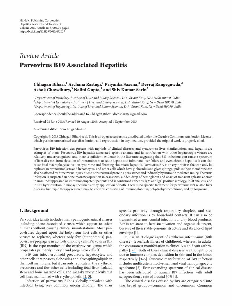
Table 3: List of reported cases of parvovirus B19 hepatitis.
Author Cases Associated condition ReferencesMartınez Gonzalez et al. (2012) One (acute hepatitis) [16]Sun and Zhang (2012) One (FHF) Aplastic anemia [17]Larsen (2011) One (acute hepatitis) [18]Hatakka et al. (2011) One (acute hepatitis) [19]Yang et al. (2012) One (acute hepatitis) DLBCL [20]Sun et al. (2011) Two (acute hepatiti) [21]Al Nahdi et al. (2010) One (recurrent acute hepatitis) [22]Mogensen et al. (2010) One (chronic hepatitis) Lymphopenia [23]Wang et al. (2009) One (chronic hepatitis) [24]Krygier et al. (2009) One (acute hepatitis) [25]Kim et al. (2009) One (acute hepatitis) [26]Pongratz et al. (2009) One (acute hepatitis) [27]Cao et al. (2009) One (fulminant hepatic failure) [28]Kishore and Sen (2009) One (fulminant hepatic failure) Coexistent A and E [29]Al-Abdwani et al. (2008) One (acute hepatitis) Aplastic anemia [30]Giørtz-Carlsen et al. (2007) One (acute hepatitis) [31]Ozcay et al. (2006) One (fulminant hepatic failure) Pure red cell aplasia [32]Aydin et al. (2006) One (acute hepatitis) [33]Toshihiro et al. (2003) One (acute hepatitis) [34]Chehal et al. (2002) One (acute hepatitis) [35]Dame et al. (2002) One (acute hepatitis) Aplastic anemia [36]Dıaz and Collazos (2000) One (acute hepatitis) [37]Lee et al. (2000) One (acute hepatitis) Post-renal-transplant immunosuppression [38]Pinho et al. (2001) One (acute hepatitis) [39]Shan et al. (2001) One (FCH) Post-renal-transplant immunosuppression [40]Alliot et al. (2001) One (acute hepatitis) HIV [41]Karetnyi et al. (1999) One (fulminant hepatic failure) [42]Drago et al. (1999) One (acute hepatitis) [43]Sokal et al. (1998) One (fulminant hepatic failure) [44]Hillingsø et al. (1998) One (acute hepatitis) [45]Hillingsø et al. (1998) One (acute hepatitis) [46]Longo et al. (1998) One (acute hepatitis) Still’s disease [47]Pardi et al. (1998) Two (acute hepatitis) Post-Liver-transplant aplastic anemia [48]Weinberg et al. (1996) One (acute hepatitis) [49]Naides et al. (1996) One (acute hepatitis) [50]Yoto et al. (1996) One (acute hepatitis) [51]Langnas et al. (1995) Six (2 acute hepatitis, 4 fulminant hepatitis) Aplastic anemia [52]Pouchot et al. (1993) One (acute hepatitis) [53]
affected patients [24]. A study by Toan et al. on 463 hepatitisB positive Vietnamese patients showed that 99/463 patients(21.4%) were positive for B19 DNA which was significantlyhigher than those of healthy controls. They also concludedthat in HBV/B19 coinfection the probability of progression tomore severe hepatitis is significantly higher [54].
The association of B19 with chronic hepatitis B and Chas also been described by Hsu et al. They found that B19serology for IgM and IgG was positive in 35.2% and 85%.2% of the cases of chronic hepatitis B with B19 DNA weredetected in 37% of the cases of chronic hepatitis B. In casesof chronic hepatitis C, IgM and IgG antibodies for B19 were
positive in 15.7% and 70.6%, and B19 DNA was detectedin 23.5% of HCV cases. Distinctive subtypes of B19 weredetected in chronic hepatitis B and C, TW-3 in chronichepatitis B andTW-9 in cases of chronic hepatitis C infection.Liver dysfunction was not associated with B19 coexistencein the chronic hepatitis cases. The study also revealed thata significant proportion of coinfection occurs in chronichepatitis cases with B19 infection [55]. It is important to notethat although liver functions are not much affected by thecoinfection of B19 [24, 55], large cohort studies are requiredto explore the pathological course of B19 in association withchronic hepatitis and their clinical outcomes.

4 Hepatitis Research and Treatment
2.3. Parvovirus B19 and Fibrosing Cholestatic Hepatitis. Therewas a single case report of fibrosing cholestatic hepatitis(FCH) due to B19 infection in a patient with renal allograftfor IgA nephropathy. During the postoperative period, thepatient developed features of acute liver failure. All viralmarkers were negative except for HBV and B19 DNA. Thepatient was given lamivudine therapy; however, his conditiongot deteriorated, and the patient subsequently died.The post-mortem liver tissue revealed FCH.On immunohistochemicalexamination, the biopsy was negative for HBsAg andHBcAg,while the PCR showed strong positivity in liver tissue for B19infection, and it was considered that B19was the cause of FCH[40].
2.4. Parvovirus B19 Coinfection with Other HepatotropicViruses. B19 coinfection with other hepatotropic viruses canlead to severe acute fulminant hepatic failure (FHF) withsevere outcome as compared to isolated B19 or other hepa-totropic virus associated FHF. Dwivedi et al. in their study of48 patients with FHF, divided them into three groups as thoseassociated with (i) B19 infection alone, (ii) one or more otherhepatotropic viral infection in the absence of B19 infection,and (iii) B19 coinfection with other hepatotropic viruses.They found that FHF caused by B19 and coinfection withother hepatitis viruses had severe jaundice, high bilirubin,high alanine aminotransferase or aspartate aminotransferaseactivity, and unfavorable outcome resulting in death of mostof these patients, compared with those with isolated B19 orother hepatitis viruses infection [56]. It was hypothesized thatB19 possibly may cause injury to hepatocytes independentlyor by producing synergistic effect when present along withother hepatitis viruses.
2.5. Parvovirus B19 and Hepatitis Associated Aplastic Anemia(HAAA). Hepatitis associated aplastic anemia (HAAA) is adistinct variant of acquired aplastic anemia (AA), in whichan acute attack of hepatitis culminates in marrow failureand pancytopenia [57]. Several hepatitis viruses such ashepatitis A, B, C, E, and G have been anticipated to beassociated with this set of symptoms. Besides the hepatitisviruses, other viruses have also been implicated as causativeagent of AA which include B19, Cytomegalovirus, EpsteinBarr virus, echovirus 3, GB virus-C, transfusion transmit-ted virus (TTV), SEN virus, and non-A-E hepatitis virus(unknown viruses). B19, an underrecognized hepatotropicvirus, is documented as an offending agent of acute hepatitis,FHF, and HAAA in immunocompromised patients [58].There have been reports of B19 related HAAA in post-liver-transplant immunocompromised patients [48, 52]. Themyelotoxic effect in B19 or other viral infections is thoughtto be due to increased circulating cytotoxic CD8+ T cellsand IFN-𝛾 secretion by these cells. Similarly, high circulatingCD8+ T cells cause the altered and defective monocyte andmacrophage differentiation, decreased level of circulatingIL-1, and increased secretion of TNF-𝛼, IFN-𝛾, and IL-2receptors which causes onlooker damage of hepatocytes andsubsequently occurrence of acute hepatitis [59, 60].
2.6. Parvovirus B19 Hepatitis in Underlying HematologicalDiseases. In patients with hemolytic anemia, B19 infectioncan cause an abrupt cessation of red cell production whichis exacerbated in case of acute infection or in compensatedstates and provokes severe anemia. Anemic crisis in heredi-tary spherocytosis and in sickle cell disease has long been rec-ognized. The bone marrow in patients with transient aplasticcrisis is characterized by an absence of maturing erythroidprecursors and presence of giant pronormoblasts. Throm-bocytopenia and pancytopenia have also been reported inpatients with acute B19 infection [2]. Transient aplastic crisis(TAC) is a self-limiting condition, and normal individualsusually recover as the neutralizing antibodies are produced,while in immunocompromised patients, neutralizing anti-body cannot be produced, and this can lead to persistent purered aplasia [1]. Zaki et al. found that the incidence of B19infection is significantly higher among childrenwith hemato-logical disorders, including hemolytic anemias, lymphomas,and leukemias on chemotherapy [61]. B19 has a tropism forthe immature proliferating pronormoblasts and is essentiallyan erythrovirus. Globoside, a neutral glycolipid that acts as acellular receptor and nonstructural protein of parvovirus, isresponsible for the apoptotic death of erythroid progenitors,and some other cells such as megakaryocytes may be lysedby restricted expression of viral proteins in the absence ofviral propagation [62]. Thus, the high propensity of B19 inhematological disease at times can also cause acute hepatitis[63].
2.7. Parvovirus B19 Hepatitis and Hemophagocytosis Lympho-histiocytosis (HLH). Hemophagocytic lymphohistiocytosis(HLH) is a hyperinflammatory condition clinically charac-terized by fever, splenomegaly, jaundice, and phagocytosisof erythrocytes, leukocytes, platelets, and their precursorsby macrophages in bone marrow and other tissues [64].Among viruses, Epstein-Barr virus, Cytomegalovirus, humanherpesvirus 6, and parvovirus B19 have been implicated tocause virus associated HLH (VAHLH) [65]. B19 related HLHis not so uncommon and can occur both in immunocompe-tent and immunocompromised hosts [66]. The hallmark ofHLH pathogenesis is T cell activation leading to stimulationof macrophages which thereby initiates hemophagocytosis.Thus, CD8+ and CD4+ T cells activation triggers markedcytokine production of TNF-𝛼, a key factor in histiocyticactivation [67]. As described above, activated T cells andreleased cytokines may also lead to hepatocyte damage andultimately hepatitis.
3. Diagnosis
3.1. Hematological Tests. In patients with evidence of clin-ically significant anemia or transient aplastic crisis (TAC),a complete blood count with reticulocyte count aids insuspecting B19 infection with the following possible scenar-ios: (1) patients infected with parvovirus B19 will have alow reticulocyte count (0-1%) and (2) in an aplastic crisis,hemoglobin levels will drop below the patient’s baseline by atleast 2 g/dL [68]. Bone marrow examination in these patients

Hepatitis Research and Treatment 5
(a) (b)
Figure 3: (a) Parvovirus B19 inclusion in Pronormoblast in bone marrow aspirate (Geimsa, 1000x), in same case as described above. (b)Clearing of nuclei in pronormoblasts, due to Parvovirus B19 inclusion (HE, 400x); an adult case of hereditary spherocytosis with acutehepatitis and sudden drop of haemoglobin, Parvovirus IgM serology positive.
reveals absence of maturing red cell with prominence ofpronormoblast having cytoplasmic blebs and intranuclearinclusions (Figure 3(a)). Bone marrow biopsy examinationshows nuclear clearing due to viral inclusions present in them[69] (Figure 3(b)).
3.2. Serology. Parvovirus serology (anti-parvovirus B19 im-munoglobulin M (IgM) and immunoglobulin G (IgG) anti-bodies) can be determined using enzyme-linked immunoas-say (ELISA), radioimmunoassay, or immunofluorescence.Results of IgM testing are maybe difficult to interpret; how-ever, reliable results can be obtained by using automatedinstruments dedicated for serological testing. Generally, IgMantibodies are detectable 3 days after infection, and IgGantibodies can be detected after 2 weeks at the time ofrecovery of hematopoiesis. IgM antibodies once formedremain detectable for months, while IgG can be detected forlifetime. In immunodeficient patients, inability to clear thevirus leads to chronic B19 infection and leads to pure red cellaplasia (PRAC). In contrast to TAC, PRAC is characterizedby very low or absent antibody levels, and they are diagnosedbest by polymerase chain reaction (PCR) [70]. Pregnantwomen exposed to parvovirus B19 should get IgG and IgMserology done as soon as possible, as infection risk for fetusremains due [71].
3.3. Polymerase Chain Reaction (PCR). PCR testing for par-vovirus B19 is routinely available with high sensitivity level.Low levels of B19 DNA can be detected for more than 4months in serum after acute infection and for years inother tissues. PCR can also be used to diagnose chronicinfection by detecting viral DNA present in the blood orother tissues/fluids. However, the interpretation pertaining topregnant women is uncertain [72].
3.4. Immunohistochemistry. Parvovirus B19monoclonal anti-body R92F6 against VP1/VP2 capsid protein antigen can alsobe detected in liver tissue and in bone marrow biopsy byimmunohistochemistry [39].
3.5. Other Potential Diagnostic Methods. Enzyme-linked im-munosorbent spot assay (ELISPOT) is a method to mea-sure the qualitative and quantitative immune response inhumans and animals. This method identifies and enumeratescytokine-producing cells at the single cell level. By havingappropriate conditions, the ELISPOT assay allows visual-ization of the secretory product of individual activated orresponding cells [27]. Each spot that develops in the assayrepresents a single reactive cell. This ELISPOT technique canbe used to detect CD4+ T cells specific for B19 viral proteinsin cases of persistent infection [27].
The diagnosis of acute or chronic infection should bemade on the basis of standard DNA hybridization or quan-titative (real-time) PCR in combination with serologic assaysfor B19-specific IgG, IgM, or both [73].
4. Pathogenesis
Themechanism bywhich parvovirus B19 infectionmay resultin hepatic injury is exactly not clear. Hepatic cell damagerelated to direct viral invasion is one possibility. Alternatively,injury may result as an indirect consequence of the immuneresponse directed against the virus.
4.1. Direct Cytopathic Effect. B19 is a single-stranded DNAvirus and has a genome length of 5.4 kb with hairpin struc-tures at each extremity. Two major open reading frames(ORFs) extend through the entire genome of virus. A non-structural protein (NS1) is found on the N-terminal region ofthe genome, and its molecular weight is 70 to 77 kDa. NS1 isthought to be essential for viral DNA replication and also forthe regulation of viral promoters. NS1 contains a consensussequence for ATP- or GTP-binding, which is associated withATPase and DNA helicase activities. NS1 is also known tobe cytotoxic for erythroid cells and is possibly related to thepathogenesis of B19 virus infection [74]. B19 can replicate onlyin the erythroid precursors and few other cells including fetalliver, isolated stem and bone marrow cells, and megakary-ocytic leukemia cell lines maintained with erythropoietin.

6 Hepatitis Research and Treatment
G1 arrest
NS1
Induces P21/WAF1, a cyclin dependent kinase inhibitor
Apoptosis through activation of caspase-3 and caspase-9
Apoptotic cell death
Parvovirus B19 Parvovirus B19
Globoside receptor
(a)
Secretion of IFN gamma, TNF alpha, interleukin-2 receptors, and reduced interleukin-1
Bone marrow derived
cytotoxic CD8 cells
accumulation
Altered and defective
monocytes and macrophages
Cytotoxic CD8 T cells in liver
through circulation
B
aa
Cell damage
and death
Hepatic microenvironment
(b)
Figure 4: Schematic presentation of direct and indirect hepatocellular injury in parvovirus B19 infection.
Hepatocytes express globoside and glycosphingolipids, theputative receptors for B19 virus. B19 virus enters hepatocytesthrough globoside and establishes a restricted infection withthe production of NS1 without the production of viralprogeny [75]. NS1 expression plays a critical role in G1arrest induced by B19 virus. Furthermore, NS1 expressionalso significantly increases p21/WAF1 expression, a cyclindependent kinase inhibitor that induces G1 arrest. Ultimately,the G1 arrested hepatocytes undergo apoptosis by activationof caspase-3 and caspase-9 [74, 76]. Diagrammatic summaryis depicted in Figure 4(a).
4.2. Indirect Immunological Effect. The hepatotoxic effectcaused by parvovirus B19 infection is thought to be due toincreased circulating CD8+ cytotoxic T cells and IFN-𝛾 andTNF-𝛼 secretion by these cells [58]. Similarly, high circulatingCD8+ T cells cause the altered and defective monocyte andmacrophage differentiation, decreased level of circulating IL-1, and increased secretion of TNF-𝛼, IFN-𝛾, and IL-2 recep-tors which causes damage of hepatocytes and subsequentlyleading to acute hepatitis [58–60]. Diagrammatic summaryis depicted in Figure 4(b).
5. Treatment
There are no specific treatment guidelines for infectioncaused by B19 virus, and most of the symptoms and elevationof liver enzymes presented during infection stage resolvewithout any treatment. In case of acute and fulminanthepatitis, combination therapy consisting of an intravenousinfusion of immunoglobulin and dehydrohydrocortisone andsubcutaneous injections of granulocyte colony-stimulatingfactor for three months has been tried [17]. For HAAA andHLH, an immunosuppressive therapy comprising antithymo-cyte Globulin (ATG), cyclosporine, and steroids has proven
to be effective [58]. Aplastic crises and PRAC are tran-siently responsive to erythropoietin, growth factors, gran-ulocyte colony-stimulating factor, granulocyte macrophagecolony stimulating factor, interleukin-3, and androgens [77].Nonresponsive HAAA should be treated by allogenic bonemarrow (BM) transplantation from HLA matched siblings[78].
6. Conclusion
There are sufficient lines of evidence in the literature thatstate that Parvovirus B19 infection can be associated withthe development of acute hepatitis, FHF, HAAA, hepatitiswith HLH, chronic hepatitis, and rarely FCH. There is asignificant rate of coexistence of B19 with chronic hepatitisB and C as suggested by the literature. This area needs to befurther explored and validated through large cohort studies.Infection with parvovirus B19 should be considered in thedifferential diagnosis in both immunocompromised andimmunocompetent patients presenting with acute hepatitisof unknown etiology particularly in cases of underlyinghemolytic diseases and immunodeficient host with aplasticanemia.The parvovirus B19 infection can be detected by pos-itive IgM serology and by PCR in infected tissues. ParvovirusB19 can cause hepatitis due to direct cytopathic and indirectimmunological injury through CD8+ cytotoxic T cells. Acombination of an intravenous infusion of immunoglobulin,dehydrohydrocortisone and cyclosporine and subcutaneousinjections of granulocyte colony-stimulating factor for threemonths has proved to be an effective therapy for parvovirusB19 hepatitis and HAAA.
Conflict of Interests
The authors declare that they have no conflict of interests.

Hepatitis Research and Treatment 7
References
[1] N. S. Young and K. E. Brown, “Mechanisms of disease: par-vovirus B19,”TheNew England Journal of Medicine, vol. 350, no.6, pp. 586–597, 2004.
[2] K. E. Brown and N. S. Young, “The simian parvoviruses,”Reviews in Medical Virology, vol. 7, pp. 211–218, 1997.
[3] T. L. Moore, “Parvovirus-associated arthritis,” Current Opinionin Rheumatology, vol. 12, no. 4, pp. 289–294, 2000.
[4] T. Chorba, P. Coccia, and R. C. Holman, “The role of parvovirusB19 in aplastic crisis and erythema infectiosum (fifth disease),”Journal of Infectious Diseases, vol. 154, no. 3, pp. 383–393, 1986.
[5] Z.He,H. Zhuang, X.Wang et al., “Retrospective analysis of non-A-E hepatitis: possible role of hepatitis B and C virus infection,”Journal of Medical Virology, vol. 69, no. 1, pp. 59–65, 2003.
[6] S. Arista, S. De Grazia, V. DiMarco, R. Di Stefano, and A. Craxı,“Parvovirus B19 and “cryptogenic” chronic hepatitis,” Journal ofHepatology, vol. 38, no. 3, pp. 375–376, 2003.
[7] M. Beghetti, A. Gervaix, C. A. Haenggeli, M. Berner, andP. C. Rimensberger, “Myocarditis associated with parvovirusB19 infection in two siblings with merosin-deficient congenitalmuscular dystrophy,”European Journal of Pediatrics, vol. 159, no.1-2, pp. 135–136, 2000.
[8] T. H. Finkel, T. J. Torok, P. J. Ferguson et al., “Chronic par-vovirus B19 infection and systemic necrotising vasculitis:opportunistic infection or aetiological agent?” The Lancet, vol.343, no. 8908, pp. 1255–1258, 1994.
[9] G. Nigro, M. Zerbini, A. Krzysztofiak et al., “Active or recentparvovirus B19 infection in childrenwithKawasaki disease,”TheLancet, vol. 343, no. 8908, pp. 1260–1261, 1994.
[10] P. J. Ferguson, F. T. Saulsbury, S. F. Dowell, T. J. Torok, D. D.Erdman, and L. J. Anderson, “Prevalence of human parvovirusB19 infection in children with Henoch-Schonlein purpura,”Arthritis and Rheumatism, vol. 39, no. 5, pp. 880–881, 1996.
[11] S. E. Gabriel, M. Espy, D. D. Erdman, J. Bjornsson, T. F.Smith, and G. G. Hunder, “The role of parvovirus B19 in thepathogenesis of giant cell arteritis: a preliminary evaluation,”Arthritis and Rheumatism, vol. 42, pp. 1255–1258, 1999.
[12] S. B. Smith, L. F. Libow, D. M. Elston, R. A. Bernert, andK. E. Warschaw, “Gloves and socks syndrome: early and latehistopathologic features,” Journal of the American Academy ofDermatology, vol. 47, no. 5, pp. 749–754, 2002.
[13] S. Kim Jacobson, J. S. Daly, G. M. Thorne, and K. McIntosh,“Chronic parvovirus B19 infection resulting in chronic fatiguesyndrome: case history and review,” Clinical Infectious Diseases,vol. 24, no. 6, pp. 1048–1051, 1997.
[14] J. R. Kerr, F. Barah, M. L. Chiswick et al., “Evidence for therole of demyelination, HLA-DR alleles, and cytokines in thepathogenesis of parvovirus B19 meningoencephalitis and itssequelae,” Journal of Neurology Neurosurgery and Psychiatry,vol. 73, no. 6, pp. 739–746, 2002.
[15] I. Mihaly, A. Trethon, Z. Aranyi et al., “Observations on humanparvovirus B19 infection diagnosed in 2011,”Orvosi Hetilap, vol.153, no. 49, pp. 1948–1957, 2012.
[16] J. Martınez Gonzalez, C. Senosiain Lalastra, F. Mesonero Gis-mero, and V. Moreira Vicente, “An exceptional cause of acutehepatitis in an adult: parvovirus B19,” Journal of Gastroenterol-ogy and Hepatology, vol. 35, no. 10, pp. 697–699, 2012.
[17] L. Sun and J.-C. Zhang, “Acute fulminant hepatitis with bonemarrow failure in an adult due to parvovirus B19 infection,”Hepatology, vol. 55, no. 1, pp. 329–330, 2012.
[18] L. Larsen, “Parvovirus B19-akut hepatitis hos immunkompetentpatient,”Ugeskrift for Laeger, vol. 173, no. 43, pp. 2719–2720, 2011.
[19] A. Hatakka, J. Klein, R. He, J. Piper, E. Tam, and A. Walkty,“Acute hepatitis as a manifestation of parvovirus B19 infection,”Journal of Clinical Microbiology, vol. 49, no. 9, pp. 3422–3424,2011.
[20] S.-H. Yang, L.-W. Lin, Y.-J. Fang, A.-L. Cheng, and S.-H.Kuo, “Parvovirus B19 infection-related acute hepatitis afterrituximab-containing regimen for treatment of diffuse large B-cell lymphoma,” Annals of Hematology, vol. 91, no. 2, pp. 291–294, 2012.
[21] L. Sun, J.-C. Zhang, and Z.-S. Jia, “Association of parvovirus B19infection with acute icteric hepatitis in adults,” ScandinavianJournal of Infectious Diseases, vol. 43, no. 6-7, pp. 547–549, 2011.
[22] N. Al Nahdi, H. Wiesinger, H. Sutherland, and E. M. Yoshida,“Recurrent idiopathic acute hepatitis-associated aplastic ane-mia/pancytopenia fourteen years after initial episode,” Annalsof Hepatology, vol. 9, no. 4, pp. 468–470, 2010.
[23] T. H. Mogensen, J. M. B. Jensen, S. Hamilton-Dutoit, and C. S.Larsen, “Chronic hepatitis caused by persistent parvovirus B19infection,” BMC Infectious Diseases, vol. 10, article 246, 2010.
[24] C. Wang, A. Heim, V. Schlaphoff et al., “Intrahepatic long-term persistence of parvovirus B19 and its role in chronic viralhepatitis,” Journal of Medical Virology, vol. 81, no. 12, pp. 2079–2088, 2009.
[25] D. S. Krygier, U. P. Steinbrecher, M. Petric et al., “ParvovirusB19 induced hepatic failure in an adult requiring liver trans-plantation,” World Journal of Gastroenterology, vol. 15, no. 32,pp. 4067–4069, 2009.
[26] B. J. Kim, K. H. Yoo, K. Li, and M. N. Kim, “Parvovirus B19infection associated with acute hepatitis in infant,” PediatricInfectious Disease Journal, vol. 28, no. 7, article 667, 2009.
[27] G. Pongratz, J. Lindner, S. Modrow, S. Schimanski, J.Scholmerich, andM. Fleck, “Persistent parvovirus B19 infectiondetected by specific CD4+ T-cell responses in a patient withhepatitis and polyarthritis,” Journal of Internal Medicine, vol.266, no. 3, pp. 296–301, 2009.
[28] Y.-H. Cao, G.-Y. Zhang, andG.-C. Zhang, “Successful treatmentwith high-dose intravenous immunoglobulin for parvovirusB19 infection associated with acute fulminant hepatitis in achinese child,” Clinical Pediatrics, vol. 48, no. 6, pp. 674–676,2009.
[29] J. Kishore and M. Sen, “Parvovirus B19-induced thrombocy-topenia and anemia in a child with fatal fulminant hepaticfailure coinfected with hepatitis A and E viruses,” Journal ofTropical Pediatrics, vol. 55, no. 5, pp. 335–337, 2009.
[30] R. M. Al-Abdwani, F. A. Khamis, A. Balkhair, M. Sacharia,and Y. A. Wali, “A child with human parvovirus B19 infectioninduced aplastic anemia and acute hepatitis: effectiveness ofimmunosuppressive therapy,” Pediatric Hematology and Oncol-ogy, vol. 25, no. 7, pp. 699–703, 2008.
[31] B. Giørtz-Carlsen, S. Rittig, and T. Thelle, “Neurological symp-toms and acute hepatitis associated with parvovirus B19,”Ugeskrift for Laeger, vol. 169, no. 47, pp. 4075–4077, 2007.
[32] F. Ozcay, Y. E. Bikmaz, O. Canan, and N. Ozbek, “HepatitisA and parvovirus B19 infections in an infant with fulminanthepatic failure,” Turkish Journal of Gastroenterology, vol. 17, no.2, pp. 148–150, 2006.
[33] M. Aydin, Y. Bulut, G. Poyrazoglu, M. Turgut, and A. Seyrek,“Detection of human parvovirus B19 in children with acutehepatitis,”Annals of Tropical Paediatrics, vol. 26, no. 1, pp. 25–28,2006.

8 Hepatitis Research and Treatment
[34] M. Toshihiro, Y. Takikawa, Y. Fukuda, S.-I. Sato, R. Endou, andK. Suzuki, “A case of acute hepatitis associated with ParvovirusB19,” Japanese Journal of Gastroenterology, vol. 100, no. 11, pp.1312–1316, 2003.
[35] A. Chehal, A. I. Sharara, H. A. Haidar, J. Haidar, and A.Bazarbachi, “Acute viral hepatitis A and parvovirus B19 infec-tions complicated by pure red cell aplasia and autoimmunehemolytic anemia,” Journal of Hepatology, vol. 37, no. 1, pp. 163–165, 2002.
[36] C. Dame, C. Hasan, U. Bode, and A. M. Eis-Hubinger, “Acuteliver disease and aplastic anemia associated with the persistenceof B19 DNA in liver and bone marrow,” Pediatric Pathology andMolecular Medicine, vol. 21, no. 1, pp. 25–29, 2002.
[37] F. Dıaz and J. Collazos, “Hepatic dysfunction due to parvovirusB19 infection,” Journal of Infection and Chemotherapy, vol. 6, no.1, pp. 63–64, 2000.
[38] P. C. Lee, C. J. Hung, H. Y. Lei, T. T. Chang, J. R.Wang, andM. S.Jan, “Parvovirus B19-related hepatitis in an immunosuppressedkidney transplant,” Nephrology Dialysis Transplantation, vol. 15,pp. 1486–1488, 2000.
[39] J. R. R. Pinho, V. A. F. Alves, A. F. Vieira et al., “Detection ofhuman parvovirus B19 in a patient with hepatitis,” BrazilianJournal of Medical and Biological Research, vol. 34, no. 9, pp.1131–1138, 2001.
[40] Y.-S. Shan, P.-C. Lee, J.-R. Wang, H.-P. Tsai, C.-M. Sung,and Y.-T. Jin, “Fibrosing cholestatic hepatitis possibly relatedto persistent parvovirus B19 infection in a renal transplantrecipient,” Nephrology Dialysis Transplantation, vol. 16, no. 12,pp. 2420–2422, 2001.
[41] C. Alliot, M. Barrios, J. Taib, and M. Brunel, “Parvovirus B19infection in an HIV-infected patient with febrile pancytopeniaand acute hepatitis,” European Journal of Clinical Microbiologyand Infectious Diseases, vol. 20, no. 1, pp. 43–45, 2001.
[42] Y. V. Karetnyi, P. R. Beck, R. S. Markin, A. N. Langnas, and S.J. Naides, “Human parvovirus B19 infection in acute fulminantliver failure,” Archives of Virology, vol. 144, no. 9, pp. 1713–1724,1999.
[43] F. Drago, M. Semino, P. Rampini, and A. Rebora, “ParvovirusB19 infection associated with acute hepatitis and a purpuricexanthem,” British Journal of Dermatology, vol. 141, no. 1, pp.160–161, 1999.
[44] E. M. Sokal, M. Melchior, C. Cornu et al., “Acute parvovirusB19 infection associated with fulminant hepatitis of favourableprognosis in young children,”The Lancet, vol. 352, no. 9142, pp.1739–1741, 1998.
[45] J. G. Hillingsø, I. P. Jensen, and L. Tom-Petersen, “ParvovirusB19 as causative agent of acute hepatitis in adults,” Ugeskrift forLaeger, vol. 160, no. 44, pp. 6355–6356, 1998.
[46] J. G. Hillingsø, I. P. Jensen, and L. Tom-Petersen, “ParvovirusB19 and acute hepatitis in adults,”The Lancet, vol. 351, no. 9107,pp. 955–956, 1998.
[47] G. Longo, M. Luppi, M. Bertesi, L. Ferrara, G. Torelli, and G.Emilia, “Still’s disease, severe thrombocytopenia, and acute hep-atitis associated with acute parvovirus B19 infection,” ClinicalInfectious Diseases, vol. 26, no. 4, pp. 994–995, 1998.
[48] D. S. Pardi, Y. Romero, L. E. Mertz, and D. D. Douglas,“Hepatitis-associated aplastic anemia and acute parvovirus B19infection: a report of two cases and a review of the literature,”The American Journal of Gastroenterology, vol. 93, no. 3, pp.468–470, 1998.
[49] J. M. Weinberg, J. T. Wolfe, A. L. Frattali, V. P. Werth, S. J.Naides, and E. M. Spiers, “Parvovirus B19 infection associated
with acute hepatitis, arthralgias, and rash,” Journal of ClinicalRheumatology, vol. 2, no. 2, pp. 85–88, 1996.
[50] S. J. Naides, Y. V. Karetnyi, L. L. W. Cooling et al., “Human par-vovirus B19 infection and hepatitis,” The Lancet, vol. 347, no.9014, pp. 1563–1564, 1996.
[51] Y. Yoto, T. Kudoh, K. Haseyama, N. Suzuki, and S. Chiba,“Human parvovirus B19 infection associated with acute hepati-tis,”The Lancet, vol. 347, no. 9005, pp. 868–869, 1996.
[52] A. N. Langnas, R. S. Markin, M. S. Cattral, and S. J. Naides,“Parvovirus B19 as a possible causative agent of fulminant liverfailure and associated aplastic anemia,” Hepatology, vol. 22, no.6, pp. 1661–1665, 1995.
[53] J. Pouchot, H. Ouakil, M. L. Debin, and P. Vinceneux, “AdultStill’s disease associated with acute human parvovirus B19infection,”The Lancet, vol. 341, no. 8855, pp. 1280–1281, 1993.
[54] N. L. Toan, L. H. Song, P. G. Kremsner et al., “Co-infection ofhuman parvovirus B19 in Vietnamese patients with hepatitis Bvirus infection,” Journal of Hepatology, vol. 45, no. 3, pp. 361–369, 2006.
[55] T.-C. Hsu, T.-Y. Chen, M.-C. Lin, B.-S. Tzang, and G. J. Tsay,“Human parvovirus B19 infection in patients with chronichepatitis B or hepatitis C infection,” Journal of Gastroenterologyand Hepatology, vol. 20, no. 5, pp. 733–738, 2005.
[56] M. Dwivedi, H. Manocha, S. Tiwari, G. Tripathi, and T. N.Dhole, “Coinfection of parvovirus b19 with other hepatitisviruses leading to fulminant hepatitis of unfavorable outcomein children,”The Pediatric Infectious Disease Journal, vol. 28, no.7, pp. 649–650, 2009.
[57] Y. Osugi, H. Yagasaki, M. Sako et al., “Antithymocyte globulinand cyclosporine for treatment of 44 children with hepatitisassociated aplastic anemia,” Haematologica, vol. 92, no. 12, pp.1687–1690, 2007.
[58] B. Rauff, M. Idrees, S. A. R. Shah et al., “Hepatitis associatedaplastic anemia: a review,” Virology Journal, vol. 8, article 87,2011.
[59] R.Andreesen,W.Brugger, C.Thomssen,A. Rehm,B. Speck, andG.W. Lohr, “Defectivemonocyte-to-macrophagematuration inpatients with aplastic anemia,” Blood, vol. 74, no. 6, pp. 2150–2156, 1989.
[60] T. Muta, Y. Tanaka, E. Takeshita et al., “Recurrence of hepatitis-associated aplastic anemia after a 10-year Interval,” InternalMedicine, vol. 47, no. 19, pp. 1733–1737, 2008.
[61] M. E. S. Zaki, S. A. Hassan, T. Seleim, and R. A. Lateef, “Par-vovirus B19 infection in childrenwith a variety of hematologicaldisorders,” Hematology, vol. 11, no. 4, pp. 261–266, 2006.
[62] S. Serke, T. F. Schwarz, H. Baurmann et al., “Productive infec-tion of in vitro generated haemopoietic progenitor cells fromnormal human adult peripheral blood with parvovirus B19:studies bymorphology, immunocytochemistry, flow-cytometryand DNA-hybridization,” British Journal of Haematology, vol.79, no. 1, pp. 6–13, 1991.
[63] T. Kudoh, Y. Yoto, N. Suzuki et al., “Human parvovirus B19-induced aplastic crisis in iron deficiency anemia,” Acta Paedi-atrica Japonica, vol. 36, no. 4, pp. 448–449, 1994.
[64] G. Janka, “Hemophagocytic lymphohistiocytosis: when theimmune system runs amok,” Klinische Padiatrie, vol. 221, no. 5,pp. 278–285, 2009.
[65] M. P. Hoang, D. B. Dawson, Z. R. Rogers, R. H. Scheuermann,and B. B. Rogers, “Polymerase chain reaction amplificationof archival material for Epstein-Barr virus, cytomegalovirus,human herpesvirus 6, and parvovirus B19 in children with bone

Hepatitis Research and Treatment 9
marrow hemophagocytosis,” Human Pathology, vol. 29, no. 10,pp. 1074–1077, 1998.
[66] K. Shirono and H. Tsuda, “Parvovirus B19-associated haemo-phagocytic syndrome in healthy adults,” British Journal of Hae-matology, vol. 89, no. 4, pp. 923–926, 1995.
[67] I.-J. Su, C.-H. Wang, A.-L. Cheng, and R.-L. Chen, “Hemo-phagocytic syndrome in Epstein-Barr virus-associated T-lym-phoproliferative disorders: disease spectrum, pathogenesis, andmanagement,” Leukemia and Lymphoma, vol. 19, no. 5-6, pp.401–406, 1995.
[68] M. M. Mustafa and K. L. McClain, “Diverse hematologic effectsof parvovirus B19 infection,” Pediatric Clinics of North America,vol. 43, no. 3, pp. 809–821, 1996.
[69] K. Smith-Whitley, H. Zhao, R. L. Hodinka et al., “Epidemiologyof human parvovirus B19 in children with sickle cell disease,”Blood, vol. 103, no. 2, pp. 422–427, 2004.
[70] The American Academy of Pediatrics Committee on InfectiousDiseases, “Parvovirus B19,” inRedBook: Report of the Committeeon Infectious Diseases, L. K. Pickering, C. J. Baker, D. W.Kimberlin, and S. S. Long, Eds., pp. 491–493, The AmericanAcademy of Peiatrics, Elk Grove Village, Ill, USA, 28th edition,2009.
[71] C. K. Fairley, J. S. Smoleniec, O. E. Caul, and E. Miller, “Obser-vational study of effect of intrauterine transfusions on outcomeof fetal hydrops after parvovirus B19 infection,”The Lancet, vol.346, no. 8986, pp. 1335–1337, 1995.
[72] M. Soderlund-Venermo, K. Hokynar, J. Nieminen, H. Rautako-rpi, and K. Hedman, “Persistence of human parvovirus B19 inhuman tissues,” Pathologie Biologie, vol. 50, no. 5, pp. 307–316,2002.
[73] G. L. Mandell, J. E. Bennet, and R. Dolin,Mandell, Douglas andBennett’s Principals and Practice of Infectious Diseases, vol. 2,Churchill Livingstone, Philadelphia, Pa, USA, 6th edition, 2005.
[74] E. Morita, A. Nakashima, H. Asao, H. Sato, and K. Sugamura,“Human parvovirus B19 nonstructural protein (NS1) inducescell cycle arrest at G1 phase,” Journal of Virology, vol. 77, no. 5,pp. 2915–2921, 2003.
[75] L. L. W. Cooling, T. A. W. Koerner, and S. J. Naides, “Multipleglycosphingolipids determine the tissue tropism of parvovirusB19,” Journal of Infectious Diseases, vol. 172, no. 5, pp. 1198–1205,1995.
[76] B. D. Poole, Y. V. Karetnyi, and S. J. Naides, “Parvovirus B19-induced apoptosis of hepatocytes,” Journal of Virology, vol. 78,no. 14, pp. 7775–7783, 2004.
[77] N. S. Young and J. Maciejewski, “The pathophysiology of ac-quired aplastic anemia,” The New England Journal of Medicine,vol. 336, no. 19, pp. 1365–1372, 1997.
[78] K. Doney, W. Leisenring, R. Storb, and F. R. Appelbaum,“Primary treatment of acquired aplastic anemia: outcomes withbonemarrow transplantation and immunosuppressive therapy,”Annals of Internal Medicine, vol. 126, no. 2, pp. 107–115, 1997.