Molecular mechanism underlying B19 virus inactivation and comparison to other parvoviruses
10
TRANSFUSION COMPLICATIONS Molecular mechanism underlying B19 virus inactivation and comparison to other parvoviruses Bernhard Mani, Marco Gerber, Patricia Lieby, Nicola Boschetti, Christoph Kempf, and Carlos Ros BACKGROUND: B19 virus (B19V) is a human patho- gen frequently present in blood specimens. Transmis- sion of the virus occurs mainly via the respiratory route, but it has also been shown to occur through the admin- istration of contaminated plasma-derived products. Parvoviridae are highly resistant to physicochemical treatments; however, B19V is more vulnerable than the rest of parvoviruses. The molecular mechanism govern- ing the inactivation of B19V and the reason for its higher vulnerability remain unknown. STUDY DESIGN AND METHODS: After inactivation of B19V by wet heat and low pH, the integrity of the viral capsid was examined by immunoprecipitation with two monoclonal antibodies directed to the N-terminal of VP1 and to a conformational epitope in VP2. The accessibil- ity of the viral DNA was quantitatively analyzed by a hybridization-extension assay and by nuclease treatment. RESULTS: The integrity of the viral particles was main- tained during the inactivation procedure; however, the capsids became totally depleted of viral DNA. The DNA-depleted capsids, although not infectious, were able to attach to target cells. Comparison studies with other members of the Parvoviridae family revealed a remarkable instability of B19V DNA in its encapsidated state. CONCLUSION: Inactivation of B19V by heat or low pH is not mediated by capsid disintegration but by the con- version of the infectious virions into DNA-depleted capsids. The high instability of the viral DNA in its encapsidated state is an exclusive feature of B19V, which explains its lower resistance to inactivation treatments. B 19 virus (B19V) is the only well documented human pathogen of the Parvoviridae family. The virus belongs to the genus Erythrovirus. In most cases, the infection is either asymptom- atic or accompanied by mild nonspecific symptoms. The most common syndrome caused by B19V is an erythema- tous rash illness named erythema infectiosum affecting children. B19V is also the causative agent for transient aplastic crisis, which may have severe effects on patients suffering from sickle cell disease and other anemic ill- nesses. Chronic infections accompanied by pure red cell aplasia and anemia affect immunocompromised patients. Furthermore, B19V may cause fetal death, autoimmune diseases, and arthropathies. 1 B19V is a widespread pathogen. The serologic evi- dence of a past infection is 40 to 60 percent for young adults and 80 to 100 percent for elder people. 1,2 Owing to its high prevalence, blood donations are frequently con- taminated with B19V. The measured incidence of con- tamination depends on the sensitivity of the detection method and ranges from 0.003 percent (immunodiffu- sion) to 1.2 percent (polymerase chain reaction [PCR]) of blood donations examined. 3-7 Because plasma pools are constituted of hundreds of donations, B19V DNA is found in the majority of plasma pools as determined by PCR. 8-10 The contamination of plasma-derived products, such as coagulation factors VIII and IX, human serum albumin, intravenous immune globulin, intramuscularly injected ABBREVIATIONS: B19V = B19 virus; MVM = minute virus of mice; PBSA = phosphate-buffered saline containing 1 percent bovine serum albumin; PLA2 = phospholipase A2. From the Department of Chemistry and Biochemistry, Univer- sity of Bern; and CSL Behring AG, Bern, Switzerland. Address reprint requests to: Carlos Ros, Department of Chemistry and Biochemistry, University of Bern, Freiestrasse 3, 3012 Bern, Switzerland; e-mail: [email protected]. Received for publication March 9, 2007; revision received April 17, 2007, and accepted April 22, 2007. doi: 10.1111/j.1537-2995.2007.01393.x TRANSFUSION 2007;47:1765-1774. Volume 47, October 2007 TRANSFUSION 1765
-
Upload
independent -
Category
Documents
-
view
0 -
download
0
Transcript of Molecular mechanism underlying B19 virus inactivation and comparison to other parvoviruses

T R A N S F U S I O N C O M P L I C A T I O N S
Molecular mechanism underlying B19 virus inactivation andcomparison to other parvoviruses
Bernhard Mani, Marco Gerber, Patricia Lieby, Nicola Boschetti, Christoph Kempf, and Carlos Ros
BACKGROUND: B19 virus (B19V) is a human patho-gen frequently present in blood specimens. Transmis-sion of the virus occurs mainly via the respiratory route,but it has also been shown to occur through the admin-istration of contaminated plasma-derived products.Parvoviridae are highly resistant to physicochemicaltreatments; however, B19V is more vulnerable than therest of parvoviruses. The molecular mechanism govern-ing the inactivation of B19V and the reason for itshigher vulnerability remain unknown.STUDY DESIGN AND METHODS: After inactivation ofB19V by wet heat and low pH, the integrity of the viralcapsid was examined by immunoprecipitation with twomonoclonal antibodies directed to the N-terminal of VP1and to a conformational epitope in VP2. The accessibil-ity of the viral DNA was quantitatively analyzed bya hybridization-extension assay and by nucleasetreatment.RESULTS: The integrity of the viral particles was main-tained during the inactivation procedure; however, thecapsids became totally depleted of viral DNA. TheDNA-depleted capsids, although not infectious, wereable to attach to target cells. Comparison studies withother members of the Parvoviridae family revealed aremarkable instability of B19V DNA in its encapsidatedstate.CONCLUSION: Inactivation of B19V by heat or low pHis not mediated by capsid disintegration but by the con-version of the infectious virions into DNA-depletedcapsids. The high instability of the viral DNA in itsencapsidated state is an exclusive feature of B19V,which explains its lower resistance to inactivationtreatments.
B19 virus (B19V) is the only well documentedhuman pathogen of the Parvoviridae family.The virus belongs to the genus Erythrovirus. Inmost cases, the infection is either asymptom-
atic or accompanied by mild nonspecific symptoms. Themost common syndrome caused by B19V is an erythema-tous rash illness named erythema infectiosum affectingchildren. B19V is also the causative agent for transientaplastic crisis, which may have severe effects on patientssuffering from sickle cell disease and other anemic ill-nesses. Chronic infections accompanied by pure red cellaplasia and anemia affect immunocompromised patients.Furthermore, B19V may cause fetal death, autoimmunediseases, and arthropathies.1
B19V is a widespread pathogen. The serologic evi-dence of a past infection is 40 to 60 percent for youngadults and 80 to 100 percent for elder people.1,2 Owing toits high prevalence, blood donations are frequently con-taminated with B19V. The measured incidence of con-tamination depends on the sensitivity of the detectionmethod and ranges from 0.003 percent (immunodiffu-sion) to 1.2 percent (polymerase chain reaction [PCR]) ofblood donations examined.3-7 Because plasma pools areconstituted of hundreds of donations, B19V DNA is foundin the majority of plasma pools as determined by PCR.8-10
The contamination of plasma-derived products, such ascoagulation factors VIII and IX, human serum albumin,intravenous immune globulin, intramuscularly injected
ABBREVIATIONS: B19V = B19 virus; MVM = minute virus of
mice; PBSA = phosphate-buffered saline containing 1 percent
bovine serum albumin; PLA2 = phospholipase A2.
From the Department of Chemistry and Biochemistry, Univer-
sity of Bern; and CSL Behring AG, Bern, Switzerland.
Address reprint requests to: Carlos Ros, Department of
Chemistry and Biochemistry, University of Bern, Freiestrasse 3,
3012 Bern, Switzerland; e-mail: [email protected].
Received for publication March 9, 2007; revision received
April 17, 2007, and accepted April 22, 2007.
doi: 10.1111/j.1537-2995.2007.01393.x
TRANSFUSION 2007;47:1765-1774.
Volume 47, October 2007 TRANSFUSION 1765

immune globulin, prothrombin complex concentrate andantithrombin III has been reported.8,9,11,12 Therefore, thereis a risk of transmitting B19V through the administrationof plasma-derived products. In these studies however, thecontamination was demonstrated with the presence ofB19V DNA with PCR, which does not necessarily prove thepresence of infectious virus. Nevertheless, direct evidenceof B19V transmission through the administration ofplasma-derived products has also been shown in severalcase studies.13-16 Moreover, patients that receive suchmedication on a regular basis show a higher prevalence ofB19V-specific antibodies than control groups.17 Alto-gether, the contamination of plasma-derived productsindicates a potential risk of a B19V infection for the treatedpatient with potentially severe consequences for pregnantwomen and anemic and immunocompromised patients.
To achieve maximal safety for plasma-derived clinicalproducts, pathogen safety guidelines have been estab-lished, as a result of which manufacturers must demon-strate the effective elimination of viral agents during themanufacturing process of their products. Virus elimina-tion is demonstrated either with the relevant pathogenitself or with one or several closely related model viruses.To date, there is no convenient cell culture infectivity testfor B19V. For this reason, animal parvoviruses such asbovine parvovirus, canine parvovirus, porcine parvovirus,or minute virus of mice (MVM) are often used for valida-tion studies regarding the inactivation of B19V. Parvovi-ruses are among the most stable viruses and have beenshown to resist many common physicochemical inactiva-tion procedures. B19V inactivation can be achieved withdry or wet heat,18-21 as well as with low or high pH,22,23 UVCirradiation,24,25 or photochemical reactions.26 Interestingly,B19V has been found to be more readily inactivated thanother parvoviruses. Whereas B19V is inactivated beyondthe detection limit after 10 minutes at 60°C or after 2 hoursat pH 4, canine parvovirus,21 MVM,27 and porcine parvovi-rus18 can withstand 1 hour at 60°C without considerableinactivation. Similarly, the treatment of MVM at pH 4 for6 hours only moderately reduces its infectivity.23 Thereason why B19V is more sensitive to inactivation thanother parvoviruses is not known. Although different inac-tivation conditions for B19V have been described, theunderlying mechanism of B19V inactivation has not yetbeen elucidated. It is generally assumed that the inactiva-tion occurs through capsid disintegration because theviral genome becomes accessible to DNases.18,27 We haveshown in a recent study, however, that after mild heattreatments, the DNA from B19V and MVM can be ren-dered accessible without capsid disintegration.28
In this study we have analyzed the B19V capsid rear-rangements occurring during the inactivation process.The results revealed a sequence of structural transitionspreceding capsid disintegration. The critical transition,which resulted in full virus inactivation, was the dissocia-
tion of the viral DNA from the still intact capsid. Compari-son studies revealed that the DNA release from intactcapsids is a common feature among parvoviruses butoccurs much more prematurely in B19V, explaining itslower resistance to inactivation procedures.
MATERIALS AND METHODS
Cells and virusesHuman UT7/EPO cells were propagated in RPMI 1640supplemented with 5 percent fetal calf serum (FCS) and2 U per mL recombinant human erythropoietin (EPO;Janssen-Cilag, Midrand, South Africa) at 37°C and5 percent CO2. UT7 cells were provided by A. Gröner (CSLBehring, Marburg, Germany). Two B19V-containingplasma samples (Genotype 1) were obtained from twoinfected individuals (S-1 and S-2) and did not containB19V-specific immunoglobulin M or immunoglobulin G(IgG) antibodies. B19V was concentrated from infectedserum by ultracentrifugation through 20 percent sucrose.The viral pellet was washed and resuspended inphosphate-buffered saline (PBS). All other parvoviruseswere derived from cell culture supernatant. H-1 parvovi-rus was provided by C. Dinsart (German Cancer ResearchCenter, Heidelberg, Germany). Porcine parvovirus wasprovided by T. Novak (CSL Behring, Marburg, Germany).
Exposure of viral particles to inactivationconditionsViral suspensions in PBS were heat-treated in thin-walltubes for 3 or 10 minutes in a preheated thermoblock. Aprobe was used to monitor the temperature of the suspen-sion. After the temperature treatment, the samples wererapidly cooled on ice and immediately used for subse-quent reactions. For pH treatments, the viral suspensionswere acidified by adding MES-buffered saline until thedesired pH was achieved and incubated for 2 hours at37°C. After the treatment, the pH of the viral suspensionwas neutralized by dilution (1:100) into PBS or in PBS con-taining 1 percent BSA (PBSA). Additionally, the heat sen-sitivity of B19V in citrate buffer, which has been recentlyreported to confer heat resistance to B19V,29 was exam-ined. The viral suspension was diluted in citrate buffer(0.5 mol/L trisodium citrate, 0.1 mol/L NaCl, pH 7) or inPBS and exposed to heat as specified above.
Infectivity assayTitration of B19V was performed by limited dilution inquadruplicate. UT7 cells were seeded on 96-well plates(3 ¥ 104 per well) in RPMI, containing 2 U per mL recom-binant human EPO and 5 percent FCS. Virus was dilutedgeometrically by the factor 10 in RPMI. An equal volume of
MANI ET AL.
1766 TRANSFUSION Volume 47, October 2007

diluted virus was added to each well and incubated at 37°Cin 5 percent CO2. After 4 days, the cell culture volume wascarefully removed and cells were fixed with a solution ofice-cold methanol:acetone (1:1, v/v) for 1 hour at 20°C.After fixation, the cells were air-dried, washed with PBSA,and incubated with a mouse antibody against B19V (1:40diluted in PBSA, clone R92F6 IgG1, Novocastra, Newcastleupon Tyne, UK) for 1 hour at room temperature. The cellswere washed with PBSA, and as secondary antibody, aconjugated F(ab′)2 fragment of goat anti-mouse immuno-globulins was added (1:50 dilution, DakoCytomation,Glostrup, Denmark) for 1 hour at room temperature. Afterfinal washings with PBSA, the cells were overlaid with50 mL of glycerin:PBS solution (1:1) and examined underfluorescence microscope. The infectivity titer was calcu-lated with the Spaerman-Kärber method.30
Assessment of B19V capsid integrityAfter exposure to heat or low pH, the integrity of the viralcapsid was examined by immunoprecipitation with twodifferent antibodies. One antibody is directed to a VP2conformational epitope (monoclonal antibody [MoAb]860-55D), which exclusively recognizes capsids and notdenatured proteins. Another antibody recognizes anepitope in the N-terminal of VP1 (MoAb 1418).31 Theimmunoprecipitation was performed overnight at 4°C inthe presence of 20 mL of protein G PLUS-agarose (SantaCruz Biotechnology, Santa Cruz, CA) and 0.5 to 1 mg anti-body in a total volume of 120 mL PBSA. The supernatantwas carefully removed, and the beads were washed threetimes with PBSA. Immunoprecipitated viral capsids wereresolved by sodium dodecyl sulfate (SDS)-10 percentpolyacrylamide gel electrophoresis (PAGE). After thetransfer to a polyvinylidene fluoride membrane, the blotwas probed with a mouse anti-B19 VPs (1:500, US Biologi-cals, Swampscott, MA), followed by a horseradishperoxidase–conjugated secondary antibody (1:20,000dilution). The viral structural proteins were visualizedwith a chemiluminescence system (Pierce, Rockford, IL).
Assessment of B19V DNA accessibilitySubsequent to the temperature or pH treatments, thepresence of externalized viral DNA was examined by ahybridization-extension assay as previ-ously described.28 Briefly, a probe con-sisting of a virus-specific 3′-end and avirus-unrelated 5′-end was hybridizedto the target viral DNA and subse-quently extended with sequenase(3.25 U, USB, Cleveland, OH). Theextended probe was purified with a PCRpurification kit (QIAquick, Qiagen,Valencia, CA) and quantified by real-
time PCR. Alternatively, the presence of externalized viralDNA was examined by the treatment of the viral suspen-sions with DNase I (10 U, Amersham Biosciences, Piscat-away, NJ) overnight at room temperature in PBScontaining 6 mmol per L MgCl2. The viral DNA was puri-fied and quantified as specified below.
Quantitative PCRThe viral DNA was quantified with a real-time PCR system(LightCycler, Roche Diagnostics, Rotkreuz, Switzerland).PCR was carried out with the FastStart DNA SYBR Greenkit (Roche Diagnostics) following the manufacturer’sinstructions. For the detection and quantification ofprobe-extended DNA generated from the hybridization-extension reaction, a forward primer specific for the 5′virus-unrelated tail of the probe and a downstream virus-specific reverse primer were used. All probes and primersused are shown in Tables 1 through 3.
Assessment of the viral DNA-capsid associationTo verify whether the exposed viral DNA is still associatedto the capsid or otherwise dissociated, the B19V capsidswere immunoprecipitated with MoAb 860-55D as indi-cated above. The amount of viral capsid protein and viralDNA present in the immunoprecipitated and supernatantfractions was analyzed by SDS-PAGE and quantitativePCR, respectively.
FACS analysisThe presence of B19V on the cell surface was quantita-tively analyzed by flow cytometry. UT7/EPO cells wereinfected with either intact or heat-inactivated B19V (100copies/cell) under conditions allowing the binding butnot the internalization of the virus (4°C). The cells werewashed three times and incubated with an anti-B19Vcapsid MoAb (5 mg/mL, 8293, Chemicon International,Temecula, CA) at 4°C for 1 hour in PBS containing2 percent FCS, followed by an incubation with fluoresceinisothiocyanate (FITC)-conjugated rat anti-mouse IgG(5 mg/mL, A85-1, BD Biosciences, San Jose, CA) at 4°C for1 hour. The cells were analyzed by flow cytometry with aflow cytometer (FACScan, Becton Dickinson, San Jose,
TABLE 1. Probes used for the hybridization-extension assay
Virus5′ virus-unrelated
sequence3′ virus-specific
sequence
B19V CGATCCGACTCACACCTGGACC........CCGCCTTATGCAAATGBPV GGGCGAAGAACGGTGGATTAA.........CGAGGACAGGTGGACCCPV GGGCGAAGAACGGTGGATTAA.........GCGGTTTGTGTGTTTAH-1 CCACAGAGGTCCAAGCACGCA.........AGCGGTTCAGAGAGTTMVM GGGGATGCGGGGAGTGTACGGGC.......GATAAGCGGTTCAGGGPPV AGGCGGTTCATGGGTGGATAG.........GTTGCTTACTTCAGTT
MOLECULAR MECHANISM OF B19V INACTIVATION
Volume 47, October 2007 TRANSFUSION 1767

CA). Data acquisition and analysis were conducted withsoftware (CellQuest Pro, BD Biosciences). The percentageof cells having B19V on their surface is indicated in theupper right quadrant of each panel.
RESULTS
B19V inactivation by heat and low-pH treatmentsTwo different conditions, 60°C for 10 minutes and pH 4 for2 hours, were evaluated for their capacity to inactivateB19V. After these treatments, an immunofluorescenceinfectivity assay was performed as described above. Theapplied heat or low-pH treatments resulted in the reduc-tion of the virus infectivity beyond the detection limit(Table 4). These results are consistent with previous dataon the inactivation of B19V.18,21,23
B19V inactivation by heat or low pH is not causedby capsid disintegrationSubsequent to the inactivation treatments by heat and lowpH, the integrity of the viral capsid was examined. Viruseswere immunoprecipitated with MoAb 860-55D againsta VP2 conformational epitope, which recognizes onlycapsids.31 The results showed that the inactivating heat
treatments did not cause capsid disas-sembly (Fig. 1A). The capsid integritywas also examined with an antibodyspecific to N-VP1. As shown in Fig. 1A,after heat inactivation of B19V, VP2could be immunoprecipitated with theantibody directed to N-VP1. Capsid dis-integration was only observed increas-ing the incubation times at 60°C(Fig. 1B) or increasing the temperature
above 60°C (Fig. 1C). As expected, treatments at 85°Cresulted in the complete destruction of the viral capsids.
Similarly to the temperature treatment, inactivationof B19V by low-pH treatment was not caused by capsiddisintegration. As shown in Fig. 1D, viral capsidsremained assembled after exposure for 2 hours at pH 4.Moreover, exposure to more severe acidic conditions(pH 3) did not cause capsid disintegration.
B19V inactivation by heat or low pH is due to therelease of the viral DNAAfter the heat and low-pH inactivation treatments, theaccessibility of the viral DNA was examined with ahybridization-extension assay, as described above. Theresults showed that while the viral capsid remainedassembled, the viral genome, however, became fullyaccessible. The amount of accessible viral DNA wassimilar to that detected after complete disintegration ofthe viral capsids at 85°C (Figs. 2A, 2B).
To determine whether the DNA that had becomeaccessible by the inactivation treatments was still associ-ated with the virus capsid or otherwise dissociated, viruseswere immunoprecipitated with the MoAb against capsids,and the DNA content in the supernatant and immunopre-cipitated fractions was determined with quantitative PCR.As expected, in the untreated virus samples, all the viralDNA was immunoprecipitated and only a minor amount ofDNA was detectable in the supernatant. Exposure ofviruses to the temperature of 60°C or higher, however,resulted in total release of the viral DNA from the capsids(Fig. 2C). The same results were obtained after inactivationat pH 4 for 2 hours (Fig. 2D), indicating that the inactiva-tion mechanism of B19V by heat or low-pH treatments wassimilarly caused by the conversion of the infectious DNA-containing virions into noninfectious empty capsids.
B19V DNA is not externalized and the infectivity ispreserved when using citrate as thermostabilizerIt has been recently reported that in the presence ofcitrate, B19V becomes resistant to inactivation by pasteuri-zation. Citrate is used as a protein stabilizer in thepreparation of some plasma-derived products.29 Themechanism by which the presence of citrate considerably
TABLE 2. Primers used for PCR after the hybridization-extensionassay
Virus Forward primer Reverse primer
B19V CGATCCGACTCACACCTGGACC CCCCGGTAAGGTCAAGCTTAGAAGCBPV GGGCGAAGAACGGTGGATTAA CCCCCACATAGTTCATAGAAGCCTCPV GGGCGAAGAACGGTGGATTAA TCCATTGCTGTTTGTGCTCCTGTAH-1 CCACAGAGGTCCAAGCACGCA CCGCCCCTCGTTGTAGAGACTTCMVM GGGGATGCGGGGAGTGTACGGGC CCAACCATCTGATCCAGTAAACATPPV AGGCGGTTCATGGGTGGATAG CCGTTTTGTGAGGCTCTCGATT
TABLE 3. Primers used for B19V genomedetection
Forward primer Reverse primer
TGGGGCAGCATGTGTTAAA CACAGGTACTCCAGGCACAG
TABLE 4. Effect of temperature and low-pHtreatments on B19V infectivity
S-1* S-2
Stock 4.75† 6pH 7.4‡ 4.85 5.35pH 4 �2.48 �2.37§ �2.48 �2.8737°C 4.1 5.160°C �2.48 �1.62 �2.48 �2.62
* S-1 and S-2 are serum samples of two infected individuals.† Titers are given in log TCID50 per mL.‡ pH and temperature treatments for 2 hours and 10 minutes,
respectively.§ Reduction of infectivity.
MANI ET AL.
1768 TRANSFUSION Volume 47, October 2007

increases the heat resistance of B19Vremains unknown. We have examinedand compared the heat sensitivity ofB19V in PBS and in a buffer containingcitrate, as specified under Materials andMethods. The results confirmed thatalthough the virus was fully inactivatedin PBS, the presence of citrate conferredheat resistance and the virus couldnot be inactivated (data not shown). Asexpected, the viral DNA became fullyaccessible after the heat treatment ofB19V in PBS but was not externalized inthe presence of citrate (Fig. 3).
The inactivated DNA-depletedcapsids preserve their capacity tobind cellsThe capacity of the heat-inactivatedB19V particles to bind the target cellswas tested. The same amount of inacti-vated and infectious B19V was added toUT7 cells under conditions that allowedonly viral binding and not internaliza-tion (4°C). Subsequently, flow cytometryanalysis was performed with a B19Vcapsid proteins antibody as describedabove. The results revealed that theheat-inactivated and the infectiousB19V bound to UT7 cells with a similarefficiency (Fig. 4).
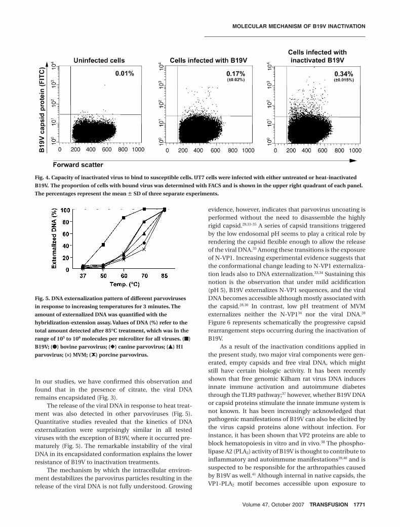
B19V shows a unique DNAexternalization pattern amongparvovirusesB19V is more readily inactivated thanother parvoviruses. To understand thereason for this difference, the external-ization of the B19V DNA was comparedto that of other parvoviruses. B19V,bovine parvovirus, canine parvovirus,H1, MVM, and porcine parvovirus wereexposed to increasing temperatures for3 minutes, and the amount of accessibleDNA was determined with thehybridization-extension assay. The rateof externalization was remarkablysimilar among all the examined virusesexcept for B19V (Fig. 5). At 50°C,approximately 40 percent of the B19Vvirions externalized their DNA, whereasbarely any externalized DNA could bedetected in the case of the other par-
Fig. 1. Effect of inactivation by heat or low pH on B19V capsid integrity. After the
exposure of B19V to different conditions, the intact capsids were immunoprecipi-
tated and analyzed by Western blot. The immunoprecipitation was performed with
an antibody directed to a VP2 conformational epitope (MoAb 860-55D), except for
the right section in A, where an antibody recognizing an epitope in the N-terminal of
VP1 (MoAb 1418) was used.31 The immunoprecipitations were performed after expo-
sure to (A) 60°C for 10 minutes, (B) increasing incubation times at 60°C, (C) increas-
ing incubation temperatures, and (D) after exposure to low pH.
MOLECULAR MECHANISM OF B19V INACTIVATION
Volume 47, October 2007 TRANSFUSION 1769

voviruses. Although 60°C treatment leads to the external-ization of nearly all the B19V genomes, the externalizationin the rest of the tested viruses was at approximately20 percent and in the range of 40 to 80 percent at 70°C.These results imply that the reason for the faster inactiva-
tion of B19V is due to the higher instability of its DNA inthe encapsidated state.
DISCUSSION
To date, the lack of an appropriate cell culture to propa-gate B19V has complicated the experimental work withthis virus. In contrast, optimal cell systems are availablefor many animal parvoviruses. For this reason, they arecommonly used in validation studies as models for B19V.For an unknown reason, however, B19V has been shown tobe more easily inactivated than the other members of theParvoviridae family.22,23 Therefore, the animal parvovi-ruses do not mimic the effect of inactivation procedureson B19V.32 Although different inactivation conditions forB19V have been described, the underlying mechanism ofthe inactivation and the reason for its higher vulner-ability to physicochemical conditions have not yet beenelucidated.
In this study we have examined the structural capsidrearrangements occurring during the inactivation of B19V.For this purpose, we have applied two different proce-dures previously shown to efficiently inactivate B19V.18,21,27
One is the exposure of the virus to heat (60°C for 10 min)and the other is the exposure to acidic conditions (pH 4for 2 hr). Our results demonstrated that the first structuraltransition determining B19V inactivation is not the disin-tegration of the capsid, which remained intact (Fig. 1), butthe loss of the viral DNA (Fig. 2). Interestingly, the heatsensitivity of B19V largely depends on the composition ofthe buffer. In a recent report, it was shown that a solutioncontaining citrate conferred heat resistance to B19V.29
Fig. 2. Effect of inactivation by heat or low pH on B19V DNA
accessibility and release. (A, B) Effect of inactivation on B19V
DNA accessibility. The externalized DNA (%) refers to the
amount detected at 85°C. (C, D) Effect of inactivation on B19V
DNA release (dissociation from the capsid). Viral DNA (%) in
relation to the input is shown.�
Fig. 3. Sensitivity of B19V DNA to DNase I after heat treatment
in PBS or in citrate buffer.
MANI ET AL.
1770 TRANSFUSION Volume 47, October 2007
In our studies, we have confirmed this observation andfound that in the presence of citrate, the viral DNAremains encapsidated (Fig. 3).
The release of the viral DNA in response to heat treat-ment was also detected in other parvoviruses (Fig. 5).Quantitative studies revealed that the kinetics of DNAexternalization were surprisingly similar in all testedviruses with the exception of B19V, where it occurred pre-maturely (Fig. 5). The remarkable instability of the viralDNA in its encapsidated conformation explains the lowerresistance of B19V to inactivation treatments.
The mechanism by which the intracellular environ-ment destabilizes the parvovirus particles resulting in therelease of the viral DNA is not fully understood. Growing
evidence, however, indicates that parvovirus uncoating isperformed without the need to disassemble the highlyrigid capsid.28,33-35 A series of capsid transitions triggeredby the low endosomal pH seems to play a critical role byrendering the capsid flexible enough to allow the releaseof the viral DNA.35 Among these transitions is the exposureof N-VP1. Increasing experimental evidence suggests thatthe conformational change leading to N-VP1 externaliza-tion leads also to DNA externalization.33,34 Sustaining thisnotion is the observation that under mild acidification(pH 5), B19V externalizes N-VP1 sequences, and the viralDNA becomes accessible although mostly associated withthe capsid.28,36 In contrast, low pH treatment of MVMexternalizes neither the N-VP134 nor the viral DNA.28
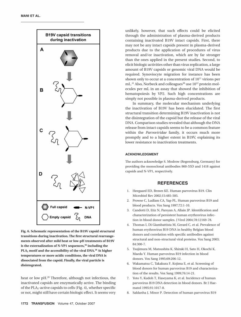
Figure 6 represents schematically the progressive capsidrearrangement steps occurring during the inactivation ofB19V.
As a result of the inactivation conditions applied inthe present study, two major viral components were gen-erated, empty capsids and free viral DNA, which mightstill have certain biologic activity. It has been recentlyshown that free genomic Kilham rat virus DNA inducesinnate immune activation and autoimmune diabetesthrough the TLR9 pathway;37 however, whether B19V DNAor capsid proteins stimulate the innate immune system isnot known. It has been increasingly acknowledged thatpathogenic manifestations of B19V can also be elicited bythe virus capsid proteins alone without infection. Forinstance, it has been shown that VP2 proteins are able toblock hematopoiesis in vitro and in vivo.38 The phospho-lipase A2 (PLA2) activity of B19V is thought to contribute toinflammatory and autoimmune manifestations39,40 and issuspected to be responsible for the arthropathies causedby B19V as well.41 Although internal in native capsids, theVP1-PLA2 motif becomes accessible upon exposure to
Fig. 4. Capacity of inactivated virus to bind to susceptible cells. UT7 cells were infected with either untreated or heat-inactivated
B19V. The proportion of cells with bound virus was determined with FACS and is shown in the upper right quadrant of each panel.
The percentages represent the mean � SD of three separate experiments.
Fig. 5. DNA externalization pattern of different parvoviruses
in response to increasing temperatures for 3 minutes. The
amount of externalized DNA was quantified with the
hybridization-extension assay. Values of DNA (%) refer to the
total amount detected after 85°C treatment, which was in the
range of 105 to 106 molecules per microliter for all viruses. (�)
B19V; (�) bovine parvovirus; (�) canine parvovirus; (�) H1
parvovirus; (¥) MVM; ( ) porcine parvovirus.
MOLECULAR MECHANISM OF B19V INACTIVATION
Volume 47, October 2007 TRANSFUSION 1771
heat or low pH.28 Therefore, although not infectious, theinactivated capsids are enzymatically active. The bindingof the PLA2-active capsids to cells (Fig. 4), whether specificor not, might still have certain biologic effect. It seems very
unlikely, however, that such effects could be elicitedthrough the administration of plasma-derived productscontaining inactivated B19V intact capsids. First, theremay not be any intact capsids present in plasma-derivedproducts due to the application of procedures of virusremoval and/or inactivation, which are by far strongerthan the ones applied in the present studies. Second, toelicit biologic activities other than virus replication, a largeamount of B19V capsids or genomic viral DNA would berequired. Synoviocyte migration for instance has beenshown only to occur at a concentration of 1011 virions permL.41 Also, Norbeck and colleagues38 use 1012 protein mol-ecules per mL in an assay that showed the inhibition ofhematopoiesis by VP2. Such high concentrations aresimply not possible in plasma-derived products.
In summary, the molecular mechanism underlyingthe inactivation of B19V has been elucidated. The firststructural transition determining B19V inactivation is notthe disintegration of the capsid but the release of the viralDNA. Comparison studies revealed that although the DNArelease from intact capsids seems to be a common featurewithin the Parvoviridae family, it occurs much morepromptly and to a higher extent in B19V, explaining itslower resistance to inactivation treatments.
ACKNOWLEDGMENT
The authors acknowledge S. Modrow (Regensburg, Germany) for
providing the monoclonal antibodies 860-55D and 1418 against
capsids and N-VP1, respectively.
REFERENCES
1. Heegaard ED, Brown KE. Human parvovirus B19. Clin
Microbiol Rev 2002;15:485-505.
2. Prowse C, Ludlam CA, Yap PL. Human parvovirus B19 and
blood products. Vox Sang 1997;72:1-10.
3. Candotti D, Etiz N, Parsyan A, Allain JP. Identification and
characterization of persistent human erythrovirus infec-
tion in blood donor samples. J Virol 2004;78:12169-78.
4. Thomas I, Di Giambattista M, Gerard C, et al. Prevalence of
human erythrovirus B19 DNA in healthy Belgian blood
donors and correlation with specific antibodies against
structural and non-structural viral proteins. Vox Sang 2003;
84:300-7.
5. Tsujimura M, Matsushita K, Shiraki H, Sato H, Okochi K,
Maeda Y. Human parvovirus B19 infection in blood
donors. Vox Sang 1995;69:206-12.
6. Wakamatsu C, Takakura F, Kojima E, et al. Screening of
blood donors for human parvovirus B19 and characteriza-
tion of the results. Vox Sang 1999;76:14-21.
7. Yoto Y, Kudoh T, Haseyama K, et al. Incidence of human
parvovirus B19 DNA detection in blood donors. Br J Hae-
matol 1995;91:1017-8.
8. Saldanha J, Minor P. Detection of human parvovirus B19
Fig. 6. Schematic representation of the B19V capsid structural
transitions during inactivation. The first structural rearrange-
ments observed after mild heat or low-pH treatments of B19V
is the externalization of N-VP1 sequences,36 including the
PLA2 motif and the accessibility of the viral DNA.28 At higher
temperatures or more acidic conditions, the viral DNA is
dissociated from the capsid. Finally, the viral particle is
disintegrated.
MANI ET AL.
1772 TRANSFUSION Volume 47, October 2007

DNA in plasma pools and blood products derived from
these pools: implications for efficiency and consistency of
removal of B19 DNA during manufacture. Br J Haematol
1996;93:714-9.
9. Schmidt I, Blumel J, Seitz H, Willkommen H, Lower J. Par-
vovirus B19 DNA in plasma pools and plasma derivatives.
Vox Sang 2001;81:228-35.
10. Zaaijer HL, Koppelman MH, Farrington CP. Parvovirus B19
viraemia in Dutch blood donors. Epidemiol Infect 2004;
132:1161-6.
11. Plentz A, Hahn J, Knoll A, Holler E, Jilg W, Modrow S. Expo-
sure of hematologic patients to parvovirus B19 as a con-
taminant of blood cell preparations and blood products.
Transfusion 2005;45:1811-5.
12. Schneider B, Becker M, Brackmann HH, Eis-Hubinger AM.
Contamination of coagulation factor concentrates with
human parvovirus B19 genotype 1 and 2. Thromb
Haemost 2004;92:838-45.
13. Blumel J, Schmidt I, Effenberger W, et al. Parvovirus B19
transmission by heat-treated clotting factor concentrates.
Transfusion 2002;42:1473-81.
14. Hino M, Ishiko O, Honda KI, et al. Transmission of symp-
tomatic parvovirus B19 infection by fibrin sealant used
during surgery. Br J Haematol 2000;108:194-5.
15. Matsui H, Sugimoto M, Tsuji S, Shima M, Giddings J,
Yoshioka A. Transient hypoplastic anemia caused by
primary human parvovirus B19 infection in a previously
untreated patient with hemophilia transfused with a
plasma-derived, monoclonal antibody-purified factor VIII
concentrate. J Pediatr Hematol Oncol 1999;21:74-6.
16. Santagostino E, Mannucci PM, Gringeri A, et al. Transmis-
sion of parvovirus B19 by coagulation factor concentrates
exposed to 100 degrees C heat after lyophilization. Transfu-
sion 1997;37:517-22.
17. Laub R, Strengers P. Parvoviruses and blood products.
Pathol Biol (Paris) 2002;50:339-48.
18. Blumel J, Schmidt I, Willkommen H, Lower J. Inactivation
of parvovirus B19 during pasteurization of human serum
albumin. Transfusion 2002;42:1011-8.
19. Miyagawa E, Yoshida T, Takahashi H, et al. Infection of the
erythroid cell line, KU812Ep6 with human parvovirus B19
and its application to titration of B19 infectivity. J Virol
Methods 1999;83:45-54.
20. Prikhod’ko GG, Vasilyeva I, Reyes H, et al. Evaluation of
a new LightCycler reverse transcription-polymerase chain
reaction infectivity assay for detection of human parvovi-
rus B19 in dry-heat inactivation studies. Transfusion 2005;
45:1011-9.
21. Yunoki M, Tsujikawa M, Urayama T, et al. Heat sensitivity
of human parvovirus B19 [published erratum appears in
Vox Sang 2003;85:67-8]. Vox Sang 2003;84:164-9.
22. Blumel J, Eis-Hubinger AM, Stuhler A, Bonsch C, Gessner
M, Lower J. Characterization of parvovirus B19 genotype 2
in KU812Ep6 cells. J Virol 2005;79:14197-206.
23. Boschetti N, Niederhauser I, Kempf C, et al. Different sus-
ceptibility of B19 virus and mice minute virus to low pH
treatment. Transfusion 2004;44:1079-86.
24. Caillet-Fauquet P, Di Giambattista M, Draps ML, et al.
Continuous-flow UVC irradiation: a new, effective, protein
activity-preserving system for inactivating bacteria and
viruses, including erythrovirus B19. J Virol Methods 2004;
118:131-9.
25. Sugawara H, Motokawa R, Abe H, et al. Inactivation of par-
vovirus B19 in coagulation factor concentrates by UVC
radiation: assessment by an in vitro infectivity assay using
CFU-E derived from peripheral blood CD34+ cells. Trans-
fusion 2001;41:456-61.
26. Lin L, Hanson CV, Alter HJ, et al. Inactivation of viruses in
platelet concentrates by photochemical treatment with
amotosalen and long-wavelength ultraviolet light. Transfu-
sion 2005;45:580-90.
27. Boschetti N, Wyss K, Mischler A, Hostettler T, Kempf C.
Stability of minute virus of mice against temperature and
sodium hydroxide. Biologicals 2003;31:181-5.
28. Ros C, Baltzer C, Mani B, Kempf C. Parvovirus uncoating in
vitro reveals a mechanism of DNA release without capsid
disassembly and striking differences in encapsidated DNA
stability. Virology 2006;345:137-47.
29. Hattori S, Yunoki M, Tsujikawa M, et al. Variability of par-
vovirus B19 to inactivation by liquid heating in plasma
products. Vox Sang 2007;92:121-4.
30. Cavalli-Sforza L. Biometrie: Grundzüge biologisch-
medizinischer Statistik. Stuttgart: G. Fischer Verlag; 1974.
31. Gigler A, Dorsch S, Hemauer A, et al. Generation of neu-
tralizing human monoclonal antibodies against parvovirus
B19 proteins. J Virol 1999;73:1974-9.
32. Kasermann F, Kempf C, Boschetti N. Strengths and limita-
tions of the model virus concept. PDA J Pharm Sci Technol
2004;58:244-9.
33. Bleker S, Pawlita M, Kleinschmidt JA. Impact of capsid
conformation and Rep-capsid interactions on adeno-
associated virus type 2 genome packaging. J Virol 2006;80:
810-20.
34. Cotmore SF, D’abramo AM Jr, Ticknor CM, Tattersall P.
Controlled conformational transitions in the MVM virion
expose the VP1 N-terminus and viral genome without par-
ticle disassembly. Virology 1999;254:169-81.
35. Mani B, Baltzer C, Valle N, et al. Low pH-dependent
endosomal processing of the incoming parvovirus minute
virus of mice virion leads to externalization of the VP1
N-terminal sequence (N-VP1), N-VP2 cleavage, and
uncoating of the full-length genome. J Virol 2006;80:1015-
24.
36. Ros C, Gerber M, Kempf C. Conformational changes in the
VP1-unique region of native human parvovirus B19 lead to
exposure of internal sequences that play a role in virus
neutralization and infectivity. J Virol 2006;80:
12017–24.
37. Zipris D, Lien E, Nair A, et al. TLR9-signaling pathways are
involved in kilham rat virus-induced autoimmune diabetes
MOLECULAR MECHANISM OF B19V INACTIVATION
Volume 47, October 2007 TRANSFUSION 1773

in the biobreeding diabetes-resistant rat. J Immunol 2007;
178:693-701.
38. Norbeck O, Tolfvenstam T, Shields LE, Westgren M,
Broliden K. Parvovirus B19 capsid protein VP2 inhibits
hematopoiesis in vitro and in vivo: implications for thera-
peutic use. Exp Hematol 2004;32:1082-7.
39. Lehmann HW, Von Landenberg P, Modrow S. Parvovirus
B19 infection and autoimmune disease. Autoimmun Rev
2003;2:218-23.
40. Von Landenberg P, Lehmann HW, Knoll A, Dorsch S,
Modrow S. Antiphospholipid antibodies in pediatric and
adult patients with rheumatic disease are associated with
parvovirus B19 infection. Arthritis Rheum 2003;48:
1939-47.
41. Lu J, Zhi N, Wong S, Brown KE. Activation of synoviocytes
by the secreted phospholipase A2 motif in the VP1-unique
region of parvovirus B19 minor capsid protein. J Infect Dis
2006;193:582-90.
MANI ET AL.
1774 TRANSFUSION Volume 47, October 2007