Unraveling the Underlying Heavy Metal Detoxification ... - MDPI
31
microorganisms Review Unraveling the Underlying Heavy Metal Detoxification Mechanisms of Bacillus Species Badriyah Shadid Alotaibi 1 , Maryam Khan 2 and Saba Shamim 2, * Citation: Alotaibi, B.S.; Khan, M.; Shamim, S. Unraveling the Underlying Heavy Metal Detoxification Mechanisms of Bacillus Species. Microorganisms 2021, 9, 1628. https://doi.org/10.3390/ microorganisms9081628 Academic Editor: Juan Carlos Gutiérrez Received: 7 June 2021 Accepted: 13 July 2021 Published: 30 July 2021 Publisher’s Note: MDPI stays neutral with regard to jurisdictional claims in published maps and institutional affil- iations. Copyright: © 2021 by the authors. Licensee MDPI, Basel, Switzerland. This article is an open access article distributed under the terms and conditions of the Creative Commons Attribution (CC BY) license (https:// creativecommons.org/licenses/by/ 4.0/). 1 Department of Pharmaceutical Sciences, College of Pharmacy, Princess Nourah Bint Abdulrahman University, Riyadh 11671, Saudi Arabia; [email protected] 2 Institute of Molecular Biology and Biotechnology (IMBB), Defence Road Campus, The University of Lahore, Lahore 55150, Pakistan; [email protected] * Correspondence: [email protected] Abstract: The rise of anthropogenic activities has resulted in the increasing release of various contaminants into the environment, jeopardizing fragile ecosystems in the process. Heavy metals are one of the major pollutants that contribute to the escalating problem of environmental pollution, being primarily introduced in sensitive ecological habitats through industrial effluents, wastewater, as well as sewage of various industries. Where heavy metals like zinc, copper, manganese, and nickel serve key roles in regulating different biological processes in living systems, many heavy metals can be toxic even at low concentrations, such as mercury, arsenic, cadmium, chromium, and lead, and can accumulate in intricate food chains resulting in health concerns. Over the years, many physical and chemical methods of heavy metal removal have essentially been investigated, but their disadvantages like the generation of chemical waste, complex downstream processing, and the uneconomical cost of both methods, have rendered them inefficient,. Since then, microbial bioremediation, particularly the use of bacteria, has gained attention due to the feasibility and efficiency of using them in removing heavy metals from contaminated environments. Bacteria have several methods of processing heavy metals through general resistance mechanisms, biosorption, adsorption, and efflux mechanisms. Bacillus spp. are model Gram-positive bacteria that have been studied extensively for their biosorption abilities and molecular mechanisms that enable their survival as well as their ability to remove and detoxify heavy metals. This review aims to highlight the molecular methods of Bacillus spp. in removing various heavy metals ions from contaminated environments. Keywords: bioremediation; Bacillus; resistance; microorganisms; biosorption; efflux; heavy metals 1. Introduction to Heavy Metals Living beings are constantly surrounded by different environments of land, air, and water, which are significant for their sustainability. The Earth is the fulcrum of all precious resources, where various fragile ecosystems work in harmony. Millions of years ago, the discovery of fire led to the revolutionary development of man, which culminated in the evolution and expansion of civilization. In the same manner, the dawn of industrialization saw the rapid growth and the impetuous exploitation of the Earth’s natural (renewable and non-renewable) sources, which gave rise to the predicament of environmental pol- lution [1]. This type of pollution has been associated with environmental contaminants of several kinds, such as organic and inorganic ions, isotopes, gases, nanoparticles, and organo-metallic compounds [2]. Regardless of these contaminants contributing greatly to environmental pollution, the presence of heavy metals in the environment is one point of grave concern over the past few decades. According to Ali and Khan [3], heavy metals are defined as those naturally occurring metals “having an atomic number and density greater than 20 and 5 g/cm -3 , respectively”. Even more so, the term “heavy metal” is now inclusive of the metallic elements as well Microorganisms 2021, 9, 1628. https://doi.org/10.3390/microorganisms9081628 https://www.mdpi.com/journal/microorganisms
-
Upload
khangminh22 -
Category
Documents
-
view
3 -
download
0
Transcript of Unraveling the Underlying Heavy Metal Detoxification ... - MDPI

microorganisms
Review
Unraveling the Underlying Heavy Metal DetoxificationMechanisms of Bacillus Species
Badriyah Shadid Alotaibi 1, Maryam Khan 2 and Saba Shamim 2,*
�����������������
Citation: Alotaibi, B.S.; Khan, M.;
Shamim, S. Unraveling the
Underlying Heavy Metal
Detoxification Mechanisms of Bacillus
Species. Microorganisms 2021, 9, 1628.
https://doi.org/10.3390/
microorganisms9081628
Academic Editor: Juan
Carlos Gutiérrez
Received: 7 June 2021
Accepted: 13 July 2021
Published: 30 July 2021
Publisher’s Note: MDPI stays neutral
with regard to jurisdictional claims in
published maps and institutional affil-
iations.
Copyright: © 2021 by the authors.
Licensee MDPI, Basel, Switzerland.
This article is an open access article
distributed under the terms and
conditions of the Creative Commons
Attribution (CC BY) license (https://
creativecommons.org/licenses/by/
4.0/).
1 Department of Pharmaceutical Sciences, College of Pharmacy, Princess Nourah Bint Abdulrahman University,Riyadh 11671, Saudi Arabia; [email protected]
2 Institute of Molecular Biology and Biotechnology (IMBB), Defence Road Campus, The University of Lahore,Lahore 55150, Pakistan; [email protected]
* Correspondence: [email protected]
Abstract: The rise of anthropogenic activities has resulted in the increasing release of variouscontaminants into the environment, jeopardizing fragile ecosystems in the process. Heavy metalsare one of the major pollutants that contribute to the escalating problem of environmental pollution,being primarily introduced in sensitive ecological habitats through industrial effluents, wastewater,as well as sewage of various industries. Where heavy metals like zinc, copper, manganese, and nickelserve key roles in regulating different biological processes in living systems, many heavy metals canbe toxic even at low concentrations, such as mercury, arsenic, cadmium, chromium, and lead, and canaccumulate in intricate food chains resulting in health concerns. Over the years, many physical andchemical methods of heavy metal removal have essentially been investigated, but their disadvantageslike the generation of chemical waste, complex downstream processing, and the uneconomical cost ofboth methods, have rendered them inefficient,. Since then, microbial bioremediation, particularly theuse of bacteria, has gained attention due to the feasibility and efficiency of using them in removingheavy metals from contaminated environments. Bacteria have several methods of processing heavymetals through general resistance mechanisms, biosorption, adsorption, and efflux mechanisms.Bacillus spp. are model Gram-positive bacteria that have been studied extensively for their biosorptionabilities and molecular mechanisms that enable their survival as well as their ability to remove anddetoxify heavy metals. This review aims to highlight the molecular methods of Bacillus spp. inremoving various heavy metals ions from contaminated environments.
Keywords: bioremediation; Bacillus; resistance; microorganisms; biosorption; efflux; heavy metals
1. Introduction to Heavy Metals
Living beings are constantly surrounded by different environments of land, air, andwater, which are significant for their sustainability. The Earth is the fulcrum of all preciousresources, where various fragile ecosystems work in harmony. Millions of years ago, thediscovery of fire led to the revolutionary development of man, which culminated in theevolution and expansion of civilization. In the same manner, the dawn of industrializationsaw the rapid growth and the impetuous exploitation of the Earth’s natural (renewableand non-renewable) sources, which gave rise to the predicament of environmental pol-lution [1]. This type of pollution has been associated with environmental contaminantsof several kinds, such as organic and inorganic ions, isotopes, gases, nanoparticles, andorgano-metallic compounds [2]. Regardless of these contaminants contributing greatly toenvironmental pollution, the presence of heavy metals in the environment is one point ofgrave concern over the past few decades.
According to Ali and Khan [3], heavy metals are defined as those naturally occurringmetals “having an atomic number and density greater than 20 and 5 g/cm−3, respectively”.Even more so, the term “heavy metal” is now inclusive of the metallic elements as well
Microorganisms 2021, 9, 1628. https://doi.org/10.3390/microorganisms9081628 https://www.mdpi.com/journal/microorganisms

Microorganisms 2021, 9, 1628 2 of 31
as metalloids, both of which are somewhat toxic to the living beings and environment, in-cluding metalloids such as selenium (Se), arsenic (As), and tellurium (Te) [4]. Nevertheless,there are many metals which are not relatively toxic to both humans and the environment,including gold (Au) [5,6]. The toxicity of metals and metalloids is influenced by their abilityto form covalent bonds, usually with organic groups resulting in the formation of lipophiliccompounds distributed widely in the Earth’s biosphere, some of which are more toxicthan the ionic form of the major metal found in those compounds, like tributyltin oxidein the case of arsenic. Heavy metals tend to facilitate their entry in human beings by oneor more of the following methods: consumption of contaminated food and drinks; atmo-spheric inhalation; drinking of contaminated water; and/or direct contact with agricultural,pharmaceutical, residential, and industrial contamination [7]. They readily persist in theenvironment and are non-biodegradable. Many of the metals are detoxified by the actionof living organisms concealing the element inside a protein or by mediating its depositionwithin intra-cellular granules which can then be stored or expelled accordingly. The gradualpersistence of heavy metals in our ecosystem means that humans will eventually come intocontact with them, which can potentially lead to their bioaccumulation in the human body.Bioaccumulation is the process through which contaminants, particularly heavy metals,are accumulated in the human body through ingestion or inhalation and can potentiallybe dangerous, as they tend to cause various physiological and biological complications. Itis important to note here that this phenomenon is relative to the toxicity of heavy metals,which is usually observed in high concentrations [8]. The sources of heavy metals in theenvironment can comprise of geological, natural, lithogenic, and anthropogenic origins.Natural sources of heavy metals in the environment comprise of soil and rock erosion,volcanic eruptions, as well as sediment run-off [9]. Many anthropogenic processes alsocontribute to the presence of heavy metals in the environments, with rapid industrializationbeing the main culprit in this case. The release of heavy metals via insecticides, pesticides,and phosphate fertilizers lead to the release of metals ions such as zinc and cadmium.Moreover, metals such as mercury, arsenic, and lead have also been used in the agriculturalfields, which leads to their accumulation in soil and underground sources, along with theiraccumulation in trophic levels of the food chain [10–12]. In addition, industrial processessuch as metal and coal mining, fossil fuel combustion, and disposal of wastes and effluentshas also greatly increased the burden of heavy metal pollution [13]. In water sources, riversand streams that are near industries and mining areas tend to become polluted by heavymetals, speeding up their precipitation towards the sedimental level due to their decreasingsolubility [14].
Contingent to their significance in biological systems, most of the heavy metals can bedifferentiated as essential and non-essential heavy metals, respectively. Essential heavymetals are characterized as necessary for biological life and its biochemical and physio-logical processes in low concentrations, as high concentrations can represent toxicity. Onthe other hand, non-essential heavy metals are those which have no reported biologicalfunction in the living systems, and only contribute to exert their toxicity in various bio-logical beings. Examples of the former include heavy metals such as manganese, iron,copper, and zinc, while heavy metals like cadmium, lead, and mercury tend to be non-essential [15,16]. Moreover, heavy metals such as manganese, iron, cobalt, nickel, copper,zinc, and molybdenum are regarded as trace elements for the biochemical and biologicalgrowth of plants. These metals have also been recognized as essential for the developmentof growth and survival in stress environments, along with the biosynthesis of variousorganic compounds and metabolites [17]. Essential heavy metals are significant due tothe fact that their deficit can lead to serious growth abnormality or stress for the livingbeing. Nevertheless, the essential heavy metals required by every biological system (i.e.,plants, animals, microorganisms, human beings) vary greatly from one to another, asthe interaction of heavy metals with each of the organisms is reported to be an intricateprocess [18,19]. Organelles and cellular components such as mitochondria, cell membrane,and enzymes tend to be affected by heavy metal concentrations, as the interaction of these

Microorganisms 2021, 9, 1628 3 of 31
metal ions and cellular proteins has reported to cause DNA damage, as well as alterationof the normal cell cycle leading to apoptosis and often carcinogenesis [6]. The damage toDNA can be mediated by two mechanisms, namely direct and indirect damage, where theformer refers to the conformational changes occurring in the biological molecules becauseof interaction with the metal ions, and the latter leads to the generation of reactive oxygenspecies, free radicals, and other oxidative stress species, respectively [20]. Heavy metalslike iron, chromium, vanadium, cadmium, and copper are reported to be involved in theformation of free radicals by following the Fenton reaction of superoxide and hydroxylions, usually in the mitochondrial and peroxisomal region of cells [21].
2. Methods of Heavy Metal Remediation
When compared to organic pollutants, heavy metals are considered to be persistentin the environment because they are non-biodegradable and cannot be broken downeasily. Incessant accumulation of heavy metals in soils may damage their ecosystem andinadvertently lead to the disturbance of various physicochemical properties of soils, suchas pH, anion/cation exchange, thermal and electrical conductivity, microbial ecosystems,and the mobility of heavy metal ions in the soil [22]. Various studies have reportedthe accumulation of heavy metals in soils and their risks to biological systems. At highconcentrations, heavy metals are able to directly and indirectly affect associated microbialecosystems [23]. Thus, it is crucial to apply efficient technologies which are ultimatelycheap, feasible, and are prone to site-specificity for the remediation of contaminatedenvironments. Over the past two decades, many remediation techniques for soil and waterhave been developed and investigated which have proven to be efficacious in mitigatingthe total content of heavy metal ions and/or their accumulation at trophic levels in thefood chain [23,24]. Physical methods such as heat treatment [25], soil replacement [26],soil washing [27], electroremediation [28] and vitrification [29,30] are employed for theirwidespread applications in the removal of various waste products. This aspect ensuresthe efficiency of physical methods in removing almost every kind of contaminant in theenvironment, though this property does not come without some disadvantages. Thecontaminants removed by physical methods need to be processed further, which adds tothe already high cost of the method used, along with their specificity. Chemical methodsfor heavy metal remediation are also employed to alter the chemical characteristics ofcontaminants that subsequently decrease their hazardous properties. Though methods likeprecipitation [31], leaching [32], extraction [33], ion exchange [34], encapsulation [35], andimmobilization [36] are observed to be efficient at field level, the generation of byproductsmay act as a major setback which leads to additional processes at downstream level. Manyof these techniques are contingent upon physical, chemical, and biological methods ofbioremediation, which may be used singularly or in combination with each other for thesame purpose. In spite of their effectiveness, many of these techniques are not feasible,environmentally-friendly, or cheap, which ultimately makes them challenging to be used.Nevertheless, new techniques are always being introduced in the search of the most suitablemethod of remediation.
3. Bioremediation—An Environmentally-Friendly Approach for the Removal ofHeavy Metals
Bioremediation is a conventional process that employs the use of plants, animals,and microorganisms for the remediation of pollutants like heavy metals. It is one of mosteffective, non-invasive, and economically feasible methods for the permanent mitigation ofheavy metal pollution and for the complete restoration of the natural environment of manyecosystems, as there is no production of secondary by-products [37]. Though methods suchas phytoremediation (bioremediation using plants), phycoremediation (bioremediationusing algae) and mycoremediation (bioremediation using fungi) have been widely reportedin literature for heavy metal removal, our review focuses on bacterial bioremediation withBacillus spp. as potentially active and efficacious agents for the removal of various heavymetal ions present in the environment.

Microorganisms 2021, 9, 1628 4 of 31
Bacteria are abundantly present in the environment, where their variety in shape, size,morphology, as well as resistance mechanisms makes them suitable for bioremediationof organic and inorganic pollutants. Biosorption mediated by bacteria is a cost-effective,economically feasible method for the removal of heavy metal ions and other pollutantsfrom contaminated sites, as many species have evolved to develop mechanisms whichmediate resistance to process heavy metal ions amidst high concentrations [38]. Over thepast years, many bacterial species have garnered widespread attention for their potentialability to remove heavy metal ions from the environment, whereas bacterial biomass(live and dead) has also been investigated for biosorption of metal ions such as copper,chromium, zinc, nickel, cadmium, and mercury, concomitant on their interactions withthe bacterial cell wall and related peptidoglycans [39–41]. Many bacterial species such asBacillus, Pseudomonas, Enterobacter, Flavobacterium, Geobacter, and Micrococcus spp. have beeninvestigated for potential use in biosorption of various heavy metal ions from contaminatedsites, by observing their surface to volume ratio and other feasible characteristics whichenhance the binding interactions between bacterial functional groups and heavy metalions [42,43]. In a recent study, the biosorption ability of many bacterial species such as B.subtilis, B. megaterium, A. niger, and Penicillium spp. were investigated for the removal ofmany heavy metal ions such as lead, chromium, and cadmium, of which Bacillus spp. wereobserved to be the most efficient [44,45].
4. Bacillus spp. as Potential Agents for Heavy Metal Removal and Their OverallSignificance
Bacillus spp. are Gram-positive, rod-shaped bacteria belonging to the phylum Fir-micutes, and are spore-forming in nature. They are generally characterized as soil mi-croorganisms which can be aerobic or facultatively anaerobic but can also be found invarious sources such as air, water, edible produce, foods, and the human gut [46]. Interms of features presented as genetic or commercial applications, Bacillus group is themost heterogenous as some species have been well characterized as opportunist pathogensand toxin producers, whereas others have widespread industrial and medicinal applica-tions [47]. One of the unique characteristics of Bacillus spp. is the formation of spores underextreme environments, which is usually triggered during a deficit of nutrients [48,49].Owing to their resilient structures, spores are able to endure severe environmental stresssuch as desiccation, high temperature, humidity, as well as radiation [50]. It is due to thisfeature they exhibit various commercial applications and are more suitable to be used thanvegetative cells, respectively [51].
The positive use of Bacillus spp. in various fields has attracted attention to theircharacteristic features, and research has enabled the utilization of these features to thebest of man’s interest. In aquaculture and fishery environments, Bacillus are utilized asbiocontrol products [52]. In medical, industrial, and environmental fields, the advantage ofusing Gram-positive bacteria like Bacillus is that they do not tend to partake in the transferof genetic material from Gram-negative bacteria. Moreover, they replicate expeditiouslyand can survive in many environmental conditions. Many species of Bacillus, such as B.subtilis, B. coagulans, B. pumilus, B. licheniformis, and B. cereus are utilized globally for manyapplications [51]. Many study findings have indicated the safety of B. subtilis for probioticuse, due to the demonstration of antimicrobial and anti-cancerous effects [53]. Bacillus spp.are also used for the production of various enzymes such as amylase, protease, cellulase,and pectinase in the food industry, [54] as well as in several supplemental nutrientssuch as vitamins and carotenoids [55,56]. Apart from these uses, Bacillus spp. are alsoextensively investigated for their role in the mitigation of heavy metals from contaminatedenvironments via biosorption, bioaccumulation, among many other techniques, due to thereported notion that contaminated sites are often dominated by Gram-positive bacteria,owing to their versatile metabolic properties and better qualities of biosorption [57,58].
Microorganisms 2021, 9, 1628 5 of 31
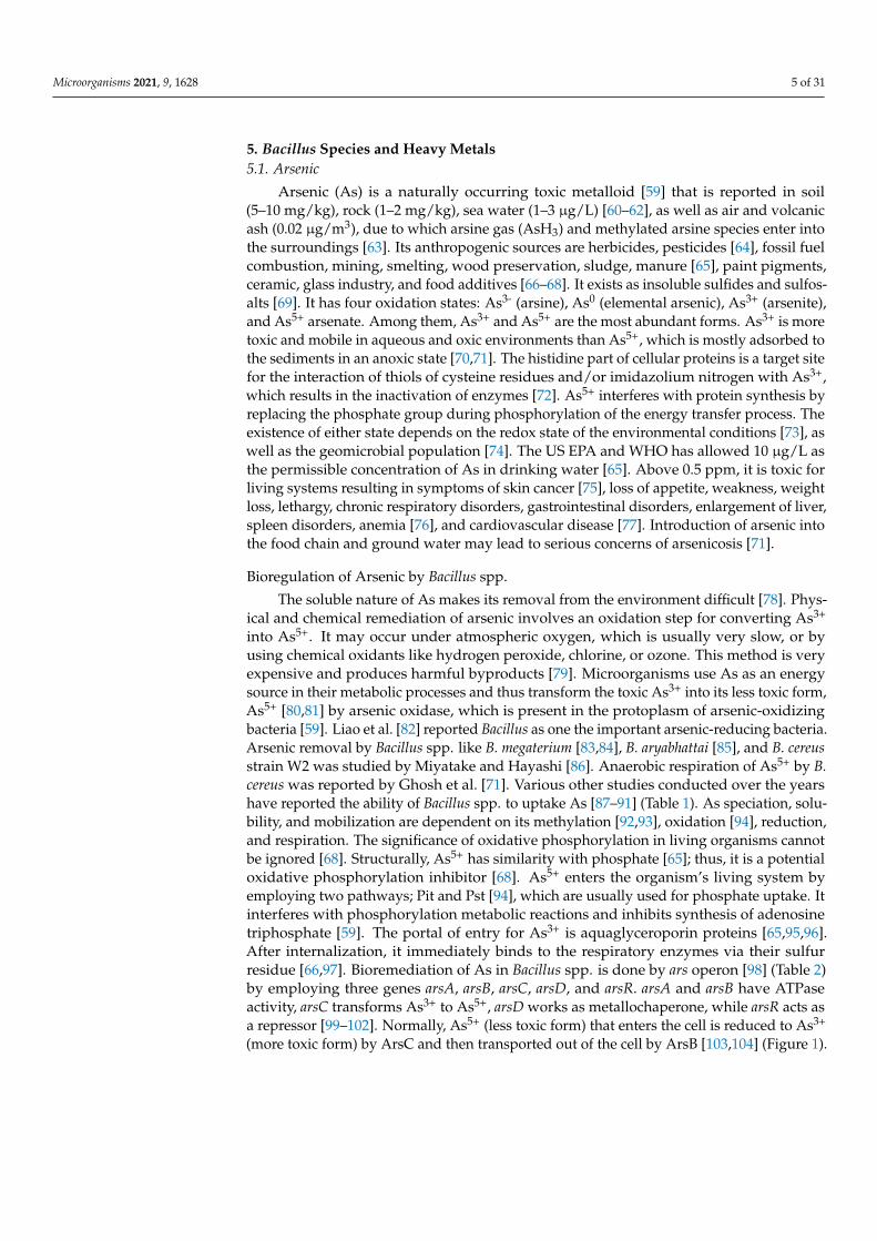
5. Bacillus Species and Heavy Metals5.1. Arsenic
Arsenic (As) is a naturally occurring toxic metalloid [59] that is reported in soil(5–10 mg/kg), rock (1–2 mg/kg), sea water (1–3 µg/L) [60–62], as well as air and volcanicash (0.02 µg/m3), due to which arsine gas (AsH3) and methylated arsine species enter intothe surroundings [63]. Its anthropogenic sources are herbicides, pesticides [64], fossil fuelcombustion, mining, smelting, wood preservation, sludge, manure [65], paint pigments,ceramic, glass industry, and food additives [66–68]. It exists as insoluble sulfides and sulfos-alts [69]. It has four oxidation states: As3- (arsine), As0 (elemental arsenic), As3+ (arsenite),and As5+ arsenate. Among them, As3+ and As5+ are the most abundant forms. As3+ is moretoxic and mobile in aqueous and oxic environments than As5+, which is mostly adsorbed tothe sediments in an anoxic state [70,71]. The histidine part of cellular proteins is a target sitefor the interaction of thiols of cysteine residues and/or imidazolium nitrogen with As3+,which results in the inactivation of enzymes [72]. As5+ interferes with protein synthesis byreplacing the phosphate group during phosphorylation of the energy transfer process. Theexistence of either state depends on the redox state of the environmental conditions [73], aswell as the geomicrobial population [74]. The US EPA and WHO has allowed 10 µg/L asthe permissible concentration of As in drinking water [65]. Above 0.5 ppm, it is toxic forliving systems resulting in symptoms of skin cancer [75], loss of appetite, weakness, weightloss, lethargy, chronic respiratory disorders, gastrointestinal disorders, enlargement of liver,spleen disorders, anemia [76], and cardiovascular disease [77]. Introduction of arsenic intothe food chain and ground water may lead to serious concerns of arsenicosis [71].
Bioregulation of Arsenic by Bacillus spp.
The soluble nature of As makes its removal from the environment difficult [78]. Phys-ical and chemical remediation of arsenic involves an oxidation step for converting As3+
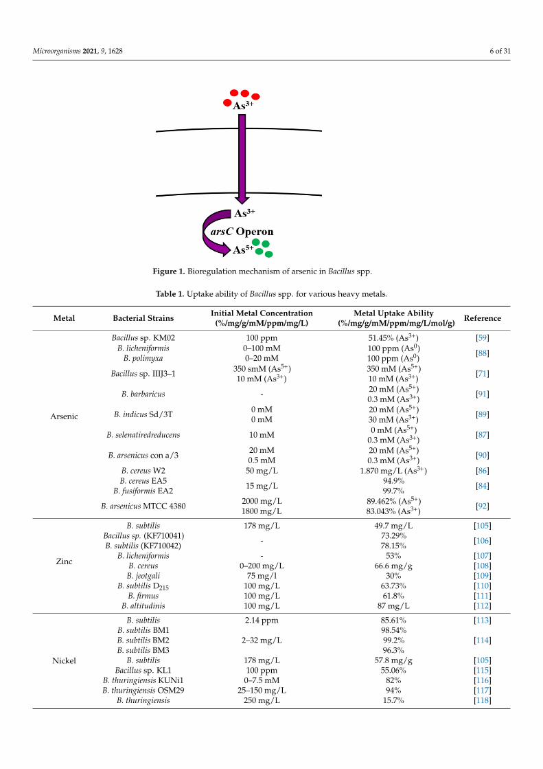
into As5+. It may occur under atmospheric oxygen, which is usually very slow, or byusing chemical oxidants like hydrogen peroxide, chlorine, or ozone. This method is veryexpensive and produces harmful byproducts [79]. Microorganisms use As as an energysource in their metabolic processes and thus transform the toxic As3+ into its less toxic form,As5+ [80,81] by arsenic oxidase, which is present in the protoplasm of arsenic-oxidizingbacteria [59]. Liao et al. [82] reported Bacillus as one the important arsenic-reducing bacteria.Arsenic removal by Bacillus spp. like B. megaterium [83,84], B. aryabhattai [85], and B. cereusstrain W2 was studied by Miyatake and Hayashi [86]. Anaerobic respiration of As5+ by B.cereus was reported by Ghosh et al. [71]. Various other studies conducted over the yearshave reported the ability of Bacillus spp. to uptake As [87–91] (Table 1). As speciation, solu-bility, and mobilization are dependent on its methylation [92,93], oxidation [94], reduction,and respiration. The significance of oxidative phosphorylation in living organisms cannotbe ignored [68]. Structurally, As5+ has similarity with phosphate [65]; thus, it is a potentialoxidative phosphorylation inhibitor [68]. As5+ enters the organism’s living system byemploying two pathways; Pit and Pst [94], which are usually used for phosphate uptake. Itinterferes with phosphorylation metabolic reactions and inhibits synthesis of adenosinetriphosphate [59]. The portal of entry for As3+ is aquaglyceroporin proteins [65,95,96].After internalization, it immediately binds to the respiratory enzymes via their sulfurresidue [66,97]. Bioremediation of As in Bacillus spp. is done by ars operon [98] (Table 2)by employing three genes arsA, arsB, arsC, arsD, and arsR. arsA and arsB have ATPaseactivity, arsC transforms As3+ to As5+, arsD works as metallochaperone, while arsR acts asa repressor [99–102]. Normally, As5+ (less toxic form) that enters the cell is reduced to As3+
(more toxic form) by ArsC and then transported out of the cell by ArsB [103,104] (Figure 1).
Microorganisms 2021, 9, 1628 6 of 31
Microorganisms 2021, 9, x FOR PEER REVIEW 6 of 32
to As3+ (more toxic form) by ArsC and then transported out of the cell by ArsB [103,104] (Figure 1).
Figure 1. Bioregulation mechanism of arsenic in Bacillus spp.
Table 1. Uptake ability of Bacillus spp. for various heavy metals.
Metal Bacterial Strains Initial Metal Concentration (%/mg/g/mM/ppm/mg/L)
Metal Uptake Ability (%/mg/g/mM/ppm/mg/L/mol/g)
Reference
Arsenic
Bacillus sp. KM02 100 ppm 51.45% (As3+) [59] B. licheniformis
B. polimyxa 0–100 mM 0–20 mM
100 ppm (As0) 100 ppm (As0)
[88]
Bacillus sp. IIIJ3–1 350 smM (As5+) 10 mM (As3+)
350 mM (As5+) 10 mM (As3+)
[71]
B. barbaricus - 20 mM (As5+) 0.3 mM (As3+)
[91]
B. indicus Sd/3T 0 mM 0 mM
20 mM (As5+) 30 mM (As3+)
[89]
B. selenatiredreducens 10 mM 0 mM (As5+)
0.3 mM (As3+) [87]
B. arsenicus con a/3 20 mM 0.5 mM
20 mM (As5+) 0.3 mM (As3+)
[90]
B. cereus W2 50 mg/L 1.870 mg/L (As3+) [86] B. cereus EA5
B. fusiformis EA2 15 mg/L
94.9% 99.7%
[84]
B. arsenicus MTCC 4380 2000 mg/L 1800 mg/L
89.462% (As5+) 83.043% (As3+)
[92]
Zinc
B. subtilis 178 mg/L 49.7 mg/L [105] Bacillus sp. (KF710041) B. subtilis (KF710042)
- 73.29% 78.15%
[106]
B. licheniformis - 53% [107] B. cereus 0–200 mg/L 66.6 mg/g [108] B. jeotgali 75 mg/l 30% [109]
B. subtilis D215 100 mg/L 63.73% [110] B. firmus 100 mg/L 61.8% [111]
B. altitudinis 100 mg/L 87 mg/L [112]
Nickel
B. subtilis 2.14 ppm 85.61% [113] B. subtilis BM1 B. subtilis BM2 B. subtilis BM3
2–32 mg/L 98.54% 99.2% 96.3%
[114]
Figure 1. Bioregulation mechanism of arsenic in Bacillus spp.
Table 1. Uptake ability of Bacillus spp. for various heavy metals.
Metal Bacterial Strains Initial Metal Concentration(%/mg/g/mM/ppm/mg/L)
Metal Uptake Ability(%/mg/g/mM/ppm/mg/L/mol/g) Reference
Arsenic
Bacillus sp. KM02 100 ppm 51.45% (As3+) [59]B. licheniformis
B. polimyxa0–100 mM0–20 mM
100 ppm (As0)100 ppm (As0)
[88]
Bacillus sp. IIIJ3–1 350 smM (As5+)10 mM (As3+)
350 mM (As5+)10 mM (As3+)
[71]
B. barbaricus - 20 mM (As5+)0.3 mM (As3+)
[91]
B. indicus Sd/3T 0 mM0 mM
20 mM (As5+)30 mM (As3+)
[89]
B. selenatiredreducens 10 mM 0 mM (As5+)0.3 mM (As3+)
[87]
B. arsenicus con a/3 20 mM0.5 mM
20 mM (As5+)0.3 mM (As3+)
[90]
B. cereus W2 50 mg/L 1.870 mg/L (As3+) [86]B. cereus EA5
B. fusiformis EA2 15 mg/L 94.9%99.7% [84]
B. arsenicus MTCC 4380 2000 mg/L1800 mg/L
89.462% (As5+)83.043% (As3+)
[92]
Zinc
B. subtilis 178 mg/L 49.7 mg/L [105]Bacillus sp. (KF710041)B. subtilis (KF710042) - 73.29%
78.15% [106]
B. licheniformis - 53% [107]B. cereus 0–200 mg/L 66.6 mg/g [108]B. jeotgali 75 mg/l 30% [109]
B. subtilis D215 100 mg/L 63.73% [110]B. firmus 100 mg/L 61.8% [111]
B. altitudinis 100 mg/L 87 mg/L [112]
Nickel
B. subtilis 2.14 ppm 85.61% [113]B. subtilis BM1B. subtilis BM2B. subtilis BM3
2–32 mg/L98.54%99.2%96.3%
[114]
B. subtilis 178 mg/L 57.8 mg/g [105]Bacillus sp. KL1 100 ppm 55.06% [115]
B. thuringiensis KUNi1 0–7.5 mM 82% [116]B. thuringiensis OSM29 25–150 mg/L 94% [117]
B. thuringiensis 250 mg/L 15.7% [118]

Microorganisms 2021, 9, 1628 7 of 31
Table 1. Cont.
Metal Bacterial Strains Initial Metal Concentration(%/mg/g/mM/ppm/mg/L)
Metal Uptake Ability(%/mg/g/mM/ppm/mg/L/mol/g) Reference
Cadmium
B. safensis 40 ppm60 ppm
83.5%98.10% [119]
B. licheniformis - 98.34% [120]B. catenulatus JB-022 150 mg/L 66% [121]
B. thuringiensis DM55 0.25 mM 79% [122]
Lead
B. pumilus MF472596 100–1000 ppm 96% [123]B. subtilis X3 200–1400 mg/L 590.49 mg/g [124]
B. cereus 5–100 mg/L 36.71 mg/g [125]Bacillus S1
Bacillus SS1975 and 100 mg/L
50 mg/mL53%, 51%
57% [126]
Bacillus sp. AS2 500 ppm 74.5 mg/g (99.5 %) [127]
Copper
B. cereus 100 ppm 54% [128]B. cereus 400 ppm 48% [129]
B. thuringiensis OSM29 25 mg/L 91.8% [117]B. licheniformis 5 gm/L 32% [130]B. thioparans 40 mg/L 27.3 mg/g [131]
B. subtilis D215 100 mg/L 67.18% [110]B. sphaericus
B. cereusBacillus sp.
17.6 mg/L44.0 mg/L88.0 mg/L
5.6 mol/g5.9 mol/g6.4 mol/g
[132]
Bacillus sp. SG-1 - 60% [133]
Chromium
B. cereus NWUAB01 100 mg/L 43% [134]B. cereus 100 mg/L 81% [135]
B. salmalaya 139SI 50 ppm 20.35 mg/g [136]B. cereus FA-3 1000 µg/ml 72% [137]B. licheniformis 15 mg/L 95% [138]Bacillus sp. B 500–4500 mg/L 47% [139]B. marisflavi 200 mg/L 5.783% [140]
B. licheniformis 300 mg/g 69.4% [141]B. thuringiensis 250 mg/L 83.3% [142]B. licheniformisB. laterosporus - 62 mg/g
72.6 mg/g [143]
B. circulansB. megaterium 0.96 mg/L 34.5%
32% [144]
Mercury
B. thuringiensis CASKS3200 mg/L400 mg/L600 mg/L
62.4%54%40%
[145]
B. licheniformis 50 mg/L 70% [146]B. cereus
BW-03(pPW-05) 5–50 ppm 96.4% [147]
B. licheniformis 100 µg/mL 70% [148]B. cereus 5 mg/L 104.1 mg/g [149]
Bacillus sp. 1–10 mg/L 7.9 mg/g [150]
ManganeseB. thuringiensis HM7 400 mg/L 95.04% [151]
B. cereus HM-5 600 mg/L 67% [152]Bacillus sp. 13.3 mg/g 55.56 mg/g [153]
Molybdenum Bacillus sp. Zeid 14 - 200 mg/L [154]Bacillus sp. strain A.rzi 0.1 mM Not reported [155]
Silver B. licheniformis R08 100 mg/L 73.6 mg/g [156]

Microorganisms 2021, 9, 1628 8 of 31
Table 2. The proteins, operons, and methods of removal employed by Bacillus spp. for the bioregulation of heavy metals.
Metal Protein(s)/Gene(s) Method(s) Reference
Arsenic ars operon (arsR, arsD, arsA, arsB, arsC)
Reduction (Detoxification)Efflux
Cell membrane binding, Adsorption on cellsurface
Complexation by exopolysaccharides
[59,81,87,99–102]
Zinc
ZurZosA
ycdHI-yceAyciABCCadACzcD
Physico-chemical adsorptionIon exchange
EffluxUptake
[108,157,158]
Nickel CzcDCitM Efflux [159,160]
Cadmiumcad operon
yvgWKinA
Efflux [122,134,161–165]
Lead pbr operon Efflux [166–168]
CopperCueR
copZA operon (CopA, CopZ, CopB)YcnJ
Efflux by chaperoneUptake [169–172]
Chromium ChrREfflux,Uptake
Enzymatic reduction (Detoxification)[173–175]
Mercury
mer operon (merR, merA, merB)MerRMerAMerB
EffluxEnzymatic reduction (Detoxification) [176–180]
Manganese
mntABCD operonMntRMntHMnePMneS
EffluxUptake [181–184]
Molybdenum modABC operon Uptake [148,185–190]
Gold Not reported Bioaccumulation [191,192]
Silver SilPsil genes Efflux [193–198]
5.2. Zinc
Zinc (Zn) is one of the most profusely abundant transition elements in the Earth’scrust, and is also widely found in biological systems, coming second only to iron. Zn(atomic number 30) is a member of group XII (previously known as II-B) of the periodictable of elements, and as analogous to all members of the group, it is also characterized asa divalent metal [199]. At room temperature, it occurs as a brittle, lustrous metal with ablue-white hue [200]. In reactions concerning hydrolysis, Zn tends to act as a Lewis acidor electrophile, which catalyzes these reactions and is thereby integrated into assortedmetallo-enzymes, transcription factors, and regulatory proteins [201]. In cells, it exhibitsantioxidative properties against the formation and mitigation of free radicals and reactiveoxygen species, which contributes to the perpetuation of protein stability. The significanceof Zn resonates with its function as an essential nutrient in living systems, augmentingits presence in both human beings and bacteria, where more than 5% of bacterial proteinsevince their dependency on Zn [202]. These manifold functions preponderate over its

Microorganisms 2021, 9, 1628 9 of 31
toxicity at higher concentrations in cells, which can often be promoted by the blockage ofprotein thiols via mis-metallation with other metals, resulting in the disruption of variousbiological functions [203].
In the environment, anthropogenic actions have shaped the presence of Zn and itscompounds in industrial and agricultural wastewaters, underscoring the production (morethan 12 million tons annually) and then consumption of Zn in a multitude of processessuch as galvanization, metallurgy, and the pharmaceutical industry, alloy metal casting,pesticides, and production of several other consumer goods [204]. Moreover, miningactivities and contamination of sludge in soils poses a considerable threat of Zn toxicity tothe sustainability and quality of crops [205], which further raises concern for the purificationof contaminated sites by effective methods. It has been reported that the removal of Znin low concentrations is mediated by physical and chemical methods, but its removalby biological agents (plants, algae, microorganisms) is a method which has been gainingattention due to its many advantages that eclipse its drawbacks. The treatment of Zn-contaminated wastewaters through plants, biomass, sawdust, mollusk shells, fruit andvegetable peels, agricultural wastes, and polysaccharides such as chitosan and pectin aspotentially effective biosorbents has been widely reported [206,207].
Bioregulation of Zinc by Bacillus spp.
The action of Bacillus spp. in removing Zn from contaminated environments has beenhighlighted in many bioremediation studies. This ability of Bacillus spp. is regulated eitherby acquiring resistance through plasmids or by evolving mechanisms of resistance [208,209].In B. subtilis, Zn uptake is regulated by the Zur family, which enables the transport of Znions via two transporter proteins. Gaballa et al. [157] also reported a third uptake systemin B. subtilis for Zn ions called ZosA (P-type ATPase), which was expressed in conditionsof oxidative stress. Efflux of Zn in high concentrations is facilitated by a CPx-type ATPaseefflux pump in B. subtilis, known as CadA [157,158] (Table 2, Figure 2). Moreover, theremoval of Zn by Bacillus sp. such as B. subtilis, B. licheniformis, B. cereus, B. jeotgali, and B.firmus was reported in recent studies [105–111] (Table 1). Khan et al. [112] also reported theremoval of Zn (87 mg/L) by B. altitudinis isolated from industrial wastewater.
Microorganisms 2021, 9, x FOR PEER REVIEW 9 of 32
In the environment, anthropogenic actions have shaped the presence of Zn and its compounds in industrial and agricultural wastewaters, underscoring the production (more than 12 million tons annually) and then consumption of Zn in a multitude of pro-cesses such as galvanization, metallurgy, and the pharmaceutical industry, alloy metal casting, pesticides, and production of several other consumer goods [204]. Moreover, min-ing activities and contamination of sludge in soils poses a considerable threat of Zn tox-icity to the sustainability and quality of crops [205], which further raises concern for the purification of contaminated sites by effective methods. It has been reported that the re-moval of Zn in low concentrations is mediated by physical and chemical methods, but its removal by biological agents (plants, algae, microorganisms) is a method which has been gaining attention due to its many advantages that eclipse its drawbacks. The treatment of Zn-contaminated wastewaters through plants, biomass, sawdust, mollusk shells, fruit and vegetable peels, agricultural wastes, and polysaccharides such as chitosan and pectin as potentially effective biosorbents has been widely reported [206,207].
Bioregulation of Zinc by Bacillus spp. The action of Bacillus spp. in removing Zn from contaminated environments has been
highlighted in many bioremediation studies. This ability of Bacillus spp. is regulated either by acquiring resistance through plasmids or by evolving mechanisms of resistance [208,209]. In B. subtilis, Zn uptake is regulated by the Zur family, which enables the transport of Zn ions via two transporter proteins. Gaballa et al. [157] also reported a third uptake system in B. subtilis for Zn ions called ZosA (P-type ATPase), which was expressed in conditions of oxidative stress. Efflux of Zn in high concentrations is facilitated by a CPx-type ATPase efflux pump in B. subtilis, known as CadA [157,158] (Table 2, Figure 2). More-over, the removal of Zn by Bacillus sp. such as B. subtilis, B. licheniformis, B. cereus, B. jeot-gali, and B. firmus was reported in recent studies [105–111] (Table 1). Khan et al. [112] also reported the removal of Zn (87 mg/L) by B. altitudinis isolated from industrial wastewater.
Figure 2. Uptake and efflux mechanism of Bacillus spp. for the regulation of zinc.
5.3. Nickel Nickel (Ni) belongs to group 10 and is the 28th element in the periodic table, discov-
ered by Swedish chemist Axel Cronstedt in its purified form for the first time in 1951. It is a hard, silvery-white transition metal which belongs to the ferromagnetic group of metals with high electrical and thermal conductivity [210]. It is the 24th most copious element found in the Earth’s crust, and the 5th most abundantly found in terms of weight. It is naturally found in its oxidation state (2+) which is analogous to most environmental and biological settings, though it may exhibit other valences as well (−1 to +4) [20]. It persists in nature in its hydroxide form at pH >6.7, while its complexes appear to be readily soluble at pH < 6.5. It is found to exist in various forms of air- and water-resistant minerals (oxides
Figure 2. Uptake and efflux mechanism of Bacillus spp. for the regulation of zinc.
5.3. Nickel
Nickel (Ni) belongs to group 10 and is the 28th element in the periodic table, discoveredby Swedish chemist Axel Cronstedt in its purified form for the first time in 1951. It is ahard, silvery-white transition metal which belongs to the ferromagnetic group of metalswith high electrical and thermal conductivity [210]. It is the 24th most copious elementfound in the Earth’s crust, and the 5th most abundantly found in terms of weight. It isnaturally found in its oxidation state (2+) which is analogous to most environmental andbiological settings, though it may exhibit other valences as well (−1 to +4) [20]. It persists

Microorganisms 2021, 9, 1628 10 of 31
in nature in its hydroxide form at pH > 6.7, while its complexes appear to be readily solubleat pH < 6.5. It is found to exist in various forms of air- and water-resistant minerals (oxidesand sulfides) [211], which give rise to Ni salts of strong (readily soluble in water) and weakacids (poorly soluble in water), respectively [212]. Natural sources of Ni in the environmentare attributable to soil and rock erosion, volcanic eruptions, meteorite emissions, air-blowndust, as well as foods [213]. Combustion of fossil fuels and leaching from rocks andsoil contribute to its presence in air and water, respectively. Moreover, anthropogenicemissions in the form of metal smelting and mining, metal refineries, Ni plating and alloyproduction, and effluent and sludge disposal into soil and water catalyze its presence inhigh concentrations in the environment [214]. The commercial use of Ni and its extensiveapplications such as production of Ni-Cd batteries, use in jewelry, orthodontic equipment,machinery, coins, food processing, clothing, and electronics promote its ubiquity in theenvironment, where it exists as sulfides, oxides, and less frequently, in its metallic form [215].Ni toxicity has been the subject of widespread research in humans, where it is highlightedthat the metal poses no considerable nutritional value in humans and poses industrial andoccupational hazardous risk [216]. Nevertheless, it has been characterized as essential forthe growth of plants, microorganisms and animals [217], where Ni-based enzymes andcofactors are reported to serve a key role in their function [218].
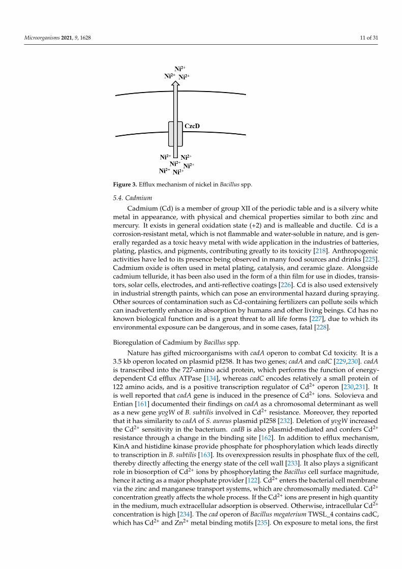
Bioregulation of Nickel by Bacillus spp.
There have been many methods of Ni removal from solid matrices, but the mosteffective are those which are capable of removing/treating Ni before it emanates into theenvironment [219]. Several physico-chemical methods have been employed over the yearsfor the removal of Ni from aqueous solutions [220]. Regardless of which of these methodshave been used in the past, newer, cheaper, and more efficient methods of adsorption havebeen used for Ni removal, such as the use of biomass, where sugarcane, corn cobs, citruspeels, and bark have been used [221]. In a study, corn hydrochar was treated with KOHand altered by treating polyethyleneimine (PEI) to increase adsorption of Ni ions onto thesurface [222]. Bioremediation by Gram-positive bacteria, such as Bacillus spp., has been thebetter method for the removal of Ni ions from Ni-contaminated media. There have beenmany studies that demonstrate the uptake and/or removal of Ni ions from contaminatedenvironments such as soils, wastewater, and rivers [105,113–115] (Table 1). B. thuringiensishas also been frequently reported to uptake and remove Ni from contaminated environ-ments [116–118]. In a recent study, B. megaterium was isolated from Ni-contaminated soilsand was able to uptake more than 500 mg Ni, where more than 3000 mg/L Ni salt waspreviously found [223]. The removal of Ni by bacteria is contingent on their inherentmechanisms of resistance, which ultimately facilitate uptake, transportation, and efflux ofthe metal ions in and out of the cell. According to Moore et al. [159], mechanisms of Nihomeostasis and regulation have not been well characterized in B. subtilis when comparedto Gram-negative bacteria such as Escherichia coli and Helicobacter pylori, though someevidence suggests their presence [224]. Members of the cation diffusion facilitator (CDF)family have long been characterized to mediate efflux of multiple metal ions, includingNi [160]. In B. subtilis, the cation diffusion transporter CzcD is reported to provide pro-tection to the cell amid high concentrations of Ni2+, Cu+, Zn2+, and Co2+ [159] (Figure 3).When these ions happen to be bound with citrate, these complexes, during favorableconditions, are taken up by the metal-dicitrate uptake system known as CitM in B. subtilis,consequently leading to an increase in toxicity to them [159] (Table 2).
Microorganisms 2021, 9, 1628 11 of 31Microorganisms 2021, 9, x FOR PEER REVIEW 11 of 32
Figure 3. Efflux mechanism of nickel in Bacillus spp.
5.4. Cadmium Cadmium (Cd) is a member of group XII of the periodic table and is a silvery white
metal in appearance, with physical and chemical properties similar to both zinc and mer-cury. It exists in general oxidation state (+2) and is malleable and ductile. Cd is a corrosion-resistant metal, which is not flammable and water-soluble in nature, and is generally re-garded as a toxic heavy metal with wide application in the industries of batteries, plating, plastics, and pigments, contributing greatly to its toxicity [218]. Anthropogenic activities have led to its presence being observed in many food sources and drinks [225]. Cadmium oxide is often used in metal plating, catalysis, and ceramic glaze. Alongside cadmium tel-luride, it has been also used in the form of a thin film for use in diodes, transistors, solar cells, electrodes, and anti-reflective coatings [226]. Cd is also used extensively in industrial strength paints, which can pose an environmental hazard during spraying. Other sources of contamination such as Cd-containing fertilizers can pollute soils which can inadvert-ently enhance its absorption by humans and other living beings. Cd has no known bio-logical function and is a great threat to all life forms [227], due to which its environmental exposure can be dangerous, and in some cases, fatal [228].
Bioregulation of Cadmium by Bacillus spp. Nature has gifted microorganisms with cadA operon to combat Cd toxicity. It is a 3.5
kb operon located on plasmid pI258. It has two genes; cadA and cadC [229,230]. cadA is transcribed into the 727-amino acid protein, which performs the function of energy-de-pendent Cd efflux ATPase [134], whereas cadC encodes relatively a small protein of 122 amino acids, and is a positive transcription regulator of Cd2+ operon [230,231]. It is well reported that cadA gene is induced in the presence of Cd2+ ions. Solovieva and Entian [161] documented their findings on cadA as a chromosomal determinant as well as a new gene yvgW of B. subtilis involved in Cd2+ resistance. Moreover, they reported that it has similar-ity to cadA of S. aureus plasmid pI258 [232]. Deletion of yvgW increased the Cd2+ sensitivity in the bacterium. cadB is also plasmid-mediated and confers Cd2+ resistance through a change in the binding site [162]. In addition to efflux mechanism, KinA and histidine ki-nase provide phosphate for phosphorylation which leads directly to transcription in B. subtilis [163]. Its overexpression results in phosphate flux of the cell, thereby directly af-fecting the energy state of the cell wall [233]. It also plays a significant role in biosorption of Cd2+ ions by phosphorylating the Bacillus cell surface magnitude, hence it acting as a major phosphate provider [122]. Cd2+ enters the bacterial cell membrane via the zinc and manganese transport systems, which are chromosomally mediated. Cd2+ concentration greatly affects the whole process. If the Cd2+ ions are present in high quantity in the me-dium, much extracellular adsorption is observed. Otherwise, intracellular Cd2+ concentra-tion is high [234]. The cad operon of Bacillus megaterium TWSL_4 contains cadC, which has
Figure 3. Efflux mechanism of nickel in Bacillus spp.
5.4. Cadmium
Cadmium (Cd) is a member of group XII of the periodic table and is a silvery whitemetal in appearance, with physical and chemical properties similar to both zinc andmercury. It exists in general oxidation state (+2) and is malleable and ductile. Cd is acorrosion-resistant metal, which is not flammable and water-soluble in nature, and is gen-erally regarded as a toxic heavy metal with wide application in the industries of batteries,plating, plastics, and pigments, contributing greatly to its toxicity [218]. Anthropogenicactivities have led to its presence being observed in many food sources and drinks [225].Cadmium oxide is often used in metal plating, catalysis, and ceramic glaze. Alongsidecadmium telluride, it has been also used in the form of a thin film for use in diodes, transis-tors, solar cells, electrodes, and anti-reflective coatings [226]. Cd is also used extensivelyin industrial strength paints, which can pose an environmental hazard during spraying.Other sources of contamination such as Cd-containing fertilizers can pollute soils whichcan inadvertently enhance its absorption by humans and other living beings. Cd has noknown biological function and is a great threat to all life forms [227], due to which itsenvironmental exposure can be dangerous, and in some cases, fatal [228].
Bioregulation of Cadmium by Bacillus spp.
Nature has gifted microorganisms with cadA operon to combat Cd toxicity. It is a3.5 kb operon located on plasmid pI258. It has two genes; cadA and cadC [229,230]. cadAis transcribed into the 727-amino acid protein, which performs the function of energy-dependent Cd efflux ATPase [134], whereas cadC encodes relatively a small protein of122 amino acids, and is a positive transcription regulator of Cd2+ operon [230,231]. Itis well reported that cadA gene is induced in the presence of Cd2+ ions. Solovieva andEntian [161] documented their findings on cadA as a chromosomal determinant as wellas a new gene yvgW of B. subtilis involved in Cd2+ resistance. Moreover, they reportedthat it has similarity to cadA of S. aureus plasmid pI258 [232]. Deletion of yvgW increasedthe Cd2+ sensitivity in the bacterium. cadB is also plasmid-mediated and confers Cd2+
resistance through a change in the binding site [162]. In addition to efflux mechanism,KinA and histidine kinase provide phosphate for phosphorylation which leads directlyto transcription in B. subtilis [163]. Its overexpression results in phosphate flux of the cell,thereby directly affecting the energy state of the cell wall [233]. It also plays a significantrole in biosorption of Cd2+ ions by phosphorylating the Bacillus cell surface magnitude,hence it acting as a major phosphate provider [122]. Cd2+ enters the bacterial cell membranevia the zinc and manganese transport systems, which are chromosomally mediated. Cd2+
concentration greatly affects the whole process. If the Cd2+ ions are present in high quantityin the medium, much extracellular adsorption is observed. Otherwise, intracellular Cd2+
concentration is high [234]. The cad operon of Bacillus megaterium TWSL_4 contains cadC,which has Cd2+ and Zn2+ metal binding motifs [235]. On exposure to metal ions, the first

Microorganisms 2021, 9, 1628 12 of 31
interaction is always with cell wall [236]. Its structure and composition play a significantrole in deciding the next step of the process. Cd2+ adsorption on the bacterial cell wall dealswith exchanging ions like Ca2+, Mg2+, and H+ ions [237]. Chelation is another processin which Cd2+ ions are exchanged with cell surface protons like –SO3H, –COOH, and–NH [238]. It involves sequestration via intracellular metallothionein (MT) [239]. Inorganicdeposition of Cd2+ in the cell wall or inside cells can take place through interaction withhydroxide, carbonate, sulfate, and phosphorus [240]. In addition to metal concentration,biosorption is dependent on cell wall composition and cell physiology [241]. In Gram-positive bacterial species, resistance to Cd2+ is achieved by cadA system that is plasmid-borne. Cd2+ enters the bacterial cell by the MIT (metal ion transporter) system [164,165](Table 2). The genes for Cd2+ resistance are mostly plasmid-mediated. According to Chenet al. [242] they are found on R plasmid along with antibiotic resistance genes, e.g., inpathogens including K. pneumoniae, P. aeruginosa, and S. aureus. These genes are reported tobe directly involved in the uptake of Cd2+ ions from the environment (Figure 4). Basha andRajaganesh [120] reported B. licheniformis to be a good biosorbent for Cd2+, as it removedmore than 98% of Cd2+. Other species such as B. catenulatus and B. safensis are also reportedto be effective in removing Cd2+ [119,121] (Table 1).
Microorganisms 2021, 9, x FOR PEER REVIEW 12 of 32
Cd2+ and Zn2+ metal binding motifs [235]. On exposure to metal ions, the first interaction is always with cell wall [236]. Its structure and composition play a significant role in de-ciding the next step of the process. Cd2+ adsorption on the bacterial cell wall deals with exchanging ions like Ca2+, Mg2+, and H+ ions [237]. Chelation is another process in which Cd2+ ions are exchanged with cell surface protons like –SO3H, –COOH, and –NH [238]. It involves sequestration via intracellular metallothionein (MT) [239]. Inorganic deposition of Cd2+ in the cell wall or inside cells can take place through interaction with hydroxide, carbonate, sulfate, and phosphorus [240]. In addition to metal concentration, biosorption is dependent on cell wall composition and cell physiology [241]. In Gram-positive bacte-rial species, resistance to Cd2+ is achieved by cadA system that is plasmid-borne. Cd2+ en-ters the bacterial cell by the MIT (metal ion transporter) system [164,165] (Table 2). The genes for Cd2+ resistance are mostly plasmid-mediated. According to Chen et al. [242] they are found on R plasmid along with antibiotic resistance genes, e.g., in pathogens including K. pneumoniae, P. aeruginosa, and S. aureus. These genes are reported to be directly involved in the uptake of Cd2+ ions from the environment (Figure 4). Basha and Rajaganesh [120] reported B. licheniformis to be a good biosorbent for Cd2+, as it removed more than 98% of Cd2+. Other species such as B. catenulatus and B. safensis are also reported to be effective in removing Cd2+ [119,121] (Table 1).
Figure 4. Mechanism of cadmium bioregulation by Bacillus spp.
5.5. Lead Lead (Pb) is a toxic heavy metal that is introduced into the environment via the
weathering of rocks. Anthropogenic sources include fossil fuels, extraction and melting of metals, battery-manufacturing industries, insecticides, pigments, and fertilizers [243]. Tet-raethyl lead (TEL) has a common application as a gasoline additive, due to which it is a source of heat and electricity [244]. It exists in two states: Pb2+ and Pb4+ [245]. Its toxicity determines its bioavailability as well as mobility in the soil. Its common forms are oxides, hydroxides, ionic form, metal oxyanion complexes [200], phosphates, and carbonates (at pH above 6). The stable and insoluble forms include oxides, sulfides, and pyromorphites [246]. Its exposure occurs by food, water, and inhalation, which affects the circulatory, gastrointestinal, reproductive, neurological, muscular, kidney, and genetic systems [123]. Dose and exposure time are prime factors [247]. The permissible level of Pb in drinking water is <10 µL/L [230].
Bioregulation of Lead by Bacillus spp. Pb-resistant Bacillus spp. have been reported previously [245]. Bacillus uses pbr op-
eron [166–168] and active transport [248] as potential strategies to combat the toxic effects of Pb (Table 2, Figure 5). Microorganisms immobilize it by adsorption, chelation, inorganic precipitation, complexation, and biosorption. These processes involve bacterial cell wall
Figure 4. Mechanism of cadmium bioregulation by Bacillus spp.
5.5. Lead
Lead (Pb) is a toxic heavy metal that is introduced into the environment via theweathering of rocks. Anthropogenic sources include fossil fuels, extraction and meltingof metals, battery-manufacturing industries, insecticides, pigments, and fertilizers [243].Tetraethyl lead (TEL) has a common application as a gasoline additive, due to which itis a source of heat and electricity [244]. It exists in two states: Pb2+ and Pb4+ [245]. Itstoxicity determines its bioavailability as well as mobility in the soil. Its common formsare oxides, hydroxides, ionic form, metal oxyanion complexes [200], phosphates, andcarbonates (at pH above 6). The stable and insoluble forms include oxides, sulfides, andpyromorphites [246]. Its exposure occurs by food, water, and inhalation, which affectsthe circulatory, gastrointestinal, reproductive, neurological, muscular, kidney, and geneticsystems [123]. Dose and exposure time are prime factors [247]. The permissible level of Pbin drinking water is <10 µL/L [230].
Bioregulation of Lead by Bacillus spp.
Pb-resistant Bacillus spp. have been reported previously [245]. Bacillus uses pbroperon [166–168] and active transport [248] as potential strategies to combat the toxiceffects of Pb (Table 2, Figure 5). Microorganisms immobilize it by adsorption, chelation,inorganic precipitation, complexation, and biosorption. These processes involve bacterialcell wall functional groups including phosphate, carboxyl, carbonyl, sulfhydryl, andhydroxyl groups, which confer a negative charge to the cell wall. Binding of Pb to any of

Microorganisms 2021, 9, 1628 13 of 31
them results in insoluble substance. On the outside environment, Pb2+ is exchanged by Naor K cations [124]. Another method is adsorption through the cell wall, as it is comprisedof organic macromolecules including polypeptides, polysaccharides, and proteins, whichhave the ability to adsorb Pb via electrostatic forces including Van der Waal’s forces,covalent or ionic bonds [124]. Pb interferes with microbial growth, morphology, andbiochemical activities by damaging the DNA, protein, and lipids and even replacing theessential ions within the enzymes [123,249]. Microbes resist Pb toxicity by extracellularprecipitation, exclusion, volatilization, biomethylation, cell surface binding, intracellularsequestration, and enhanced siderophore production [123]. Much like other Gram-positivebacteria, Bacillus spp. also employ one or several of these methods to remove Pb from thecontaminated environments [125–127] (Table 1).
Microorganisms 2021, 9, x FOR PEER REVIEW 13 of 32
functional groups including phosphate, carboxyl, carbonyl, sulfhydryl, and hydroxyl groups, which confer a negative charge to the cell wall. Binding of Pb to any of them re-sults in insoluble substance. On the outside environment, Pb2+ is exchanged by Na or K cations [124]. Another method is adsorption through the cell wall, as it is comprised of organic macromolecules including polypeptides, polysaccharides, and proteins, which have the ability to adsorb Pb via electrostatic forces including Van der Waal’s forces, co-valent or ionic bonds [124]. Pb interferes with microbial growth, morphology, and bio-chemical activities by damaging the DNA, protein, and lipids and even replacing the es-sential ions within the enzymes [123,249]. Microbes resist Pb toxicity by extracellular pre-cipitation, exclusion, volatilization, biomethylation, cell surface binding, intracellular se-questration, and enhanced siderophore production [123]. Much like other Gram-positive bacteria, Bacillus spp. also employ one or several of these methods to remove Pb from the contaminated environments [125–127] (Table 1).
Figure 5. pbr operon involved in the regulation of lead resistance in Bacillus spp.
5.6. Copper Copper (Cu) is categorized into the group I-B, and period 4 of the periodic table [200].
It is a soft, diamagnetic, malleable, and ductile metal with remarkable electrical and ther-mal conductivity. It acts as a soft and intermediate Lewis acid and tends to bind to soft bases (hydride, alkyl, thiol, phosphine) and auxiliary ligands to Cu2+, such as sulfate and nitrate [250]. Apart from being widespread in the environment thanks to anthropogenic actions, it also exists naturally in the form of minerals such as sulfides, carbonates, and oxides. The discovery and use of Cu dates back to ancient times, with its use spanning more than five thousand years. It exists in either of its two oxidation states, which can be the oxidized, divalent cupric form (Cu2+) or the reduced, monovalent cuprous form (Cu+) [251]. The significance of Cu in biological systems is pivotal; it serves an important role as a micronutrient in several biological processes in both prokaryotic and eukaryotic organ-isms. However, this stands only for lower concentrations of the metal; higher concentra-tions tend to induce cell toxicity, resulting in intracellular damage including changes in DNA, respiration, and overall growth [252]. Moreover, Cu is essentially required as a co-factor in more than thirty known enzymes, due to its ability to reversibly interconvert from its less to more required forms very easily [253]. Elevated levels of Cu exposure are a deep-rooted cause of environmental pollution by Cu, the fundamental reason being an-thropogenic processes. Industries using Cu or its compounds, Cu mining, burning of fos-sil fuels, inadequate treatment of wastewater, accumulation in dumps, production of phosphate-containing fertilizer, and natural processes such as erosion, volcanic eruptions, forest wildfires, and decay are all processes which greatly contribute to its presence in the environment. Furthermore, its production is also a source of direct Cu pollution, capable of harming the fragile ecosystems of soil, water, and air, respectively [254].
Figure 5. pbr operon involved in the regulation of lead resistance in Bacillus spp.
5.6. Copper
Copper (Cu) is categorized into the group I-B, and period 4 of the periodic table [200].It is a soft, diamagnetic, malleable, and ductile metal with remarkable electrical andthermal conductivity. It acts as a soft and intermediate Lewis acid and tends to bind to softbases (hydride, alkyl, thiol, phosphine) and auxiliary ligands to Cu2+, such as sulfate andnitrate [250]. Apart from being widespread in the environment thanks to anthropogenicactions, it also exists naturally in the form of minerals such as sulfides, carbonates, andoxides. The discovery and use of Cu dates back to ancient times, with its use spanning morethan five thousand years. It exists in either of its two oxidation states, which can be theoxidized, divalent cupric form (Cu2+) or the reduced, monovalent cuprous form (Cu+) [251].The significance of Cu in biological systems is pivotal; it serves an important role as amicronutrient in several biological processes in both prokaryotic and eukaryotic organisms.However, this stands only for lower concentrations of the metal; higher concentrationstend to induce cell toxicity, resulting in intracellular damage including changes in DNA,respiration, and overall growth [252]. Moreover, Cu is essentially required as a co-factorin more than thirty known enzymes, due to its ability to reversibly interconvert fromits less to more required forms very easily [253]. Elevated levels of Cu exposure area deep-rooted cause of environmental pollution by Cu, the fundamental reason beinganthropogenic processes. Industries using Cu or its compounds, Cu mining, burning offossil fuels, inadequate treatment of wastewater, accumulation in dumps, production ofphosphate-containing fertilizer, and natural processes such as erosion, volcanic eruptions,forest wildfires, and decay are all processes which greatly contribute to its presence in theenvironment. Furthermore, its production is also a source of direct Cu pollution, capableof harming the fragile ecosystems of soil, water, and air, respectively [254].
Bioregulation of Copper by Bacillus spp.
Like many other heavy metal ions, Cu is considered to be essential for Bacillus subtilis,while concentrations exceeding normal amounts can be toxic for the cells. Species like

Microorganisms 2021, 9, 1628 14 of 31
B. thuringiensis, B. cereus, B. licheniformis, and B. sphaericus are also involved in the removalof Cu, when their concentrations exceed the required limit [117,128–133,255,256] (Table 1).In correlation with other bacterial species, Cu in the cytosol is regulated by CueR [257].B. subtilis CueR is responsible for regulating the copZA operon which encodes both Cuchaperone and a P-type ATPase for Cu efflux, the latter of which is a member of the integralfamily which exports metal out of the cell [169]. In the former, CopZ plays a key role as Cuchaperone in transferring Cu over to CopA [170], which contributes to uptake of Cu, whileCopB is accountable for Cu efflux and detoxification [171] (Table 2, Figure 1). Chillappagariet al. [172] reported that YcnJ was associated with Cu uptake function of copZA operon inB. subtilis, where their model proposed that the protein works in conjunction with otherCop proteins to facilitate Cu transport in and out of the cell (Figure 6).
Microorganisms 2021, 9, x FOR PEER REVIEW 14 of 32
Bioregulation of Copper by Bacillus spp. Like many other heavy metal ions, Cu is considered to be essential for Bacillus subtilis,
while concentrations exceeding normal amounts can be toxic for the cells. Species like B. thuringiensis, B. cereus, B. licheniformis, and B. sphaericus are also involved in the removal of Cu, when their concentrations exceed the required limit [117,128–133,255,256] (Table 1). In correlation with other bacterial species, Cu in the cytosol is regulated by CueR [257]. B. subtilis CueR is responsible for regulating the copZA operon which encodes both Cu chaperone and a P-type ATPase for Cu efflux, the latter of which is a member of the inte-gral family which exports metal out of the cell [169]. In the former, CopZ plays a key role as Cu chaperone in transferring Cu over to CopA [170], which contributes to uptake of Cu, while CopB is accountable for Cu efflux and detoxification [171] (Table 2, Figure 1). Chillappagari et al. [172] reported that YcnJ was associated with Cu uptake function of copZA operon in B. subtilis, where their model proposed that the protein works in con-junction with other Cop proteins to facilitate Cu transport in and out of the cell (Figure 6).
Figure 6. Uptake and efflux mechanism of copper by Bacillus spp.
5.7. Chromium Chromium (Cr) is the 7th most abundant element on Earth, found widely in its crust.
It belongs to the group VI-B in the periodic table and is characterized as a redox transition metal (3d) with variable valences (−2 to +6). According to the WHO and US EPA, the per-missible limit of Cr in drinking water is >50 µg/L [258], whereas in soil, concentrations of >90 mg/kg are considered to be safe [259]. Among its many oxidation states, Cr mainly exists in its trivalent (Cr3+) and hexavalent (Cr6+) ions which are the most stable forms found naturally [260]. The toxicity and absorption are primarily dependent upon the oxi-dation state of the metal, which is also affiliated with its concentration in biological sys-tems [261]. The hexavalent form is reported to be relatively more toxic than the former, which remains fairly innocuous and is associated with lipid and sugar metabolism [262]. On the other hand, Cr6+ has been reported to act as a carcinogen and a mutagen, which is absorbed readily into the human food chain [263]. Cr3+ has a greater affinity for organic solutions and is able to form hydroxide, oxide, and sulfate complexes in nature, while Cr6+ tends to persist more in the environment and biological systems owing to its emission due to anthropogenic actions [264]. Furthermore, Cr6+ occurs as a potent oxidizing agent, as part of many solutions, but it is most commonly found in the form of oxyanion chromate (Cr2O42−), which is the only ion present in pH <7. Compounds of Cr6+ pose a significant risk as environmental contaminants due to their elevated toxicity, as high concentrations lead to changes in the structure and variety of microbial systems present in various envi-ronments [265]. Nevertheless, Cr3+ appears to be accountable for most of the Cr toxicity at intracellular levels. Concentrations of Cr in the environment are imputed to anthropo-genic actions, from their production by various industries, such as tanning, electroplating,
Figure 6. Uptake and efflux mechanism of copper by Bacillus spp.
5.7. Chromium
Chromium (Cr) is the 7th most abundant element on Earth, found widely in itscrust. It belongs to the group VI-B in the periodic table and is characterized as a redoxtransition metal (3d) with variable valences (−2 to +6). According to the WHO and USEPA, the permissible limit of Cr in drinking water is >50 µg/L [258], whereas in soil,concentrations of >90 mg/kg are considered to be safe [259]. Among its many oxidationstates, Cr mainly exists in its trivalent (Cr3+) and hexavalent (Cr6+) ions which are the moststable forms found naturally [260]. The toxicity and absorption are primarily dependentupon the oxidation state of the metal, which is also affiliated with its concentration inbiological systems [261]. The hexavalent form is reported to be relatively more toxicthan the former, which remains fairly innocuous and is associated with lipid and sugarmetabolism [262]. On the other hand, Cr6+ has been reported to act as a carcinogen and amutagen, which is absorbed readily into the human food chain [263]. Cr3+ has a greateraffinity for organic solutions and is able to form hydroxide, oxide, and sulfate complexes innature, while Cr6+ tends to persist more in the environment and biological systems owingto its emission due to anthropogenic actions [264]. Furthermore, Cr6+ occurs as a potentoxidizing agent, as part of many solutions, but it is most commonly found in the form ofoxyanion chromate (Cr2O4
2−), which is the only ion present in pH < 7. Compounds ofCr6+ pose a significant risk as environmental contaminants due to their elevated toxicity,as high concentrations lead to changes in the structure and variety of microbial systemspresent in various environments [265]. Nevertheless, Cr3+ appears to be accountable formost of the Cr toxicity at intracellular levels. Concentrations of Cr in the environmentare imputed to anthropogenic actions, from their production by various industries, suchas tanning, electroplating, chemical industry, smelting, and the leather industry [259], tobeing discharged into the environment in the form of untreated residues, wastewater, andeffluent sludge. Moreover, the mining of Cr from its ores also contributes greatly to itsenvironmental burden, threatening many fragile ecosystems [266].

Microorganisms 2021, 9, 1628 15 of 31
Bioregulation of Chromium by Bacillus spp.
The removal of Cr from contaminated environments has been achieved in the pastthrough many physico-chemical techniques. However, one of the most efficient processesremains microbial bioremediation, as most microorganisms tend to survive even in highlyheavy metal-contaminated environments. Moreover, bioremediation can be utilized totreat and remove these ions in order to combat environmental pollution, which rings truefor most of the microbes used to treat Cr6+ which are isolated from tannery effluents andsewage [267]. Cr3+ is relatively non-toxic, compared with Cr6+, and is insoluble, and istherefore much easier to remove through precipitation, whereas Cr6+ tends to persist innature [268]. Microbes in Cr-contaminated environments have inherent resistance whichenables their survival by evasion of metal stress via metal efflux, uptake, or detoxificationthrough reducing immobilization of metal ions [173] (Table 2). Among the methods ofmicrobial biodegradation, enzyme-regulated biotransformation of Cr6+ from its toxic tonon-toxic state (Cr3+) by bacteria is considered to be a cheap and efficient method of Crremoval from contaminated soil and wastewater. In Gram-positive and negative bacte-ria, this reduction of chromate is arbitrated by an enzyme, ChrR (chromate reductase),which is exclusively found in Cr-resistant bacteria and is not often affiliated with plas-mids [174]. This reduction can be mediated both aerobically and anaerobically, throughthe cytosolic component and membrane-bound component, respectively [175]. How-ever, in Bacillus spp., reduction of chromate ions is regulated through the aerobic process,which transpires through the transfer of electrons from the hexavalent to trivalent formof Cr; this occurs through the formation of an unstable intermediate (Cr5+), regulated byNADH/NADPH [269] (Figure 7). B. licheniformis was reported to remove 95% and 69.4% ofCr in various studies [138,141,270]. Nayak et al. [135] reported that B. cereus removed morethan 81% of Cr. Other Bacillus spp. have also been reported to be effective in mitigating Crpollution [121,136,137,139,140,142–144,271,272] (Table 1).
Microorganisms 2021, 9, x FOR PEER REVIEW 15 of 32
chemical industry, smelting, and the leather industry [259], to being discharged into the environment in the form of untreated residues, wastewater, and effluent sludge. Moreo-ver, the mining of Cr from its ores also contributes greatly to its environmental burden, threatening many fragile ecosystems [266].
Bioregulation of Chromium by Bacillus spp. The removal of Cr from contaminated environments has been achieved in the past
through many physico-chemical techniques. However, one of the most efficient processes remains microbial bioremediation, as most microorganisms tend to survive even in highly heavy metal-contaminated environments. Moreover, bioremediation can be utilized to treat and remove these ions in order to combat environmental pollution, which rings true for most of the microbes used to treat Cr6+ which are isolated from tannery effluents and sewage [267]. Cr3+ is relatively non-toxic, compared with Cr6+, and is insoluble, and is therefore much easier to remove through precipitation, whereas Cr6+ tends to persist in nature [268]. Microbes in Cr-contaminated environments have inherent resistance which enables their survival by evasion of metal stress via metal efflux, uptake, or detoxification through reducing immobilization of metal ions [173] (Table 2). Among the methods of microbial biodegradation, enzyme-regulated biotransformation of Cr6+ from its toxic to non-toxic state (Cr3+) by bacteria is considered to be a cheap and efficient method of Cr removal from contaminated soil and wastewater. In Gram-positive and negative bacteria, this reduction of chromate is arbitrated by an enzyme, ChrR (chromate reductase), which is exclusively found in Cr-resistant bacteria and is not often affiliated with plasmids [174]. This reduction can be mediated both aerobically and anaerobically, through the cytosolic component and membrane-bound component, respectively [175]. However, in Bacillus spp., reduction of chromate ions is regulated through the aerobic process, which tran-spires through the transfer of electrons from the hexavalent to trivalent form of Cr; this occurs through the formation of an unstable intermediate (Cr5+), regulated by NADH/NADPH [269] (Figure 7). B. licheniformis was reported to remove 95% and 69.4% of Cr in various studies [138,141,270]. Nayak et al. [135] reported that B. cereus removed more than 81% of Cr. Other Bacillus spp. have also been reported to be effective in miti-gating Cr pollution [121,136,137,139,140,142–144,271,272] (Table 1).
Figure 7. Uptake mechanism of Bacillus spp. for cadmium and its regulation.
5.8. Mercury Mercury (Hg) is a shiny, silvery metal at room temperature, which belongs to Group
XII and is the 80th element in the periodic table [273]. It is one of the most toxic elements on Earth with adverse health effects to all living beings including humans. It is tradition-ally grouped together with lead and cadmium for the “big three heavy metal poisons”
Figure 7. Uptake mechanism of Bacillus spp. for cadmium and its regulation.
5.8. Mercury
Mercury (Hg) is a shiny, silvery metal at room temperature, which belongs to GroupXII and is the 80th element in the periodic table [273]. It is one of the most toxic elements onEarth with adverse health effects to all living beings including humans. It is traditionallygrouped together with lead and cadmium for the “big three heavy metal poisons” whichare not reported to be involved in any essential biological function [200]. Once introducedinto various environments, Hg tends to accumulate rapidly, with it being cumulativelypresent in soils, sediments, water, inside living things, and in the atmosphere. Worldwide,levels of Hg pollution have greatly increased at atmospheric level due to the various miningand industrial processes through which the metal can be released into the nearby soilsand sediments [274]. Due to its tendency to accumulate in the ecosystem, it can remainsuspended in the atmosphere for a time period of approximately 24 months [275]. Major

Microorganisms 2021, 9, 1628 16 of 31
anthropogenic activities contribute more significantly to Hg release than natural processes,which can aid in its transmission worldwide [274]. Further aiding its spread, Hg existsnaturally in its elemental (Hg0), organic, and inorganic (Hg1+, Hg2+) forms, which areinterconvertible in different environments, due to which it can insert itself deep into sourcesof soil, sediment, air, and water, respectively [276]. When elemental Hg is released into theenvironment, it tends to make small, tightly packed, sphere-shaped droplets in a processknown as vaporization, due to the massive surface tension and vapor pressure [277]. On theother hand, the inorganic forms of Hg are more ubiquitously found, due to their economicsignificance and widespread applications in several industries which are the major sourcesof Hg emission into the environment [278].
Bioregulation of Mercury by Bacillus spp.
Bacillus and its various species have been positively associated with the bioremediationof Hg, as reported by various studies conducted over the years [145–150,279] (Table 1). Thehigh toxicity of Hg leads to its dangerous effect on biological systems. Faced with highconcentrations of toxic Hg, bacteria are equipped with several mechanisms in order toensure their survival. The presence of mercury resistance genes and operons, particularlythe mer operon, are reported in both Gram-positive and negative bacteria [208]. In bacteria,two types of operons exist for resistance against Hg, with one being a narrow spectrummer operon and the other being a broad-spectrum operon. The reversible detoxificationof Hg from its toxic to non-toxic state is induced inside the cell, but the egress of Hgoutside the cell is facilitated by diffusion [232]. Once the metal ions are out of the cell,they can again undergo oxidation by other bacterial species [160]. This phenomenon hasbeen investigated in many bacteria, where its activation via MerR, an activator protein,is responsible for the detection of Hg. The reduction of inorganic Hg2+ and organic Hgis facilitated through the enzyme mercuric reductase and the lyase enzyme, encoded bythe merA and merB gene, respectively [176] (Figure 8). Furthermore, the different forms ofHg may serve a function in its regulation of transportation, and bioaccumulation, whichmay be a cause of Hg toxicity in habitats and ecosystems near sites where Hg is minedor emitted [280]. Apart from Hg transformation by enzymes, it can be bioremediated bythe action of metallothioneins by accumulating Hg ions in an inactive form [177] (Table 2).Though the mechanism of Hg resistance is far better understood in Gram-negative bacteria,Gram-positive bacteria also share the same mer operon, although some similar geneticsequences and some disparity among bacterial species is widely reported. In B. cereus, merAgene was identified in the USA which is usually reported among Bacillus group [178,179].This occurrence among Bacillus spp. has been attributed to the location of these geneson transposons [281]. Bacillus spp. isolated from soils were also reported to possess Hg-resistant genes which facilitated the reduction and eventual bioremediation of Hg [180].Furthermore, B. cereus was genetically engineered to harbor Hg resistance via a mer operonfrom another Bacillus sp., B. thuringiensis, for better biosorption and volatilization of Hgfrom contaminated sites [177].
Microorganisms 2021, 9, x FOR PEER REVIEW 17 of 32
Figure 8. Uptake and reduction mechanisms for mercury by Bacillus spp.
5.9. Manganese Manganese (Mn) is a transition metal belonging to Group VII (atomic number 25) of
the periodic table, and is the 12th most abundantly found element on Earth [282]. It is widely distributed in nature as an essential trace element significant to all living beings. In the environment, Mn is not found as a free element, rather as a component of different naturally found minerals in the form of various oxides [283]. Mn is found in several oxi-dation states, of which the divalent form is reported to be the most common [284]. It is also able to exhibit all valences from 1 to 7, though 1 and 5 are very rare [185]. The essen-tiality of Mn lies in its significance as a co-factor and regulator of various processes in biological systems. Therefore, its toxicity levels can be low in living beings, but unusually high concentrations can cause detrimental effects to the nervous system in humans [186]. Many minerals are comprised of Mn nodules, which can be found in various environ-ments such as soil, sediment, and in water, depending on geographical position, which is also a deciding factor for the size of the nodules. There are many industrial applications of Mn, making Mn the 4th most used metal in industries on the basis of tonnage. Mn is most used in the steel industry, followed by the chemical and battery industry, in which more than 90% of the world’s Mn is utilized in the desulfurization and reinforcement of steel [282].
Bioregulation of Manganese by Bacillus spp. The essentiality of Mn mitigates the overall toxicity of the metal in biological systems,
although some toxicological conditions can be observed in adults and children alike, due to high Mn concentrations [187]. However, Mn found in high concentrations in the envi-ronment is difficult to degrade. Usually, the removal methods for Mn involve chemical techniques and the addition of chemical reagents which oxidize Mn by oxidation or aera-tion from the contaminated sites, but these methods are deemed to be very expensive, and not very efficient. The use of these methods to remove Mn from the environment also leads to the production of secondary metabolites [188]. The removal and regulation of Mn by microorganisms is mediated by metal-specific regulators that regulate low or sufficient concentrations of Mn inside the cell. This system is well characterized in bacteria, espe-cially Gram-positive bacteria such as Bacillus spp., due to the fact that many studies have reported Mn bioremediation by Bacillus [44,151–153], whereas the Mn homeostasis mech-anisms have been studied extensively over the years (Table 1). In B. subtilis, Mn concen-tration is regulated by MntR, an Mn-specific metallo-regulator related to the DtxR/IdeR family [181], which serves important roles in the sensing of metals such as iron (Fe) and Mn in several bacterial species [189]. It has also been suggested by Helmann in his review [285] that MntR is not only involved in the regulation of Mn uptake, but also in some conditions can sense and regulate Fe concentrations and their eventual homeostasis in the
Figure 8. Uptake and reduction mechanisms for mercury by Bacillus spp.

Microorganisms 2021, 9, 1628 17 of 31
5.9. Manganese
Manganese (Mn) is a transition metal belonging to Group VII (atomic number 25) ofthe periodic table, and is the 12th most abundantly found element on Earth [282]. It iswidely distributed in nature as an essential trace element significant to all living beings.In the environment, Mn is not found as a free element, rather as a component of differentnaturally found minerals in the form of various oxides [283]. Mn is found in severaloxidation states, of which the divalent form is reported to be the most common [284]. Itis also able to exhibit all valences from 1 to 7, though 1 and 5 are very rare [185]. Theessentiality of Mn lies in its significance as a co-factor and regulator of various processes inbiological systems. Therefore, its toxicity levels can be low in living beings, but unusuallyhigh concentrations can cause detrimental effects to the nervous system in humans [186].Many minerals are comprised of Mn nodules, which can be found in various environmentssuch as soil, sediment, and in water, depending on geographical position, which is also adeciding factor for the size of the nodules. There are many industrial applications of Mn,making Mn the 4th most used metal in industries on the basis of tonnage. Mn is most usedin the steel industry, followed by the chemical and battery industry, in which more than90% of the world’s Mn is utilized in the desulfurization and reinforcement of steel [282].
Bioregulation of Manganese by Bacillus spp.
The essentiality of Mn mitigates the overall toxicity of the metal in biological systems,although some toxicological conditions can be observed in adults and children alike,due to high Mn concentrations [187]. However, Mn found in high concentrations in theenvironment is difficult to degrade. Usually, the removal methods for Mn involve chemicaltechniques and the addition of chemical reagents which oxidize Mn by oxidation or aerationfrom the contaminated sites, but these methods are deemed to be very expensive, and notvery efficient. The use of these methods to remove Mn from the environment also leadsto the production of secondary metabolites [188]. The removal and regulation of Mn bymicroorganisms is mediated by metal-specific regulators that regulate low or sufficientconcentrations of Mn inside the cell. This system is well characterized in bacteria, especiallyGram-positive bacteria such as Bacillus spp., due to the fact that many studies have reportedMn bioremediation by Bacillus [44,151–153], whereas the Mn homeostasis mechanismshave been studied extensively over the years (Table 1). In B. subtilis, Mn concentration isregulated by MntR, an Mn-specific metallo-regulator related to the DtxR/IdeR family [181],which serves important roles in the sensing of metals such as iron (Fe) and Mn in severalbacterial species [189]. It has also been suggested by Helmann in his review [285] thatMntR is not only involved in the regulation of Mn uptake, but also in some conditionscan sense and regulate Fe concentrations and their eventual homeostasis in the cells. B.subtilis is reported to require Mn for growth and to regulate free and labile concentrationsof Mn inside the bacterial cell. Moreover, the uptake of Mn into Mn-starved B. subtiliscells demonstrated an ephemeral inhibition in growth, which resumed only after theconcentration of the metal inside the cell reverted to normal [182]. In conditions of Mnlimitation, B. subtilis MntR is de-repressed, which leads to the expression of two Mn uptakesystems [183], namely the ATP-dependent Mn ABC transporter (MntABCD operon), andMntH, a proton-coupled symporter [184] (Table 2). When there is an excess of Mn inbacterial cells, MntR regulates the expression of Mn efflux pumps, MneP and MneS [286].Under conditions of extreme excess of Mn in B. subtilis cells, the yybP–ykoY riboswitchis activated inside the cell, which senses the excessive concentrations of Mn and in turnactivates Mn-sensing genes that regulate homeostasis in the cell [203] (Figure 9).
Microorganisms 2021, 9, 1628 18 of 31
Microorganisms 2021, 9, x FOR PEER REVIEW 18 of 32
cells. B. subtilis is reported to require Mn for growth and to regulate free and labile con-centrations of Mn inside the bacterial cell. Moreover, the uptake of Mn into Mn-starved B. subtilis cells demonstrated an ephemeral inhibition in growth, which resumed only after the concentration of the metal inside the cell reverted to normal [182]. In conditions of Mn limitation, B. subtilis MntR is de-repressed, which leads to the expression of two Mn up-take systems [183], namely the ATP-dependent Mn ABC transporter (MntABCD operon), and MntH, a proton-coupled symporter [184] (Table 2). When there is an excess of Mn in bacterial cells, MntR regulates the expression of Mn efflux pumps, MneP and MneS [286]. Under conditions of extreme excess of Mn in B. subtilis cells, the yybP–ykoY riboswitch is activated inside the cell, which senses the excessive concentrations of Mn and in turn ac-tivates Mn-sensing genes that regulate homeostasis in the cell [203] (Figure 9).
Figure 9. Uptake mechanisms of Bacillus spp. for the regulation of manganese inside bacterial cell.
5.10. Molybdenum Molybdenum (Mo) is a member of Group VI of the periodic table which also houses
similar metals such as chromium and tungsten. Though Mo has been characterized as a metal since the Middle Ages, its pure form was produced for the first time in 1893 [287]. Mo is not naturally found in its metallic state, rather it is found in conjunction with other elements. It is naturally found in minerals, soils, rocks, and water. In high or moderate concentrations of Mo, the metal readily forms various complexes comprising of poly-molybdates. In its natural state, Mo occurs as a silvery-white metal which in its powder form can give off a black or greyish hue [287]. Mo exists in oxidation states ranging from 2- to +6, though it is significant in states of +4, +5, +6. It is an essential trace element re-quired for biological life and evolution. The different oxidation states of Mo are important as they partake in various redox reactions and act as cofactors for different enzymes. Nev-ertheless, exposure to Mo can cause toxicity, which can result in several health defects in biological systems [190]. The industrial applications of Mo are extensive, such as its usage as an additive substance in commercial lubricants, catalysts, corrosion inhibitors, and as a vital component in tungsten production. Most of the Mo produced is used in the steel and welding industry, where it is used for the reinforcement of steel and other alloys [288]. In analytical chemistry, Mo has long been used in several tests and techniques as a color-imetric agent. The natural and anthropogenic processes that contribute to the introduction of Mo into the environment include soil leaching, soil run-off, sedimentation, as well as burning of fossil fuels, mining, and mine wastes [289].
Figure 9. Uptake mechanisms of Bacillus spp. for the regulation of manganese inside bacterial cell.
5.10. Molybdenum
Molybdenum (Mo) is a member of Group VI of the periodic table which also housessimilar metals such as chromium and tungsten. Though Mo has been characterized as ametal since the Middle Ages, its pure form was produced for the first time in 1893 [287].Mo is not naturally found in its metallic state, rather it is found in conjunction withother elements. It is naturally found in minerals, soils, rocks, and water. In high ormoderate concentrations of Mo, the metal readily forms various complexes comprising ofpolymolybdates. In its natural state, Mo occurs as a silvery-white metal which in its powderform can give off a black or greyish hue [287]. Mo exists in oxidation states ranging from 2-to +6, though it is significant in states of +4, +5, +6. It is an essential trace element requiredfor biological life and evolution. The different oxidation states of Mo are important as theypartake in various redox reactions and act as cofactors for different enzymes. Nevertheless,exposure to Mo can cause toxicity, which can result in several health defects in biologicalsystems [190]. The industrial applications of Mo are extensive, such as its usage as anadditive substance in commercial lubricants, catalysts, corrosion inhibitors, and as a vitalcomponent in tungsten production. Most of the Mo produced is used in the steel andwelding industry, where it is used for the reinforcement of steel and other alloys [288]. Inanalytical chemistry, Mo has long been used in several tests and techniques as a colorimetricagent. The natural and anthropogenic processes that contribute to the introduction of Mointo the environment include soil leaching, soil run-off, sedimentation, as well as burningof fossil fuels, mining, and mine wastes [289].
Bioregulation of Molybdenum by Bacillus spp.
In bacteria, the transportation of Mo may be facilitated by sulfate transport systems,but the major uptake system has been characterized as the inducible ABC-type transporter(modABC operon), which is also able to facilitate the uptake of tungsten [160,290] (Figure 1).This transport system has been reported to regulate the uptake (high affinity) of Mo fromthe environment [291,292], and has been employed by Bacillus spp. to uptake Mo [154,155](Table 1). The main protein of this system is ModA, which is a substrate-binding periplasmicprotein primarily interacting with other proteins of the system such as ModB and ModCthat facilitate the transport of Mo via hydrolysis of ATP [293]. This system has been wellcharacterized in Gram-negative bacteria such as E. coli, though similar mod genes havebeen identified in many bacterial species, including B. subtilis [294,295] (Table 2). Moreover,high molybdate affinity uptake has been reported to be mediated by ModA protein in B.subtilis in Mo-limited conditions [296].
5.11. Gold
Gold (Au) resides in group XI of the periodic table, with an atomic number of 79. It isone of the rarest elements on this Earth, and its inertness allows it to exist in mineralized

Microorganisms 2021, 9, 1628 19 of 31
zones where it may exist in one of its many well-characterized geological forms [297]. It iscategorized as a non-essential element, incapable of forming free ions in aqueous solutionsand highly toxic to living beings under high concentrations [191,298]. Naturally foundAu is usually present in the form of alloys with other indigenous metals. Under surfaceconditions, Au is found in the form of metal colloid, aurous, and auric forms, with valencesof 0, +1, and +3, respectively, whereas its state is dependent upon the thermodynamicreactions taking place in its presence. Moreover, Au readily forms complexes with ligandswhich are organic in nature, a trait synonymous with its native group in the periodictable [297].
Bioregulation of Gold by Bacillus spp.
Though the resistance mechanisms of Au uptake and efflux in Gram-negative bacteriasuch as Cupriavidus metallidurans are well known [191,299], the mechanisms are poorlyunderstood in Gram-positive bacteria. Nevertheless, they are capable of mediating pre-cipitation and biomineralization of Au complexes in natural environmental settings [300](Table 2). This role offers some insight for microbes in mediating Au mobility and pro-moting bacterioform and secondary Au grains [301]. As is the case with other metals,complexes of Au also tend to be toxic for bacterial species at high concentrations, whichmay lead to the generation of free radicals and disruption of enzyme activity [302]. Microbe-mediated solubilization of Au is dependent upon the microbe’s oxidation ability, as well asits ability to facilitate ligands which can then bind to Au ions for their stability via complexor colloid formation. This phenomenon is true for many bacterial species, which residein different environmental settings. In particular, heterotrophic bacteria have been linkedto Au solubilization in soils rich in organic matter. B. subtilis, along with other Bacillusspp., has been previously reported to solubilize Au as Au-amino acid complexes [303,304].Moreover, it was observed that the reduction conditions generated by ETC of bacteriaresulted in the precipitation of Au on an extracellular level, along with the formation ofiron sulfide by hydrogen sulfide, causing Au removal from solution through the processof reductive adsorption [305]. Very early studies on B. subtilis do report this extracellularreduction of Au from chloride solution, as an outcome of selective adsorption [306]. Someother studies also report the accumulation of Au by Bacillus spp., which is inherentlypossible due to the cell membrane structure and its bound proteins [192,307]. Apart fromthe cell membrane, the enzyme-mediated hydrolysis of ATP in Bacillus spp. also playsa significant role, as the regulation of intracellular metabolism has been reported to beassociated with the bioaccumulation of Au [191,307].
5.12. Silver
Silver (Ag) is a silvery-white metal which is found naturally in its metal state as wellas in ores. The metal itself is insoluble in water but becomes soluble once bound to saltssuch as nitrates. Some compounds of Ag are stable in air and water, but the rest appear tobe sensitive to light. In its natural form, Ag is found with sulfide or closely bound in asso-ciation with other metal sulfides [308]. With two stable isotopes, it exists as a monovalention but readily forms complexes with other metals. Seldom occurring as a singular metal,it is often recovered from the environment from its ores, whereas the secondary productionis generated from old Ag scrap and Ag-containing products, such as photo-films, wastes,batteries, jewelry, coins, and dental equipment. Atmospheric emissions are attributable tosmelting procedures, manufacturing and recycling of photographic films, combustion offossil fuels, as well as cloud seeding [309]. Along with the environmental effects of Ag inthe environment, the anti-bacterial and biological use of Ag and its nanoparticles have beenextensively researched for many years. The most widely known use of Ag in medicine is itsuse as a topical antimicrobial agent for the treatment of burns, which has since translatedinto its usage in many clinical dressings, eye drops, and even dentures [310].

Microorganisms 2021, 9, 1628 20 of 31
Bioregulation of Silver by Bacillus spp.
In the course of removing Ag or surviving amidst Ag in the environment, bacterialspecies display high sensitivity to Ag due to its probable non-specific toxic nature, andits ability to permeate and affect bacterial metabolism, transport, and ion exchange sys-tems [311]. Therefore, bacterial resistance to Ag and its mechanisms usually is dependenton its binding proteins and affiliated efflux pumps in Gram-negative and/or positivebacteria [193–195]. Molecular bioregulation and resistance mechanisms active against Agin Gram-negative bacteria are well studied, with two Ag efflux systems (SilCBA and SilP)active in transporting Ag out of the cell while the mechanisms in Gram-positive bacteria,to the best of our knowledge, are poorly elucidated, with reports of only MRSA isolatesdemonstrating resistance against Ag, courtesy of Sil genes [196–198] (Table 2). However,one study reported the maximum uptake (73.6 mg g−1) of Ag by B. licheniformis fromaqueous solution, which denoted an active uptake system in Bacillus, mediated eitherthrough efflux pumps or resistance genes [156] (Table 1).
6. Conclusions
Biological methods, particularly bacterial-mediated bioremediation, are a cost-effective,feasible, and environmentally friendly approach for the treatment of contaminated soil andgroundwater, industrial wastewater, effluents, and sludge. Many bacterial species, suchas Bacillus spp. are viable options for bioremediation, as their molecular mechanisms aresuggestive of the resistance, transport, efflux and detoxification systems that work in highand low concentrations of heavy metals to maintain and regulate metal homeostasis at thecellular level. In order to utilize these mechanisms for the efficient removal of contaminants,including heavy metals, further research is required in order to study and characterizepreviously unknown molecular mechanisms of resistance, which can aid in elucidating therole of Bacillus spp. in bioremediation at a much deeper level.
Author Contributions: Conceptualization, S.S.; methodology, M.K.; software, S.S.; validation, S.S.;formal analysis, S.S.; investigation, M.K.; resources, M.K.; data curation, M.K.; writing—original draftpreparation, M.K.; writing—review and editing, B.S.A.; visualization, S.S.; supervision, S.S.; projectadministration, S.S.; funding acquisition, B.S.A. All authors have read and agreed to the publishedversion of the manuscript.
Institutional Review Board Statement: Not Applicable.
Informed Consent Statement: Not Applicable.
Data Availability Statement: Not Applicable.
Acknowledgments: This research was funded by the Deanship of the Scientific Research at PrincessNourah bint Abdulrahman University through the Fast-Track Path of Research Funding Program.
Conflicts of Interest: Authors declare no conflict of interest.
References1. Gautam, P.K.; Gautam, R.K.; Banerjee, S.; Chattopadhyaya, M.C.; Pandey, J.D. Heavy Metals in the Environment: Fate, Transport,
Toxicity and Remediation Technologies. In Heavy Metals: Sources, Toxicity and Remediation Techniques; Pathania, D., Ed.; NovaScience Publishers: Hauppauge, NY, USA, 2016; pp. 1–27.
2. Masindi, V.; Muedi, K.L. Environmental Contamination by Heavy Metals. In Heavy Metals; Saleh, H.E.-D., Aglan, R.F., Eds.;IntechOpen Publishers: London, UK, 2018; pp. 115–132. [CrossRef]
3. Ali, H.; Khan, E. What are heavy metals? Long-standing controversy over the scientific use of the term “heavy metals”-proposalof a comprehensive definition. Toxicol. Environ. Chem. 2018, 100, 6–19. [CrossRef]
4. Presentato, A.; Piacenza, E.; Turner, R.J.; Zannoni, D.; Cappelletti, M. Processing of Metals and Metalloids by Actinobacteria: CellResistance Mechanisms and Synthesis of Metal(loid)-Based Nanostructures. Microorganisms 2020, 8, 2027. [CrossRef]
5. Duffus, J.H. Heavy metals—A meaningless term? Pure Appl. Chem. 2002, 74, 793–807. [CrossRef]6. Tchounwou, P.B.; Yedjou, C.G.; Patlolla, A.K.; Sutton, D.J. Heavy metal toxicity and the environment. Exp. Suppl. 2012, 101,
133–164. [CrossRef]7. Martin, Y.E.; Johnson, E.A. Biogeosciences survey: Studying interactions of the biosphere with the lithosphere, hydrosphere and
atmosphere. Prog. Phys. Geogr. 2012, 36, 833–852. [CrossRef]

Microorganisms 2021, 9, 1628 21 of 31
8. Ali, H.; Khan, E.; Ilahi, I. Environmental chemistry and ecotoxicology of hazardous heavy metals: Environmental persistence,toxicity, and bioaccumulation. J. Chem. 2019, 2019, 730305. [CrossRef]
9. Nagajyoti, P.C.; Lee, K.D.; Sreekanth, T.V.M. Heavy metals, occurrence and toxicity for plants: A review. Environ. Chem. Lett. 2010,8, 199–216. [CrossRef]
10. He, Z.L.; Yang, X.E.; Stoffella, P.J. Trace elements in agroecosystems and impacts on the environment. J. Trace Elem. Med. Biol.2005, 19, 125–140. [CrossRef]
11. Muchuweti, M.; Birkett, J.W.; Chinyanga, E.; Zvauya, R.; Scrimshaw, M.D.; Lester, J.N. Heavy metal content of vegetables irrigatedwith mixtures of wastewater and sewage sludge in Zimbabwe: Implications for human health. Agric. Ecosyst. Environ. 2006, 112,41–48. [CrossRef]
12. Gupta, N.; Khan, D.K.; Santra, S.C. Heavy metal accumulation in vegetables grown in a long-term wastewater-irrigatedagricultural land of tropical India. Environ. Monit. Assess. 2012, 184, 6673–6682. [CrossRef]
13. Selvi, A.; Rajasekar, A.; Theerthagiri, J.; Ananthaselvam, A.; Sathishkumar, K.; Madhavan, J.; Rahman, P.K.S.M. Integratedremediation processes toward heavy metal removal/recovery from various environments-A review. Front. Environ. Sci. 2019, 7,66–81. [CrossRef]
14. Briffa, J.; Sinagra, E.; Blundell, R. Heavy metal pollution in the environment and their toxicological effects on humans. Heliyon2020, 6, 1–26. [CrossRef]
15. Türkmen, M.; Türkmen, A.; Tepe, Y.; Töre, Y.; Ates, A. Determination of metals in fish species from Aegean and Mediterraneanseas. Food Chem. 2009, 113, 233–237. [CrossRef]
16. Ramírez, R. The gastropod Osilinus atrata as a bioindicator of Cd, Cu, Pb and Zn contamination in the coastal waters of theCanary Islands. Chem. Ecol. 2013, 29, 208–220. [CrossRef]
17. Rahim, M.; Ullah, I.; Khan, A.; Haris, M.R.H.M. Health risk from heavy metals via consumption of food crops in the vicinity ofDistrict Shangla. J. Chem. Soc. Pak. 2016, 38, 177–185.
18. Appenroth, K.-J. What are “heavy metals” in Plant Sciences? Acta Physiol. Plant. 2010, 32, 615–619. [CrossRef]19. Chalkiadaki, O.; Dassenakis, M.; Lydakis-Simantiris, N. Bioconcentration of Cd and Ni in various tissues of two marine bivalves
living in different habitats and exposed to heavily polluted seawater. Chem. Ecol. 2014, 30, 726–742. [CrossRef]20. Valko, M.; Morris, H.; Cronin, M. Metals, toxicity and oxidative stress. Curr. Med. Chem. 2005, 12, 1161–1208. [CrossRef]21. Engwa, G.A.; Ferdinand, P.U.; Nwalo, F.N.; Unachukwu, N.M. Mechanism and Health Effects of Heavy Metal Toxicity in Humans.
In Poisoning in the Modern World—New Tricks for an Old Dog? Karcioglu, O., Arslan, B., Eds.; IntechOpen Publishers: London, UK,2019; pp. 1–24. [CrossRef]
22. Shahid, M.; Arshad, M.; Kaemmerer, M.; Pinelli, E. Long-term field metal extraction by pelargonium: Phytoextraction efficiencyin relation to plant maturity. Int. J. Phytoremed. 2012, 14, 493–505. [CrossRef] [PubMed]
23. Minnikova, T.V.; Denisova, T.V.; Mandzhieva, S.S.; Kolesnikov, S.I.; Minkina, T.M.; Chaplygin, V.A.; Burachevskaya, M.V.;Sushkova, S.N.; Bauer, T.V. Assessing the effect of heavy metals from the Novocherkassk power station emissions on thebiological activity of soils in the adjacent areas. J. Geochem. Explor. 2017, 174, 70–78. [CrossRef]
24. Murtaza, G.; Murtaza, B.; Niazi, K.N.; Sabir, M. Soil Contaminants: Sources, Effects, and Approaches for Remediation. InImprovement of Crops in the Era of Climatic Changes; Ahmad, P., Wani, R.M., Azooz, M.M., Phan Tran, L.-S., Eds.; Springer:New York, NY, USA, 2014; pp. 171–196. [CrossRef]
25. Shi, W.; Liu, C.; Ding, D.; Lei, Z.; Yang, Y.; Feng, C.; Zhang, Z. Immobilization of heavy metals in sewage sludge by usingsubcritical water technology. Bioresour. Technol. 2013, 137, 18–24. [CrossRef] [PubMed]
26. Yao, Z.; Li, J.; Xie, H.; Yu, C. Review on remediation technologies of soil contaminated by heavy metals. Proceed. Environ. Sci.2012, 16, 722–729. [CrossRef]
27. Zhou, D.M.; Hao, X.Z.; Xue, Y. Advances in remediation technologies of contaminated soils. Ecol. Environ. Sci. 2004, 13, 234–242.28. Elicker, C.; Filho, P.S.; Castagno, K. Electroremediation of heavy metals in sewage sludge. Braz. J. Chem. Eng. 2014, 31, 365–371.
[CrossRef]29. Navarro, A.; Cardellach, E.; Cañadas, I.; Rodríguez, J. Solar thermal vitrification of mining contaminated soils. Int. J. Min. Process.
2013, 119, 65–74. [CrossRef]30. Khalid, S.; Shahid, M.; Niazi, N.K.; Murtaza, B.; Bibi, I.; Dumate, C. A comparison of technologies for remediation of heavy metal
contaminated soils. J. Geochem. Explor. 2017, 182, 247–268. [CrossRef]31. Albers, C.N.; Jacobsen, O.S.; Flores, E.M.M.; Johnsen, A.R. Arctic and subarctic natural soils emit chloroform and brominated
analogues by alkaline hydrolysis of trihaloacetyl compounds. Environ. Sci. Technol. 2017, 51, 6131–6138. [CrossRef]32. Alghanmi, S.I.; Al Sulami, A.F.; El-Zayat, T.A.; Alhogbi, B.G.; Salam, M.A. Acid leaching of heavy metals from contaminated
soil collected from Jeddah, Saudi Arabia: Kinetic and thermodynamics studies. Int. Soil Water Conserv. Res. 2015, 3, 196–208.[CrossRef]
33. Lee, M.; Paik, I.S.; Do, W.; Kim, I.; Lee, Y.; Lee, S. Soil washing of As-contaminated stream sediments in the vicinity of anabandoned mine in Korea. Environ. Geochem. Health 2007, 29, 319–329. [CrossRef]
34. Li, Z.; Wang, L.; Meng, J.; Liu, X.; Xu, J.; Wang, F.; Brookes, P. Zeolite-supported nanoscale zero-valent iron: New findings onsimultaneous adsorption of Cd(II), Pb(II), and As(III) in aqueous solution and soil. J. Hazard. Mater. 2018, 344, 1–11. [CrossRef]
Microorganisms 2021, 9, 1628 22 of 31
35. Venkatachalam, P.; Jayaraj, M.; Manikandan, R.; Geetha, N.; Rene, E.R.; Sharma, N.C.; Sahi, S.V. Zinc oxide nanoparticles(ZnONPs) alleviate heavy metal-induced toxicity in Leucaena leucocephala seedlings: A physiochemical analysis. Plant. Physiol.Biochem. 2017, 110, 59–69. [CrossRef]
36. Venegas, A.; Rigol, A.; Vidal, M. Viability of organic wastes and biochars as amendments for the remediation of heavy metal-contaminated soils. Chemosphere 2015, 119, 190–198. [CrossRef] [PubMed]
37. Abbas, S.H.; Ismail, I.M.; Mostafa, T.M.; Sulaymon, A.H. Biosorption of heavy metals: A review. J. Chem. Sci. Technol. 2014, 3,74–102.
38. Mustapha, M.U.; Halimoon, N. Microorganisms and biosorption of heavy metals in the environment: A review paper. J. Microb.Biochem. Technol. 2015, 7, 253–256. [CrossRef]
39. Kang, S.Y.; Lee, J.U.; Kim, K.M. Biosorption of Cr(III) and Cr(VI) onto the cell surface of Pseudomonas aeruginosa. Biochem. Eng. J.2007, 36, 54–58. [CrossRef]
40. Akhtar, K.; Akhtar, M.W.; Khalid, A.M. Removal and recovery of uranium from aqueous solutions by Trichoderma harzianum.Water Res. 2007, 41, 1366–1378. [CrossRef]
41. Ozer, A.; Ozer, D. Comparative study of the biosorption of Pb(II), Ni(II) and Cr(VI) ions onto S. cerevisiae: Determination ofbiosorption heats. J. Hazard. Mater. 2003, 100, 219–229. [CrossRef] [PubMed]
42. Mosa, K.A.; Saadoun, I.; Kumar, K.; Helmy, M.; Dhankher, O.P. Potential biotechnological strategies for the cleanup of heavymetals and metalloids. Front. Plant. Sci. 2016, 7, 303. [CrossRef] [PubMed]
43. Kapahi, M.; Sachdeva, S. Bioremediation options for heavy metal pollution. J. Health Pollut. 2019, 9, 191203. [CrossRef] [PubMed]44. Abioye, O.P.; Oyewole, O.A.; Oyeleke, S.B.; Adeyemi, M.O.; Orukotan, A.A. Biosorption of lead, chromium and cadmium in
tannery effluent using indigenous microorganisms. Braz. J. Biol. Sci. 2018, 5, 25–32. [CrossRef]45. Tarekegn, M.M.; Salilih, F.Z.; Ishetu, A.I. Microbes used as a tool for bioremediation of heavy metal from the environment. Cog.
Food Agric. 2020, 6. [CrossRef]46. Kotb, E. Purification and partial characterization of serine fibrinolytic enzyme from Bacillus megaterium KSK-07 isolated from
kishk, a traditional Egyptian fermented food. Appl. Biochem. Microbiol. 2015, 51, 34–43. [CrossRef]47. Elshaghabee, F.M.F.; Rokana, N.; Gulhane, R.D.; Sharma, C.; Panwar, H. Bacillus as potential probiotics: Status, concerns, and
future perspectives. Front. Microbiol. 2017, 8, 1490. [CrossRef]48. Setlow, P.; Wang, S.; Li, Y.-Q. Germination of spores of the orders Bacillales and Clostridiales. Annu. Rev. Microbiol. 2017, 71,
459–477. [CrossRef]49. Cai, D.; Rao, Y.; Zhan, Y.; Wang, Q.; Chen, S. Engineering Bacillus for efficient production of heterologous protein: Current
progress, challenge and prospect. J. Appl. Microbiol. 2019, 126, 1632–1642. [CrossRef] [PubMed]50. Wolken, W.A.M.; Tramper, J.; van der Werf, M.J. What Can Spores Do for Us? Trends Biotechnol. 2003, 21, 338–345. [CrossRef]51. Sanders, M.E.; Morelli, L.; Tompkins, T.A. Sporeformers as human probiotics: Bacillus, Sporolactobacillus and Brevibacillus. Comp.
Rev. Food Sci. Food Saf. 2003, 2, 101–110. [CrossRef] [PubMed]52. Hong, H.A.; Duc, L.H.; Cutting, S.M. The Use of Bacterial Spore Formers as Probiotics. FEMS Microbiol. Rev. 2005, 29, 813–835.
[CrossRef]53. Lee, N.-K.; Kim, W.-S.; Paik, H.-D. Bacillus strains as human probiotics: Characterization, safety, microbiome, and probiotic carrier.
Food Sci. Biotechnol. 2019, 28, 1297–1305. [CrossRef]54. Ouattara, H.G.; Reverchon, S.; Niamke, S.L.; Nasser, W. Regulation of the synthesis of pulp degrading enzymes in Bacillus isolated
from cocoa fermentation. Food Microbiol. 2017, 63, 255–262. [CrossRef]55. Tanaka, K.; Takanaka, S.; Yoshida, K. A second-generation Bacillus cell factory for rare inositol production. Bioengineered 2014, 5,
331–334. [CrossRef]56. Takano, H. The regulatory mechanism underlying light-inducible production of carotenoids in nonphototrophic bacteria. Biosci.
Biotechnol. Biochem. 2016, 80, 1264–1273. [CrossRef]57. Chikere, C.B.; Okpokwasili, G.C.; Chikere, B.O. Bacterial diversity in a tropical crude oil-polluted soil undergoing bioremediation.
Afr. J. Biotechnol. 2009, 8, 2535–2540. [CrossRef]58. Nwinyi, O.C.; Kanu, I.A.; Tunde, A.; Ajanaku, K.O. Characterization of diesel degrading bacterial species from contaminated
tropical ecosystem. Braz. Arch. Biol. Technol. 2014, 57, 789–796. [CrossRef]59. Dey, U.; Chatterjee, S.; Mondal, N.K. Isolation and characterization of arsenic-resistant bacteria and possible application in
bioremediation. Biotech. Rep. 2016, 10, 1–7. [CrossRef] [PubMed]60. Matschullat, J. Arsenic in the geosphere—A review. Sci. Total Environ. 2000, 249, 297–312. [CrossRef]61. WHO. Environmental Health Criteria: Arsenic and Arsenic Compounds; WHO: Geneva, Switzerland, 2001; p. 224.62. Oritz-Escobar, M.E.; Hue, N.V.; Cutler, W.G. Recent developments on arsenic: Contamination and remediation. In Recent Research
Developments in Bioenergetics; Transworld Research Network: Trivandrum, India, 2006; Volume 4, pp. 1–32.63. Wilcox, D.E. Arsenic. Can This Toxic Metalloid Sustain Life? In Interrelations between Essential Metal Ions and Human Diseases.
Metal Ions in Life Sciences; Sigel, A., Sigel, H., Sigel, R., Eds.; Springer: Dordrecht, The Netherlands, 2013; pp. 475–498. [CrossRef]64. Mandal, B.K.; Suzuki, K.T. Arsenic around the world: A review. Talanta 2002, 58, 201–235. [CrossRef]65. Hue, N.V. Bioremediation of Arsenic Toxicity. In Arsenic Toxicity: Prevention and Treatment; Chakrabarty, N., Ed.; CRC Press: Boca
Raton, FL, USA, 2015.

Microorganisms 2021, 9, 1628 23 of 31
66. Cervantes, C.; Ji, G.; Ramírez, J.L.; Silver, S. Resistance to arsenic compounds in microorganisms. FEMS Microbiol. Rev. 1994, 15,355–367. [CrossRef] [PubMed]
67. Ratnaike, R.N. Acute and chronic arsenic toxicity. Postgrad. Med. J. 2003, 79, 391–396. [CrossRef]68. Lim, K.T.; Shukor, M.Y.; Wasoh, H. Physical, chemical, and biological methods for the removal of arsenic compounds. Biomed. Res.
Int. 2014, 2014, 503784. [CrossRef] [PubMed]69. Elangovan, D.; Chalakh, M.L. Arsenic pollution in West Bengal. Tech. Dig. 2006, 9, 31–35.70. Smedley, P.L.; Kinniburgh, D.G. A review of the source, behavior and distribution of arsenic in natural waters. Appl. Geochem.
2002, 17, 517–568. [CrossRef]71. Ghosh, S.; Mohapatra, B.; Satyanrayana, T.; Sar, P. Molecular and taxonomic characterization of arsenic (As) transforming Bacillus
sp. strain IIIJ3–1 isolated from As-contaminated groundwater of Brahmaputra river basin, India. BMC Microbiol. 2020, 20, 256–275.[CrossRef]
72. Mateos, L.M.; Ordonez, E.; Letek, M.; Gil, J.A. Corynebacterium glutamicum as a model bacteria for the bioremediation of arsenic.Int. Microbiol. 2006, 9, 2007–2015.
73. Zhu, Y.G.; Yoshinaga, M.; Zhao, F.J.; Rosen, B.P. Earth abides arsenic biotransformations. Ann. Rev. Earth Planet. Sci. 2014, 42,443–467. [CrossRef] [PubMed]
74. Paul, D.; Kazy, S.K.; Gupta, A.K.; Pal, T.; Sar, P. Diversity, metabolic properties and arsenic mobilization potential of indigenousbacteria in arsenic contaminated groundwater of West Bengal, India. PLoS ONE 2015, 19, e0118735. [CrossRef]
75. Khan, A.H.; Rasul, S.B.; Munir, A.; Habibuddowla, M.; Alauddin, M.; Newaz, S.S.; Hussan, A. Appraisal of a simple arsenicremoval method for groundwater of Bangladesh. J. Environ. Sci. Health 2000, 35, 1021–1041. [CrossRef]
76. Banerjee, S.; Datta, S.; Chattyopadhyay, D.; Sarkar, P. Arsenic accumulating and transforming bacteria isolated from contaminatedsoil for potential use in bioremediation. J. Environ. Sci. Health Part A 2011, 46, 1736–1747. [CrossRef]
77. Smith, A.H.; Lingas, E.O.; Rahman, M. Contamination of drinking water by arsenic in Bangladesh: A public health emergency.Bull. World Health Org. 2000, 78, 1093–1103. [PubMed]
78. Rhine, E.D.; Phelps, C.D.; Young, L.Y. Anaerobic arsenic oxidation by novel denitrifying isolates. Environ. Microbiol. 2006, 8,899–908. [CrossRef] [PubMed]
79. Simeonova, D.D.; Micheva, K.; Muller, D.A.E.; Lagarde, F.; Lett, M.C.; Groudeva, V.I.; Lieremont, D. Arsenite oxidation in batchreactors with alginate-immobilized ULPAs1 strain. Biotechnol. Bioeng. 2005, 91, 441–446. [CrossRef] [PubMed]
80. Kamaluddin, S.P.; Arunkumar, K.R.; Avudainayagam, S.; Ramasamy, K. Bioremediation of chromium contaminated environments.IndianJ. Exp. Biol. 2003, 41, 972–985.
81. Anyanwu, C.U.; Ugwu, C.E. Incidence of arsenic resistant bacteria isolated from a sewage treatment plant. Int. J. Basic Appl. Sci.2010, 10, 64–78.
82. Liao, V.H.-C.; Chu, Y.-J.; Su, Y.-C.; Hsiao, S.-Y.; Wei, C.-C.; Liu, C.-W.; Liao, C.-M.; Shen, W.-C.; Chang, F.-J. Arsenite-oxidizing andarsenate-reducing bacteria associated with arsenic-rich groundwater in Taiwan. J. Contam. Hydrol. 2011, 123, 20–29. [CrossRef]
83. Miyatke, M.; Hayashi, S. Characteristics of arsenic removal from aqueous solution by Bacillus megaterium strain UM-123. J.Environ. Biotechnol. 2009, 9, 123–129.
84. Mohamed, E.A.H.; Farag, A.G. Arsenic removal from aqueous solutions by different Bacillus and Lysinibacillus species. Bioremediat.J. 2015, 19, 269–276. [CrossRef]
85. Singh, N.; Gupta, S.; Marwa, N.; Pandey, V.; Verma, P.C.; Rathaur, S.; Singh, N. Arsenic mediated modifications in Bacillusaryabhattai and their biotechnological application for arsenic bioremediation. Chemosphere 2016, 164, 524–534. [CrossRef] [PubMed]
86. Miyatake, M.; Hayashi, S. Characteristics of arsenic removal by Bacillus cereus strain W2. Resour. Process. 2011, 58, 101–107.[CrossRef]
87. Blum, J.S.; Bindi, A.B.; Buzzelli, J.; Stolz, J.F.; Oremland, R.S. Bacillus arsenicoselenatis, sp. nov. and Bacillus selenitireducens sp. nov.:Two haloalkaliphiles from mono Lake, California that respire oxyanions of selenium and arsenic. Arch. Microbiol. 1998, 171, 19–30.[CrossRef] [PubMed]
88. Anderson, C.R.; Cook, G.M. Isolation and characterization of arsenate-reducing bacteria from arsenic contaminated sites in NewZealand. Curr. Microbiol. 2004, 48, 341–347. [CrossRef] [PubMed]
89. Suresh, K.; Parabagaran, S.R.; Sengupta, S.; Shivaji, S. Bacillus indicus sp. nov. an arsenic-resistant bacterium isolated from anaquifer in West Bengal, India. Int. J. Syst. Evol. Microbiol. 2004, 54, 1369–1375. [CrossRef]
90. Shivaji, S.; Suresh, K.; Chaturvedi, P.; Dube, S.; Sengupta, S. Bacillus arsenicus sp. nov., an arsenic-resistant bacterium isolatedfrom a siderite concretion in West Bengal, India. Int. J. Syst. Evol. Microbiol. 2005, 55, 1123–1127. [CrossRef] [PubMed]
91. Jiménez, G.; Blanch, A.R.; Tamames, J.; Rosselló-Mora, R. Complete genome sequence of Bacillus toyonensis BCT-7112T, the activeingredient of the feed additive preparation Toyocerin. Genome Announc. 2013, 1, 1–2. [CrossRef] [PubMed]
92. Podder, M.S.; Majumder, C.B. Biosorptive performance of Bacillus arsenicus MTCC 4380 biofilm supported on sawdust/MnFe2O4composite for the removal of As(III) and As(V). Water Conser. Sci. Eng. 2016, 1, 103–125. [CrossRef]
93. Andreoni, V.; Zanchi, R.; Cavalca, L.; Corsini, A.; Romagnoli, C.; Canzi, E. Arsenite oxidation in Ancylobacter dichloromethanicusAs3–1b strain: Detection of genes involved inn arsenite oxidation and CO2 fixation. Curr. Microbiol. 2012, 65, 212–218. [CrossRef][PubMed]
94. Santini, J.M.; Stolz, J.F.; Macy, J.M. Isolation of a new arsenate-respiring bacterium-physiological and phylogenetic studies.Geomicrobiol. J. 2002, 19, 41–52. [CrossRef]

Microorganisms 2021, 9, 1628 24 of 31
95. Rosen, B.P. Biochemistry of arsenic detoxification. FEBS Lett. 2002, 529, 86–92. [CrossRef]96. Liu, Z.; Shen, J.; Carbrey, J.M.; Mukhopadhyay, R.; Agre, P.; Rosen, B.P. Arsenite transport by mammalian aquaglyceroporins
AQP7 and AQP 9. Proc. Natl. Acad. Sci. USA 2002, 99, 6053–6058. [CrossRef]97. Oremland, R.S.; Stolz, J.F. Arsenic, microbes and contaminated aquifers. Trends Microbiol. 2005, 13, 45–49. [CrossRef]98. Villegas-Torres, M.F.; Bedoya-Reina, O.C.; Salazar, C.; Vives-Florez, M.J.; Dussan, J. Horizontal arsC gene transfer among
microorganisms isolated from arsenic polluted soil. Int. Biodeter. Biodegrad. 2011, 65, 147–152. [CrossRef]99. Rosen, B.P. Families of arsenic transporters. Trends Microbiol. 1999, 7, 207–212. [CrossRef]100. Yang, H.C.; Fu, H.L.; Lin, Y.F.; Rosen, B.P. Pathways of arsenic uptake and efflux. Curr. Top. Membr. 2012, 69, 325–358. [CrossRef]101. Mukhopadhyay, R.; Rosen, B.P.; Phung, L.T.; Silver, S. Microbial arsenic: From geocycles to genes and enzymes. FEMS Microbiol.
Rev. 2002, 26, 311–325. [CrossRef] [PubMed]102. Zhou, T.; Radaev, S.; Rosen, B.P.; Gatti, D.L. Structure of the ArsA ATPase: The catalytic subunit of a heavy metal resistance pump.
EMBO J. 2000, 19, 4838–4845. [CrossRef] [PubMed]103. Afkar, E. Localization of the dissimilatory arsenate reductase in Sulfurospiriullum barnesii strain Ses-2. Am. J. Agric. Biol. Sci. 2012,
7, 97–105. [CrossRef]104. Musingarimi, W.; Tuffin, M.; Cowan, D. Characterisation of the arsenic resistance genes in Bacillus sp. UWC isolated from
maturing fly ash acid mine drainage Neutralised soilds. S. Afr. J. Sci. 2010, 106, 59–63. [CrossRef]105. Wierzba, S. Biosorption of lead(II), zinc(II) and nickel(II) from industrial wastewater by Stenotrophomonas maltophilia and Bacillus
subtilis. Pol. J. Chem. Technol. 2015, 17. [CrossRef]106. Singh, P.P.; Chopra, A.K. Removal of Zn2+ and Pb2+ using new isolates of Bacillus spp. PPS03 and Bacillus subtilis PPS04 from
Paper mill effluents using indigenously designed Bench-top Bioreactor. J. Appl. Nat. Sci. 2014, 6, 47–56. [CrossRef]107. Kamika, I.; Momba, M.N. Assessing the resistance and bioremediation ability of selected bacterial and protozoan species to heavy
metals in metal-rich industrial wastewater. BMC Microbiol. 2013, 13, 28. [CrossRef] [PubMed]108. Joo, J.H.; Hassan, S.H.; Oh, S.E. Comparative study of biosorption of Zn2+ by Pseudomonas aeruginosa and Bacillus cereus. Int.
Biodeter. Biodegrad. 2010, 64, 734–741. [CrossRef]109. Green-Ruiz, C.; Rodriguez-Tirado, V.; Gomez-Gil, B. Cadmium and zinc removal from aqueous solutions by Bacillus jeotgali: pH,
salinity and temperature effects. Bioresour. Technol. 2008, 99, 3864–3870. [CrossRef] [PubMed]110. Sabae, S.Z.; Hazaa, M.; Hallim, S.A.; Awny, N.M.; Daboor, S.M. Bioremediation of Zn+2, Cu+2 and Fe+2 using Bacillus subtilis
D215 and Pseudomonas putida biovar A D225. Biosci. Res. 2006, 3, 189–204.111. Salehizadeh, H.; Shojaosadati, S.A. Removal of metal ions from aqueous solution by polysaccharide produced from Bacillus firmus.
Water Res. 2003, 37, 4231–4235. [CrossRef]112. Khan, M.; Ijaz, M.; Chotana, G.A.; Murtaza, G.; Malik, A.; Shamim, S. Bacillus altitudinis MT422188: A potential agent for zinc
bioremediation. Bioremediat. J. 2021. [CrossRef]113. Mardiyono; Sajidan; Masykuri, M.; Setyono, P. Bioremediation of nickel heavy metals in electroplating industrial liquid waste
with Bacillus subtilis. AIP Conf. Proc. 2019, 2202. [CrossRef]114. Al-Gheethi, A.; Mohamed, R.; Noman, E.; Ismail, N.; Kadir, O.A. Removal of heavy metal ions from aqueous solutions using
Bacillus subtilis biomass pre-treated by supercritical carbon dioxide. Clean Soil Air Water 2017, 45, 1700356. [CrossRef]115. Taran, M.; Sisakhtnezhad, S.; Azin, T. Biological removal of nickel (II) by Bacillus KL1 in different conditions: Optimization by
Taguchi statistical approach. Pol. J. Chem. Technol. 2015, 17, 29–32. [CrossRef]116. Das, P.; Sinha, S.; Mukherjee, S.K. Nickel Bioremediation potential of Bacillus thuringiensis KUNi1 and some environmental factors
in nickel removal. Bioremediat. J. 2014, 18, 169–177. [CrossRef]117. Oves, M.; Khan, M.S.; Zaidi, A. Biosorption of heavy metals by Bacillus thuringiensis strain OSM29 originating from industrial
effluent contaminated north Indian soil. Saudi J. Biol. Sci. 2013, 20, 121–129. [CrossRef]118. Öztürk, A. Removal of nickel from aqueous solution by the bacterium Bacillus thuringiensis. J. Hazard. Mater. 2007, 147, 518–523.
[CrossRef]119. Priyalaxmi, R.; Murugan, A.; Raja, P.; Raj, K.D. Bioremediation of cadmium by Bacillus safensis (JX126862), a marine bacterium
isolated from mangrove sediments. Int. J. Curr. Microbiol. Appl. Sci. 2014, 3, 326–335.120. Basha, S.A.; Rajaganesh, K. Microbial bioremediation of heavy metals from textile industry dye effluents using isolated bacterial
strains. Int. J. Curr. Microbiol. Appl. Sci. 2014, 3, 785–794.121. Kim, S.Y.; Jin, M.R.; Chung, C.H.; Yun, Y.-S.; Jahng, K.Y.; Yu, K.-Y. Biosorption of cationic basic dye and cadmium by the novel
biosorbent Bacillus catenulatus JB-022 strain. J. Biosci. Bioeng. 2015, 119, 433–439. [CrossRef]122. El-Helow, E.R.; Sabry, S.A.; Amer, R.M. Cadmium biosorption by a cadmium resistant strain of Bacillus thuringiensis: Regulation
and optimization of cell surface affinity for metal cations. BioMetals 2000, 13, 273–280. [CrossRef] [PubMed]123. Sahoo, S.; Goli, D. Bioremediation of lead by a halophilic bacteria Bacillus pumilus isolated from the mangrove regions of
Karnataka. Int. J. Sci. Res. 2020, 9, 1337–1343. [CrossRef]124. Qiao, W.; Zhang, Y.; Xia, H.; Luo, Y.; Liu, S.; Wang, S.; Wang, W. Bioimmobilization of lead by Bacillus subtilis X3 biomass isolated
from lead mine soil under promotion of multiple adsorption mechanisms. R. Soc. Open Sci. 2019, 6, 181701. [CrossRef]125. Pan, J.-H.; Liu, R.-X.; Tang, H.-X. Surface reaction of Bacillus cereus biomass and its biosorption for lead and copper ions. J. Environ.
Sci. 2007, 19, 403–408. [CrossRef]

Microorganisms 2021, 9, 1628 25 of 31
126. Arifiyanto, A.; Apriyanti, F.D.; Purwaningsih, P.; Kalqutny, S.H.; Agustina, D.; Surtiningdih, T.; Shovitri, M.; Zulaika, E. Lead (Pb)bioaccumulation; genera Bacillus isolated S1 and SS19 as a case study. AIP Conf. Proc. 2017, 1854, 020003. [CrossRef]
127. Cephidian, A.; Makhdoumi, A.; Mashreghi, M.; Mahmudy Gharaie, M.H. Removal of anthropogenic lead pollutions by a potentBacillus species AS2 isolated from geogenic contaminated site. Int. J. Environ. Sci. Technol. 2016, 13, 2135–2142. [CrossRef]
128. Raj, A.S.; Muthukumar, P.V.; Bharathiraja, B.; Priya, M. Comparative biosorption capacity of copper and chromium by Bacilluscereus. Int. J. Eng. Technol. 2018, 7, 442–444. [CrossRef]
129. Rohini, B.; Jayalakshmi, S. Bioremediation potential of Bacillus cereus against copper and other heavy metals. Int. J. Adv. Res. Biol.Sci. 2015, 2, 200–209.
130. Karakagh, R.M.; Chorom, M.; Motamedi, H.; Kalkhajeh, Y.K.Y.; Oustan, S. Biosorption of Cd and Ni by inactivated bacteriaisolated from agricultural soil treated with sewage sludge. Ecohydrol. Hydrobiol. 2012, 12, 191–198. [CrossRef]
131. Rodríguez-Tirado, V.; Green-Ruiz, C.; Gómez-Gil, B. Cu and Pb biosorption on Bacillus thioparans strain u3 in aqueous solution:Kinetic and equilibrium studies. Chem. Eng. J. 2012, 181, 352–359. [CrossRef]
132. da Costa, A.C.A.; Duta, F.P. Bioaccumulation of copper, zinc, cadmium and lead by Bacillus sp., Bacillus cereus, Bacillus sphaericusand Bacillus subtilis. Braz. J. Microbiol. 2001, 32. [CrossRef]
133. He, L.M.; Tebo, B.M. Surface charge properties of and Cu(II) Adsorption by Spores of the Marine Bacillus sp. Strain SG-1. Appl.Environ. Microbiol. 1998, 64, 1123–1129. [CrossRef] [PubMed]
134. Ayangbenro, A.S.; Babalola, O.O. Genomic analysis of Bacillus cereus NWUAB01 and its heavy metal removal from polluted soil.Sci. Rep. 2020, 10, 19660–19671. [CrossRef]
135. Nayak, A.K.; Panda, S.S.; Basu, A.; Dhal, N.K. Enhancement of toxic Cr (VI), Fe, and other heavy metals phytoremediation by thesynergistic combination of native Bacillus cereus strain and Vetiveria zizanioides L. Int. J. Phytoremediat. 2018, 20, 682–691. [CrossRef]
136. Dadrasnia, A.; Wei, K.S.C.; Shahsavari, N.; Azirun, M.S.; Ismail, S. Biosorption potential of Bacillus salmalaya Strain 139SI forremoval of Cr(VI) from aqueous solution. Int. J. Environ. Res. Public Health 2015, 12, 15321–15338. [CrossRef]
137. Singh, N.; Verma, T.; Gaur, R. Detoxification of hexavalent chromium by an indigenous facultative anaerobic Bacillus cereus strainisolated from tannery effluent. Afr. J. Biotechnol. 2013, 12, 1091–1103.
138. Samarth, D.P.; Chandekar, C.J.; Bhadekar, R. Biosorption of heavy metals from aqueous solution using Bacillus licheniformis. Int. J.Pure Appl. Sci. Technol. 2012, 10, 12–19.
139. Chaturvedi, M.K. Studies on chromate removal by chromium-resistant Bacillus sp. isolated from tannery effluent. J. Environ. Prot.2011, 2, 76. [CrossRef]
140. Mishra, S.; Doble, M. Novel chromium tolerant microorganisms: Isolation, characterization and their biosorption capacity.Ecotoxicol. Environ. Saf. 2008, 71, 874–879. [CrossRef] [PubMed]
141. Zhou, M.; Liu, Y.; Zeng, G.; Li, X.; Xu, W.; Fan, T. Kinetic and equilibrium studies of Cr (VI) biosorption by dead Bacilluslicheniformis biomass. World J. Microbiol. Biotechnol. 2007, 23, 43–48. [CrossRef]
142. Sahin, Y.; Öztürk, A. Biosorption of chromium (VI) ions from aqueous solution by the bacterium Bacillus thuringiensis. Process.Biochem. 2005, 40, 1895–1901. [CrossRef]
143. Zouboulis, A.; Loukidou, M.; Matis, K. Biosorption of toxic metals from aqueous solutions by bacteria strains isolated frommetal-polluted soils. Process. Biochem. 2004, 39, 909–916. [CrossRef]
144. Srinath, T.; Verma, T.; Ramteke, P.; Garg, S. Chromium (VI) biosorption and bioaccumulation by chromate resistant bacteria.Chemosphere 2002, 48, 427–435. [CrossRef]
145. Saranya, K.; Sundaramanickam, A.; Shekhar, S.; Swaminathan, S. Biosorption of mercury by Bacillus thuringiensis (CASKS3)isolated from mangrove sediments of southeast coast India. Ind. J. Geo-Mar. Sci. 2019, 48, 143–150.
146. Upadhyay, K.H.; Vaishnav, A.M.; Tipre, D.R.; Patel, B.C.; Dave, S.R. Kinetics and mechanisms of mercury biosorption by anexopolysaccharide producing marine isolate Bacillus licheniformis. 3 Biotech 2017, 7, 313. [CrossRef]
147. Dash, H.R.; Das, S. Bioremediation of inorganic mercury through volatilization and biosorption by transgenic Bacillus cereusBW-03(pPW-05). Int. Biodeter. Biodegrad. 2015, 103, 179–185. [CrossRef]
148. Muneer, B.; Iqbal, M.J.; Shakoori, F.R.; Shakoori, A.R. Tolerance and biosorption of mercury by microbial consortia: Potential usein bioremediation of wastewater. Pak. J. Zool. 2013, 45, 247–254.
149. Sinha, A.; Khare, S.K. Mercury bioremediation by mercury accumulating Enterobacter sp. cells and its alginate immobilizedapplication. Biodegradation 2012, 23, 25–34. [CrossRef] [PubMed]
150. Green-Ruiz, C. Mercury (II) removal from aqueous solutions by nonviable Bacillus sp. from a tropical estuary. Bioresour. Technol.2006, 97, 1907–1911. [CrossRef] [PubMed]
151. Huang, H.; Zhao, Y.; Xu, Z.; Ding, Y.; Zhou, X.; Dong, M. A high Mn(II)-tolerance strain, Bacillus thuringiensis HM7, isolated frommanganese ore and its biosorption characteristics. PeerJ 2020, 8, e8589. [CrossRef]
152. Zhenggang, X.; Yi, D.; Huimin, H.; Liang, W.; Yunlin, Z.; Guiyan, Y. Biosorption characteristics of Mn (II) by Bacillus cereus StrainHM-5 isolated from soil contaminated by manganese ore. Pol. J. Environ. Stud. 2019, 463–472. [CrossRef]
153. Hasan, H.A.; Abdullah, S.R.S.; Kofli, N.T.; Kamaruddin, S.K. Biosorption of manganese in drinking water by isolated bacteria. J.Appl. Sci. 2010, 10, 2653–2657. [CrossRef]
154. Adnan, A.S.M.; Abu Zeid, I.M.; Ahmad, S.A.; Halmi, M.I.E.; Abdullah, S.R.S.; Masdor, N.A.; Shukor, M.S.; Shukor, M.Y. Amolybdenum-reducing Bacillus sp. strain Zeid 14 in soils from Sudan that could grow on amides and acetonitrile. Malays. J. SoilSci. 2016, 20, 111–134.

Microorganisms 2021, 9, 1628 26 of 31
155. Othman, A.R.; Bakar, N.A.; Halmi, M.I.; Johari, W.L.; Ahmad, S.A.; Jirangon, H.; Syed, M.A.; Shukor, M.Y. Kinetics of molybdenumreduction to molybdenum blue by Bacillus sp. strain A.rzi. Biomed. Res. Int. 2013, 2013, 371058. [CrossRef]
156. Sun, D.; Li, X.; Zhang, G. Biosorption of Ag(I) from aqueous solution by Bacillus licheniformis strain R08. Appl. Mech. Mater. 2013,295–298, 129–134. [CrossRef]
157. Gaballa, A.; Wang, T.; Ye, R.W.; Helmann, J.D. Functional analysis of the Bacillus subtilis Zur regulon. J. Bacteriol. 2002, 184,6508–6514. [CrossRef]
158. Mikhaylina, A.; Ksibe, A.Z.; Scanlan, D.J.; Blindauer, C.A. Bacterial zinc uptake regulator proteins and their regulons. Biochem.Soc. Trans. 2018, 46, 983–1001. [CrossRef]
159. Moore, C.M.; Gaballa, A.; Hui, M.; Ye, R.W.; Helmann, J.D. Genetic and physiological responses of Bacillus subtilis to metal ionstress. Mol. Microbiol. 2005, 57, 27–40. [CrossRef]
160. Nies, D.H. Efflux-mediated heavy metal resistance in prokaryotes. FEMS Microbiol. Rev. 2003, 27, 313–339. [CrossRef]161. Solovieva, I.M.; Entian, K.-D. Investigation of the yvgW Bacillus subtilis chromosomal gene involved in Cd2+ ion resistance. FEMS
Microbiol. Lett. 2002, 208, 105–109. [CrossRef] [PubMed]162. Zhang, H.; Zhou, Y.; Bao, H.; Zhang, L.; Wang, R.; Zhou, X. Plasmid-borne cadmium resistant determinants are associated with
the susceptibility of Listeria monocytogenes to bacteriophage. Microbiol. Res. 2015, 172, 1–6. [CrossRef]163. Hoch, J.A. Regulation of the phsophorelay and the initiation of sporulation in Bacillus subtilis. Ann. Rev. Microbiol. 1993, 47,
441–465. [CrossRef]164. Herzberg, M.; Bauer, L.; Kirsten, A.; Nies, D.H. Interplay between seven secondary metal uptake system is required for full metal
resistance of Cupriavidus metallidurans. Metallomics 2016, 8, 313–326. [CrossRef] [PubMed]165. Yu, X.; Ding, Z.; Ji, Y.; Zhao, J.; Liu, X.; Tian, J.; Wu, N.; Fan, Y. An operon consisting of a P-type ATPase gene and a transcriptional
regulator gene responsible for cadmium resistances in Bacillus vietamensis 151–6 and Bacillus marisflavi 151–25. BMC Microbiol.2020, 20, 18–30. [CrossRef]
166. Hynninen, A.; Touze, T.; Pitkanen, L.; Lecreulx, D.M.; Virta, M. An efflux transporter PbrA and a phosphatase PbrB cooperate inlead-resistance mechanism in bacteria. Mol. Microbiol. 2009, 74, 384–394. [CrossRef] [PubMed]
167. Silver, S. Bacterial resistance to toxic metal ions—A review. Gene 1996, 179, 9–19. [CrossRef]168. Silver, S.; Phung, L.T. A bacterial view of periodic table: Genes and proteins for toxic inorganic ions. J. Ind. Microbiol. Biotechnol.
2005, 32, 587–605. [CrossRef]169. Gaballa, A.; Helmann, J.D. Bacillus subtilis CPx-type ATPases: Characterization of Cd, Zn, Co and Cu efflux systems. Biometals
2003, 16, 497–505. [CrossRef] [PubMed]170. Smaldone, G.T.; Helmann, J.D. CsoR regulates the copper efflux operon copZA in Bacillus subtilis. Microbiology 2007, 153,
4123–4128. [CrossRef] [PubMed]171. Odermatt, A.; Suter, H.; Krapf, R.; Solioz, M. An ATPase operon involved in copper resistance by Enterococcus hirae. Ann. N. Y.
Acad. Sci. 1992, 671, 484–486. [CrossRef] [PubMed]172. Chillappagari, S.; Miethke, M.; Trip, H.; Kuipers, O.P.; Marahiel, M.A. Copper acquisition is mediated by YcnJ and regulated by
YcnK and CsoR in Bacillus subtilis. J. Bacteriol. 2009, 191, 2362–2370. [CrossRef]173. Dhal, B.; Thatoi, H.; Das, N.; Pandey, B.D. Reduction of hexavalent chromium by Bacillus sp. isolated from chromite mine soils
and characterization of reduced product. J. Chem. Technol. Biotechnol. 2010, 85, 1471–1479. [CrossRef]174. Cervantes, C.; Campos-Garcia, J.; Devars, S.; Gutierrez-Corona, F.; Loza-Tavera, H.; Torres-Guzman, J.C.; Moreno-Sanchez, R.
Interactions of chromium with microorganisms and plants. FEMS Microbiol. Rev. 2001, 25, 335–347. [CrossRef]175. Aguilar-Barajas, E.; Paluscio, E.; Cervantes, C.; Rensing, C. Expression of chromate resistance genes from Shewanella sp. strain
ANA-3 in Escherichia coli. FEMS Microbiol. Lett. 2008, 285, 97–100. [CrossRef]176. Permina, E.A.; Kazakov, A.E.; Kalinina, O.V.; Gelfand, M.S. Comparative genomics of regulation of heavy metal resistance in
Eubacteria. BMC Microbiol. 2006, 6, 49. [CrossRef] [PubMed]177. Das, S.; Dash, H.R.; Chakraborty, J. Genetic basis and importance of metal resistant genes in bacteria for bioremediation of
contaminated environments with toxic metal pollutants. Appl. Microbiol. Biotechnol. 2016, 100, 2967–2984. [CrossRef]178. Bogdanova, E.S.; Bass, I.A.; Minakhin, L.S.; Petrova, M.A.; Mindlin, S.Z.; Volodin, A.A.; Kalyaeva, E.S.; Tiedje, J.M.; Hobman, J.L.;
Brown, N.L.; et al. Horizontal spread of mer operons among Gram-positive bacteria in natural environments. Microbiology 1998,144, 609–620. [CrossRef]
179. Narita, M.; Chiba, K.; Nishizawa, H.; Ishii, H.; Huang, C.-C.; Kawabata, Z.; Silver, S.; Endo, G. Diversity of mercury resistancedeterminants among Bacillus strains isolated from sediment of Minamata Bay. FEMS Microbiol. Lett. 2003, 223, 73–82. [CrossRef]
180. Medina, J.A.C.; Farias, J.E.; Hernndez, A.C.; Martinez, R.G.; Valdes, S.S.; Silva, G.H.; Jones, G.H.; Campos-Guillen, J. Isolation andcharacterization of mercury resistant Bacillus sp. from soils with an extensive history as substrates for mercury Extraction inMexico. Geomicrobiol. J. 2013, 30, 454–461. [CrossRef]
181. Guedon, E.; Helmann, J.D. Origins of metal ion selectivity in the DtxR/MntR family of metalloregulators. Mol. Microbiol. 2003, 48,495–506. [CrossRef]
182. Fisher, S.; Buxbaum, L.; Toth, K.; Eisenstadt, E.; Silver, S. Regulation of manganese accumulation and exchange in Bacillus subtilisW23. J. Bacteriol. 1973, 113, 1373–1380. [CrossRef]
183. Glasfeld, A.; Guedon, E.; Helmann, J.D.; Brennan, R.G. Structure of the manganese bound manganese transport regulator ofBacillus subtilis. Nat. Struct. Biol. 2003, 10, 652–657. [CrossRef] [PubMed]

Microorganisms 2021, 9, 1628 27 of 31
184. Guedon, E.; Moore, C.M.; Que, Q.; Wang, T.; Ye, R.W.; Helmann, J.D. The global transcriptional response of Bacillus subtilis tomanganese involves the MntR, Fur, TnrA and sigmaB regulons. Mol. Microbiol. 2003, 49, 1477–1491. [CrossRef] [PubMed]
185. Meaden, G.T. The general physical properties of manganese metal. Met. Rev. 1968, 13, 97–114. [CrossRef]186. Langenhoff, A.A.M.; Bronwers-Ceiler, D.L.; Engelberting, J.H.L.; Quist, J.J.; Wolkenfelt, J.P.N.; Zehnder, A.J.B.; Schraa, G. Microbial
reduction of manganese coupled to toluene oxidation. FEMS Microbiol. Ecol. 1997, 22, 119–127. [CrossRef]187. Zhongxing, S.; Guimin, W.; Yonggen, J. Relationship between environmental manganese exposure and children’s neurological
behavior. Shanghai J. Prev. Med. 2017, 29, 288–291.188. Zeng, X.-C.; Chao, S.-H.; Zhu, M.-L.; Fan, X.-T.; Jiang, Y.-X.; Cao, H.-B. The contents of five trace elements in Panaxnotoginseng and
the associated health risk. China Environ. Sci. 2016, 36, 293.189. Merchant, A.T.; Spatafora, G.A. A role for the DtxR family of metalloregulators in gram-positive pathogenesis. Mol. Oral Microbiol.
2014, 29, 1–10. [CrossRef] [PubMed]190. Huang, X.; Shin, J.H.; Pinochet-Barros, A.; Su, T.T.; Helmann, J.D. Bacillus subtilis MntR coordinates the transcriptional regulation
of manganese uptake and efflux systems. Mol. Microbiol. 2016, 103, 253–268. [CrossRef] [PubMed]191. Reith, F.; Lengke, M.F.; Falconer, D.; Craw, D.; Southam, G. The geomicrobiology of gold. ISME J. 2007, 1, 567–584. [CrossRef]
[PubMed]192. Karamushka, V.I.; Ulberg, Z.R.; Gruzina, T.G.; Podolska, V.I.; Pertsov, N.V. Study of the role of surface structural components of
microorganisms in heterocoagulation with colloidal gold particles. Prikl. Biokhim. Microbiol. 1987, 23, 697–702.193. Silver, S. Bacterial silver resistance: Molecular biology and uses and misuses of silver compounds. FEMS Microbiol. Rev. 2003, 27,
341–353. [CrossRef]194. Woods, E.J.; Cochrane, C.A.; Percival, S.L. Prevalence of silver resistance genes in bacteria isolated from human and horse wounds.
Vet. Microbiol. 2009, 138, 325–329. [CrossRef]195. Elkrewi, E.; Randall, C.P.; Ooi, N.; Cottell, J.L.; O’Neill, A.J. Cryptic silver resistance is prevalent and readily activated in certain
Gram-negative pathogens. J. Antimicrob. Chemother. 2017, 72, 3043–3046. [CrossRef]196. Loh, J.V.; Percival, S.L.; Woods, E.J.; Williams, N.J.; Cochrane, C.A. Silver resistance in MRSA isolated from wound and nasal
sources in humans and animals. Int. Wound J. 2009, 6, 32–38. [CrossRef]197. Randall, C.P.; Gupta, A.; Jackson, N.; Busse, D.; O’Neill, A.J. Silver resistance in Gram-negative bacteria: A dissection of
endogenous and exogenous mechanisms. J. Antimicrob. Chemother. 2015, 70, 1037–1046. [CrossRef]198. Kedziora, A.; Wernecki, M.; Korzekwa, K.; Speruda, M.; Gerasymchuk, Y.; Łukowiak, A.; Bugla-Płoskonska, G. Consequences of
long-term bacteria’s exposure to silver nanoformulations with different physicochemical properties. Int. J. Nanomed. 2020, 15,199–213. [CrossRef] [PubMed]
199. Jensen, W.B. The place of Zinc, Cadmium, and Mercury in the Periodic Table. J. Chem. Ed. 2003, 80, 952–961. [CrossRef]200. Wuana, R.A.; Okieimen, F.E. Heavy Metals in contaminated soils: A review of sources, chemistry, risks and best available
strategies for remediation. Int. Sch. Res. Not. 2011, 2011, 402647. [CrossRef]201. Cerasi, M.; Ammendola, S.; Battistoni, A. Competition for zinc binding in the host-pathogen interaction. Front. Cell. Infect.
Microbiol. 2013, 3, 108–121. [CrossRef] [PubMed]202. Rahman, M.T.; Karim, M.M. Metallothionein: A potential link in the regulation of zinc in nutritional immunity. Biol. Trace Elem.
Res. 2018, 182, 1–13. [CrossRef] [PubMed]203. Chandrangsu, P.; Rensing, C.; Helmann, J.D. Metal homeostasis and resistance in bacteria. Nat. Rev. Microbiol. 2017, 15, 338–350.
[CrossRef]204. Zinicovscaia, I.; Yushin, N.; Grozdov, D.; Abdusamadzoda, D.; Safonov, A.; Rodlovskaya, E. Zinc-containing effluent treatment
using Shewanella xiamenensis biofilm formed on zeolite. Materials 2021, 14, 1760. [CrossRef]205. Jain, D.; Kour, R.; Bhojiya, A.A.; Meena, R.H.; Singh, A.; Mohanty, S.R.; Rajpurohit, D.; Ameta, K.D. Zinc tolerant plant growth
promoting bacteria alleviates phytotoxic effects of zinc on maize through zinc immobilization. Sci. Rep. 2020, 10, 1385–1398.[CrossRef]
206. Zhou, C.; Gong, X.; Han, J.; Guo, R. Removal of Pb(II) and Zn(II) from aqueous solutions by raw crab shell: A comparative study.Water Environ. Res. 2016, 88, 374–383. [CrossRef]
207. Rodrigues, A.C.D.; do Amaral Sobrinho, N.M.B.; dos Santos, F.S.; dos Santos, A.M.; Pereira, A.C.C.; Lima, E.S.A. Biosorption oftoxic metals by water lettuce (Pistia stratiotes) biomass. Water Air Soil Pollut. 2017, 228, 156. [CrossRef]
208. Silver, S. Plasmid-determined metal resistance mechanisms: Range and overview. Plasmid 1991, 27, 1–3. [CrossRef]209. Krishna, M.P.; Varghese, R.; Babu, V.A.; Jyothy, S.; Hatha, A.A.M. Bioremediation of Zinc Using Bacillus sp. Isolated from Metal
Contaminated Industrial Zone. In Prospects in Bioscience: Addressing the Issues; Sabu, A., Augustine, A., Eds.; Springer India: Delhi,India, 2013; pp. 11–18. [CrossRef]
210. Das, K.K.; Das, S.N.; Dhundasi, S.A. Nickel, its adverse health effects & oxidative stress. Ind. J. Med. Res. 2008, 128, 412–425.211. Schaumlöffel, D. Nickel species: Analysis and toxic effects. J. Trace Elem. Med. Biol. 2012, 26, 1–6. [CrossRef]212. Das, K.; Reddy, R.; Bagoji, I.; Das, S.; Bagali, S.; Mullur, L.; Khodnapur, J.; Biradar, M. Primary concept of nickel toxicity—An
overview. J. Basic Clin. Physiol. Pharm. 2019, 30, 141–152. [CrossRef]213. Cempel, M.; Nikel, G. Nickel: A review of its sources and environmental toxicology. Pol. J. Environ. Stud. 2006, 15, 375–382.214. Beattie, H.; Keen, C.; Coldwell, M.; Tan, E.; Morton, J.; McAlinden, J.; Smith, P. The use of bio-monitoring to assess exposure in
the electroplating industry. J. Expo. Sci. Environ. Epidemiol. 2017, 27, 47–55. [CrossRef]

Microorganisms 2021, 9, 1628 28 of 31
215. International Agency for Research on Cancer (IARC). Nickel and Nickel Compounds Monograph; IARC Monographs on theEvaluation of Carcinogenic Risks to Humans; WHO Press: Geneva, Switzerland, 2017; pp. 169–218.
216. Buxton, S.; Garman, E.; Heim, K.E.; Lyons-Darden, T.; Schlekat, C.E.; Taylor, M.D.; Oller, A.R. Concise review of nickel humanhealth toxicology and ecotoxicology. Inorganics 2019, 7, 89. [CrossRef]
217. Boer, J.L.; Mulrooney, S.B.; Hausinger, R.P. Nickel-dependent metalloenzymes. Arch. Biochem. Biophys. 2014, 544, 142–152.[CrossRef]
218. Genchi, G.; Carocci, A.; Lauria, G.; Sinicropi, M.S.; Catalano, A. Nickel: Human health and environmental toxicology. Int. J.Environ. Res. Public Health 2020, 17, 679. [CrossRef] [PubMed]
219. Cagnetta, G.; Huang, J.; Yu, G. A mini-review on mechanochemical treatment of contaminated soil: From laboratory to large-scale.Crit. Rev. Environ. Sci. Technol. 2018, 48, 723–771. [CrossRef]
220. Moghbeli, M.R.; Khajeh, A.; Alikhani, M. Nanosilica reinforced ion-exchange polyHIPE type membrane for removal of nickelions: Preparation, characterization and adsorption studies. Chem. Eng. J. 2017, 309, 552–562. [CrossRef]
221. Li, X.; Wang, Y.; Cui, X.; Lou, Z.; Shan, W.; Xiong, Y.; Fan, Y. Recovery of silver from nickel electrolyte using corn stalk-basedsulfur-bearing adsorbent. Hydrometallurgy 2018, 176, 192–200. [CrossRef]
222. Vakili, M.; Rafatullah, M.; Yuan, J.; Zwain, H.M.; Mojiri, A.; Gholami, Z.; Gholami, F.; Wang, W.; Giwa, A.S.; Yu, Y.; et al. Nickelion removal from aqueous solutions through the adsorption process: A review. Rev. Chem. Eng. 2020, 20190047. [CrossRef]
223. Njokua, K.L.; Akinyede, O.R.; Obidi, O.F. Microbial remediation of heavy metals contaminated media by Bacillus megaterium andRhizopus stolonifer. Sci. Afr. 2020, 10, e00545. [CrossRef]
224. Krom, B.P.; Huttinga, H.; Warner, J.B.; Lolkema, J.S. Impact of the Mg2+-citrate transporter CitM on heavy metal toxicity inBacillus subtilis. Arch. Microbiol. 2002, 178, 370–375. [CrossRef]
225. Sinicropi, M.S.; Amantea, D.; Caruso, A.; Saturnino, C. Chemical and biological properties of toxic metals and use of chelatingagents for the pharmacological treatment of metal poisoning. Arch. Toxicol. 2010, 84, 501–520. [CrossRef] [PubMed]
226. Lokhande, B.; Patil, P.S.; Uplane, M.D. Studies on cadmium oxide sprayed thin films deposited through non-aqueous medium.Mater. Chem. Phys. 2004, 84, 238–242. [CrossRef]
227. Yuan, Z.; Luo, T.; Liu, X.; Hua, H.; Zhuang, Y.; Zhang, X.; Zhang, L.; Zhang, Y.; Xu, W.; Ren, R. Tracing anthropogenic cadmiumemissions: From sources to pollution. Sci. Total Environ. 2019, 676, 87–96. [CrossRef] [PubMed]
228. Kumar, S.; Sharma, A. Cadmium toxicity: Effects on human reproduction and fertility. Rev. Environ. Health 2019, 34, 327–338.[CrossRef]
229. Parsons, C.; Lee, S.; Kathariou, S. Dissemination and conservation of cadmium and arsenic resistance determinants in Listeria andother Gram-positive bacteria. Mol. Microbiol. 2020, 113, 560–569. [CrossRef]
230. Endo, G.; Silver, S. CadC, the transcriptional regulatory protein of the cadmium resistance system of Staphylococcus aureus plasmidpI258. J. Bacteriol. 1995, 177, 4437–4441. [CrossRef]
231. Yoon, K.P.; Silver, S. A second gene in the Staphylococcus aureus cad A cadmium resistance determinant of plasmid pI258. J.Bacteriol. 1991, 173, 7636–7642. [CrossRef]
232. Silver, S.; Phung, L.T. Bacterial plasmid-mediated heavy metal resistances: New surprises. Annu. Rev. Microbiol. 1996, 50, 753–897.[CrossRef]
233. Lang, W.K.; Glassey, K.; Archibald, A.R. Influence of phosphate supply on teichoic acid and teichuronic acid content of Bacillussubtilis cell walls. J. Bacteriol. 1982, 151, 367–375. [CrossRef]
234. Abbas, S.Z.; Rafatullah, M.; Hossain, K.; Ismail, N.; Tajarudin, H.A.; Khalil, H.P.S.A. A review on mechanism and futureperspectives of cadmium-resistant bacteria. Int. J. Environ. Sci. Technol. 2017, 15, 243–262. [CrossRef]
235. Kumari, W.M.N.H.; Thiruchittampalam, S.; Weerasinghe, M.S.S.; Chandrasekharan, N.V.; Wijayarathna, C.D. Characterization ofa Bacillus megaterium strain with metal bioremediation potential and in silico discovery of novel cadmium binding motifs in theregulator, CadC. Appl. Microbiol. Biotechnol. 2021, 105, 2573–2586. [CrossRef]
236. Peraferrer, C.; Martinez, M.; Poch, J.; Villaescusa, I. Toxicity of metal-ethylene diaminetetraacetic acid solution as a function ofchemical speciation: An approach for toxicity assessment. Arch. Environ. Contam. Toxicol. 2012, 63, 484–494. [CrossRef] [PubMed]
237. Rukhsana, F.; Butterly, C.R.; Baldock, J.A.; Xu, J.M.; Tang, C. Model organic compounds differ in priming effects on alkalinityrelease in soils through carbon and nitrogen mineralization. Soil Biol. Biochem. 2012, 51, 35–43. [CrossRef]
238. Blanco, A. Immobilization of Nonviable Cyanobacteria and Their Use for Heavy Metal Adsorption from Water in Environmental Biotechnol-ogy and Cleaner Bioprocesses; Oluguin, E.J., Sanchez, E.J., Hernandez, E., Eds.; Taylor and Francis: Philadelphia, PA, USA, 2000;p. 135.
239. Qin, W.; Zhao, J.; Yu, X.; Liu, Z.; Chu, Z.; Tian, J.; Wu, N. Improving cadmium resistance in Escherichia coli through continuousgenome evolution. Front. Microbiol. 2019, 10, 278. [CrossRef] [PubMed]
240. Özdemir, S.; Kilinc, E.; Poli, A.; Nicolaus, B.; Guven, K. Biosorption of Cd, Cu, Ni, Mn and Zn from aqueous solutions bythermophilic bacteria, Geobacillus toebii subsp. Decanicus and Geobacillus thermoleovorans sub. sp. Stromboliensis: Equilibrium,kinetic and thermodynamic studies. Chem. Eng. J. 2009, 152, 195–206. [CrossRef]
241. Li, J.; Liu, Y.-R.; Zhang, L.-M.; He, J.-Z. Sorption mechanism and distribution of cadmium by different microbial species. J. Environ.Manag. 2019, 237, 552–559. [CrossRef]
242. Chen, M.; Li, Y.; Zhang, L.; Wang, J.; Zhenf, C.; Zhang, X. Analysis of gene expression provides insights into the mechanism ofcadmium tolerance in Acidthiobacillus ferrooxidans. Curr. Microbiol. 2015, 70, 290–297. [CrossRef]
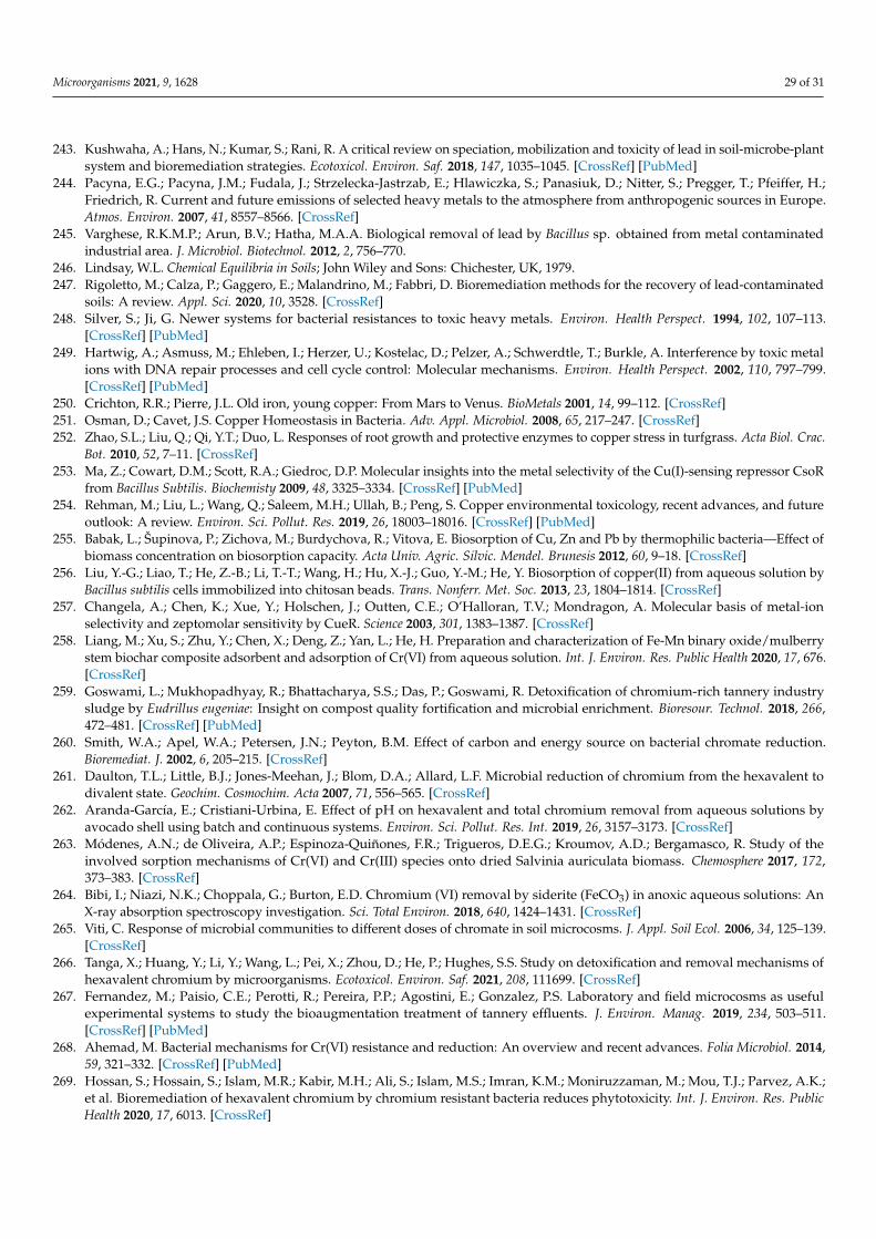
Microorganisms 2021, 9, 1628 29 of 31
243. Kushwaha, A.; Hans, N.; Kumar, S.; Rani, R. A critical review on speciation, mobilization and toxicity of lead in soil-microbe-plantsystem and bioremediation strategies. Ecotoxicol. Environ. Saf. 2018, 147, 1035–1045. [CrossRef] [PubMed]
244. Pacyna, E.G.; Pacyna, J.M.; Fudala, J.; Strzelecka-Jastrzab, E.; Hlawiczka, S.; Panasiuk, D.; Nitter, S.; Pregger, T.; Pfeiffer, H.;Friedrich, R. Current and future emissions of selected heavy metals to the atmosphere from anthropogenic sources in Europe.Atmos. Environ. 2007, 41, 8557–8566. [CrossRef]
245. Varghese, R.K.M.P.; Arun, B.V.; Hatha, M.A.A. Biological removal of lead by Bacillus sp. obtained from metal contaminatedindustrial area. J. Microbiol. Biotechnol. 2012, 2, 756–770.
246. Lindsay, W.L. Chemical Equilibria in Soils; John Wiley and Sons: Chichester, UK, 1979.247. Rigoletto, M.; Calza, P.; Gaggero, E.; Malandrino, M.; Fabbri, D. Bioremediation methods for the recovery of lead-contaminated
soils: A review. Appl. Sci. 2020, 10, 3528. [CrossRef]248. Silver, S.; Ji, G. Newer systems for bacterial resistances to toxic heavy metals. Environ. Health Perspect. 1994, 102, 107–113.
[CrossRef] [PubMed]249. Hartwig, A.; Asmuss, M.; Ehleben, I.; Herzer, U.; Kostelac, D.; Pelzer, A.; Schwerdtle, T.; Burkle, A. Interference by toxic metal
ions with DNA repair processes and cell cycle control: Molecular mechanisms. Environ. Health Perspect. 2002, 110, 797–799.[CrossRef] [PubMed]
250. Crichton, R.R.; Pierre, J.L. Old iron, young copper: From Mars to Venus. BioMetals 2001, 14, 99–112. [CrossRef]251. Osman, D.; Cavet, J.S. Copper Homeostasis in Bacteria. Adv. Appl. Microbiol. 2008, 65, 217–247. [CrossRef]252. Zhao, S.L.; Liu, Q.; Qi, Y.T.; Duo, L. Responses of root growth and protective enzymes to copper stress in turfgrass. Acta Biol. Crac.
Bot. 2010, 52, 7–11. [CrossRef]253. Ma, Z.; Cowart, D.M.; Scott, R.A.; Giedroc, D.P. Molecular insights into the metal selectivity of the Cu(I)-sensing repressor CsoR
from Bacillus Subtilis. Biochemisty 2009, 48, 3325–3334. [CrossRef] [PubMed]254. Rehman, M.; Liu, L.; Wang, Q.; Saleem, M.H.; Ullah, B.; Peng, S. Copper environmental toxicology, recent advances, and future
outlook: A review. Environ. Sci. Pollut. Res. 2019, 26, 18003–18016. [CrossRef] [PubMed]255. Babak, L.; Šupinova, P.; Zichova, M.; Burdychova, R.; Vitova, E. Biosorption of Cu, Zn and Pb by thermophilic bacteria—Effect of
biomass concentration on biosorption capacity. Acta Univ. Agric. Silvic. Mendel. Brunesis 2012, 60, 9–18. [CrossRef]256. Liu, Y.-G.; Liao, T.; He, Z.-B.; Li, T.-T.; Wang, H.; Hu, X.-J.; Guo, Y.-M.; He, Y. Biosorption of copper(II) from aqueous solution by
Bacillus subtilis cells immobilized into chitosan beads. Trans. Nonferr. Met. Soc. 2013, 23, 1804–1814. [CrossRef]257. Changela, A.; Chen, K.; Xue, Y.; Holschen, J.; Outten, C.E.; O’Halloran, T.V.; Mondragon, A. Molecular basis of metal-ion
selectivity and zeptomolar sensitivity by CueR. Science 2003, 301, 1383–1387. [CrossRef]258. Liang, M.; Xu, S.; Zhu, Y.; Chen, X.; Deng, Z.; Yan, L.; He, H. Preparation and characterization of Fe-Mn binary oxide/mulberry
stem biochar composite adsorbent and adsorption of Cr(VI) from aqueous solution. Int. J. Environ. Res. Public Health 2020, 17, 676.[CrossRef]
259. Goswami, L.; Mukhopadhyay, R.; Bhattacharya, S.S.; Das, P.; Goswami, R. Detoxification of chromium-rich tannery industrysludge by Eudrillus eugeniae: Insight on compost quality fortification and microbial enrichment. Bioresour. Technol. 2018, 266,472–481. [CrossRef] [PubMed]
260. Smith, W.A.; Apel, W.A.; Petersen, J.N.; Peyton, B.M. Effect of carbon and energy source on bacterial chromate reduction.Bioremediat. J. 2002, 6, 205–215. [CrossRef]
261. Daulton, T.L.; Little, B.J.; Jones-Meehan, J.; Blom, D.A.; Allard, L.F. Microbial reduction of chromium from the hexavalent todivalent state. Geochim. Cosmochim. Acta 2007, 71, 556–565. [CrossRef]
262. Aranda-García, E.; Cristiani-Urbina, E. Effect of pH on hexavalent and total chromium removal from aqueous solutions byavocado shell using batch and continuous systems. Environ. Sci. Pollut. Res. Int. 2019, 26, 3157–3173. [CrossRef]
263. Módenes, A.N.; de Oliveira, A.P.; Espinoza-Quiñones, F.R.; Trigueros, D.E.G.; Kroumov, A.D.; Bergamasco, R. Study of theinvolved sorption mechanisms of Cr(VI) and Cr(III) species onto dried Salvinia auriculata biomass. Chemosphere 2017, 172,373–383. [CrossRef]
264. Bibi, I.; Niazi, N.K.; Choppala, G.; Burton, E.D. Chromium (VI) removal by siderite (FeCO3) in anoxic aqueous solutions: AnX-ray absorption spectroscopy investigation. Sci. Total Environ. 2018, 640, 1424–1431. [CrossRef]
265. Viti, C. Response of microbial communities to different doses of chromate in soil microcosms. J. Appl. Soil Ecol. 2006, 34, 125–139.[CrossRef]
266. Tanga, X.; Huang, Y.; Li, Y.; Wang, L.; Pei, X.; Zhou, D.; He, P.; Hughes, S.S. Study on detoxification and removal mechanisms ofhexavalent chromium by microorganisms. Ecotoxicol. Environ. Saf. 2021, 208, 111699. [CrossRef]
267. Fernandez, M.; Paisio, C.E.; Perotti, R.; Pereira, P.P.; Agostini, E.; Gonzalez, P.S. Laboratory and field microcosms as usefulexperimental systems to study the bioaugmentation treatment of tannery effluents. J. Environ. Manag. 2019, 234, 503–511.[CrossRef] [PubMed]
268. Ahemad, M. Bacterial mechanisms for Cr(VI) resistance and reduction: An overview and recent advances. Folia Microbiol. 2014,59, 321–332. [CrossRef] [PubMed]
269. Hossan, S.; Hossain, S.; Islam, M.R.; Kabir, M.H.; Ali, S.; Islam, M.S.; Imran, K.M.; Moniruzzaman, M.; Mou, T.J.; Parvez, A.K.;et al. Bioremediation of hexavalent chromium by chromium resistant bacteria reduces phytotoxicity. Int. J. Environ. Res. PublicHealth 2020, 17, 6013. [CrossRef]

Microorganisms 2021, 9, 1628 30 of 31
270. Congeevaram, S.; Dhanarani, S.; Park, J.; Dexilin, M.; Thamaraiselvi, K. Biosorption of chromium and nickel by heavy metalresistant fungal and bacterial isolates. J. Hazard. Mater. 2007, 146, 270–277. [CrossRef] [PubMed]
271. Kumar, R.; Bhatia, D.; Singh, R.; Rani, S.; Bishnoi, N.R. Sorption of heavy metals from electroplating effluent using immobilizedbiomass Trichoderma viride in a continuous packed-bed column. Int. Biodeter. Biodegrad. 2011, 65, 1133–1139. [CrossRef]
272. Vijayaraghavan, K.; Yun, Y.S. Bacterial biosorbents and biosorption. Biotechnol. Adv. 2008, 26, 266–291. [CrossRef] [PubMed]273. Habashi, F. Mercury, Physical and Chemical Properties. In Encyclopedia of Metalloproteins; Kretsinger, R.H., Uversky, V.N.,
Permyakov, E.A., Eds.; Springer: New York, NY, USA, 2013. [CrossRef]274. Driscoll, C.T.; Mason, R.P.; Chan, H.M.; Jacob, D.J.; Pirrone, N. Mercury as a global pollutant: Sources, pathways, and effects.
Environ. Sci. Technol. 2013, 47, 4967–4983. [CrossRef] [PubMed]275. Velásquez-Riaño, M.; Benavides-Otaya, H.D. Bioremediation techniques applied to aqueous media contaminated with mercury.
Crit. Rev. Biotechnol. 2016, 36, 1124–1130. [CrossRef]276. Beckers, F.; Rinklebe, J. Cycling of mercury in the environment: Sources, fate, and human health implications: A review. Crit. Rev.
Environ. Sci. Technol. 2017, 47, 693–794. [CrossRef]277. Wagner-Dobler, I. Pilot plant for bioremediation of mercury containing industrial wastewater. Appl. Microbiol. Biotechnol. 2003, 62,
124–133. [CrossRef] [PubMed]278. Kumari, S.; Amit Jamwal, R.; Mishra, N.; Singh, D.K. Recent developments in environmental mercury bioremediation and its
toxicity: A review. Environ. Nanotechnol. Monit. Manag. 2020, 13, 100283. [CrossRef]279. Imam, S.S.A.; Rajpoot, I.K.; Gajjar, B.; Sachdeva, A. Comparative study of heavy metal bioremediation in soil by Bacillus subtilis
and Saccharomyces cerevisiae. Ind. J. Sci. Technol. 2016, 9. [CrossRef]280. Yan-de, J.; Zhen-li, H.; Xiao-e, Y. Role of soil rhizobacteria in phytoremediation of heavy metal contaminated soils. J. Zhejiang
Univ. Sci. 2007, 8, 192–207. [CrossRef]281. Stapleton, P.; Pike, R.; Mullany, P.; Lucas, V.; Roberts, G.; Rowbury, R.; Wilson, M.; Richards, H. Mercuric resistance genes in
gram-positive oral bacteria. FEMS Microbiol. Lett. 2004, 236, 213–220. [CrossRef]282. Das, A.P.; Sukla, L.B.; Pradhan, N.; Nayak, S. Manganese biomining: A review. Bioresour. Technol. 2011, 102, 7381–7387. [CrossRef]283. Gadd, G.M. Metals, minerals and microbes: Geomicrobiology and bioremediation. Microbiology 2010, 156, 609–664. [CrossRef]284. Ehrlich, H.L.; Newman, D.K. Geomicrobiology, 5th ed.; CRC/Taylor and Francis: Boca Raton, FL, USA, 2009. [CrossRef]285. Helmann, J.D. Specificity of metal sensing: Iron and manganese homeostasis in Bacillus subtilis. Biol. Chem. 2014, 289, 28112–28120.
[CrossRef] [PubMed]286. Paruthiyil, S.; Pinochet-Barros, A.; Huang, X.; Helmann, J.D. Bacillus subtilis TerC family proteins help prevent manganese
intoxication. J. Bacteriol. 2020, 202, e00624-19. [CrossRef] [PubMed]287. Barceloux, D.G. Molybdenum. Clin. Toxicol. 1999, 37, 231–237. [CrossRef]288. Morrison, S.J.; Mushovic, P.S.; Niesen, P.L. Early breakthrough of molybdenum and uranium in a permeable reactive barrier.
Environ. Sci. Technol. 2006, 40, 2018–2024. [CrossRef] [PubMed]289. Zhang, M.; Reardon, E.J. Removal of B, Cr, Mo, and Se from wastewater by incorporation into hydrocalumite and ettringite.
Environ. Sci. Technol. 2003, 37, 2947–2952. [CrossRef]290. Grunden, A.M.; Shanmugam, K.T. Molybdate transport and regulation in bacteria. Arch. Microbiol. 1997, 168, 345–354. [CrossRef]291. Périnet, S.; Jeukens, J.; Kukavica-Ibrulj, I.; Ouellet, M.M.; Charette, S.J.; Levesque, R.C. Molybdate transporter ModABC is
important for Pseudomonas aeruginosa chronic lung infection. BMC Res. Notes 2016, 9, 23. [CrossRef]292. Xia, Z.; Lei, L.; Zhang, H.Y.; Wei, H.L. Characterization of the ModABC molybdate transport system of Pseudomonas putida in
nicotine degradation. Front. Microbiol. 2018, 9, 3030. [CrossRef]293. Davidson, A.L.; Dassa, E.; Orelle, C.; Chen, J. Structure, function, and evolution of bacterial ATP-binding cassette systems.
Microbiol. Mol. Biol. Rev. 2008, 72, 317–364. [CrossRef]294. Self, W.T.; Grunden, A.M.; Hasona, A.; Shanmugam, K.T. Molybdate transport. Res. Microbiol. 2001, 152, 311–321. [CrossRef]295. Zhong, Q.; Kobe, B.; Kappler, U. Molybdenum enzymes and how they support virulence in pathogenic bacteria. Front. Microbiol.
2020, 11, 3185. [CrossRef]296. Ge, X.; Thorgersen, M.P.; Poole, F.L.; Deutschbauer, A.M.; Chandonia, J.M.; Novichkov, P.S.; Gushgari-Doyle, S.; Lui, L.M.;
Nielsen, T.; Chakraborty, R.; et al. Characterization of a metal-resistant Bacillus strain with a high molybdate affinity ModA fromcontaminated sediments at the oak ridge reservation. Front. Microbiol. 2020, 11, 2543. [CrossRef]
297. Boyle, R.W. The geochemistry of gold and its deposits. Geol. Surv. Can. Bull. 1979, 280, 583.298. Karthikeyan, S.; Beveridge, T.J. Pseudomonas aeruginosa biofilms react with and precipitate toxic soluble gold. Environ. Microbiol.
2002, 4, 667–675. [CrossRef] [PubMed]299. Bütof, L.; Wiesemann, N.; Herzberg, M.; Altzschner, M.; Holleitner, A.; Reith, F.; Nies, D.H. Synergistic gold-copper detoxification
at the core of gold biomineralisation in Cupriavidus metallidurans. Metallomics 2018, 10, 278–286. [CrossRef] [PubMed]300. Watterson, J.R. Preliminary evidence for the involvement of budding bacteria in the origin of Alaskan placer gold. Geology 1992,
20, 315–318. [CrossRef]301. Hough, R.M.; Butt, C.R.M.; Reddy, S.M.; Verrall, M. Gold nuggets: Supergene or hypogene? Aust. J. Earth Sci. 2007, 54, 959–964.
[CrossRef]302. Nies, D.H. Microbial heavy metal resistance. Appl. Microbiol. Biotechnol. 1999, 51, 730–750. [CrossRef]

Microorganisms 2021, 9, 1628 31 of 31
303. Korobushkina, E.D.; Mineyev, G.G.; Praded, G.P. Mechanism of the microbiological process of dissolution of gold. Mikrobiologiia1976, 45, 535–538.
304. Korobushkina, E.D.; Karavaiko, G.I.; Korobushkin, I.M. Biochemistry of gold. In Environmental Biogeochemistry Ecological Bulletin;Hallberg, R., Ed.; Oikos Editorial Office: Stockholm, Sweden, 1983; pp. 325–333.
305. Lengke, M.F.; Southam, G. The deposition of elemental gold from gold(I)-thiosulfate complex mediated by sulfate-reducingbacterial conditions. Econ. Geol. 2007, 102, 109–126. [CrossRef]
306. Gee, A.R.; Dudeney, A.W.L. Adsorption and crystallization of gold on biological surfaces. In Biohydrometallurgy; Kelly, D.P.,Norris, P.R., Eds.; Science & Technology Letters: London, UK, 1988; pp. 437–451.
307. Ulberg, Z.R.; Karamushka, V.I.; Vidybida, A.K. Interaction of energized bacteria calls with particles of colloidal gold: Peculiaritiesand kinetic model of the process. Biochim. Biophys. Acta 1992, 1134, 89–95. [CrossRef]
308. Agency for Toxic Substances and Disease Registry (ATSDR). Toxicological Profile for Silver; US Department of Health and HumanServices, Public Health Service, Agency for Toxic Substances and Disease Registry (TP-90–24): Atlanta, GA, USA, 1990.
309. Eisler, R. Silver Hazards to Fish, Wildlife and Invertebrates: A Synoptic Review; Department of the Interior, National Biological Service:Washington, DC, USA, 1997; pp. 1–44.
310. Becker, R.O. Silver ions in the treatment of local infections. Met.-Based Drugs 1999, 6, 297–300. [CrossRef] [PubMed]311. Wilkinson, L.J.; White, R.J.; Chipman, J.K. Silver and nanoparticles of silver in wound dressings: A review of efficacy and safety. J.
Wound Care 2011, 20, 543–549. [CrossRef] [PubMed]