Molecular activity underlying working memory
11
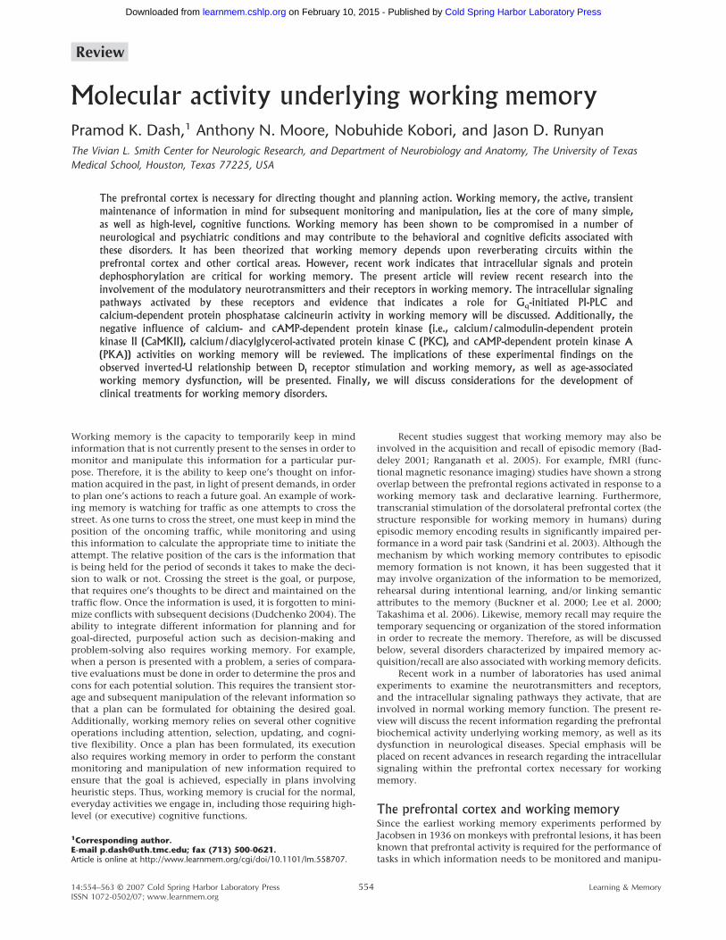
Molecular activity underlying working memory Pramod K. Dash, 1 Anthony N. Moore, Nobuhide Kobori, and Jason D. Runyan The Vivian L. Smith Center for Neurologic Research, and Department of Neurobiology and Anatomy, The University of Texas Medical School, Houston, Texas 77225, USA The prefrontal cortex is necessary for directing thought and planning action. Working memory, the active, transient maintenance of information in mind for subsequent monitoring and manipulation, lies at the core of many simple, as well as high-level, cognitive functions. Working memory has been shown to be compromised in a number of neurological and psychiatric conditions and may contribute to the behavioral and cognitive deficits associated with these disorders. It has been theorized that working memory depends upon reverberating circuits within the prefrontal cortex and other cortical areas. However, recent work indicates that intracellular signals and protein dephosphorylation are critical for working memory. The present article will review recent research into the involvement of the modulatory neurotransmitters and their receptors in working memory. The intracellular signaling pathways activated by these receptors and evidence that indicates a role for G q -initiated PI-PLC and calcium-dependent protein phosphatase calcineurin activity in working memory will be discussed. Additionally, the negative influence of calcium- and cAMP-dependent protein kinase (i.e., calcium/calmodulin-dependent protein kinase II (CaMKII), calcium/diacylglycerol-activated protein kinase C (PKC), and cAMP-dependent protein kinase A (PKA)) activities on working memory will be reviewed. The implications of these experimental findings on the observed inverted-U relationship between D 1 receptor stimulation and working memory, as well as age-associated working memory dysfunction, will be presented. Finally, we will discuss considerations for the development of clinical treatments for working memory disorders. Working memory is the capacity to temporarily keep in mind information that is not currently present to the senses in order to monitor and manipulate this information for a particular pur- pose. Therefore, it is the ability to keep one’s thought on infor- mation acquired in the past, in light of present demands, in order to plan one’s actions to reach a future goal. An example of work- ing memory is watching for traffic as one attempts to cross the street. As one turns to cross the street, one must keep in mind the position of the oncoming traffic, while monitoring and using this information to calculate the appropriate time to initiate the attempt. The relative position of the cars is the information that is being held for the period of seconds it takes to make the deci- sion to walk or not. Crossing the street is the goal, or purpose, that requires one’s thoughts to be direct and maintained on the traffic flow. Once the information is used, it is forgotten to mini- mize conflicts with subsequent decisions (Dudchenko 2004). The ability to integrate different information for planning and for goal-directed, purposeful action such as decision-making and problem-solving also requires working memory. For example, when a person is presented with a problem, a series of compara- tive evaluations must be done in order to determine the pros and cons for each potential solution. This requires the transient stor- age and subsequent manipulation of the relevant information so that a plan can be formulated for obtaining the desired goal. Additionally, working memory relies on several other cognitive operations including attention, selection, updating, and cogni- tive flexibility. Once a plan has been formulated, its execution also requires working memory in order to perform the constant monitoring and manipulation of new information required to ensure that the goal is achieved, especially in plans involving heuristic steps. Thus, working memory is crucial for the normal, everyday activities we engage in, including those requiring high- level (or executive) cognitive functions. Recent studies suggest that working memory may also be involved in the acquisition and recall of episodic memory (Bad- deley 2001; Ranganath et al. 2005). For example, fMRI (func- tional magnetic resonance imaging) studies have shown a strong overlap between the prefrontal regions activated in response to a working memory task and declarative learning. Furthermore, transcranial stimulation of the dorsolateral prefrontal cortex (the structure responsible for working memory in humans) during episodic memory encoding results in significantly impaired per- formance in a word pair task (Sandrini et al. 2003). Although the mechanism by which working memory contributes to episodic memory formation is not known, it has been suggested that it may involve organization of the information to be memorized, rehearsal during intentional learning, and/or linking semantic attributes to the memory (Buckner et al. 2000; Lee et al. 2000; Takashima et al. 2006). Likewise, memory recall may require the temporary sequencing or organization of the stored information in order to recreate the memory. Therefore, as will be discussed below, several disorders characterized by impaired memory ac- quisition/recall are also associated with working memory deficits. Recent work in a number of laboratories has used animal experiments to examine the neurotransmitters and receptors, and the intracellular signaling pathways they activate, that are involved in normal working memory function. The present re- view will discuss the recent information regarding the prefrontal biochemical activity underlying working memory, as well as its dysfunction in neurological diseases. Special emphasis will be placed on recent advances in research regarding the intracellular signaling within the prefrontal cortex necessary for working memory. The prefrontal cortex and working memory Since the earliest working memory experiments performed by Jacobsen in 1936 on monkeys with prefrontal lesions, it has been known that prefrontal activity is required for the performance of tasks in which information needs to be monitored and manipu- 1 Corresponding author. E-mail [email protected]; fax (713) 500-0621. Article is online at http://www.learnmem.org/cgi/doi/10.1101/lm.558707. Review 14:554–563 © 2007 Cold Spring Harbor Laboratory Press Learning & Memory 554 ISSN 1072-0502/07; www.learnmem.org Cold Spring Harbor Laboratory Press on February 10, 2015 - Published by learnmem.cshlp.org Downloaded from
-
Upload
independent -
Category
Documents
-
view
0 -
download
0
Transcript of Molecular activity underlying working memory
Molecular activity underlying working memoryPramod K. Dash,1 Anthony N. Moore, Nobuhide Kobori, and Jason D. RunyanThe Vivian L. Smith Center for Neurologic Research, and Department of Neurobiology and Anatomy, The University of TexasMedical School, Houston, Texas 77225, USA
The prefrontal cortex is necessary for directing thought and planning action. Working memory, the active, transientmaintenance of information in mind for subsequent monitoring and manipulation, lies at the core of many simple,as well as high-level, cognitive functions. Working memory has been shown to be compromised in a number ofneurological and psychiatric conditions and may contribute to the behavioral and cognitive deficits associated withthese disorders. It has been theorized that working memory depends upon reverberating circuits within theprefrontal cortex and other cortical areas. However, recent work indicates that intracellular signals and proteindephosphorylation are critical for working memory. The present article will review recent research into theinvolvement of the modulatory neurotransmitters and their receptors in working memory. The intracellular signalingpathways activated by these receptors and evidence that indicates a role for Gq-initiated PI-PLC andcalcium-dependent protein phosphatase calcineurin activity in working memory will be discussed. Additionally, thenegative influence of calcium- and cAMP-dependent protein kinase (i.e., calcium/calmodulin-dependent proteinkinase II (CaMKII), calcium/diacylglycerol-activated protein kinase C (PKC), and cAMP-dependent protein kinase A(PKA)) activities on working memory will be reviewed. The implications of these experimental findings on theobserved inverted-U relationship between D1 receptor stimulation and working memory, as well as age-associatedworking memory dysfunction, will be presented. Finally, we will discuss considerations for the development ofclinical treatments for working memory disorders.
Working memory is the capacity to temporarily keep in mindinformation that is not currently present to the senses in order tomonitor and manipulate this information for a particular pur-pose. Therefore, it is the ability to keep one’s thought on infor-mation acquired in the past, in light of present demands, in orderto plan one’s actions to reach a future goal. An example of work-ing memory is watching for traffic as one attempts to cross thestreet. As one turns to cross the street, one must keep in mind theposition of the oncoming traffic, while monitoring and usingthis information to calculate the appropriate time to initiate theattempt. The relative position of the cars is the information thatis being held for the period of seconds it takes to make the deci-sion to walk or not. Crossing the street is the goal, or purpose,that requires one’s thoughts to be direct and maintained on thetraffic flow. Once the information is used, it is forgotten to mini-mize conflicts with subsequent decisions (Dudchenko 2004). Theability to integrate different information for planning and forgoal-directed, purposeful action such as decision-making andproblem-solving also requires working memory. For example,when a person is presented with a problem, a series of compara-tive evaluations must be done in order to determine the pros andcons for each potential solution. This requires the transient stor-age and subsequent manipulation of the relevant information sothat a plan can be formulated for obtaining the desired goal.Additionally, working memory relies on several other cognitiveoperations including attention, selection, updating, and cogni-tive flexibility. Once a plan has been formulated, its executionalso requires working memory in order to perform the constantmonitoring and manipulation of new information required toensure that the goal is achieved, especially in plans involvingheuristic steps. Thus, working memory is crucial for the normal,everyday activities we engage in, including those requiring high-level (or executive) cognitive functions.
Recent studies suggest that working memory may also beinvolved in the acquisition and recall of episodic memory (Bad-deley 2001; Ranganath et al. 2005). For example, fMRI (func-tional magnetic resonance imaging) studies have shown a strongoverlap between the prefrontal regions activated in response to aworking memory task and declarative learning. Furthermore,transcranial stimulation of the dorsolateral prefrontal cortex (thestructure responsible for working memory in humans) duringepisodic memory encoding results in significantly impaired per-formance in a word pair task (Sandrini et al. 2003). Although themechanism by which working memory contributes to episodicmemory formation is not known, it has been suggested that itmay involve organization of the information to be memorized,rehearsal during intentional learning, and/or linking semanticattributes to the memory (Buckner et al. 2000; Lee et al. 2000;Takashima et al. 2006). Likewise, memory recall may require thetemporary sequencing or organization of the stored informationin order to recreate the memory. Therefore, as will be discussedbelow, several disorders characterized by impaired memory ac-quisition/recall are also associated with working memory deficits.
Recent work in a number of laboratories has used animalexperiments to examine the neurotransmitters and receptors,and the intracellular signaling pathways they activate, that areinvolved in normal working memory function. The present re-view will discuss the recent information regarding the prefrontalbiochemical activity underlying working memory, as well as itsdysfunction in neurological diseases. Special emphasis will beplaced on recent advances in research regarding the intracellularsignaling within the prefrontal cortex necessary for workingmemory.
The prefrontal cortex and working memorySince the earliest working memory experiments performed byJacobsen in 1936 on monkeys with prefrontal lesions, it has beenknown that prefrontal activity is required for the performance oftasks in which information needs to be monitored and manipu-
1Corresponding author.E-mail [email protected]; fax (713) 500-0621.Article is online at http://www.learnmem.org/cgi/doi/10.1101/lm.558707.
Review
14:554–563 © 2007 Cold Spring Harbor Laboratory Press Learning & Memory554ISSN 1072-0502/07; www.learnmem.org
Cold Spring Harbor Laboratory Press on February 10, 2015 - Published by learnmem.cshlp.orgDownloaded from
lated (Jacobsen 1936), but not tasks that require the simple main-tenance of information over time. More recent brain-imagingstudies in humans using positron-emission tomography (PET)and fMRI have shown increased blood flow within the humanprefrontal cortex during the delay-period of working memorytasks (Jonides et al. 1993; Petrides et al. 1993; D’Esposito et al.1995). The prefrontal regions most often associated with workingmemory are the mid-dorsolateral, the orbitofrontal, and the me-sial frontal regions. Interestingly, which of these different pre-frontal regions is involved in the directing and maintaining ofthought appears to be based on the type of information and thegoal.
Research using monkey and rat models has yielded much ofour present knowledge regarding prefrontal activity, neurotrans-mission, and the intracellular signaling involved in workingmemory. Behavioral paradigms such as the delayed match-to-sample and delayed nonmatch-to-sample tasks have been shownto test the capacity for working memory in animals and in hu-mans. In the delayed match-to-sample task, subjects are shown asample (e.g., an object) to remember. Following a brief presenta-tion of the object, it is removed from the field of view. After adelay of a few seconds, two objects (the previously seen objectand a novel object) are presented. The subject is required to cor-rectly identify the previously seen sample in order to receive areward. This and other working memory tasks require the subject(1) to attend to particular items in order to acquire specific in-formation, (2) to hold this information for a period of secondsduring the delay period, and (3) to use this information for thepurpose of making a correct response. Neurophysiologic studiesin monkeys in the 1970s and 80s revealed that working memorywas linked to maintained or persistent neuronal activity in theprefrontal cortex during the delay period (“delay-period activ-ity”) of these working memory tasks. Specific neurons were ob-served to be active during the delay period, and that this activityterminated when the participant completed the task. These cellswere first identified in the dorsolateral PFC (dlPFC) of monkeysperforming delay match-to-sample tasks (Fuster and Alexander1971; Kojima and Goldman-Rakic 1984), but have since beenobserved in other brain areas. For example, neuronal activityduring the delay period has been recorded in the inferior tempo-ral cortex (Miller et al. 1993). However, while delay-period activ-ity in the dlPFC is maintained in the presence of distracters,delay-period cells in the inferior temporal cortex cease to firewhen the subject is presented with a distracting stimulus. In ad-dition to the inferior temporal cortex, neuronal activity duringthe delay period has also been observed in other structures in-cluding the auditory cortex, the medial geniculate body, the hip-pocampus, the posterior parietal cortex, and the somatosensorycortex (Watanabe and Niki 1985; Koch and Fuster 1989; Sakurai1990; Zhou and Fuster 1997). While the role of delay-period cellsin working memory has not been fully elucidated, it has beenobserved that ∼70% of delay-period activity recorded in an ocu-lomotor delayed response task represented transient informationstorage (Funahashi et al. 1993).
Catecholamines in working memoryAs is the case for most of the brain, the major excitatory neuro-transmitter for the prefrontal cortex is glutamate and the majorinhibitory neurotransmitter is GABA. While these neurotrans-mitters are necessary for prefrontal neuronal activity and are in-volved in the focal specificity of this activity, modulatory neu-rotransmission in the PFC has been shown to play a prominentrole in working memory. Prefrontal dopamine neurotransmis-sion, in particular, has been shown to be important for workingmemory in animals and in humans. The seminal work by Bro-
zoski et al. first demonstrated the involvement of catecholamines(dopamine and norepinephrine) in working memory. In thisstudy, it was observed that depletion of PFC catecholamines bydirect injection of 6-hydroxy-dopamine (6OHDA) caused pro-found working memory impairments in monkeys (Brozoski et al.1979). These impairments are comparable to those seen follow-ing lesion of the PFC, indicating the prominent role for catechol-amines in working memory. Since this initial observation of therequirement of dopamine, a large body of research has examinedthe role of dopamine, and the role of specific dopamine receptorsubtypes in working memory. For instance, depletion of PFC cat-echolamines has been shown to impair working memory in rats(Simon et al. 1979), whereas iontophoretic application of dopa-mine into the PFC has been observed to increase delay-periodactivity (number of spikes/sec) in monkeys performing workingmemory tasks (Sawaguchi 2001). Furthermore, it has recentlybeen observed in rats and in humans that prefrontal dopaminelevels transiently increase during working memory, reiteratingthe importance of this modulatory neurotransmitter for workingmemory (Phillips et al. 2004; Aalto et al. 2005).
While dopamine is required for normal working memory,subsequent studies have shown that excessive dopamine cancause working memory impairments. For example, infusion ofhigh levels of dopamine D1 receptor agonists into the prefrontalcortex has been demonstrated to impair working memory (Zahrtet al. 1997; Runyan et al. 2005). Dose-response studies have re-vealed that working memory performance requires an optimalrange of dopamine (or D1 receptor stimulation). These studiesshowed that prefrontal D1 receptor stimulation follows an in-verted-U-shaped dose-response curve with both insufficient andexcessive stimulation resulting in working memory impairments(Williams and Goldman-Rakic 1995; Arnsten and Goldman-Rakic 1998; Runyan et al. 2005). Consistent with this, it has beendemonstrated that the working memory deficits observed in con-ditions associated with elevated dopamine levels (e.g., stress) arereduced by administration of D1 receptor antagonists. Similarly,conditions associated with reduced dopamine levels (e.g., Parkin-son’s Disease) show benefit from the use of D1 agonists (or L-DOPA, a brain-permeable precursor for dopamine) (Cools 2006).In addition to dopamine, this inverted-U-shaped response curvehas been identified for norepinephrine, a catecholamine derivedfrom dopamine.
Receptors for modulatory neurotransmittersDopamine receptors are generally divided into two families: D1-like (D1, D5) and D2-like (D2, D3, and D4) based upon their abilityto either increase (D1-like) or decrease (D2-like) cAMP levels. Theinfluence of dopamine on prefrontal neurons is mediated largelyby D1 receptors, which are more abundant than D2 receptors inthese cells (Lidow et al. 1991). For example, it has been shownthat D1 antagonists (which also inhibit D5 receptors), but not D2
antagonists, suppress PFC delay-period activity and disrupt work-ing memory in monkeys and in rats (Lidow et al. 1991; Sawagu-chi and Goldman-Rakic 1991; Didriksen 1995; Aultman and Mo-ghaddam 2001). Similarly, prefrontal infusion of low levels of D1
agonists, but not D2 agonists, improves working memory (Arn-sten et al. 1994; Runyan et al. 2005). Interestingly, D4 receptors,which are also abundantly expressed in the PFC (Meador-Woodruff et al. 1996), have been observed to be involved inworking memory (Zhang et al. 2004).
D1 receptors are present in close conjunction with glutama-tergic synapses on the distal dendritic spines of prefrontal neu-rons (Smiley and Goldman-Rakic 1993; Williams and Goldman-Rakic 1993). By comparison, D5 receptors, which have similarintracellular activities as D1, are largely extrasynaptic (Paspalas
Working memory
555www.learnmem.org Learning & Memory
Cold Spring Harbor Laboratory Press on February 10, 2015 - Published by learnmem.cshlp.orgDownloaded from
and Goldman-Rakic 2004). As most agonists/antagonists act onboth D1 and D5 receptors, it is difficult to determine the contri-bution of each of these receptor subtypes to working memory.D1 receptors are expressed on both pyramidal neurons and onGABAergic interneurons (Muly et al. 1998) within the prefrontalcortex. Due to this distribution, dopamine has been shown to notonly stimulate the activity of pyramidal neurons, but also toeither enhance or depress (in a concentration-dependent man-ner) the excitability of fast-spiking (FS) inhibitory interneurons(Gao and Goldman-Rakic 2003). Since inhibitory neurons withinthe prefrontal cortex are thought to restrict the spatial extent ofneuronal activity, the presence of D1 receptors on inhibitory neu-rons suggests that they play an important role in promoting fo-cal, persistent excitability (Trantham-Davidson et al. 2004).
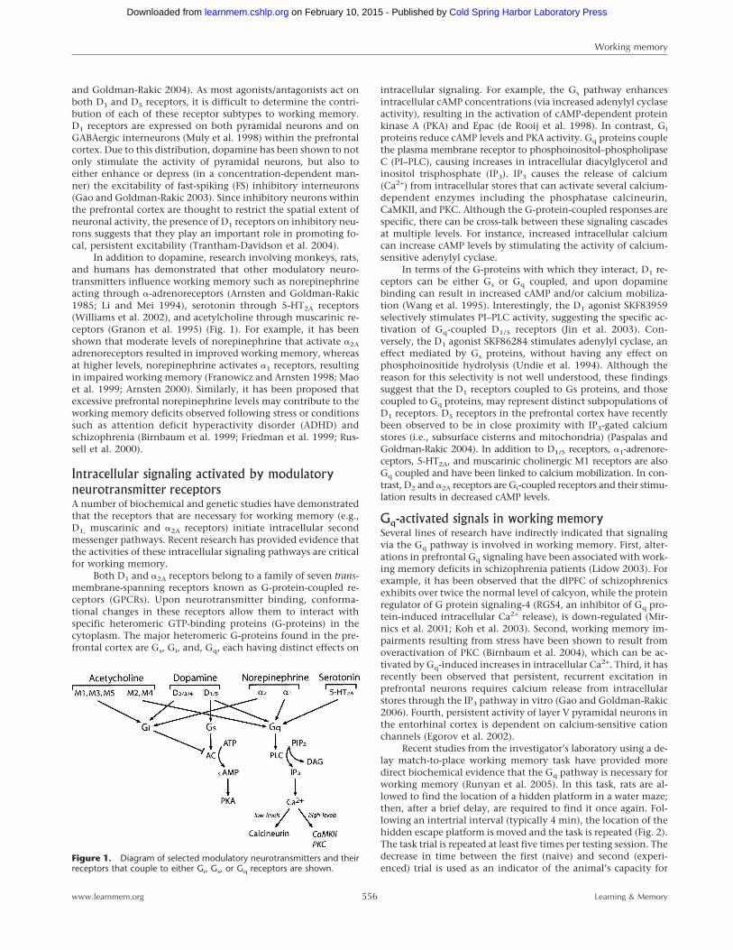
In addition to dopamine, research involving monkeys, rats,and humans has demonstrated that other modulatory neuro-transmitters influence working memory such as norepinephrineacting through �-adrenoreceptors (Arnsten and Goldman-Rakic1985; Li and Mei 1994), serotonin through 5-HT2A receptors(Williams et al. 2002), and acetylcholine through muscarinic re-ceptors (Granon et al. 1995) (Fig. 1). For example, it has beenshown that moderate levels of norepinephrine that activate �2A
adrenoreceptors resulted in improved working memory, whereasat higher levels, norepinephrine activates �1 receptors, resultingin impaired working memory (Franowicz and Arnsten 1998; Maoet al. 1999; Arnsten 2000). Similarly, it has been proposed thatexcessive prefrontal norepinephrine levels may contribute to theworking memory deficits observed following stress or conditionssuch as attention deficit hyperactivity disorder (ADHD) andschizophrenia (Birnbaum et al. 1999; Friedman et al. 1999; Rus-sell et al. 2000).
Intracellular signaling activated by modulatoryneurotransmitter receptorsA number of biochemical and genetic studies have demonstratedthat the receptors that are necessary for working memory (e.g.,D1, muscarinic and �2A receptors) initiate intracellular secondmessenger pathways. Recent research has provided evidence thatthe activities of these intracellular signaling pathways are criticalfor working memory.
Both D1 and �2A receptors belong to a family of seven trans-membrane-spanning receptors known as G-protein-coupled re-ceptors (GPCRs). Upon neurotransmitter binding, conforma-tional changes in these receptors allow them to interact withspecific heteromeric GTP-binding proteins (G-proteins) in thecytoplasm. The major heteromeric G-proteins found in the pre-frontal cortex are Gs, Gi, and, Gq, each having distinct effects on
intracellular signaling. For example, the Gs pathway enhancesintracellular cAMP concentrations (via increased adenylyl cyclaseactivity), resulting in the activation of cAMP-dependent proteinkinase A (PKA) and Epac (de Rooij et al. 1998). In contrast, Gi
proteins reduce cAMP levels and PKA activity. Gq proteins couplethe plasma membrane receptor to phosphoinositol–phospholipaseC (PI–PLC), causing increases in intracellular diacylglycerol andinositol trisphosphate (IP3). IP3 causes the release of calcium(Ca2+) from intracellular stores that can activate several calcium-dependent enzymes including the phosphatase calcineurin,CaMKII, and PKC. Although the G-protein-coupled responses arespecific, there can be cross-talk between these signaling cascadesat multiple levels. For instance, increased intracellular calciumcan increase cAMP levels by stimulating the activity of calcium-sensitive adenylyl cyclase.
In terms of the G-proteins with which they interact, D1 re-ceptors can be either Gs or Gq coupled, and upon dopaminebinding can result in increased cAMP and/or calcium mobiliza-tion (Wang et al. 1995). Interestingly, the D1 agonist SKF83959selectively stimulates PI–PLC activity, suggesting the specific ac-tivation of Gq-coupled D1/5 receptors (Jin et al. 2003). Con-versely, the D1 agonist SKF86284 stimulates adenylyl cyclase, aneffect mediated by Gs proteins, without having any effect onphosphoinositide hydrolysis (Undie et al. 1994). Although thereason for this selectivity is not well understood, these findingssuggest that the D1 receptors coupled to Gs proteins, and thosecoupled to Gq proteins, may represent distinct subpopulations ofD1 receptors. D5 receptors in the prefrontal cortex have recentlybeen observed to be in close proximity with IP3-gated calciumstores (i.e., subsurface cisterns and mitochondria) (Paspalas andGoldman-Rakic 2004). In addition to D1/5 receptors, �1-adrenore-ceptors, 5-HT2A, and muscarinic cholinergic M1 receptors are alsoGq coupled and have been linked to calcium mobilization. In con-trast, D2 and �2A receptors are Gi-coupled receptors and their stimu-lation results in decreased cAMP levels.
Gq-activated signals in working memorySeveral lines of research have indirectly indicated that signalingvia the Gq pathway is involved in working memory. First, alter-ations in prefrontal Gq signaling have been associated with work-ing memory deficits in schizophrenia patients (Lidow 2003). Forexample, it has been observed that the dlPFC of schizophrenicsexhibits over twice the normal level of calcyon, while the proteinregulator of G protein signaling-4 (RGS4, an inhibitor of Gq pro-tein-induced intracellular Ca2+ release), is down-regulated (Mir-nics et al. 2001; Koh et al. 2003). Second, working memory im-pairments resulting from stress have been shown to result fromoveractivation of PKC (Birnbaum et al. 2004), which can be ac-tivated by Gq-induced increases in intracellular Ca2+. Third, it hasrecently been observed that persistent, recurrent excitation inprefrontal neurons requires calcium release from intracellularstores through the IP3 pathway in vitro (Gao and Goldman-Rakic2006). Fourth, persistent activity of layer V pyramidal neurons inthe entorhinal cortex is dependent on calcium-sensitive cationchannels (Egorov et al. 2002).
Recent studies from the investigator’s laboratory using a de-lay match-to-place working memory task have provided moredirect biochemical evidence that the Gq pathway is necessary forworking memory (Runyan et al. 2005). In this task, rats are al-lowed to find the location of a hidden platform in a water maze;then, after a brief delay, are required to find it once again. Fol-lowing an intertrial interval (typically 4 min), the location of thehidden escape platform is moved and the task is repeated (Fig. 2).The task trial is repeated at least five times per testing session. Thedecrease in time between the first (naive) and second (experi-enced) trial is used as an indicator of the animal’s capacity for
Figure 1. Diagram of selected modulatory neurotransmitters and theirreceptors that couple to either Gi, Gs, or Gq receptors are shown.
Working memory
556www.learnmem.org Learning & Memory
Cold Spring Harbor Laboratory Press on February 10, 2015 - Published by learnmem.cshlp.orgDownloaded from
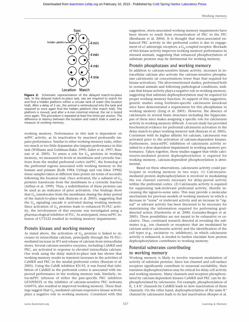
working memory. Performance in this task is dependent onmPFC activity, as its inactivation by mucimol profoundly im-pairs performance. Similar to other working memory tasks, eithertoo much or too little dopamine also impairs performance in thistask (Williams and Goldman-Rakic 1995; Zahrt et al. 1997; Run-yan et al. 2005). To assess a role for Gq proteins in workingmemory, we measured its levels in membrane and cytosolic frac-tions from the medial prefrontal cortex (mPFC, the homolog ofthe prefrontal regions associated with working memory in thehuman and primate [Kolb 1984; Uylings and van Eden 1990])tissue samples taken at different time points (in terms of seconds)following the location trial. Once activated, the � subunit of Gq
proteins translocate from the membrane to a cytosolic fraction(Arthur et al. 1999). Thus, a redistribution of these proteins canbe used as an indicator of prior activation. Our findings showthat Gq translocates from the membrane during the delay periodof the match-to-place task (Runyan et al. 2005), suggesting thatthe Gq signaling cascade is activated during working memory.Since activation of Gq proteins leads to enhanced PI–PLC activ-ity, the involvement of this enzyme was investigated using apharmacological inhibitor of PLC. As anticipated, intra-mPFC in-fusion of U73122 resulted in working memory impairments.
Protein kinases and working memoryAs stated above, the activation of Gq proteins is linked to in-creases in intracellular calcium, principally through the PI–PLC-mediated increase in IP3 and release of calcium from intracellularstores. Several calcium-sensitive enzymes, including CaMKII andPKC, are activated in response to elevated intracellular calcium.Our work using the delay match-to-place task has shown thatworking memory results in transient increases in the activities ofCaMKII and PKC in the medial prefrontal cortex (Runyan et al.2005). Using the CaMK inhibitor KN-92, it was found that inhi-bition of CaMKII in the prefrontal cortex is associated with im-proved performance in the working memory task. Similarly, in-tra-mPFC infusion of either the pan-specific PKC inhibitorGF109203X or the inhibitor of calcium-sensitive PKC isoformsGö6976, also resulted in improved working memory. These find-ings suggest that Gq-mediated calcium-responsive kinase activityplays a negative role in working memory. Consistent with this
suggestion, stress-associated working memory impairments havebeen shown to result from overactivation of PKC in the PFC(Birnbaum et al. 2004). It is thought that stress-associated en-hanced PKC activity in the prefrontal cortex is due to engage-ment of �1 adrenergic receptors, a Gq-coupled receptor. Blockadeof this kinase activity improves working memory performance instressed animals, suggesting that enhanced phosphorylation ofsubstrate proteins may be detrimental for working memory.
Protein phosphatases and working memoryIn addition to calcium-sensitive kinase activity, increases in in-tracellular calcium also activate the calcium-sensitive phospha-tase calcineurin (at concentrations lower than that required forkinase activation). The abovementioned studies, performed bothin normal animals and following pathological conditions, indi-cate that kinase activity plays a negative role in working memory,suggesting that substrate dephosphorylation may be required forproper working memory function. In support of this suggestion,genetic studies using forebrain-specific calcineurin knockoutmice have demonstrated a requirement for this phosphatase inworking memory (Zeng et al. 2001). However, the absence ofcalcineurin in several brain structures including the hippocam-pus of these mice makes assigning a specific role for calcineurinactivity in working memory difficult. A recent study has providedbiochemical evidence for prefrontal calcineurin activation in thedelay match-to-place working memory task (Runyan et al. 2005).Consistent with its higher affinity for calcium, calcineurin wasactivated prior to the activation of calcium-dependent kinases.Furthermore, intra-mPFC inhibition of calcineurin activity re-sulted in a dose-dependent impairment in working memory per-formance. Taken together, these studies suggest that while calci-neurin-mediated protein dephosphorylation is required forworking memory, calcium-dependent phosphorylation is detri-mental.
Based on these observations, calcineurin activity could par-ticipate in working memory in two ways. (1) Calcineurin-mediated protein dephosphorylation is involved in modulatingthe ion channel currents necessary for delay-period activitywithin the prefrontal cortex. (2) Calcineurin activity is requiredfor suppressing task-irrelevant prefrontal activity, thereby in-creasing the signal-to-noise ratio. This may provide a molecularmechanism for previous models of working memory in which adecrease in “noise” or irrelevant activity and an increase in “sig-nal” or relevant activity has been theorized to be necessary formaintaining the information required for performing goal-directed action (Durstewitz et al. 2000; Gonzalez-Burgos et al.2005). These possibilities are not meant to be exhaustive or ex-clusive. Thus, continued research directed at revealing the sub-strates (e.g., ion channels or receptors) that are modulated bycalcium and/or calcineurin activity and the identification of thecell types (e.g., excitatory vs. inhibitory), in which calcineurinactivity is enhanced, is needed to further elucidate how proteindephosphorylation contributes to working memory.
Potential substrates contributingto working memoryWorking memory is likely to involve transient modulation ofactivity of substrate proteins. Since ion channel and cell-surfacereceptors significantly contribute to neuronal excitability, theirtransient dephosphorylation may be critical for delay cell activityand working memory. Many channels and receptors phosphory-lated by calcium-dependent kinases CaMKII and PKC can be de-phosphorylated by calcineurin. For example, phosphorylation ofKv 1.4 K+ channels by CaMKII leads to slow inactivation of thesechannels. On the other hand, dephosphorylation of the same K+
channel by calcineurin leads to its fast inactivation (Roeper et al.
Figure 2. Schematic representation of the delayed match-to-placetask. In the delayed match-to-place task, rats are required to search forand find a hidden platform within a circular tank of water (the locationtrial). After a delay of 5 sec, the animal is reintroduced into the tank andrequired to once again find the hidden platform (the match trial). Theplatform is moved, and after a 4-min intertrial interval, the rat is testedonce again. This procedure is repeated at least five times per session. Thedifference in latency between the location and match trials is used as ameasure of working memory.
Working memory
557www.learnmem.org Learning & Memory
Cold Spring Harbor Laboratory Press on February 10, 2015 - Published by learnmem.cshlp.orgDownloaded from
1997). The activity of L-type Ca2+ channels in the hippocampusis enhanced via dephosphorylation by calcineurin (Norris et al.2002), and the voltage-dependent Kv2.1 K(+) channel, when de-phosphorylated by calcineurin, translocates from a clustered to amore uniform distribution (Misonou et al. 2004). Additionally,there is recent evidence that suggests that D1 receptors are physi-cally linked with calcineurin, and that calcineurin actively de-phosphorylates D1 receptors (Adlersberg et al. 2004). A list ofseveral channels and receptors whose activity is modulated byphosphorylation/dephosphorylation is provided in Table 1.
Although the substrates that are dephosphorylated in vivohave not been identified, in vitro neurophysiologic studies havedemonstrated that D1 receptor activity can modulate the func-tion of several ion channels and receptors. For example, D1
stimulation can increase the excitability of prefrontal pyramidalneurons through inhibition of several K+ currents (Dong andWhite 2003). In addition, D1 receptor stimulation in prefrontalneurons has also been reported to modulate a persistent sodiumcurrent (INap) that is dependent on PKC, but not PKA, activity(Gorelova and Yang 2000). Prefrontal D1 receptor suppression ofL-type Ca2+ currents has also been shown to require Ca2+-dependent molecular activity (Young and Yang 2004). Similarly,the excitability of prefrontal neurons has been observed to in-crease as a result of interaction between D1 and NMDA receptors,and this interaction requires Ca2+-dependent signaling (Wangand O’Donnell 2001). In addition to direct influences on excit-atory neurons, modulation of ion channels and receptors in in-hibitory neurons can also influence working memory (Rao et al.2000). Concomitant with this, it has been reported that D1 re-ceptor stimulation increases the activity of fast-spiking interneu-rons through the modulation of potassium currents (Gorelova etal. 2002).
In addition to D1 receptors, 5-HT2 receptors (which are alsoinvolved in working memory) have been shown to inhibit L-typeCa2+ currents in prefrontal pyramidal neurons. This occurs as aresult of phospholipase C activation, mobilization of intracellu-lar calcium, and the activation of calcineurin (Day et al. 2002).Recently, it has been demonstrated that stimulation of prefrontal�2a receptors improves working memory performance by reduc-ing cAMP and closure of hyperpolarization-activated cyclicnucleotide-gated (HCN) channels (Wang et al. 2007).
Inverted-U response curve and calcium-mediatedintracellular signalingAs described above, working memory performance has beendemonstrated to obey an inverted U-shaped response curve as aresult of increasing D1 stimulation or dopamine levels. This re-lationship has been demonstrated not only in experimental ani-mals such as rats and monkeys, but also in human volunteers.
Based on the distribution of D1 receptors on both excitatoryand inhibitory neurons, a mechanism for the observed in-verted-U dose response curve for dopamine has been proposed.
Goldman-Rakic and colleagues suggested that low concentra-tions of dopamine activate D1 receptors located on both the pre-frontal excitatory and inhibitory neurons. However, at theselower concentrations, D1 stimulation is more effective at enhanc-ing the activity of excitatory neurons, thus facilitating delay cellactivity and working memory. In contrast, higher levels of dopa-mine result in a plateau in the enhancement of excitatory activ-ity, yet still facilitate inhibitory neuron activity. This increasedinhibitory activity reduces the persistent activity of excitatoryneurons necessary for working memory (Goldman-Rakic et al.2000).
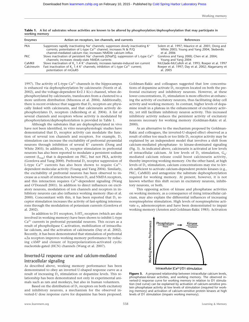
As an alternative to the mechanism proposed by Goldman-Rakic and colleagues, the inverted-U-shaped effect observed as aresult of either too much or too little D1 receptor activity may beexplained by an independent model that involves a shift fromcalcium-mediated phosphatase- to kinase-dominated signaling(Fig. 3). As indicated above, calcineurin is activated at low levelsof intracellular calcium. At low levels of D1 stimulation, Gq-mediated calcium release could boost calcineurin activity,thereby improving working memory. On the other hand, at highlevels of D1 stimulation, calcium concentrations may rise to lev-els sufficient to activate calcium-dependent protein kinases (e.g.,PKC, CaMKII) and antagonize the substrate dephosphorylationrequired for working memory. At present, however, it is notknown whether this shift occurs in excitatory neurons, inhibi-tory neurons, or both.
This opposing action of kinase and phosphatase activitieson working memory, as a consequence of rising intracellular cal-cium, may also explain the differential influences of increasingnorepinephrine stimulation. High levels of norepinephrine acti-vate �1 adrenoreceptors and have been demonstrated to impairworking memory (Arnsten and Goldman-Rakic 1985). Activation
Figure 3. A proposed relationship between intracellular calcium levels,phosphatase-kinase activities, and working memory. The observed in-verted-U response curve for working memory in relation to D1 stimula-tion (red curve) can be explained by activation of calcium-sensitive pro-tein phosphatase activity at low levels of stimulation (required for work-ing memory) and activation of calcium-sensitive protein kinases at highlevels of D1 stimulation (impairs working memory).
Table 1. A list of substrates whose activities are known to be altered by phosphorylation/dephosphorylation that may participate inworking memory
Enzymes Action on receptors, ion channels, and currents References
PKA Suppresses rapidly inactivating Na+ channels; suppresses slowly inactivating K+
currents; potentiation of L-type Ca2+ channel; increases N & P/Qchannel-mediated calcium rise; increases NMDAr calcium influx
Solem et al. 1997; Maurice et al. 2001; Dong andWhite 2003; Young and Yang 2004; Skeberdiset al. 2006
PKC Slows inactivation of persistent Na+ channels [I(NaP)]; suppression of L-type Ca2+
channels; increases steady-state NMDA currentsGorelova and Yang 2000; Chen et al. 2004;
Young and Yang 2004CaMKII Slows inactivation of Kv 1.4 K+ channels; increases kainate-induced ion current McGlade-McCulloh et al. 1993; Roeper et al. 1997Calcineurin Fast inactivation of Kv 1.4 K+ channels; inhibition of L-type Ca2+ current;
potentiation of mGluR5Roeper et al. 1997; Day et al. 2002; Alagarsamy et
al. 2005
Working memory
558www.learnmem.org Learning & Memory
Cold Spring Harbor Laboratory Press on February 10, 2015 - Published by learnmem.cshlp.orgDownloaded from
of these receptors has been linked to PKC activation, and PKCinhibition improves working memory in conditions associatedwith high norepinephrine levels such as stress (Birnbaum et al.2004).
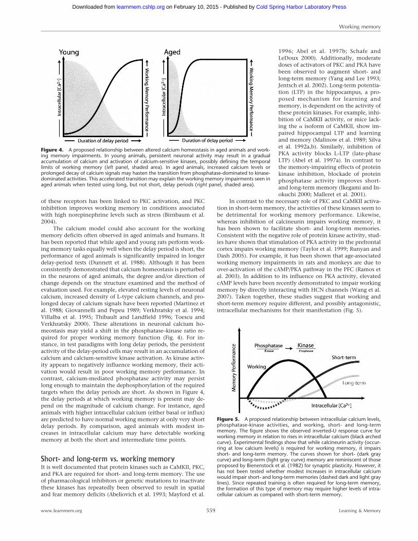
The calcium model could also account for the workingmemory deficits often observed in aged animals and humans. Ithas been reported that while aged and young rats perform work-ing memory tasks equally well when the delay period is short, theperformance of aged animals is significantly impaired in longerdelay-period tests (Dunnett et al. 1988). Although it has beenconsistently demonstrated that calcium homeostasis is perturbedin the neurons of aged animals, the degree and/or direction ofchange depends on the structure examined and the method ofevaluation used. For example, elevated resting levels of neuronalcalcium, increased density of L-type calcium channels, and pro-longed decay of calcium signals have been reported (Martinez etal. 1988; Giovannelli and Pepeu 1989; Verkhratsky et al. 1994;Villalba et al. 1995; Thibault and Landfield 1996; Toescu andVerkhratsky 2000). These alterations in neuronal calcium ho-meostasis may yield a shift in the phosphatase–kinase ratio re-quired for proper working memory function (Fig. 4). For in-stance, in test paradigms with long delay periods, the persistentactivity of the delay-period cells may result in an accumulation ofcalcium and calcium-sensitive kinase activation. As kinase activ-ity appears to negatively influence working memory, their acti-vation would result in poor working memory performance. Incontrast, calcium-mediated phosphatase activity may persistlong enough to maintain the dephosphorylation of the requiredtargets when the delay periods are short. As shown in Figure 4,the delay periods at which working memory is present may de-pend on the magnitude of calcium change. For instance, agedanimals with higher intracellular calcium (either basal or influx)are predicted to have normal working memory at only very shortdelay periods. By comparison, aged animals with modest in-creases in intracellular calcium may have detectable workingmemory at both the short and intermediate time points.
Short- and long-term vs. working memoryIt is well documented that protein kinases such as CaMKII, PKC,and PKA are required for short- and long-term memory. The useof pharmacological inhibitors or genetic mutations to inactivatethese kinases has repeatedly been observed to result in spatialand fear memory deficits (Abeliovich et al. 1993; Mayford et al.
1996; Abel et al. 1997b; Schafe andLeDoux 2000). Additionally, moderatedoses of activators of PKC and PKA havebeen observed to augment short- andlong-term memory (Yang and Lee 1993;Jentsch et al. 2002). Long-term potentia-tion (LTP) in the hippocampus, a pro-posed mechanism for learning andmemory, is dependent on the activity ofthese protein kinases. For example, inhi-bition of CaMKII activity, or mice lack-ing the � isoform of CaMKII, show im-paired hippocampal LTP and learningand memory (Malinow et al. 1989; Silvaet al. 1992a,b). Similarly, inhibition ofPKA activity blocks L-LTP (late-phaseLTP) (Abel et al. 1997a). In contrast tothe memory-impairing effects of proteinkinase inhibition, blockade of proteinphosphatase activity improves short-and long-term memory (Ikegami and In-okuchi 2000; Malleret et al. 2001).
In contrast to the necessary role of PKC and CaMKII activa-tion in short-term memory, the activities of these kinases seem tobe detrimental for working memory performance. Likewise,whereas inhibition of calcineurin impairs working memory, ithas been shown to facilitate short- and long-term memories.Consistent with the negative role of protein kinase activity, stud-ies have shown that stimulation of PKA activity in the prefrontalcortex impairs working memory (Taylor et al. 1999; Runyan andDash 2005). For example, it has been shown that age-associatedworking memory impairments in rats and monkeys are due toover-activation of the cAMP/PKA pathway in the PFC (Ramos etal. 2003). In addition to its influence on PKA activity, elevatedcAMP levels have been recently demonstrated to impair workingmemory by directly interacting with HCN channels (Wang et al.2007). Taken together, these studies suggest that working andshort-term memory require different, and possibly antagonistic,intracellular mechanisms for their manifestation (Fig. 5).
Figure 4. A proposed relationship between altered calcium homeostasis in aged animals and work-ing memory impairments. In young animals, persistent neuronal activity may result in a gradualaccumulation of calcium and activation of calcium-sensitive kinases, possibly defining the temporallimits of working memory (left panel, shaded area). In aged animals, increased calcium levels orprolonged decay of calcium signals may hasten the transition from phosphatase-dominated to kinase-dominated activities. This accelerated transition may explain the working memory impairments seen inaged animals when tested using long, but not short, delay periods (right panel, shaded area).
Figure 5. A proposed relationship between intracellular calcium levels,phosphatase-kinase activities, and working, short- and long-termmemory. The figure shows the observed inverted-U response curve forworking memory in relation to rises in intracellular calcium (black archedcurve). Experimental findings show that while calcineurin activity (occur-ring at low calcium levels) is required for working memory, it impairsshort- and long-term memory. The curves shown for short- (dark graycurve) and long-term (light gray curve) memory are reminiscent of thoseproposed by Bienenstock et al. (1982) for synaptic plasticity. However, ithas not been tested whether modest increases in intracellular calciumwould impair short- and long-term memories (dashed dark and light graylines). Since repeated training is often required for long-term memory,the formation of this type of memory may require higher levels of intra-cellular calcium as compared with short-term memory.
Working memory
559www.learnmem.org Learning & Memory
Cold Spring Harbor Laboratory Press on February 10, 2015 - Published by learnmem.cshlp.orgDownloaded from
An antagonistic intracellular mechanism may have func-tional implications. As described above, working memory in-volves the transient maintenance of information for subsequentmanipulation in order to guide goal-directed behaviors. Once theobjective is achieved, there is no need to store this transientinformation in a longer form. It is therefore possible that usingopposing biochemical mechanisms (e.g., kinases are required forlong-term memory, but appear to be detrimental to workingmemory) may ensure that information held in working memoryis not stored in more enduring forms. This enables a differentia-tion of circumstances, in which one forms long-term memoriesfrom those circumstances in which one has to merely transientlykeep in mind and use particular information momentarily.
Disorders of working memorySeveral types of neurological disorders are marked by deficits inworking memory. Since the late 1800s, initially through studiesof brain-injured patients, it has been known that damage to thePFC impairs the individual’s ability to make appropriate and di-rected actions. Subsequent studies have revealed that PFC dam-age results in deficits in working memory, which can give rise tomany of the behavioral abnormalities observed in these earlierstudies. These patients are easily distracted, are impaired at fo-cusing and maintaining attention on sensory stimuli, have poorconcentration and organization of thought, and are more sus-ceptible to proactive interference (Woods and Knight 1986;Godefroy and Rousseaux 1996; Thompson-Schill et al. 2002). Inaddition to PFC damage, deficits in working memory are alsoassociated with schizophrenia, ADHD, Alzheimer’s disease (AD),
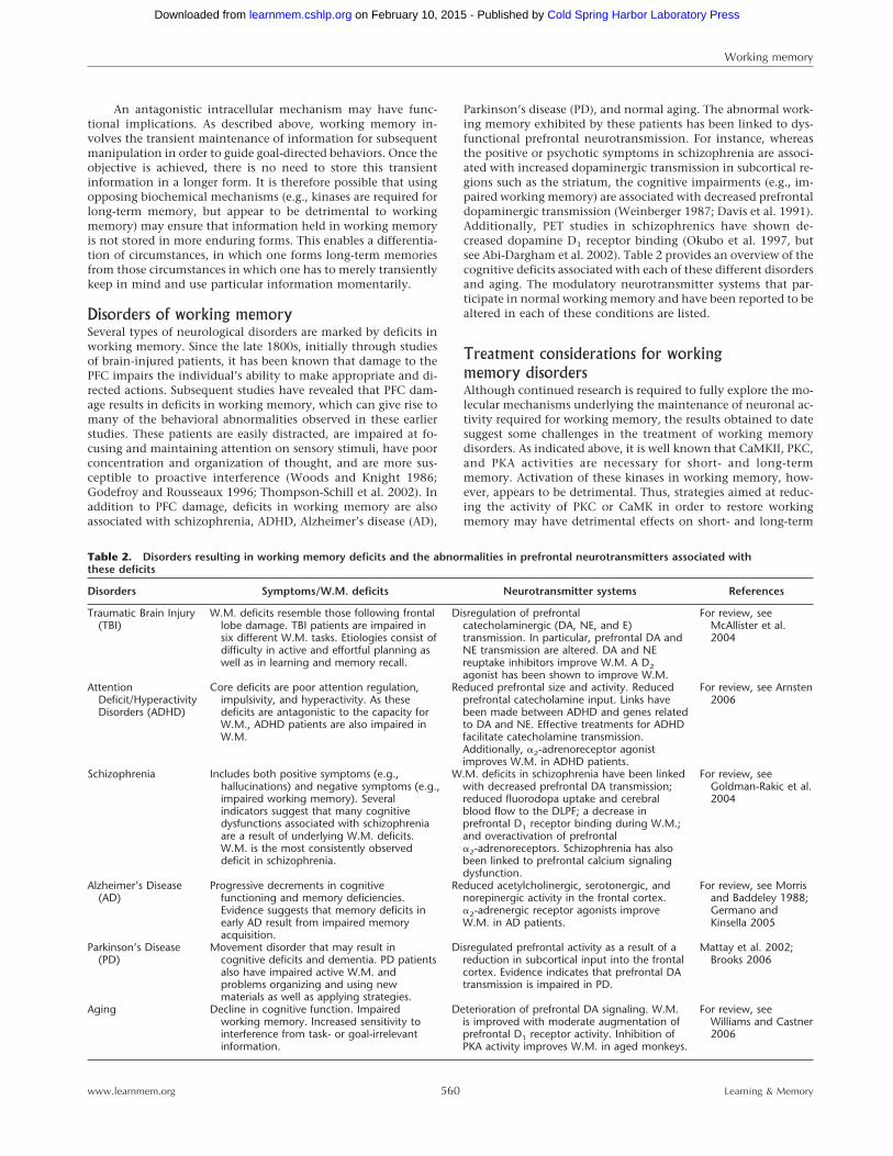
Parkinson’s disease (PD), and normal aging. The abnormal work-ing memory exhibited by these patients has been linked to dys-functional prefrontal neurotransmission. For instance, whereasthe positive or psychotic symptoms in schizophrenia are associ-ated with increased dopaminergic transmission in subcortical re-gions such as the striatum, the cognitive impairments (e.g., im-paired working memory) are associated with decreased prefrontaldopaminergic transmission (Weinberger 1987; Davis et al. 1991).Additionally, PET studies in schizophrenics have shown de-creased dopamine D1 receptor binding (Okubo et al. 1997, butsee Abi-Dargham et al. 2002). Table 2 provides an overview of thecognitive deficits associated with each of these different disordersand aging. The modulatory neurotransmitter systems that par-ticipate in normal working memory and have been reported to bealtered in each of these conditions are listed.
Treatment considerations for workingmemory disordersAlthough continued research is required to fully explore the mo-lecular mechanisms underlying the maintenance of neuronal ac-tivity required for working memory, the results obtained to datesuggest some challenges in the treatment of working memorydisorders. As indicated above, it is well known that CaMKII, PKC,and PKA activities are necessary for short- and long-termmemory. Activation of these kinases in working memory, how-ever, appears to be detrimental. Thus, strategies aimed at reduc-ing the activity of PKC or CaMK in order to restore workingmemory may have detrimental effects on short- and long-term
Table 2. Disorders resulting in working memory deficits and the abnormalities in prefrontal neurotransmitters associated withthese deficits
Disorders Symptoms/W.M. deficits Neurotransmitter systems References
Traumatic Brain Injury(TBI)
W.M. deficits resemble those following frontallobe damage. TBI patients are impaired insix different W.M. tasks. Etiologies consist ofdifficulty in active and effortful planning aswell as in learning and memory recall.
Disregulation of prefrontalcatecholaminergic (DA, NE, and E)transmission. In particular, prefrontal DA andNE transmission are altered. DA and NEreuptake inhibitors improve W.M. A D2agonist has been shown to improve W.M.
For review, seeMcAllister et al.2004
AttentionDeficit/HyperactivityDisorders (ADHD)
Core deficits are poor attention regulation,impulsivity, and hyperactivity. As thesedeficits are antagonistic to the capacity forW.M., ADHD patients are also impaired inW.M.
Reduced prefrontal size and activity. Reducedprefrontal catecholamine input. Links havebeen made between ADHD and genes relatedto DA and NE. Effective treatments for ADHDfacilitate catecholamine transmission.Additionally, �2-adrenoreceptor agonistimproves W.M. in ADHD patients.
For review, see Arnsten2006
Schizophrenia Includes both positive symptoms (e.g.,hallucinations) and negative symptoms (e.g.,impaired working memory). Severalindicators suggest that many cognitivedysfunctions associated with schizophreniaare a result of underlying W.M. deficits.W.M. is the most consistently observeddeficit in schizophrenia.
W.M. deficits in schizophrenia have been linkedwith decreased prefrontal DA transmission;reduced fluorodopa uptake and cerebralblood flow to the DLPF; a decrease inprefrontal D1 receptor binding during W.M.;and overactivation of prefrontal�2-adrenoreceptors. Schizophrenia has alsobeen linked to prefrontal calcium signalingdysfunction.
For review, seeGoldman-Rakic et al.2004
Alzheimer’s Disease(AD)
Progressive decrements in cognitivefunctioning and memory deficiencies.Evidence suggests that memory deficits inearly AD result from impaired memoryacquisition.
Reduced acetylcholinergic, serotonergic, andnorepinergic activity in the frontal cortex.�2-adrenergic receptor agonists improveW.M. in AD patients.
For review, see Morrisand Baddeley 1988;Germano andKinsella 2005
Parkinson’s Disease(PD)
Movement disorder that may result incognitive deficits and dementia. PD patientsalso have impaired active W.M. andproblems organizing and using newmaterials as well as applying strategies.
Disregulated prefrontal activity as a result of areduction in subcortical input into the frontalcortex. Evidence indicates that prefrontal DAtransmission is impaired in PD.
Mattay et al. 2002;Brooks 2006
Aging Decline in cognitive function. Impairedworking memory. Increased sensitivity tointerference from task- or goal-irrelevantinformation.
Deterioration of prefrontal DA signaling. W.M.is improved with moderate augmentation ofprefrontal D1 receptor activity. Inhibition ofPKA activity improves W.M. in aged monkeys.
For review, seeWilliams and Castner2006
Working memory
560www.learnmem.org Learning & Memory
Cold Spring Harbor Laboratory Press on February 10, 2015 - Published by learnmem.cshlp.orgDownloaded from
memory. As has been suggested by Arnsten and colleagues re-garding the potential negative influences of PKA stimulation onworking memory (when utilized as a means of improving long-term memories) (Arnsten et al. 2005), it now appears that globalinhibition of calcium-sensitive kinases to improve workingmemory may have similar complicating influences on long-termmemory. However, it has not yet been tested whether the degreeof kinase inhibition necessary to improve working memory inpathological situations is sufficient to impair short- or long-termmemories. Likewise, strategies to augment phosphatase activity,while potentially improving working memory, may have nega-tive influences on longer lasting forms of memory, depending onthe degree of activation. Although yet to be experimentallytested, low doses of drugs such as SKF83959 that have little effecton adenylyl cyclase and PKA activity, but stimulate PLC-coupledD1 receptors (Undie et al. 1994; Jin et al. 2003), may offer a meansof treating working memory deficits without overt influences onother cognitive processes. Alternatively, therapies aimed at thespecific downstream substrates (e.g., ion channels) that are re-quired for, or impair, working memory may have clinical utility.For example, intra-mPFC infusion of the HCN channel blockerZD7288 has been demonstrated to enhance spatial workingmemory in rats (Wang et al. 2007). As research in this area con-tinues, the challenges associated with the treatment of workingmemory disorders will become defined, and new strategies toovercome these deficits will be revealed.
AcknowledgmentsWe thank Drs. James Knierim and Harel Shouval for their invalu-able comments on this manuscript. The work performed in ourlaboratories was made possible by grants from NIH (NS35457,NS049160, MH072933, NS052313).
ReferencesAalto, S., Bruck, A., Laine, M., Nagren, K., and Rinne, J.O. 2005. Frontal
and temporal dopamine release during working memory andattention tasks in healthy humans: A positron emission tomographystudy using the high-affinity dopamine D2 receptor ligand [11C]FLB457. J. Neurosci. 25: 2471–2477.
Abel, T., Nguyen, P.V., Barad, M., Deuel, T.A., Kandel, E.R., andBourtchouladze, R. 1997a. Genetic demonstration of a role for PKAin the late phase of LTP and in hippocampus-based long-termmemory. Cell 88: 615–626.
Abel, T., Nguyen, P.V., Barad, M., Deuel, T.A., Kandel, E.R., andBourtchouladze, R. 1997b. Genetic demonstration of a role for PKAin the late phase of LTP and in hippocampus-based long-termmemory. Cell 88: 615–626.
Abeliovich, A., Paylor, R., Chen, C., Kim, J.J., Wehner, J.M., andTonegawa, S. 1993. PKC� mutant mice exhibit mild deficits inspatial and contextual learning. Cell 75: 1263–1271.
Abi-Dargham, A., Mawlawi, O., Lombardo, I., Gil, R., Martinez, D.,Huang, Y., Hwang, D.R., Keilp, J., Kochan, L., Van Heertum, R., et al.2002. Prefrontal dopamine D1 receptors and working memory inschizophrenia. J. Neurosci. 22: 3708–3719.
Adlersberg, M., Hsiung, S.C., Glickstein, S.B., Liu, K.P., Tamir, H., andSchmauss, C. 2004. Regulation of dopamine D-receptor activation invivo by protein phosphatase 2B (calcineurin). J. Neurochem.90: 865–873.
Alagarsamy, S., Saugstad, J., Warren, L., Mansuy, I.M., Gereau, R.W., andConn, P.J. 2005. NMDA-induced potentiation of mGluR5 ismediated by activation of protein phosphatase 2B/calcineurin.Neuropharmacology 49 (Suppl. 1): 135–145.
Arnsten, A.F. 2000. Through the looking glass: Differential noradenergicmodulation of prefrontal cortical function. Neural Plast. 7: 133–146.
Arnsten, A.F. 2006. Fundamentals of attention-deficit/hyperactivitydisorder: Circuits and pathways. J. Clin. Psychiatry 67 (Suppl.8): 7–12.
Arnsten, A.F. and Goldman-Rakic, P.S. 1985. Alpha 2-adrenergicmechanisms in prefrontal cortex associated with cognitive decline inaged nonhuman primates. Science 230: 1273–1276.
Arnsten, A.F. and Goldman-Rakic, P.S. 1998. Noise stress impairsprefrontal cortical cognitive function in monkeys: Evidence for ahyperdopaminergic mechanism. Arch. Gen. Psychiatry 55: 362–368.
Arnsten, A.F., Cai, J.X., Murphy, B.L., and Goldman-Rakic, P.S. 1994.Dopamine D1 receptor mechanisms in the cognitive performance ofyoung adult and aged monkeys. Psychopharmacology 116: 143–151.
Arnsten, A.F., Ramos, B.P., Birnbaum, S.G., and Taylor, J.R. 2005.Protein kinase A as a therapeutic target for memory disorders:Rationale and challenges. Trends Mol. Med. 11: 121–128.
Arthur, J.M., Collinsworth, G.P., Gettys, T.W., and Raymond, J.R. 1999.Agonist-induced translocation of Gq/11� immunoreactivity directlyfrom plasma membrane in MDCK cells. Am. J. Physiol.276: F528–F534.
Aultman, J.M. and Moghaddam, B. 2001. Distinct contributions ofglutamate and dopamine receptors to temporal aspects of rodentworking memory using a clinically relevant task. Psychopharmacology153: 353–364.
Baddeley, A. 2001. The concept of episodic memory. Philos. Trans. R.Soc. Lond. B Biol. Sci. 356: 1345–1350.
Bienenstock, E.L., Cooper, L.N., and Munro, P.W. 1982. Theory for thedevelopment of neuron selectivity: Orientation specificity andbinocular interaction in visual cortex. J. Neurosci. 2: 32–48.
Birnbaum, S., Gobeske, K.T., Auerbach, J., Taylor, J.R., and Arnsten, A.F.1999. A role for norepinephrine in stress-induced cognitive deficits:Alpha-1-adrenoceptor mediation in the prefrontal cortex. Biol.Psychiatry 46: 1266–1274.
Birnbaum, S.G., Yuan, P.X., Wang, M., Vijayraghavan, S., Bloom, A.K.,Davis, D.J., Gobeske, K.T., Sweatt, J.D., Manji, H.K., and Arnsten,A.F. 2004. Protein kinase C overactivity impairs prefrontal corticalregulation of working memory. Science 306: 882–884.
Brooks, D.J. 2006. Dopaminergic action beyond its effects on motorfunction: Imaging studies. J. Neurol. 253 (Suppl. 4): iv8–iv15.
Brozoski, T.J., Brown, R.M., Rosvold, H.E., and Goldman, P.S. 1979.Cognitive deficit caused by regional depletion of dopamine inprefrontal cortex of rhesus monkey. Science 205: 929–932.
Buckner, R.L., Logan, J., Donaldson, D.I., and Wheeler, M.E. 2000.Cognitive neuroscience of episodic memory encoding. Acta Psychol.105: 127–139.
Chen, G., Greengard, P., and Yan, Z. 2004. Potentiation of NMDAreceptor currents by dopamine D1 receptors in prefrontal cortex.Proc. Natl. Acad. Sci. 101: 2596–2600.
Cools, R. 2006. Dopaminergic modulation of cognitivefunction-implications for L-DOPA treatment in Parkinson’s disease.Neurosci. Biobehav. Rev. 30: 1–23.
D’Esposito, M., Detre, J.A., Alsop, D.C., Shin, R.K., Atlas, S., andGrossman, M. 1995. The neural basis of the central executive systemof working memory. Nature 378: 279–281.
Davis, K.L., Kahn, R.S., Ko, G., and Davidson, M. 1991. Dopamine inschizophrenia: A review and reconceptualization. Am. J. Psychiatry148: 1474–1486.
Day, M., Olson, P.A., Platzer, J., Striessnig, J., and Surmeier, D.J. 2002.Stimulation of 5-HT(2) receptors in prefrontal pyramidal neuronsinhibits Ca(v)1.2 L type Ca2+ currents via a PLC�/IP3/calcineurinsignaling cascade. J. Neurophysiol. 87: 2490–2504.
de Rooij, J., Zwartkruis, F.J., Verheijen, M.H., Cool, R.H., Nijman, S.M.,Wittinghofer, A., and Bos, J.L. 1998. Epac is a Rap1guanine-nucleotide-exchange factor directly activated by cyclic AMP.Nature 396: 474–477.
Didriksen, M. 1995. Effects of antipsychotics on cognitive behaviour inrats using the delayed non-match to position paradigm. Eur. J.Pharmacol. 281: 241–250.
Dong, Y. and White, F.J. 2003. Dopamine D1-class receptors selectivelymodulate a slowly inactivating potassium current in rat medialprefrontal cortex pyramidal neurons. J. Neurosci. 23: 2686–2695.
Dudchenko, P.A. 2004. An overview of the tasks used to test workingmemory in rodents. Neurosci. Biobehav. Rev. 28: 699–709.
Dunnett, S.B., Evenden, J.L., and Iversen, S.D. 1988. Delay-dependentshort-term memory deficits in aged rats. Psychopharmacology96: 174–180.
Durstewitz, D., Seamans, J.K., and Sejnowski, T.J. 2000.Neurocomputational models of working memory. Nat. Neurosci.3 (Suppl): 1184–1191.
Egorov, A.V., Hamam, B.N., Fransen, E., Hasselmo, M.E., and Alonso,A.A. 2002. Graded persistent activity in entorhinal cortex neurons.Nature 420: 173–178.
Franowicz, J.S. and Arnsten, A.F. 1998. The �-2a noradrenergic agonist,guanfacine, improves delayed response performance in young adultrhesus monkeys. Psychopharmacology 136: 8–14.
Friedman, J.I., Temporini, H., and Davis, K.L. 1999. Pharmacologicstrategies for augmenting cognitive performance in schizophrenia.Biol. Psychiatry 45: 1–16.
Funahashi, S., Chafee, M.V., and Goldman-Rakic, P.S. 1993. Prefrontalneuronal activity in rhesus monkeys performing a delayedanti-saccade task. Nature 365: 753–756.
Fuster, J.M. and Alexander, G.E. 1971. Neuron activity related to
Working memory
561www.learnmem.org Learning & Memory
Cold Spring Harbor Laboratory Press on February 10, 2015 - Published by learnmem.cshlp.orgDownloaded from
short-term memory. Science 173: 652–654.Gao, W.J. and Goldman-Rakic, P.S. 2003. Selective modulation of
excitatory and inhibitory microcircuits by dopamine. Proc. Natl.Acad. Sci. 100: 2836–2841.
Gao, W.J. and Goldman-Rakic, P.S. 2006. NMDA receptor-mediatedepileptiform persistent activity requires calcium release fromintracellular stores in prefrontal neurons. Exp. Neurol. 197: 495–504.
Germano, C. and Kinsella, G.J. 2005. Working memory and learning inearly Alzheimer’s disease. Neuropsychol. Rev. 15: 1–10.
Giovannelli, L. and Pepeu, G. 1989. Effect of age on K+-inducedcytosolic Ca2+ changes in rat cortical synaptosomes. J. Neurochem.53: 392–398.
Godefroy, O. and Rousseaux, M. 1996. Divided and focused attention inpatients with lesion of the prefrontal cortex. Brain Cogn.30: 155–174.
Goldman-Rakic, P.S., Muly III, E.C., and Williams, G.V. 2000. D1receptors in prefrontal cells and circuits. Brain Res. Brain Res. Rev.31: 295–301.
Goldman-Rakic, P.S., Castner, S.A., Svensson, T.H., Siever, L.J., andWilliams, G.V. 2004. Targeting the dopamine D1 receptor inschizophrenia: Insights for cognitive dysfunction.Psychopharmacology 174: 3–16.
Gonzalez-Burgos, G., Kroener, S., Seamans, J.K., Lewis, D.A., andBarrionuevo, G. 2005. Dopaminergic modulation of short-termsynaptic plasticity in fast-spiking interneurons of primatedorsolateral prefrontal cortex. J. Neurophysiol. 94: 4168–4177.
Gorelova, N.A. and Yang, C.R. 2000. Dopamine D1/D5 receptoractivation modulates a persistent sodium current in rat prefrontalcortical neurons in vitro. J. Neurophysiol. 84: 75–87.
Gorelova, N., Seamans, J.K., and Yang, C.R. 2002. Mechanisms ofdopamine activation of fast-spiking interneurons that exertinhibition in rat prefrontal cortex. J. Neurophysiol. 88: 3150–3166.
Granon, S., Poucet, B., Thinus-Blanc, C., Changeux, J.P., and Vidal, C.1995. Nicotinic and muscarinic receptors in the rat prefrontalcortex: Differential roles in working memory, response selection andeffortful processing. Psychopharmacology 119: 139–144.
Ikegami, S. and Inokuchi, K. 2000. Antisense DNA against calcineurinfacilitates memory in contextual fear conditioning by lowering thethreshold for hippocampal long-term potentiation induction.Neuroscience 98: 637–646.
Jacobsen, C. 1936. Studies of cerebral function in primates. Comp.Psychol. Monogr. 13: 3–60.
Jentsch, J.D., Olausson, P., Nestler, E.J., and Taylor, J.R. 2002.Stimulation of protein kinase a activity in the rat amygdalaenhances reward-related learning. Biol. Psychiatry 52: 111–118.
Jin, L.Q., Goswami, S., Cai, G., Zhen, X., and Friedman, E. 2003.SKF83959 selectively regulates phosphatidylinositol-linked D1dopamine receptors in rat brain. J. Neurochem. 85: 378–386.
Jonides, J., Smith, E.E., Koeppe, R.A., Awh, E., Minoshima, S., andMintun, M.A. 1993. Spatial working memory in humans as revealedby PET. Nature 363: 623–625.
Koch, K.W. and Fuster, J.M. 1989. Unit activity in monkey parietalcortex related to haptic perception and temporary memory. Exp.Brain Res. 76: 292–306.
Koh, P.O., Bergson, C., Undie, A.S., Goldman-Rakic, P.S., and Lidow,M.S. 2003. Up-regulation of the D1 dopamine receptor-interactingprotein, calcyon, in patients with schizophrenia. Arch. Gen.Psychiatry 60: 311–319.
Kojima, S. and Goldman-Rakic, P.S. 1984. Functional analysis ofspatially discriminative neurons in prefrontal cortex of rhesusmonkey. Brain Res. 291: 229–240.
Kolb, B. 1984. Functions of the frontal cortex of the rat: A comparativereview. Brain Res. 320: 65–98.
Lee, A.C., Robbins, T.W., and Owen, A.M. 2000. Episodic memory meetsworking memory in the frontal lobe: Functional neuroimagingstudies of encoding and retrieval. Crit. Rev. Neurobiol. 14: 165–197.
Li, B.M. and Mei, Z.T. 1994. Delayed-response deficit induced by localinjection of the � 2-adrenergic antagonist yohimbine into thedorsolateral prefrontal cortex in young adult monkeys. Behav. NeuralBiol. 62: 134–139.
Lidow, M.S. 2003. Calcium signaling dysfunction in schizophrenia: Aunifying approach. Brain Res. Brain Res. Rev. 43: 70–84.
Lidow, M.S., Goldman-Rakic, P.S., Gallager, D.W., and Rakic, P. 1991.Distribution of dopaminergic receptors in the primate cerebralcortex: Quantitative autoradiographic analysis using [3H]raclopride,[3H]spiperone and [3H]SCH23390. Neuroscience 40: 657–671.
Malinow, R., Schulman, H., and Tsien, R.W. 1989. Inhibition ofpostsynaptic PKC or CaMKII blocks induction but not expression ofLTP. Science 245: 862–866.
Malleret, G., Haditsch, U., Genoux, D., Jones, M.W., Bliss, T.V.,Vanhoose, A.M., Weitlauf, C., Kandel, E.R., Winder, D.G., andMansuy, I.M. 2001. Inducible and reversible enhancement of
learning, memory, and long-term potentiation by genetic inhibitionof calcineurin. Cell 104: 675–686.
Mao, Z.M., Arnsten, A.F., and Li, B.M. 1999. Local infusion of an �-1adrenergic agonist into the prefrontal cortex impairs spatial workingmemory performance in monkeys. Biol. Psychiatry 46: 1259–1265.
Martinez, A., Vitorica, J., and Satrustegui, J. 1988. Cytosolic free calciumlevels increase with age in rat brain synaptosomes. Neurosci. Lett.88: 336–342.
Mattay, V.S., Tessitore, A., Callicott, J.H., Bertolino, A., Goldberg, T.E.,Chase, T.N., Hyde, T.M., and Weinberger, D.R. 2002. Dopaminergicmodulation of cortical function in patients with Parkinson’s disease.Ann. Neurol. 51: 156–164.
Maurice, N., Tkatch, T., Meisler, M., Sprunger, L.K., and Surmeier, D.J.2001. D1/D5 dopamine receptor activation differentially modulatesrapidly inactivating and persistent sodium currents in prefrontalcortex pyramidal neurons. J. Neurosci. 21: 2268–2277.
Mayford, M., Bach, M.E., Huang, Y.Y., Wang, L., Hawkins, R.D., andKandel, E.R. 1996. Control of memory formation through regulatedexpression of a CaMKII transgene. Science 274: 1678–1683.
McAllister, T.W., Flashman, L.A., Sparling, M.B., and Saykin, A.J. 2004.Working memory deficits after traumatic brain injury:Catecholaminergic mechanisms and prospects for treatment––Areview. Brain Inj. 18: 331–350.
McGlade-McCulloh, E., Yamamoto, H., Tan, S.E., Brickey, D.A., andSoderling, T.R. 1993. Phosphorylation and regulation of glutamatereceptors by calcium/calmodulin-dependent protein kinase II. Nature362: 640–642.
Meador-Woodruff, J.H., Damask, S.P., Wang, J., Haroutunian, V., Davis,K.L., and Watson, S.J. 1996. Dopamine receptor mRNA expression inhuman striatum and neocortex. Neuropsychopharmacology 15: 17–29.
Miller, E.K., Li, L., and Desimone, R. 1993. Activity of neurons inanterior inferior temporal cortex during a short-term memory task. J.Neurosci. 13: 1460–1478.
Mirnics, K., Middleton, F.A., Stanwood, G.D., Lewis, D.A., and Levitt, P.2001. Disease-specific changes in regulator of G-protein signaling 4(RGS4) expression in schizophrenia. Mol. Psychiatry 6: 293–301.
Misonou, H., Mohapatra, D.P., Park, E.W., Leung, V., Zhen, D.,Misonou, K., Anderson, A.E., and Trimmer, J.S. 2004. Regulation ofion channel localization and phosphorylation by neuronal activity.Nat. Neurosci. 7: 711–718.
Morris, R.G. and Baddeley, A.D. 1988. Primary and working memoryfunctioning in Alzheimer-type dementia. J. Clin. Exp. Neuropsychol.10: 279–296.
Muly III, E.C., Szigeti, K., and Goldman-Rakic, P.S. 1998. D1 receptor ininterneurons of macaque prefrontal cortex: distribution andsubcellular localization. J. Neurosci. 18: 10553–10565.
Norris, C.M., Blalock, E.M., Chen, K.C., Porter, N.M., and Landfield,P.W. 2002. Calcineurin enhances L-type Ca(2+) channel activity inhippocampal neurons: Increased effect with age in culture.Neuroscience 110: 213–225.
Okubo, Y., Suhara, T., Suzuki, K., Kobayashi, K., Inoue, O., Terasaki, O.,Someya, Y., Sassa, T., Sudo, Y., Matsushima, E., et al. 1997.Decreased prefrontal dopamine D1 receptors in schizophreniarevealed by PET. Nature 385: 634–636.
Paspalas, C.D. and Goldman-Rakic, P.S. 2004. Microdomains fordopamine volume neurotransmission in primate prefrontal cortex. J.Neurosci. 24: 5292–5300.
Petrides, M., Alivisatos, B., Meyer, E., and Evans, A.C. 1993. Functionalactivation of the human frontal cortex during the performance ofverbal working memory tasks. Proc. Natl. Acad. Sci. 90: 878–882.
Phillips, A.G., Ahn, S., and Floresco, S.B. 2004. Magnitude of dopaminerelease in medial prefrontal cortex predicts accuracy of memory on adelayed response task. J. Neurosci. 24: 547–553.
Ramos, B.P., Birnbaum, S.G., Lindenmayer, I., Newton, S.S., Duman,R.S., and Arnsten, A.F. 2003. Dysregulation of protein kinase asignaling in the aged prefrontal cortex: New strategy for treatingage-related cognitive decline. Neuron 40: 835–845.
Ranganath, C., Cohen, M.X., and Brozinsky, C.J. 2005. Workingmemory maintenance contributes to long-term memory formation:Neural and behavioral evidence. J. Cogn. Neurosci. 17: 994–1010.
Rao, S.G., Williams, G.V., and Goldman-Rakic, P.S. 2000. Destructionand creation of spatial tuning by disinhibition: GABAA blockade ofprefrontal cortical neurons engaged by working memory. J. Neurosci.20: 485–494.
Roeper, J., Lorra, C., and Pongs, O. 1997. Frequency-dependentinactivation of mammalian A-type K+ channel KV1.4 regulated byCa2+/calmodulin-dependent protein kinase. J. Neurosci.17: 3379–3391.
Runyan, J.D. and Dash, P.K. 2005. Distinct prefrontal molecularmechanisms for information storage lasting seconds versus minutes.Learn. Mem. 12: 232–238.
Runyan, J.D., Moore, A.N., and Dash, P.K. 2005. A role for prefrontal
Working memory
562www.learnmem.org Learning & Memory
Cold Spring Harbor Laboratory Press on February 10, 2015 - Published by learnmem.cshlp.orgDownloaded from
calcium-sensitive protein phosphatase and kinase activities inworking memory. Learn. Mem. 12: 103–110.
Russell, V., Allie, S., and Wiggins, T. 2000. Increased noradrenergicactivity in prefrontal cortex slices of an animal model forattention-deficit hyperactivity disorder—The spontaneouslyhypertensive rat. Behav. Brain Res. 117: 69–74.
Sakurai, Y. 1990. Cells in the rat auditory system have sensory-delaycorrelates during the performance of an auditory working memorytask. Behav. Neurosci. 104: 856–868.
Sandrini, M., Cappa, S.F., Rossi, S., Rossini, P.M., and Miniussi, C. 2003.The role of prefrontal cortex in verbal episodic memory: rTMSevidence. J. Cogn. Neurosci. 15: 855–861.
Sawaguchi, T. 2001. The effects of dopamine and its antagonists ondirectional delay-period activity of prefrontal neurons in monkeysduring an oculomotor delayed-response task. Neurosci. Res.41: 115–128.
Sawaguchi, T. and Goldman-Rakic, P.S. 1991. D1 dopamine receptors inprefrontal cortex: Involvement in working memory. Science251: 947–950.
Schafe, G.E. and LeDoux, J.E. 2000. Memory consolidation of auditorypavlovian fear conditioning requires protein synthesis and proteinkinase A in the amygdala. J. Neurosci. 20: RC96.
Silva, A.J., Paylor, R., Wehner, J.M., and Tonegawa, S. 1992a. Impairedspatial learning in �-calcium-calmodulin kinase II mutant mice.Science 257: 206–211.
Silva, A.J., Stevens, C.F., Tonegawa, S., and Wang, Y. 1992b. Deficienthippocampal long-term potentiation in �-calcium-calmodulin kinaseII mutant mice. Science 257: 201–206.
Simon, H., Scatton, B., and Le Moal, M. 1979. Definitive disruption ofspatial delayed alternation in rats after lesions in the ventralmesencephalic tegmentum. Neurosci. Lett. 15: 319–324.
Skeberdis, V.A., Chevaleyre, V., Lau, C.G., Goldberg, J.H., Pettit, D.L.,Suadicani, S.O., Lin, Y., Bennett, M.V., Yuste, R., Castillo, P.E., et al.2006. Protein kinase A regulates calcium permeability of NMDAreceptors. Nat. Neurosci. 9: 501–510.
Smiley, J.F. and Goldman-Rakic, P.S. 1993. Heterogeneous targets ofdopamine synapses in monkey prefrontal cortex demonstrated byserial section electron microscopy: A laminar analysis using thesilver-enhanced diaminobenzidine sulfide (SEDS) immunolabelingtechnique. Cereb. Cortex 3: 223–238.
Solem, M., McMahon, T., and Messing, R.O. 1997. Protein kinase Aregulates regulates inhibition of N- and P/Q-type calcium channelsby ethanol in PC12 cells. J. Pharmacol. Exp. Ther. 282: 1487–1495.
Takashima, A., Jensen, O., Oostenveld, R., Maris, E., van de Coevering,M., and Fernandez, G. 2006. Successful declarative memoryformation is associated with ongoing activity during encoding in adistributed neocortical network related to working memory: Amagnetoencephalography study. Neuroscience 139: 291–297.
Taylor, J.R., Birnbaum, S., Ubriani, R., and Arnsten, A.F. 1999.Activation of cAMP-dependent protein kinase A in prefrontal corteximpairs working memory performance. J. Neurosci. 19: RC23.
Thibault, O. and Landfield, P.W. 1996. Increase in single L-type calciumchannels in hippocampal neurons during aging. Science272: 1017–1020.
Thompson-Schill, S.L., Jonides, J., Marshuetz, C., Smith, E.E., D’Esposito,M., Kan, I.P., Knight, R.T., and Swick, D. 2002. Effects of frontal lobedamage on interference effects in working memory. Cogn. Affect.Behav. Neurosci. 2: 109–120.
Toescu, E.C. and Verkhratsky, A. 2000. Parameters of calciumhomeostasis in normal neuronal ageing. J. Anat. 197: 563–569.
Trantham-Davidson, H., Neely, L.C., Lavin, A., and Seamans, J.K. 2004.Mechanisms underlying differential D1 versus D2 dopamine receptorregulation of inhibition in prefrontal cortex. J. Neurosci.24: 10652–10659.
Undie, A.S., Weinstock, J., Sarau, H.M., and Friedman, E. 1994. Evidencefor a distinct D1-like dopamine receptor that couples to activation ofphosphoinositide metabolism in brain. J. Neurochem. 62: 2045–2048.
Uylings, H.B. and van Eden, C.G. 1990. Qualitative and quantitativecomparison of the prefrontal cortex in rat and in primates,including humans. Prog. Brain Res. 85: 31–62.
Verkhratsky, A., Shmigol, A., Kirischuk, S., Pronchuk, N., and Kostyuk,P. 1994. Age-dependent changes in calcium currents and calciumhomeostasis in mammalian neurons. Ann. N. Y. Acad. Sci.747: 365–381.
Villalba, M., Pereira, R., Martinez-Serrano, A., and Satrustegui, J. 1995.Altered cell calcium regulation in synaptosomes and brain cells ofthe 30-month-old rat: Prominent effects in hippocampus. Neurobiol.Aging 16: 809–816.
Wang, J. and O’Donnell, P. 2001. D1 dopamine receptors potentiatenmda-mediated excitability increase in layer V prefrontal corticalpyramidal neurons. Cereb. Cortex 11: 452–462.
Wang, H.Y., Undie, A.S., and Friedman, E. 1995. Evidence for thecoupling of Gq protein to D1-like dopamine sites in rat striatum:Possible role in dopamine-mediated inositol phosphate formation.Mol. Pharmacol. 48: 988–994.
Wang, M., Ramos, B.P., Paspalas, C.D., Shu, Y., Simen, A., Duque, A.,Vijayraghavan, S., Brennan, A., Dudley, A., Nou, E., et al. 2007.�2A-adrenoceptors strengthen working memory networks byinhibiting cAMP-HCN channel signaling in prefrontal cortex. Cell129: 397–410.
Watanabe, T. and Niki, H. 1985. Hippocampal unit activity and delayedresponse in the monkey. Brain Res. 325: 241–254.
Weinberger, D.R. 1987. Implications of normal brain development forthe pathogenesis of schizophrenia. Arch. Gen. Psychiatry 44: 660–669.
Williams, G.V. and Castner, S.A. 2006. Under the curve: Critical issuesfor elucidating D1 receptor function in working memory.Neuroscience 139: 263–276.
Williams, S.M. and Goldman-Rakic, P.S. 1993. Characterization of thedopaminergic innervation of the primate frontal cortex using adopamine-specific antibody. Cereb. Cortex 3: 199–222.
Williams, G.V. and Goldman-Rakic, P.S. 1995. Modulation of memoryfields by dopamine D1 receptors in prefrontal cortex. Nature376: 572–575.
Williams, G.V., Rao, S.G., and Goldman-Rakic, P.S. 2002. Thephysiological role of 5-HT2A receptors in working memory. J.Neurosci. 22: 2843–2854.
Woods, D.L. and Knight, R.T. 1986. Electrophysiologic evidence ofincreased distractibility after dorsolateral prefrontal lesions. Neurology36: 212–216.
Yang, H.C. and Lee, E.H. 1993. Protein kinase C activation facilitatesmemory retention in rats. Chin. J. Physiol. 36: 115–123.
Young, C.E. and Yang, C.R. 2004. Dopamine D1/D5 receptor modulatesstate-dependent switching of soma-dendritic Ca2+ potentials viadifferential protein kinase A and C activation in rat prefrontalcortical neurons. J. Neurosci. 24: 8–23.
Zahrt, J., Taylor, J.R., Mathew, R.G., and Arnsten, A.F. 1997.Supranormal stimulation of D1 dopamine receptors in the rodentprefrontal cortex impairs spatial working memory performance. J.Neurosci. 17: 8528–8535.
Zeng, H., Chattarji, S., Barbarosie, M., Rondi-Reig, L., Philpot, B.D.,Miyakawa, T., Bear, M.F., and Tonegawa, S. 2001. Forebrain-specificcalcineurin knockout selectively impairs bidirectional synapticplasticity and working/episodic-like memory. Cell 107: 617–629.
Zhang, K., Grady, C.J., Tsapakis, E.M., Andersen, S.L., Tarazi, F.I., andBaldessarini, R.J. 2004. Regulation of working memory by dopamineD4 receptor in rats. Neuropsychopharmacology 29: 1648–1655.
Zhou, Y.D. and Fuster, J.M. 1997. Neuronal activity of somatosensorycortex in a cross-modal (visuo-haptic) memory task. Exp. Brain Res.116: 551–555.
Received February 16, 2007; accepted in revised form June 7, 2007.
Working memory
563www.learnmem.org Learning & Memory
Cold Spring Harbor Laboratory Press on February 10, 2015 - Published by learnmem.cshlp.orgDownloaded from
10.1101/lm.558707Access the most recent version at doi: 2007 14: 554-563 Learn. Mem.
Pramod K. Dash, Anthony N. Moore, Nobuhide Kobori, et al. Molecular activity underlying working memory
References
http://learnmem.cshlp.org/content/14/8/554.full.html#ref-list-1
This article cites 126 articles, 43 of which can be accessed free at:
ServiceEmail Alerting
click here.top right corner of the article or
Receive free email alerts when new articles cite this article - sign up in the box at the
http://learnmem.cshlp.org/subscriptionsgo to: Learning & Memory To subscribe to
Copyright © 2007, Cold Spring Harbor Laboratory Press
Cold Spring Harbor Laboratory Press on February 10, 2015 - Published by learnmem.cshlp.orgDownloaded from