
OUTLOOK OF LIVER ENZYMES IN HEPATITIS INFECTION
21
OUTLOOK OF LIVER ENZYMES IN HEPATITIS B VIRAL INFECTION 1
Transcript of OUTLOOK OF LIVER ENZYMES IN HEPATITIS INFECTION

OUTLOOK OF LIVER ENZYMES INHEPATITIS B VIRAL INFECTION
1

OUTLINES INTRODUCTION
EPIDEMIOLOGY OF HEPATITIS B VIRAI INFECTION
ANATOMY OF THE LIVER
FUNCTIONS OF THE LIVER
PATHOPHYSIOLOGY OF HEPATITIS B VIRAL INFECTION
LIVER ENZYMES
LIVER TRANSAMINASES
LIVER ENZYME PATTERNS AND RESPONSE TO HBV
INFECTION
SIGNS AND SYMPTOMS OF HBV INFECTION
DIAGNOSIS OF HEPATITIS B VIRAL INFECTION
CONCLUSION
RECOMMENDATIONS
REFERENCES
2

INTRODUCTION
Hepatitis is a medical condition defined by theinflammation of the liver and characterized by thepresence of inflammatory cells in the tissue of theorgan. The condition can be self-limiting (healing onits own) or can progress to fibrosis (scarring) andcirrhosis.
Hepatitis may occur with limited or no symptoms, butoften leads to jaundice, anorexia (poor appetite) andmalaise. Hepatitis is acute when it lasts less thansix months and chronic when it persists longer.Worldwide hepatitis viruses are the most common causeof the condition, but hepatitis can be caused byother infections, toxic substances (notably alcohol,certain medications, some industrial organic solventsand plants), and autoimmune diseases
Hepatitis B virus, abbreviated HBV, is a species ofthe genus Orthohepadnavirus, which is likewise a part ofthe Hepadnaviridae family of viruses. This virus causesthe disease hepatitis B.
Hepatitis B Virus Infection
In 1965, Blumbery reported the discovery of hepatitisB surface antigen (HBsAg) or Australian antigen andits antibody (HbsAb). Few years later, Dane (1970)visualised HBV virion in his experiment. Hepatitis Bvirus is a hepatotropic virus with incubation periodof 30-90days. Progress has been made regarding its
3

epidemiology, virology, natural history andtreatment.
Hepatitis B virus infection is an infectious illnessof the liver caused by the hepatitis B virus (HBV)that affects hominoidea, including humans. About athird of the world population has been infected atone point in their lives, including 350 million whoare chronic carriers.
The virus is transmitted by exposure to infectiousblood or body fluids such as semen and vaginalfluids, while viral DNA has been detected in thesaliva, tears, and urine of chronic carriers.Perinatal infection is a major route of infection inendemic (mainly developing) countries. Other riskfactors for developing HBV infection include workingin a healthcare setting, transfusions, dialysis,acupuncture, tattooing, sharing razors ortoothbrushes with an infected person, travel incountries where it is endemic, and residence in aninstitution. However, hepatitis B viruses cannot bespread by holding hands, sharing eating utensils ordrinking glasses, kissing, hugging, coughing,sneezing, or breastfeeding.
The acute illness causes liver inflammation,vomiting, jaundice, and, rarely, death. Chronichepatitis B may eventually cause cirrhosis and livercancer—a disease with poor response to all but a fewcurrent therapies. The infection is preventable byvaccination.
Hepatitis B virus is a hepadnavirus—hepa fromhepatotropic (attracted to the liver) and dna because itis a DNA virus—and it has a circular genome ofpartially double-stranded DNA. The viruses replicatethrough an RNA intermediate form by reverse
4
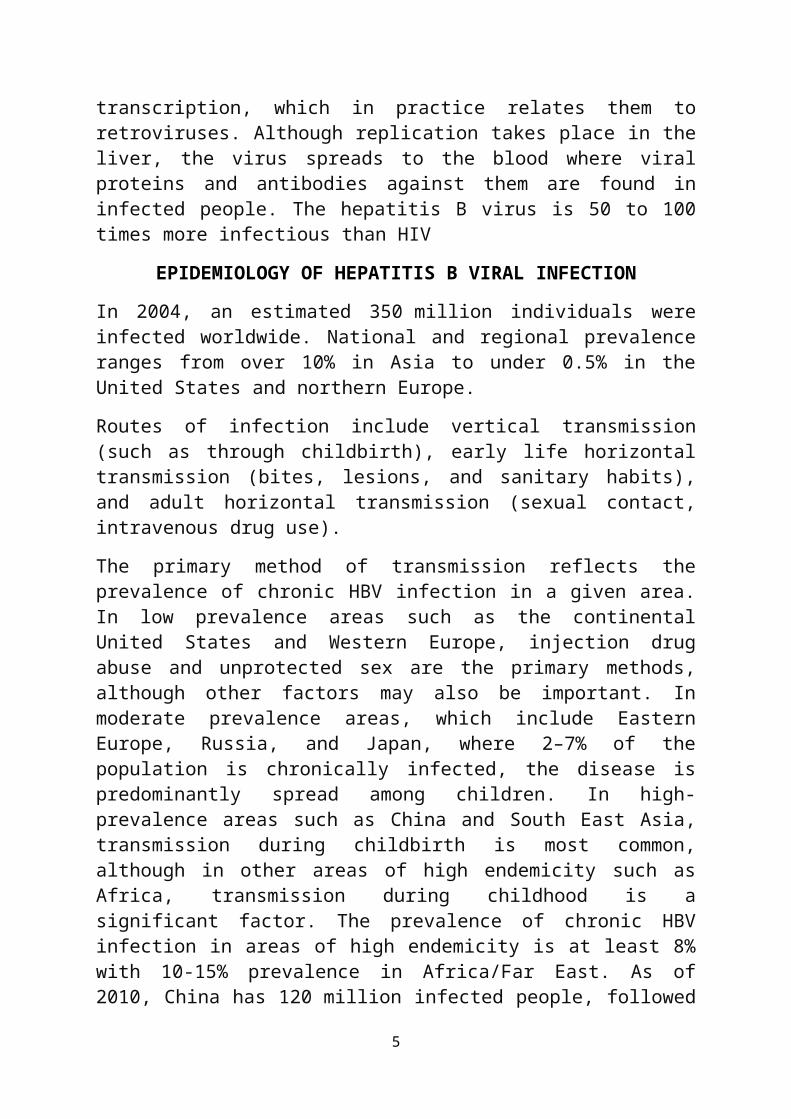
transcription, which in practice relates them toretroviruses. Although replication takes place in theliver, the virus spreads to the blood where viralproteins and antibodies against them are found ininfected people. The hepatitis B virus is 50 to 100times more infectious than HIV
EPIDEMIOLOGY OF HEPATITIS B VIRAL INFECTION
In 2004, an estimated 350 million individuals wereinfected worldwide. National and regional prevalenceranges from over 10% in Asia to under 0.5% in theUnited States and northern Europe.
Routes of infection include vertical transmission(such as through childbirth), early life horizontaltransmission (bites, lesions, and sanitary habits),and adult horizontal transmission (sexual contact,intravenous drug use).
The primary method of transmission reflects theprevalence of chronic HBV infection in a given area.In low prevalence areas such as the continentalUnited States and Western Europe, injection drugabuse and unprotected sex are the primary methods,although other factors may also be important. Inmoderate prevalence areas, which include EasternEurope, Russia, and Japan, where 2–7% of thepopulation is chronically infected, the disease ispredominantly spread among children. In high-prevalence areas such as China and South East Asia,transmission during childbirth is most common,although in other areas of high endemicity such asAfrica, transmission during childhood is asignificant factor. The prevalence of chronic HBVinfection in areas of high endemicity is at least 8%with 10-15% prevalence in Africa/Far East. As of2010, China has 120 million infected people, followed
5

by India and Indonesia with 40 million and12 million, respectively. According to World HealthOrganization (WHO), an estimated 600,000 people dieevery year related to the infection.
In the United States about 19,000 new cases occurredin 2011 down nearly 90% from 1990.
In Federal Teaching Hospital Abakaliki, out of atotal of 76 samples, collected in 2014, 15 werepositive for Hepatitis B virus infection representingabout 20% of the population. Also in 2013, out of 356samples received in 2013, 95 were positiverepresenting about 27% of the population. This wasmore in females than in males; a ratio of 9:7.
ANATOMY OF THE LIVER
The liver is a reddish brown organ with four lobes ofunequal size and shape. A human liver normally weighs1.44–1.66 kg (3.2–3.7 lb), and is a soft, pinkish-brown, triangular organ. It is both the largestinternal organ (the skin being the largest organoverall) and the largest gland in the human body. Itis located in the right upper quadrant of theabdominal cavity, resting just below the diaphragm.The liver lies to the right of the stomach andoverlies the gallbladder. It is connected to twolarge blood vessels, one called the hepatic arteryand one called the portal vein. The hepatic arterycarries blood from the aorta, whereas the portal veincarries blood containing digested nutrients from theentire gastrointestinal tract and also from thespleen and pancreas. These blood vessels subdivideinto capillaries, which then lead to a lobule. Eachlobule is made up of millions of hepatic cells which
6

are the basic metabolic cells. Lobules are thefunctional units of the liver.
FUNCTIONS OF THE LIVER
The functions of the liver are:
Synthesis of products like amino acids,carbohydrates etc.
Breakdown of certain substances like insulin,hormones
The liver also stores multitude of substanceslike glucose in the form glycogen.
The liver performs immunological functions.
The liver produces certain substances likealbumin etc.
7

PATHOPHYSIOLOGY OF HEPATITIS B VIRAL INFECTION
Hepatitis B virus primarily interferes with thefunctions of the liver by replicating in liver cells,known as hepatocytes. A functional receptor is NTCP.There is evidence that the receptor in the closelyrelated duck hepatitis B virus is carboxypeptidase D.The virions bind to the host cell via the preS domainof the viral surface antigen and are subsequentlyinternalized by endocytosis. HBV-preS-specificreceptors are expressed primarily on hepatocytes;however, viral DNA and proteins have also beendetected in extrahepatic sites, suggesting thatcellular receptors for HBV may also exist onextrahepatic cells.
During HBV infection, the host immune response causesboth hepatocellular damage and viral clearance.Although the innate immune response does not play asignificant role in these processes, the adaptiveimmune response, in particular virus-specificcytotoxic T lymphocytes(CTLs), contributes to most ofthe liver injury associated with HBV infection. CTLseliminate HBV infection by killing infected cells andproducing antiviral cytokines, which are then used topurge HBV from viable hepatocytes. Although liverdamage is initiated and mediated by the CTLs,antigen-nonspecific inflammatory cells can worsenCTL-induced immunopathology, and platelets activated
8
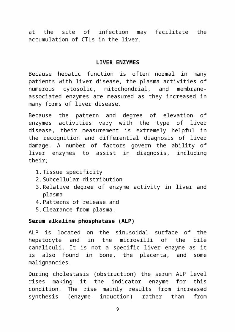
at the site of infection may facilitate theaccumulation of CTLs in the liver.
LIVER ENZYMES
Because hepatic function is often normal in manypatients with liver disease, the plasma activities ofnumerous cytosolic, mitochondrial, and membrane-associated enzymes are measured as they increased inmany forms of liver disease.
Because the pattern and degree of elevation ofenzymes activities vary with the type of liverdisease, their measurement is extremely helpful inthe recognition and differential diagnosis of liverdamage. A number of factors govern the ability ofliver enzymes to assist in diagnosis, includingtheir;
1.Tissue specificity2.Subcellular distribution3.Relative degree of enzyme activity in liver and
plasma4.Patterns of release and 5.Clearance from plasma.
Serum alkaline phosphatase (ALP)
ALP is located on the sinusoidal surface of thehepatocyte and in the microvilli of the bilecanaliculi. It is not a specific liver enzyme as itis also found in bone, the placenta, and somemalignancies.
During cholestasis (obstruction) the serum ALP levelrises making it the indicator enzyme for thiscondition. The rise mainly results from increasedsynthesis (enzyme induction) rather than from
9

obstructed outflow. The increase in cholestasis isusually to values greater than 300 UL whereas inhepatocellular disease the levels remain normal orare only slightly increased (to less than 300 U/L).
Subjects presenting with an isolated increased serumALP may be difficult to evaluate if the clinicalpicture is unhelpful. The ideal approach is todetermine the tissue of origin by separating andmeasuring the ALP isoenzymes using electrophoretictechniques (page 179). If this test is not availableestimation of the serum y-glutamyltransferase can behelpful -- a normal serum level makes liver diseaseunlikely; on the other hand a high value does notnecessarily indicate liver disease because of enzymeinduction (see below). A useful test, if available,is the serum 5'-nucleotidase which originatespredominantly from the liver and is usually onlyincreased in subjects with liver disease.
LIVER TRANSAMINASES
Traditionally the LFT enzyme profile has included onetransaminase, either aspartate aminotransferase (AST)or alanine aminotransferase (ALT). The transaminasesare located in the hepatocyte and high plasma levelsindicate release from the cells during damage(hepatocellular disease). Values in excess of 400 U/Loccur in hepatocellular disease whilst in cholestasisthe level is normal or only slightly raised (<400U/L.).
AST has a wide distribution and a raised serum valueis a non-specific event with the possibilitiesincluding destruction of hepatocytes, myocardium,skeletal muscle, or erythrocytes. ALT also has a widedistribution in the body but a raised serum level ismost likely to reflect liver cell damage, i.e., for
10

clinical purposes a high serum ALT indicates liverpathology.
In the hepatocyte ALT is located only in the cytosolwhereas AST is found in the cytosol and in themitochondria and thus it has been suggested,particularly in the German literature, that in the
context of liver disease a serum AST value in excessof the serum ALT value indicates a severe diseaseprocess. The reasoning behind this is as follows:Most of the raised transaminases in liver diseasereflects 'seepage' of enzymes through damaged cellmembranes, i.e., the cytosol components escapeleaving the mitochondria behind. Hence the serum ALTlevel will exceed the AST level. If damage is severeand many hepatocytes are destroyed then, in additionto the cytosol component, the mitochondrial enzymesare released as well, giving an AST value higher thanthe ALT value.
There is good evidence in the literature, andpersonal experience confirms this, that alcoholicliver disease is associated with a high AST:ALTratio; and it is further suggested that when theserum transaminase concentrations are raised, butless than ten-fold the upper reference limit (i.e.,<400 UL), an AST:ALT ratio greater than 2 is strongevidence for alcoholic liver disease. A paper byCohen and Kaplan in 1969 showed that of 104 patientswith alcoholic liver disease, 70% had ratios above 2,whereas of 167 patients with non-alcoholic liverdisease only 9.6% has ratios above 2 (using a cut-offof 1.5 the proportions were 85% and 25%respectively).
The cause of the high ratio is unclear; it isunlikely to be due to the mitochondrial AST component
11

because liver biopsies show the same pattern asserum.
A suggested reason is reduced hepatic pyridoxal-5-phosphate which is a co-factor for both enzymes butits deficiency has a greater impact on ALT activity.
Serum y-glutamyltransferase (GGT)
GGT, which catalyses the transfer of y-glutamylgroups from one peptide to another, is involved inthe transport of amino acids across cell membranes.The liver, kidney, pancreas, and prostate are themain sources of GGT. Several isoenzymes have beendescribed but have not yet been shown to be ofclinical value. GGT is associated with the cellmembrane fraction, which includes the microsomes,stimulation of which induces the synthesis of a hostof enzymes. GGT can be induced by drugs such asphenytoin and phenobarbital and other xenobioticssuch as alcohol.
The plasma level is increased in both hepatocellularand cholestatic liver disease and is a sensitivemarker of hepatic disease. However, two othercharacteristics can cause difficulties ininterpretation. Firstly, on odd occasions, severeliver disease can be associated with normal
serum GGTP levels and secondly, increased serumlevels, due to enzyme induction, can present in theabsence of liver disease (as occurs in chronicalcoholism, with the drugs phenytoin sodium,
barbiturates, warfarin, and amitriptyline, and indisorders such as obesity, diabetes mellitus, andhypertriglyceridaemia). Despite these problems, it isa useful test which often provides the firstindication of a patient’s increased alcohol
12

consumption, even though its specificity for thisdisorder is low.
LIVER ENZYME PATTERNS AND RESPONSE TO HBV INFECTION
Acute hepatitis refers to an injury directed againstthe hepatocytes. The injury may be mediated eitherdirectly or indirectly. Direct injury occurs withcertain drugs, such as acetaminophen, or withischaemia. Indirect injury is immunologicallymediated injury that occurs with hepatitis virues andethanol. With indirect injury, there is a;
1.Gradual rise in cytosolic enzymes (i.e. AST, ALTand Lactate Dehydrogenase).
2.A plateau phase and3.Gradual resolution of enzyme elevation
Acute Hepatitis B virus infection leads to thenecrosis of the hepatocytes resulting in the releaseof the Transaminases, lactate dehydrogenase andalkaline phosphatise.
Increase in AST and ALT in acute hepatitis B virusinfection is usually is 8-50 times of the upperreference limit while the Alkaline phophatase isusually mildly elevated, and is less than 3 times theupper reference limit in 90% of acute hepatitis Bvirus infection (Dufour, 2006).
ALT is typically higher than AST because of slowerclearance. Enzyme elevation typically peak beforepeak bilirubin occurs, and remain increased for anaverage of 4 to 5 weeks (longer for ALT than ASTbecause of its longer half-life).
Lactate dehydrogenase is usually more affected indirect injury to the liver example in theacetamenophen induced drug injury, and may not be
13

significantly affected by indirect injury to theliver including that caused by hepatitis B virus.
ALP is usually mildly elevated, and is less thanthree times the upper reference limit in 90% of casesof acute hepatitis.
Chronic hepatitis B infection is characterised byongoing inflammatory damage to hepatocytes (lastinglonger than six months), often accompanied byhepatocyte regeneration and scaring. Most cases ofchronic hepatitis b virus infection are diagnosedbecause of increased transaminases (ALT and AST)typically 1 to 5 times upper reference limits.Aminotransferases may sometimes be normal eitherintermittently or for a prolonged period in chronichepatitis B virus infection. Reversal of the AST:ALTratio to greater than one suggest a coexistingalcohol abuse or development of cirrhosis.
The reference ranges of the liver enzymes includingALT, AST, and ALP usually depend on the method used,the type of kit used and on the specific geographicallocation. Below is a table showing the referenceranges of the liver enzymes.
AST 1-12IU/L
ALT 1-12IU/L
ALP 9-35IU/L
SIGNS AND SYMPTOMS OF HBV INFECTION
Acute infection with hepatitis B virus is associatedwith acute viral hepatitis – an illness that beginswith general ill-health, loss of appetite, nausea,vomiting, body aches, mild fever, and dark urine, andthen progresses to development of jaundice. It hasbeen noted that itchy skin has been an indication as
14

a possible symptom of all hepatitis virus types. Theillness lasts for a few weeks and then graduallyimproves in most affected people. A few people mayhave more severe liver disease (fulminant hepaticfailure), and may die as a result. The infection maybe entirely asymptomatic and may go unrecognized.
Chronic infection with hepatitis B virus either maybe asymptomatic or may be associated with a chronicinflammation of the liver (chronic hepatitis),leading to cirrhosis over a period of several years.This type of infection dramatically increases theincidence of hepatocellular carcinoma (liver cancer).Across Europe hepatitis B and C cause approximately50% hepatocellular carcinomas. Chronic carriers areencouraged to avoid consuming alcohol as it increasestheir risk for cirrhosis and liver cancer. HepatitisB virus has been linked to the development ofmembranous glomerulonephritis (MGN).
Symptoms outside of the liver are present in 1–10% ofHBV-infected people and include serum-sickness–likesyndrome, acute necrotizing vasculitis (polyarteritisnodosa), membranous glomerulonephritis, and papularacrodermatitis of childhood (Gianotti–Crostisyndrome). The serum-sickness–like syndrome occurs inthe setting of acute hepatitis B, often preceding theonset of jaundice. The clinical features are fever,skin rash, and polyarteritis. The symptoms oftensubside shortly after the onset of jaundice, but canpersist throughout the duration of acute hepatitis B.About 30–50% of people with acute necrotizingvasculitis (polyarteritis nodosa) are HBV carriers.HBV-associated nephropathy has been described inadults but is more common in children. Membranousglomerulonephritis is the most common form. Other
15
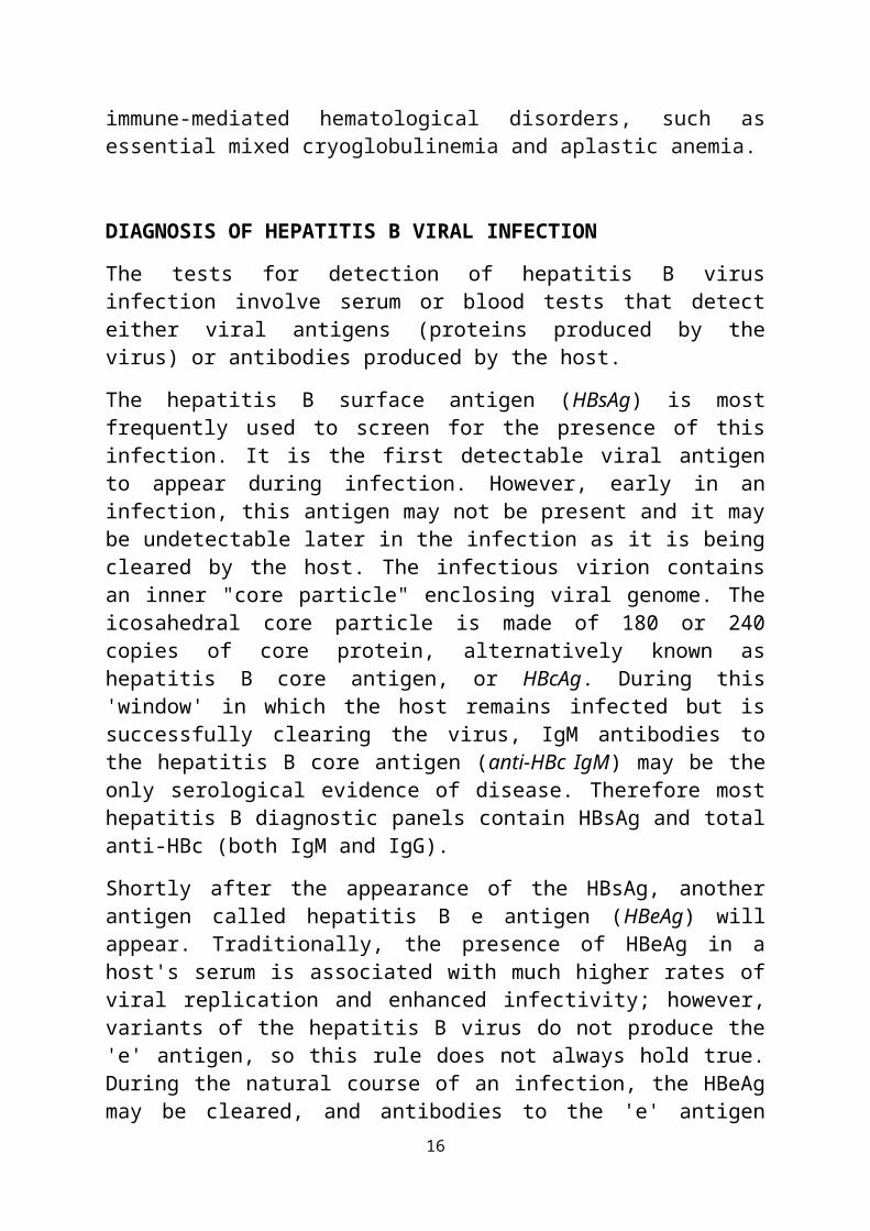
immune-mediated hematological disorders, such asessential mixed cryoglobulinemia and aplastic anemia.
DIAGNOSIS OF HEPATITIS B VIRAL INFECTION
The tests for detection of hepatitis B virusinfection involve serum or blood tests that detecteither viral antigens (proteins produced by thevirus) or antibodies produced by the host.
The hepatitis B surface antigen (HBsAg) is mostfrequently used to screen for the presence of thisinfection. It is the first detectable viral antigento appear during infection. However, early in aninfection, this antigen may not be present and it maybe undetectable later in the infection as it is beingcleared by the host. The infectious virion containsan inner "core particle" enclosing viral genome. Theicosahedral core particle is made of 180 or 240copies of core protein, alternatively known ashepatitis B core antigen, or HBcAg. During this'window' in which the host remains infected but issuccessfully clearing the virus, IgM antibodies tothe hepatitis B core antigen (anti-HBc IgM) may be theonly serological evidence of disease. Therefore mosthepatitis B diagnostic panels contain HBsAg and totalanti-HBc (both IgM and IgG).
Shortly after the appearance of the HBsAg, anotherantigen called hepatitis B e antigen (HBeAg) willappear. Traditionally, the presence of HBeAg in ahost's serum is associated with much higher rates ofviral replication and enhanced infectivity; however,variants of the hepatitis B virus do not produce the'e' antigen, so this rule does not always hold true.During the natural course of an infection, the HBeAgmay be cleared, and antibodies to the 'e' antigen
16

(anti-HBe) will arise immediately afterwards. Thisconversion is usually associated with a dramaticdecline in viral replication.
CONCLUSION
Hepatitis B virus infection is an inflammation of theliver caused by the Hepatitis B virus. It is veryinfectious and can be transmitted by various meansincluding, sexual contact, blood transfusion, sharingof sharp objects, even with less intimate contactswith the carriers of the virus. This is especiallytrue because the virus can penetrate the unbrokenskin. Symptoms of acute infection with the virusinclude, general ill-health, loss of appetite,nausea, vomiting, body aches, mild fever, and darkurine, and then progresses to development ofjaundice. Hepatitis B virus infection normally causesa raise in the liver enzymes including, AST, ALT, andsometimes ALP. Hence diagnosis of the infection canalso include a raise in the serum level of theseenzymes. Vaccines for the virus are available.
RECOMMENDATION
Since most cases of hepatitis B virus infectionis asymptomatic, i recommend that people go forhepatitis B screening whether or not they feelthe symptoms or not. Because asymptomatichepatitis B virus infection can lead to cirrhosis
17

or liver failure after a period of about 20years.
It is also recommended that every sample that isreceived in the laboratory be treated with therequired precautions since they may contain verycontagious infections like hepatitis B viruswhich can break through the unbroken skinbarrier.
Every healthcare personnel and unvaccinatedadults should be vaccinated, and also receivebooster doses of hepatitis B vaccines.
I equally recommend that pregnant mothers go forhepatitis B screening as part of their antenatalscreening tests before delivery.
There should be proper waste disposal managementin the laboratory to ensure safe disposal ofhepatitis b infected materials.
REFERENCES
Alpert E, Isselbacher KJ, Schur PH (1971). "Thepathogenesis of arthritis associated with viralhepatitis. Complement-component studies". The NewEngland Journal of Medicine 285 (4): 185–9.
Barker LF, Shulman NR, Murray R, Hirschman RJ, RatnerF, Diefenbach WC, Geller HM (1996). "Transmissionof serum hepatitis. 1970". Journal of the AmericanMedical Association 276 (10): 841–844.
Beck J, Nassal M (2007). "Hepatitis B virusreplication". World J. Gastroenterol. 13 (1): 48–64.
Bruss V (2007). "Hepatitis B virus morphogenesis".World J. Gastroenterol. 13 (1): 65–73.
18

Chang MH (2007). "Hepatitis B virus infection". SeminFetal Neonatal Med 12 (3): 160–167.
Coopstead, Lee-Ellen C. (2010). Pathophysiology.Missouri: Saunders. pp. 886–887.
Dienstag JL (1981). "Hepatitis B as an immune complexdisease". Seminars in Liver Disease 1 (1): 45–57.
El-Serag, Hashem B. (2011). "Hepatocellularcarcinoma". New England Journal of Medicine 365 (12):1118–1127.
El-Serag, HB; Rudolph, KL (2007). "Hepatocellularcarcinoma: epidemiology and molecularcarcinogenesis". Gastroenterology 132 (7): 2557–76.
Gan SI, Devlin SM, Scott-Douglas NW, Burak KW (2005)."Lamivudine for the treatment of membranousglomerulopathy secondary to chronic hepatitis Binfection". Canadian journal of gastroenterology = Journalcanadien de gastroenterologie 19 (10): 625–9.
Gocke DJ, Hsu K, Morgan C, Bombardieri S, Lockshin M,Christian CL (1970). "Association betweenpolyarteritis and Australia antigen". Lancet 2(7684): 1149–53.
Harrison T (2009). Desk Encyclopedia of General Virology.Boston: Academic Press. p. 455.
Howard, C. R. (1986). "The Biology ofHepadnaviruses". Journal of General Virology 67 (7):1215–1235.
Kay A, Zoulim F (2007). "Hepatitis B virus geneticvariability and evolution". Virus research 127 (2):164–176.
Kidd-Ljunggren K, Holmberg A, Bläckberg J, LindqvistB (2006). "High levels of hepatitis B virus DNA
19

in body fluids from chronic carriers". The Journal ofHospital Infection 64 (4): 352–7.
Kramvis A, Kew M, François G (2005). "Hepatitis Bvirus genotypes". Vaccine 23 (19): 2409–23.
Lai KN, Li PK, Lui SF, et al. (1991). "Membranousnephropathy related to hepatitis B virus inadults". The New England Journal of Medicine 324 (21):1457–63.
Li W, Miao X, Qi Z, Zeng W, Liang J, Liang Z (2010)."Hepatitis B virus X protein upregulatesHSP90alpha expression via activation of c-Myc inhuman hepatocarcinoma cell line, HepG2". Virol. J. 7:45.
Liang TJ (2009). "Hepatitis B: the virus anddisease". Hepatology (Baltimore, Md.) 49 (5 Suppl):S13–21.
Locarnini, S. (2004). "Molecular Virology ofHepatitis B Virus". Seminars in Liver Disease 24: 3–10.
Pungpapong S, Kim WR, Poterucha JJ (2007). "NaturalHistory of Hepatitis B Virus Infection: an Updatefor Clinicians". Mayo Clinic Proceedings 82 (8): 967–975.
Schilsky, M. L. (2013). "Hepatitis B "360"".Transplantation Proceedings 45 (3): 982–985.
Sleisenger, MH; Feldman M, Friedman LS (2006).Fordtran's gastrointestinal and liver disease: pathophysiology,diagnosis, management (8th ed.). Philadelphia:Saunders.
Takekoshi Y, Tanaka M, Shida N, Satake Y, Saheki Y,Matsumoto S (1978). "Strong association betweenmembranous nephropathy and hepatitis-B surface
20

antigenaemia in Japanese children". Lancet 2(8099): 1065–8.
21