Dynamic Localization of Trypanosoma brucei Mitochondrial DNA Polymerase ID
Upload
khangminh22Category
view
3download
0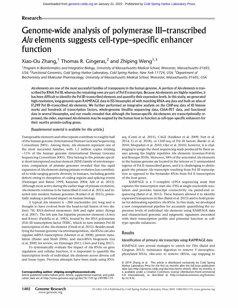
Genome-wide analysis of polymerase III–transcribedAlu elements suggests cell-type–specific enhancerfunction
Xiao-Ou Zhang,1 Thomas R. Gingeras,2 and Zhiping Weng1,3
1Program in Bioinformatics and Integrative Biology, University of Massachusetts Medical School, Worcester, Massachusetts 01605,USA; 2Functional Genomics, Cold Spring Harbor Laboratory, Cold Spring Harbor, New York 11724, USA; 3Department ofBiochemistry and Molecular Pharmacology, University of Massachusetts Medical School, Worcester, Massachusetts 01605, USA
Alu elements are one of the most successful families of transposons in the human genome. A portion of Alu elements is tran-
scribed by RNA Pol III, whereas the remaining ones are part of Pol II transcripts. Because Alu elements are highly repetitive, it
has been difficult to identify the Pol III–transcribed elements and quantify their expression levels. In this study, we generated
high-resolution, long-genomic-span RAMPAGE data in 155 biosamples all with matching RNA-seq data and built an atlas of
17,249 Pol III–transcribed Alu elements. We further performed an integrative analysis on the ChIP-seq data of 10 histone
marks and hundreds of transcription factors, whole-genome bisulfite sequencing data, ChIA-PET data, and functional
data in several biosamples, and our results revealed that although the human-specific Alu elements are transcriptionally re-
pressed, the older, expressed Alu elements may be exapted by the human host to function as cell-type–specific enhancers for
their nearby protein-coding genes.
[Supplemental material is available for this article.]
Transposable elements andother repeats contribute to roughlyhalfof the human genome (InternationalHumanGenome SequencingConsortium 2001). Among them, Alu elements represent one ofthe most successful families, with 1.2 million copies totaling∼11% of the human genome (International Human GenomeSequencing Consortium 2001). They belong to the primate-specif-ic short interspersednuclear element (SINE) familyof retrotranspo-sons; comparison of primate genomes revealed that the rapidexpansionofAlu elements duringprimate evolutionhas contribut-ed to wide-ranging genetic diversity in humans, including geneticdefects owing to disruption of coding regions and splicing events(Deininger and Batzer 1999; Kazazian 2004; Ade et al. 2013).Althoughmore active during the earlier stage of primate evolution,Alu elements continue to be transcribed (Conti et al. 2015) and in-serted into modern human genomes (Konkel et al. 2015), poten-tially making a profound impact on human biology.
A typical Alu element is ∼280 nucleotides (nt) long and isthought to have evolved from the head-to-tail fusion of two dis-tinct 7SL RNA-derived monomers (left and right arms) (Kriegset al. 2007). The left arm has bipartite promoter elements (A-boxand B-box) (Paolella et al. 1983), bound by the RNA polymerase(Pol) III transcription factor TFIIIC, which in turn initiates Pol IIItranscription of the Alu element (Orioli et al. 2012). Besides modi-fying the humangenome via retrotransposition,AluRNAs can alsoregulate mRNA transcription (Mariner et al. 2008), protein trans-lation (Hasler and Strub 2006), and microRNA biogenesis (Guet al. 2009; for review, see Deininger 2011; Chen and Yang 2017).
To systematically evaluate the impact of Alu RNAs on generegulation and cellular function, it is important to quantify thetranscription levels of individual Alu elements across diverse celland tissue types. Previous attempts have been made using RNA-
seq (Conti et al. 2015), CAGE (Faulkner et al. 2009; Fort et al.2014; Li et al. 2018), or ChIP-seq of Pol III factors (Barski et al.2010; Moqtaderi et al. 2010; Oler et al. 2010); however, it is chal-lenging to assign the short sequencing reads produced by these as-says among the highly repetitive Alu elements (Goerner-Potvinand Bourque 2018). Moreover, 58% of the annotated Alu elementsin the human genome are located in the introns or 3′ untranslatedregions of Pol II–transcribed genes, and it is challenging to distin-guish the primary Alu transcripts resulting from Pol III transcrip-tion as opposed to the bystander RNAs from Pol II transcriptionof the host genes.
RAMPAGE is a 5′-complete cDNA sequencing assay thatcaptures the transcription start site (TSS) at single-nucleotide reso-lution and provides transcript connectivity via paired-end se-quencing (Batut et al. 2013). These features allowed it to identifyexpressed transposons in flies (Batut et al. 2013) and to hold prom-ise for delineating repetitive Alu RNAs. In this study, we developeda new computational pipeline for accurately quantifying the ex-pression levels of individual Alu elements using RAMPAGE dataand characterized genomic and epigenetic signatures associatedwith their transcription profile and potential function as cell-type–specific enhancers.
Results
Identification of primary Alu transcripts using RAMPAGE data
RAMPAGE uses several strategies to enrich for TSSs (Batut andGingeras 2013): terminator digestion to remove 5′-monophos-phorylated RNAs, ribo-zero to remove rRNAs, cap trapping to
Corresponding author: [email protected] published online before print. Article, supplemental material, and publi-cation date are at http://www.genome.org/cgi/doi/10.1101/gr.249789.119.
© 2019 Zhang et al. This article is distributed exclusively by Cold SpringHarbor Laboratory Press for the first six months after the full-issue publicationdate (see http://genome.cshlp.org/site/misc/terms.xhtml). After six months, itis available under a Creative Commons License (Attribution-NonCommercial4.0 International), as described at http://creativecommons.org/licenses/by-nc/4.0/.
Research
1402 Genome Research 29:1402–1414 Published by Cold Spring Harbor Laboratory Press; ISSN 1088-9051/19; www.genome.orgwww.genome.org
Cold Spring Harbor Laboratory Press on January 31, 2022 - Published by genome.cshlp.orgDownloaded from

enrich for 5′-capped RNAs, and template switching to enrichfor 5′-triphosphorylated and 5′-capped RNAs. Like other Pol III–transcribed RNAs, Alu RNAs have a 5′-triphosphorylated end butlack the 5′-cap structure (Reddy 1988; Burke et al. 2016).Although the cap trapping assay enriches for RNAs with a 5′-cap,the assay still captures uncapped 5′-triphosphorylated transcripts(Takahashi et al. 2012). Thus, Alu RNAs are enriched byRAMPAGE, although not as enriched as Pol II–transcribed RNAswith 5′-capped ends. Furthermore, with single-nucleotide resolu-tion for identifying TSS and connectivity between the TSS andthe downstream transcript, RAMPAGE holds great promise for an-notating repetitive Alu elements.
As part of the ENCODE Project, we produced RAMPAGE andRNA-seq data in 155 biosamples with high quality and reproduc-ibility (Supplemental Figs. S1, S2A; Supplemental Table S1;Supplemental Material). To use RAMPAGE peaks to identify indi-vidual primary Alu RNAs transcribed by Pol III, we must overcometwo challenges. First, a full-size Alu element contains two repeatedarms both evolved from the 7SL RNA (Supplemental Fig. S2B), andthe 1.2 million copies of Alu elements in the human genome havehighly similar sequences; thus, it is challenging to assign short se-quencing reads to the bodies of individual Alu elements. Instead,we relied on the downstream unique sequences captured byRAMPAGE: Pol III transcription initiates at the 5′-end of each Aluusing the internal promoter elements A-box and B-box locatedin the left arm (Supplemental Fig. S2B) and terminates down-stream from the Alu body because the body lacks internal termina-tion elements (Erwin et al. 2014). Furthermore, paired-endRAMPAGE reads link the downstream sequences to the TSSs ofthe corresponding Alu elements, so we designed our computation-al pipeline to capture this information (Fig. 1A; SupplementalMethods). Second, the Pol III–transcribed Alu RNAs are generallyexpressed at low levels in somatic cells (Paulson and Schmid1986; Conti et al. 2015), and these weak signals can be contami-nated by the typically much stronger Pol II transcription signalseven when they are not near a TSS. We developed two measure-ments—entropy (E) and effective length (L) to filter out false-pos-itive RAMPAGE peaks (Fig. 1A; Supplemental Methods). ForGENCODE-annotated genes, TSS-overlapping RAMPAGE peakshave much higher entropies than the other peaks, and an entropycutoff of 2.5 clearly separated the two populations of peaks(Supplemental Fig. S2C); thus, we applied the same entropy cutoffto RAMPAGE peaks that annotated Alu elements. As primary Alutranscripts are generally unspliced (Conti et al. 2015) and the frag-ment sizes of RAMPAGE libraries are <1 kb (Batut and Gingeras2013), we used an effective length cutoff of 1000 nt to further filterout RAMPAGE peaks with improperly long genomic spans(Supplemental Fig. S2D).
Thus, our computational pipeline took advantage of the twounique features of RAMPAGE technique (TSS identification andconnection to the downstream transcripts) and the features of pri-mary Alu elements (unspliced and transcribed past its body). Thispipeline allowed us to comprehensively identify Pol III–tran-scribed Alu elements using only RAMPAGE data. Figure 1B showsan example intergenic AluSx1 element, which is transcribed byPol III from its 5′-end (bound by its main subunits POLR3A andTFIIIC) to downstream (RNA-seq signal). One RAMPAGE peakwith 19 reads precisely annotates the TSS of this AluSx1 elementwith the RAMPAGE read pairs further depicting its transcriptionalprofile (Fig. 1B). Supplemental Figure S2E shows four more exam-ples with unfiltered RAMPAGE reads, two intergenic and twointronic.
We performed a series of computational tests and confirmedthat the expressed Alu elements identified by our RAMPAGE pipe-line resulted from Pol III transcription (Fig. 1C,D; SupplementalFigs. S3A–D, S4; Supplemental Material). We also compared theexpressed Alu elements identified by our RAMPAGE pipeline withthe expressed Alu elements using RNA-seq and ChIP-seq data(Moqtaderi et al. 2010; Conti et al. 2015), and our method showedhigher sensitivity and specificity than the earlier approaches(Fig. 1E–G; Supplemental Fig. S3E,F; Supplemental Material).
Transcribed Alu elements show high tissue specificity
We applied our pipeline to the RAMPAGE data in 155 biosamples(derived from 27 cell lines, 16 primary cell types, and 45 tissues,with some cell and tissue types having multiple biosamples asdetailed in Supplemental Table S1) and identified 17,249 Alu ele-ments that were expressed in at least one biosample (Fig. 2A; Sup-plemental Table S2), which accounted for 1.44% of the 1,194,734annotated Alu elements in the human genome. This result is con-sistent with a previous RNA-seq study reporting that only a limitednumber of Alu elements were transcribed into primary transcripts(Conti et al. 2015). Roughly twice as many expressed Alu elementswere detected in tissues than in cell lines or primary cells (medianN=181, 91, 82, respectively) (Supplemental Fig. S5A, left), reflect-ing the heterogeneous cell type compositions in the tissue sam-ples. The expression levels of the expressed Alu elements wereslightly higher in primary cells than in cell lines and tissues (medi-an=0.76, 0.73, and 0.71 RPM, respectively) (Supplemental Fig.S5A, right). Most of the expressed Alu elements (10,622 of17,249; 62%) were transcribed in a single biosample (Fig. 2B), andthe Alu elements expressed in multiple biosamples had higher ex-pression levels than those expressed in a single biosample (Supple-mental Fig. S5B). We defined Alu elements expressed in three ormore biosamples as robustly transcribed Alu elements henceforth.
Retrotransposons are generally expressed with high tissuespecificity (Faulkner et al. 2009). We asked whether transcribedAlu elements showed tissue specificity. We first clustered the104 tissue biosamples by their expression profiles of Alu elements,and biologically related biosamples clearly grouped together(Fig. 2C; Supplemental Fig. S5C; Supplemental Table S3). Wethen perform a detailed analysis on a subset of 52 RAMPAGEdata sets from 13 tissues of four individuals. Because most of theexpressed Alu elements were transcribed in only one biosample,the overall correlations between biosamples were low, yet similartissues still clustered together (Fig. 2D). For example, gastroesoph-ageal sphincter, esophagus muscularis mucosa, and sigmoid co-lon, which are all components of the digestive system, clusteredtogether with high correlations. Furthermore, biosamples fromthe same tissue but different individuals had substantially highercorrelations than biosamples from different tissues of the sameindividual (median Pearson correlation coefficient 0.41 vs. 0.10;P-value=1.10×10⁻¹⁴) (Fig. 2D). We also observed distinct expres-sion patterns for Alu elements in specific tissue types (e.g., testis)(Supplemental Fig. S5D,E) or associated with specific factors (e.g.,DICER1) (Supplemental Material). Together, these results indicatethat the expression profile of Alu elements reflects the regulatoryprograms of the specific tissue type.
Expression of primary Alu transcripts relies more on the genomic
context than on primary sequences
We asked whether expressed Alu elements tended to be youngerand had stronger regulatory sequence motifs than unexpressed
Pol III–transcribed Alu elements
Genome Research 1403www.genome.org
Cold Spring Harbor Laboratory Press on January 31, 2022 - Published by genome.cshlp.orgDownloaded from
E
F
BA
C
D
G
Figure 1. Genome-wide identification of expressed Alu elements using RAMPAGE data. (A) A computational pipeline to call RAMPAGE peaks and anno-tate expressed Alu elements. Paired-end RAMPAGE reads (R1 and R2, black; N, read pairs in total) with uniquely mapping R2 readswere first clustered to callpeaks at the 5′-end of R1, and the resulting peaks (blue bars) were further filtered with entropy (E) and effective length (L) to annotate expressed Alu el-ements (orange bar) (for more details, see SupplementalMethods). (B) An expressed AluSx1 element in an intergenic region. RAMPAGE peakmarks the TSSof the AluSx1 element precisely, with RAMPAGE read pairs linking the TSS to downstream positions. RNA-seq and ChIP-seq data sets of POLR3A and TFIIICfurther confirm the expression of this AluSx1 element. (C) POLR3A (left) and POLR2A (right) ChIP-seq signals are shownwith respect to the TSS of expressedAlu elements (orange line), protein-coding and lncRNA genes transcribed by Pol II (pink line), and unexpressed Alu elements (gray line). The signals wereaveraged over all genes in each set. (D) RNA-seq signal in the ±100-bp window (in 10-nt bins) centered on the TSSs of robustly expressed Alu elementsidentified by RAMPAGE (identified in three or more biosamples). To avoid overlap with the TSSs of Pol II–transcribed genes, only intronic and intergenicAlu elements were included. Each row of the heatmap corresponds to one such Alu element in one biosamplewith 10 or more nonzero RNA-seq signal bins,and the dot and bars correspond to the median and first and third quartiles of all Alu elements. (E) Only 84 of the 1593 Alu elements that overlap POLR3Aand TFIIIC ChIP-seq peaks (Moqtaderi et al. 2010) are expressed according to RAMPAGE or RNA-seq data in K562 cells. On the other hand, 297 expressedAlu elements (by RAMPAGE or RNA-seq; four by both) do not overlap POLR3A and TFIIIC peaks. (F) Heatmap of normalized read densities for POLR3A andTFIIIC ChIP-seq data around the TSSs of expressed Alu elements identified by RAMPAGE, RNA-seq (Conti et al. 2015), or both techniques in K562 cells. ChIP-seq peaks (Moqtaderi et al. 2010) are labeled on the right of the corresponding heatmaps. (G) Among the 219 expressed Alu elements (by RAMPAGE orRNA-seq) that do not overlap POLR3A or TFIIIC peaks, the Alu elements identified by RAMPAGE show significantly higher POLR3A and TFIIIC signals thanthe Alu elements defined by RNA-seq. Wilcoxon rank-sum test P-values are shown.
Zhang et al.
1404 Genome Researchwww.genome.org
Cold Spring Harbor Laboratory Press on January 31, 2022 - Published by genome.cshlp.orgDownloaded from

elements. Alu elements are classified into three subfamilies withdecreasing evolutionary ages, AluJ, AluS, and AluY, and it hasbeen proposed that AluY elements, being the youngest and leastdegenerated in sequences, might represent the most retrotranspo-sitionally active subfamily (Bennett et al. 2008). Indeed,AluY is theonly known subfamily currently active in retrotransposition in thehuman genome (Konkel et al. 2015).
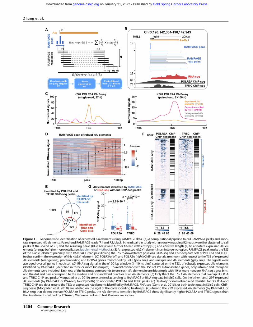
In contrast to expectation, there was a slight depletion of ex-pressed AluS and AluY elements compared with the oldest subfam-ily, AluJ (Supplemental Fig. S6A). Furthermore, we did not observeany difference in evolutionary divergence between expressed andunexpressed Alu elements within each subfamily (SupplementalFig. S6B). To investigate this problem in greater depth, we classifiedexpressed Alu elements into five groups, B0–B4, depending onwhether they were in four other primate genomes (chimpanzee,gorilla, orangutan, or macaque), with B4 defining the human-specific Alu elements (N=103), i.e., the Alu elements that did
not exist in the other four primate genomes, and B0–B3 were col-lectively designated nonhuman-specific (Fig. 3A; SupplementalFig. S6C; Supplemental Table S4; Supplemental Methods). As ex-pected, the majority of human-specific Alu elements (88%) werein the AluY class (Fig. 3B); nevertheless, these human-specificAluY elements (N=91) had significantly lower sequence diver-gence than the remaining AluY elements (median= 1.6 vs. 6.7;P-value=2.42×10−50) (Fig. 3C), whereas the differences weremuch less apparent for the AluS and AluJ subfamilies (Fig. 3C). In-deed, the human-specific Alu elements were expressed at signifi-cantly lower levels than nonhuman-specific Alu elements (P-value= 0.04) (Supplemental Fig. S6D, left), and our results becomemore significant when we contrasted human-specific AluY ele-ments against nonhuman-specific AluY elements (P-value =1.83×10⁻⁴) (Fig. 3D, left) or human-specific AluS and AluJ elements(P-value=1.15×10⁻³) (Fig. 3D, right). To test whether our map-ping strategy might be biased against younger elements, we
BA
C D
Figure 2. Expressed Alu elements show high tissue specificity. (A) Venn diagram showing expressed Alu elements defined using RAMPAGE data in celllines (red), primary cells (green), and tissues (blue). (B) Histogram showing counts of Alu elements expressed in different numbers of biosamples. Notethat Alu elements identified in three or more biosamples were defined as robustly expressed Alu elements (robust Alu elements). (C) Dendrogram resultingfrom agglomerative hierarchical clustering of tissue biosamples based on their Alu expression. Each leaf of the tree represents one RAMPAGE tissue sample,and subtrees dominated by one tissue type are highlighted. (D) Correlationmatrix of expressed Alu elements across libraries belonging to 13 tissues of fourindividuals. Note that correlations between biosamples in the same tissue but from different individuals are significantly higher than correlations betweenbiosamples from the same individual but different tissues. Wilcoxon rank-sum test P-values are shown.
Pol III–transcribed Alu elements
Genome Research 1405www.genome.org
Cold Spring Harbor Laboratory Press on January 31, 2022 - Published by genome.cshlp.orgDownloaded from
modified our pipeline by permitting multiple-mapping reads. Theresults still showed that human-specific Alu elements showed low-er expression levels than nonhuman-specific ones (P-value =0.02)(Supplemental Fig. S6E, left), especially in the AluY subfamily (P-value =3.54×10⁻⁵) (Supplemental Fig. S6E, right). These resultssuggest that compared with other expressed Alu elements, hu-man-specificAluY elements are relatively repressed in human cells.
Furthermore, our B0–B4 classification can better capture the po-tential activities of Alu elements than the subfamily classification,as we did not detect a significant difference between AluY andAluS/J subfamilies (Supplemental Fig. S6D, right).
To understand whether expressed and unexpressed Alu ele-ments were regulated differently, we performed de novo motiffinding on these two sets of elements separately (Supplemental
E F
BA
C D
G H
Figure 3. Expressed Alu elements show distinct genomic context and sequence features. (A) The primate phylogeny. B4 denotes the human-specificbranch, and B0–B3 denote nonhuman-specific branches. (B) The AluY subfamily accounted for a large proportion of human-specific Alu elements.(C ) Sequence divergence distributions of human-specific (solid lines) and nonhuman-specific (dashed lines) Alu elements in each Alu subfamily. Notethat human-specific AluY elements show lower sequence divergence than nonhuman-specific AluY elements. (D) Human-specific AluY elements showedlower expression levels (measured by RPM) than nonhuman-specific AluY elements (left) and human-specific AluS/J elements (right). Wilcoxon rank-sumtest P-values are shown. (E) Robustly expressed (red) and expressed (orange) Alu elements are closer to the TSS of Pol II–transcribed genes than are unex-pressed (gray) Alu elements.Wilcoxon rank-sum test P-values are shown. (F) Robustly expressed (red) and expressed (orange) Alu elements aremore likely tobe located in gene-rich regions than are unexpressed (gray) Alu elements. Wilcoxon rank-sum test P-values are shown. (G) Receiver operating characteristic(ROC) curve of random forest classifiers for distinguishing robustly expressed or expressed against unexpressed Alu elements using genomic context andsequence features. (AUC) Area under the curve. (H) The top 20most important features of the random forest classifiers for distinguishing robustly expressed(red bars) or expressed (orange bars) Alu elements against unexpressed Alu elements, ordered by feature importance.
Zhang et al.
1406 Genome Researchwww.genome.org
Cold Spring Harbor Laboratory Press on January 31, 2022 - Published by genome.cshlp.orgDownloaded from

Methods). We identified highly significant A-box and B-box mo-tifs, but they were nearly identical between the two sets of ele-ments (Supplemental Fig. S6F,G). Thus, we concluded that theprimary sequence of an Alu element is not a strong determinantof its transcriptional activity.
Oler et al. (2010) reported that for Pol III–transcribed genes,especially tRNA genes, genomic context was an important factorfor determining whether they could be efficiently transcribedand that expressed tRNA genes generally resided near the TSSs ofPol II–transcribed genes. We tested whether expressed Alu ele-ments also showed such characteristics. Indeed, 65% of robustlyexpressed, 60% of expressed, and only 45% of unexpressed Alu el-ements reside within 10 kb of the TSSs of Pol II–transcribed genes,and the differences among the three groups are highly significant(Fig. 3E). Furthermore, there are significantly more genes aroundrobustly expressed and expressed than around unexpressed Alu el-ements (Fig. 3F). Robustly expressed Alu elements tend to havemore nearby Alu elements (regardless of their expression; within10 kb) than unexpressed Alu elements do (Supplemental Fig.S6H), likely because the former congregate around genes. Whenwe performed this analysis on a per-biosample basis, the differencewas significant in 39 of the biosamples (Wilcoxon rank-sum testFDR-adjusted P-value<0.05). These results are consistent withour results that expressed Alu elements are enriched in the intronsof Pol II–transcribed genes (Supplemental Material).
To discern the relative importance of the various aforemen-tioned sequence and genomic context features in determiningthe transcription status of an Alu element, we trained random for-est classifiers using these features (in total 42 features) (Supplemen-tal Methods) to discriminate expressed from unexpressed Aluelements. We could distinguish robustly expressed or expressedAlu elements from unexpressed Alu elements at the area underthe receiver operating characteristic (ROC) curve (AUC) of 0.74and 0.69, respectively (AUC=0.5 for random performance) (Fig.3G). The top two most important features for the random forestclassifiers were the distance to nearby genes (gene distance) andthe number of nearby genes (gene count) (Fig. 3H), consistentwith our analysis described above concluding that the genomiccontext, but less of the primary sequence, of an Alu element deter-mines whether it is expressed.
Alu expression corresponds to open chromatin and active histone
modifications
As described above, most Alu elements were expressed in only onebiosample. Figure 4A shows the overlaps among Alu elements thatwere expressed in the three cell lines K562, GM12878, and PC-3;nine elements were expressed in all three cell lines, whereas 416elements were expressed in only one cell line. To explore possiblemechanisms for this cell-type–specific expression, we examinedchromatin accessibility (DNase-seq), ChIP-seq of the histonevariant H2AZ1 (previously known as H2AZ) and 10 histone mod-ifications, and whole-genome bisulfite sequencing of DNA meth-ylation in these three cell lines (DNA-methylation data were notavailable in PC-3).
Low DNA methylation has been associated with active tran-scription of Alu elements (Jordà et al. 2017). Indeed, the levels ofDNA methylation at expressed Alu elements in K562 orGM12878 cells were significantly lower than the levels at unex-pressed Alu elements (Fig. 4B). However, it is intriguing that thelevels at Alu elements expressed in other samples were even higherthan the levels at Alu elements unexpressed in any of the 155 bio-
samples (Fig. 4B), suggesting that DNA methylation is a mecha-nism that actively represses Alu elements in a cell-type–specificmanner.
We further observed that the levels of DNase, H2AZ1, andfour histone marks (H3K4me2, H3K4me1, H3K27ac, andH3K9ac) were higher at the Alu elements specifically expressedin each cell line than at the Alu elements specifically expressedin the other two cell lines (Fig. 4C). These epigeneticmarks are typ-ically enriched at enhancers and promoters (Heintzman et al.2007; Calo and Wysocka 2013). The differences were weaker orinsignificant for the other six histone marks (H3K4me3,H3K79me2, H3K36me3, H4K20me1, H3K9me3, and H3K27me3)(Supplemental Fig. S7), which are enriched at transcribed promot-ers, gene bodies transcribed by Pol II, or repressed promoters, butnot at enhancers (Barski et al. 2007). Thus, our results suggestthat expressed Alu elements show open chromatin and epigeneticsignatures of active enhancers.
To quantify the relative importance of these epigenetic sig-nals in predicting cell-type–specific expression of Alu elements,we constructed random forest classifiers using these signals asfeatures (Supplemental Methods), supplemented by the five best-performing genetic features identified in the previous section: intotal, 18 features for K562 and GM12878 and 17 features for PC-3 (sans DNA methylation). These classifiers achieved highly accu-rate predictions (AUC=0.91, 0.88, and 0.93 for K562, GM12878,and PC-3, respectively). The relative importance of the features isshown in Figure 4D, with enhancer features ranked at the top, fur-ther supporting our hypothesis that expressed Alu elements haveenhancer-like chromatin signatures.
Expressed Alu elements may act as cell-type–specific enhancers
for nearby genes
As shown above, Alu elements are expressed in a tissue-specificmanner, and the expressed Alu elements show epigenetic signa-tures consistent with active regulatory elements in the correspond-ing cell types; furthermore, expressed Alu elements tend to be nearthe TSSs of Pol II genes. Thus, we asked whether expressed Alu el-ements might function as enhancers for their neighboring Pol IIgenes in a cell-type–specific manner.
Across the 116 biosamples with at least 50 expressed Alu ele-ments each, the Pol II genes near (TSS located at ≤10 kb) the ex-pressed Alu elements in a biosample were significantly morehighly expressed thanwere the genes near theAlu elements not ex-pressed in that biosample butwere expressed in another biosamplewith RAMPAGE data (median fold-change across 116 biosamples =2.01) (Fig. 5A). The conclusion remained the same when only thenearest gene was used (Supplemental Fig. S8A). Accordingly, thegenes near expressed Alu elements showed significantly higherDNase signals at their TSSs than genes near Alu elements unex-pressed in that biosample but expressed in other biosamples (me-dian fold-change across 38 biosamples also with DNase-seq data=1.76) (Supplemental Fig. S8B). To understand the potential func-tions of these nearby genes, we performed Gene Ontology (GO)analysis on the genes near expressed Alu elements in five tissuesby combining the RAMPAGE data in the biosamples that belongedto each of these tissues (Supplemental Table S3). Our analysis re-vealed GO terms highly specific to each tissue: axon and neuronalprojection for the brain, cardiac muscle for the heart, metabolicprocesses for the liver, alveolar lamellar body for the lung, and im-mune responses for the spleen (Fig. 5B; Supplemental Table S5).We further performed motif enrichment analysis on the Alu
Pol III–transcribed Alu elements
Genome Research 1407www.genome.org
Cold Spring Harbor Laboratory Press on January 31, 2022 - Published by genome.cshlp.orgDownloaded from

elements expressed in each of the five tissues and detected somemaster transcription factors known to be involved in the biologicalprocesses that largely define the identities of the respective tissues(Supplemental Fig. S8C; Supplemental Table S6; SupplementalMaterial). These results are compatible with the hypothesis thatexpressed Alu elements may contribute to the transcriptional reg-
ulation of neighboring genes that have essential functions forthe specific tissues.
Next, we investigated whether expressed Alu elements wereenriched in annotated cis-regulatory elements, especially enhanc-ers (Supplemental Table S7). We first considered the ENCODERegistry of 1.31 M candidate cis-regulatory elements (cCREs),
BA
C
D
Figure 4. Cell-type–specific Alu expression corresponds to cell-type–specific histone modifications. (A) Cell-type–specific Alu expression in K562 (blue),GM12878 (red), and PC-3 (yellow) cells. (B) DNA methylation profile of expressed and unexpressed Alu elements. Wilcoxon rank-sum test P-values areshown. (C ) Average signal of DNase-seq as well as ChIP-seq signals of H2AZ1, H3K4me2, H3K4me1, H3K27ac, and H3K9ac per cell type (rows) in the±1-kb regions centered on the TSSs of Alu elements specifically expressed in each of the three cell types. Student’s t-test P-values are shown for comparingthe average signals in the corresponding cell type against the other two cell types (e.g., DNase signals in K562 cells for K562-specific Alu elements againstDNase signals in K562 cells for GM12878-specific and PC-3-specific Alu elements). (D) Features in random forest models for distinguishing cell-type–spe-cific Alu expression in K562 (blue), GM12878 (red), and PC-3 (yellow) cells against 150 randomly sampled Alu elements expressed in other cell types. Thefeatures are ordered by their importance.
Zhang et al.
1408 Genome Researchwww.genome.org
Cold Spring Harbor Laboratory Press on January 31, 2022 - Published by genome.cshlp.orgDownloaded from
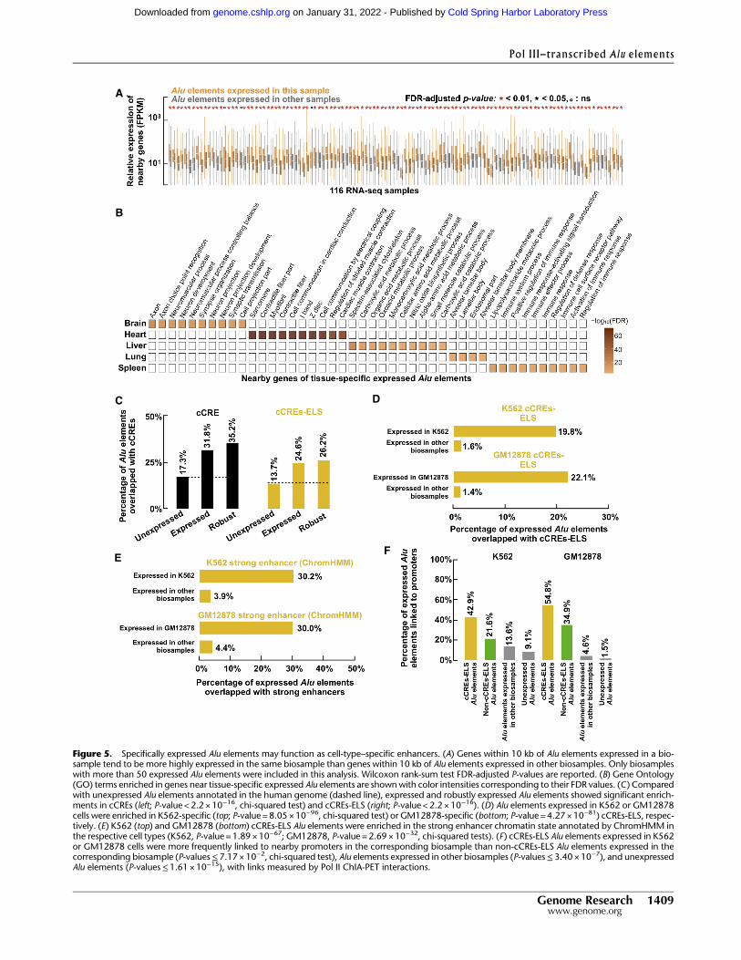
EF
B
A
C D
Figure 5. Specifically expressed Alu elements may function as cell-type–specific enhancers. (A) Genes within 10 kb of Alu elements expressed in a bio-sample tend to be more highly expressed in the same biosample than genes within 10 kb of Alu elements expressed in other biosamples. Only biosampleswith more than 50 expressed Alu elements were included in this analysis. Wilcoxon rank-sum test FDR-adjusted P-values are reported. (B) Gene Ontology(GO) terms enriched in genes near tissue-specific expressed Alu elements are shownwith color intensities corresponding to their FDR values. (C) Comparedwith unexpressed Alu elements annotated in the human genome (dashed line), expressed and robustly expressed Alu elements showed significant enrich-ments in cCREs (left; P-value < 2.2 × 10⁻¹⁶, chi-squared test) and cCREs-ELS (right; P-value < 2.2 × 10⁻¹⁶). (D) Alu elements expressed in K562 or GM12878cells were enriched in K562-specific (top; P-value = 8.05 × 10−96, chi-squared test) or GM12878-specific (bottom; P-value = 4.27× 10−81) cCREs-ELS, respec-tively. (E) K562 (top) and GM12878 (bottom) cCREs-ELS Alu elements were enriched in the strong enhancer chromatin state annotated by ChromHMM inthe respective cell types (K562, P-value = 1.89 × 10−67; GM12878, P-value = 2.69× 10⁻³², chi-squared tests). (F) cCREs-ELS Alu elements expressed in K562or GM12878 cells were more frequently linked to nearby promoters in the corresponding biosample than non-cCREs-ELS Alu elements expressed in thecorresponding biosample (P-values≤7.17 ×10⁻², chi-squared test), Alu elements expressed in other biosamples (P-values≤3.40 ×10⁻⁷), and unexpressedAlu elements (P-values≤1.61 ×10⁻¹⁵), with links measured by Pol II ChIA-PET interactions.
Pol III–transcribed Alu elements
Genome Research 1409www.genome.org
Cold Spring Harbor Laboratory Press on January 31, 2022 - Published by genome.cshlp.orgDownloaded from
which were defined using DNase-seq and H3K4me4, H3K27ac,and CTCF ChIP-seq data in hundreds of human biosamples andcontained 125,798 and 90,692 cCREs predicted to be active inK562 or GM12878 cells, respectively. Compared with unexpressedAlu elements, expressed and robustly expressed Alu elementsshowed 1.84- and 2.03-fold enrichment for cCREs, and similar lev-els of enrichment were observed when the subset of cCREs withenhancer-like signatures (cCREs-ELS) was considered (P-values <2.2 ×10⁻¹⁶) (Fig. 5C). When comparing Alu elements expressedin K562 or GM12878 cells with Alu elements expressed in otherbiosamples, we observed 12.3- and 15.7-fold enrichment forcCREs-ELS annotated in the corresponding cell types (P-values≤4.27×10−81) (Fig. 5D). We next considered the strong enhancersin K562 and GM12878 defined by ChromHMMwith a large panelof histone modifications (Ernst et al. 2011) and observed a 7.74-and 6.82-fold enrichment for Alu elements expressed in K562 orGM12878 cells, respectively, versus Alu elements expressed in oth-er biosamples (chi-squared test P-value=1.89×10−67 and 2.69×10⁻³² for K562 and GM12878, respectively) (Fig. 5E).
To assess the likelihood that expressedAlu elementsmight actas enhancers for their nearby genes, we further examined whetherAlu elements expressed in K562 or GM12878 formed chromatininteractions with their neighboring genes using Pol II ChIA-PETdata in these two cell types. Compared with Alu elements ex-pressed in other biosamples or unexpressed Alu elements, thereis a significant enrichment for expressed Alu elements in thesetwo cell types to form chromatin interactions with neighboringprotein-coding genes, especially those expressed Alu elementsthat were also cCREs-ELS in the corresponding cell type (Fig. 5F).Moderate enrichment of POLR2A ChIP-seq signals was also ob-served at these expressed Alu elements in the corresponding celltypes where they were also cCREs-ELS (Supplemental Fig. S8D).Taken together, these results suggest that expressed Alu elementsmight act as enhancers to regulate neighboring genes that func-tion specifically in the corresponding cell types.
Specific binding of TFs associated with cell-type–specific
expression of Alu elements
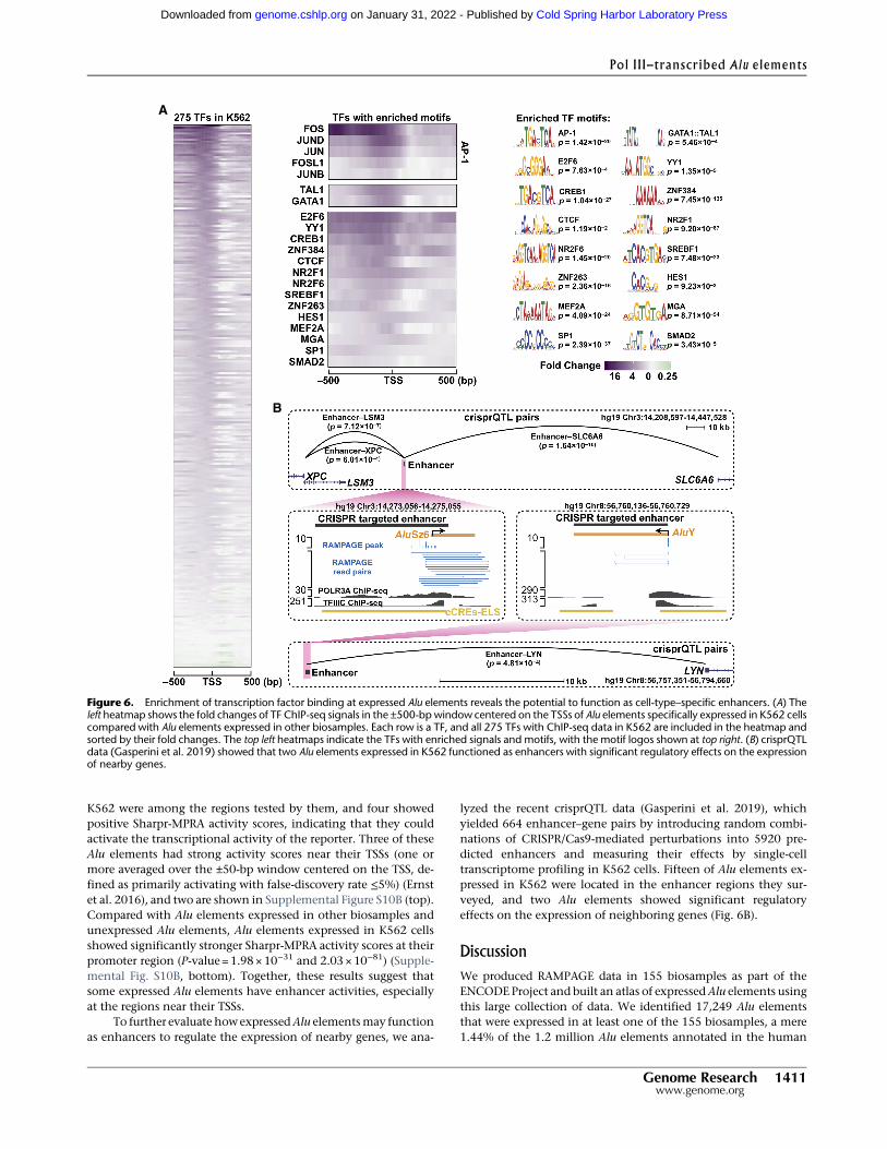
Having observed that expressed Alu elements had characteristicchromatin features of active enhancers and were enriched in chro-matin interactions with their nearby genes, which were alsoexpressed and functioned specifically in the corresponding tissues,we asked whether these expressed Alu elements were boundby transcription factors in the corresponding cell types. TheENCODE Consortium had performed ChIP-seq experiments on alarge number of TFs, including 275 TFs in K562 cells and 141 TFsin GM12878 cells. Thus, we tested whether there was a significantenrichment of TF binding in Alu elements expressed in each ofthese two cell types compared with Alu elements expressed in oth-er biosamples.
We observed significant enrichments at the upstream regionsof expressed Alu elements for most of the TFs with ChIP-seq data(260 TFs in K562 cells [Fig. 6A, left]; 130 TFs in GM12878 cells[Supplemental Fig. S9A, left]). However, only a small subset ofthese TFs also showed enrichment for their motifs at expressedAlu elements (21 TFs in K562 cells [Fig. 6A, middle]; 16 TFs inGM12878 cells [Supplemental Fig. S9A, right]; SupplementalTable S6).We reasoned that the TFs with enrichedmotifs bind spe-cifically to the expressed Alu elements, whereas the other TFs maycobind via protein–protein interactions or bind nonspecifically fa-cilitated by the favorable chromatin conditions; thus, we focused
our subsequent analysis on the subset of TFs with enrichedmotifs.Previous studies provided evidence that some of these TFs couldenhance the transcription of Alu elements (Ullu and Weiner1985; Chesnokov and Schmid 1996; Conti et al. 2015). YY1, SP1,and the MEF2 family of TFs were shown to bind Alu elements dur-ing developmental processes (Oei et al. 2004) and macrophage re-sponses to tuberculosis infection (Bouttier et al. 2016). AP-1(heterodimer of FOS and JUN) binds to Pol III genes (Raha et al.2010; Ahuja and Kumar 2017) and recruits EP300 to increase his-tone acetylation and stabilize TFIIIC at their core promoters(Mertens and Roeder 2008; Ahuja and Kumar 2017), promotingPol III transcription at these genes. Thus, AP-1 and EP300 mayalso activate Alu transcription. Indeed, we detected enrichedChIP-seq signals for FOS and EP300 at theAlu elements specificallyexpressed in K562 or GM12878 cells (Supplemental Fig. S9B). Westratified expressed Alu elements into three sets—with high, medi-um, and lowFOSChIP-seq signals, and the EP300 levels scaled pos-itively with FOS occupancy (Supplemental Fig. S9C), providingevidence that AP-1 might recruit EP300 to regulate cell-type–spe-cific expression of Alu elements. In addition to activating Pol IIItranscription at these Alu loci, these TFs may activate Pol II tran-scription at nearby genes.
Whenwe compared the TFs that showed enrichments of bothChIP-seq signals and sequence motifs at expressed Alu elements inK562 versus GM12878 (Fig. 6A vs. Supplemental Fig. S9A), someTFs (e.g., GATA1::TAL1 in K562 cells and PAX5 in GM12878 cells)were enriched in one cell type but not in the other, and suchTFs were ranked as top master transcription factors associatedwith cell proliferation and lineage commitment (SupplementalMaterial), suggesting these cell-type–specific Alu elements canattract the binding of master TFs, which in turn activate lineage-specific transcriptional programs.
Functional data support enhancer activities at expressed
Alu elements
We further looked for self-transcribing active regulatory region se-quencing (STARR-seq) and massively parallel reporter assay(Sharpr-MPRA) data supporting that expressed Alu elements mayfunction as active enhancers. Barakat et al. (2018) performedSTARR-seq on the regions bound by NANOG, POU5F1,H3K4me1, or H3K27ac, identified by ChIP-seq, in primed and na-ive embryonic stem cells (ESCs). The Alu elements expressed inESCs (Supplemental Methods) had significantly more STARR-seqreads than did Alu elements expressed in other biosamples(Wilcoxon rank-sum test P-value= 2.42× 10⁻³ in primed ESCsand 1.19×10⁻⁴ in naive ESCs) and unexpressed Alu elements (P-value =2.17×10⁻⁴ in primed ESCs and 1.12×10⁻⁶ in naive ESCs)(Supplemental Fig. S10A, top). When we overlapped Alu elementsexpressed in ESCs with active enhancers defined by Barakat et al.(reads per plasmid [RPP] ≥138), we observed 4.1- and 4.6-fold en-richment over Alu elements expressed in other biosamples (chi-squared test P-value=3.00×10⁻⁷ and 1.11×10⁻⁷ in primed andnaive ESCs, respectively) and 5.9- and 6.6-folds of enrichmentover unexpressed Alu elements (P-value=1.76×10⁻¹⁴ and 6.17×10⁻¹⁹ in primed and naive ESCs, respectively) (Supplemental Fig.S10A, bottom). These functional data support that expressed Aluelements may function as enhancers in ESCs.
In another study, Ernst et al. (2016) developed the Sharpr-MPRA technique to test more than 15,000 regions (4.6 millionnt in total), which were annotated as enhancers by ChromHMM,at 5-nt resolution in K562 cells. Five Alu elements expressed in
Zhang et al.
1410 Genome Researchwww.genome.org
Cold Spring Harbor Laboratory Press on January 31, 2022 - Published by genome.cshlp.orgDownloaded from
K562 were among the regions tested by them, and four showedpositive Sharpr-MPRA activity scores, indicating that they couldactivate the transcriptional activity of the reporter. Three of theseAlu elements had strong activity scores near their TSSs (one ormore averaged over the ±50-bp window centered on the TSS, de-fined as primarily activating with false-discovery rate ≤5%) (Ernstet al. 2016), and two are shown in Supplemental Figure S10B (top).Compared with Alu elements expressed in other biosamples andunexpressed Alu elements, Alu elements expressed in K562 cellsshowed significantly stronger Sharpr-MPRA activity scores at theirpromoter region (P-value=1.98×10⁻³¹ and 2.03×10−81) (Supple-mental Fig. S10B, bottom). Together, these results suggest thatsome expressed Alu elements have enhancer activities, especiallyat the regions near their TSSs.
To further evaluate howexpressedAlu elementsmay functionas enhancers to regulate the expression of nearby genes, we ana-
lyzed the recent crisprQTL data (Gasperini et al. 2019), whichyielded 664 enhancer–gene pairs by introducing random combi-nations of CRISPR/Cas9-mediated perturbations into 5920 pre-dicted enhancers and measuring their effects by single-celltranscriptome profiling in K562 cells. Fifteen of Alu elements ex-pressed in K562 were located in the enhancer regions they sur-veyed, and two Alu elements showed significant regulatoryeffects on the expression of neighboring genes (Fig. 6B).
Discussion
We produced RAMPAGE data in 155 biosamples as part of theENCODE Project and built an atlas of expressedAlu elements usingthis large collection of data. We identified 17,249 Alu elementsthat were expressed in at least one of the 155 biosamples, a mere1.44% of the 1.2 million Alu elements annotated in the human
B
A
Figure 6. Enrichment of transcription factor binding at expressed Alu elements reveals the potential to function as cell-type–specific enhancers. (A) Theleft heatmap shows the fold changes of TF ChIP-seq signals in the ±500-bp window centered on the TSSs of Alu elements specifically expressed in K562 cellscompared with Alu elements expressed in other biosamples. Each row is a TF, and all 275 TFs with ChIP-seq data in K562 are included in the heatmap andsorted by their fold changes. The top left heatmaps indicate the TFs with enriched signals and motifs, with the motif logos shown at top right. (B) crisprQTLdata (Gasperini et al. 2019) showed that two Alu elements expressed in K562 functioned as enhancers with significant regulatory effects on the expressionof nearby genes.
Pol III–transcribed Alu elements
Genome Research 1411www.genome.org
Cold Spring Harbor Laboratory Press on January 31, 2022 - Published by genome.cshlp.orgDownloaded from
genome, and 61.6% of these 17,249 Alu elements were expressedin only one biosample. These results indicate that Pol III–tran-scribed Alu expression is overall very low and highly cell-type spe-cific. Contrary to the expectation that the youngest Alu elements—those AluY elements that only exist in the human genome,which are the least divergent from the consensus Alu sequence—should be most expressed, these youngest Alu elements are signifi-cantly less expressed than olderAlu elements.With the caveat thatour results might be influenced by the read-mapping strategy inour pipeline (although we did test allowing multiple-mappingreads and still obtained the same conclusion), these results suggestthat humans are highly effective in suppressing young Alu ele-ments, whichmay still possess the capability of retrotransposition.One activemechanism for repressing Alu expression is DNAmeth-ylation (Kochanek et al. 1993, 1995; Bakshi et al. 2016), and ac-cordingly, we found that the Alu elements that were notexpressed in a particular biosample (but were expressed in otherbiosamples) had significantly higher DNA methylation levels inthat biosample than did Alu elements that were not expressed inany of the 155 biosamples that we surveyed.
Are the few expressed Alu elements escapees of the active re-pressive mechanism in the host human cells, or alternatively,can they possibly serve some biological functions? We performeda series of analyses, and our results suggest the latter: Expressed Aluelementsmay, in some instances, function as cell-type–specific en-hancers for nearby protein-coding genes. Expressed Alu elementsare significantly more likely to be intronic and exonic than inter-genic with respect to genomic Alu elements (2.17% vs. 0.43%,P-value<2.2 ×10⁻¹⁶). We compared the genetic and epigeneticfeatures at expressed and unexpressed Alu elements and foundthat distance to Pol II genes, chromatin accessibility, and activehistone marks characteristic of active enhancers were predictiveof cell-type–specificAlu expression. Furthermore, biosamples in re-lated tissues clustered together by theirAlu expression profiles, andthe protein-coding genes near expressed Alu elements tended tobe expressed and function toward the tissue specificity of thecorresponding biosamples. We also observed that expressed Aluelements were significantly enriched in enhancers defined usingepigenetic signals and that ChIA-PET data further supported thechromatin interaction between expressed Alu elements and theirneighboring genes in the matching cell types. Expressed Aluelements are significantly bound by many transcription factorsthat work with RNA Pol II, and the binding is cell-type specific.Finally, we found some functional data (STARR-seq, Sharpr-MPRA, and crisprQTL) in ESCs and K562 cells, indicating thatsome Alu elements expressed in these cell types functioned asenhancers to regulate expression of nearby genes. Because Aluelements are highly repetitive, there have been few studies on in-dividual elements. The atlas of expressed Alu elements that result-ed from our study will stimulate more studies targeting individualelements.
A recent study showed that Alu elements could explain a sig-nificant amount of disease heritability, especially blood traits(Hormozdiari et al. 2018). Another study analyzed nucleosome oc-cupancy, histonemodification, and sequencemotif features at Aluelements genome-wide and concluded that Alu elements showedcharacteristics of enhancers (Su et al. 2014). These studies didnot investigate whether these potentially functional Alu elementswere expressed in the corresponding cell type and consequentlydid not distinguish Alu elements transcribed by Pol III versus byPol II. Here, we focused on the Alu elements transcribed by PolIII and further concluded that the Alu elements transcribed in a
biosample by Pol III are more likely to be enhancers than are Aluelements not transcribed by Pol III in the corresponding biosam-ple. Does Pol III play a role in the enhancer functions of theseAlu elements that they transcribe? Policarpi et al. (2017) showedthat in cortical neurons a subset of SINEs recruited Pol III transcrip-tion in a stimulus-dependent manner. They performed in-depthexperiments on one such enhancer-like SINE near the Fos genein the mouse and showed that its Pol III transcript interactedwith Pol II at the Fos promoter and was required for the functionsof cortical neurons. Thus, we propose that the expressed Alu ele-ments thatwe identified in this study could act in a similarmannerto enhance the transcription of neighboring protein-coding genes.This model is further supported by ChIP-seq data that showedPol II occupancy at Pol III loci, although that study did not inves-tigate Alu elements (Barski et al. 2010). On the other hand, themoderate enrichment of Pol II ChIP-seq signals at expressed Aluelements in the specific cell types (Supplemental Fig. S8D) suggeststhat Pol II may facilitate Pol III in its transcription of Alu elementsby modifying the local chromatin structure as proposed for otherPol III–transcribed genes (Barski et al. 2010; Raha et al. 2010).
In summary, wehave identified 17,249 Pol III–transcribedAluelements thatwere expressed in at least one of 155 biosamples. Ourintegrative analyses showed that although the approximately 100human-specific Alu elements are actively repressed in transcrip-tion, the older expressed Alu elements may have been exaptedby the human host to function as enhancers for their neighboringprotein-coding genes.
Methods
A computational pipeline for annotating primary Alu transcriptsusing RAMPAGE data
We developed a new computational pipeline by combining theunique features of the RAMPAGE assay and primary Alu elementsto precisely annotated the TSSs of Pol III transcribed Alu elements.
Characterization of Pol III–transcribed Alu elements
Tissue specificity, evolutionary conservation, genomic context,and sequence features were systematically characterized andcompared between expressed and unexpressed Alu elements.DNase-seq, ChIP-seq of the histone variant H2AZ1 and 10 histonemodifications, and whole-genome bisulfite sequencing of DNAmethylation were used to profile the cell-specific expression ofPol III–transcribedAlu elements. Random forest classifiers were im-plemented to train models for predicting the transcriptional statesof Alu elements.
Functional analysis of expressed Alu elements as active enhancers
Expressed Alu elements were overlapped with cCREs and ENCODEChromHMM annotations (Ernst et al. 2011). STARR-seq (Barakatet al. 2018) and Sharpr-MPRA (Ernst et al. 2016) datawere analyzedto assess the enhancer activity of expressed Alu elements. Pol IIChIA-PET data (Li et al. 2012; Tang et al. 2015) and crisprQTLdata (Gasperini et al. 2019)were used to evaluate the regulatory im-pacts of expressed Alu elements on the expression of nearby genes.
Software availability
The source code of our Alu identification pipeline is includedin the Supplemental Material as Supplemental Code and canalso be accessed at the GitHub (https://github.com/kepbod/rampage_alu).
Zhang et al.
1412 Genome Researchwww.genome.org
Cold Spring Harbor Laboratory Press on January 31, 2022 - Published by genome.cshlp.orgDownloaded from

Acknowledgments
We thank members of the Weng laboratory for helpful commentson this manuscript. This project was funded by National Institutesof Health (NIH) grant U24-HG009446 to Z.W. and NIH grant U54-HG004557 to T.R.G.
Author contributions: X.-O.Z. and Z.W. conceived and de-signed the project. T.R.G. led the production of all RAMPAGEand RNA-seq data. Z.W. supervised the project. X.-O.Z. performedthe bioinformatics analysis. X.-O.Z. and Z.W. analyzed the dataand wrote the paper with contributions from T.R.G.
References
Ade C, Roy-Engel AM, Deininger PL. 2013. Alu elements: an intrinsic sourceof human genome instability. Curr Opin Virol 3: 639–645. doi:10.1016/j.coviro.2013.09.002
Ahuja R, Kumar V. 2017. Stimulation of Pol III-dependent 5S rRNA and U6snRNA gene expression by AP-1 transcription factors. FEBS J 284: 2066–2077. doi:10.1111/febs.14104
Bakshi A, Herke SW, Batzer MA, Kim J. 2016. DNAmethylation variation ofhuman-specific Alu repeats. Epigenetics 11: 163–173. doi:10.1080/15592294.2015.1130518
Barakat TS, Halbritter F, Zhang M, Rendeiro AF, Perenthaler E, Bock C,Chambers I. 2018. Functional dissection of the enhancer repertoire inhuman embryonic stem cells. Cell Stem Cell 23: 276–288.e8. doi:10.1016/j.stem.2018.06.014
Barski A, Cuddapah S, Cui K, Roh T-Y, Schones DE, Wang Z, Wei G,Chepelev I, Zhao K. 2007. High-resolution profiling of histone methyl-ations in the human genome. Cell 129: 823–837. doi:10.1016/j.cell.2007.05.009
Barski A, Chepelev I, Liko D, Cuddapah S, Fleming AB, Birch J, Cui K,WhiteRJ, Zhao K. 2010. Pol II and its associated epigenetic marks are present atPol III–transcribed noncoding RNA genes. Nat Struct Mol Biol 17: 629–634. doi:10.1038/nsmb.1806
Batut P, Gingeras TR. 2013. RAMPAGE: promoter activity profiling bypaired-end sequencing of 5′-complete cDNAs. Curr Protoc Mol Biol104: 25B.11.1–25B.11.16. doi:10.1002/0471142727.mb25b11s104
Batut P, Dobin A, Plessy C, Carninci P, Gingeras TR. 2013. High-fidelity pro-moter profiling reveals widespread alternative promoter usage andtransposon-driven developmental gene expression. Genome Res 23:169–180. doi:10.1101/gr.139618.112
Bennett EA, Keller H, Mills RE, Schmidt S, Moran JV, Weichenrieder O,Devine SE. 2008. Active Alu retrotransposons in the human genome.Genome Res 18: 1875–1883. doi:10.1101/gr.081737.108
Bouttier M, Laperriere D, Memari B, Mangiapane J, Fiore A, Mitchell E,Verway M, Behr MA, Sladek R, Barreiro LB, et al. 2016. Alu repeats astranscriptional regulatory platforms in macrophage responses to M. tu-berculosis infection. Nucleic Acids Res 44: 10571–10587. doi:10.1093/nar/gkw782
Burke JM, Kincaid RP, Nottingham RM, Lambowitz AM, Sullivan CS. 2016.DUSP11 activity on triphosphorylated transcripts promotes Argonauteassociation with noncanonical viral microRNAs and regulates steady-state levels of cellular noncoding RNAs. Genes Dev 30: 2076–2092.doi:10.1101/gad.282616.116
Calo E, Wysocka J. 2013. Modification of enhancer chromatin: what,how, and why? Mol Cell 49: 825–837. doi:10.1016/j.molcel.2013.01.038
Chen LL, Yang L. 2017. ALUternative regulation for gene expression. TrendsCell Biol 27: 480–490. doi:10.1016/j.tcb.2017.01.002
Chesnokov I, Schmid CW. 1996. Flanking sequences of an Alu source stim-ulate transcription in vitro by interacting with sequence-specific tran-scription factors. J Mol Evol 42: 30–36. doi:10.1007/BF00163208
Conti A, Carnevali D, Bollati V, Fustinoni S, Pellegrini M, Dieci G. 2015.Identification of RNA polymerase III-transcribed Alu loci by computa-tional screening of RNA-Seq data. Nucleic Acids Res 43: 817–835.doi:10.1093/nar/gku1361
Deininger P. 2011. Alu elements: know the SINEs. Genome Biol 12: 236.doi:10.1186/gb-2011-12-12-236
Deininger PL, Batzer MA. 1999. Alu repeats and human disease. Mol GenetMetab 67: 183–193. doi:10.1006/mgme.1999.2864
Ernst J, Kheradpour P, Mikkelsen TS, Shoresh N, Ward LD, Epstein CB,Zhang X, Wang L, Issner R, Coyne M, et al. 2011. Mapping and analysisof chromatin state dynamics in nine human cell types. Nature 473: 43–49. doi:10.1038/nature09906
Ernst J, Melnikov A, Zhang X, Wang L, Rogov P, Mikkelsen TS, Kellis M.2016. Genome-scale high-resolution mapping of activating and repres-
sive nucleotides in regulatory regions. Nat Biotechnol 34: 1180–1190.doi:10.1038/nbt.3678
Erwin JA, Marchetto MC, Gage FH. 2014. Mobile DNA elements in the gen-eration of diversity and complexity in the brain. Nat Rev Neurosci 15:497–506. doi:10.1038/nrn3730
Faulkner GJ, Kimura Y, Daub CO, Wani S, Plessy C, Irvine KM, Schroder K,Cloonan N, Steptoe AL, Lassmann T, et al. 2009. The regulated retro-transposon transcriptome of mammalian cells. Nat Genet 41: 563–571. doi:10.1038/ng.368
Fort A, Hashimoto K, Yamada D, Salimullah M, Keya CA, Saxena A, BonettiA, Voineagu I, Bertin N, Kratz A, et al. 2014. Deep transcriptome profil-ing of mammalian stem cells supports a regulatory role for retrotranspo-sons in pluripotency maintenance. Nat Genet 46: 558–566. doi:10.1038/ng.2965
Gasperini M, Hill AJ, McFaline-Figueroa JL, Martin B, Kim S, Zhang MD,Jackson D, Leith A, Schreiber J, Noble WS, et al. 2019. A genome-wideframework for mapping gene regulation via cellular genetic screens.Cell 176: 377–390.e19. doi:10.1016/j.cell.2018.11.029
Goerner-Potvin P, Bourque G. 2018. Computational tools to unmask trans-posable elements. Nat Rev Genet 19: 688–704. doi:10.1038/s41576-018-0050-x
Gu TJ, Yi X, Zhao XW, Zhao Y, Yin JQ. 2009. Alu-directed transcriptionalregulation of some novel miRNAs. BMC Genomics 10: 563. doi:10.1186/1471-2164-10-563
Hasler J, Strub K. 2006. Alu RNP and Alu RNA regulate translation initiationin vitro. Nucleic Acids Res 34: 2374–2385. doi:10.1093/nar/gkl246
Heintzman ND, Stuart RK, Hon G, Fu Y, Ching CW, Hawkins RD, BarreraLO, Van Calcar S, Qu C, Ching KA, et al. 2007. Distinct and predictivechromatin signatures of transcriptional promoters and enhancers inthe human genome. Nat Genet 39: 311–318. doi:10.1038/ng1966
Hormozdiari FI, van de Geijn B, Nasser J, Weissbrod O, Gazal S, Ju CJ-T,O’Connor L, Hujoel MLA, Engreitz J, Hormozdiari F, et al. 2018.Functional disease architectures reveal unique biological role of trans-posable elements. bioRxiv doi:10.1101/482281
International Human Genome Sequencing Consortium. 2001. Initial se-quencing and analysis of the human genome. Nature 409: 860–921.doi:10.1038/35057062
JordàM,Díez-Villanueva A,Mallona I,Martín B, Lois S, BarreraV, EstellerM,Vavouri T, Peinado MA. 2017. The epigenetic landscape of Alu repeatsdelineates the structural and functional genomic architecture of coloncancer cells. Genome Res 27: 118–132. doi:10.1101/gr.207522.116
Kazazian HH. 2004. Mobile elements: drivers of genome evolution. Science303: 1626–1632. doi:10.1126/science.1089670
Kochanek S, Renz D, Doerfler W. 1993. DNA methylation in the Alu se-quences of diploid and haploid primary human cells. EMBO J 12:1141–1151. doi:10.1002/j.1460-2075.1993.tb05755.x
Kochanek S, Renz D, Doerfler W. 1995. Transcriptional silencing of humanAlu sequences and inhibition of protein binding in the box B regulatoryelements by 5′-CG-3′ methylation. FEBS Lett 360: 115–120. doi:10.1016/0014-5793(95)00068-K
KonkelMK,Walker JA, Hotard AB, RanckMC, Fontenot CC, Storer J, StewartC,Marth GT, Genomes C, BatzerMA. 2015. Sequence analysis and char-acterization of active human Alu subfamilies based on the 1000Genomes Pilot Project. Genome Biol Evol 7: 2608–2622. doi:10.1093/gbe/evv167
Kriegs JO, Churakov G, Jurka J, Brosius J, Schmitz J. 2007. Evolutionary his-tory of 7SL RNA-derived SINEs in Supraprimates. Trends Genet 23: 158–161. doi:10.1016/j.tig.2007.02.002
Li G, Ruan X, Auerbach RK, Sandhu KS, Zheng M,Wang P, Poh HM, Goh Y,Lim J, Zhang J, et al. 2012. Extensive promoter-centered chromatin in-teractions provide a topological basis for transcription regulation. Cell148: 84–98. doi:10.1016/j.cell.2011.12.014
Li C, Lenhard B, Luscombe NM. 2018. Integrated analysis sheds light onevolutionary trajectories of young transcription start sites in the humangenome. Genome Res 28: 676–688. doi:10.1101/gr.231449.117
Mariner PD, Walters RD, Espinoza CA, Drullinger LF, Wagner SD, Kugel JF,Goodrich JA. 2008. Human Alu RNA is a modular transacting repressorof mRNA transcription during heat shock.Mol Cell 29: 499–509. doi:10.1016/j.molcel.2007.12.013
Mertens C, Roeder RG. 2008. Different functional modes of p300 in activa-tion of RNA polymerase III transcription from chromatin templates.MolCell Biol 28: 5764–5776. doi:10.1128/MCB.01262-07
Moqtaderi Z, Wang J, Raha D, White RJ, Snyder M, Weng Z, Struhl K. 2010.Genomic binding profiles of functionally distinct RNA polymerase IIItranscription complexes in human cells. Nat Struct Mol Biol 17: 635–640. doi:10.1038/nsmb.1794
Oei SL, Babich VS, Kazakov VI, Usmanova NM, Kropotov AV, Tomilin NV.2004. Clusters of regulatory signals for RNA polymerase II transcriptionassociated with Alu family repeats and CpG islands in human promot-ers. Genomics 83: 873–882. doi:10.1016/j.ygeno.2003.11.001
Pol III–transcribed Alu elements
Genome Research 1413www.genome.org
Cold Spring Harbor Laboratory Press on January 31, 2022 - Published by genome.cshlp.orgDownloaded from
Oler AJ, Alla RK, Roberts DN, Wong A, Hollenhorst PC, Chandler KJ,Cassiday PA, Nelson CA, Hagedorn CH, Graves BJ, et al. 2010. HumanRNA polymerase III transcriptomes and relationships to Pol II promoterchromatin and enhancer-binding factors. Nat Struct Mol Biol 17: 620–628. doi:10.1038/nsmb.1801
Orioli A, Pascali C, PaganoA, TeichmannM,Dieci G. 2012. RNA polymeraseIII transcription control elements: themes and variations. Gene 493:185–194. doi:10.1016/j.gene.2011.06.015
Paolella G, LuceroMA,MurphyMH, Baralle FE. 1983. The Alu family repeatpromoter has a tRNA-like bipartite structure. EMBO J 2: 691–696. doi:10.1002/j.1460-2075.1983.tb01486.x
Paulson KE, Schmid CW. 1986. Transcriptional inactivity of Alu repeats inHeLa cells. Nucleic Acids Res 14: 6145–6158. doi:10.1093/nar/14.15.6145
Policarpi C, Crepaldi L, Brookes E, Nitarska J, French SM, Coatti A, Riccio A.2017. Enhancer SINEs link Pol III to Pol II transcription in neurons. CellRep 21: 2879–2894. doi:10.1016/j.celrep.2017.11.019
Raha D,Wang Z,Moqtaderi Z,Wu L, Zhong G, GersteinM, Struhl K, SnyderM. 2010. Close association of RNA polymerase II and many transcrip-tion factors with Pol III genes. Proc Natl Acad Sci 107: 3639–3644.doi:10.1073/pnas.0911315106
Reddy R. 1988. Compilation of small RNA sequences. Nucleic Acids Res 16(Suppl): r71–r85. doi:10.1093/nar/16.suppl.r71
SuM, Han D, Boyd-Kirkup J, Yu X, Han JD. 2014. Evolution of Alu elementstoward enhancers. Cell Rep 7: 376–385. doi:10.1016/j.celrep.2014.03.011
Takahashi H, Lassmann T, Murata M, Carninci P. 2012. 5′ end–centeredexpression profiling using cap-analysis gene expression and next-generation sequencing. Nat Protoc 7: 542–561. doi:10.1038/nprot.2012.005
Tang Z, Luo OJ, Li X, Zheng M, Zhu JJ, Szalaj P, Trzaskoma P, Magalska A,Wlodarczyk J, Ruszczycki B, et al. 2015. CTCF-Mediated human 3D ge-nome architecture reveals chromatin topology for transcription. Cell163: 1611–1627. doi:10.1016/j.cell.2015.11.024
Ullu E, Weiner AM. 1985. Upstream sequences modulate the internal pro-moter of the human 7SL RNA gene. Nature 318: 371–374. doi:10.1038/318371a0
Received February 22, 2019; accepted in revised form July 24, 2019.
Zhang et al.
1414 Genome Researchwww.genome.org
Cold Spring Harbor Laboratory Press on January 31, 2022 - Published by genome.cshlp.orgDownloaded from

10.1101/gr.249789.119Access the most recent version at doi:2019 29: 1402-1414 originally published online August 14, 2019Genome Res.
Xiao-Ou Zhang, Thomas R. Gingeras and Zhiping Weng
specific enhancer function−suggests cell-type elements Alutranscribed −Genome-wide analysis of polymerase III
Material
Supplemental
http://genome.cshlp.org/content/suppl/2019/08/14/gr.249789.119.DC1
References
http://genome.cshlp.org/content/29/9/1402.full.html#ref-list-1
This article cites 52 articles, 9 of which can be accessed free at:
License
Commons Creative
.http://creativecommons.org/licenses/by-nc/4.0/described at a Creative Commons License (Attribution-NonCommercial 4.0 International), as
). After six months, it is available underhttp://genome.cshlp.org/site/misc/terms.xhtmlfirst six months after the full-issue publication date (see This article is distributed exclusively by Cold Spring Harbor Laboratory Press for the
ServiceEmail Alerting
click here.top right corner of the article or
Receive free email alerts when new articles cite this article - sign up in the box at the
https://genome.cshlp.org/subscriptionsgo to: Genome Research To subscribe to
© 2019 Zhang et al.; Published by Cold Spring Harbor Laboratory Press
Cold Spring Harbor Laboratory Press on January 31, 2022 - Published by genome.cshlp.orgDownloaded from
Copyright © 2022 FDOKUMEN