Structural and Functional Characterization of Sapovirus RNA-Dependent RNA Polymerase
15
Published Ahead of Print 22 November 2006. 2007, 81(4):1858. DOI: 10.1128/JVI.01462-06. J. Virol. A. Tucker and Jacques Rohayem Julia Gebhardt, Alexander Gorbalenya, Bruno Canard, Paul Stephen W. B. Fullerton, Martina Blaschke, Bruno Coutard, Polymerase of Sapovirus RNA-Dependent RNA Structural and Functional Characterization http://jvi.asm.org/content/81/4/1858 Updated information and services can be found at: These include: REFERENCES http://jvi.asm.org/content/81/4/1858#ref-list-1 at: This article cites 60 articles, 36 of which can be accessed free CONTENT ALERTS more» articles cite this article), Receive: RSS Feeds, eTOCs, free email alerts (when new http://journals.asm.org/site/misc/reprints.xhtml Information about commercial reprint orders: http://journals.asm.org/site/subscriptions/ To subscribe to to another ASM Journal go to: on November 22, 2013 by guest http://jvi.asm.org/ Downloaded from on November 22, 2013 by guest http://jvi.asm.org/ Downloaded from
Transcript of Structural and Functional Characterization of Sapovirus RNA-Dependent RNA Polymerase

Published Ahead of Print 22 November 2006. 2007, 81(4):1858. DOI: 10.1128/JVI.01462-06. J. Virol.
A. Tucker and Jacques RohayemJulia Gebhardt, Alexander Gorbalenya, Bruno Canard, Paul Stephen W. B. Fullerton, Martina Blaschke, Bruno Coutard, Polymeraseof Sapovirus RNA-Dependent RNA Structural and Functional Characterization
http://jvi.asm.org/content/81/4/1858Updated information and services can be found at:
These include:
REFERENCEShttp://jvi.asm.org/content/81/4/1858#ref-list-1at:
This article cites 60 articles, 36 of which can be accessed free
CONTENT ALERTS more»articles cite this article),
Receive: RSS Feeds, eTOCs, free email alerts (when new
http://journals.asm.org/site/misc/reprints.xhtmlInformation about commercial reprint orders: http://journals.asm.org/site/subscriptions/To subscribe to to another ASM Journal go to:
on Novem
ber 22, 2013 by guesthttp://jvi.asm
.org/D
ownloaded from
on N
ovember 22, 2013 by guest
http://jvi.asm.org/
Dow
nloaded from
JOURNAL OF VIROLOGY, Feb. 2007, p. 1858–1871 Vol. 81, No. 40022-538X/07/$08.00�0 doi:10.1128/JVI.01462-06Copyright © 2007, American Society for Microbiology. All Rights Reserved.
Structural and Functional Characterization of SapovirusRNA-Dependent RNA Polymerase�
Stephen W. B. Fullerton,1† Martina Blaschke,2† Bruno Coutard,3 Julia Gebhardt,2Alexander Gorbalenya,4 Bruno Canard,3 Paul A. Tucker,1 and Jacques Rohayem2*
European Molecular Biology Laboratory, Hamburg, Germany1; Institut fur Virologie, The Calicilab, Medizinisch-Theoretisches Zentrum,Dresden, Germany2; Laboratoire Architecture et Fonction des Macromolecules Biologiques UMR6098 CNRS and
Universite de la Mediterannee, Marseilles, France3; and Department of Medical Microbiology,Leiden University Medical Center, Leiden, The Netherlands4
Received 11 July 2006/Accepted 13 November 2006
Sapoviruses are one of the major agents of acute gastroenteritis in childhood. They form a tight geneticcluster (genus) in the Caliciviridae family that regroups both animal and human pathogenic strains. Nopermissive tissue culture has been developed for human sapovirus, limiting its characterization to surrogatesystems. We report here on the first extensive characterization of the key enzyme of replication, the RNA-dependent RNA polymerase (RdRp) associated with the 3Dpol-like protein. Enzymatically active sapovirus3Dpol and its defective mutant were expressed in Escherichia coli and purified. The overall structure of thesapovirus 3Dpol was determined by X-ray crystallography to 2.32-Å resolution. It revealed a right hand foldtypical for template-dependent polynucleotide polymerases. The carboxyl terminus is located within the activesite cleft, as observed in the RdRp of some (norovirus) but not other (lagovirus) caliciviruses. Sapovirus 3Dpol
prefers Mn2� over Mg2� but may utilize either as a cofactor in vitro. In a synthetic RNA template-dependentreaction, sapovirus 3Dpol synthesizes a double-stranded RNA or labels the template 3� terminus by terminaltransferase activity. Initiation of RNA synthesis occurs de novo on heteropolymeric templates or in a primer-dependent manner on polyadenylated templates. Strikingly, this mode of initiation of RNA synthesis was alsodescribed for norovirus, but not for lagovirus, suggesting structural and functional homologies in the RNA-dependent RNA polymerase of human pathogenic caliciviruses. This first experimental evidence makes sapo-virus 3Dpol an attractive target for developing drugs to control calicivirus infection in humans.
Sapovirus (SV) is a global agent of viral gastroenteritis,mainly infecting children under 5 years of age and being one ofthe most common agents of viral gastroenteritis among infants inJapan (9). Sapovirus infections have also been reported in all agegroups (18, 23, 24, 33, 41, 52), and outbreaks of sapovirus gastro-enteritis have been described in child day care centers (38), el-derly persons’ homes (23), and hospitals (24, 53).
Sapovirus belongs to the Caliciviridae family, which includesfour genera (norovirus, sapovirus, lagovirus, and vesivirus).The human pathogenic caliciviruses have been so far exclu-sively limited to the norovirus and sapovirus genera. Bothgenera also include animal pathogenic strains, although animalpathogenic caliciviruses are mainly represented in the vesivirusand lagovirus genera. Phylogenetic analysis of the Caliciviridaebased on the complete capsid gene shows that sapovirus strainscluster separately within the Caliciviridae (19). This genusseems to be more distantly related to the human norovirusthan to the lagovirus (19). The sapovirus genus is divided infive genogroups (15). Strains belonging to genogroups 1, 2, 4,and 5 infect humans, with the genogroup 1 strains being pre-dominantly detected over the years (18). Animal sapovirusstrains, i.e., porcine strains, cluster within genogroup 3 (15).
The porcine enteric calicivirus remains so far the only strain ofthe sapovirus species that can be propagated in cultured cells(7, 8).
Sapovirus is a nonenveloped RNA virus with a single-stranded positive-sense genome of approximately 7.4 kb (7,320to 7,431 nucleotides [nt]) (20). The viral genome consists oftwo open reading frames encoding the nonstructural proteinsand the capsid protein or virion protein 1 (VP1) encoded inopen reading frame 1 (ORF-1) and a protein termed VP2which is encoded in ORF-2 that is located at the 3� end of thegenome (Fig. 1). ORF-1 is predicted to encode a singlepolyprotein that is cotranslationally processed by the viral pro-tease, resulting in the appearance of nonstructural proteinsrequired for replication of the viral genome. Among those, theRNA-dependent RNA polymerase (3Dpol-like, further re-ferred to as 3Dpol) is predicted to play a key role in thereplication of the genome, as well as in the synthesis andamplification of a subgenomic RNA. The subgenomic RNAcorresponds to the region of the genome that encodes VP1 andVP2 as well as the 3� end (Fig. 1). Downstream from ORF-2,a 55- to 82-nt untranslated region is present (20), which isfollowed by a poly(A) tail of variable length.
So far, the structure and function of the viral enzymes in-volved in replication of the sapovirus genome remain unchar-acterized. Recently, Oka et al. have described the processing ofsapovirus polyprotein precursor in a cell-free system (46). Inthat study, the 3Cpro3Dpol protein precursor was recoveredafter processing of the ORF-1-encoded polyprotein, suggesting
* Corresponding author. Mailing address: Institut fur Virologie, TheCalicilab, Fiedlerstr. 42, D-01307 Dresden, Germany. Phone: 49 3514586200. Fax: 49 351 4586314. E-mail: [email protected].
† Both authors contributed equally to the study.� Published ahead of print on 22 November 2006.
1858
on Novem
ber 22, 2013 by guesthttp://jvi.asm
.org/D
ownloaded from
that the 3Cpro3Dpol protein precursor is one of the active formsof the viral polymerase.
In our study, we have examined the structural and functionalfeatures of sapovirus 3Dpol polymerase. The three-dimensionalstructure of the sapovirus 3Dpol was solved using X-ray crys-tallography and displays a “right hand” shape, with the Cterminus of the enzyme entering the channel containing theactive site. Furthermore, we present the first experimentalevidence for the RNA-dependent RNA polymerase (RdRp)and terminal transferase activities associated with sapovirus3Dpol, showing that initiation of RNA synthesis by sapovirus3Dpol on heteropolymeric RNA template occurs de novo. Weconclude that sapovirus 3Dpol displays structural and functionalfeatures similar to the RNA-dependent RNA polymerase ofnorovirus (3Dpol), a related human pathogenic calicivirus.
MATERIALS AND METHODS
Bioinformatics analysis. Full-genome sequences of a representative set ofsapoviruses and other caliciviruses were retrieved from the GenBank databaseand put into the Viralis database (A. E. Gorbalenya, unpublished data). Theywere aligned using the MUSCLE (13) and HMMER2.3.2 (12) programs, takinginto consideration also the phylogenic structure of the Caliciviridae family andcleavage site annotations in the calicivirus genome files. A manually adjustedalignment of the 3Cpro3Dpol area was used for predicting the location of 3Dpol
domain.Generation of synthetic RNA templates. Sapovirus clone pJG-Sap01 (GenBank
accession number AY694184), which was characterized by phylogenetic analysisbased on the complete sequence of its capsid gene as belonging to sapovirus GGI(Manchester-like), was used for generation of a DNA fragment of 2,259 bp in lengthcorresponding to the complete subgenomic RNA of sapovirus (nt 5170 to 7429). TheDNA fragment was generated by PCR amplification, using a T7 promoter sequencefused at its 3� end to a gene-specific sequence as a forward primer and a gene-specificprimer complementary to the 3� end of the sequence as a reverse primer. Foramplification of DNA, primers 86-Sap-T7-for, consisting of the T7 promoter se-quence fused at its 3� end to a gene-specific sequence (5�-CAGAGATGCATAATACGACTCACTATAGGGAGAGCCACCATGGAGGGCAATGGCTCCAACCCAGAGC-3�), and 48-Sap-ORF-2-rev (5�-AGGGACGGCGACAATCGCTTAA
TTGTC-3�) were used. Reaction conditions, amplification cycles, and identificationof the products were performed as previously described for generation of norovirussynthetic subgenomic RNA (50, 51).
For in vitro transcription, the Megascript T7 Kit (Ambion) was used accordingto the manufacturer’s instructions. The reaction mix consisted of 1 �g purifiedcDNA, 2 �l of 10� T7 reaction buffer, 20 U of RNase inhibitor (RNAsin, 40U/�l; Promega), 7.5 mM (each) ATP, CTP, GTP, and UTP, 10 U T7 polymerase,and RNase-DNase-free water to a final volume of 20 �l. The reaction was runfor 2 h at 37°C, then 20 U DNase I (Ambion) was added, and the reactionmixture was incubated at 37°C for 30 min. poly(A)-tailing of synthesized RNAwas performed with the poly(A)-tailing kit (Ambion) according to manufac-turer’s instructions. The reaction products were then purified with theMEGAclear kit (Ambion) according to the manufacturer’s instructions. TheRNA concentration in the sample was determined by measuring the UVabsorbance at 280 nm.
Expression and purification of sapovirus 3Dpol. The DNA corresponding tothe sapovirus 3Dpol coding sequence was generated by PCR from sapovirusfull-length cDNA clone pJG-Sap01 (GenBank accession number AY694184) asdescribed above. The PCR product was molecularly cloned into the pDEST14vector (Gateway) according to the manufacturer’s instructions and using primersVZ7-Sap-3D-H6C-for (5�-GGGGACAAGTTTGTACAAAAAAGCAGGCTTCGAAGGAGATGCCACCATGAAAGATGAATTTCAATGGAAGGGTTTGCCCGTGG-3) and VZ8-Sap-3D-H6C-rev (5�-GGGGACCACTTTGTACAAGAAAGCTGGGTCTTATTAGTGATGGTGATGGTGATGCTCCATCTCAAACACTATTTTGTGGGTTCC-3�), yielding clone pVZ-Sap-3D-H6C. The3Dpol active site mutant was generated from the pVZ-Sap-3D-H6C clone bysite-directed mutagenesis using the QuikChange site-directed mutagenesis kit(Stratagene) and primers 91-Sap-�pol-for (5�-GTCCACACGTACGGTGGTGGTTGCATGTATAGT-3�) and 92-Sap-�pol-rev (5�-ACTATACATGCAACCACCACCGTACGTGTGGAC-3�) according to the manufacturer’s instructions,yielding the pVZ-Sap-3D-H6C-D347GD348G clone. The 3Dpol active site mutantbears substitutions in the GDD motif of both aspartates with glycine(GD347GD348G). Sapovirus 3Dpol and 3Dpol active site mutants were expressed inEscherichia coli. For this purpose, E. coli Rosetta2 (DE3)pLysS cells (Novagen)were transformed with the pVZ-Sap-3D-H6C and pVZ-Sap-3D-H6C-D343GD344G clones. Cells were grown at 37°C in 2YT medium with ampicillin(100 mg/liter) and chloramphenicol (34 mg/liter). Upon reaching an opticaldensity at 600 nm of 0.6, protein expression was induced by adding isopropyl-�-D-thiogalactopyranoside (IPTG) to a final concentration of 1 mM. Cultures werethen incubated at 25°C overnight. Cell pellets obtained from 500-ml cultureswere washed once in 4 ml phosphate-buffered saline and 1% Triton X-100
FIG. 1. Genome organization of sapovirus clone pJG-SapI (GenBank accession number AY694184) and expression of sapovirus 3D-likepolymerase (3Dpol). The complete sapovirus clone pJG-Sap01 (7,429 bp in length) and the ORF-1 (6,842 bp in length) and ORF-2 (497 bp inlength) are shown. The putative cleavage sites at the interface 3Cpro/3Dpol (Q1207/D) and 3Dpol/VP1 (E1721/G) are indicated. Both the 3Dpol wildtype and 3Dpol active site mutant were expressed. The domain VZ10SV-AY694184-11-3DL-RdRp predicted to encompass the active RNA-dependent RNA polymerase of sapovirus (aa position 1207 to 1721 in the ORF-1 polyprotein of clone pJG-Sap01) is shown. The recombinantproteins display a His6 tag at the C terminus or N terminus. For generation of the synthetic subgenomic RNA, a template DNA encompassing thecomplete VP1 and VP2 genes was used. Synthetic subgenomic RNA was generated by T7-mediated in vitro transcription followed by poly(A)-tailing yielding a polyadenylated product of about 2,259 nucleotides in length.
VOL. 81, 2007 STRUCTURE AND FUNCTION OF SAPOVIRUS 3Dpol 1859
on Novem
ber 22, 2013 by guesthttp://jvi.asm
.org/D
ownloaded from

(Merck). Cells were treated with DNase (10 U/ml) for 15 min at 37°C, thensonicated on ice, and resuspended in binding buffer (20 mM Tris-HCl [pH 7.9],500 mM NaCl, 5 mM imidazole). After centrifugation at 4,300 rpm at 4°C for 40min, the cleared lysate was obtained. The His6-tagged protein was bound on aNi-nitrilotriacetic acid (NTA)-Sepharose resin (Novagen) preequilibrated withthe binding buffer. The bound protein was washed with the binding buffercontaining 60 mM imidazole and eluted with the binding buffer containing 500mM imidazole. The eluted protein was then dialyzed against buffer A (25 mMTris-HCl [pH 8.0], 1 mM �-mercaptoethanol, 100 mM NaCl, 5 mM MgCl2, 10%glycerol, and 0.1% Triton X-100). The protein concentration was determinedwith the BCA protein assay kit (Pierce) based on the Biuret reaction. Thefraction containing the recombinant protein was diluted in glycerol to a finalvolume of 50% and stored at �20°C.
Crystallization of sapovirus 3Dpol. The DNA corresponding to sapovirus3Dpol coding sequence was generated by PCR from sapovirus full-length cDNAclone pJG-Sap01 (GenBank accession number AY694184) as described above.The PCR product was molecularly cloned into the pDEST14 vector (Gateway)according to the manufacturer’s instructions and using primers VZ5-Sap-3D-H6N-for (5�-GGGGACAAGTTTGTACAAAAAAGCAGGCTTCGAAGGAGATGCCACCATGAAACATCACCACACCATCACGATGAATTTCAATGGAAGGGTTTGCCCGTGG-3�) and VZ6-Sap-3D-H6N-rev (5�-GGGGACCACTTTGTACAAGAAAGCTGGGTCTTATTACTCCATCTCAAACACTATTTTGTGGGTTCC-3�), yielding clone pVZ-Sap-3D-H6N. A sequence correspondingto the ribosome binding site as well that for a hexahistidine tag were included atthe 5� end of the gene. A multiparameter expression screening in Escherichia coliwas performed using a sparse matrix (1), followed by a dot blot procedure (61).The best expression condition [Rosetta2 (DE3)pLysS (Novagen) strain using2YT medium at 37°C for 3 h after induction with 0.5 mM IPTG] was determinedfor protein production. The cell pellet was harvested by centrifugation and thenresuspended in lysis buffer (5 g cell pellet per 30 ml) containing 50 mM Tris, pH7.8, 300 mM NaCl, and 5 mM imidazole. The lysis solution was sonicated on icefor 2 min with 3-s pulses. The lysate was clarified by centrifugation at 22,000 rpmfor 1 h. The supernatant was retained and filtered through a 0.4-�m filter, andthe N-terminal His6 tag was utilized in nickel-affinity chromatography (Ni-NTA)with the sapovirus RdRp eluting over a 0 to 300 mM imidazole gradient. Thepurified protein was further polished on a Sephadex 200 gel filtration columnwith a buffer containing 50 mM bicine, pH 8.8, 300 mM NaCl before concen-tration to 7 mg/ml, as measured by using the theoretical extinction coefficientdetermined at 280 nm. The protein sample was centrifuged at 100,000 � g for40 min to remove any aggregate arising from the concentration procedure.Subsequent crystallization trials were carried out at the EMBL high-through-put crystallization facility Hamburg (Hamburg, Germany), with the initialtrials leading to optimized conditions of 20% polyethylene glycol (PEG) 4k,0.25 M ammonium sulfate, 0.1 M citrate, pH 5.5, and a protein concentrationof 5 mg/ml.
The diffraction data (Table 1) were collected on beam line BW7B at theEMBL Hamburg Outstation at DESY (Hamburg, Germany). The diffractiondata were measured to 2.32-Å resolution, and the data were processed and scaledusing the MOSFLM and SCALA programs of the CCP4 program package (14,31). The phases were obtained by molecular replacement using the programAMORE (42) and the rabbit hemorrhagic disease virus (RHDV) RdRp (PDBidentification no. 1KHV) as the model (43). The crystals were primitive ortho-rhombic in space group P212121, with unit cell dimensions of a � 58.43, b �93.88, and c � 94.77 Å, and contain one molecule in the asymmetric unit. Themodel was built using consecutive rounds of ARP/wARP (29), manual building,and refinement with REFMAC5 (39). The coordinates and structure factors havebeen deposited with the PDB with code 2CKW. A summary of the model qualityis given in Table 1.
Micrococcal nuclease treatment of purified sapovirus 3Dpol. Purified sapovirus3Dpol was treated with micrococcal nuclease, as described previously for noro-virus 3Dpol (50), to remove RNA or DNA fragments that might act as primers inthe 3Dpol assay. The activity of the polymerase was subsequently assessed in theRNA-dependent RNA polymerase assay.
RNA-dependent RNA polymerase assay. The RNA-dependent RNA polymer-ase activity of sapovirus 3Dpol was assessed in vitro. The reaction mixture con-sisted of 1 �g synthetic RNA template (2,259 nt in length, yielding a finalconcentration of 0.024 �M), 50 mM HEPES (pH 7.0), 3 mM magnesium acetate,4 mM DTT, and 6 �M ZnCl2, 50 U of RNase inhibitor (RNAsin; Promega), 0.4mM (each) ATP, CTP, and GTP, 0.1 mM UTP as well as 0.07 �M [�-32P]UTP(3,000 Ci/mmol; Hartmann Analytic) when [�-32P]UMP incorporation was as-sessed, and RNase-DNase free water to a final volume of 50 �l. In each reaction,3 �M purified SV 3Dpol was added, and the reaction was carried out at 37°C for2 h. It was stopped by adding 50 �l of stop solution (4 M ammonium acetate, 100
mM EDTA) and purified with the MEGAclear kit (Ambion) according to themanufacturer’s instructions.
Reaction products were separated on agarose gels under nondenaturing ordenaturing conditions, as described previously (50). They were visualized usingeither UV transillumination or autoradiography (50), and incorporation of[�-32P]UMP was determined by precipitation of the SV 3Dpol products as de-scribed previously (50).
For primer-dependent initiation of RNA synthesis by norovirus 3Dpol onhomopolymeric RNA templates, 1 �g homopolymeric poly(U) RNA, poly(G)RNA, poly(C) RNA or poly(A) RNA was used as template, together with 3 �Moligo(A)20, oligo(C)20, oligo(G)20, or oligo(U)20 RNA primer under the condi-tions described above. Similarly, primer-dependent initiation of RNA synthesisby sapovirus 3Dpol on polyadenylated subgenomic RNA (0.024 �M) was inves-tigated in the presence of 3 �M oligo(U)20 RNA primer. In parallel, initiation ofRNA synthesis by sapovirus 3Dpol was investigated under the same conditions asdescribed except that anti-subgenomic RNA (0.024 �M) was used in the absence(primer-independent) of an RNA primer.
Nuclease treatment of sapovirus 3Dpol reaction product. The 3Dpol reactionproducts of 1 �g RNA template (final concentration of 0.024 �M) were incu-bated with S1 nuclease (Promega) and 1 �l 10� S1 nuclease buffer (500 mMsodium acetate [pH 4.5], 45 mM ZnSO4) with low (50 mM) or high (250 mM)NaCl concentrations and RNase-DNase free water to a final volume of 10 �l.The reaction mixture was incubated at 37°C for 60 min and then stopped by theaddition of 1 �l of 1 M EDTA. The reaction mixture was resuspended indenaturing loading buffer, heated at 65°C (or boiled when appropriate) for 5 min,then flash chilled on ice, loaded onto agarose gels, and autoradiographed and/orvisualized under UV-light after ethidium bromide staining.
Assessing the terminal transferase activity of sapovirus 3Dpol. The sameconditions described for assessment of sapovirus 3Dpol activity were used within vitro-transcribed subgenomic RNA as a template, except that instead of anucleoside triphosphate (NTP) mixture, only [�-32P]ATP, [�-32P]GTP,[�-32P]CTP, or [�-32P]UTP was added separately (each at a final concentrationof 0.07 �M, 3,000 Ci/mmol) to the reaction mixture. Incorporation of[�-32P]AMP, [�-32P]GMP, [�-32P]CMP, or [�-32P]UMP radioactivity as well asvisualization of the products was performed as described above.
TABLE 1. Crystallographic data for the sapovirus RNA-dependentRNA polymerase
Parameter Resulta
Wavelength (Å) ............................................................ 0.961No. of total reflections ................................................ 105,410No. of unique reflections ............................................ 22,387Rmerge
b (%) ................................................................... 11.5 (36.4)I/I ................................................................................. 5.2 (2)Highest resolution shell (Å) ....................................... 2.46–2.32Rwork
c (%) ..................................................................... 17.3Rfree
c (%)....................................................................... 22.9a (Å) .............................................................................. 58.43b (Å) .............................................................................. 93.88c (Å)............................................................................... 94.77Space group .................................................................. P212121Resolution limits (Å)................................................... 43.93–2.32Completeness (%)........................................................ 99.5 (99.4)Mean multiplicity ......................................................... 3.9No. of residues in most favored regions ................... 377 (90.4%)No. of residues in additional allowed
regions ....................................................................... 36 (8.6%)No. of residues in generously allowed
regions ....................................................................... 3 (0.7%)No. of residues in disallowed regions........................1 (0.2%) ASN 288Average B values.......................................................... 20Bond length RMS deviation (Å)................................ 0.022Bond angle RMS deviation (degrees) ....................... 1.95
a Value in parentheses refers to the high-resolution shell.b Rmerge � h i (Ii(h) � �I(h)�)/h i Ii(h), where Ii(h) is the ith intensity
measurement and �I(h)� is the mean of all measurements of I(h).c Rwork and Rfree � h(F(h)obs � F(h)calc)/hF(h)obs .
1860 FULLERTON ET AL. J. VIROL.
on Novem
ber 22, 2013 by guesthttp://jvi.asm
.org/D
ownloaded from
RESULTS
Domain design. Prior analysis of polyprotein processingshowed that 3Dpol is proteolytically released as part of a largerand stable 3Cpro3Dpol protein in sapovirus (46). To predict the3Cpro/3Dpol domain border in sapovirus, a calicivirus-wideamino acid alignment of 3Cpro3Dpol was produced, as de-scribed in Materials and Methods. It was used to align the3Cpro/3Dpol cleavage site in those caliciviruses, where 3Cpro3Dpol isprocessed to 3Cpro and 3Dpol, and identify its tentative coun-terpart in the 3Cpro/3Dpol junction area of sapoviruses. Usingthis approach, the N terminus of 3Dpol was predicted to startwith Asp1207 (Fig. 1).
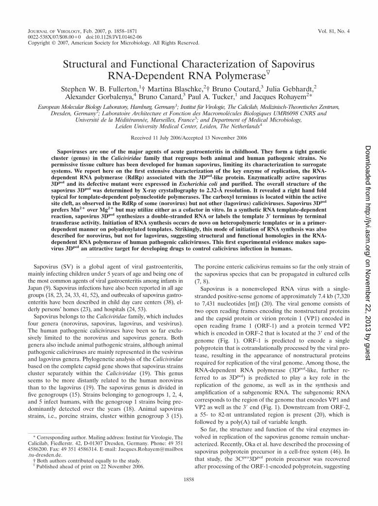
Heterologous expression and purification of recombinantsapovirus 3Dpol. A fusion protein bearing a His6 tag at its Cterminus or at its N terminus was overexpressed in E. coli andpurified by Ni-NTA affinity chromatography. A soluble sapo-virus protein of about 57 kDa was obtained (Fig. 2). Analo-gously, an active site mutant of sapovirus 3Dpol (m3Dpol) wasexpressed and purified. It displayed the same characteristics in
FIG. 2. Expression and purification of sapovirus 3Dpol in E. coli. So-dium dodecyl sulfate-polyacrylamide gel electrophoresis analysis of puri-fied recombinant sapovirus 3Dpol expressed in E. coli and carrying aC-terminal His6 tag. 3Dpol, wild-type sapovirus 3D-like polymerase;m3Dpol, active site sapovirus 3Dpol mutant (YGD343GD344G); M, molec-ular mass marker (in kDa).
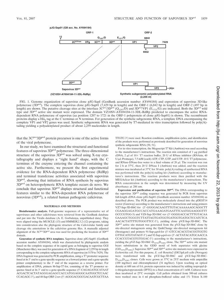
FIG. 3. Structure of sapovirus 3Dpol. (A) The structure is colored from the N terminus (blue) to the C terminus (red). The three major domains of thumb,fingers, and palm are clearly visible with the N-terminal extension (blue) reaching across from the top of the fingers to the thumb domain (red). (B) A ribbondiagram of the sapovirus 3Dpol. The �-helices are represented by � 1 to 15 (aqua) and the �-sheets are represented by � 1 to 4 (lilac).
VOL. 81, 2007 STRUCTURE AND FUNCTION OF SAPOVIRUS 3Dpol 1861
on Novem
ber 22, 2013 by guesthttp://jvi.asm
.org/D
ownloaded from
terms of solubility and migration during sodium dodecyl sul-fate-polyacrylamide gel electrophoresis analysis (Fig. 2).
Structure of sapovirus 3Dpol. The structure of the 3Dpol
from sapovirus has been determined to 2.32 Å resolution (PDB
identification no. 2CKW). The model was refined to an Rfactor of 17.3% and free R factor of 22.9% and contains 488amino acids (amino acids [aa] 1 to 375 and 385 to 496) and 271water molecules. The N-terminal His6 tag, 19 C-terminal
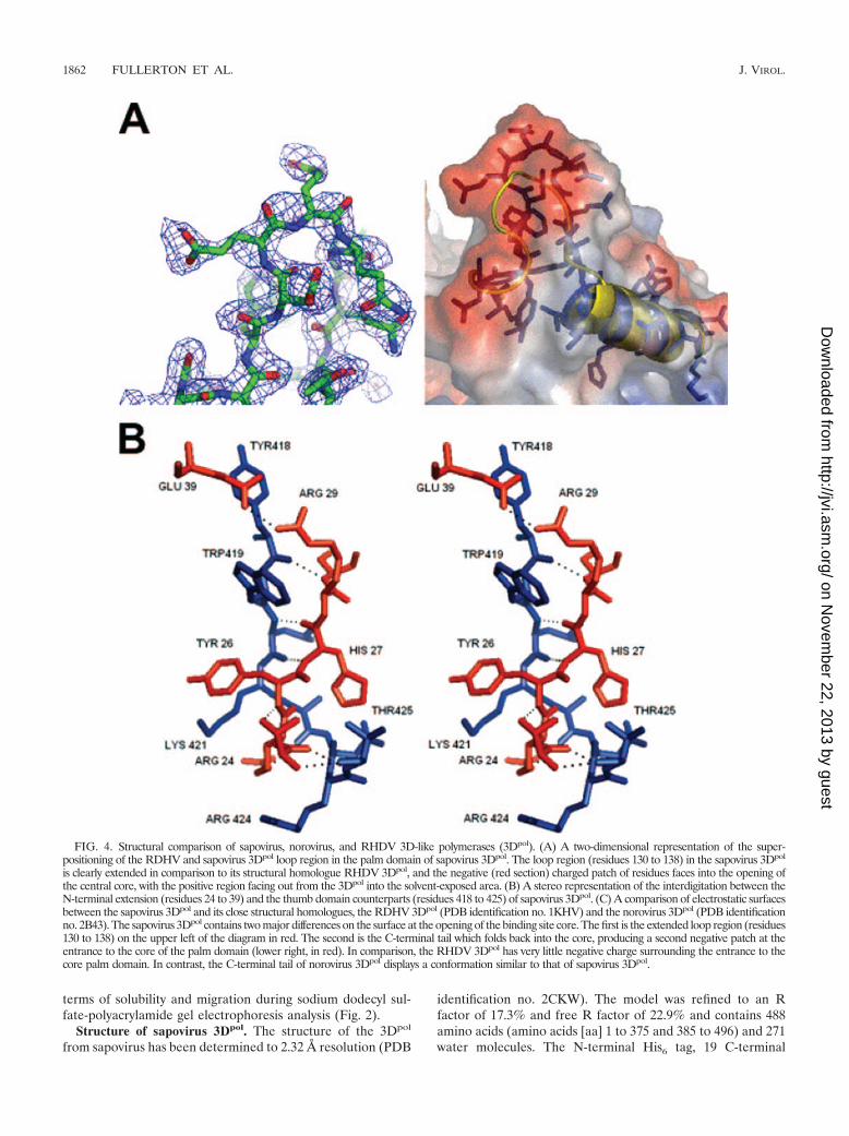
FIG. 4. Structural comparison of sapovirus, norovirus, and RHDV 3D-like polymerases (3Dpol). (A) A two-dimensional representation of the super-positioning of the RDHV and sapovirus 3Dpol loop region in the palm domain of sapovirus 3Dpol. The loop region (residues 130 to 138) in the sapovirus 3Dpol
is clearly extended in comparison to its structural homologue RHDV 3Dpol, and the negative (red section) charged patch of residues faces into the opening ofthe central core, with the positive region facing out from the 3Dpol into the solvent-exposed area. (B) A stereo representation of the interdigitation between theN-terminal extension (residues 24 to 39) and the thumb domain counterparts (residues 418 to 425) of sapovirus 3Dpol. (C) A comparison of electrostatic surfacesbetween the sapovirus 3Dpol and its close structural homologues, the RDHV 3Dpol (PDB identification no. 1KHV) and the norovirus 3Dpol (PDB identificationno. 2B43). The sapovirus 3Dpol contains two major differences on the surface at the opening of the binding site core. The first is the extended loop region (residues130 to 138) on the upper left of the diagram in red. The second is the C-terminal tail which folds back into the core, producing a second negative patch at theentrance to the core of the palm domain (lower right, in red). In comparison, the RHDV 3Dpol has very little negative charge surrounding the entrance to thecore palm domain. In contrast, the C-terminal tail of norovirus 3Dpol displays a conformation similar to that of sapovirus 3Dpol.
1862 FULLERTON ET AL. J. VIROL.
on Novem
ber 22, 2013 by guesthttp://jvi.asm
.org/D
ownloaded from
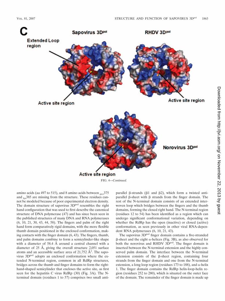
amino acids (aa 497 to 515), and 8 amino acids between pro375and lys385 are missing from the structure. These residues can-not be modeled because of poor experimental electron density.The domain structure of sapovirus 3Dpol resembles the righthand configuration that was used to first describe the canonicalstructure of DNA polymerase (47) and has since been seen inthe published structures of many DNA and RNA polymerases(6, 10, 21, 30, 43, 44, 58). The fingers and palm of the righthand form comparatively rigid domains, with the more flexiblethumb domain positioned in the enclosed conformation, mak-ing contacts with the finger domain (6, 43). The fingers, thumb,and palm domains combine to form a semicylinder-like shapewith a diameter of 58.4 Å around a central channel with adiameter of 25 Å, giving the overall structure 2,051 surfaceatoms and an accessible surface area of 21,752 Å2. The sapo-virus 3Dpol adopts an enclosed conformation where the ex-tended N-terminal region, common in all RdRp structures,bridges across the thumb and finger domains to form the right-hand-shaped semicylinder that encloses the active site, as firstseen for the hepatitis C virus RdRp (30) (Fig. 3A). The N-terminal domain (residues 1 to 57) comprises two small anti-
parallel �-strands (�1 and �2), which form a twisted anti-parallel �-sheet with � strands from the finger domain. Therest of the N-terminal domain consists of an extended inter-woven loop which bridges between the fingers and the thumbdomains, forming the closed right hand. The N-terminal region(residues 12 to 54) has been identified as a region which canundergo significant conformational variation, depending onwhether the RdRp has the open (inactive) or closed (active)conformation, as seen previously in other viral RNA-depen-dent RNA polymerases (6, 10, 21, 43).
The sapovirus 3Dpol finger domain contains a five-stranded�-sheet and the eight �-helices (Fig. 3B), as also observed forboth the norovirus and RHDV 3Dpol. The finger domain isinserted between the N-terminal extension and the highly con-served palm domain. The interface between the N-terminalextension consists of the �-sheet region, containing fourstrands from the finger domain and one from the N-terminalextension, a long loop region (residues 173 to 188), and �-helix1. The finger domain contains the RdRp helix-loop-helix re-gion (residues 252 to 280), which is situated on the outer faceof the domain. The remainder of the finger domain is made up
FIG. 4—Continued.
VOL. 81, 2007 STRUCTURE AND FUNCTION OF SAPOVIRUS 3Dpol 1863
on Novem
ber 22, 2013 by guesthttp://jvi.asm
.org/D
ownloaded from
of five �-helices (�2 to 6). The sapovirus 3Dpol structure dis-plays the same short loop segment between the third andfourth �-helices reported to be essential for the use of anRNA-primer duplex as a polymerization template (22).
The �-helices 2, 6, and 8 form the finger-palm interface with�-helix 10 in the palm domain. The most striking differencebetween the finger domains in the sapovirus 3Dpol and theRHDV 3Dpol is an extended loop region 130 to 138 which ispositioned on the opposite side of the central cavity adjacent tothe C-terminal residues. This extended loop region containsthree additional residues compared with the norovirus andRHDV 3Dpol models and is positioned further toward theopening of the central cavity, forming a negative patch on oneside of the opening to the active site (Fig. 4A).
The palm domain has the highest structural conservation,with an �/� fold containing a three-stranded anti-parallel�-sheet, consisting of a single � strand and a � hairpin lockedbetween �-helix 10 and �-helix 11 on the internal side of thepolymerase and �-helix 7 on the outer side. The palm domaincontains the catalytically important GDD sequence, which ispresent in all known RdRps of positive-stranded RNA viruses.The GDD motif (Gly346, Asp347, and Asp348) is thought tobe involved in metal ion interactions linked to nucleotidyltransfers during RNA elongation (44). However, no metals areobserved in this model. Soaking crystals with either Mn2� orMg2� did not result in observable density for the metals indifference maps. The structure of a sapovirus 3Dpol active sitemutant (GD347GD348G) at 3-Å resolution (data to be pub-lished) shows the C� trace to be approximately the same as thenative structure, indicating that the loss of activity is the resultof the loss of the enzymatically active aspartate side chains andis not the result of a conformational change. The interfacebetween the palm region and the thumb is supplied via anextended loop (residues 388 to 403) and a loosely arranged�-hairpin structure (residues 404 to 409) on which the thumbdomain is believed to hinge.
The thumb domain contains four �-helices interconnectedby two extended loops, one linking �-helix 12 with 13 and the
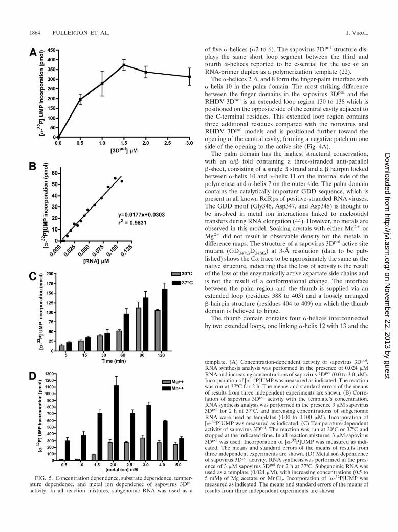
FIG. 5. Concentration dependence, substrate dependence, temper-ature dependence, and metal ion dependence of sapovirus 3Dpol
activity. In all reaction mixtures, subgenomic RNA was used as a
template. (A) Concentration-dependent activity of sapovirus 3Dpol.RNA synthesis analysis was performed in the presence of 0.024 �MRNA and increasing concentrations of sapovirus 3Dpol (0.0 to 3.0 �M).Incorporation of [�-32P]UMP was measured as indicated. The reactionwas run at 37°C for 2 h. The means and standard errors of the meansof results from three independent experiments are shown. (B) Corre-lation of sapovirus 3Dpol activity with the template’s concentration.RNA synthesis analysis was performed in the presence 3 �M sapovirus3Dpol for 2 h at 37°C, and increasing concentrations of subgenomicRNA were used as templates (0.00 to 0.100 �M). Incorporation of[�-32P]UMP was measured as indicated. (C) Temperature-dependentactivity of sapovirus 3Dpol. The reaction was run at 30°C or 37°C andstopped at the indicated time. In all reaction mixtures, 3 �M sapovirus3Dpol was used. Incorporation of [�-32P]UMP was measured as indi-cated. The means and standard errors of the means of results fromthree independent experiments are shown. (D) Metal ion dependenceof sapovirus 3Dpol activity. RNA synthesis was performed in the pres-ence of 3 �M sapovirus 3Dpol for 2 h at 37°C. Subgenomic RNA wasused as a template (0.024 �M), with increasing concentrations (0.5 to5 mM) of Mg acetate or MnCl2. Incorporation of [�-32P]UMP wasmeasured as indicated. The means and standard errors of the means ofresults from three independent experiments are shown.
1864 FULLERTON ET AL. J. VIROL.
on Novem
ber 22, 2013 by guesthttp://jvi.asm
.org/D
ownloaded from
other linking �-helix 14 with 15. The thumb domain interactswith the N-terminal extension via �-helix 12 and the loopcontaining residues 418 to 435 which link helix 12 and 13. Thepart of the loop containing residues 418 to 425 forms a smalltwo-stranded �-sheet with residues 24 to 28 from the N-termi-nal extension. This interaction involves seven hydrogen bondsbetween the two domains (Fig. 4B), resulting in the formationof a complex interdigitation of the side chains, which locks theright hand into the enclosed conformation (Fig. 4B). The sidechains of Arg25, Tyr26, and Arg28 interdigitate with the sidechains of Arg424, Lys421, and Trp419. Glu39 forms an addi-tional hydrogen bond with Arg29, the side chains of which arelocated between the Tyr418 and Trp419 side chains (Fig. 4B).These interactions form an interlocked bridge between thethumb and N-terminal extension that resembles the fingers ofjoined hands. The C terminus of sapovirus 3Dpol points towardthe channel containing the active site and is situated oppositeto the extended loop region (residues 130 to 138), forming twonegatively charged patches on either side of the mouth of theactive site channel. These negatively charged patches may helpregulate the RNA binding to the positively charged centralcore (Fig. 4C).
Biochemical characterization of sapovirus 3Dpol activity.RNA synthesis by SV 3Dpol was characterized by measuringthe relationship between incorporation of [�-32P]UMP andenzyme concentration in the reaction mixture. As shown in Fig.5A, the amount of UMP incorporated increased with the con-centration of sapovirus 3Dpol up to 1.5 �M, where the reactionreached its saturation phase. Sapovirus 3Dpol activity displayeda clear correlation to the template’s concentration over a broadlinear range (0.000 to 0.100 �M; r2 � 0.9831; 95% confidenceintervals, 0.9726 to 0.9974; P � 0.0001) (Fig. 5B). The temper-ature dependence of 3Dpol activity was also determined. Sapo-virus 3Dpol was found to display a higher activity at 37°Ccompared to 30°C (Fig. 5C). To address the metal ion depen-dence of sapovirus 3Dpol, the enzyme’s activity was measuredin the presence of increasing concentrations of Mg2� or Mn2�,using subgenomic RNA as a template. Over a concentrationrange from 0.5 to 5 mM, sapovirus 3Dpol displayed a clearpreference for Mn2� over Mg2� (Fig. 5D). However, sapovirus3Dpol was also active in the presence of Mg2�, indicating thatsapovirus 3Dpol displays flexibility with respect to the use ofMg2� or Mn2� as a cofactor.
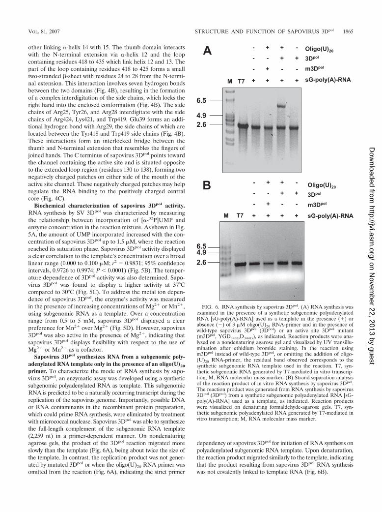
Sapovirus 3Dpol synthesizes RNA from a subgenomic poly-adenylated RNA template only in the presence of an oligo(U)20
primer. To characterize the mode of RNA synthesis by sapo-virus 3Dpol, an enzymatic assay was developed using a syntheticsubgenomic polyadenylated RNA as template. This subgenomicRNA is predicted to be a naturally occurring transcript during thereplication of the sapovirus genome. Importantly, possible DNAor RNA contaminants in the recombinant protein preparation,which could prime RNA synthesis, were eliminated by treatmentwith micrococcal nuclease. Sapovirus 3Dpol was able to synthesizethe full-length complement of the subgenomic RNA template(2,259 nt) in a primer-dependent manner. On nondenaturingagarose gels, the product of the 3Dpol reaction migrated moreslowly than the template (Fig. 6A), being about twice the size ofthe template. In contrast, the replication product was not gener-ated by mutated 3Dpol or when the oligo(U)20 RNA primer wasomitted from the reaction (Fig. 6A), indicating the strict primer
dependency of sapovirus 3Dpol for initiation of RNA synthesis onpolyadenylated subgenomic RNA template. Upon denaturation,the reaction product migrated similarly to the template, indicatingthat the product resulting from sapovirus 3Dpol RNA synthesiswas not covalently linked to template RNA (Fig. 6B).
FIG. 6. RNA synthesis by sapovirus 3Dpol. (A) RNA synthesis wasexamined in the presence of a synthetic subgenomic polyadenylatedRNA [sG-poly(A)-RNA] used as a template in the presence (�) orabsence (�) of 3 �M oligo(U)20 RNA-primer and in the presence ofwild-type sapovirus 3Dpol (3Dpol) or an active site 3Dpol mutant(m3Dpol, YGD343GD344G), as indicated. Reaction products were ana-lyzed on a nondenaturing agarose gel and visualized by UV transillu-mination after ethidium bromide staining. In the reaction usingm3Dpol instead of wild-type 3Dpol, or omitting the addition of oligo-(U)20 RNA-primer, the residual band observed corresponds to thesynthetic subgenomic RNA template used in the reaction. T7, syn-thetic subgenomic RNA generated by T7-mediated in vitro transcrip-tion; M, RNA molecular mass marker. (B) Strand separation analysisof the reaction product of in vitro RNA synthesis by sapovirus 3Dpol.The reaction product was generated from RNA synthesis by sapovirus3Dpol (3Dpol) from a synthetic subgenomic polyadenylated RNA [sG-poly(A)-RNA] used as a template, as indicated. Reaction productswere visualized on denaturing formaldehyde-agarose gels. T7, syn-thetic subgenomic polyadenylated RNA generated by T7-mediated invitro transcription; M, RNA molecular mass marker.
VOL. 81, 2007 STRUCTURE AND FUNCTION OF SAPOVIRUS 3Dpol 1865
on Novem
ber 22, 2013 by guesthttp://jvi.asm
.org/D
ownloaded from

Sapovirus 3Dpol initiates RNA synthesis de novo on anti-subgenomic heteropolymeric RNA templates. To assess theability of sapovirus 3Dpol to initiate RNA synthesis on anti-subgenomic RNA, sapovirus anti-subgenomic RNA was syn-thesized by in vitro transcription. Sapovirus 3Dpol was able toreplicate anti-subgenomic RNA in the absence of an RNAprimer (Fig. 7A). Strand separation analysis of the sapovirus3Dpol replication product allowed complete resolution of thedouble-stranded RNA on denaturing gels, indicating that thereplication product did not consist of two RNA strands covalentlylinked, as would had been expected in the case of a back-priminginitiation of sapovirus 3Dpol RNA synthesis (Fig. 7B).
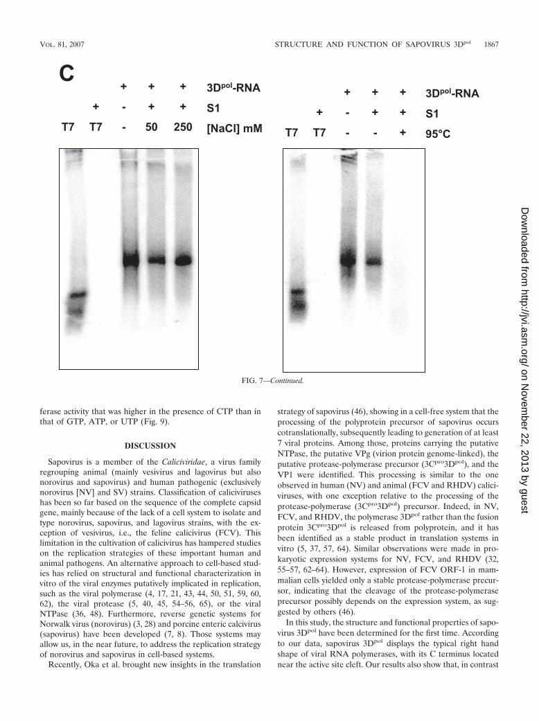
To further characterize the nature of the product of sapo-virus 3Dpol synthesis, treatment with nuclease S1 was per-formed. In contrast to the exogenous single-stranded RNAtemplate, the sapovirus 3Dpol product was not susceptible to S1nuclease treatment, indicating that it is double-stranded RNA(Fig. 7C). However, decreasing NaCl concentrations (50 mM)in the S1 nuclease reaction allowed partial digestion of thesynthesized RNA, as expected in the case of low-ionic-strengthbuffer (Fig. 7C). Denaturation of SV 3Dpol products by heatingat 95°C followed by incubation with S1 nuclease allowed theircomplete digestion, indicating that sapovirus 3Dpol synthesisproduct consists of the cRNA strands (Fig. 7D).
Sapovirus 3Dpol initiates RNA synthesis on homopolymericRNA templates. Initiation of RNA synthesis by sapovirus 3Dpol
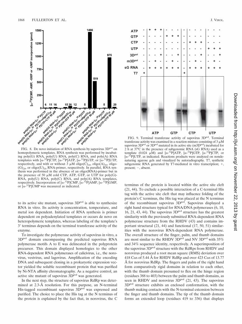
was examined on homopolymeric RNA templates. In the ab-sence of an oligonucleotide primer, no incorporation of[�-32P]AMP, [�-32P]CMP, [�-32P]GMP, or [�-32P]UMP wasseen (Fig. 8). In contrast, addition of a complementary oligo-nucleotide RNA-primer to the reaction led to incorporation of[�-32P]GMP and [�-32P]UMP on homopolymeric poly(C)-RNA and poly(A) RNA templates, respectively. Interestingly,addition of cold GTP to the reaction mixture (50 �M) allowedincorporation of [�-32P]GMP even in the absence of an oligo-nucleotide RNA-primer. This was only observed on poly(C)templates, indicating that synthesis of RNA from poly(C) ho-mopolymeric templates can take place in a primer-indepen-dent manner. For poly(G), poly(U), and poly(A) RNA tem-plates, no incorporation of [�-32P]CMP, [�-32P]AMP, or[�-32P]UMP was seen in the absence of a primer, even afteraddition of high concentrations of cold CTP, ATP, or UTP,respectively.
Our results indicate that sapovirus 3Dpol initiates RNA syn-thesis on poly(C) or poly(A) homopolymeric RNA templatesin a primer-dependent manner. In addition, de novo initiationis possible on poly(C)-RNA templates in the presence of highconcentrations of GTP.
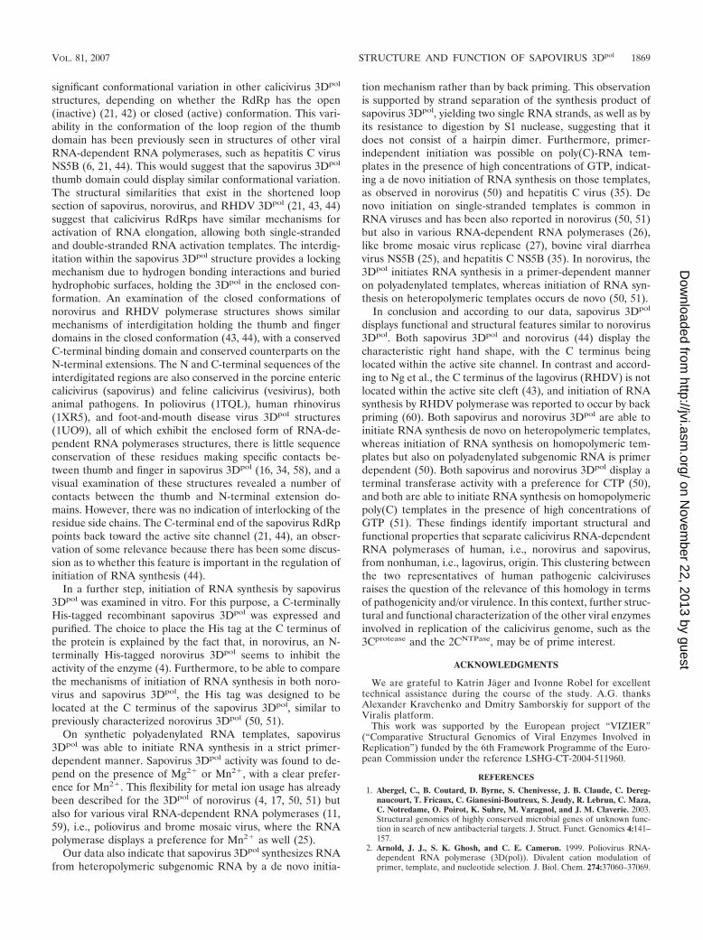
Sapovirus 3Dpol exhibits a terminal transferase activity invitro. Terminal transferase activity of RNA polymerase hasalready been reported for RNA viruses, e.g., hepatitis C virus(49) and poliovirus (2), adding nucleotides to the 3� ends ofnewly synthesized RNAs. This activity could potentially beused by RNA viruses as a mechanism to restore the 3� initia-tion site of RNA synthesis (49) and is one of the prerequisitesfor initiation of RNA synthesis by back priming. To character-ize the terminal transferase activity of sapovirus 3Dpol, theenzyme was incubated with subgenomic RNA template in areaction mixture containing the �-32P-labeled version of onlyone of the NTPs. Sapovirus 3Dpol displayed terminal trans-
FIG. 7. De novo initiation of RNA synthesis on sapovirus anti-sub-genomic RNA. (A) RNA synthesis was examined in the presence of asynthetic anti-subgenomic RNA (Anti-sG RNA) used as a template andin the presence wild-type sapovirus 3Dpol (3Dpol) or an active site 3Dpol
mutant (m3Dpol, YGD343GD344G), as indicated. Reaction products wereanalyzed on nondenaturing agarose gel and visualized by UV transillu-mination after ethidium bromide staining. In the reaction using m3Dpol
instead of wild-type 3Dpol, the residual band observed corresponds to thesynthetic anti-subgenomic RNA template used in the reaction. T7, syn-thetic anti-subgenomic RNA generated by T7-mediated in vitro transcrip-tion; M, RNA molecular mass marker; �, present; �, absent. (B) Strandseparation analysis of the reaction product of in vitro RNA synthesis bysapovirus 3Dpol. The reaction product was generated from RNA synthesisby sapovirus 3Dpol from a synthetic anti-subgenomic RNA (Anti-sGRNA) used as a template, as indicated. Reaction products were visualizedon denaturing formaldehyde-agarose gels. T7, synthetic anti-subgenomicRNA generated by T7-mediated in vitro transcription; M, RNA molecu-lar mass marker. (C) The sapovirus 3Dpol synthesis product (3Dpol-RNA)was generated from synthetic anti-subgenomic RNA (T7) used as a tem-plate. The RNA synthesis product was then incubated in the presence (�)or absence (�) of S1 nuclease (S1), in the presence of low-salt (50 mMNaCl) or high-salt (250 mM NaCl) concentrations, or after heating (95°C,5 min) and rapid chilling on ice for 5 min, as indicated. Reactions productswere analyzed on nondenaturing agarose gels and visualized by autora-diography. 3Dpol-RNA, sapovirus 3Dpol synthesis product using anti-sub-genomic RNA as a template. T7, synthetic anti-subgenomic RNA gener-ated by T7-mediated in vitro transcription.
1866 FULLERTON ET AL. J. VIROL.
on Novem
ber 22, 2013 by guesthttp://jvi.asm
.org/D
ownloaded from
ferase activity that was higher in the presence of CTP than inthat of GTP, ATP, or UTP (Fig. 9).
DISCUSSION
Sapovirus is a member of the Caliciviridae, a virus familyregrouping animal (mainly vesivirus and lagovirus but alsonorovirus and sapovirus) and human pathogenic (exclusivelynorovirus [NV] and SV) strains. Classification of caliciviruseshas been so far based on the sequence of the complete capsidgene, mainly because of the lack of a cell system to isolate andtype norovirus, sapovirus, and lagovirus strains, with the ex-ception of vesivirus, i.e., the feline calicivirus (FCV). Thislimitation in the cultivation of calicivirus has hampered studieson the replication strategies of these important human andanimal pathogens. An alternative approach to cell-based stud-ies has relied on structural and functional characterization invitro of the viral enzymes putatively implicated in replication,such as the viral polymerase (4, 17, 21, 43, 44, 50, 51, 59, 60,62), the viral protease (5, 40, 45, 54–56, 65), or the viralNTPase (36, 48). Furthermore, reverse genetic systems forNorwalk virus (norovirus) (3, 28) and porcine enteric calcivirus(sapovirus) have been developed (7, 8). Those systems mayallow us, in the near future, to address the replication strategyof norovirus and sapovirus in cell-based systems.
Recently, Oka et al. brought new insights in the translation
strategy of sapovirus (46), showing in a cell-free system that theprocessing of the polyprotein precursor of sapovirus occurscotranslationally, subsequently leading to generation of at least7 viral proteins. Among those, proteins carrying the putativeNTPase, the putative VPg (virion protein genome-linked), theputative protease-polymerase precursor (3Cpro3Dpol), and theVP1 were identified. This processing is similar to the oneobserved in human (NV) and animal (FCV and RHDV) calici-viruses, with one exception relative to the processing of theprotease-polymerase (3Cpro3Dpol) precursor. Indeed, in NV,FCV, and RHDV, the polymerase 3Dpol rather than the fusionprotein 3Cpro3Dpol is released from polyprotein, and it hasbeen identified as a stable product in translation systems invitro (5, 37, 57, 64). Similar observations were made in pro-karyotic expression systems for NV, FCV, and RHDV (32,55–57, 62–64). However, expression of FCV ORF-1 in mam-malian cells yielded only a stable protease-polymerase precur-sor, indicating that the cleavage of the protease-polymeraseprecursor possibly depends on the expression system, as sug-gested by others (46).
In this study, the structure and functional properties of sapo-virus 3Dpol have been determined for the first time. Accordingto our data, sapovirus 3Dpol displays the typical right handshape of viral RNA polymerases, with its C terminus locatednear the active site cleft. Our results also show that, in contrast
FIG. 7—Continued.
VOL. 81, 2007 STRUCTURE AND FUNCTION OF SAPOVIRUS 3Dpol 1867
on Novem
ber 22, 2013 by guesthttp://jvi.asm
.org/D
ownloaded from
to its active site mutant, sapovirus 3Dpol is able to synthesizeRNA in vitro. Its activity is concentration, temperature, andmetal ion dependent. Initiation of RNA synthesis is primerdependent on polyadenylated templates or occurs de novo onheteropolymeric templates, whereas labeling of the template’s3� terminus depends on the terminal transferase activity of theenzyme.
To investigate the polymerase activity of sapovirus in vitro, a3Dpol domain encompassing the predicted sapovirus RNApolymerase motifs A to E was delineated in the polyproteinprecursor. This domain displayed homologies to the otherRNA-dependent RNA polymerases of calicivirus, i.e., the noro-virus, vesivirus, and lagovirus. Amplification of the encodingDNA and subsequent cloning in a prokaryotic expression vec-tor yielded the soluble recombinant protein that was purifiedby Ni-NTA affinity chromatography. As a negative control, anactive site mutant of sapovirus 3Dpol was generated.
In the next step, the structure of sapovirus RdRp was deter-mined at 2.3-Å resolution. For this purpose, an N-terminalHis-tagged recombinant sapovirus 3Dpol was expressed andpurified. The choice to place the His tag at the N terminus ofthe protein is explained by the fact that, in norovirus, the C
terminus of the protein is located within the active site cleft(21, 44). To exclude a possible interaction of a C-terminal Histag with the active site cleft that may influence folding of theprotein’s C terminus, the His tag was placed at the N terminusof the recombinant sapovirus 3Dpol. Sapovirus displayed aright hand structure typical for RNA/DNA polymerases (6, 10,16, 21, 43, 44). The sapovirus 3Dpol structure has the greatestsimilarity with the previously submitted RNA-dependent RNApolymerase structure from the RHDV (43) and contains im-portant structural (21, 44) and functional (17, 50, 51) similar-ities with the norovirus RNA-dependent RNA polymerase.The overall structure of the finger, palm, and thumb domainsare most similar to the RHDV 3Dpol and NV 3Dpol with 35%and 34% sequence identity, respectively. A superimposition ofthe sapovirus 3Dpol structure with the RdRps from RHDV andnorovirus produced a root mean square (RMS) deviation over418 C�s of 5.44 Å for RHDV RdRp and over 423 C�s of 13.77Å for norovirus RdRp. The fingers and palm of the right handform comparatively rigid domains in relation to each other,with the thumb domain presumed to flex on the hinge region(residues 388 to 403) between the palm and thumb domains, asseen in RHDV and norovirus 3Dpol (21, 43). The sapovirus3Dpol structure exhibits an enclosed conformation, with thethumb making contacts with the N-terminal extension betweenthe finger and thumb domains. The tip of the thumb domainforms an extended loop (residues 419 to 336) that displays
FIG. 8. De novo initiation of RNA synthesis by sapovirus 3Dpol onhomopolymeric templates. RNA synthesis was performed by incubat-ing poly(G) RNA, poly(U) RNA, poly(C) RNA, and poly(A) RNAtemplates with [�-32P]CTP, [�-32P]ATP, [�-32P]GTP, or [�-32P]UTP,respectively, and with or without 3 �M oligo(C)20, oligo(A)20, oligo-(G)20, or oligo(U)20 RNA-primer, respectively. In parallel, RNA syn-thesis was performed in the absence of an oligo(RNA)-primer but inthe presence of 50 �M cold CTP, ATP, GTP, or UTP for poly(G)-RNA, poly(U) RNA, poly(C) RNA, and poly(A) RNA templates,respectively. Incorporation of [�-32P]CMP, [�-32P]AMP, [�-32P]GMP,or [�-32P]UMP was measured as indicated.
FIG. 9. Terminal transferase activity of sapovirus 3Dpol. Terminaltransferase activity was examined in a reaction mixture consisting of 3 �Msapovirus 3Dpol or 3Dpol mutated in its active site (m3Dpol) incubated for2 h at 37°C in the presence of subgenomic RNA (sG RNA) used as atemplate (0.024 �M) and [�-32P]ATP, [�-32P]GTP, [�-32P]CTP, or[�-32P]UTP, as indicated. Reactions products were analyzed on nonde-naturing agarose gels and visualized by autoradiography. T7, syntheticsubgenomic RNA generated by T7-mediated in vitro transcription; �,present; �, absent.
1868 FULLERTON ET AL. J. VIROL.
on Novem
ber 22, 2013 by guesthttp://jvi.asm
.org/D
ownloaded from
significant conformational variation in other calicivirus 3Dpol
structures, depending on whether the RdRp has the open(inactive) (21, 42) or closed (active) conformation. This vari-ability in the conformation of the loop region of the thumbdomain has been previously seen in structures of other viralRNA-dependent RNA polymerases, such as hepatitis C virusNS5B (6, 21, 44). This would suggest that the sapovirus 3Dpol
thumb domain could display similar conformational variation.The structural similarities that exist in the shortened loopsection of sapovirus, norovirus, and RHDV 3Dpol (21, 43, 44)suggest that calicivirus RdRps have similar mechanisms foractivation of RNA elongation, allowing both single-strandedand double-stranded RNA activation templates. The interdig-itation within the sapovirus 3Dpol structure provides a lockingmechanism due to hydrogen bonding interactions and buriedhydrophobic surfaces, holding the 3Dpol in the enclosed con-formation. An examination of the closed conformations ofnorovirus and RHDV polymerase structures shows similarmechanisms of interdigitation holding the thumb and fingerdomains in the closed conformation (43, 44), with a conservedC-terminal binding domain and conserved counterparts on theN-terminal extensions. The N and C-terminal sequences of theinterdigitated regions are also conserved in the porcine entericcalicivirus (sapovirus) and feline calicivirus (vesivirus), bothanimal pathogens. In poliovirus (1TQL), human rhinovirus(1XR5), and foot-and-mouth disease virus 3Dpol structures(1UO9), all of which exhibit the enclosed form of RNA-de-pendent RNA polymerases structures, there is little sequenceconservation of these residues making specific contacts be-tween thumb and finger in sapovirus 3Dpol (16, 34, 58), and avisual examination of these structures revealed a number ofcontacts between the thumb and N-terminal extension do-mains. However, there was no indication of interlocking of theresidue side chains. The C-terminal end of the sapovirus RdRppoints back toward the active site channel (21, 44), an obser-vation of some relevance because there has been some discus-sion as to whether this feature is important in the regulation ofinitiation of RNA synthesis (44).
In a further step, initiation of RNA synthesis by sapovirus3Dpol was examined in vitro. For this purpose, a C-terminallyHis-tagged recombinant sapovirus 3Dpol was expressed andpurified. The choice to place the His tag at the C terminus ofthe protein is explained by the fact that, in norovirus, an N-terminally His-tagged norovirus 3Dpol seems to inhibit theactivity of the enzyme (4). Furthermore, to be able to comparethe mechanisms of initiation of RNA synthesis in both noro-virus and sapovirus 3Dpol, the His tag was designed to belocated at the C terminus of the sapovirus 3Dpol, similar topreviously characterized norovirus 3Dpol (50, 51).
On synthetic polyadenylated RNA templates, sapovirus3Dpol was able to initiate RNA synthesis in a strict primer-dependent manner. Sapovirus 3Dpol activity was found to de-pend on the presence of Mg2� or Mn2�, with a clear prefer-ence for Mn2�. This flexibility for metal ion usage has alreadybeen described for the 3Dpol of norovirus (4, 17, 50, 51) butalso for various viral RNA-dependent RNA polymerases (11,59), i.e., poliovirus and brome mosaic virus, where the RNApolymerase displays a preference for Mn2� as well (25).
Our data also indicate that sapovirus 3Dpol synthesizes RNAfrom heteropolymeric subgenomic RNA by a de novo initia-
tion mechanism rather than by back priming. This observationis supported by strand separation of the synthesis product ofsapovirus 3Dpol, yielding two single RNA strands, as well as byits resistance to digestion by S1 nuclease, suggesting that itdoes not consist of a hairpin dimer. Furthermore, primer-independent initiation was possible on poly(C)-RNA tem-plates in the presence of high concentrations of GTP, indicat-ing a de novo initiation of RNA synthesis on those templates,as observed in norovirus (50) and hepatitis C virus (35). Denovo initiation on single-stranded templates is common inRNA viruses and has been also reported in norovirus (50, 51)but also in various RNA-dependent RNA polymerases (26),like brome mosaic virus replicase (27), bovine viral diarrheavirus NS5B (25), and hepatitis C NS5B (35). In norovirus, the3Dpol initiates RNA synthesis in a primer-dependent manneron polyadenylated templates, whereas initiation of RNA syn-thesis on heteropolymeric templates occurs de novo (50, 51).
In conclusion and according to our data, sapovirus 3Dpol
displays functional and structural features similar to norovirus3Dpol. Both sapovirus 3Dpol and norovirus (44) display thecharacteristic right hand shape, with the C terminus beinglocated within the active site channel. In contrast and accord-ing to Ng et al., the C terminus of the lagovirus (RHDV) is notlocated within the active site cleft (43), and initiation of RNAsynthesis by RHDV polymerase was reported to occur by backpriming (60). Both sapovirus and norovirus 3Dpol are able toinitiate RNA synthesis de novo on heteropolymeric templates,whereas initiation of RNA synthesis on homopolymeric tem-plates but also on polyadenylated subgenomic RNA is primerdependent (50). Both sapovirus and norovirus 3Dpol display aterminal transferase activity with a preference for CTP (50),and both are able to initiate RNA synthesis on homopolymericpoly(C) templates in the presence of high concentrations ofGTP (51). These findings identify important structural andfunctional properties that separate calicivirus RNA-dependentRNA polymerases of human, i.e., norovirus and sapovirus,from nonhuman, i.e., lagovirus, origin. This clustering betweenthe two representatives of human pathogenic calcivirusesraises the question of the relevance of this homology in termsof pathogenicity and/or virulence. In this context, further struc-tural and functional characterization of the other viral enzymesinvolved in replication of the calicivirus genome, such as the3Cprotease and the 2CNTPase, may be of prime interest.
ACKNOWLEDGMENTS
We are grateful to Katrin Jager and Ivonne Robel for excellenttechnical assistance during the course of the study. A.G. thanksAlexander Kravchenko and Dmitry Samborskiy for support of theViralis platform.
This work was supported by the European project “VIZIER”(“Comparative Structural Genomics of Viral Enzymes Involved inReplication”) funded by the 6th Framework Programme of the Euro-pean Commission under the reference LSHG-CT-2004-511960.
REFERENCES
1. Abergel, C., B. Coutard, D. Byrne, S. Chenivesse, J. B. Claude, C. Dereg-naucourt, T. Fricaux, C. Gianesini-Boutreux, S. Jeudy, R. Lebrun, C. Maza,C. Notredame, O. Poirot, K. Suhre, M. Varagnol, and J. M. Claverie. 2003.Structural genomics of highly conserved microbial genes of unknown func-tion in search of new antibacterial targets. J. Struct. Funct. Genomics 4:141–157.
2. Arnold, J. J., S. K. Ghosh, and C. E. Cameron. 1999. Poliovirus RNA-dependent RNA polymerase (3D(pol)). Divalent cation modulation ofprimer, template, and nucleotide selection. J. Biol. Chem. 274:37060–37069.
VOL. 81, 2007 STRUCTURE AND FUNCTION OF SAPOVIRUS 3Dpol 1869
on Novem
ber 22, 2013 by guesthttp://jvi.asm
.org/D
ownloaded from
3. Asanaka, M., R. L. Atmar, V. Ruvolo, S. E. Crawford, F. H. Neill, and M. K.Estes. 2005. Replication and packaging of Norwalk virus RNA in culturedmammalian cells. Proc. Natl. Acad. Sci. USA 102:10327–10332.
4. Belliot, G., S. V. Sosnovtsev, K. O. Chang, V. Babu, U. Uche, J. J. Arnold,C. E. Cameron, and K. Y. Green. 2005. Norovirus proteinase-polymerase andpolymerase are both active forms of RNA-dependent RNA polymerase.J. Virol. 79:2393–2403.
5. Belliot, G., S. V. Sosnovtsev, T. Mitra, C. Hammer, M. Garfield, and K. Y.Green. 2003. In vitro proteolytic processing of the MD145 norovirus ORF1nonstructural polyprotein yields stable precursors and products similar tothose detected in calicivirus-infected cells. J. Virol. 77:10957–10974.
6. Biswal, B. K., M. M. Cherney, M. Wang, L. Chan, C. G. Yannopoulos, D.Bilimoria, O. Nicolas, J. Bedard, and M. N. James. 2005. Crystal structuresof the RNA-dependent RNA polymerase genotype 2a of hepatitis C virusreveal two conformations and suggest mechanisms of inhibition by non-nucleoside inhibitors. J. Biol. Chem. 280:18202–18210.
7. Chang, K. O., S. S. Sosnovtsev, G. Belliot, Q. Wang, L. J. Saif, and K. Y.Green. 2005. Reverse genetics system for porcine enteric calicivirus, a pro-totype sapovirus in the Caliciviridae. J. Virol. 79:1409–1416.
8. Chang, K. O., S. V. Sosnovtsev, G. Belliot, Y. Kim, L. J. Saif, and K. Y.Green. 2004. Bile acids are essential for porcine enteric calicivirus replicationin association with down-regulation of signal transducer and activator oftranscription 1. Proc. Natl. Acad. Sci. USA 101:8733–8738.
9. Chiba, S., S. Nakata, K. Numata-Kinoshita, and S. Honma. 2000. Sapporovirus: history and recent findings. J. Infect. Dis. 181(Suppl. 2):S303–S308.
10. Choi, K. H., J. M. Groarke, D. C. Young, R. J. Kuhn, J. L. Smith, D. C.Pevear, and M. G. Rossmann. 2004. The structure of the RNA-dependentRNA polymerase from bovine viral diarrhea virus establishes the role ofGTP in de novo initiation. Proc. Natl. Acad. Sci. USA 101:4425–4430.
11. Crotty, S., D. Gohara, D. K. Gilligan, S. Karelsky, C. E. Cameron, and R.Andino. 2003. Manganese-dependent polioviruses caused by mutationswithin the viral polymerase. J. Virol. 77:5378–5388.
12. Eddy, S. R. 1998. Profile hidden Markov models. Bioinformatics 14:755–763.13. Edgar, R. C. 2004. MUSCLE: multiple sequence alignment with high accu-
racy and high throughput. Nucleic Acids Res. 32:1792–1797.14. Evans, P. R. 1993. Data collection and processing, p. 114–122. In Proceed-
ings of the CCP4 study weekend, 29 to 30 January 1993. Science and Engi-neering Research Council Daresbury Laboratory, Daresbury, Warrington,Cheshire, United Kingdom.
15. Farkas, T., W. M. Zhong, Y. Jing, P. W. Huang, S. M. Espinosa, N. Martinez,A. L. Morrow, G. M. Ruiz-Palacios, L. K. Pickering, and X. Jiang. 2004.Genetic diversity among sapoviruses. Arch. Virol. 149:1309–1323.
16. Ferrer-Orta, C., A. Arias, R. Perez-Luque, C. Escarmis, E. Domingo, and N.Verdaguer. 2004. Structure of foot-and-mouth disease virus RNA-dependentRNA polymerase and its complex with a template-primer RNA. J. Biol.Chem. 279:47212–47221.
17. Fukushi, S., S. Kojima, R. Takai, F. B. Hoshino, T. Oka, N. Takeda, K.Katayama, and T. Kageyama. 2004. Poly(A)- and primer-independent RNApolymerase of norovirus. J. Virol. 78:3889–3896.
18. Gallimore, C. I., M. Iturriza-Gomara, D. Lewis, D. Cubitt, H. Cotterill, andJ. J. Gray. 2006. Characterization of sapoviruses collected in the UnitedKingdom from 1989 to 2004. J. Med. Virol. 78:673–682.
19. Green, K. Y., T. Ando, M. S. Balayan, T. Berke, I. N. Clarke, M. K. Estes,D. O. Matson, S. Nakata, J. D. Neill, M. J. Studdert, and H. J. Thiel. 2000.Taxonomy of the caliciviruses. J. Infect. Dis. 181(Suppl. 2):S322–S330.
20. Green, K. Y., R. M. Chanock, and A. Z. Kapikian. 2001. Human caliciviruses,p. 841–874. In D. M. Knipe, P. M. Howley, D. E. Griffin, R. A. Lamb, M. A.Martin, B. Roizman, and S. E. Straus (ed.), Fields virology, 4th ed. LippincottWilliams and Wilkins, Philadelphia, PA.
21. Hogbom, M., J. Rohayem, T. Unge, and A. Jones. 22 September 2005,posting date. Crystal structure of the Norwalk virus RNA dependentRNA polymerase from strain Hu/NLV/Dresden174/1997/GE. PDB ID:2B43. http://www.pdb.org/.
22. Hong, Z., C. E. Cameron, M. P. Walker, C. Castro, N. Yao, J. Y. N. Lau, andW. Zhong. 2001. A novel mechanism to ensure terminal initiation by hepa-titis C virus NS5B polymerase. Virology 285:6–11.
23. Humphrey, T. J., J. G. Cruickshank, and W. D. Cubitt. 1984. An outbreak ofcalicivirus associated gastroenteritis in an elderly persons home. A possiblezoonosis? J. Hyg. (London) 93:293–299.
24. Johansson, P. J., K. Bergentoft, P. A. Larsson, G. Magnusson, A. Widell, M.Thorhagen, and K. O. Hedlund. 2005. A nosocomial sapovirus-associatedoutbreak of gastroenteritis in adults. Scand. J. Infect. Dis. 37:200–204.
25. Kao, C. C., A. M. Del Vecchio, and W. Zhong. 1999. De novo initiation ofRNA synthesis by a recombinant flaviviridae RNA-dependent RNA poly-merase. Virology 253:1–7.
26. Kao, C. C., P. Singh, and D. J. Ecker. 2001. De novo initiation of viralRNA-dependent RNA synthesis. Virology 287:251–260.
27. Kao, C. C., and J. H. Sun. 1996. Initiation of minus-strand RNA synthesis bythe brome mosaicvirus RNA-dependent RNA polymerase: use of oligoribo-nucleotide primers. J. Virol. 70:6826–6830.
28. Katayama, K., G. S. Hansman, T. Oka, S. Ogawa, and N. Takeda. 2006.
Investigation of norovirus replication in a human cell line. Arch. Virol.151:1291–1308.
29. Lamzin, V. S., A. Perrakis, and K. S. Wilson. 2001. The ARP/WARP suitefor automated construction and refinement of protein models. In M. G.Rossmann and E. Arnold (ed.), Int. tables for crystallography, vol. F. Crys-tallography of biological macromolecules Kluwer Academic Publishers, Dor-drecht, The Netherlands.
30. Lesburg, C. A., M. B. Cable, E. Ferrari, Z. Hong, A. F. Mannarino, and P. C.Weber. 1999. Crystal structure of the RNA-dependent RNA polymerasefrom hepatitis C virus reveals a fully encircled active site. Nat. Struct. Biol.6:937–943.
31. Leslie, A. G. W. 1992. Joint ccp4 and ESF-EACMB newsletter on proteincrystallography. Daresbury Laboratory, Warrington, United Kingdom.
32. Liu, B. L., G. J. Viljoen, I. N. Clarke, and P. R. Lambden. 1999. Identificationof further proteolytic cleavage sites in the Southampton calicivirus polypro-tein by expression of the viral protease in E. coli. J. Gen. Virol. 80(Pt 2):291–296.
33. Lopman, B. A., D. W. Brown, and M. Koopmans. 2002. Human calicivirusesin Europe. J. Clin. Virol. 24:137–160.
34. Love, R. A., K. A. Maegley, X. Yu, R. A. Ferre, L. K. Lingardo, W. Diehl, H. E.Parge, P. S. Dragovich, and S. A. Fuhrman. 2004. The crystal structure of theRNA-dependent RNA polymerase from human rhinovirus: a dual functiontarget for common cold antiviral therapy. Structure 12:1533–1544.
35. Luo, G., R. K. Hamatake, D. M. Mathis, J. Racela, K. L. Rigat, J. Lemm, andR. J. Colonno. 2000. De novo initiation of RNA synthesis by the RNA-dependent RNA polymerase (NS5B) of hepatitis C virus. J. Virol. 74:851–863.
36. Marin, M. S., R. Casais, J. M. Alonso, and F. Parra. 2000. ATP binding andATPase activities associated with recombinant rabbit hemorrhagic diseasevirus 2C-like polypeptide. J. Virol. 74:10846–10851.
37. Martin Alonso, J. M., R. Casais, J. A. Boga, and F. Parra. 1996. Processingof rabbit hemorrhagic disease virus polyprotein. J. Virol. 70:1261–1265.
38. Matson, D. O., M. K. Estes, R. I. Glass, A. V. Bartlett, M. Penaranda, E.Calomeni, T. Tanaka, S. Nakata, and S. Chiba. 1989. Human calicivirus-associated diarrhea in children attending day care centers. J. Infect. Dis.159:71–78.
39. Murshudov, G. N., A. A. Vagin, and E. J. Dodson. 1997. Refinement ofmacromolecular structures by the maximum-likelihood method. Acta Crys-tallogr. D 53:240–255.
40. Nakamura, K., Y. Someya, T. Kumasaka, G. Ueno, M. Yamamoto, T. Sato,N. Takeda, T. Miyamura, and N. Tanaka. 2005. A norovirus protease struc-ture provides insights into active and substrate binding site integrity. J. Virol.79:13685–13693.
41. Nakata, S., S. Chiba, H. Terashima, and T. Nakao. 1985. Prevalence ofantibody to human calicivirus in Japan and Southeast Asia determined byradioimmunoassay. J. Clin. Microbiol. 22:519–521.
42. Navazza, J. 1994. AMoRe: an automated package for molecular replace-ment. N. Acta Crystallogr. A 50:157–163.
43. Ng, K. K., M. M. Cherney, A. L. Vazquez, A. Machin, J. M. Alonso, F. Parra,and M. N. James. 2002. Crystal structures of active and inactive conforma-tions of a caliciviral RNA-dependent RNA polymerase. J. Biol. Chem. 277:1381–1387.
44. Ng, K. K., N. Pendas-Franco, J. Rojo, J. A. Boga, A. Machin, J. M. Alonso,and F. Parra. 2004. Crystal structure of norwalk virus polymerase reveals thecarboxyl terminus in the active site cleft. J. Biol. Chem. 279:16638–16645.
45. Oka, T., K. Katayama, S. Ogawa, G. S. Hansman, T. Kageyama, T. Miyamura,and N. Takeda. 2005. Cleavage activity of the sapovirus 3C-like protease inEscherichia coli. Arch. Virol. 150:2539–2548.
46. Oka, T., K. Katayama, S. Ogawa, G. S. Hansman, T. Kageyama, H. Ushijima,T. Miyamura, and N. Takeda. 2005. Proteolytic processing of sapovirus ORF1polyprotein. J. Virol. 79:7283–7290.
47. Ollis, D. L., P. Brick, R. Hamlin, N. G. Xuong, and T. A. Steitz. 1985.Structure of large fragment of Escherichia coli DNA polymerase I com-plexed with dTMP. Nature 313:762–766.
48. Pfister, T., and E. Wimmer. 2001. Polypeptide p41 of a Norwalk-like virus isa nucleic acid-independent nucleoside triphosphatase. J. Virol. 75:1611–1619.
49. Ranjith-Kumar, C. T., J. Gajewski, L. Gutshall, D. Maley, R. T. Sarisky, andC. C. Kao. 2001. Terminal nucleotidyl transferase activity of recombinantFlaviviridae RNA-dependent RNA polymerases: implication for viral RNAsynthesis. J. Virol. 75:8615–8623.
50. Rohayem, J., K. Jager, I. Robel, U. Scheffler, A. Temme, and W. Rudolph.2006. Characterization of norovirus 3Dpol RNA-dependent RNA polymer-ase activity and initiation of RNA synthesis. J. Gen. Virol. 87:2621–2630.
51. Rohayem, J., I. Robel, K. Jager, U. Scheffler, and W. Rudolph. 2006. Protein-primed and de novo initiation of RNA synthesis by norovirus 3Dpol. J. Virol.80:7060–7069.
52. Sakuma, Y., S. Chiba, R. Kogasaka, H. Terashima, S. Nakamura, K. Horino,and T. Nakao. 1981. Prevalence of antibody to human calicivirus in generalpopulation of northern Japan. J. Med. Virol. 7:221–225.
53. Simor, A. E., M. McArthur, A. McGeer, S. Shurtleff, and M. Jabar. 1990.Calicivirus gastroenteritis in a geriatric long-term care facility–Ontario. Can.Dis. Wkly. Rep. 16:239–240, 243.
1870 FULLERTON ET AL. J. VIROL.
on Novem
ber 22, 2013 by guesthttp://jvi.asm
.org/D
ownloaded from
54. Someya, Y., N. Takeda, and T. Miyamura. 2005. Characterization of thenorovirus 3C-like protease. Virus Res. 110:91–97.
55. Someya, Y., N. Takeda, and T. Miyamura. 2000. Complete nucleotide se-quence of the chiba virus genome and functional expression of the 3C-likeprotease in Escherichia coli. Virology 278:490–500.
56. Someya, Y., N. Takeda, and T. Miyamura. 2002. Identification of active-siteamino acid residues in the Chiba virus 3C-like protease. J. Virol. 76:5949–5958.
57. Sosnovtseva, S. A., S. V. Sosnovtsev, and K. Y. Green. 1999. Mapping of thefeline calicivirus proteinase responsible for autocatalytic processing of thenonstructural polyprotein and identification of a stable proteinase-polymer-ase precursor protein. J. Virol. 73:6626–6633.
58. Thompson, A. A., and O. B. Peersen. 2004. Structural basis for proteolysis-dependent activation of the poliovirus RNA-dependent RNA polymerase.EMBO J. 23:3462–3471.
59. Vazquez, A. L., J. M. Alonso, and F. Parra. 2000. Mutation analysis of theGDD sequence motif of a calicivirus RNA-dependent RNA polymerase.J. Virol. 74:3888–3891.
60. Vazquez, A. L., J. M. Martin Alonso, R. Casais, J. A. Boga, and F. Parra.1998. Expression of enzymatically active rabbit hemorrhagic disease virus
RNA-dependent RNA polymerase in Escherichia coli. J. Virol. 72:2999–3004.
61. Vincentelli, R., S. Canaan, J. Offant, C. Cambillau, and C. Bignon. 2005.Automated expression and solubility screening of His-tagged proteins in96-well format. Anal. Biochem. 346:77–84.
62. Wei, L., J. S. Huhn, A. Mory, H. B. Pathak, S. V. Sosnovtsev, K. Y. Green,and C. E. Cameron. 2001. Proteinase-polymerase precursor as the activeform of feline calicivirus RNA-dependent RNA polymerase. J. Virol. 75:1211–1219.
63. Wirblich, C., M. Sibilia, M. B. Boniotti, C. Rossi, H. J. Thiel, and G. Meyers.1995. 3C-like protease of rabbit hemorrhagic disease virus: identification ofcleavage sites in the ORF1 polyprotein and analysis of cleavage specificity.J. Virol. 69:7159–7168.
64. Wirblich, C., H. J. Thiel, and G. Meyers. 1996. Genetic map of the calicivirusrabbit hemorrhagic disease virus as deduced from in vitro translation studies.J. Virol. 70:7974–7983.
65. Zeitler, C. E., M. K. Estes, and B. V. Venkataram Prasad. 2006. X-raycrystallographic structure of the Norwalk virus protease at 1.5-A resolution.J. Virol. 80:5050–5058.
VOL. 81, 2007 STRUCTURE AND FUNCTION OF SAPOVIRUS 3Dpol 1871
on Novem
ber 22, 2013 by guesthttp://jvi.asm
.org/D
ownloaded from