Stimulation of Hepatitis C Virus (HCV) Nonstructural Protein 3 (NS3) Helicase Activity by the NS3...
12
10.1128/JVI.79.14.8687-8697.2005. 2005, 79(14):8687. DOI: J. Virol. Luo Fenghua Yuan, Pei-Yong Shi, Cheng Kao and Guangxiang Chen Zhang, Zhaohui Cai, Young-Chan Kim, Ranjith Kumar, by HCV RNA-Dependent RNA Polymerase Activity by the NS3 Protease Domain and Nonstructural Protein 3 (NS3) Helicase Stimulation of Hepatitis C Virus (HCV) http://jvi.asm.org/content/79/14/8687 Updated information and services can be found at: These include: REFERENCES http://jvi.asm.org/content/79/14/8687#ref-list-1 at: This article cites 61 articles, 34 of which can be accessed free CONTENT ALERTS more» articles cite this article), Receive: RSS Feeds, eTOCs, free email alerts (when new http://journals.asm.org/site/misc/reprints.xhtml Information about commercial reprint orders: http://journals.asm.org/site/subscriptions/ To subscribe to to another ASM Journal go to: on August 25, 2014 by guest http://jvi.asm.org/ Downloaded from on August 25, 2014 by guest http://jvi.asm.org/ Downloaded from
-
Upload
independent -
Category
Documents
-
view
3 -
download
0
Transcript of Stimulation of Hepatitis C Virus (HCV) Nonstructural Protein 3 (NS3) Helicase Activity by the NS3...
10.1128/JVI.79.14.8687-8697.2005.
2005, 79(14):8687. DOI:J. Virol. LuoFenghua Yuan, Pei-Yong Shi, Cheng Kao and Guangxiang Chen Zhang, Zhaohui Cai, Young-Chan Kim, Ranjith Kumar, by HCV RNA-Dependent RNA PolymeraseActivity by the NS3 Protease Domain andNonstructural Protein 3 (NS3) Helicase Stimulation of Hepatitis C Virus (HCV)
http://jvi.asm.org/content/79/14/8687Updated information and services can be found at:
These include:
REFERENCEShttp://jvi.asm.org/content/79/14/8687#ref-list-1at:
This article cites 61 articles, 34 of which can be accessed free
CONTENT ALERTS more»articles cite this article),
Receive: RSS Feeds, eTOCs, free email alerts (when new
http://journals.asm.org/site/misc/reprints.xhtmlInformation about commercial reprint orders: http://journals.asm.org/site/subscriptions/To subscribe to to another ASM Journal go to:
on August 25, 2014 by guest
http://jvi.asm.org/
Dow
nloaded from
on August 25, 2014 by guest
http://jvi.asm.org/
Dow
nloaded from

JOURNAL OF VIROLOGY, July 2005, p. 8687–8697 Vol. 79, No. 140022-538X/05/$08.00�0 doi:10.1128/JVI.79.14.8687–8697.2005Copyright © 2005, American Society for Microbiology. All Rights Reserved.
Stimulation of Hepatitis C Virus (HCV) Nonstructural Protein 3 (NS3)Helicase Activity by the NS3 Protease Domain and by HCV
RNA-Dependent RNA PolymeraseChen Zhang,1 Zhaohui Cai,1 Young-Chan Kim,2 Ranjith Kumar,2 Fenghua Yuan,1 Pei-Yong Shi,3
Cheng Kao,2 and Guangxiang Luo1*Department of Microbiology, Immunology and Molecular Genetics, University of Kentucky College of Medicine,
Lexington, Kentucky 405361; Department of Biochemistry and Biophysics, Texas A&M University,College Station, Texas 77843-21282; and Wadsworth Center, New York State
Department of Health, Albany, New York 122013
Received 17 January 2005/Accepted 22 March 2005
Hepatitis C virus (HCV) nonstructural protein 3 (NS3) possesses multiple enzyme activities. The N-terminalone-third of NS3 primarily functions as a serine protease, while the remaining two-thirds of NS3 serve as ahelicase and nucleoside triphosphatase. Whether the multiple enzyme activities of NS3 are functionallyinterdependent and/or modulated by other viral NS proteins remains unclear. We performed biochemicalstudies to examine the functional interdependence of the NS3 protease and helicase domains and the modu-lation of NS3 helicase by NS5B, an RNA-dependent RNA polymerase (RdRp). We found that the NS3 proteasedomain of the full-length NS3 (NS3FL) enhances the NS3 helicase activity. Additionally, HCV RdRp stimulatesthe NS3FL helicase activity by more than sevenfold. However, the helicase activity of the NS3 helicase domainwas unaffected by HCV RdRp. Glutathione S-transferase pull-down as well as fluorescence anisotropy resultsrevealed that the NS3 protease domain is required for specific NS3 and NS5B interaction. These findingssuggest that HCV RdRp regulates the functions of NS3 during HCV replication. In contrast, NS3FL does notincrease NS5B RdRp activity in vitro, which is contrary to a previously published report that the HCV NS3enhances NS5B RdRp activity.
Hepatitis C virus (HCV) infection causes liver diseases suchas chronic hepatitis, cirrhosis, and hepatocellular carcinoma. Itinfects approximately 170 million people worldwide, and themajority of HCV-exposed individuals become persistently in-fected (51, 57). HCV belongs to the Hepacivirus genus of theFlaviviridae family and is divided into six major genotypes andnumerous subtypes based on genome sequence heterogeneity(13, 48). The viral genome is a single-stranded positive-senseRNA that encodes a single open reading frame (7, 8). Upontranslation, the polyprotein is processed by both cellular andviral proteases into individual structural and nonstructural(NS) proteins (47). The structural proteins (E1, E2, C, andprobably p7) are proteolytically cleaved from the N-terminalportion of the viral polyprotein by cellular signal peptidases(17, 31, 52), while the NS proteins (NS2, NS3, NS4A, NS4B,NS5A, and NS5B) are produced by the polyprotein processingat the NS2–NS3 junction by the NS2-3 metalloprotease and atthe downstream sites of NS3 by the NS3 serine protease (1, 10,21, 32, 33).
HCV structural proteins are required for virus entry, assem-bly, and egression, while the NS proteins play many importantroles in viral polyprotein processing, RNA replication, andpathogenesis (2, 3, 39, 49). A significant advance in the under-standing of HCV replication was the establishment of sub-genomic replicons of HCV, which can replicate in the cultured
cells (5, 34). These studies revealed that NS3, NS4A, NS4B,NS5A, and NS5B proteins are sufficient for RNA replication.Mutational analysis of replicable HCV RNAs further impliedthat the NS3 to -5B proteins likely represent the core set ofviral proteins necessary for HCV RNA replication in vivo (12,29, 36). These proteins colocalize with each other and with thereplicating HCV RNA predominantly in the perinuclear mem-brane regions (12, 54). Also, subcellular fractionation studiesdemonstrated that each of the NS3 to -5B proteins was local-ized in the same membrane fraction (12, 54). These findingssuggest that the NS3 to -5B proteins form a membrane-boundmultiprotein complex where HCV RNA replicates. The inter-action of these proteins and the effects of those interactions onviral RNA replication remain poorly understood. It is clear,however, that NS3 and NS5B are essential for HCV replicationin vivo (29).
NS3 is a multifunctional protein possessing protease, heli-case, and nucleoside triphosphatase (NTPase) activities lo-cated in two functionally distinct domains (1, 10, 16, 21, 22, 25,26, 30, 56). The N-terminal one-third of NS3 primarily servesas a serine protease that associates with NS4A to proteolyti-cally process the viral polyprotein at the junctions betweenNS3/4A, 4A/4B, 4B/5A, and 5A/5B (10). This region is alsorequired for the NS2-3 metalloprotease responsible for cleav-age at the NS2–NS3 junction (16, 21, 59). The C-terminaltwo-thirds of NS3 have helicase and NTPase activities (25, 26).A number of biochemical studies demonstrated that the pro-tease and helicase domains of NS3 are active when expressedseparately in vitro (25, 26, 38, 55). However, there is no evi-dence to suggest that the protease and helicase domains of
* Corresponding author. Mailing address: Department of Microbi-ology, Immunology and Molecular Genetics, University of KentuckyCollege of Medicine, 800 Rose Street, Lexington, KY 40536. Phone:(859) 257-5577. Fax: (859) 257-8994. E-mail: [email protected].
8687
on August 25, 2014 by guest
http://jvi.asm.org/
Dow
nloaded from

NS3 are proteolytically cleaved during HCV replication in thecell. There is circumstantial evidence suggesting that the NS3protease domain might modulate its helicase activity in vitro(22, 23, 40). This observation, however, remains to be con-firmed.
NS5B is the viral RNA-dependent RNA polymerase (RdRp)responsible for catalyzing RNA polymerization during HCVreplication (4). Replication of HCV RNA takes place in themembrane-bound replication complex (11, 12, 41, 54). BothNS3 and NS5B are assumed to function together during HCVRNA replication. One model is that the NS3 helicase couldunwind RNA secondary structures and/or a double-strandedRNA (dsRNA) intermediate before RNA synthesis by NS5B(43). NS3 was reported to interact with NS5B and with NS4Ain the cell (24). A recent report by Piccininni et al. suggestedthat the HCV NS3 protein interacts with NS4B and NS5B infar Western blot assays. Furthermore, they observed that NS3could stimulate NS5B RdRp activity (44). Whether the HCVRdRp modulates NS3 helicase activity has not been experi-mentally examined.
In this study, we expressed and purified a full-length NS3(NS3FL), a truncated NS3 helicase domain (NS3H), and NS5Bof HCV genotype 1b. The enzyme activities of these recombi-nant proteins were directly compared in vitro. We show thatthe NS3FL was about fivefold more active than NS3H in he-licase but not NTPase activity, suggesting that the NS3 pro-tease domain contributes to helicase activity. Additionally,HCV RdRp (NS5B) was found to stimulate the NS3 helicasebut not NTPase activity. In contrast, NS5B failed to stimulatethe helicase of NS3H, suggesting that the NS3 protease isrequired for helicase stimulation by NS5B. GlutathioneS-transferase (GST) pull-down experiments and fluorescencespectroscopy analysis were performed to determine whetherthe NS3 protease domain mediates a protein-protein interac-tion between NS3 and NS5B. Interestingly, NS5B was found tospecifically interact with NS3FL but not NS3H. These findingssuggest a possible coordination between the HCV RdRp ac-tivity and helicase activity through the NS3 protease domain.In contrast, the NS3-NS5B interaction did not significantlyaffect NS5B RdRp activity in vitro on HCV-specific or nonviralRNA templates.
MATERIALS AND METHODS
DNA construction. Both cDNAs encoding NS3FL and the C-terminal 450amino acids of the NS3 helicase domain (NS3H) of HCV genotype 1b wereamplified by PCR using pBR322/I377-NS3-3�/S1179I (37) as a template anddifferent sets of synthetic oligonucleotide primers. 1b-His-NS3 (5�-CGCGCCATGGCACACCACCACCACCACCACGCGCCTATTACGGCCTACTC-3�)and 1b-NS3-EcoRI (5�-GGAATTCTACGTGACGACCTCCAGGTCAG-3�)were used for PCR amplification of NS3FL cDNA. The PCR DNA was cut byrestriction enzymes NcoI and EcoRI and cloned into pET21d vector (Novagen)that was also digested with NcoI and EcoRI. The NS3H cDNA was amplified byPCR using NS3H-BspHI (5�-GCGCAAGCTTCATGAGGTCCCCGGTCTTCACGGAC-3�) and NS3-His (5�-CGCGAAGCTTCAGTGGTGGTGGTGGTGGTGCGTGACGACCTCCAGGTCAG-3�) as primers. PCR DNA was digestedwith BspHI (NcoI compatible) and HindIII and inserted into the vector pET-21dsimilarly cut by the same enzymes. The resulting DNA constructs were desig-nated pET-21d-NS3FL and pET-21d-NS3H, respectively. An amino acid muta-tion from cysteine to histidine at residue 292 (C292H) was introduced by two-step PCR using two sets of primers. The 5�-end portion of the DNA wasamplified using primers NS3-NcoI (5�-CATGCCATGGCGCCTATTACGGCCTAC-3�) and C292H/3 (5�-GTGGTGCTCATCACATATTATGATGC-3�),while the 3� portion was amplified with C292H/5 (5�-TAATATGTGATGAGC
ACCACTCAACTGACTC-3�) and NS3-His as primers and pET-21d-NS3FL asa template. For the second-round PCR, both 5� and 3� PCR DNAs were used astemplates and NS3-NcoI and NS3-His were used as primers. The PCR DNA wasdigested with NcoI and HindIII and cloned into pET21d vector between NcoIand HindIII sites, resulting in pET21d/NS3FL-C292H. The NS5B cDNA wasalso amplified by PCR using NS5B-BspHI (5�-CGGGATCCTCATGAGCATGTCCTACACATGGACAGGC-3�) and NS5B�21-XhoI (5�-CGCGCTCGAGTCAGTGGTGGTGGTGGTGGTGGCGGGGTCGGGCACGAGACAGGC-3�)as primers and pBR322/I377-NS3-3�/S1179I as a template. PCR DNA was di-gested with BspHI (compatible with NcoI site) and XhoI and inserted intopET21d vector between the NcoI and XhoI sites. A His6 tag was added to eitherthe N or C termini of NS3 and NS5B proteins to facilitate protein purification.
To express a GST-NS5B fusion protein, the cDNA of NS5B of genotype 1bwas amplified by PCR using pBR322/I377-NS3-3�/S1179I as a template andNS5B-BspHI and NS5B�21-XhoI as primers. The NS5B PCR product wasdigested by BamHI and XhoI and inserted into a pGEX-4T-1 vector (AmershamPharmacia Biotech), which was similarly digested with BamHI and XhoI, result-ing in a construct designated pGEX-4T/NS5B.
The West Nile virus (WNV) RdRp (amino acids 266 to 905) cDNA was PCRamplified from an infectious cDNA clone of a New York strain of WNV (53) andcloned into the expression vector pET-28a (Novagen) between the restrictionenzyme sites NdeI and XhoI, resulting in a construct designated pET-28a-WNV-RdRp. The WNV RdRp expressed from pET-28a-WN-RdRp contained a His6
tag at the N terminus.Protein expression and purification. NS3FL, NS3H, HCV NS5B�21, and
WNV RdRp were expressed in Escherichia coli strain BL21 (Novagen). Thecultures were grown in LB medium at 37°C until the optical density at 600 nm(OD600) reached 1.0 and induced by addition of isopropyl-�-D-thiogalactopyr-anoside (IPTG) to 1 mM. After an additional incubation at 18°C for 20 h, cellswere harvested by centrifugation at 6,000 � g for 10 min and resuspended in alysis buffer containing 50 mM Na3PO4, pH 8.0, 300 mM NaCl, 5 mM 2-mercap-toethanol, 10% glycerol, 0.5% Igepal CA630, and a protease inhibitor cocktailconsisting of 1 mM phenylmethylsulfonyl fluoride, 0.5 �g/ml leupeptin, pepstatinA, and 2 mM benzamidine. Cells were lysed by freezing and thawing, followed bysonication. Cell debris was removed by centrifugation at 12,000 � g for 30 min.The supernatant was further clarified by passing though a 0.45-�m filter (Corn-ing) and then loaded onto a HiTrap chelating column charged with NiSO4
(Amersham Pharmacia Biotech). The bound protein was eluted with an imida-zole solution in a 100-to-500 mM linear gradient. Selected fractions through Ni2�
column chromatography were analyzed on a 10% sodium dodecyl sulfate (SDS)-polyacrylamide gel. Protein concentrations were determined by spectrophotom-etry using the calculated extinction coefficients for each protein. For expressionof a GST-NS5B fusion protein, E. coli strain BL21pLysS(DE3) transformed withpGEX-4T/NS5B was grown at 37°C in LB medium containing 100 �g/ml ofampicillin to an OD600 of 0.6. The protein expression was induced by addition of0.4 mM IPTG at 25°C for 8 h. Bacteria were harvested, pelleted down, andresuspended in phosphate-buffered saline (PBS) buffer, containing 1% TritonX-100, and protease inhibitor cocktail described above. Lysates clarified as de-scribed above were loaded onto a GSTrap HP column (Amersham PharmaciaBiotech). The column was washed with PBS buffer. The bound GST-NS5B fusionprotein was eluted with an elution buffer (50 mM Tris-HCl and 10 mM reducedglutathione; pH 8.0). The pooled GST fusion protein fractions were concen-trated by polyethylene glycol 8000 and then loaded onto a fast performanceliquid chromatography (FPLC) Superdex-200 gel filtration column that wasequilibrated in FPLC buffer (50 mM Tris-HCl, pH 7.5, 10% glycerol, and 5%�-mercaptoethanol containing 150 mM KCl). The GST fusion protein was con-firmed by Western blot analysis. The purity of the isolated proteins was examinedby electrophoresis on a 10% SDS-polyacrylamide gel electrophoresis (SDS-PAGE) gel and stained with Coomassie blue. The control GST protein was alsoexpressed and purified using the same protocol. The purified proteins weredivided into aliquots and stored at �80°C.
The WNV RdRp was expressed in E. coli BL21 in LB medium at 30°C to anOD600 of about 0.8 and induced by addition of 1 mM IPTG at 30°C for 4 h.Otherwise, the purification scheme was the same as for HCV NS5B.
Western blot analysis. Purified NS3H, NS3FL, and NS5B�21 proteins wereresolved by electrophoresis in a 12% SDS-PAGE gel and then transferred ontoa nitrocellulose membrane. The HCV proteins were determined by Western blotanalysis using monoclonal antibodies against NS3 and NS5B proteins. Proteinswere visualized by using a chemiluminescence kit (Roche) with horseradishperoxidase-conjugated goat anti-mouse antibodies (Pierce) as secondary anti-bodies.
Preparation of NS3 helicase substrate. A short dsDNA was formed by hybrid-ization of two synthetic oligonucleotides, a 30-nucleotide (nt) releasing strand
8688 ZHANG ET AL. J. VIROL.
on August 25, 2014 by guest
http://jvi.asm.org/
Dow
nloaded from
(5�-TGGTACTCCTCACACCTGGGCGGCGGTTAA-3�) and a 54-nt DNA(5�-GACTACGTACTGTTAACCGCCGCCCAGGTGTGAGGAGTACCAGGCCAGATCTGC-3�). The 30 nt were labeled with 32P at the 5� end by use of[�-32P]ATP (MP Biomedicals) and T4 polynucleotide kinase (New EnglandBiolabs) at 37°C for 1 h. Unincorporated [�-32P]ATP was removed by passingthrough a Sephadex G-50 column (Roche). The radiolabeled 30-nt DNA wasused to anneal to an equal molar amount of longer-strand DNA (54 nt) to forma partial dsDNA. NS3 helicase substrate has a 12-nt overhang at both the 5� and3� ends of the longer-strand DNA.
Helicase assay. The NS3 helicase was assayed in a 10-�l reaction mixturecontaining 20 mM HEPES, pH 7.5, 2 mM dithiothreitol, 100 �g/ml bovine serumalbumin, 3 mM MgCl2, various amounts of enzyme and substrate as indicated,and 0.5 pmol of a 30-nt capture oligonucleotide (5�-TTAACCGCCGCCCAGGTGTGAGGAGTACCA-3�). The reaction was initiated by addition of 5 mMATP and incubated at 37°C for 30 min and then stopped by addition of a glycerolloading buffer containing 20 mM EDTA and 0.5% SDS. The products wereanalyzed in a 12% native polyacrylamide gel and quantitated with a Phospho-rImager (Molecular Dynamics). The unwound products were calculated as thepercentage of the total DNA substrate. A synthetic NS4A core peptide (KK-GSVVIVGRIILSGRPAIVP-KK) was used for its effect on the NS3FL helicaseactivity. To determine the effects of NS5B on NS3 helicase activity, increasingamounts of NS5B were added into the helicase reaction mixture. As controls,increasing amounts of WNV RdRp or HCV NS5B�21 itself were incubated witha partially duplex DNA substrate under the same reaction conditions as above.
NTPase assay. The protocol of Cui et al. was used to assay NTPase activity (9).Briefly, the 10-�l reaction mixture contained 20 mM HEPES, 3 mM MgCl2, 2mM dithiothreitol, 100 �g/ml bovine serum albumin, 0.5 mM cold ATP spikedwith 1 �Ci [�-32P]ATP (4,500 Ci/mmol; MP Biomedicals), and NS3FL or NS3Hat the indicated concentrations. The reaction mixture was incubated at 37°C for30 min and then stopped by the addition of EDTA to 50 mM. One-tenth of thereaction mixture was spotted onto a thin-layer chromatography plate coated withpolyethyleneimine cellulose (J.T. Baker). The radioactive phosphate hydrolyzedfrom [�-32P]ATP was separated by chromatography using 0.375 M potassiumphosphate (pH 3.5) as a running buffer. The hydrolyzed phosphate was quanti-fied with a PhosphorImager (Molecular Dynamics).
GST pull-down assay. GST protein (2 �g) or a GST-NS5B fusion protein (2�g) was incubated with 20 �l of a 50% slurry of glutathione-agarose gel at 4°Cfor 2 h with rotation. After washing with PBST buffer (phosphate-buffered salinecontaining 1% Triton X-100), GST-bound resins were incubated with 2 �g ofHis-tagged NS3 protein in 300 �l of PBS buffer containing 0.5% Igepal CA630at 4°C for 2 h and then washed with NETN 300 buffer (20 mM Tris-HCl, pH 7.4,0.1 mM EDTA, and 300 mM NaCl). Proteins bound to the glutathione-agarosegel (Pierce) were resuspended in a loading buffer, denatured, and loaded into a10% SDS-PAGE gel. Proteins were subsequently transferred onto a membrane,followed by Western blot analysis using an anti-NS3H monoclonal antibody.
Analysis of NS3FL-NS5B interaction using fluorescence spectroscopy. Fluo-rescence measurements were made at room temperature (22 to 23°C) with aPerkin-Elmer luminescence spectrometer LS55 and cuvettes with an optical pathlength of 0.4 cm. Equilibrium anisotropy measurements were performed with aversion of the HCV NS5B�21 protein which was engineered to have a tetracys-teine motif capable of binding to the FlAsH dye (Y. C. Kim, unpublished data).When dialyzed with the FlAsH dye, approximately 15% of the protein willincorporate a dye molecule. After removal of the free dye, NS5B�21 at 0.2 �Mwas titrated with increasing concentrations of NS3FL in buffer A (50 mMHEPES, pH 7.5, 50 mM NaCl, 5 mM MgCl2, and 0.002% Tween 20). Measure-ments were taken with an integration time of 1 second and a slit width of 5 nmof both excitation (500 nm) and emission (530 nm). Anisotropy values wererecorded after 20 s after each addition of NS3FL to allow the sample to reachequilibrium. Anisotropy measurements were repeated 10 times for each sample,and the resulting values were averaged.
Binding data were analyzed by nonlinear least square fitting using Kaleida-Graph software (Synergy Software, Reading, PA). The Hill equation was used todetermine the Kd as follows: �A Bmax xn/(xn � Kd
n) (Hill equation). In thisequation, �A is the value of anisotropy change by the ligand binding, Bmax is thevalue of maximum anisotropy change, x is the total concentration of the inputNS3FL, and the exponential term (n) is the Hill coefficient, which can be used toestimate the extent cooperative binding.
RdRp activity assays. Standard RdRp assays consisted of 0.125 �M of tem-plate RNA with 0.08 �M of NS5B in a 20-�l reaction mixture containing 20 mMsodium glutamate (pH 8.2), 4 mM MgCl2, 12.5 mM dithiothreitol, 0.5% (vol/vol)Triton X-100. Reactions with LE21 as template had 200 �M GTP, 100 �M ATPand UTP, and 250 nM [-32P]CTP (Amersham Inc.). Where indicated, NS3FLand NS3H were at 1� (1:1) and 2� (1:2) that of NS5B. Assays were performed
with both NS5B�21 and full-length NS5B (NS5BFL). The assay using templateH121 was performed only with NS5B�21 at 1:1 and 1:2 molar ratios of NS5B toNS3. One part of the reaction was performed with 200 �M GTP, 100 �M ATPand UTP, and 500 nM [-32P]CTP, and the other was with 200 �M GTP, 100 �MATP and CTP, and 500 nM [-32P]UTP. In all cases, the reaction mixture wasadjusted for the final buffer content. RNA synthesis reaction mixtures wereincubated at 25°C for 60 min and stopped by phenol-chloroform extractionfollowed by ethanol precipitation in the presence of 5 �g of glycogen and 0.5 Mammonium acetate. Products were separated by electrophoresis on denaturing(7.5 M urea) polyacrylamide gels. Gels were wrapped in plastic and exposed tofilm at �80°C.
RESULTS
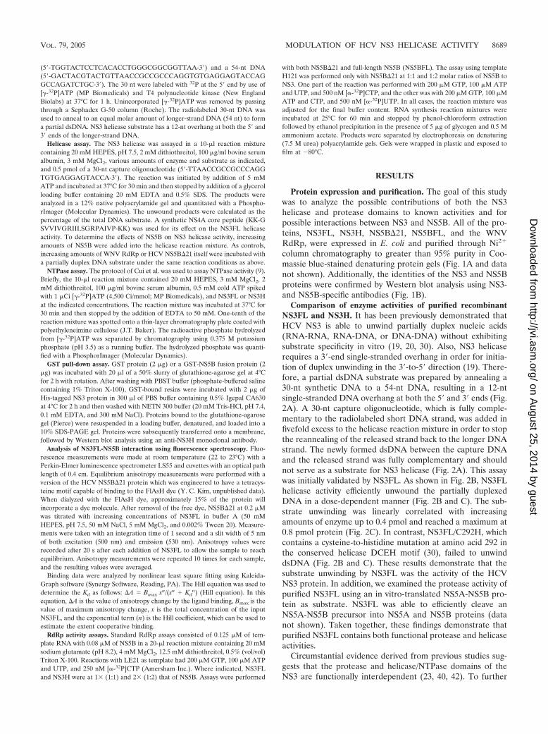
Protein expression and purification. The goal of this studywas to analyze the possible contributions of both the NS3helicase and protease domains to known activities and forpossible interactions between NS3 and NS5B. All of the pro-teins, NS3FL, NS3H, NS5B�21, NS5BFL, and the WNVRdRp, were expressed in E. coli and purified through Ni2�
column chromatography to greater than 95% purity in Coo-massie blue-stained denaturing protein gels (Fig. 1A and datanot shown). Additionally, the identities of the NS3 and NS5Bproteins were confirmed by Western blot analysis using NS3-and NS5B-specific antibodies (Fig. 1B).
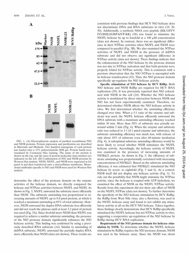
Comparison of enzyme activities of purified recombinantNS3FL and NS3H. It has been previously demonstrated thatHCV NS3 is able to unwind partially duplex nucleic acids(RNA-RNA, RNA-DNA, or DNA-DNA) without exhibitingsubstrate specificity in vitro (19, 20, 30). Also, NS3 helicaserequires a 3�-end single-stranded overhang in order for initia-tion of duplex unwinding in the 3�-to-5� direction (19). There-fore, a partial dsDNA substrate was prepared by annealing a30-nt synthetic DNA to a 54-nt DNA, resulting in a 12-ntsingle-stranded DNA overhang at both the 5� and 3� ends (Fig.2A). A 30-nt capture oligonucleotide, which is fully comple-mentary to the radiolabeled short DNA strand, was added infivefold excess to the helicase reaction mixture in order to stopthe reannealing of the released strand back to the longer DNAstrand. The newly formed dsDNA between the capture DNAand the released strand was fully complementary and shouldnot serve as a substrate for NS3 helicase (Fig. 2A). This assaywas initially validated by NS3FL. As shown in Fig. 2B, NS3FLhelicase activity efficiently unwound the partially duplexedDNA in a dose-dependent manner (Fig. 2B and C). The sub-strate unwinding was linearly correlated with increasingamounts of enzyme up to 0.4 pmol and reached a maximum at0.8 pmol protein (Fig. 2C). In contrast, NS3FL/C292H, whichcontains a cysteine-to-histidine mutation at amino acid 292 inthe conserved helicase DCEH motif (30), failed to unwinddsDNA (Fig. 2B and C). These results demonstrate that thesubstrate unwinding by NS3FL was the activity of the HCVNS3 protein. In addition, we examined the protease activity ofpurified NS3FL using an in vitro-translated NS5A-NS5B pro-tein as substrate. NS3FL was able to efficiently cleave anNS5A-NS5B precursor into NS5A and NS5B proteins (datanot shown). Taken together, these findings demonstrate thatpurified NS3FL contains both functional protease and helicaseactivities.
Circumstantial evidence derived from previous studies sug-gests that the protease and helicase/NTPase domains of theNS3 are functionally interdependent (23, 40, 42). To further
VOL. 79, 2005 MODULATION OF HCV NS3 HELICASE ACTIVITY 8689
on August 25, 2014 by guest
http://jvi.asm.org/
Dow
nloaded from
determine the effect of the protease domain on the enzymeactivities of the helicase domain, we directly compared thehelicase and NTPase activities between NS3FL and NS3H. Asshown in Fig. 3, NS3FL unwound the substrate more efficientlythan NS3H. The substrate unwinding was proportional to in-creasing amounts of NS3FL up to 0.8 pmol protein, at which itreached a maximum unwinding at 85% of total substrate. How-ever, NS3H unwound the duplex DNA substrate less efficientlyand did not reach the plateau until at least 3.2 pmol of proteinwas used (Fig. 3A). Since fivefold more NS3H than NS3FL wasrequired to achieve a similar substrate unwinding, the presenceof the NS3 protease domain significantly enhances the NS3helicase activity. This finding was confirmed by using a previ-ously described RNA substrate (14). Similar to unwinding ofdsDNA substrate, NS3FL unwound the partially duplex RNAmore efficiently than NS3H (data not shown). These results are
consistent with previous findings that HCV NS3 helicase doesnot discriminate DNA and RNA substrates in vitro (19, 20,30). Additionally, a synthetic NS4A core peptide (KK-GSVVIVGRIILSGRPAIVP-KK) (58) was found to stimulate theNS3FL helicase by up to fourfold at a 100 �M concentration(data not shown). In contrast, there was no significant differ-ence in their NTPase activities when NS3FL and NS3H werecompared in parallel (Fig. 3B). We also examined the NTPaseactivities of NS3FL and NS3H in the presence of dsDNAsubstrate and did not observe any significant difference inNTPase activity (data not shown). These findings indicate thatthe enhancement of the NS3 helicase by the protease domainwas not due to NTPase activation and that both proteins wereproperly folded for NTPase activity. This is consistent with aprevious observation that the NS3 NTPase is uncoupled withits helicase translocation (43). Thus, the NS3 protease domainspecifically up-regulates the NS3 helicase activity.
Specific stimulation of NS3 helicase by HCV RdRp. BothNS3 helicase and NS5B RdRp are required for HCV RNAreplication (29). It was previously reported that NS3 colocal-ized with NS5B in the cell (24). Whether the NS3 helicaseactivity is modulated by direct interaction between NS5B andNS3 has not been experimentally examined. Therefore, wedetermined whether NS5B affects the NS3 helicase activity invitro. We first determined whether the unwinding efficiencychanged over time. When a 5:1 ratio of the enzyme and sub-strate was used, the NS3FL helicase efficiently unwound theDNA substrate with a maximum unwinding efficiency reachedwithin 30 min. More than 50% of substrate was actually un-wound within 5 min (Fig. 4). When the enzyme and substrateratio was reduced to 1:1 (0.1 pmol enzyme and substrate), thesubstrate unwinding efficiency was much less, with release ofonly about 10% of substrate even after 60-minute incubation(Fig. 4). A suboptimal substrate unwinding efficiency would bemore likely to reveal whether NS5B stimulates the NS3FLhelicase activity. Accordingly, the helicase activity of NS3FLwas examined in the presence of increasing amounts ofNS5B�21 protein. As shown in Fig. 5, the efficiency of sub-strate unwinding was proportionally correlated with increasingconcentrations of NS5B�21. Based on the substrate unwindingefficiency, it was estimated that NS5B�21 stimulated the NS3helicase by seven- to eightfold (Fig. 5 and 6). As a control,NS5B itself did not display any helicase activity (Fig. 5). Torule out the possibility that NS5B might stimulate the NTPaseactivity, since the helicase is coupled with ATP hydrolysis, weexamined the effect of NS5B on the NS3FL NTPase activity.Results from this experiment did not show any effect of NS5Bon the NS3FL NTPase (data not shown). To further determinethe specificity of the NS3 helicase stimulation by HCV RdRp,the RdRp from West Nile virus, also a flavivirus, was used inthe NS3FL helicase assay and found to not exhibit any stimu-latory activity at all on the HCV NS3 helicase. Taken together,these findings clearly demonstrate that HCV RdRp specificallystimulated the NS3FL helicase but not NTPase activity in vitro,suggesting a cooperative up-regulation of the NS3 helicase byRdRp during HCV RNA replication.
Requirement of the NS3 protease domain for helicase stim-ulation by NS5B. To determine whether the NS3FL helicasestimulation by RdRp requires the NS3 protease domain, NS3Hwas used in the helicase assay. Since the NS3H was about
FIG. 1. (A) Electrophoresis analysis of purified recombinant NS3and NS5B proteins. Protein expression and purification are describedin Materials and Methods. Two hundred nanograms of each proteinwas loaded onto a 12% polyacrylamide–SDS gel. Protein bands werevisualized by Coomassie blue staining. The name of the protein isshown on the top, and the sizes of protein molecular mass markers areindicated on the left. (B) Confirmation of NS3 and NS5B proteins byWestern blot analysis. NS3H, NS3FL, and NS5B were separated as forpanel A and then transferred onto a nitrocellulose membrane. Mono-clonal antibodies specific to NS3 and NS5B were used for Western blotanalysis.
8690 ZHANG ET AL. J. VIROL.
on August 25, 2014 by guest
http://jvi.asm.org/
Dow
nloaded from
fivefold lower in helicase activity than NS3FL, an approxi-mately equivalent substrate unwinding activity of NS3H toNS3FL was used in the helicase assay. Contrary to NS3FL, theNS3H helicase activity was not significantly affected by theaddition of increasing amounts of NS5B�21 (Fig. 6). Theseresults demonstrate that the NS3 protease domain was re-quired for helicase stimulation by NS5B. This finding alsosuggests that the NS3 protease domain might mediate a spe-cific protein-protein interaction between NS3FL and NS5B.
NS3-NS5B interaction revealed by GST pull-down. To de-termine a protein-protein interaction between NS3 and NS5B,a GST pull-down experiment was performed. NB5B bound toglutathione-agarose was incubated with either NS3FL orNS3H protein. GST protein alone was used as a control in this
experiment. Coprecipitated proteins were detected by Westernblot analysis using a monoclonal antibody against the NS3helicase domain. As shown in Fig. 7, NS5B specifically pulleddown the NS3FL but not NS3H (Fig. 7), suggesting that theprotease domain was required for NS3 and NS5B interaction.GST did not pull down any NS3 protein, demonstrating thatthe NS3 protease mediated a specific NS3-NS5B interaction.
Determination of NS5B and NS3FL interaction by fluores-cence spectroscopy. To more quantitatively analyze the inter-action between NS5B and NS3FL, we used a recombinantNS5B�21 molecule that has been engineered to bind theFlAsH dye (18). This protein is fully capable of de novo initi-ating RNA synthesis and does not appear to be significantlyaffected in any of the known activities (Y. C. Kim, unpublished
FIG. 2. (A) Schematic of the helicase assay. A synthetic 30-nt oligonucleotide was annealed to a 54-nt DNA to form a partially duplex DNAsubstrate. Upon duplex unwinding by NS3 helicase, the 5�-end 32P-radiolabeled release strand (indicated by P* at the 5� end) is trapped by acomplementary capture oligonucleotide. (B) Titration of the NS3FL helicase. A 0.1-pmol aliquot of dsDNA substrate was incubated withincreasing amounts of NS3FL or an inactive helicase mutant, NS3FL/C292H, at 37°C for 30 min. The unwound substrate products were analyzedin a native 12% polyacrylamide gel. The percentage of substrate unwound, indicated at the bottom, was determined by from the quantitative dataderived from phosphorimager results. Amounts of NS3FL used are indicated on the top. (C) Correlation of NS3FL helicase activity with enzymeconcentrations. The quantitative data are derived from panel B.
FIG. 3. (A) Comparison of helicase activities of NS3FL and NS3H. The helicase assay was the same as that in Fig. 2. (B) Comparison of theNTPase activities of NS3FL and NS3H. The NTPase activity is expressed as the percentage of radioactive phosphate converted from [�-32P]ATP.The enzyme concentrations (x axis) are plotted against the percentage of conversion of phosphate from ATP (y axis). E, NS3FL; Œ, NS3H.
VOL. 79, 2005 MODULATION OF HCV NS3 HELICASE ACTIVITY 8691
on August 25, 2014 by guest
http://jvi.asm.org/
Dow
nloaded from

data). However, having a dye-labeled version of NS5B�21 al-lowed us to examine the properties of the protein even in thepresence of other molecules that could otherwise affect thespectroscopic properties of the solution. In the presence ofincreasing NS3FL, we observed a significant change in theanisotropy of NS5B�21 that started to saturate when NS3FLprotein reached about 5 �M (Fig. 8). The binding isotherm wasused to derive the affinity of the NS5B-NS3FL interaction, witha Kd of approximately 2 �M. In contrast, the Kd for interactionbetween NS5B�21 and NS3H was higher than 50 �M (Fig. 8).Therefore, these results from fluorescence anisotropy are inagreement with those from the GST pull-down assays (Fig. 7),demonstrating that the NS3 protease domain is required for atighter interaction with NS5B.
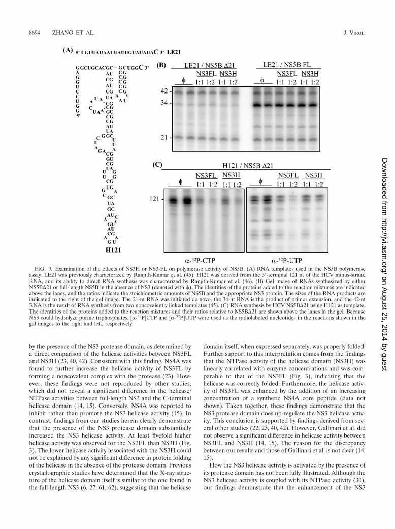
Effect of NS3 on RdRp activity of NS5B. In order to examinewhether the HCV NS3 could affect RNA synthesis by the HCVRNA polymerase in vitro, we used two previously character-ized RNA templates that are both capable of directing de novoinitiation (Fig. 9A) (45, 46). A short 21-nt RNA named LE21can form a quasi-stable intramolecular hairpin with a single-stranded 3� sequence that is capable of directing de novoinitiation of RNA synthesis. When the polymerase reaches the
FIG. 4. (A) Time course of NS3FL helicase activity. A 0.1-pmol aliquot of dsDNA substrate was incubated with either 0.1 pmol or 0.5 pmolNS3FL enzyme (as indicated at the bottom) at 37°C. At different time points (indicated on the top), the reactions were stopped by addition of aloading buffer containing 20 mM EDTA and 0.5% SDS. The unwound products were analyzed in a 12% native polyacrylamide gel, visualized byautoradiograph, and quantified by use of a phosphorimager. (B) Relation of NS3FL helicase activity to incubation time. The quantitative data frompanel A were used to plot the incubation time (x axis, in min) against the percentage of unwound substrate (y axis). F, 0.1 pmol NS3FL; Œ, 0.5pmol NS3FL.
FIG. 5. Stimulation of NS3FL helicase activity by HCV RdRp. Apartially duplex DNA substrate was incubated with 0.1 pmol NS3FLand increasing amounts of NS5B�21 at 37°C for 30 min (left panel). Ascontrols, the helicase substrate was incubated with increasing amountsof NS5B in the absence of NS3FL (right panel). The unwound sub-strate products were resolved in a 12% native polyacrylamide gel,visualized by autoradiograph, and quantified with a phosphorimager.The percentage of unwound substrate is shown at the bottom, and theamounts of NS5B are indicated on the top.
FIG. 6. Specificity of NS3 helicase stimulation by HCV RdRp. Aviral RdRp derived from WNV was used as a nonspecific control tocompare with HCV RdRp in stimulation of the NS3FL helicase. ThedsDNA substrate was incubated with 0.1 pmol NS3FL or 0.5 pmolNS3H and increasing amounts of either HCV NS5B or WNV NS5 at37°C for 30 min. A 0.5-pmol aliquot of NS3H was used here, whichachieved an unwinding efficiency approximately equivalent to 0.1 pmolNS3FL, since the helicase activity of NS3H is about fivefold lower thanthat of NS3FL. The unwound products were separated by electro-phoresis in a 12% native polyacrylamide gel, visualized by autoradio-graph, and quantified with a phosphorimager. �, 0.1 pmol NS3FL plusNS5B; Œ, 0.5 pmol NS3H plus NS5B; E, 0.1 pmol NS3FL plus WNVNS5.
8692 ZHANG ET AL. J. VIROL.
on August 25, 2014 by guest
http://jvi.asm.org/
Dow
nloaded from

5� end of LE21, it could either terminate RNA synthesis, thusproducing a 21-nt de novo-initiated RNA, or use a secondtemplate to generate a recombinant RNA product of 42 nt. Inaddition, two molecules of LE21 can partially anneal to directprimer-dependent RNA synthesis, generating a 34-nt product.The addition of either NS3H or NS3FL did not significantlyalter the type or the amount of products synthesized byNS5B�21. To ensure that the lack of the C-terminal 21 aminoacids did not prevent a possible effect of NS3, we also tested afull-length NS5B protein. Again, no significant effect on RNAsynthesis was observed in the presence of NS3H or NS3FL. Insome experiments, we did observe a 20 to 30% decrease in the
amount of products made from LE21 in the presence ofNS3FL (R. Kumar, unpublished data).
The lack of an effect on RNA synthesis from LE21 could bebecause it lacks a sufficiently structured region that wouldrequire the function of an RNA helicase. To examine thispossibility, we used the 121-nt RNA derived from the 3� end ofthe HCV minus-strand RNA genomes as a template. Unex-pectedly, we found that the addition of NS3FL or NS3H athigher concentrations inhibited RNA synthesis by NS5B�21 inour standard reaction. One possible reason for this is that theATPase activity of NS3 could decrease the concentration ofthe already low concentration of radiolabeled nucleotide (0.3�M final concentration) and that the longer length of H121exacerbated the inhibitory effect in comparison to productsynthesis from LE21. We have also tested RNA synthesesusing [-32P]CTP and [-32P]UTP as radiolabels and observedthat NS3H or NS3FL also inhibited RNA synthesis from H121.One alternative interpretation for the inhibition of RNA syn-thesis is that the NS3 competes with NS5B for RNA template,since NS3 possesses an RNA-binding activity.
DISCUSSION
A consensus has not been reached as to whether the twodistinctive NS3 enzymes are functionally interdependent be-tween the protease and helicase domains. A number of studieshave demonstrated that both the full-length NS3 and the trun-cated protease domain alone have comparable protease activ-ities in vitro, suggesting that the NS3 protease does not dependon the presence of the NS3 helicase domain (14, 15, 22, 50, 55).We have evidence from this study to show that both NS3FLand NS3H have comparable NTPase activities. Therefore,some activities of NS3 can be functionally independent. How-ever, contradicting evidence exists to either support or opposea role of the NS3 protease domain in the modulation of itshelicase activity (14, 15, 23, 40, 42). Several studies have re-vealed that the NS3 helicase activity is significantly enhanced
FIG. 7. Determination of NS3FL-NS5B interaction by GST pull-down. A GST-NS5B fusion protein or GST alone (as a control) wasfirst incubated with glutathione-agarose gel. After washing, the GST orGST-NS5B-bound gel was subsequently incubated with NS3FL orNS3H, respectively. The NS3 proteins pulled down by NS5B were thendetermined by Western blot analysis using a monoclonal anti-NS3Hantibody. Purified recombinant NS3FL and NS3H were used as posi-tive controls for Western blot analysis. The sizes of protein molecularmass markers are indicated on the left.
FIG. 8. Analysis of NS5B-NS3 interaction using fluorescence anisotropy. (A) A comparison of the interaction of between fluorescent-labeledNS5B and either NS3H or NS3FL. Each change in anisotropy value represents the mean and one deviation from 10 measurements. (B) Fluo-rescence anisotropy experiment to determine the affinity of the NS5B-NS3FL interaction. NS5B bound to the fluorescent dye FlAsH wasdetermined in the presence of increasing concentrations of NS3FL. Each data point represents the average of 10 independent measurements. TheKd for the interaction was derived from the binding isotherm using the Hill equation, as described in Materials and Methods.
VOL. 79, 2005 MODULATION OF HCV NS3 HELICASE ACTIVITY 8693
on August 25, 2014 by guest
http://jvi.asm.org/
Dow
nloaded from
by the presence of the NS3 protease domain, as determined bya direct comparison of the helicase activities between NS3FLand NS3H (23, 40, 42). Consistent with this finding, NS4A wasfound to further increase the helicase activity of NS3FL byforming a noncovalent complex with the protease (23). How-ever, these findings were not reproduced by other studies,which did not reveal a significant difference in the helicase/NTPase activities between full-length NS3 and the C-terminalhelicase domain (14, 15). Conversely, NS4A was reported toinhibit rather than promote the NS3 helicase activity (15). Incontrast, findings from our studies herein clearly demonstratethat the presence of the NS3 protease domain substantiallyincreased the NS3 helicase activity. At least fivefold higherhelicase activity was observed for the NS3FL than NS3H (Fig.3). The lower helicase activity associated with the NS3H couldnot be explained by any significant difference in protein foldingof the helicase in the absence of the protease domain. Previouscrystallographic studies have determined that the X-ray struc-ture of the helicase domain itself is similar to the one found inthe full-length NS3 (6, 27, 61, 62), suggesting that the helicase
domain itself, when expressed separately, was properly folded.Further support to this interpretation comes from the findingsthat the NTPase activity of the helicase domain (NS3H) waslinearly correlated with enzyme concentrations and was com-parable to that of the NS3FL (Fig. 3), indicating that thehelicase was correctly folded. Furthermore, the helicase activ-ity of NS3FL was enhanced by the addition of an increasingconcentration of a synthetic NS4A core peptide (data notshown). Taken together, these findings demonstrate that theNS3 protease domain does up-regulate the NS3 helicase activ-ity. This conclusion is supported by findings derived from sev-eral other studies (22, 23, 40, 42). However, Gallinari et al. didnot observe a significant difference in helicase activity betweenNS3FL and NS3H (14, 15). The reason for the discrepancybetween our results and those of Gallinari et al. is not clear (14,15).
How the NS3 helicase activity is activated by the presence ofits protease domain has not been fully illustrated. Although theNS3 helicase activity is coupled with its NTPase activity (30),our findings demonstrate that the enhancement of the NS3
FIG. 9. Examination of the effects of NS3H or NS3-FL on polymerase activity of NS5B. (A) RNA templates used in the NS5B polymeraseassay. LE21 was previously characterized by Ranjith-Kumar et al. (45). H121 was derived from the 3�-terminal 121 nt of the HCV minus-strandRNA, and its ability to direct RNA synthesis was characterized by Ranjith-Kumar et al. (46). (B) Gel image of RNAs synthesized by eitherNS5B�21 or full-length NS5B in the absence of NS3 (denoted with �). The identities of the proteins added to the reaction mixtures are indicatedabove the lanes, and the ratios indicate the stoichiometric amounts of NS5B and the appropriate NS3 protein. The sizes of the RNA products areindicated to the right of the gel image. The 21-nt RNA was initiated de novo, the 34-nt RNA is the product of primer extension, and the 42-ntRNA is the result of RNA synthesis from two noncovalently linked templates (45). (C) RNA synthesis by HCV NS5B�21 using H121 as template.The identities of the proteins added to the reaction mixtures and their ratios relative to NS5B�21 are shown above the lanes in the gel. BecauseNS3 could hydrolyze purine triphosphates, [-32P]CTP and [-32P]UTP were used as the radiolabeled nucleotides in the reactions shown in thegel images to the right and left, respectively.
8694 ZHANG ET AL. J. VIROL.
on August 25, 2014 by guest
http://jvi.asm.org/
Dow
nloaded from
helicase by the protease domain was not due to activation ofthe NTPase activity. NS3FL was found to exhibit a substan-tially higher helicase activity than NS3H, but it did not affectthe level of its NTPase activity (Fig. 3). These findings suggestthat the protease domain can directly activate the helicasewithout an effect on the NTPase activity. Consistent with anearlier observation that NS4A was able to enhance functionalNS3-RNA complex formation (42), an NS4A core peptide wasalso found to significantly enhance the NS3 helicase activity(data not shown). NS4A is known to bind tightly to the pro-tease domain of NS3 and to stabilize the NS3 structure (28, 35,60), which is required for protease activity. Likewise, stabiliza-tion of NS3 by NS4A may also convert NS3 into a more activehelicase. The molecular basis underlying the helicase enhance-ment by the protease domain is probably best explained by theatomic structure of the full-length NS3 (62). The protease andhelicase domains are segregated and covalently connected by ashort strand. A number of amino acid residues of each domainare involved in the surface interaction at the domain interface.Additionally, there are extensive hydrogen bond interactionsbetween the region containing the protease active site and theC-terminal six residues of the helicase domain (62). Theseinteractions probably stabilize the enzyme in a conformationoptimal for helicase activity. Furthermore, the nucleic acid-binding sites on the protease surface may also contribute to theenhancement of the helicase activity by binding to the helicasesubstrate (62). Mutagenesis analysis to alter interactions be-tween the NS3 protease and helicase domains will likely pro-vide insights into the understanding of the structural basis forthe functional dependence of the NS3 helicase on the proteasedomain.
In addition to the NS3 protease, HCV RdRp (NS5B) wasfound to strikingly stimulate the NS3 helicase activity by aspecific protein-protein interaction with the NS3 protease do-main (Fig. 7 and 8). This novel finding is strongly supported byseveral lines of evidence described in this report. First, NS5Bstimulation of NS3FL helicase activity occurs in a dose-depen-dent manner (Fig. 5 and 6). In contrast, a nonspecific controlRdRp derived from WNV had no effect on the HCV NS3helicase activity (Fig. 6). Additionally, stimulation of the NS3helicase by NS5B is strictly dependent on the presence of theprotease domain, since NS5B failed to stimulate the helicaseactivity of the NS3H that did not contain the protease domain(Fig. 6). Similar to the NS3 protease domain, NS5B did notaffect the NTPase activity of NS3FL (data not shown). Fur-thermore, it appeared that NS5B could inhibit the NS3 pro-tease activity when an in vitro-translated NS5A-NS5B sub-strate was used in a protease assay (Z. Cai et al., unpublishedresult). These findings suggest that the protease domain isrequired for mediating the NS3-NS5B interaction. Such a pro-tein-protein interaction was indeed confirmed by both GSTpull-down assay and fluorescence spectroscopy analysis. NS5Bwas able to specifically pull down NS3FL but not the NS3Hprotein (Fig. 7). Likewise, fluorescence anisotropy analysisdemonstrated that NS5B tightly interacted with NS3FL but notNS3H (Fig. 8). A specific interaction between NS5B and theNS3 protease domain appears to be a prerequisite for NS3helicase stimulation by HCV RdRp. It is noteworthy that ourresults are contrary to a previous finding that NS5B couldinteract with the NS3 helicase domain alone, as shown by a
membrane-binding assay (44). One possible explanation is thatthe difference may be attributed to a difference in the affinity ofthe NS5B interaction between NS3H and NS3FL.
It is not clear yet how NS5B interacts with the NS3 proteasedomain and how such an interaction subsequently activates theNS3 helicase activity. Understanding of these fundamentalquestions will inevitably help to illustrate the molecular mech-anism of HCV RNA replication. As discussed above, the Cterminus of NS3 forms hydrogen bond interactions with theprotease domain near the enzyme active site after cis-cleavageby the NS3 protease (62). The N-terminal portion of NS5Bmay interact with the protease domain through the proteaseactive site in a manner similar to the C-terminal portion ofNS3, as revealed by the structure of a single-chain NS3-NS4Amolecule (62). The NS5B N terminus is the P�-side product ofthe cleavage by the NS3 serine protease at the NS5A and NS5Bjunction. There could be other regions involved in the inter-action between these two molecules. Upon interaction with theprotease domain, NS5B may cause a conformational rear-rangement of the NS3 helicase, which could increase substrateunwinding.
Replication of most positive-strand RNA viruses takes placein the membrane-bound replication complex consisting of viralRNA, most NS proteins, and cellular proteins as well. The viralreplicase proteins were found to interact with each other in theintracellular sites where viral RNA is replicated (11, 12, 41,54). Therefore, it is conceivable that the replicase proteinsaffect/regulate each other’s properties through protein-proteininteractions. The HCV NS3 represents the first viral RNAhelicase whose function is stimulated by a viral RNA polymer-ase. Our findings also provide an explanation for the existenceof two apparently functionally independent distinct activities(protease and helicase/NTPase) within one NS3 molecule. TheNS3 protease domain functions not only as an enzyme but alsoas a mediator for a specific interaction with NS5B, which en-hances the NS3 helicase activity.
As to the role of NS3 in RNA synthesis in vitro, our resultscontrast with those previously reported by Piccininni et al., whoobserved an unexpected multimeric product made from theinput template in the presence of NS3FL (44). This differenceis especially notable because both studies used a templatederived from the 3� end of minus-strand HCV RNA. We spec-ulate that the difference is due to the ratio of NS5B to templateused in our respective reaction mixtures. Piccininni et al. used6.4-fold more enzyme than template RNA, while our reactionmixtures had an equal or higher molar ratio of enzyme totemplate. The multimeric product in the presence of NS3,observed in the study by Piccininni et al., is inconsistent with denovo RNA synthesis during HCV RNA replication. The pri-mary mode of RNA synthesis during HCV replication in vivois initiated by a de novo mechanism. Despite the differences,neither study observed that NS3FL or NS3H could increase denovo RNA synthesis (Fig. 9) (43). In fact, both studies showedthat in the presence of 0.5 mM ATP or below, NS3 actuallydecreased RNA synthesis by NS5B, possibly due to the NTPaseactivity of NS3 decreasing the NTP concentration needed forefficient RNA synthesis or the RNA-binding activity of NS3that competes for the RNA template with NS5B.
In summary, the NS3 helicase activity is differentially mod-ulated by the presence of the NS3 protease domain as well as
VOL. 79, 2005 MODULATION OF HCV NS3 HELICASE ACTIVITY 8695
on August 25, 2014 by guest
http://jvi.asm.org/
Dow
nloaded from
the viral RNA polymerase. Perhaps NS3 initially functions as aprotease responsible for viral polyprotein processing. Oncerecruited into the HCV replication complex containing NS5B,NS3 switches to serving as the helicase/NTPase that is requiredfor HCV RNA replication.
ACKNOWLEDGMENTS
We thank Jackie Wright-Minogue at Schering-Plough Research In-stitute for kindly providing us with synthetic NS4A core peptides andKyung-Soo Chang for the NS3H and NS5B monoclonal antibodies.
This work was supported by a grant from the National Institutes ofHealth (CA093712 to G.L.) and in part by a grant from the KentuckyScience and Engineering Foundation (KSEF-148-502-02-18 to G.L.).C.K. acknowledges funding from National Science Foundation grantMCB0332259. P.-Y.S. was in part supported by a developmentalproject from Northeast Biodefense Center.
Chen Zhang and Zhaohui Cai contributed equally to this work.
REFERENCES
1. Bartenschlager, R., L. Ahlborn-Laake, J. Mous, and H. Jacobsen. 1993.Nonstructural protein 3 of the hepatitis C virus encodes a serine-type pro-teinase required for cleavage at the NS3/4 and NS4/5 junctions. J. Virol.67:3835–3844.
2. Bartosch, B., J. Dubuisson, and F. L. Cosset. 2003. Infectious hepatitis Cvirus pseudo-particles containing functional E1-E2 envelope protein com-plexes. J. Exp. Med. 197:633–642.
3. Baumert, T. F., S. Ito, D. T. Wong, and T. J. Liang. 1998. Hepatitis C virusstructural proteins assemble into virus-like particles in insect cells. J. Virol.72:3827–3836.
4. Behrens, S. E., L. Tomei, and R. De Francesco. 1996. Identification andproperties of the RNA-dependent RNA polymerase of hepatitis C virus.EMBO J. 15:12–22.
5. Blight, K. J., A. A. Kolykhalov, and C. M. Rice. 2000. Efficient initiation ofHCV RNA replication in cell culture. Science 290:1972–1975.
6. Cho, H. S., N. C. Ha, L. W. Kang, K. M. Chung, S. H. Back, S. K. Jang, andB. H. Oh. 1998. Crystal structure of RNA helicase from genotype 1b hepatitisC virus. A feasible mechanism of unwinding duplex RNA. J. Biol. Chem.273:15045–15052.
7. Choo, Q. L., G. Kuo, A. J. Weiner, L. R. Overby, D. W. Bradley, and M.Houghton. 1989. Isolation of a cDNA clone derived from a blood-bornenon-A, non-B viral hepatitis genome. Science 244:359–362.
8. Choo, Q. L., K. H. Richman, J. H. Han, K. Berger, C. Lee, C. Dong, C.Gallegos, D. Coit, R. Medina-Selby, P. J. Barr, et al. 1991. Genetic organi-zation and diversity of the hepatitis C virus. Proc. Natl. Acad. Sci. USA88:2451–2455.
9. Cui, T., R. J. Sugrue, Q. Xu, A. K. Lee, Y. C. Chan, and J. Fu. 1998.Recombinant dengue virus type 1 NS3 protein exhibits specific viral RNAbinding and NTPase activity regulated by the NS5 protein. Virology 246:409–417.
10. De Francesco, R., and C. Steinkuhler. 2000. Structure and function of thehepatitis C virus NS3-NS4A serine proteinase. Curr. Top. Microbiol. Immu-nol. 242:149–169.
11. Egger, D., B. Wolk, R. Gosert, L. Bianchi, H. E. Blum, D. Moradpour, andK. Bienz. 2002. Expression of hepatitis C virus proteins induces distinctmembrane alterations including a candidate viral replication complex. J. Vi-rol. 76:5974–5984.
12. El-Hage, N., and G. Luo. 2003. Replication of hepatitis C virus RNA occursin a membrane-bound replication complex containing nonstructural viralproteins and RNA. J. Gen. Virol. 84:2761–2769.
13. Forns, X., R. Thimme, S. Govindarajan, S. U. Emerson, R. H. Purcell, F. V.Chisari, and J. Bukh. 2000. Hepatitis C virus lacking the hypervariableregion 1 of the second envelope protein is infectious and causes acuteresolving or persistent infection in chimpanzees. Proc. Natl. Acad. Sci. USA97:13318–13323.
14. Gallinari, P., D. Brennan, C. Nardi, M. Brunetti, L. Tomei, C. Steinkuhler,and R. De Francesco. 1998. Multiple enzymatic activities associated withrecombinant NS3 protein of hepatitis C virus. J. Virol. 72:6758–6769.
15. Gallinari, P., C. Paolini, D. Brennan, C. Nardi, C. Steinkuhler, and R. DeFrancesco. 1999. Modulation of hepatitis C virus NS3 protease and helicaseactivities through the interaction with NS4A. Biochemistry 38:5620–5632.
16. Grakoui, A., D. W. McCourt, C. Wychowski, S. M. Feinstone, and C. M. Rice.1993. A second hepatitis C virus-encoded proteinase. Proc. Natl. Acad. Sci.USA 90:10583–10587.
17. Grakoui, A., C. Wychowski, C. Lin, S. M. Feinstone, and C. M. Rice. 1993.Expression and identification of hepatitis C virus polyprotein cleavage prod-ucts. J. Virol. 67:1385–1395.
18. Griffin, B. A., S. R. Adams, J. Jones, and R. Y. Tsien. 2000. Fluorescent
labeling of recombinant proteins in living cells with FlAsH. Methods Enzy-mol. 327:565–578.
19. Gwack, Y., D. W. Kim, J. H. Han, and J. Choe. 1996. Characterization ofRNA binding activity and RNA helicase activity of the hepatitis C virus NS3protein. Biochem. Biophys. Res. Commun. 225:654–659.
20. Gwack, Y., D. W. Kim, J. H. Han, and J. Choe. 1997. DNA helicase activityof the hepatitis C virus nonstructural protein 3. Eur. J. Biochem. 250:47–54.
21. Hijikata, M., H. Mizushima, T. Akagi, S. Mori, N. Kakiuchi, N. Kato, T.Tanaka, K. Kimura, and K. Shimotohno. 1993. Two distinct proteinaseactivities required for the processing of a putative nonstructural precursorprotein of hepatitis C virus. J. Virol. 67:4665–4675.
22. Hong, Z., E. Ferrari, J. Wright-Minogue, R. Chase, C. Risano, G. Seelig,C. G. Lee, and A. D. Kwong. 1996. Enzymatic characterization of hepatitis Cvirus NS3/4A complexes expressed in mammalian cells by using the herpessimplex virus amplicon system. J. Virol. 70:4261–4268.
23. Howe, A. Y., R. Chase, S. S. Taremi, C. Risano, B. Beyer, B. Malcolm, andJ. Y. Lau. 1999. A novel recombinant single-chain hepatitis C virus NS3-NS4A protein with improved helicase activity. Protein Sci. 8:1332–1341.
24. Ishido, S., T. Fujita, and H. Hotta. 1998. Complex formation of NS5B withNS3 and NS4A proteins of hepatitis C virus. Biochem. Biophys. Res. Com-mun. 244:35–40.
25. Jin, L., and D. L. Peterson. 1995. Expression, isolation, and characterizationof the hepatitis C virus ATPase/RNA helicase. Arch. Biochem. Biophys.323:47–53.
26. Kim, D. W., Y. Gwack, J. H. Han, and J. Choe. 1995. C-terminal domain ofthe hepatitis C virus NS3 protein contains an RNA helicase activity. Bio-chem. Biophys. Res. Commun. 215:160–166.
27. Kim, J. L., K. A. Morgenstern, J. P. Griffith, M. D. Dwyer, J. A. Thomson,M. A. Murcko, C. Lin, and P. R. Caron. 1998. Hepatitis C virus NS3 RNAhelicase domain with a bound oligonucleotide: the crystal structure providesinsights into the mode of unwinding. Structure 6:89–100.
28. Kim, J. L., K. A. Morgenstern, C. Lin, T. Fox, M. D. Dwyer, J. A. Landro,S. P. Chambers, W. Markland, C. A. Lepre, E. T. O’Malley, S. L. Harbeson,C. M. Rice, M. A. Murcko, P. R. Caron, and J. A. Thomson. 1996. Crystalstructure of the hepatitis C virus NS3 protease domain complexed with asynthetic NS4A cofactor peptide. Cell 87:343–355. [Erratum, 89:159, 1997.]
29. Kolykhalov, A. A., K. Mihalik, S. M. Feinstone, and C. M. Rice. 2000.Hepatitis C virus-encoded enzymatic activities and conserved RNA elementsin the 3� nontranslated region are essential for virus replication in vivo.J. Virol. 74:2046–2051.
30. Kwong, A. D., J. L. Kim, and C. Lin. 2000. Structure and function of hepatitisC virus NS3 helicase. Curr. Top. Microbiol. Immunol. 242:171–196.
31. Lin, C., B. D. Lindenbach, B. M. Pragai, D. W. McCourt, and C. M. Rice.1994. Processing in the hepatitis C virus E2-NS2 region: identification of p7and two distinct E2-specific products with different C termini. J. Virol.68:5063–5073.
32. Lin, C., B. M. Pragai, A. Grakoui, J. Xu, and C. M. Rice. 1994. Hepatitis Cvirus NS3 serine proteinase: trans-cleavage requirements and processingkinetics. J. Virol. 68:8147–8157.
33. Lin, C., and C. M. Rice. 1995. The hepatitis C virus NS3 serine proteinaseand NS4A cofactor: establishment of a cell-free trans-processing assay. Proc.Natl. Acad. Sci. USA 92:7622–7626.
34. Lohmann, V., F. Korner, J. Koch, U. Herian, L. Theilmann, and R. Barten-schlager. 1999. Replication of subgenomic hepatitis C virus RNAs in ahepatoma cell line. Science 285:110–113.
35. Love, R. A., H. E. Parge, J. A. Wickersham, Z. Hostomsky, N. Habuka, E. W.Moomaw, T. Adachi, and Z. Hostomska. 1996. The crystal structure ofhepatitis C virus NS3 proteinase reveals a trypsin-like fold and a structuralzinc binding site. Cell 87:331–342.
36. Luo, G. 2004. Molecular virology of hepatitis C virus. Birkhauser, Basel,Switzerland.
37. Luo, G., S. Xin, and Z. Cai. 2003. Role of the 5�-proximal stem-loop struc-ture of the 5� untranslated region in replication and translation of hepatitisC virus RNA. J. Virol. 77:3312–3318.
38. Markland, W., R. A. Petrillo, M. Fitzgibbon, T. Fox, R. McCarrick, T.McQuaid, J. R. Fulghum, W. Chen, M. A. Fleming, J. A. Thomson, and S. P.Chambers. 1997. Purification and characterization of the NS3 serine pro-tease domain of hepatitis C virus expressed in Saccharomyces cerevisiae.J. Gen. Virol. 78:39–43.
39. McKeating, J. A., L. Q. Zhang, C. Logvinoff, M. Flint, J. Zhang, J. Yu, D.Butera, D. D. Ho, L. B. Dustin, C. M. Rice, and P. Balfe. 2004. Diversehepatitis C virus glycoproteins mediate viral infection in a CD81-dependentmanner. J. Virol. 78:8496–8505.
40. Morgenstern, K. A., J. A. Landro, K. Hsiao, C. Lin, Y. Gu, M. S. Su, and J. A.Thomson. 1997. Polynucleotide modulation of the protease, nucleosidetriphosphatase, and helicase activities of a hepatitis C virus NS3-NS4Acomplex isolated from transfected COS cells. J. Virol. 71:3767–3775.
41. Mottola, G., G. Cardinali, A. Ceccacci, C. Trozzi, L. Bartholomew, M. R.Torrisi, E. Pedrazzini, S. Bonatti, and G. Migliaccio. 2002. Hepatitis C virusnonstructural proteins are localized in a modified endoplasmic reticulum ofcells expressing viral subgenomic replicons. Virology 293:31–43.
42. Pang, P. S., E. Jankowsky, P. J. Planet, and A. M. Pyle. 2002. The hepatitis
8696 ZHANG ET AL. J. VIROL.
on August 25, 2014 by guest
http://jvi.asm.org/
Dow
nloaded from
C viral NS3 protein is a processive DNA helicase with cofactor enhancedRNA unwinding. EMBO J. 21:1168–1176.
43. Paolini, C., R. De Francesco, and P. Gallinari. 2000. Enzymatic properties ofhepatitis C virus NS3-associated helicase. J. Gen. Virol. 81:1335–1345.
44. Piccininni, S., A. Varaklioti, M. Nardelli, B. Dave, K. D. Raney, and J. E.McCarthy. 2002. Modulation of the hepatitis C virus RNA-dependent RNApolymerase activity by the non-structural (NS) 3 helicase and the NS4Bmembrane protein. J. Biol. Chem. 277:45670–45679.
45. Ranjith-Kumar, C. T., Y. C. Kim, L. Gutshall, C. Silverman, S. Khandekar,R. T. Sarisky, and C. C. Kao. 2002. Mechanism of de novo initiation by thehepatitis C virus RNA-dependent RNA polymerase: role of divalent metals.J. Virol. 76:12513–12525.
46. Ranjith-Kumar, C. T., J. L. Santos, L. L. Gutshall, V. K. Johnston, J.Lin-Goerke, M. J. Kim, D. J. Porter, D. Maley, C. Greenwood, D. L. Earn-shaw, A. Baker, B. Gu, C. Silverman, R. T. Sarisky, and C. Kao. 2003.Enzymatic activities of the GB virus-B RNA-dependent RNA polymerase.Virology 312:270–280.
47. Reed, K. E., and C. M. Rice. 2000. Overview of hepatitis C virus genomestructure, polyprotein processing, and protein properties. Curr. Top. Micro-biol. Immunol. 242:55–84.
48. Robertson, B., G. Myers, C. Howard, T. Brettin, J. Bukh, B. Gaschen, T.Gojobori, G. Maertens, M. Mizokami, O. Nainan, S. Netesov, K. Nishioka,T. Shin i, P. Simmonds, D. Smith, L. Stuyver, A. Weiner, et al. 1998.Classification, nomenclature, and database development for hepatitis C virus(HCV) and related viruses: proposals for standardization. Arch. Virol. 143:2493–2503.
49. Sakai, A., M. S. Claire, K. Faulk, S. Govindarajan, S. U. Emerson, R. H.Purcell, and J. Bukh. 2003. The p7 polypeptide of hepatitis C virus is criticalfor infectivity and contains functionally important genotype-specific se-quences. Proc. Natl. Acad. Sci. USA 100:11646–11651.
50. Sali, D. L., R. Ingram, M. Wendel, D. Gupta, C. McNemar, A. Tsarbopoulos,J. W. Chen, Z. Hong, R. Chase, C. Risano, R. Zhang, N. Yao, A. D. Kwong,L. Ramanathan, H. V. Le, and P. C. Weber. 1998. Serine protease of hepatitisC virus expressed in insect cells as the NS3/4A complex. Biochemistry 37:3392–3401.
51. Seeff, L. B., and J. H. Hoofnagle. 2002. National Institutes of Health Con-sensus Development Conference: management of hepatitis C, 2002. Hepa-tology 36:S1–S2.
52. Selby, M. J., Q. L. Choo, K. Berger, G. Kuo, E. Glazer, M. Eckart, C. Lee, D.Chien, C. Kuo, and M. Houghton. 1993. Expression, identification and sub-cellular localization of the proteins encoded by the hepatitis C viral genome.J. Gen. Virol. 74:1103–1113.
53. Shi, P. Y., M. Tilgner, M. K. Lo, K. A. Kent, and K. A. Bernard. 2002.Infectious cDNA clone of the epidemic West Nile virus from New York City.J. Virol. 76:5847–5856.
54. Shi, S. T., K. J. Lee, H. Aizaki, S. B. Hwang, and M. M. Lai. 2003. HepatitisC virus RNA replication occurs on a detergent-resistant membrane thatcofractionates with caveolin-2. J. Virol. 77:4160–4168.
55. Steinkuhler, C., L. Tomei, and R. De Francesco. 1996. In vitro activity ofhepatitis C virus protease NS3 purified from recombinant baculovirus-in-fected Sf9 cells. J. Biol. Chem. 271:6367–6373.
56. Suzich, J. A., J. K. Tamura, F. Palmer-Hill, P. Warrener, A. Grakoui, C. M.Rice, S. M. Feinstone, and M. S. Collett. 1993. Hepatitis C virus NS3 proteinpolynucleotide-stimulated nucleoside triphosphatase and comparison withthe related pestivirus and flavivirus enzymes. J. Virol. 67:6152–6158.
57. World Health Organization. 1998. W.H.O. concerns: hepatitis C. Lancet351:1415.
58. Wright-Minogue, J., N. Yao, R. Zhang, N. J. Butkiewicz, B. M. Baroudy, J. Y.Lau, and Z. Hong. 2000. Cross-genotypic interaction between hepatitis Cvirus NS3 protease domains and NS4A cofactors. J. Hepatol. 32:497–504.
59. Wu, Z., N. Yao, H. V. Le, and P. C. Weber. 1998. Mechanism of autoprote-olysis at the NS2-NS3 junction of the hepatitis C virus polyprotein. TrendsBiochem. Sci. 23:92–94.
60. Yan, Y., Y. Li, S. Munshi, V. Sardana, J. L. Cole, M. Sardana, C.Steinkuehler, L. Tomei, R. De Francesco, L. C. Kuo, and Z. Chen. 1998.Complex of NS3 protease and NS4A peptide of BK strain hepatitis C virus:a 2.2 Å resolution structure in a hexagonal crystal form. Protein Sci. 7:837–847.
61. Yao, N., T. Hesson, M. Cable, Z. Hong, A. D. Kwong, H. V. Le, and P. C.Weber. 1997. Structure of the hepatitis C virus RNA helicase domain. Nat.Struct. Biol. 4:463–467.
62. Yao, N., P. Reichert, S. S. Taremi, W. W. Prosise, and P. C. Weber. 1999.Molecular views of viral polyprotein processing revealed by the crystal struc-ture of the hepatitis C virus bifunctional protease-helicase. Struct. Fold Des.7:1353–1363.
VOL. 79, 2005 MODULATION OF HCV NS3 HELICASE ACTIVITY 8697
on August 25, 2014 by guest
http://jvi.asm.org/
Dow
nloaded from