Comparison of different HCV viral load and genotyping assays
Upload
independentCategory
view
1download
0
BRIEF REVIEW
Interaction of the hepatitis C virus (HCV) core with cellular genesin the development of HCV-induced steatosis
Mahwish Khan • Shah Jahan • Saba Khaliq •
Bushra Ijaz • Waqar Ahmad • Baila Samreen •
Sajida Hassan
Received: 31 March 2010 / Accepted: 31 August 2010 / Published online: 15 September 2010
� Springer-Verlag 2010
Abstract Hepatitis C virus (HCV) has chronically infected
a large number of patients, leading to the development
of steatosis, cirrhosis and, ultimately, hepatocellular carci-
noma. The pathogenesis of HCV has not been fully
explained, although steatosis is considered to contribute
greatly to liver fibrosis progression, modulating host-cell
lipid metabolism. Suspected underlying molecular mecha-
nisms include interactions between HCV proteins and
intracellular lipid metabolic pathways. Recent studies have
suggested that the nucleocapsid of HCV (core) acts as a
pathogenic factor involved in lipid droplet accumulation,
changes in lipogenic gene expression and/or the activity of
lipogenic proteins in a genotype-specific manner. In this
review, we have tried to summarize the current knowledge
regarding HCV-induced steatosis and the regulation of
expression of host genes and receptors that aid in the viral life
cycle and promote liver diseases.
Introduction
Hepatitis C virus (HCV) is a major health concern, with an
estimated 3% of the world’s population (*300 million
individuals) chronically infected with this viral pathogen
[6]. HCV causes acute and chronic hepatitis, which leads to
fibrosis, steatosis, insulin resistance (IR), cirrhosis and
hepatocellular carcinoma (HCC) in a significant number of
patients [21, 58, 130]. Epidemiological studies reveal that
about 20–30% ofchronic HCV infections are robustly
associated with hepatic steatosis, type II diabetes, IR and
cardiovascular disease [3, 20]. Fatty liver disease, the most
frequent cause of abnormal liver function, is a pathological
condition ranging from simple fat accumulation (hepatic
steatosis) to hepatic steatosis with inflammation (steato-
hepatitis), which leads to fibrosis and HCC [118]. Two
forms of steatosis, metabolic and HCV-induced, occur.
Metabolic steatosis occurs in the setting of obesity,
hyperlipidemia, and IR, whereas HCV-induced steatosis is
known as the sole route for a direct cytopathic effect
caused by HCV [42]. Hepatic steatosis is a frequent his-
tological feature of chronic HCV infection, and there is
increasing evidence that HCV infection by itself is an
independent predictor of steatosis [113].
Numerous studies illustrate the involvement of HCV in
steatosis. However, the fundamental cellular events involved
are still poorly understood due to the unavailability of an
efficient experimental model. Some insights into the path-
ways of steatohepatitis are defined by impaired lipid accu-
mulation due to hepatic loss of adiponectin receptors, which
play an important role in fatty acid accumulation by elevat-
ing the expression level of the enzymes AMP-activated
protein kinase (AMPK), acetyl-CoA carboxylase (ACC),
fatty acid synthase (FAS), liver gluconeogenic enzyme and
phosphoenol pyruvate carboxy kinase (PEPCK) due to HCV
infection. In addition, transcription factors such as sterol
regulatory element binding protein (SREBP) and peroxi-
somal proliferator activator receptors (PPARs) are also key
players in HCV-induced steatosis. SREBP activates the
enzymes involved in the fatty acid/cholesterol synthesis
pathway, such as ACC, FAS and 3-hydroxy-3-methylglu-
taryl CoA reductase (HMGR). PPARs controls fatty acid
oxidation, and its deficiency results in defective fatty acid
oxidation [80, 90].
M. Khan � S. Jahan � S. Khaliq � B. Ijaz � W. Ahmad �B. Samreen � S. Hassan (&)
Applied and Functional Genomics Lab,
Centre of Excellence in Molecular Biology,
University of the Punjab, 87-West Canal Bank Road Thokar
Niaz Baig, Lahore, Pakistan
e-mail: [email protected]
123
Arch Virol (2010) 155:1735–1753
DOI 10.1007/s00705-010-0797-7
Apart from abovementioned players, additional host
factors that are involved in HCV-induced steatotic path-
ways are discussed in this review, not only for a more
complete understanding of the crosstalk between HCV core
and host genes that may result in the development of ste-
atosis but also for identifying potential therapeutic targets.
Recently, it has been investigated whether genetic poly-
morphism in the HCV core protein contributes to the
development of steatosis, but the mechanism involved in
the development of steatosis is not fully understood [73,
177]. The main objective of this review is to discuss the
molecular mechanism by which the HCV core interacts
with cellular genes in the development of steatosis and the
effect of genetic polymorphisms. This review also dis-
cusses the interaction of different cellular genes that are
involved in HCV-core-induced steatosis, providing a
molecular basis for this disease.
HCV-induced steatosis
Hepatic steatosis is the accumulation of triglycerides in
hepatocytes and a frequent histological finding in chronic
hepatitis C (CHC). Hepatic steatosis can develop secondary
to obesity, diabetes mellitus, alcohol abuse, protein mal-
nutrition, acute starvation, carbohydrate overload and CHC
infection. Sanyal et al. and Vidali et al. found a relationship
between oxidative and hepatic steatosis in the progression
of CHC [150, 187]. In CHC patients, the prevalence of
steatosis ranges from 40 to 80% (mean 55%). HCV is a
major cause of hepatic steatosis, as epidemiological studies
reveal that the prevalence of HCV-associated steatosis is
2.5 times higher than that observed in other liver diseases,
e.g., 26% in hepatitis B and 17% in autoimmune liver
diseases [11, 96]. Steatosis has been detected in 30–70% of
HCV patients and associated with worsening fibrosis,
probability of response to interferon therapy, and the risk of
developing HCC [48, 197]. Moreover, the majority of
patients with steatosis, approximately 78%, have mild
steatosis affecting fewer than 30% of their hepatocytes
[35], but in chronic HCV infection and in prolonged cases
of steatosis, it leads to fibrosis and HCC. As reported,
steatosis, which is common and more severe in HCV
genotype 3 patients, is possibly due to the presence of
steatogenic sequences within the genome of viruses of this
genotype, the burden of HCV RNA load in the liver, and
the sustained virological response (SVR) to treatment with
pegylated interferon-a and ribavirin [88, 107, 127, 146].
HCV, which belongs to the family Flaviviridae, has a
positive single-stranded RNA genome of 9.6 kb [37, 142].
The HCV genome has a single open reading frame
encoding a large polyprotein of 3,000 amino acids, which
is processed by cellular signalase and viral protease,
yielding the viral structural (core, E1, E2 and possibly P7)
and non-structural (NS2, NS3, NS4A, NS4B, NS5A, NS5B)
proteins [142, 149].
Mechanism of HCV-associated steatosis
The interaction of HCV proteins with hepatic cellular
components contributes to interference with lipid and car-
bohydrate metabolism, resulting in the release of cytokines,
namely, tumor necrosis factor-a (TNF-a) and interleukin
(IL-6 and IL-8), insulin resistance (IR), inflammation,
oxidative stress and steatosis [161]. The mechanism of
triglyceride accumulation induced by infection with HCV
is multifactorial [35]. As reported, lipid metabolism and
signaling can be modulated by HCV at three levels: firstly
by impaired lipoprotein secretion, secondly by increased
lipogenesis and thirdly by impaired fatty acid degradation.
Impaired secretion of lipoproteins from infected hepato-
cytes was the first mechanism proposed to explain HCV-
induced steatosis [35].
The HCV core is known as an inducer of oxidative
stress, steatosis and HCC [115]. In several studies, both cell
culture and transgenic mouse models have been used to
investigate whether the HCV core protein is sufficient to
induce lipid accumulation in liver, and it has been found
that the core protein causes intracellular lipid accumulation
as well as malignant transformation in these models [14,
79, 141]. It has been found that genotype 3a is more effi-
cient at causing fat accumulation in hepatocytes than the
HCV genotype 1a. Expression of the genotype 3 core
protein results in about three times more fat accumulation
than that of genotype 1 [1]. According to clinical data
based on transgenic mice as an experimental model, HCV
core protein has been shown to inhibit microsomal tri-
glyceride transfer protein (MTP) activity [132]. This
enzyme plays a rate-limiting role in very-low-density
lipoprotein (VLDL) assembly and ApoB secretion. Thus,
its inhibition results in the accumulation of triglycerides,
which causes steatosis. Moreover, data on human liver
suggest that the MTP mRNA level is reduced in the liver of
chronic HCV patients, particularly in genotype 3 patients
with steatosis [108].
In accordance with another proposed mechanism, the
HCV core as well as NS proteins may accumulate and
interact with mitochondria and the endoplasmic reticulum,
thus inducing liver damage by the production of reactive
oxygen species (ROS) such as carbon tetrachloride [91]. The
production of these ROS results in the peroxidation of
membrane lipids and structural proteins that are involved in
the trafficking and secretion apparatus, blocking VLDL
secretion and causing mitochondrial dysfunction. DNA and
cellular protein damage further aggravate oxidative stress,
which leads to steatosis. Moreover, ROS production causes
1736 M. Khan et al.
123
Kupffer cells to burst, resulting in the release of the cytokines
TNF-a, IL-6 and IL-8 [161]. TNF-a further downregulates
adiponectin protein receptors, thus inducing IR and steatosis.
HCV core and NS proteins also upregulate the SREBP-1c
signaling pathway [174, 195]. In the nucleus, SREBP-1c
transcription activates the enzymes ACC, FAS, sterol CoA
dehydrogenase 4 (SCD4), which are required for lipogenesis
[61], and favors the production of saturated and monosatu-
rated fatty acid and intracellular accumulation of triglycer-
ides in the liver. HCV core protein also binds to the active
DNA-binding domain of retinoid X receptor-a (RXR-a),
a transcriptional regulator that controls many cellular
functions and lipid synthesis and upregulates the enzymes
cellular retinol-binding protein II and acyl CoA oxidase
(AOX), resulting in increased oxidative stress and decreased
b-oxidation, which may cause steatosis [184]. PPAR-a is an
important transcription factor for regulation of several genes
that are responsible for fatty acid degradation in cellular
organelles involved in lipid homeostasis and insulin sensi-
tivity. Liver cells transfected with HCV core protein show
reduced expression of PPAR-a, which might be responsible
for progression of steatosis [184]. PPAR-a mRNA levels are
significantly lower in the liver of HCV patients infected with
genotype 3 than in those infected with HCV genotype 1 [35].
Moreover, it has been observed that RXR-a and PPAR-aare involved in downregulation of carnitine palmitoyl
transferase-1 (CPT-1). This is a rate-limiting enzyme that
increases fatty acid transport into the mitochondria for
b-oxidation, which is the main catabolic pathway of fatty
acid and AOX [30]. Reduced expression of CPT-1 in the
liver of HCV patients has been observed [204]. Down-
regulation of CPT-1 causes mitochondrial dysfunction and
further activates the substitution pathways of lipid oxida-
tion in peroxisomes and the ER. A reduced level of CPT-1
leads to formation of 4-hydroxynonenal and malondialde-
hyde, which further exacerbate the oxidative stress that
leads to steatosis [26]. Microarray studies also reveal a
significant role of HCV in induction of transcription of
several genes involved in lipid metabolism in the liver [22,
158]. Among these is soluble CD4 (SCD4), a rate-limiting
enzyme in the synthesis of monosaturated fats [22].
Reduced expression of SCD4 in the liver of obese ob/ob
mice has been shown to ameliorate hepatic steatosis
significantly.
HCV model system
Studying the mechanism of HCV pathogenesis is quite
complicated due to the lack of a suitable animal model and
a competent in vitro cell culture system for sustaining the
complete HCV life cycle and enabling the production of
infectious virus particles. In fact, humans are the only
known natural hosts for HCV. Studying HCV in a human
model is not ideal because of the lack of information about
the exact time period, route and source of infection, the
unavailability of liver samples, and the fact that the acute
stage of the infection is not obvious at an early time [15,
160]. Chimpanzees can be infected experimentally and are
still considered to be the best animal model available in a
clinically controlled environment, because their genetic
structure is closest to that of humans, HCV RNA is
detectable within a few days of infection, and liver samples
are readily available before and after infection. However,
due to many differences in HCV disease outcome and
patterns, such as a lower HCV chronicity rate, the absence
of fibrosis and cirrhosis, limited development of hepato-
cellular carcinoma, and inadequate therapy outcomes in
chimpanzees makes them inadequate for understanding the
complete behavior of HCV virus in humans [43, 99]. They
are also not widely applicable due to ethical and economic
concerns [27, 181]. Other primate models, like Callithri-
chidae and tree shrews have also been used to study HCV.
These models are also too expensive and not available in
most laboratories [176, 203].
As an alternative approach, the use of transgenic mice
with the potential to be infected with HCV has met with
moderate success [69, 82, 89, 106, 217]. Unfortunately, the
existing mouse models have limited usefulness for drug
screening and the study of HCV biology because they are
expensive and technically challenging. Transgenic animals
are tolerant to the transgenic protein, leading to an insuf-
ficient immune response and uncontrolled overexpression
of viral proteins. A Cre/loxP recombination system has
been developed to eliminate this problem and has allowed
the expression of core, E1, E2 and NS2 protein in a con-
trolled environment [189].
The recent introduction of a cDNA expression system
and subgenomic replicons in an Huh-7 cell line have
allowed researchers to study various aspects of the viral life
cycle [85, 95, 190]. Moreover, Huh-7 cells are the most
widely used for the study of liver-associated diseases and
are fundamental to the development of the HCV infectious
cell culture system [93, 214]. These models also have
some drawbacks, such as low efficiency of HCV replication
and artificial culture conditions that may influence final
results [124]. Cellular models, such as pseudo-particles,
subgenomic replicons, immortalized hepatocytes, produc-
tive replicons and hepatocyte cultures, have been used in
different studies [16, 60]. Pseudo-particles are not natural
virons, and they do not replicate. Subgenomic replicons
have abnormal antiviral activity and no drug metabolism
[19]. Immortalized cells are cell-cycle-dependent, and
hepatocyte cultures using serum infections are still debat-
able [213]. Data collected from serum infection in primary
hepatocytes do not confirm the role of HCV-LP or HCV-pp
[110].
Role of HCV core in steatosis 1737
123
Most of the present-day knowledge about HCV
pathogenesis discussed here is obtained from cell culture
and mouse models, which, to some extent, display the HCV
life cycle and host-virus interactions. The use of these
systems has many drawbacks, as overexpression of viral
proteins, either in cell culture or in transgenic mice, pro-
duces ER stress [39, 94]. Cells respond to ER stress by
activating two major pathways: the unfolded protein
response (UPR) and the ER overload response (EOR). An
insufficient response may convert physiological mecha-
nisms into pathogenesis of liver disorders like viral hepa-
titis, inflammation, steatosis and insulin resistance [74].
Viruses inhibit these responses to translate viral protein and
virus production [193, 194]. The HCV structural proteins
core and E2 accumulate and assemble in the ER. HCV core
protein depletes calcium stored in the ER, which leads to
apoptosis [18]. Christen et al. reported that the core protein
activates phosphate 2A, which inhibits interferon-c sig-
naling [31]. UPR expression has also been observed in
HCV-infected cells [33, 154] and in cells expressing HCV
viral proteins [186], but Deng et al. [36] did not see this
phenomenon. While Tardif et al. [179] demonstrated that
HCV induces ER stress but suppresses XBP (X-box-bind-
ing protein)-1 transcription factor and the downstream
ERAD (ER-associated protein degradation) pathway to
downregulate degradation of misfolded ER proteins and
facilitate its own replication. ER stress also results in the
activation of SREBP and results in non-HCV-specific
SREBP-induced fatty acid synthesis. Detailed study is
required to determine the role of UPR in the regulation and
maintenance of homeostasis of the ER.
Pathogenic effects of steatosis in HCC progression
Until recently, it has been unclear whether the two forms of
steatosis, i.e., metabolic and virus-related steatosis, induce
fibrosis progression and which of them is more efficient. A
few studies have shown that steatosis is more closely
associated with fibrosis progression in HCV patients
infected with genotype 3, i.e., patients with virus-induced
steatosis [147, 197]. However, other investigators have
reported that metabolic steatosis is significantly associated
with more severe fibrosis [56, 127]. Unfortunately, none
of these studies could clearly distinguish which form of
steatosis induces fibrosis, so it is possible that these two
forms of steatosis act synergistically.
HCV proteins interact with mitochondria and the endo-
plasmic reticulum of liver cells, generating ROS, which
disrupts the host cellular machinery, causing mitochondrial
dysfunction, exacerbating oxidative stress, and leading to
the release of TNF-a [161]. TNF-a induces IR by inhibiting
tyrosine phosphorylation of insulin receptors IRS-1 and
IRS-2, which downregulates adiponectin [10, 125, 185].
A high level of TNF-a downregulates adiponectin, which
induces IR in CHC patients [53, 83, 185]. IR is known to be
a major factor in HCV-induced steatosis, specifically in
genotype 1 patients [161]. Clinical studies have demon-
strated that chronic HCV infection can result in steatosis, IR
and impaired IRS-1/PI3 kinase (phosphotidyl inositol-3)
responses [3, 10, 66]. HCV core protein is mainly impli-
cated in the pathogenesis of steatosis and IR [79, 91, 113].
Recent studies suggest that NS5A can co-localize with the
core protein on lipid droplets and interacts with apolipo-
proteins, signifying a role in lipid metabolism contributing
to the stimulation of hepatic IR [143, 163]. Several studies
have revealed that IR is also associated with non-alcoholic
steatotic hepatits (NASH) and with a high risk of develop-
ment of HCC in CHC patients [41, 123], but the mechanism
through which hepatic steatosis leads to HCC is still
controversial.
HCC is one of the most common causes of malignancy-
related death in Africa and Asia [79]. Oxidative stress and
steatosis are collectively believed to play a pivotal role in
the development of liver injury or HCC in chronic HCV
infection [115]. HCV genotype 3a is mostly involved in
oxidative stress, lipid peroxidation and HCV-induced ste-
atosis, which contribute to the development of HCC in CHC
patients [114, 146]. The additive effects of oxidative stress
caused by the inflammatory process and induced by HCV
proteins may further exert synergistic effects with altera-
tions in intracellular signaling systems, such as mitogen-
activated protein kinase, that are also induced by HCV
proteins. These synergistic effects may be responsible for
rare characteristics, that is, the high incidence and multi-
centric nature of hepatocarcinogenesis in HCV infection.
Viral and host factors affecting steatosis in HCV
infection
Numerous studies have revealed that both host and viral
factors may contribute to the development of steatosis,
varying with HCV genotype [112]. Host factors that are
responsible for the development of steatosis are alcohol
consumption, being overweight, hyperlipidaemia, diabetes
and IR, while the viral factor for steatosis is HCV genotype
3 and its structural as well as non-structural genes (core,
NS4B and NS5A). Hui et al. and Sheikh et al. found that
increased body mass index (BMI) is associated with the
severity of steatosis in HCV non-3-genotype patients, while
the overall steatosis does not significantly associate with
BMI [66, 161]. Some specific viral genetic polymorphism
may also play a vital role in the pathogenesis of steatosis.
Moreover, hyperhomocysteinemia induces ER stress, which
further downregulates the endogenous sterol response
1738 M. Khan et al.
123
pathway through SREBP, increasing hepatic biosynthesis
and uptake of cholesterol and triglycerides, which causes
steatosis [3]. A decreased apolipoprotein B (ApoB) con-
centration is considered to be an independent risk factor for
severity of steatosis in HCV genotype 3 patients [157], and
disappearance of steatosis correlates with the normalization
of ApoB and cholesterol levels [138]. These findings sug-
gest that steatosis is viral or cytopathic in genotype 3 and
metabolic in non-genotype 3 patients.
Genetic polymorphism in HCV core protein induces
steatosis
It is well known that the HCV core protein interacts with
several pathways, including lipid metabolism, in the
development of steatosis. In recent studies, it has been
demonstrated that the core gene of genotype 3 more effi-
ciently induces steatosis than that of genotype 1, but the
mechanism underlying this process of inducing steatosis is
still not clear. One of the factors in the production of ste-
atosis in hepatocytes may be variation in the HCV core
region. Jhaveri et al. [73] identified amino acid substitu-
tions at position 182 and 186 of the HCV genotype 3a core
protein that affect lipid metabolism and contribute to the
development of steatosis. Moreover, it was observed that
mutations introduced at positions 182 and 186 resulted in a
decrease in the amount of lipid accumulation, which sug-
gests that a domain of the HCV core protein plays a vital
role in regulating lipid metabolism or trafficking. A sig-
nificant association has been found between the prevalence
of steatosis and amino acid substitutions in the viruses of
patients with steatosis. Amino acid substitution at the
sequence YATG (1b) and FATG (3a) of the HCV core
gene has been found to be important for FAS activation in a
genome-specific manner [72]. Previously, Hourioux et al.
[64] showed a greater involvement of these HCV 3a amino
acid sequences in lipid accumulation and steatosis in a cell
culture system. Tachi et al. [177] reported that 46.7% of
HCV patients with steatosis had the amino acid glutamine
at position 70, and only 9.1% with this mutation were
without steatosis, while arginine was found to be a major
amino acid 70 of genotype 3a, which is also defined as a
non-Q group. Hence, HCV genotype 1b with the amino
acid 70/Q, which is not common in genotype 3a, enhanced
the lipid accumulation that causes steatosis with substitu-
tion in amino acid 70 of the HCV core region. Therefore,
polymorphism of the HCV core protein could be one of the
main reasons for development of hepatic steatosis in CHC.
Association of the HCV core with lipid droplets
Core and E1 glycoprotein have signal peptide translational
sequences that direct the viral polyprotein to the ER
membrane. Release of the core protein from the polyprotein
involves proteolysis at this signal peptide by two cellular
enzymes, signal peptidase and signal peptide peptidase
[67, 105, 149]. The release of the protein from the ER is
targeted to lipid droplets, which is also a critical step for
viral protein cleavage [105]. In mutation studies, it has been
demonstrated that the truncated form of the core protein
(domain 2 absent) is unable to attach to lipid droplets and
that key residues binding to lipid droplets reside within D2
[59]. The earliest report describing a core-lipid droplet
association under infectious conditions came from exami-
nation of liver biopsies isolated from HCV-infected chim-
panzees [163]. In the cell, the core protein is co-localized
with lipid droplets in Japanese fulminant hepatitis-1
(JFH1)-infected or -producing cells and also at punctuate
sites juxtaposed to the organelles at later stages of infec-
tion, suggesting that it is targeted first to loading site on
droplets, from which it progressively covers the whole
organelle [23, 145, 159].
The precise purpose of attachment of core protein to
lipid droplets as part of the HCV life cycle is not fully
clear. Non-structural proteins and viral RNA are found in
association with lipid droplets coated with the core protein,
whereas removing core protein decreases association with
lipid droplets and the number of RNA synthesis sites
[8, 109, 145, 180]. Moreover, mutation in the D2 domain or
core signal peptidase eliminates detection of HCV RNA
near lipid droplets [109, 180]. The association between
core protein and lipid droplets shows the necessary com-
ponents required to initiate viral assembly at lipid droplets.
Mutations in non-structural gene NS5A domain 3 also
eliminate the interaction of viral RNA with lipid droplets,
which may be required for the viral replication complex
[103]. Since lipid droplets in hepatocytes provide the bulk
of the lipid incorporated into nascent VLDL, which may
lead to steatosis [98, 199], it has been proposed that this
pathway may be utilized by the virus as a mechanism for
transport of virions out of the cell [7]. Moreover, by
inhibiting expression of apolipoprotein B (ApoB), apoli-
poprotein E (ApoE) and microsomal triglyceride transfer
protein (MTP), the assembly and release of infectious virus
was blocked [29, 47, 65, 117]. Both ApoB and ApoE are
structural components of very-low-density lipoprotein
(VLDL) [49, 87, 162], while MTP is required for transfer
of triglycerides to the ER lumen for VLDL assembly [139].
Role of HCV non-structural proteins in steatosis
The role of the HCV core in the development of steatosis
is well established. Nevertheless, the nonstructural pro-
teins NS4B and NS5A have also been shown to regulate
lipogenesis. Park et al. [126] has shown that NS4B
synergistically enhanced the transcriptional activities of
Role of HCV core in steatosis 1739
123
HCV-core-induced SERBP-1 and FAS via Akt signaling
pathway, but the other non-structural proteins play no role
in its regulation in Huh-7 cells. They observed a genotype-
specific response, as the expression was higher in the 1b
than in the 2a (JFH-1) expression system. Presently, Akt
[46] and liver X receptor [57] are thought to regulate
SERBP expression; however, the mechanism is not fully
known.
NS5A also induces a range of pathological conditions in
host cells. Wang et al. [191] has observed lipid accumulation
and production of ROS in NS5A transgenic mice. In addition
to lipid accumulation, high levels of NFkB and STAT3 were
also observed in hepatocytes of NS5A transgenic mice. This
may play a role in steatosis and hepatocellular carcinoma.
Kim et al. [81] have reported recently that NS5A uses
multiple strategies that produce PPAR-c-related lipogenesis.
These studies show that non-structural proteins, in addition
to the HCV core, also take part in the development of stea-
tosis. However, future studies are required to fully under-
stand the exact nature of their role in steatosis.
Role of host cell receptors in HCV-associated steatosis
An interaction between the HCV proteins and cellular
receptors acting as transcriptional regulators with many
cellular functions has been proposed, depending on their
importance in steatosis progression with lipid metabolism.
Adiponectin and its receptors
Adiponectin is a 30-kDa, insulin-sensitizing, soluble matrix
protein that is abundantly expressed in white adipose tissue
[97]. Adiponectin improves hepatic insulin sensitivity,
decreases lipid accumulation in macrophages and has anti-
inflammatory properties, such as induction of IL-10 [201,
210]. In mice, administration of adiponectin reduces hepa-
tomegaly, steatosis and attenuates inflammation [202].
Adiponectin is reported to exert its action via its two
receptors, adiponectin receptor1 (Adipo R1) and Adipo R2.
In mice, Adipo R1 is expressed abundantly in skeletal
muscles, while Adipo R2 is considered the primary tran-
script in liver [206]. The expression of both receptors has
been reported to be regulated by insulin in animal models
and cell culture systems [183], but the role of these recep-
tors in human liver disease is still unclear. Adipo R1 is
reported to be associated with AMPK activation, whereas
Adipo R2 is associated with PPAR-a. Moreover, adenovi-
rus-mediated overexpression of both receptors in liver
inverted the phenomena of IR associated with steatosis
[207]. Adiponectin in human serum depends on metabolic
activity, and is present as LMW (low-molecular-weight)
and HMW (high-molecular-weight) multimers composed of
hexamers. HMW has been found to be responsible for
hepatic and whole-body insulin sensitivity and the anti-
inflammatory action of adiponectin. Wang et al. [192]
hypothesized that, in chronic HCV patients, the anti-
inflammatory property of adiponectin might leesen liver
disease severity. They found that insulin sensitivity is cor-
related with HMW adiponectin only in HCV patients with
genotype 3, whereas in HCV genotype 1 infection, the
serum adiponectin level correlated inversely with steatosis.
Recently, Wedemeyer et al. [196] reported that both the
serum adiponectin level and the free fatty acid (FFA) level
increased in chronic HCV infection, while it decreased in
obesity. Moreover, HCV core protein may provoke stea-
tosis and production of adiponectin. In contrast, several
studies have demonstrated that the serum adiponectin level
decreases in NASH, type II diabetes mellitus and coronary
artery diseases [50, 63, 66, 198]. The adiponectin level
correlated inversely with BMI, percentage of body fat,
fasting insulin concentration and plasma triglyceride level
[17].
A low serum adiponectin concentration and an increased
collagen IV and IR-HOMA (Insulin Resistance Homeo-
static Model Assessment) index are the indirect indexes to
differentiate simple steatosis from early-stage NASH
[164]. Adiponectin is believed to protect hepatocytes from
triglyceride accumulation by increasing b-oxidation of free
fatty acid and/or decreasing de novo free fatty acid (FFA)
production in hepatocytes [211]. Steatosis may activate
hepatocytes to upregulate FAS/CD95 and thus increase
vulnerability to apoptosis, inflammation and fibrosis.
Adiponectin protects hepatocytes from FFA-triggered
CD95 expression and induction of apoptosis [196]. Death-
receptor-mediated apoptosis occur through the CD95/
CD95 ligand system, which is significant for non-alcoholic
fatty acid liver disease (NAFLD) and HCV-related liver
injury [13]. It has been demonstrated that both CD95
expression and secretion of CD95 ligand by T cells and
Kupffer cells are greatly enhanced in chronic HCV patients
[13, 136]. Interestingly, it has been observed that the
adiponectin level is significantly reduced in patients with
NAFLD, while in CHC it correlates inversely with the
development of steatosis. Moreover, impairment of serum
adiponectin secretion also depends upon the HCV genotype
[40, 135].
The biological effect of adiponectin and its receptors
and their hepato-protective role in fatty liver diseases
suggest that controlling the level of adiponectin receptors,
specifically adipo-R2, might be an important therapeutic
target for the treatment of fatty liver diseases. Since very
little information is available on the subject of interaction
between steatosis and adiponectin receptors expression in
human hepatic cells, further studies are necessary in order
to understand the molecular mechanism that regulates
Adipo R2 protein turnover in HCV-induced steatosis.
1740 M. Khan et al.
123
PPAR-a in pathogenesis
PPAR-a is a member of the nuclear hormone receptor
superfamily that is required for the differentiation of nor-
mal adipocytes [182]. The major function of nuclear tran-
scription factor PPAR-a is to control fatty acid oxidation
and activation. For the transcriptional activation of the
adiponectin gene, PPAR-a plays an important role,
enhancing mRNA expression and the serum adiponectin
concentration [38, 71]. PPAR-a deficiency results in
defective fatty acid oxidation in the liver [80, 90]. Trans-
fection of liver cells with the gene for the HCV core protein
leads to reduced expression of PPAR-a, which further
downregulates several genes involved in the fatty acid
pathway [30], e.g., AOX and CPT-1, which are rate lim-
iting enzymes of mitochondrial b-oxidation [184].
PPAR-a mRNA expression was found to be significantly
reduced in the liver of chronic HCV patients infected with
genotype 3 compared to HCV genotype 1 [35, 37]. To
understand the role of PPAR-a in pathogenesis of steatosis
and HCC, Tanaka et al. [178] conducted a study in which
they mated a core gene transgenic mouse with a PPAR-aknockout (KO) mouse and observed that one-third of
transgenic mice with PPAR-a and an intact core gene
developed HCC at the age of 9 or 24 months. A thorough
understanding of the physiological role of PPAR-a and
HCV protein interaction in modulating lipid metabolism
and HCC may be helpful in developing therapies against
HCV-induced steatosis.
Retinoid X receptor-alpha (RXR-a)
Retinoid X receptor (RXR), a member of the nuclear
hormone receptor superfamily of ligand-controlling tran-
scription factors, controls gene expression by binding
cooperatively as a dimer to hormone responsive elements
[100]. RXR forms homodimers and is involved in 9-cis
retinoic acid-mediated gene activation. It also interacts
with either trans-retinoic acid receptor and PPAR [100,
101]. There are three isoforms of RXR, namely, RXR a, band c, with RXR a most abundantly expressed in the liver,
regulating cell proliferation and differentiation.
Zhu et al. have reported that HCV-induced steatosis
aggravates oxidative stress. For instance, HCV core protein
may interfere with the oxidant/antioxidant state in the liver
and may interacts with RXR-a, a transcription regulator
that controls cell proliferation and differentiation and lipid
metabolism, thus inducing HCC [216]. HCV core protein
could possibly compete with p50 and p65 for direct inter-
action with RXR a, influencing signaling of NF-jB, a
transcriptional regulator that controls many cellular func-
tions and lipid synthesis, and up regulate the enzymes
cellular retinol binding protein II and acyl CoA oxidase
(AOX), resulting in increased oxidative stress and
decreased b-oxidation, which may causes steatosis [184].
Role of LDL-R in pathogenesis
Liver is a key element in the control of plasma cholesterol,
regulated by hepatic LDL-Rs [24]. The mechanism by
which HCV binds to and enters cells appears to be complex
[4]. Several cellular receptors have been proposed to med-
iated the entry of HCV into cells, including the CD81
receptor [137], the scavenger receptor class B type I
receptor [152], and the LDL-R [111]. The LDL-R is an
endocytic receptor that transports lipoproteins, mainly the
cholesterol-rich lipoprotein LDL, into cells through recep-
tor-mediated endocytosis [32, 120]. This process involves
the cell-surface receptor recognizing an LDL particle,
followed by its internalization through clathrin-coated pits
[173, 202]. It has been suggested that HCV might enter cells
via the LDL-R [4, 111].
Some studies have reported a higher prevalence
of hypocholesterolemia and hypobetalipoproteinemia in
HCV- infected patients than in control groups [134, 157].
Sidorkiewicz et al. [171] reported that a high level of LDL-C
in serum might be responsible for inhibition of HCV binding
to LDL-R on PBMCs, which in turn leads to restriction on
HCV propagation in PBMCs in vivo. Perlemuter et al. [132]
demonstrated that hepatic overexpression of HCV core
protein interferes with the hepatic assembly and secretion of
VLDL and inhibits MTP activity, which results in lipid
accumulation and leads to steatosis.
Role of host genes in HCV-induced steatosis
AMPK
AMPK is a serine/threonine protein kinase, a heterotrimeric
protein that serves as a sensor of the cellular energy level [54]
and mediates cellular adaptation to environmental or nutri-
tional stress factors and changes in energy metabolism [55].
AMPK is activated by increased cellular AMP levels, a
marker of decreased cellular energy stores. Activated AMPK
inhibits energy-consuming biosynthetic pathways, for
instance, fatty acid and sterol synthesis, whereas it acti-
vates ATP-producing catabolic pathways, such as fatty acid
b oxidation and inhibits ATP-consuming processes such as
lipogenesis, directly, by phosphorylating regulatory pro-
teins, and indirectly, by affecting expression level of genes in
these pathways [54]. AMPK phosphorylates and inactivates
ACC directly, thus decreasing malonyl CoA formation,
increasing fatty acids transport into the mitochondria and
b-oxidation and restoring the energy balance in the cell
[200]. Moreover, AMPK activation inhibits ACC activity
indirectly by suppression of the SREBP-1c gene [215].
Role of HCV core in steatosis 1741
123
Hepatic AMPK can be activated by nutrient deprivation
and starvation [9], hypoxia and ischemia [131], oxidative/
hyperosmotic stress [55] and chronic alcohol consumption
[211]. It can also be activated by adiponectin [205] and the
antidiabetic drugs metformin [215] and thiazolidinediones
(TZDs) [148]. Treatment of ob/ob mice with metformin
significantly reduced hepatic steatosis, and its administra-
tion in humans with NASH enhanced LFT levels and
decreased liver size [102, 149]. Kohjima et al. [84] found
that AMPK gene expression was unchanged, or increased
in NAFLD so that SREBP-1c and ACC expression also
increased in parallel, which revealed negative feedback
regulation through AMPK. In other words, negative feed-
back regulation of SREBP-1c through AMPK failed in
NAFLD.
ACC
A biotin-containing multifunctional enzyme system, ACC
catalyzes the synthesis of malonyl-CoA, which is both an
intermediate in fatty acid synthesis and an allosteric
inhibitor of CPT1 [116]. CPT1 controls the transfer of
long-chain acyl-CoAs from the cytosol into the mito-
chondria, where they are oxidized. Malonyl-CoA is there-
fore a key physiological regulator of both fatty acid
synthesis [188] and oxidation [104]. Two isoforms of ACC,
ACC1 and ACC2, are present in rodents and humans.
ACC1 is highly expressed in liver and adipose tissue,
whereas ACC2 is predominantly expressed in heart and
skeletal muscle and, to a lesser extent, in the liver [2]. The
primary structural difference between the two is an extra
N-terminal hydrophobic domain in ACC2 that appears to
facilitate ACC2 localization to the mitochondrial mem-
brane [2], where it is believed to regulate local malonyl-
CoA levels, CPT1 activity, and fat oxidation.
Enzymatic activity of ACC1 is significantly enhanced in
HCV-core-expressing cells. Thus, the higher expression of
ACC1 contributes to the increased fatty acid biosynthesis
in HCV-core-expressing cells [45]. Suppression of ACC1
inhibits lipogenesis, while ACC2 reduction has no effect on
lipogenesis. Savage et al. [151] observed increased
expression of ACC in the liver of NAFLD patients, indi-
cating enhanced fatty acid synthesis in hepatocytes, which
further stimulates fatty acid accumulation leading to stea-
tosis. Moreover, the SREBP-1c gene in the liver of NAFLD
positively upregulated ACC gene expression [84].
SREBP
SREBP plays an important role in regulation of lipid syn-
thesis and cholesterol metabolism [25]. The human genome
encodes three isoforms of SREBP: SREBP-1a, SREBP-1c
and SREBP-2 [25]. SREBP-1c regulates genes that are
involved in the lipid synthesis pathway [168], whereas
SREBP-2 specifically regulate genes involved in choles-
terol synthesis [62], and these two forms of SREBPs are
expressed in the liver. SREBP-1a is an activator of both the
fatty acid synthesis and cholesterol pathways and is
expressed at reduced levels in the liver of adult mice, rats
and humans [61, 167].
It has been observed that the fatty acid synthesis rate
increases in the insulin-resistant liver. The ability of insulin
to transcriptionally activate the pathway of lipogenesis is
mediated by a membrane-bound transcription factor called
SREBP-1c [44, 168]. SREBP-1c belongs to the basic helix-
loop-helix-leucine zipper family of transcription factors
[25] and is a major regulator of fatty acid synthesis. Its
expression is markedly higher in NAFLD, up to fivefold
higher than control [84]. Horton et al. [61] demonstrated
that SREBP transcription in nucleus activates all genes
involved in lipogenesis, and overexpression of these pro-
teins in transgenic mouse liver caused the development of
fatty liver by increasing lipogenesis [166]. Insulin stimu-
lates the transcription and proteolytic maturation of hepatic
SREBP, which results in an increased rate of de novo fatty
acids biosynthesis [168, 195]. A positive correlation was
found between IRS-1 and SREBP, whereas IRS-2 expres-
sion was found to be decreased by 50%. IRS-2 exerts its
effect on lipid metabolism. In contrast, IRS-1 is probably
involved in glucose metabolism. Specifically, knockdown
of IRS-2 results in up-regulation of SREBP and FAS [84].
Current studies have revealed that HCV infection
enhances the proteolytic cleavage of SREBP precursors in
hepatic cells [195], and HCV NS2 and NS4B proteins can
upregulate SREBP-1c at the transcriptional level [126].
Interestingly, NS4B-induced SREBP activation requires
the activation of the Akt signaling pathway [126]. Conse-
quently, the promoter activity of FAS, one of the target
genes of SREBP-1c, is up-regulated upon expression of
NS2 [121] and NS4B [126] as well as the HCV core protein
[72]. Lerat et al. have observed that several HCV proteins
in transgenic mice play a role in the synthesis of triglyc-
erides through the induction of SREBP-1c, independent of
ER stress. Transgenic mice have low plasma triglyceride
levels, with development of hepatic steatosis, as observed
in infected patients [92].
A role of SREBP-1c in accumulation of triglycerides in
insulin-resistant liver of ob/ob mice has been observed
[53]. It has also been found that, in the liver of ob/ob mice,
inactivation of this gene resulted in an approximately 50
percent reduction in triglyceride accumulation [155]. This
shows that SREBP plays an important role in the devel-
opment of hepatic steatosis in insulin-resistant animals.
SREBP upregulates the enzymes ACC and FAS, which are
involved in lipogenesis. ACC catalyzes the formation of
malonyl CoA [2], and increased production of malonyl
1742 M. Khan et al.
123
CoA results in decreased fatty acid oxidation due to the
inhibition of CPT-1, which transports the fatty acids into the
mitochondria [104], and decreased fatty acid b-oxidation
causes accumulation of hepatic triglycerides. SREBP is
downregulated by AMPK [84].
Furthermore, mouse and chimpanzee models have been
used to investigate the effect of HCV core and NS proteins
on SREBP gene regulation, and it has been observed that
HCV proteins interfere with SREBP processing, which
leads to steatosis [122, 195]. A better understanding of the
genes that are involved in lipid biosynthesis and their
complex molecular interaction with HCV proteins may
help in developing novel therapeutic drugs that reduce IR
and steatosis.
FAS
FAS, a multifactorial protein that is directly linked to
intracellular lipid synthesis, plays a central role in tri-
glyceride accumulation in liver cells by catalyzing the
conversion of acetyl CoA and malonyl CoA to saturated
fatty acid, i.e., palmitic acid (C16:0), which is further
converted to triglycerides after etherification [156]. FAS
expression is also positively regulated by SREBP-1c [84,
167]. It has been reported that alterations in the SREBP-
1-FAS pathway can result in steatosis and diabetes [61].
Jackel-Cram and colleagues [72] have reported that HCV
3a is a stronger steatogenic factor than 1b, as expression of
the HCV-3a core protein enhanced FAS promoter activity
more than the core protein of genotype 1b. They further
reported that a single amino acid, phenylalanine at position
164 of the HCV 3a core protein, is critical for upregulation
of FAS promoter activity. A short amino acid stretch,
YATG in1b and FATG in 3a, is responsible for FAS
upregulation. Indeed, replacing the phenylalanine with
tyrosine in the HCV 3a core protein significantly reduced
the level of FAS promoter activity. Although these findings
reveal the involvement of phenylalanine of HCV-3a in
FAS activation, the molecular mechanism underlying this
process is not known. Therefore, further research is
required to clarify this issue. Recent confirmation of the
upregulation of FAS by HCV has also been provided using
the in vitro infectious system [208]. An increased intra-
hepatic activity of enzymes involved in lipogenesis such as
ATP citrate lyase, which is also regulated by SREBP-1c,
was observed in chimpanzees experimentally infected with
HCV [174].
PEPCK
PEPCK is a member of the phosphoenolpyruvate car-
boxykinase (GTP) family and a key enzyme of gluconeo-
genesis in the liver. PEPCK is a mitochondrial enzyme that
catalyzes the conversion of oxaloacetate to phosphoenol-
pyruvate in the presence of GTP. The hepatic PEPCK
enzyme has no known allosteric or covalent modifiers. Its
mRNA has a short life of 30 min, and thus the level of this
enzyme and its activity are mostly determined by changes
in the rate of transcription of the gene [76, 170]. Hepatic
PEPCK gene expression is normally suppressed by insulin,
but in the case of hepatic insulin resistance, suppression is
released and PEPCK gene expression increases. Adipo-
nectin receptors (Adipo R1 and Adipo R2) displayed a
significant association with hepatic PEPCK gene expres-
sion. Adipo R1 is strongly and positively associated with
PEPCK, while Adipo R2 is negatively associated with
PEPCK [75]. Despite these findings, the role of HCV
proteins in the regulation of PEPCK gene expression and
its effects on lipiogenesis are unclear.
HMGR
Cholesterol synthesis in the liver is controlled by the
microsomal enzyme HMGR. This enzyme catalyzes the
reduction of HMGR CoA to mevalonate, a rate-limiting
step in the synthesis of cholesterol and nonsterol isopre-
noids [51]. HMGR is the primary means for controlling the
level of cholesterol biosynthesis. Moreover, the enzyme is
controlled by four distinct mechanisms: feedback inhibi-
tion, control of gene expression, rate of enzyme degrada-
tion and phosphorylation/dephosphorylation. Sidorkiewicz
et al. [171] showed that modulation of the mevalonate
pathway is associated with the presence of HCV RNA in
PBMCs, which results in up-regulation of HMGR expres-
sion. The cytosolic HMGR-CoA concentration is sensitive
to changes in the supply/demand of acetyl-CoA, and
HMGR increases in liver cells in response to a shortage of
cholesterol precursors caused by ablation or inhibition of
enzyme HMG-CoA synthase [140, 153]. Morbid obesity is
also associated with an increased level of HMGR mRNA
[172]. Horton et al. [62] reported that SREBP-2 activates
genes involved in cholesterol synthesis, such as HMG-CoA
synthase and reductase. Overexpression of SREBPs in
transgenic mice increases the level of HMGR mRNA
[165]. However, the transcriptional control of HMGR by
SREBP is not fully understood.
Under certain physiological conditions, upregulation of
HMGR may be a natural response to prevent an early
decline in cholesterogenic flux, which affects the activity
of pathways that produce and utilize acetyl-CoA. Con-
versely, downregulation of HMGR may produce changes
in non-cholesterogenic pathways, which tend to increase
flux into acetyl-CoA. This association indicates that a
doubling of the acetyl-CoA concentration would cause an
eight-fold increase in the HMG-CoA concentration [153].
It is known that the SREBP gene controls both the lipid and
Role of HCV core in steatosis 1743
123
cholesterol pathways; however, the coordination between
these two pathways is still controversial. Thus, more
investigations are needed to understand the contribution of
genes that are involved in the mevalonate pathway in the
progression of HCV-induced steatosis.
Relationship between HCV-induced steatosis
and insulin resistance
HCV infection is thought to be responsible for insulin
resistance in patients with chronic liver diseases with
fibrosis. Insulin resistance is a common metabolic disorder
in the pre-diabetic state. Diabetes is more often seen with
HCV infection (20–25%) and hepatitis B (10%) than with
any other liver diseases [212]. HCV seems to cause insulin
resistance by targeting intracellular insulin signaling,
mainly the serine phosphorylation of the insulin receptor-1
(IRS-1) pathway [12]. Transgenic mice expressing HCV
core protein develop insulin resistance, which does not
occur in wild-type animals [169]. During HCV replication,
HCV core protein induces mitochondrial permeability
transition, calcium accumulation, stimulation of electron
transport and ROS production, as well as promoting glu-
tathione depletion and release of cytochrome C [86, 175].
Moreover, HCV core protein inhibits PPAR-a and c, the
main players of steatosis, which are expressed in hepato-
cytes and adipocytes, promoting IRS-1 degradation and
insulin resistance [35]. The HCV core protein induces the
overproduction of TNF-a, which is responsible for phos-
phorylation of serine residues of IRS-1, IRS-2 and down-
regulation of glucose transporter gene expression. TNF
promotes hyperinsulinemia and hyperglycaemia and has
been linked to an increased risk of development of diabetes
and HCC [70]. A high level of TNF-a downregulates
adiponectin, which induces IR in CHC patients [53, 83,
185]. HCV core protein interferes with in vitro insulin
signaling by genotype-specific mechanisms. Pazienza et al.
[128] studied the effect of the transient expression of the
core protein of genotypes 3a and 1b on insulin signaling
and found that the IRS-1 protein level was significantly
reduced in Huh-7 cells expressing the core protein of both
genotypes 3a and 1b. The core protein of genotype 3a pro-
moted IRS-1 degradation through the downregulation of
PPAR-c and by upregulating the suppressor of cytokine
signal 7 (SOCS-7), whereas the core protein of genotype 1b
targeted rapamycin (mTOR), demonstrating a genotype-
specific interaction between viral core protein and IRS-1
degradation and steatosis [128]. Suppressor of cytokine
signaling protein (SOCS) upset intracellular insulin signal-
ing by inhibiting the phosporylation of Akt, phosphatidyl
inositol 3 kinase and Glut-4. High-level expression of SOCS-
3 also has a role in insulin and interferon resistance [133].
Insulin resistance was not seen in transgenic mice that were
unable to express SOCS-3 and expressed the HCV core
protein [79]. HCV itself induces insulin resistance through
several factors that are also implicated in interferon resis-
tance, allowing the virus to resist antiviral treatment and to
promote fibrosis progression [144]. Contrary to these
observations, Pazienza and coworkers have shown that
SOCS-1 and SOCS-3 mRNA levels did not change after
transfection with both core proteins from genotypes 1b and
3a. However, cells transfected with core protein of HCV
genotype 3a had higher levels of expression of SOCS-7 than
those expressing core protein 1b. IRS-1 downregulation by
SOCS-7 has been confirmed using siRNA in core 3a-trans-
fected cells. The mechanism of IRS-1 degradation by
genotype 3a seems to be quite different from that of genotype
1b [128]. Recently, Pazieza et al. [129] explored whether the
expression of SOCS-1, SOCS-3 and SOCS-7 is activated by
STAT3 in the in vitro model of Huh-7 cells expressing the
HCV core protein of genotype 3a but concluded that, in
contrast of the other members of the SOCS family (1 and 3),
which are regulated by STAT3 activation, SOCS-7 expres-
sion appears to be STAT3-independent and is instead regu-
lated by PPAR-gamma. All of these studies indicate that
HCV-core-induced insulin resistance and steatosis are rela-
ted to the genes involved in pathogenesis caused by HCV.
HCV life cycle and therapeutic approaches for steatosis
HCV infection leads to imbalances in lipid metabolism,
resulting in increased lipogenesis, reduced secretion and
b-oxidation and steatosis. The lipid content of the infected
hepatocytes is critical for viral replication. Viral core and
NS5A protein interactions with lipid storage organelles and
association with ER membranes are important elements for
viral replication [8, 23].
Several lipids are required for the virus life cycle, and
inhibitors of lipid and fatty acid biosynthesis pathways can
be used as therapy. HCV co-localizes with VLDL and
controls its secretion for its own secretion. Treatment of
Huh-7 cells producing infectious particles with an inhibitor
of MTP or siRNA reduces the amount of HCV released as it
lowers the levels of the proteins apoB, apoE and MTP,
which are required for VLDL assembly [29, 47, 65]. CD81,
LDL and SR-B1 receptors are involved in HCV entry. HCV
particles isolated from HCV-infected patients are found to
be correlated with LDL receptor activity [110]. However,
HCV pseudo-particles containing E1 and E2 proteins do not
require LDL receptor for their entry. Affected patients have
an elevated level of LDL, and inhibition of LDL receptor
and HCV binding can protect from viral infection. The
efficacy of HCV treatment can be monitored by measuring
the serum lipoprotein concentration. It has been reported
1744 M. Khan et al.
123
that patients with genotype 1 and 2 infections and with high
LDL and cholesterol levels respond better to anti-HCV
therapy [52]. HCV particles are found as a heterogeneous
population of varying densities in blood [119]. These par-
ticles are rich in apolipoproteins and cholesterol and
resemble VLDL particles. They are precipitated on treat-
ment with antibodies against ApoB and ApoE. HCV-
induced steatosis and insulin resistance are ways for the
virus to escape interferon therapy and produce fibrosis. Ye
et al. observed that inhibition of HMG CoA reductase using
a lovastation drug inhibited RNA replication, and similar
results have been obtained by others [68, 77]. The direct
role of cholestrol in HCV replication is still debatable, as
different results are obtained in different studies. Ye et al.
[209] observed that addition of cholesterol to levostatin-
treated cells did not help to rescue HCV replication. How-
ever, Aizaki et al. and Kapadia et al. reported that depletion
of cholesterol by treating with beta cyclodextrin either did
not effect replication or reduced it to 50% [5, 78]. This
variation is because of the different HCV genotypes used in
these studies. HCV replication occurs in association with
the membrane-bounded structure. These vesicles containing
HCV replicons are rich in apoB, apoE and, which are
required for VLDL assembly. Targeting ApoB using siRNA
or inhibiting MTP, reduced secretion of both HCV and
VLDL in HCV infected Huh-7 cells [58].
The current therapies against HCV can be less effective,
depending on the genotype and because of toxic effects.
Various therapies targeting HCV proteins are being
developed, but these are limited due to the high mutation
rate of the virus. New therapies are urgently needed to
overcome this deadly virus. Inhibitors of MTP are used in
clinical trials, such as BMS-201038 [28] and CP-346086
[34], lower the plasma LDL, cholesterol and triglycerides
in treated humans and rodents when given for short time,
duration but long-term treatment results in a high amino
transferase level and fat accumulation. Therefore, these
drugs have not been approved for long-term treatment. The
significant crosstalk between HCV proteins and the lipid
metabolic pathway can be a potential antiviral strategy.
Several studies are being conducted to clarify the rela-
tionship between HCV and the lipid/cholesterol metabolic
pathways, and also to find potential therapeutic targets.
However, various aspects of disease still need to be
investigated.
Conclusion and future direction
Irrespective of the steatosis grade, HCV-induced steatosis
is associated with several diseases, leading to HCC and IR
in CHC patients. HCV proteins, like those of many viruses,
have developed the ability to modulate intracellular
metabolism and signaling of apoptosis and steatosis, ulti-
mately leading to hepatic fibrosis and hepatocellular car-
cinoma. Despite the well-defined morphological features,
evaluation of the cellular mechanism involved in the
development of steatosis may help in the production of
therapeutic drugs against virus-induced steatosis. Experi-
mental studies have shown that the core protein is capable
of inducing lipid accumulation in hepatocytes. This review
discusses the possible mechanism by which the HCV core
interacts with host genes involved in fatty acid synthesis,
the oxidation pathway and alterations in the cellular lipid
metabolism, inducing steatosis. It is the first review giving
detailed information about all of theindividual genes that
are involved in lipid metabolism and the interaction of
these genes with other genes involved in this pathway.
Some insights into the pathways of steatohepatitis are
provided by impaired lipid accumulation due to HCV core
protein downregulating the adiponectin receptors, which
further reduce PPAR-a levels and upregulate the expres-
sion of the enzymes AMPK, ACC, FAS, liver gluconeo-
genic enzyme, and PEPCK, which results in activation of
cell growth signaling and induces HCC. On the other hand,
downregulation of the adiponectin receptor also activates
hepatic stellate cells (HSC), increases net collagen syn-
thesis and lead to fibrosis and HCC. Adipo R2 catalyzes a
reaction in which AMPK is converted into acetyl CoA.
Acetyl CoA is converted into malonyl Co-A by ACC, and
increases in malonyl CoA and inhibition of CPT-1 result in
a decrease in b-oxidation, which causes steatosis. HMGR
catalyzes the reduction of HMGR CoA to mevalonate,
which is then converted to cholesterol. Regulation of
cholesterol and LDL is controlled by the LDL receptor. In
addition, transcription factors such as SREBP and PPARs
are also key players in HCV-induced steatosis. SREBP
activates the enzymes involved in fatty acid/cholesterol
synthesis pathway such as ACC, FAS and HMGR (Fig. 1).
Although a large amount of information has been obtained,
more studies are required to fully understand and correlate
the expression level of host genes and the severity of
steatosis. All of this information reveals that the interac-
tions of different cellular genes involved in HCV-induced
steatosis provide the molecular basis of this disease. HCV
genotype 3 is more often associated with viral steatosis,
and virus-related steatosis may be a risk factor for devel-
oping other diseases such as fibrosis, IR and HCC in a
substantial number of HCV patients. Large clinical studies
and comprehensive studies are required to understand the
effect of genetic polymorphisms in the HCV core region at
different positions on intracellular lipid accumulation and
clinical progression towards steatosis. Attractive pharma-
cological approaches include new molecules that could
block the receptor ligand binding of PPARs, adiponectin,
RXRa, and LDL-Rs and control gene expression of
Role of HCV core in steatosis 1745
123
AMPK, ACC, SREBP, PEPCK, FAS, which regulate lipid
metabolic pathways along with viral gene silencing.
Therefore, based on the evidence reported so far, any
strategy enabling fatty livers to increase their resistance to
stress will protect them from inflammation and will nec-
essarily improve the basal function of these organs.
References
1. Abid K, Pazienza V, de Gottardi A, Rubbia-Brandt L, Conne B,
Pugnale P, Rossi C, Mangia A, Negro F (2005) An in vitro
model of hepatitis C virus genotype 3a-associated triglycerides
accumulation. J Hepatol 42:744–751
2. Abu-Elheiga L, Brinkley WR, Zhong L, Chirala SS,
Woldegiorgis G, Wakil SJ (2000) The subcellular localization of
acetyl-CoA carboxylase 2. Proc Natl Acad Sci USA 97:1444–
1449
3. Adinolfi LE, Ingrosso D, Cesaro G, Cimmino A, D’Anto M,
Capasso R, Zappia V, Ruggiero G (2005) Hyperhomocystein-
emia and the MTHFR C677T polymorphism promote steatosis
and fibrosis in chronic hepatitis C patients. Hepatology
41:995–1003
4. Agnello V, Abel G, Elfahal M, Knight GB, Zhang QX (1999)
Hepatitis C virus and other flaviviridae viruses enter cells via
low density lipoprotein receptor. Proc Natl Acad Sci USA
96:12766–12771
5. Aizaki H, Lee KJ, Sung VM, Ishiko H, Lai MM (2004) Char-
acterization of the hepatitis C virus RNA replication complex
associated with lipid rafts. Virology 324:450–461
6. Alter MJ (2007) Epidemiology of hepatitis C virus infection.
World J Gastroenterol 13:2436–2441
7. Andre P, Perlemuter G, Budkowska A, Brechot C, Lotteau V
(2005) Hepatitis C virus particles and lipoprotein metabolism.
Semin Liver Dis 25:93–104
8. Appel N, Zayas M, Miller S, Krijnse-Locker J, Schaller T,
Friebe P, Kallis S, Engel U, Bartenschlager R (2008) Essential
role of domain III of nonstructural protein 5A for hepatitis C
virus infectious particle assembly. PLoS Pathog 4:e1000035
9. Assifi MM, Suchankova G, Constant S, Prentki M, Saha AK,
Ruderman NB (2005) AMP-activated protein kinase and coor-
dination of hepatic fatty acid metabolism of starved/carbohy-
drate-refed rats. Am J Physiol Endocrinol Metab 289:E794–E800
10. Aytug S, Reich D, Sapiro LE, Bernstein D, Begum N (2003)
Impaired IRS-1/PI3-kinase signaling in patients with HCV: a
mechanism for increased prevalence of type 2 diabetes. Hepa-
tology 38:1384–1392
11. Bach N, Thung SN, Schaffner F (1992) The histological features
of chronic hepatitis C and autoimmune chronic hepatitis: a
comparative analysis. Hepatology 15:572–577
12. Banerjee S, Saito K, Ait-Goughoulte M, Meyer K, Ray RB,
Ray R (2008) Hepatitis C virus core protein upregulates serine
phosphorylation of insulin receptor substrate-1 and impairs the
downstream akt/protein kinase B signaling pathway for insulin
resistance. J Virol 82:2606–2612
13. Bantel H, Schulze-Osthoff K (2003) Apoptosis in hepatitis C
virus infection. Cell Death Differ 10(Suppl 1):S48–S58
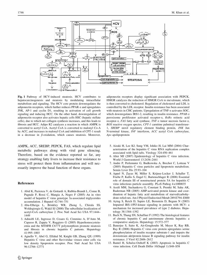
Fig. 1 Pathway of HCV-induced steatosis. HCV contributes to
hepatocarcinogenesis and steatosis by modulating intracellular
metabolism and signaling. The HCV core protein downregulates the
adiponectin receptors, which further reduces PPAR-a and upregulates
JNK, AP-1 and cyclin D1, resulting in activation of cell growth
signaling and inducing HCC. On the other hand, downregulation of
adiponectin receptor also activates hepatic cells HSC (hepatic stellate
cells), due to which net collagen synthesis increases, and this leads to
fibrosis and HCC. Adipo R2 catalyzes a reaction in which AMPK is
converted to acetyl CoA. Acetyl CoA is converted to malonyl Co-A
by ACC, and increases in malonyl CoA and inhibition of CPT-1 result
in a decrease in b-oxidation, which causes steatosis. Moreover,
adiponectin receptors display significant association with PEPCK.
HMGR catalyzes the reduction of HMGR CoA to mevalonate, which
is then converted to cholesterol. Regulation of cholesterol and LDL is
controlled by the LDL receptor. Insulin resistance has been associated
with steatosis in CHC patients. Upregulation of TNF-a activates SOC,
which downregulates IRS1-2, resulting in insulin resistance. PPAR-aperoxisome proliferator activated receptor-a, RxRa retinoic acid
receptor-a, FAS fatty acid synthase, TNF-a tumor necrosis factor-a,
ROS reactive oxygen species, CPT-1 carnitine palmitoyl transferase-
1, SREBP sterol regulatory element binding protein, JNK Jun
N-terminal kinase, INF interferon, ACC acetyl CoA carboxylase,
Apo apolipoprotein
1746 M. Khan et al.
123
14. Barba G, Harper F, Harada T, Kohara M, Goulinet S, Matsuura Y,
Eder G, Schaff Z, Chapman MJ, Miyamura T, Brechot C (1997)
Hepatitis C virus core protein shows a cytoplasmic localization
and associates to cellular lipid storage droplets. Proc Natl Acad Sci
USA 94:1200–1205
15. Bartenschlager R, Lohmann V (2001) Novel cell culture systems
for the hepatitis C virus. Antiviral Res 52:1–17
16. Bartenschlager R, Sparacio S (2007) Hepatitis C virus molecular
clones and their replication capacity in vivo and in cell culture.
Virus Res 127:195–207
17. Beltowski J (2003) Adiponectin and resistin—new hormones of
white adipose tissue. Med Sci Monit 9:RA55–RA61
18. Benali-Furet NL, Chami M, Houel L, De Giorgi F, Vernejoul F,
Lagorce D, Buscail L, Bartenschlager R, Ichas F, Rizzuto R,
Paterlini-Brechot P (2005) Hepatitis C virus core triggers
apoptosis in liver cells by inducing ER stress and ER calcium
depletion. Oncogene 24:4921–4933
19. Benedicto I, Molina-Jimenez F, Barreiro O, Maldonado-
Rodriguez A, Prieto J, Moreno-Otero R, Aldabe R, Lopez-
Cabrera M, Majano PL (2008) Hepatitis C virus envelope
components alter localization of hepatocyte tight junction-
associated proteins and promote occludin retention in the
endoplasmic reticulum. Hepatology 48:1044–1053
20. Bevan P (2001) Insulin signalling. J Cell Sci 114:1429–1430
21. Bieche I, Asselah T, Laurendeau I, Vidaud D, Degot C, Paradis V,
Bedossa P, Valla DC, Marcellin P, Vidaud M (2005) Molecular
profiling of early stage liver fibrosis in patients with chronic
hepatitis C virus infection. Virology 332:130–144
22. Bigger CB, Guerra B, Brasky KM, Hubbard G, Beard MR,
Luxon BA, Lemon SM, Lanford RE (2004) Intrahepatic gene
expression during chronic hepatitis C virus infection in chim-
panzees. J Virol 78:13779–13792
23. Boulant S, Targett-Adams P, McLauchlan J (2007) Disrupting
the association of hepatitis C virus core protein with lipid
droplets correlates with a loss in production of infectious virus.
J Gen Virol 88:2204–2213
24. Brown MS, Goldstein JL (1986) A receptor-mediated pathway
for cholesterol homeostasis. Science 232:34–47
25. Brown MS, Goldstein JL (1997) The SREBP pathway: regula-
tion of cholesterol metabolism by proteolysis of a membrane-
bound transcription factor. Cell 89:331–340
26. Browning JD, Horton JD (2004) Molecular mediators of hepatic
steatosis and liver injury. J Clin Invest 114:147–152
27. Bukh J (2004) A critical role for the chimpanzee model in the
study of hepatitis C. Hepatology 39:1469–1475
28. Chandler CE, Wilder DE, Pettini JL, Savoy YE, Petras SF,
Chang G, Vincent J, Harwood HJ Jr (2003) CP-346086: an MTP
inhibitor that lowers plasma cholesterol and triglycerides in
experimental animals and in humans. J Lipid Res 44:1887–1901
29. Chang KS, Jiang J, Cai Z, Luo G (2007) Human apolipoprotein
e is required for infectivity and production of hepatitis C virus in
cell culture. J Virol 81:13783–13793
30. Cheng Y, Dharancy S, Malapel M, Desreumaux P (2005)
Hepatitis C virus infection down-regulates the expression of
peroxisome proliferator-activated receptor alpha and carnitine
palmitoyl acyl-CoA transferase 1A. World J Gastroenterol
11:7591–7596
31. Christen V, Treves S, Duong FH, Heim MH (2007) Activation
of endoplasmic reticulum stress response by hepatitis viruses up-
regulates protein phosphatase 2A. Hepatology 46:558–565
32. Chung NS, Wasan KM (2004) Potential role of the low-density
lipoprotein receptor family as mediators of cellular drug uptake.
Adv Drug Deliv Rev 56:1315–1334
33. Ciccaglione AR, Marcantonio C, Tritarelli E, Equestre M,
Vendittelli F, Costantino A, Geraci A, Rapicetta M (2007)
Activation of the ER stress gene gadd153 by hepatitis C virus
sensitizes cells to oxidant injury. Virus Res 126:128–138
34. Cuchel M, Bloedon LT, Szapary PO, Kolansky DM, Wolfe ML,
Sarkis A, Millar JS, Ikewaki K, Siegelman ES, Gregg RE,
Rader DJ (2007) Inhibition of microsomal triglyceride transfer
protein in familial hypercholesterolemia. N Engl J Med 356:
148–156
35. de Gottardi A, Pazienza V, Pugnale P, Bruttin F, Rubbia-Brandt
L, Juge-Aubry CE, Meier CA, Hadengue A, Negro F (2006)
Peroxisome proliferator-activated receptor-alpha and -gamma
mRNA levels are reduced in chronic hepatitis C with steatosis
and genotype 3 infection. Aliment Pharmacol Ther 23:107–114
36. Deng L, Adachi T, Kitayama K, Bungyoku Y, Kitazawa S, Ishido
S, Shoji I, Hotta H (2008) Hepatitis C virus infection induces
apoptosis through a Bax-triggered, mitochondrion-mediated,
caspase 3-dependent pathway. J Virol 82:10375–10385
37. Dharancy S, Malapel M, Perlemuter G, Roskams T, Cheng Y,
Dubuquoy L, Podevin P, Conti F, Canva V, Philippe D,
Gambiez L, Mathurin P, Paris JC, Schoonjans K, Calmus Y,
Pol S, Auwerx J, Desreumaux P (2005) Impaired expression of
the peroxisome proliferator-activated receptor alpha during
hepatitis C virus infection. Gastroenterology 128:334–342
38. Diez JJ, Iglesias P (2003) The role of the novel adipocyte-derived
hormone adiponectin in human disease. Eur J Endocrinol 148:
293–300
39. Dimcheff DE, Faasse MA, McAtee FJ, Portis JL (2004) Endo-
plasmic reticulum (ER) stress induced by a neurovirulent mouse
retrovirus is associated with prolonged BiP binding and reten-
tion of a viral protein in the ER. J Biol Chem 279:33782–33790
40. Durante-Mangoni E, Zampino R, Marrone A, Tripodi MF,
Rinaldi L, Restivo L, Cioffi M, Ruggiero G, Adinolfi LE (2006)
Hepatic steatosis and insulin resistance are associated with
serum imbalance of adiponectin/tumour necrosis factor-alpha in
chronic hepatitis C patients. Aliment Pharmacol Ther 24:1349–
1357
41. El-Serag HB, Tran T, Everhart JE (2004) Diabetes increases the
risk of chronic liver disease and hepatocellular carcinoma.
Gastroenterology 126:460–468
42. Eugene JY, Hu K-Q (2006) HCV infection and hepatic steatosis.
Int J Med Sci 3:53–56
43. Feitelson MA (2002) Hepatitis C virus: from laboratory to
clinic. Cambridge University Press, New York
44. Foretz M, Guichard C, Ferre P, Foufelle F (1999) Sterol regu-
latory element binding protein-1c is a major mediator of insulin
action on the hepatic expression of glucokinase and lipogenesis-
related genes. Proc Natl Acad Sci USA 96:12737–12742
45. Fukasawa M, Tanaka Y, Sato S, Ono Y, Nitahara-Kasahara Y,
Suzuki T, Miyamura T, Hanada K, Nishijima M (2006)
Enhancement of de novo fatty acid biosynthesis in hepatic cell
line Huh7 expressing hepatitis C virus core protein. Biol Pharm
Bull 29:1958–1961
46. Furuta K, Sato S, Yamauchi T, Kakumu S (2008) Changes in
intrahepatic gene expression profiles from chronic hepatitis to
hepatocellular carcinoma in patients with hepatitis C virus
infection. Hepatol Res 38:673–682
47. Gastaminza P, Cheng G, Wieland S, Zhong J, Liao W, Chisari FV
(2008) Cellular determinants of hepatitis C virus assembly,
maturation, degradation, and secretion. J Virol 82:2120–2129
48. Gerber MA, Krawczynski K, Alter MJ, Sampliner RE,
Margolis HS (1992) Histopathology of community acquired
chronic hepatitis C. The Sentinel Counties Chronic Non-A,
Non-B Hepatitis Study Team. Mod Pathol 5:483–486
49. Gibbons GF, Wiggins D, Brown AM, Hebbachi AM (2004)
Synthesis and function of hepatic very-low-density lipoprotein.
Biochem Soc Trans 32:59–64
Role of HCV core in steatosis 1747
123
50. Goldstein BJ, Scalia R (2004) Adiponectin: a novel adipokine
linking adipocytes and vascular function. J Clin Endocrinol
Metab 89:2563–2568
51. Goldstein JL, Brown MS (1990) Regulation of the mevalonate
pathway. Nature 343:425–430
52. Gopal K, Johnson TC, Gopal S, Walfish A, Bang CT, Suwandhi P,
Pena-Sahdala HN, Clain DJ, Bodenheimer HC Jr, Min AD (2006)
Correlation between beta-lipoprotein levels and outcome of
hepatitis C treatment. Hepatology 44:335–340
53. Halse R, Pearson SL, McCormack JG, Yeaman SJ, Taylor R
(2001) Effects of tumor necrosis factor-alpha on insulin action
in cultured human muscle cells. Diabetes 50:1102–1109
54. Hardie DG (2003) Minireview: the AMP-activated protein
kinase cascade: the key sensor of cellular energy status. Endo-
crinology 144:5179–5183
55. Hardie DG (2004) The AMP-activated protein kinase pathway—
new players upstream and downstream. J Cell Sci 117:5479–
5487
56. Hezode C, Roudot-Thoraval F, Zafrani ES, Dhumeaux D,
Pawlotsky JM (2004) Different mechanisms of steatosis in
hepatitis C virus genotypes 1 and 3 infections. J Viral Hepat
11:455–458
57. Higuchi N, Kato M, Shundo Y, Tajiri H, Tanaka M, Yamashita
N, Kohjima M, Kotoh K, Nakamuta M, Takayanagi R, Enjoji M
(2008) Liver X receptor in cooperation with SREBP-1c is a
major lipid synthesis regulator in nonalcoholic fatty liver dis-
ease. Hepatol Res 38:1122–1129
58. Hoofnagle JH (2002) Course and outcome of hepatitis C.
Hepatology 36:S21–S29
59. Hope RG, McLauchlan J (2000) Sequence motifs required for
lipid droplet association and protein stability are unique to the
hepatitis C virus core protein. J Gen Virol 81:1913–1925
60. Horscroft N, Lai VC, Cheney W, Yao N, Wu JZ, Hong Z,
Zhong W (2005) Replicon cell culture system as a valuable tool
in antiviral drug discovery against hepatitis C virus. Antivir
Chem Chemother 16:1–12
61. Horton JD, Goldstein JL, Brown MS (2002) SREBPs: activators
of the complete program of cholesterol and fatty acid synthesis
in the liver. J Clin Invest 109:1125–1131
62. Horton JD, Shah NA, Warrington JA, Anderson NN, Park SW,
Brown MS, Goldstein JL (2003) Combined analysis of oligo-
nucleotide microarray data from transgenic and knockout mice
identifies direct SREBP target genes. Proc Natl Acad Sci USA
100:12027–12032
63. Hotta K, Funahashi T, Arita Y, Takahashi M, Matsuda M,
Okamoto Y, Iwahashi H, Kuriyama H, Ouchi N, Maeda K,
Nishida M, Kihara S, Sakai N, Nakajima T, Hasegawa K, Mur-
aguchi M, Ohmoto Y, Nakamura T, Yamashita S, Hanafusa T,
Matsuzawa Y (2000) Plasma concentrations of a novel, adipose-
specific protein, adiponectin, in type 2 diabetic patients. Arte-
rioscler Thromb Vasc Biol 20:1595–1599
64. Hourioux C, Patient R, Morin A, Blanchard E, Moreau A,
Trassard S, Giraudeau B, Roingeard P (2007) The genotype
3-specific hepatitis C virus core protein residue phenylalanine 164
increases steatosis in an in vitro cellular model. Gut 56:1302–1308
65. Huang H, Sun F, Owen DM, Li W, Chen Y, Gale M Jr, Ye J
(2007) Hepatitis C virus production by human hepatocytes
dependent on assembly and secretion of very low-density lipo-
proteins. Proc Natl Acad Sci USA 104:5848–5853
66. Hui JM, Hodge A, Farrell GC, Kench JG, Kriketos A, George J
(2004) Beyond insulin resistance in NASH: TNF-alpha or
adiponectin? Hepatology 40:46–54
67. Hussy P, Langen H, Mous J, Jacobsen H (1996) Hepatitis C
virus core protein: carboxy-terminal boundaries of two pro-
cessed species suggest cleavage by a signal peptide peptidase.
Virology 224:93–104
68. Ikeda M, Abe K, Yamada M, Dansako H, Naka K, Kato N (2006)
Different anti-HCV profiles of statins and their potential for
combination therapy with interferon. Hepatology 44:117–125
69. Ilan E, Arazi J, Nussbaum O, Zauberman A, Eren R, Lubin I,
Neville L, Ben-Moshe O, Kischitzky A, Litchi A, Margalit I,
Gopher J, Mounir S, Cai W, Daudi N, Eid A, Jurim O, Czerniak
A, Galun E, Dagan S (2002) The hepatitis C virus (HCV)-
Trimera mouse: a model for evaluation of agents against HCV.
J Infect Dis 185:153–161
70. Im SS, Kwon SK, Kim TH, Kim HI, Ahn YH (2007) Regulation
of glucose transporter type 4 isoform gene expression in muscle
and adipocytes. IUBMB Life 59:134–145
71. Iwaki M, Matsuda M, Maeda N, Funahashi T, Matsuzawa Y,
Makishima M, Shimomura I (2003) Induction of adiponectin, a
fat-derived antidiabetic and antiatherogenic factor, by nuclear
receptors. Diabetes 52:1655–1663
72. Jackel-Cram C, Babiuk LA, Liu Q (2007) Up-regulation of fatty
acid synthase promoter by hepatitis C virus core protein:
genotype-3a core has a stronger effect than genotype-1b core.
J Hepatol 46:999–1008
73. Jhaveri R, McHutchison J, Patel K, Qiang G, Diehl AM (2008)
Specific polymorphisms in hepatitis C virus genotype 3 core
protein associated with intracellular lipid accumulation. J Infect
Dis 197:283–291
74. Ji C (2008) Dissection of endoplasmic reticulum stress signaling
in alcoholic and non-alcoholic liver injury. J Gastroenterol
Hepatol 23(Suppl 1):S16–S24
75. Jonsson JR, Moschen AR, Hickman IJ, Richardson MM,
Kaser S, Clouston AD, Powell EE, Tilg H (2005) Adiponectin
and its receptors in patients with chronic hepatitis C. J Hepatol
43:929–936
76. Kaiser S (1998) Cell volume regulates liver phosphoenolpyr-
uvate carboxykinase and fructose-1, 6-bisphosphatase genes.
Am J Physiol 274:G509–G517
77. Kapadia SB, Chisari FV (2005) Hepatitis C virus RNA repli-
cation is regulated by host geranylgeranylation and fatty acids.
Proc Natl Acad Sci USA 102:2561–2566
78. Kapadia SB, Barth H, Baumert T, McKeating JA, Chisari FV
(2007) Initiation of hepatitis C virus infection is dependent on
cholesterol and cooperativity between CD81 and scavenger
receptor B type I. J Virol 81:374–383
79. Kawaguchi T, Yoshida T, Harada M, Hisamoto T, Nagao Y, Ide T,
Taniguchi E, Kumemura H, Hanada S, Maeyama M, Baba S,
Koga H, Kumashiro R, Ueno T, Ogata H, Yoshimura A, Sata M
(2004) Hepatitis C virus down-regulates insulin receptor sub-
strates 1 and 2 through up-regulation of suppressor of cytokine
signaling 3. Am J Pathol 165:1499–1508
80. Kersten S, Seydoux J, Peters JM, Gonzalez FJ, Desvergne B,
Wahli W (1999) Peroxisome proliferator-activated receptor
alpha mediates the adaptive response to fasting. J Clin Invest
103:1489–1498
81. Kim K, Kim KH, Ha E, Park JY, Sakamoto N, Cheong J (2009)
Hepatitis C virus NS5A protein increases hepatic lipid accu-
mulation via induction of activation and expression of PPAR-
gamma. FEBS Lett 583:2720–2726
82. Kneteman NM, Weiner AJ, O’Connell J, Collett M, Gao T,
Aukerman L, Kovelsky R, Ni ZJ, Zhu Q, Hashash A, Kline J,
Hsi B, Schiller D, Douglas D, Tyrrell DL, Mercer DF (2006)
Anti-HCV therapies in chimeric scid-Alb/uPA mice parallel
outcomes in human clinical application. Hepatology 43:1346–
1353
83. Knobler H, Schattner A (2005) TNF-{alpha}, chronic hepatitis
C and diabetes: a novel triad. Qjm 98:1–6
84. Kohjima M, Higuchi N, Kato M, Kotoh K, Yoshimoto T,
Fujino T, Yada M, Yada R, Harada N, Enjoji M, Takayanagi R,
Nakamuta M (2008) SREBP-1c, regulated by the insulin and
1748 M. Khan et al.
123
AMPK signaling pathways, plays a role in nonalcoholic fatty
liver disease. Int J Mol Med 21:507–511
85. Kolykhalov AA, Agapov EV, Blight KJ, Mihalik K,
Feinstone SM, Rice CM (1997) Transmission of hepatitis C by
intrahepatic inoculation with transcribed RNA. Science 277:
570–574
86. Korenaga M, Wang T, Li Y, Showalter LA, Chan T, Sun J,
Weinman SA (2005) Hepatitis C virus core protein inhibits
mitochondrial electron transport and increases reactive oxygen
species (ROS) production. J Biol Chem 280:37481–37488
87. Kuipers F, Jong MC, Lin Y, Eck M, Havinga R, Bloks V,
Verkade HJ, Hofker MH, Moshage H, Berkel TJ, Vonk RJ,
Havekes LM (1997) Impaired secretion of very low density
lipoprotein-triglycerides by apolipoprotein E- deficient mouse
hepatocytes. J Clin Invest 100:2915–2922
88. Kumar D, Farrell GC, Fung C, George J (2002) Hepatitis C virus
genotype 3 is cytopathic to hepatocytes: Reversal of hepatic
steatosis after sustained therapeutic response. Hepatology
36:1266–1272
89. Labonte P, Begley S, Guevin C, Asselin MC, Nassoury N,
Mayer G, Prat A, Seidah NG (2009) PCSK9 impedes hepatitis C
virus infection in vitro and modulates liver CD81 expression.
Hepatology 50:17–24
90. Leone TC, Weinheimer CJ, Kelly DP (1999) A critical role for
the peroxisome proliferator-activated receptor alpha (PPARal-
pha) in the cellular fasting response: the PPARalpha-null mouse
as a model of fatty acid oxidation disorders. Proc Natl Acad Sci
USA 96:7473–7478
91. Lerat H, Honda M, Beard MR, Loesch K, Sun J, Yang Y,
Okuda M, Gosert R, Xiao SY, Weinman SA, Lemon SM (2002)
Steatosis and liver cancer in transgenic mice expressing
the structural and nonstructural proteins of hepatitis C virus.
Gastroenterology 122:352–365
92. Lerat H, Kammoun HL, Hainault I, Merour E, Higgs MR,
Callens C, Lemon SM, Foufelle F, Pawlotsky JM (2009)
Hepatitis C virus proteins induce lipogenesis and defective tri-
glyceride secretion in transgenic mice. J Biol Chem 284:33466–
33474
93. Lindenbach BD, Evans MJ, Syder AJ, Wolk B, Tellinghuisen
TL, Liu CC, Maruyama T, Hynes RO, Burton DR, McKeating
JA, Rice CM (2005) Complete replication of hepatitis C virus in
cell culture. Science 309:623–626
94. Liu N, Kuang X, Kim HT, Stoica G, Qiang W, Scofield VL,
Wong PK (2004) Possible involvement of both endoplasmic
reticulum- and mitochondria-dependent pathways in MoMuLV-
ts1-induced apoptosis in astrocytes. J Neurovirol 10:189–198
95. Lohmann V, Korner F, Koch J, Herian U, Theilmann L,
Bartenschlager R (1999) Replication of subgenomic hepatitis C
virus RNAs in a hepatoma cell line. Science 285:110–113
96. Lonardo A, Loria P, Adinolfi LE, Carulli N, Ruggiero G (2006)
Hepatitis C and steatosis: a reappraisal. J Viral Hepat 13:73–80
97. Maeda K, Okubo K, Shimomura I, Funahashi T, Matsuzawa Y,
Matsubara K (1996) cDNA cloning and expression of a novel
adipose specific collagen-like factor, apM1 (AdiPose Most
abundant Gene transcript 1). Biochem Biophys Res Commun
221:286–289
98. Magnusson B, Asp L, Bostrom P, Ruiz M, Stillemark-Billton P,
Linden D, Boren J, Olofsson SO (2006) Adipocyte differentia-
tion-related protein promotes fatty acid storage in cytosolic
triglycerides and inhibits secretion of very low-density lipo-
proteins. Arterioscler Thromb Vasc Biol 26:1566–1571
99. Major ME, Dahari H, Mihalik K, Puig M, Rice CM, Neumann
AU, Feinstone SM (2004) Hepatitis C virus kinetics and host
responses associated with disease and outcome of infection in
chimpanzees. Hepatology 39:1709–1720
100. Mangelsdorf DJ, Evans RM (1995) The RXR heterodimers and
orphan receptors. Cell 83:841–850
101. Mangelsdorf DJ, Thummel C, Beato M, Herrlich P, Schutz G,
Umesono K, Blumberg B, Kastner P, Mark M, Chambon P,
Evans RM (1995) The nuclear receptor superfamily: the second
decade. Cell 83:835–839
102. Marchesini G, Marzocchi R, Agostini F, Bugianesi E (2005)
Nonalcoholic fatty liver disease and the metabolic syndrome.
Curr Opin Lipidol 16:421–427
103. Masaki T, Suzuki R, Murakami K, Aizaki H, Ishii K, Murayama
A, Date T, Matsuura Y, Miyamura T, Wakita T, Suzuki T (2008)
Interaction of hepatitis C virus nonstructural protein 5A with
core protein is critical for the production of infectious virus
particles. J Virol 82:7964–7976
104. McGarry JD, Brown NF (1997) The mitochondrial carnitine
palmitoyltransferase system. From concept to molecular analy-
sis. Eur J Biochem 244:1–14
105. McLauchlan J, Lemberg MK, Hope G, Martoglio B (2002)
Intramembrane proteolysis promotes trafficking of hepatitis C
virus core protein to lipid droplets. EMBO J 21:3980–3988
106. Mercer DF, Schiller DE, Elliott JF, Douglas DN, Hao C, Rinfret
A, Addison WR, Fischer KP, Churchill TA, Lakey JR, Tyrrell
DL, Kneteman NM (2001) Hepatitis C virus replication in mice
with chimeric human livers. Nat Med 7:927–933
107. Mihm S, Fayyazi A, Hartmann H, Ramadori G (1997) Analysis
of histopathological manifestations of chronic hepatitis C virus
infection with respect to virus genotype. Hepatology 25:735–739
108. Mirandola S, Realdon S, Iqbal J, Gerotto M, Dal Pero F, Bor-
toletto G, Marcolongo M, Vario A, Datz C, Hussain MM,
Alberti A (2006) Liver microsomal triglyceride transfer protein
is involved in hepatitis C liver steatosis. Gastroenterology
130:1661–1669
109. Miyanari Y, Atsuzawa K, Usuda N, Watashi K, Hishiki T,
Zayas M, Bartenschlager R, Wakita T, Hijikata M, Shimotohno K
(2007) The lipid droplet is an important organelle for hepatitis C
virus production. Nat Cell Biol 9:1089–1097
110. Molina S, Castet V, Fournier-Wirth C, Pichard-Garcia L, Avner R,
Harats D, Roitelman J, Barbaras R, Graber P, Ghersa P, Smolarsky
M, Funaro A, Malavasi F, Larrey D, Coste J, Fabre JM, Sa-Cunha
A, Maurel P (2007) The low-density lipoprotein receptor plays a
role in the infection of primary human hepatocytes by hepatitis C
virus. J Hepatol 46:411–419
111. Monazahian M, Bohme I, Bonk S, Koch A, Scholz C, Grethe S,
Thomssen R (1999) Low density lipoprotein receptor as a can-
didate receptor for hepatitis C virus. J Med Virol 57:223–229
112. Monto A, Alonzo J, Watson JJ, Grunfeld C, Wright TL (2002)
Steatosis in chronic hepatitis C: relative contributions of obesity,
diabetes mellitus, and alcohol. Hepatology 36:729–736
113. Moriya K, Yotsuyanagi H, Shintani Y, Fujie H, Ishibashi K,
Matsuura Y, Miyamura T, Koike K (1997) Hepatitis C virus
core protein induces hepatic steatosis in transgenic mice. J Gen
Virol 78(Pt 7):1527–1531
114. Moriya K, Fujie H, Shintani Y, Yotsuyanagi H, Tsutsumi T,
Ishibashi K, Matsuura Y, Kimura S, Miyamura T, Koike K
(1998) The core protein of hepatitis C virus induces hepato-
cellular carcinoma in transgenic mice. Nat Med 4:1065–1067
115. Moriya K, Nakagawa K, Santa T, Shintani Y, Fujie H, Miyoshi
H, Tsutsumi T, Miyazawa T, Ishibashi K, Horie T, Imai K,
Todoroki T, Kimura S, Koike K (2001) Oxidative stress in the
absence of inflammation in a mouse model for hepatitis C virus-
associated hepatocarcinogenesis. Cancer Res 61:4365–4370
116. Munday MR (2002) Regulation of mammalian acetyl-CoA
carboxylase. Biochem Soc Trans 30:1059–1064
117. Nahmias Y, Goldwasser J, Casali M, van Poll D, Wakita T,
Chung RT, Yarmush ML (2008) Apolipoprotein B-dependent
Role of HCV core in steatosis 1749
123
hepatitis C virus secretion is inhibited by the grapefruit flavo-
noid naringenin. Hepatology 47:1437–1445
118. Neuschwander-Tetri BA, Caldwell SH (2003) Nonalcoholic ste-
atohepatitis: summary of an AASLD Single Topic Conference.
Hepatology 37:1202–1219
119. Nielsen SU, Bassendine MF, Martin C, Lowther D, Purcell PJ,
King BJ, Neely D, Toms GL (2008) Characterization of hepatitis
C RNA-containing particles from human liver by density and
size. J Gen Virol 89:2507–2517
120. Nykjaer A, Willnow TE (2002) The low-density lipoprotein
receptor gene family: a cellular Swiss army knife? Trends Cell
Biol 12:273–280
121. Oem JK, Jackel-Cram C, Li YP, Kang HN, Zhou Y, Babiuk LA,
Liu Q (2008) Hepatitis C virus non-structural protein-2 activates
CXCL-8 transcription through NF-kappaB. Arch Virol 153:293–
301
122. Oem JK, Jackel-Cram C, Li YP, Zhou Y, Zhong J, Shimano H,
Babiuk LA, Liu Q (2008) Activation of sterol regulatory
element-binding protein 1c and fatty acid synthase transcription
by hepatitis C virus non-structural protein 2. J Gen Virol
89:1225–1230
123. Ohata K, Hamasaki K, Toriyama K, Matsumoto K, Saeki A,
Yanagi K, Abiru S, Nakagawa Y, Shigeno M, Miyazoe S,
Ichikawa T, Ishikawa H, Nakao K, Eguchi K (2003) Hepatic
steatosis is a risk factor for hepatocellular carcinoma in patients
with chronic hepatitis C virus infection. Cancer 97:3036–3043
124. Olsavsky KM, Page JL, Johnson MC, Zarbl H, Strom SC,
Omiecinski CJ (2007) Gene expression profiling and differen-
tiation assessment in primary human hepatocyte cultures,
established hepatoma cell lines, and human liver tissues. Toxicol
Appl Pharmacol 222:42–56
125. Ozes ON, Akca H, Mayo LD, Gustin JA, Maehama T, Dixon JE,
Donner DB (2001) A phosphatidylinositol 3-kinase/Akt/mTOR
pathway mediates and PTEN antagonizes tumor necrosis factor
inhibition of insulin signaling through insulin receptor substrate-
1. Proc Natl Acad Sci USA 98:4640–4645
126. Park CY, Jun HJ, Wakita T, Cheong JH, Hwang SB (2009)
Hepatitis C virus nonstructural 4B protein modulates sterol
regulatory element-binding protein signaling via the AKT
pathway. J Biol Chem 284:9237–9246
127. Patton HM, Patel K, Behling C, Bylund D, Blatt LM, Vallee M,
Heaton S, Conrad A, Pockros PJ, McHutchison JG (2004) The
impact of steatosis on disease progression and early and sus-
tained treatment response in chronic hepatitis C patients.
J Hepatol 40:484–490
128. Pazienza V, Clement S, Pugnale P, Conzelman S, Foti M,
Mangia A, Negro F (2007) The hepatitis C virus core protein of
genotypes 3a and 1b downregulates insulin receptor substrate 1
through genotype-specific mechanisms. Hepatology 45:1164–
c1171
129. Pazienza V, Vinciguerra M, Andriulli A, Mangia A (2010)
Hepatitis C virus core protein genotype 3a increases SOCS-7
expression through PPAR-{gamma} in Huh-7 cells. J Gen Virol
91:1678–1686
130. Pekow JR, Bhan AK, Zheng H, Chung RT (2007) Hepatic ste-
atosis is associated with increased frequency of hepatocellular
carcinoma in patients with hepatitis C-related cirrhosis. Cancer
109:2490–2496
131. Peralta C, Bartrons R, Riera L, Manzano A, Xaus C, Gelpi E,
Rosello-Catafau J (2000) Hepatic preconditioning preserves
energy metabolism during sustained ischemia. Am J Physiol
Gastrointest Liver Physiol 279:G163–G171
132. Perlemuter G, Sabile A, Letteron P, Vona G, Topilco A,
Chretien Y, Koike K, Pessayre D, Chapman J, Barba G, Brechot
C (2002) Hepatitis C virus core protein inhibits microsomal
triglyceride transfer protein activity and very low density
lipoprotein secretion: a model of viral-related steatosis. FASEB
J 16:185–194
133. Persico M, Capasso M, Persico E, Svelto M, Russo R, Spano D,
Croce L, La Mura V, Moschella F, Masutti F, Torella R,
Tiribelli C, Iolascon A (2007) Suppressor of cytokine signaling
3 (SOCS3) expression and hepatitis C virus-related chronic
hepatitis: Insulin resistance and response to antiviral therapy.
Hepatology 46:1009–1015
134. Petit JM, Benichou M, Duvillard L, Jooste V, Bour JB, Minello
A, Verges B, Brun JM, Gambert P, Hillon P (2003) Hepatitis C
virus-associated hypobetalipoproteinemia is correlated with
plasma viral load, steatosis, and liver fibrosis. Am J Gastroen-
terol 98:1150–1154
135. Petit JM, Minello A, Jooste V, Bour JB, Galland F, Duvillard L,
Verges B, Olsson NO, Gambert P, Hillon P (2005) Decreased
plasma adiponectin concentrations are closely related to stea-
tosis in hepatitis C virus-infected patients. J Clin Endocrinol
Metab 90:2240–2243
136. Pianko S, Patella S, Ostapowicz G, Desmond P, Sievert W
(2001) Fas-mediated hepatocyte apoptosis is increased by hep-
atitis C virus infection and alcohol consumption, and may be
associated with hepatic fibrosis: mechanisms of liver cell injury
in chronic hepatitis C virus infection. J Viral Hepat 8:406–413
137. Pileri P, Uematsu Y, Campagnoli S, Galli G, Falugi F, Petracca R,
Weiner AJ, Houghton M, Rosa D, Grandi G, Abrignani S (1998)
Binding of hepatitis C virus to CD81. Science 282:938–941
138. Poynard T, Ratziu V, McHutchison J, Manns M, Goodman Z,
Zeuzem S, Younossi Z, Albrecht J (2003) Effect of treatment
with peginterferon or interferon alfa-2b and ribavirin on stea-
tosis in patients infected with hepatitis C. Hepatology 38:75–85
139. Raabe M, Veniant MM, Sullivan MA, Zlot CH, Bjorkegren J,
Nielsen LB, Wong JS, Hamilton RL, Young SG (1999) Analysis
of the role of microsomal triglyceride transfer protein in the liver
of tissue-specific knockout mice. J Clin Invest 103:1287–1298
140. Ramachandran CK, Gray SL, Melnykovych G (1980) Cyto-
plasmic 3-hydroxy-3-methylglutaryl coenzyme A synthase and
the regulation of sterol synthesis in tissue culture cells. Biochim
Biophys Acta 618:439–448
141. Ray RB, Meyer K, Ray R (1996) Suppression of apoptotic cell
death by hepatitis C virus core protein. Virology 226:176–182
142. Reed KE, Rice CM (2000) Overview of hepatitis C virus genome
structure, polyprotein processing, and protein properties. Curr
Top Microbiol Immunol 242:55–84
143. Reyes GR (2002) The nonstructural NS5A protein of hepatitis C
virus: an expanding, multifunctional role in enhancing hepatitis
C virus pathogenesis. J Biomed Sci 9:187–197
144. Romero-Gomez M, Del Mar Viloria M, Andrade RJ, Salmeron J,
Diago M, Fernandez-Rodriguez CM, Corpas R, Cruz M,
Grande L, Vazquez L, Munoz-De-Rueda P, Lopez-Serrano P,
Gila A, Gutierrez ML, Perez C, Ruiz-Extremera A, Suarez E,
Castillo J (2005) Insulin resistance impairs sustained response rate
to peginterferon plus ribavirin in chronic hepatitis C patients.
Gastroenterology 128:636–641
145. Rouille Y, Helle F, Delgrange D, Roingeard P, Voisset C, Blan-
chard E, Belouzard S, McKeating J, Patel AH, Maertens G,
Wakita T, Wychowski C, Dubuisson J (2006) Subcellular local-
ization of hepatitis C virus structural proteins in a cell culture
system that efficiently replicates the virus. J Virol 80:2832–2841
146. Rubbia-Brandt L, Quadri R, Abid K, Giostra E, Male PJ,
Mentha G, Spahr L, Zarski JP, Borisch B, Hadengue A, Negro F
(2000) Hepatocyte steatosis is a cytopathic effect of hepatitis C
virus genotype 3. J Hepatol 33:106–115
147. Rubbia-Brandt L, Fabris P, Paganin S, Leandro G, Male PJ,
Giostra E, Carlotto A, Bozzola L, Smedile A, Negro F (2004)Steatosis affects chronic hepatitis C progression in a genotype
specific way. Gut 53:406–412
1750 M. Khan et al.
123
148. Saha AK, Avilucea PR, Ye JM, Assifi MM, Kraegen EW,
Ruderman NB (2004) Pioglitazone treatment activates AMP-
activated protein kinase in rat liver and adipose tissue in vivo.
Biochem Biophys Res Commun 314:580–585
149. Santolini E, Migliaccio G, La Monica N (1994) Biosynthesis
and biochemical properties of the hepatitis C virus core protein.
J Virol 68:3631–3641
150. Sanyal AJ (2002) AGA technical review on nonalcoholic fatty
liver disease. Gastroenterology 123:1705–1725
151. Savage DB, Choi CS, Samuel VT, Liu ZX, Zhang D, Wang A,
Zhang XM, Cline GW, Yu XX, Geisler JG, Bhanot S, Monia
BP, Shulman GI (2006) Reversal of diet-induced hepatic stea-
tosis and hepatic insulin resistance by antisense oligonucleotide
inhibitors of acetyl-CoA carboxylases 1 and 2. J Clin Invest
116:817–824
152. Scarselli E, Ansuini H, Cerino R, Roccasecca RM, Acali S,
Filocamo G, Traboni C, Nicosia A, Cortese R, Vitelli A (2002)
The human scavenger receptor class B type I is a novel candi-
date receptor for the hepatitis C virus. EMBO J 21:5017–5025
153. Schnitzer-Polokoff R, von Gunten C, Logel J, Torget R,
Sinensky M (1982) Isolation and characterization of a mam-
malian cell mutant defective in 3-hydroxy-3-methylglutaryl
coenzyme A synthase. J Biol Chem 257:472–476
154. Sekine-Osajima Y, Sakamoto N, Mishima K, Nakagawa M,
Itsui Y, Tasaka M, Nishimura-Sakurai Y, Chen CH, Kanai T,
Tsuchiya K, Wakita T, Enomoto N, Watanabe M (2008) Devel-
opment of plaque assays for hepatitis C virus-JFH1 strain and
isolation of mutants with enhanced cytopathogenicity and repli-
cation capacity. Virology 371:71–85
155. Sekiya M, Yahagi N, Matsuzaka T, Najima Y, Nakakuki M,
Nagai R, Ishibashi S, Osuga J, Yamada N, Shimano H (2003)
Polyunsaturated fatty acids ameliorate hepatic steatosis in obese
mice by SREBP-1 suppression. Hepatology 38:1529–1539
156. Semenkovich CF (1997) Regulation of fatty acid synthase
(FAS). Prog Lipid Res 36:43–53
157. Serfaty L, Andreani T, Giral P, Carbonell N, Chazouilleres O,
Poupon R (2001) Hepatitis C virus induced hypobetalipopro-
teinemia: a possible mechanism for steatosis in chronic hepatitis
C. J Hepatol 34:428–434
158. Shackel NA, McGuinness PH, Abbott CA, Gorrell MD,
McCaughan GW (2002) Insights into the pathobiology of
hepatitis C virus-associated cirrhosis: analysis of intrahepatic
differential gene expression. Am J Pathol 160:641–654
159. Shavinskaya A, Boulant S, Penin F, McLauchlan J,
Bartenschlager R (2007) The lipid droplet binding domain of
hepatitis C virus core protein is a major determinant for efficient
virus assembly. J Biol Chem 282:37158–37169
160. Sheehy P, Mullan B, Moreau I, Kenny-Walsh E, Shanahan F,
Scallan M, Fanning LJ (2007) In vitro replication models for the
hepatitis C virus. J Viral Hepat 14:2–10
161. Sheikh MY, Choi J, Qadri I, Friedman JE, Sanyal AJ (2008)
Hepatitis C virus infection: molecular pathways to metabolic
syndrome. Hepatology 47:2127–2133
162. Shelness GS, Sellers JA (2001) Very-low-density lipoprotein
assembly and secretion. Curr Opin Lipidol 12:151–157
163. Shi ST, Lee KJ, Aizaki H, Hwang SB, Lai MM (2003) Hepatitis
C virus RNA replication occurs on a detergent-resistant mem-
brane that cofractionates with caveolin-2. J Virol 77:4160–4168
164. Shimada M, Kawahara H, Ozaki K, Fukura M, Yano H,
Tsuchishima M, Tsutsumi M, Takase S (2007) Usefulness of a
combined evaluation of the serum adiponectin level, HOMA-IR,
and serum type IV collagen 7S level to predict the early stage of
nonalcoholic steatohepatitis. Am J Gastroenterol 102:1931–1938
165. Shimano H, Horton JD, Hammer RE, Shimomura I, Brown MS,
Goldstein JL (1996) Overproduction of cholesterol and fatty
acids causes massive liver enlargement in transgenic mice
expressing truncated SREBP-1a. J Clin Invest 98:1575–1584
166. Shimano H, Horton JD, Shimomura I, Hammer RE, Brown MS,
Goldstein JL (1997) Isoform 1c of sterol regulatory element
binding protein is less active than isoform 1a in livers of
transgenic mice and in cultured cells. J Clin Invest 99:846–854
167. Shimano H (2001) Sterol regulatory element-binding proteins
(SREBPs): transcriptional regulators of lipid synthetic genes.
Prog Lipid Res 40:439–452
168. Shimomura I, Bashmakov Y, Horton JD (1999) Increased levels
of nuclear SREBP-1c associated with fatty livers in two mouse
models of diabetes mellitus. J Biol Chem 274:30028–30032
169. Shintani Y, Fujie H, Miyoshi H, Tsutsumi T, Tsukamoto K,
Kimura S, Moriya K, Koike K (2004) Hepatitis C virus infection
and diabetes: direct involvement of the virus in the development
of insulin resistance. Gastroenterology 126:840–848
170. Shulman GI (2000) Cellular mechanisms of insulin resistance.
J Clin Invest 106:171–176
171. Sidorkiewicz M, Jozwiak B, Durys B, Majda-Stanislawska E,
Piekarska A, Kosciuk N, Ciechowicz J, Majewska E, Bartkowiak J
(2009) Mevalonate pathway modulation is associated with hepa-
titis C virus RNA presence in peripheral blood mononuclear cells.
Virus Res 145:141–144
172. Stahlberg D, Rudling M, Angelin B, Bjorkhem I, Forsell P,
Nilsell K, Einarsson K (1997) Hepatic cholesterol metabolism in
human obesity. Hepatology 25:1447–1450
173. Steer CJ (1996) Receptor-mediated endocytosis: mechanism,
biologic function, and molecular properties. In: Zakim D, Boyer
TD (eds) Hepatology: a text-book of liver disease volume I. WB
Saunders Company, USA, pp 49–214
174. Su AI, Pezacki JP, Wodicka L, Brideau AD, Supekova L,
Thimme R, Wieland S, Bukh J, Purcell RH, Schultz PG, Chisari
FV (2002) Genomic analysis of the host response to hepatitis C
virus infection. Proc Natl Acad Sci USA 99:15669–15674
175. Suzuki T, Aizaki H, Murakami K, Shoji I, Wakita T (2007)
Molecular biology of hepatitis C virus. J Gastroenterol 42:411–423
176. Tabor E (1989) Nonhuman primate models for non-A, non-B
hepatitis. Cancer Detect Prev 14:221–225
177. Tachi Y, Katano Y, Honda T, Hayashi K, Ishigami M, Itoh A,
Hirooka Y, Nakano I, Samejima Y, Goto H (2010) Impact of
amino acid substitutions in the hepatitis C virus genotype 1b
core region on liver steatosis and hepatic oxidative stress in
patients with chronic hepatitis C. Liver Int 30(4):554–559
178. Tanaka N, Moriya K, Kiyosawa K, Koike K, Gonzalez FJ,
Aoyama T (2008) PPARalpha activation is essential for HCV
core protein-induced hepatic steatosis and hepatocellular carci-
noma in mice. J Clin Invest 118:683–694
179. Tardif KD, Mori K, Kaufman RJ, Siddiqui A (2004) Hepatitis C
virus suppresses the IRE1-XBP1 pathway of the unfolded pro-
tein response. J Biol Chem 279:17158–17164
180. Targett-Adams P, Boulant S, McLauchlan J (2008) Visualiza-
tion of double-stranded RNA in cells supporting hepatitis C
virus RNA replication. J Virol 82:2182–2195
181. Thimme R, Bukh J, Spangenberg HC, Wieland S, Pemberton J,
Steiger C, Govindarajan S, Purcell RH, Chisari FV (2002) Viral
and immunological determinants of hepatitis C virus clearance,
persistence, and disease. Proc Natl Acad Sci USA 99:15661–
15668
182. Tontonoz P, Hu E, Spiegelman BM (1994) Stimulation of adi-
pogenesis in fibroblasts by PPAR gamma 2, a lipid-activated
transcription factor. Cell 79:1147–1156
183. Tsuchida A, Yamauchi T, Ito Y, Hada Y, Maki T, Takekawa S,
Kamon J, Kobayashi M, Suzuki R, Hara K, Kubota N, Terauchi
Y, Froguel P, Nakae J, Kasuga M, Accili D, Tobe K, Ueki K,
Nagai R, Kadowaki T (2004) Insulin/Foxo1 pathway regulates
Role of HCV core in steatosis 1751
123
expression levels of adiponectin receptors and adiponectin
sensitivity. J Biol Chem 279:30817–30822
184. Tsutsumi T, Suzuki T, Shimoike T, Suzuki R, Moriya K,
Shintani Y, Fujie H, Matsuura Y, Koike K, Miyamura T (2002)
Interaction of hepatitis C virus core protein with retinoid X
receptor alpha modulates its transcriptional activity. Hepatology
35:937–946
185. Uysal KT, Wiesbrock SM, Marino MW, Hotamisligil GS (1997)
Protection from obesity-induced insulin resistance in mice
lacking TNF-alpha function. Nature 389:610–614
186. Vandermeeren AM, Gomez CE, Patino C, Domingo-Gil E,
Guerra S, Gonzalez JM, Esteban M (2008) Subcellular forms
and biochemical events triggered in human cells by HCV
polyprotein expression from a viral vector. Virol J 5:102
187. Vidali M, Tripodi MF, Ivaldi A, Zampino R, Occhino G, Res-
tivo L, Sutti S, Marrone A, Ruggiero G, Albano E, Adinolfi LE
(2008) Interplay between oxidative stress and hepatic steatosis
in the progression of chronic hepatitis C. J Hepatol 48:399–406
188. Wakil SJ, Stoops JK, Joshi VC (1983) Fatty acid synthesis and
its regulation. Annu Rev Biochem 52:537–579
189. Wakita T, Taya C, Katsume A, Kato J, Yonekawa H, Kanegae
Y, Saito I, Hayashi Y, Koike M, Kohara M (1998) Efficient
conditional transgene expression in hepatitis C virus cDNA
transgenic mice mediated by the Cre/loxP system. J Biol Chem
273:9001–9006
190. Wakita T, Pietschmann T, Kato T, Date T, Miyamoto M, Zhao
Z, Murthy K, Habermann A, Krausslich HG, Mizokami M,
Bartenschlager R, Liang TJ (2005) Production of infectious
hepatitis C virus in tissue culture from a cloned viral genome.
Nat Med 11:791–796
191. Wang AG, Lee DS, Moon HB, Kim JM, Cho KH, Choi SH, Ha
HL, Han YH, Kim DG, Hwang SB, Yu DY (2009) Non-struc-
tural 5A protein of hepatitis C virus induces a range of liver
pathology in transgenic mice. J Pathol 219:253–262
192. Wang AY, Hickman IJ, Richards AA, Whitehead JP, Prins JB,
Macdonald GA (2005) High molecular weight adiponectin
correlates with insulin sensitivity in patients with hepatitis C
genotype 3, but not genotype 1 infection. Am J Gastroenterol
100:2717–2723
193. Wang T, Weinman SA (2006) Causes and consequences of
mitochondrial reactive oxygen species generation in hepatitis C.
J Gastroenterol Hepatol 21(Suppl 3):S34–S37
194. Waris G, Tardif KD, Siddiqui A (2002) Endoplasmic reticulum
(ER) stress: hepatitis C virus induces an ER-nucleus signal
transduction pathway and activates NF-kappaB and STAT-3.
Biochem Pharmacol 64:1425–1430
195. Waris G, Felmlee DJ, Negro F, Siddiqui A (2007) Hepatitis C
virus induces proteolytic cleavage of sterol regulatory element
binding proteins and stimulates their phosphorylation via oxi-
dative stress. J Virol 81:8122–8130
196. Wedemeyer I, Bechmann LP, Odenthal M, Jochum C,
Marquitan G, Drebber U, Gerken G, Gieseler RK, Dienes HP,
Canbay A (2009) Adiponectin inhibits steatotic CD95/Fas
up-regulation by hepatocytes: therapeutic implications for hep-
atitis C. J Hepatol 50:140–149
197. Westin J, Nordlinder H, Lagging M, Norkrans G, Wejstal R (2002)
Steatosis accelerates fibrosis development over time in hepatitis C
virus genotype 3 infected patients. J Hepatol 37:837–842
198. Weyer C, Funahashi T, Tanaka S, Hotta K, Matsuzawa Y,
Pratley RE, Tataranni PA (2001) Hypoadiponectinemia in
obesity and type 2 diabetes: close association with insulin
resistance and hyperinsulinemia. J Clin Endocrinol Metab
86:1930–1935
199. Wiggins D, Gibbons GF (1996) Origin of hepatic very-low-
density lipoprotein triacylglycerol: the contribution of cellular
phospholipid. Biochem J 320(Pt 2):673–679
200. Winder WW, Hardie DG (1996) Inactivation of acetyl-CoA
carboxylase and activation of AMP-activated protein kinase in
muscle during exercise. Am J Physiol 270:E299–E304
201. Wolf AM, Wolf D, Rumpold H, Enrich B, Tilg H (2004)
Adiponectin induces the anti-inflammatory cytokines IL-10 and
IL-1RA in human leukocytes. Biochem Biophys Res Commun
323:630–635
202. Xu A, Wang Y, Keshaw H, Xu LY, Lam KS, Cooper GJ (2003)
The fat-derived hormone adiponectin alleviates alcoholic and
nonalcoholic fatty liver diseases in mice. J Clin Invest 112:
91–100
203. Xu X, Chen H, Cao X, Ben K (2007) Efficient infection of tree
shrew (Tupaia belangeri) with hepatitis C virus grown in cell
culture or from patient plasma. J Gen Virol 88:2504–2512
204. Yamaguchi A, Tazuma S, Nishioka T, Ohishi W, Hyogo H,
Nomura S, Chayama K (2005) Hepatitis C virus core protein
modulates fatty acid metabolism and thereby causes lipid
accumulation in the liver. Dig Dis Sci 50:1361–1371
205. Yamauchi T, Kamon J, Minokoshi Y, Ito Y, Waki H, Uchida S,
Yamashita S, Noda M, Kita S, Ueki K, Eto K, Akanuma Y,
Froguel P, Foufelle F, Ferre P, Carling D, Kimura S, Nagai R,
Kahn BB, Kadowaki T (2002) Adiponectin stimulates glucose
utilization and fatty-acid oxidation by activating AMP-activated
protein kinase. Nat Med 8:1288–1295
206. Yamauchi T, Kamon J, Ito Y, Tsuchida A, Yokomizo T, Kita S,
Sugiyama T, Miyagishi M, Hara K, Tsunoda M, Murakami K,
Ohteki T, Uchida S, Takekawa S, Waki H, Tsuno NH, Shibata Y,
Terauchi Y, Froguel P, Tobe K, Koyasu S, Taira K, Kitamura T,
Shimizu T, Nagai R, Kadowaki T (2003) Cloning of adiponectin
receptors that mediate antidiabetic metabolic effects. Nature
423:762–769
207. Yamauchi T, Nio Y, Maki T, Kobayashi M, Takazawa T,
Iwabu M, Okada-Iwabu M, Kawamoto S, Kubota N, Kubota T,
Ito Y, Kamon J, Tsuchida A, Kumagai K, Kozono H, Hada Y,
Ogata H, Tokuyama K, Tsunoda M, Ide T, Murakami K,
Awazawa M, Takamoto I, Froguel P, Hara K, Tobe K, Nagai R,
Ueki K, Kadowaki T (2007) Targeted disruption of AdipoR1 and
AdipoR2 causes abrogation of adiponectin binding and metabolic
actions. Nat Med 13:332–339
208. Yang W, Huang M (2009) Studying HCV RNA synthesis in vitro
with replication complexes. Methods Mol Biol 510:177–184
209. Ye J, Wang C, Sumpter R Jr, Brown MS, Goldstein JL, Gale M
Jr (2003) Disruption of hepatitis C virus RNA replication
through inhibition of host protein geranylgeranylation. Proc Natl
Acad Sci USA 100:15865–15870
210. Yokota T, Oritani K, Takahashi I, Ishikawa J, Matsuyama A,
Ouchi N, Kihara S, Funahashi T, Tenner AJ, Tomiyama Y,
Matsuzawa Y (2000) Adiponectin, a new member of the family
of soluble defense collagens, negatively regulates the growth of
myelomonocytic progenitors and the functions of macrophages.
Blood 96:1723–1732
211. You M, Matsumoto M, Pacold CM, Cho WK, Crabb DW (2004)
The role of AMP-activated protein kinase in the action of eth-
anol in the liver. Gastroenterology 127:1798–1808
212. Zein CO, Levy C, Basu A, Zein NN (2005) Chronic hepatitis C
and type II diabetes mellitus: a prospective cross-sectional
study. Am J Gastroenterol 100:48–55
213. Zekri AR, Bahnassy AA, El-Din HM, Salama HM (2009)
Consensus siRNA for inhibition of HCV genotype-4 replication.
Virol J 6:13
214. Zhong J, Gastaminza P, Cheng G, Kapadia S, Kato T, Burton DR,
Wieland SF, Uprichard SL, Wakita T, Chisari FV (2005) Robust
hepatitis C virus infection in vitro. Proc Natl Acad Sci USA
102:9294–9299
215. Zhou G, Myers R, Li Y, Chen Y, Shen X, Fenyk-Melody J,
Wu M, Ventre J, Doebber T, Fujii N, Musi N, Hirshman MF,
1752 M. Khan et al.
123
Goodyear LJ, Moller DE (2001) Role of AMP-activated protein
kinase in mechanism of metformin action. J Clin Invest
108:1167–1174
216. Zhu N, Khoshnan A, Schneider R, Matsumoto M, Dennert G,
Ware C, Lai MM (1998) Hepatitis C virus core protein binds to
the cytoplasmic domain of tumor necrosis factor (TNF) receptor
1 and enhances TNF-induced apoptosis. J Virol 72:3691–3697
217. Zhu Q, Oei Y, Mendel DB, Garrett EN, Patawaran MB,
Hollenbach PW, Aukerman SL, Weiner AJ (2006) Novel robust
hepatitis C virus mouse efficacy model. Antimicrob Agents
Chemother 50:3260–3268
Role of HCV core in steatosis 1753
123
Copyright © 2022 FDOKUMEN