Causation and Dominance: A Study of Finnish Causative Verbs Expressing Social Dominance
Upload
independentCategory
view
0download
0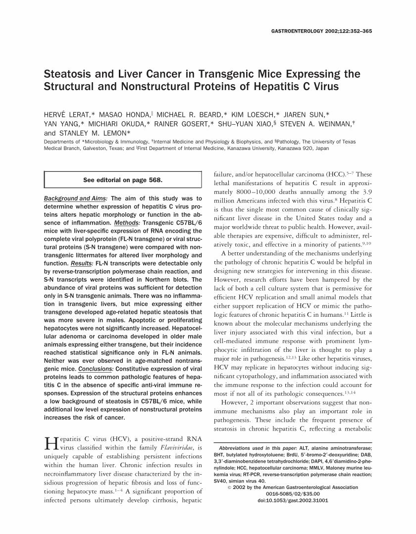
Steatosis and Liver Cancer in Transgenic Mice Expressing theStructural and Nonstructural Proteins of Hepatitis C Virus
HERVE LERAT,* MASAO HONDA,\ MICHAEL R. BEARD,* KIM LOESCH,* JIAREN SUN,*YAN YANG,* MICHIARI OKUDA,* RAINER GOSERT,* SHU–YUAN XIAO,§ STEVEN A. WEINMAN,‡
and STANLEY M. LEMON*Departments of *Microbiology & Immunology, ‡Internal Medicine and Physiology & Biophysics, and §Pathology, The University of TexasMedical Branch, Galveston, Texas; and \First Department of Internal Medicine, Kanazawa University, Kanazawa 920, Japan
See editorial on page 568.
Background and Aims: The aim of this study was todetermine whether expression of hepatitis C virus pro-teins alters hepatic morphology or function in the ab-sence of inflammation. Methods: Transgenic C57BL/6mice with liver-specific expression of RNA encoding thecomplete viral polyprotein (FL-N transgene) or viral struc-tural proteins (S-N transgene) were compared with non-transgenic littermates for altered liver morphology andfunction. Results: FL-N transcripts were detectable onlyby reverse-transcription polymerase chain reaction, andS-N transcripts were identified in Northern blots. Theabundance of viral proteins was sufficient for detectiononly in S-N transgenic animals. There was no inflamma-tion in transgenic livers, but mice expressing eithertransgene developed age-related hepatic steatosis thatwas more severe in males. Apoptotic or proliferatinghepatocytes were not significantly increased. Hepatocel-lular adenoma or carcinoma developed in older maleanimals expressing either transgene, but their incidencereached statistical significance only in FL-N animals.Neither was ever observed in age-matched nontrans-genic mice. Conclusions: Constitutive expression of viralproteins leads to common pathologic features of hepa-titis C in the absence of specific anti-viral immune re-sponses. Expression of the structural proteins enhancesa low background of steatosis in C57BL/6 mice, whileadditional low level expression of nonstructural proteinsincreases the risk of cancer.
Hepatitis C virus (HCV), a positive-strand RNAvirus classified within the family Flaviviridae, is
uniquely capable of establishing persistent infectionswithin the human liver. Chronic infection results innecroinflammatory liver disease characterized by the in-sidious progression of hepatic fibrosis and loss of func-tioning hepatocyte mass.1–4 A significant proportion ofinfected persons ultimately develop cirrhosis, hepatic
failure, and/or hepatocellular carcinoma (HCC).5–7 Theselethal manifestations of hepatitis C result in approxi-mately 8000–10,000 deaths annually among the 3.9million Americans infected with this virus.8 Hepatitis Cis thus the single most common cause of clinically sig-nificant liver disease in the United States today and amajor worldwide threat to public health. However, avail-able therapies are expensive, difficult to administer, rel-atively toxic, and effective in a minority of patients.9,10
A better understanding of the mechanisms underlyingthe pathology of chronic hepatitis C would be helpful indesigning new strategies for intervening in this disease.However, research efforts have been hampered by thelack of both a cell culture system that is permissive forefficient HCV replication and small animal models thateither support replication of HCV or mimic the patho-logic features of chronic hepatitis C in humans.11 Little isknown about the molecular mechanisms underlying theliver injury associated with this viral infection, but acell-mediated immune response with prominent lym-phocytic infiltration of the liver is thought to play amajor role in pathogenesis.12,13 Like other hepatitis viruses,HCV may replicate in hepatocytes without inducing sig-nificant cytopathology, and inflammation associated withthe immune response to the infection could account formost if not all of its pathologic consequences.13,14
However, 2 important observations suggest that non-immune mechanisms also play an important role inpathogenesis. These include the frequent presence ofsteatosis in chronic hepatitis C, reflecting a metabolic
Abbreviations used in this paper: ALT, alanine aminotransferase;BHT, butylated hydroxytoluene; BrdU, 5*-bromo-2*-deoxyuridine; DAB,3,3*-diaminobenzidene tetrahydrochloride; DAPI, 4,6*diamidino-2-phe-nylindole; HCC, hepatocellular carcinoma; MMLV, Maloney murine leu-kemia virus; RT-PCR, reverse-transcription polymerase chain reaction;SV40, simian virus 40.
© 2002 by the American Gastroenterological Association0016-5085/02/$35.00
doi:10.1053/gast.2002.31001
GASTROENTEROLOGY 2002;122:352–365

abnormality that is not observed as often in other in-flammatory conditions, such as autoimmune hepatitis orchronic hepatitis B.15–19 Some lines of evidence alsosuggest a distinctive synergy between the liver injuryassociated with chronic HCV infection and that accom-panying alcohol ingestion.20,21 This suggests that HCVinfection uniquely exacerbates alcohol-related metabolicdisturbances.
In addition to these clinical observations, a consider-able number of in vitro studies have suggested that theexpression of various HCV proteins in cultured cells maylead to altered lipid metabolism and transport, dysregu-lation of the cell cycle, either increased or decreasedsusceptibility to apoptosis, and cellular transforma-tion.22–27 However, few data convincingly support a rolefor any of these effects in human disease. In part, thisabsence of data stems from the lack of suitable animalmodels. The chimpanzee (Pan troglodytes) is the onlyanimal species that is clearly permissive for HCV infec-tion.28,29 However, unlike humans, infected chimpanzeestypically develop minimal liver disease as a result of viralreplication.
Given the absence of alternative small animal models,transgenic mice represent a potentially useful model fordetermining the long-term consequences of HCV pro-tein expression in vivo. Here, we describe in detail 2transgenic mouse lineages that express HCV proteinsunder control of the liver-specific murine albumin pro-moter/enhancer.30 Because mice can be expected to beimmunologically tolerant to transgenic proteins that aretranscriptionally regulated by this promoter, this exper-imental strategy offers the advantage of providing infor-mation on the direct, physiologic impact of HCV proteinexpression in the absence of confounding factors associ-ated with immunologic responses to the viral proteins.The transgenic animals described in this report expressRNAs encoding either the full-length HCV polyproteinor the structural proteins of a genotype 1b HCV,31,32
including the core, and the E1 and E2-p7 envelopeproteins. The phenotypes of these transgenic lineagesprovide strong evidence for nonimmune mechanisms ofliver injury in chronic hepatitis C and suggest an impor-tant role for the nonstructural proteins of HCC in hep-atocellular carcinogenesis.
Materials and MethodsProduction of Transgenic Mice
The plasmid pGEMAlbSVPA-B1(2) contains the mu-rine albumin enhancer/promoter30 with a downstream SV40intron/polyadenylation cassette. Complementary DNA (cDNA)corresponding to the full-length polyprotein-coding region
(core to NS5B, nts 342–9378) or the structural protein-codingregion only (core to E2/p7, nts 342–2771) of the genotype 1b,HCV-N strain of HCV31,32 was inserted into this vector be-tween newly engineered XhoI sites flanking the albuminenhancer/promoter and SV40 sequence, creating pAlbSVPA-HCV and pAlbSVPA-HCV-S, respectively. Purified plasmidDNA (transcriptional units only) was injected into the eggs ofF2 hybrid zygotes generated from mating F1 hybrid male andfemale mice (C3H/HeJ 3 C57BL/6). Offspring were screenedfor the presence of the transgene by Southern analysis of DNAextracted from tail biopsies after digestion with HindIII. DNAfrom 15 of 55 potential founder (F0) animals obtained afterinjection of the full-length FL-N construct (pAlbSVPA-HCV),and 12 of 38 potential F0 animals obtained after injection ofthe structural protein construct S-N (pAlbSVPA-HCV-S) con-tained the cognate transgene.
Transgenic F0 founder animals were mated with normalC57BL/6 mice to produce F1 and subsequent generations ofanimals. Mice were housed in a controlled, specific, pathogen-free environment, 4 per microfiltered cage, except when ani-mals were mating. Animals were fed regular sterile mousechow and water. All procedures involving animals were ap-proved by the Institutional Animal Care and Use Committee.The preliminary pathologic findings (steatosis and HCC) inone of these transgenic lineages (FL-N/35) have been reportedelsewhere,33 but a detailed comparison of the frequency ofthese findings in transgenic and nontransgenic animals has notbeen published.
Transgene mRNA Detection
Total RNA was extracted from approximately 100 mgof liver tissue pulverized in liquid nitrogen using a phenol-freetotal RNA isolation method (RNAqueous; Ambion, Austin,TX). For Northern blots, total RNA was fractionated in anagarose gel and passively transferred onto positively chargednylon membranes (BrightStar; Ambion) following the North-ern MAX (Ambion) protocol. RNA was cross-linked to themembranes by ultraviolet (UV) irradiation at 254 nm (UVStratalinker; Stratagene, La Jolla, CA). Hybridization of pso-ralen-biotin–conjugated DNA probes complementary to nts407–860 of HCV-N and subsequent visualization of the blotwith a streptavidin–alkaline phosphatase conjugate were car-ried out according to the BrightStar Biodetect NonisotopicDetection Kit protocol (Ambion). After washing, membraneswere incubated with CDP-Star chemiluminescent substrate(Ambion) and exposed to film.
For reverse-transcription polymerase chain reaction (RT-PCR), total RNA was purified by 2 cycles of RQ1 DNasedigestion, each followed by LiCl precipitation of RNA. Ninesets of oligonucleotide primers were used to amplify segmentsof the HCV genome spanning the entire open reading frame.RT was carried out with MMLV reverse transcriptase using asingle-tube, 2-compartment RT-PCR procedure (Clontech,Palo Alto, CA).34 Parallel PCR reactions were performed with-out a prior RT step to confirm that the template for PCRamplification products was RNA. PCR products were sepa-
February 2002 LIVER INJURY IN HEPATITIS C TRANSGENIC MICE 353
rated in agarose gels and passively transferred to BrightStarnylon membranes before hybridization to HCV-specific DNAprobes.
Necropsy and Histopathology Procedures
Animals were killed by CO2 asphyxiation, and livertissue was immediately snap-frozen in liquid nitrogen forRNA isolation and placed in 10% formalin or OCT cryostatembedding compound (Tissue-Tek, Torrance, CA) for histo-logic analyses or stored in 5 mmol/L butylated hydroxytoluene(BHT) for lipid peroxide determinations. Venous blood wasstored frozen for subsequent automated alanine aminotransfer-ase (ALT) measurements.
H&E- and trichrome-stained, paraffin-embedded liver sec-tions were examined by a pathologist (S.-Y.X.) who wasblinded to the status of the animal. The degree of steatosis wasestimated from digitized images of frozen liver sections stainedwith oil red O. These images were acquired with a NikonDiaphot inverted microscope using a blue filter to enhancecontrast and an 8-bit CCD camera (Hamamatsu Instruments,Japan). The percentage of the area occupied by oil red O–stained lipid droplets was calculated using Image-1 software(Universal Imaging Co., West Chester, PA), averaging 3–5separate, randomly selected 403 fields.
Immunohistochemical Detection ofViral Antigen
HCV core protein was detected by immunohistochem-ical analysis of formalin-fixed transgenic liver tissue sections.Endogenous peroxidase activity was blocked by immersingsections in 3% H2O2 in phosphate-buffered saline with 0.1%Tween 20 for 15 minutes, followed by antigen retrieval with0.3% pepsin in 0.01 mol/L HCl at 37°C for 15 minutes. Amurine monoclonal antibody to the HCV capsid protein,27D5G5 (generously provided by G. Inchauspe of INSERMUnite 271, Lyon, France) was used as the primary antibody ata dilution of 1:100, followed by incubation with a biotinylatedgoat anti-mouse immunoglobulin as the secondary antibody(Dako, Carpinteria, CA). Slides were incubated with strepta-vidin–horseradish peroxidase conjugate, followed by 3-amino-9-ethylcarbazole as substrate (Dako), and counterstained withhematoxylin before mounting.
For detection of the E2 protein, frozen sections of the liverwere fixed in acetone, blocked with normal goat serum andavidin/biotin (Avidin/Biotin Blocking kit; Vector Laborato-ries, Burlingame, CA), and incubated at 4°C overnight with a1:500 dilution of a rabbit antibody raised to a peptide identicalin sequence to the hypervariable region 1 of the E2 protein ofHCV-N (S. M. Lemon, unpublished observations, January,1997). Specificity was confirmed by the parallel staining oftissues with a rabbit antibody specific for a salivary protein ofthe sand fly, Lutzomyia longipalpis. Endogenous peroxidase ac-tivity was depleted with 1% H2O2 and 0.1% sodium azide,and sections were incubated with biotinylated goat anti-rabbitimmunoglobulin (Ig) G (1:200) at room temperature for 2
hours. Following incubation with avidin-conjugated peroxi-dase (Vectastain Elite ABC; Vector Laboratories), and develop-ment in 3,39-diaminobenzidine tetrahydrochloride (DAB) with0.1% H2O2, nuclei were counterstained with hematoxylin.
Apoptosis Assay
In situ detection of DNA fragmentation was carriedout using a fluorescence-based assay (Fluorescein-FragEL, On-cogene Research Products, Boston, MA). Frozen liver sections(5 mm) were fixed in 4% formaldehyde and rehydrated in 20mmol/L Tris-HCL, pH 7.6, 140 mmol/L NaCl. Sections weredigested with proteinase K (20 mg/mL) for 10 minutes andincubated with terminal deoxynucleotide transferase and flu-orescein-labeled nucleoside triphosphates at 37°C for 90 min-utes, then washed in buffer and placed under coverslips inmounting fluid containing 4,6-diamidino-2-phenylindole(DAPI) for counterstaining of nuclei. Slides were examined atan excitation wavelength of 465–495 or 330–380 nmol/L.Numbers of apoptotic nuclei were counted in each of 10randomly selected 403 fields.
Immunocytochemical Detection ofProliferating Hepatocytes
5-Bromo-29-deoxyuridine (BrdU) was administered tomice by intraperitoneal injection (100 mg/kg) 5 hours beforenecropsy. The liver and small intestines (the latter included asa positive control containing S-phase epithelial cells) wereremoved at necropsy and fixed in 10% neutral buffered for-malin for paraffin embedding and preparation of sections.Endogenous peroxidase activity was blocked by immersingsections in 0.3% H2O2 in methanol for 30 minutes, followedby digestion with 0.05% pepsin in 0.01N HCl. After incu-bation in 2N HCl for 30 minutes at 37°C, slides were washedfor 10 minutes in 0.1 mol/L borax buffer (pH 8.5) and murinemonoclonal anti-BrdU IgG (Becton Dickinson, FranklinLakes, NJ) applied at a 1:500 dilution overnight at 4°C. Afterfurther washing, slides were incubated with a biotinylatedF(ab9)2 fragment of sheep anti-mouse IgG (1:100 dilution) for1 hour, followed by DAB as chromagen. Sections were scoredfor the number of cells incorporating BrdU as described forapoptosis.
Intrahepatic Lipid Peroxides
Malonaldehyde and 4-hydroxynonenal concentrationswere determined in liver tissue homogenates by a colorimetricassay (LPO-586; OXIS International, Portland, OR). The pro-tein content of the homogenates was determined using aCoomassie blue assay (Pierce, Rockford, IL).
ResultsTransgene Expression in FL-N and S-NTransgenic Mice
Transgenic mice were designed to express eitherthe full-length HCV-N open reading frame (FL-N) or
354 LERAT ET AL. GASTROENTEROLOGY Vol. 122, No. 2
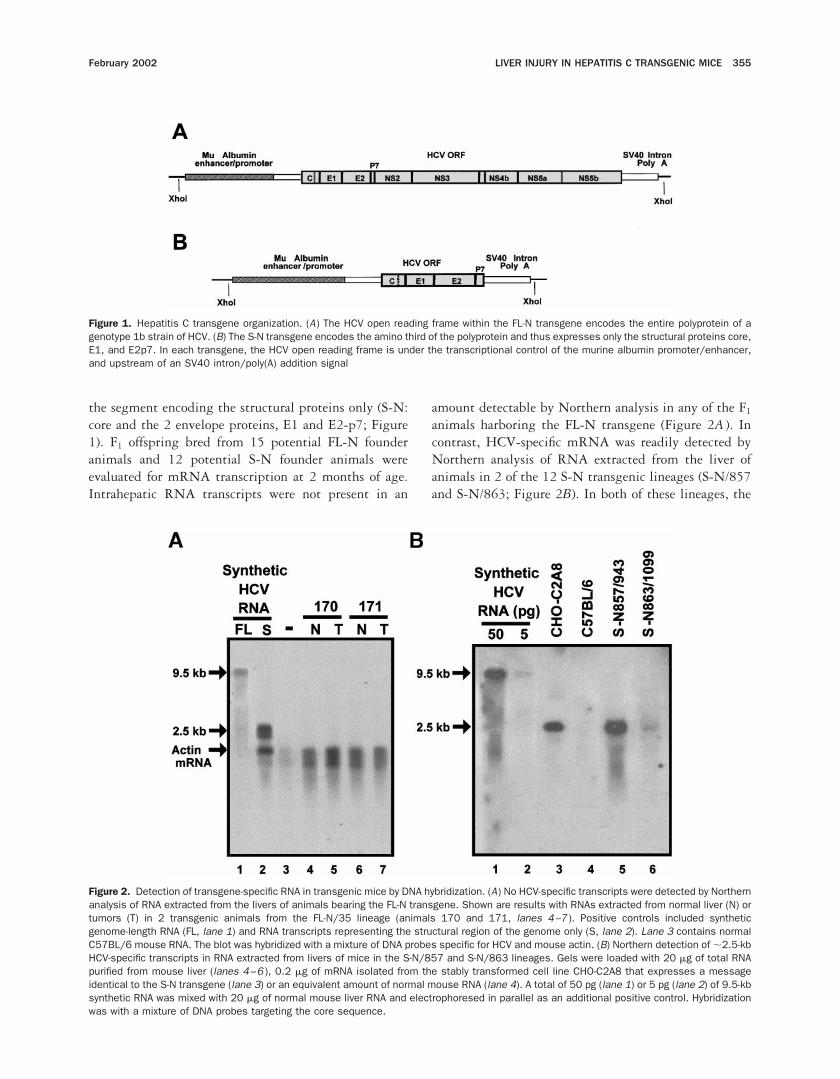
the segment encoding the structural proteins only (S-N:core and the 2 envelope proteins, E1 and E2-p7; Figure1). F1 offspring bred from 15 potential FL-N founderanimals and 12 potential S-N founder animals wereevaluated for mRNA transcription at 2 months of age.Intrahepatic RNA transcripts were not present in an
amount detectable by Northern analysis in any of the F1
animals harboring the FL-N transgene (Figure 2A ). Incontrast, HCV-specific mRNA was readily detected byNorthern analysis of RNA extracted from the liver ofanimals in 2 of the 12 S-N transgenic lineages (S-N/857and S-N/863; Figure 2B). In both of these lineages, the
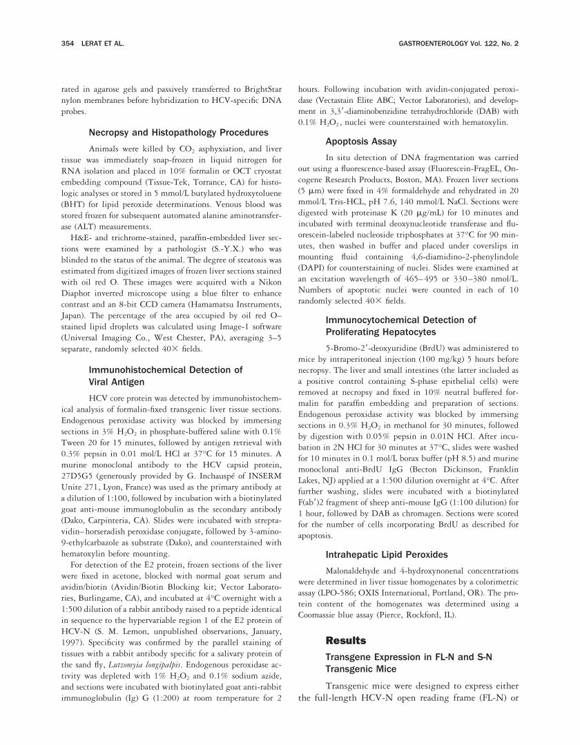
Figure 1. Hepatitis C transgene organization. (A) The HCV open reading frame within the FL-N transgene encodes the entire polyprotein of agenotype 1b strain of HCV. (B) The S-N transgene encodes the amino third of the polyprotein and thus expresses only the structural proteins core,E1, and E2p7. In each transgene, the HCV open reading frame is under the transcriptional control of the murine albumin promoter/enhancer,and upstream of an SV40 intron/poly(A) addition signal
Figure 2. Detection of transgene-specific RNA in transgenic mice by DNA hybridization. (A) No HCV-specific transcripts were detected by Northernanalysis of RNA extracted from the livers of animals bearing the FL-N transgene. Shown are results with RNAs extracted from normal liver (N) ortumors (T) in 2 transgenic animals from the FL-N/35 lineage (animals 170 and 171, lanes 4–7 ). Positive controls included syntheticgenome-length RNA (FL, lane 1) and RNA transcripts representing the structural region of the genome only (S, lane 2). Lane 3 contains normalC57BL/6 mouse RNA. The blot was hybridized with a mixture of DNA probes specific for HCV and mouse actin. (B) Northern detection of ;2.5-kbHCV-specific transcripts in RNA extracted from livers of mice in the S-N/857 and S-N/863 lineages. Gels were loaded with 20 mg of total RNApurified from mouse liver (lanes 4–6 ), 0.2 mg of mRNA isolated from the stably transformed cell line CHO-C2A8 that expresses a messageidentical to the S-N transgene (lane 3) or an equivalent amount of normal mouse RNA (lane 4). A total of 50 pg (lane 1) or 5 pg (lane 2) of 9.5-kbsynthetic RNA was mixed with 20 mg of normal mouse liver RNA and electrophoresed in parallel as an additional positive control. Hybridizationwas with a mixture of DNA probes targeting the core sequence.
February 2002 LIVER INJURY IN HEPATITIS C TRANSGENIC MICE 355

dominant RNA band was of the expected length (;2.5kb), and identical in size to transcripts present in a stabletransformed cell line expressing the identical HCV se-quence (CHO-C2A8 cells).26 Thus, there was no signif-icant splicing of transcripts expressed from the S-Ntransgene.
Although Northern analyses of the FL-N lineages didnot show HCV-specific RNAs, transcription of the trans-gene was confirmed in 4 of these lineages by RT-PCRusing primers designed to amplify the core-E1 region(Table 1). To determine whether a full-length transcriptwas produced, 9 separate PCR reactions were carried outusing primer sets designed to span the entire HCV
polyprotein-coding region and template cDNA producedby RT of RNA from animals in the FL-N/35 lineage(Figure 3). A specific amplification product of the ex-pected size was generated in each reaction, but only afterRT of the RNA. Although these results do not excludethe possibility that a proportion of the transcripts areinappropriately spliced, they provide compelling evi-dence for the presence of full-length RNA encoding theentire polyprotein in these animals. In addition to the 2S-N lineages in which RNA was detectable by Northernanalysis (Figure 2B), liver samples from mice in 3 otherS-N lineages also contained HCV-specific RNA detect-able by RT-PCR (Table 1).
Table 1. Summary of HCV Transgenic Mouse Lineages
Transgene andlineage
Transgene mRNA expression
Transgenic animalsevaluated (n)
Observed histopathology
Northernanalysis RT-PCR
Hepaticsteatosisa
Hepatocellularcarcinoma
Full-length ORFFL-N/35 2 1 73 1 1FL-N/984 2 1 1 ND 1FL-N/986 2 1 10 1 2FL-N/964 2 1 5 ND 2
Structural proteinsS-N/863 1 1 89 1 1S-N/857 111 1 4 ND 2S-N/866 2 1 2 ND 1S-N/883 2 1 1 ND 1S-N/888 2 1 3 ND 2
aND, not determined, insufficient numbers of animals available.
Figure 3. HCV-specific transcripts span the entire polyprotein-coding region in livers of mice from the FL-N/35 lineage. RT-PCR was carried outwith 9 nested pairs of oligonucleotide primers amplifying genome segments spanning the entire polyprotein-coding region. The template materialwas 1 mg of total RNA that had been subjected to 2 cycles of DNAse digestion followed by precipitation with lithium chloride to remove cellularDNA. A schematic showing the organization of the polyprotein-coding region is shown at the bottom of the figure. Above it are depicted the cDNAsegments amplified by individual primer pairs, the 59 map location of primers (Table 1), and the length of the expected PCR amplimer. Above eachis the result of DNA hybridizations of Southern blots of PCR amplimers obtained with RNA from livers of transgenic (transgene1) or nontransgenic(transgene2) mice. Reactions were run with (reverse transcriptase1) or without (reverse transcriptase2) a prior RT reaction to show thatHCV-specific sequences were amplified from RNA.
356 LERAT ET AL. GASTROENTEROLOGY Vol. 122, No. 2

HCV Protein Expression
We used the core and E2 proteins as markers forprotein expression since both are encoded by the FL-Nand S-N transgenes (Figure 1). The immunohistochem-ical detection of core protein in formalin-fixed, trans-genic liver tissue is shown in Figure 4A and B. Cyto-plasmic staining for core antigen was prominent inpericentral hepatocytes in liver from the S-N/857 lineage(Figure 4B). However, the core protein was not expressedat detectable levels in animals from the S-N/863 lineage,which showed a lower abundance of S-N transcripts thanthe S-N/857 lineage as determined by Northern analysis(Figure 2B). Similarly, core protein could not be detectedin liver tissues from the FL-N lineages (data not shown)in which a low abundance of the full-length open readingframe message was detectable only by RT-PCR (Figures2A and 3). The presence of a detectable abundance of the
;21-kilodalton core protein in the S-N/857 lineage, andits absence in the other lineages, was confirmed byimmunoblot analysis of liver tissue homogenates. Inparallel immunoblots of human tissue, the core proteinwas detectable in only 1 of 3 patients with chronichepatitis C (data not shown).
Detection of the E2 protein proved a more sensitiveindicator of protein expression. This protein was readilydetectable by immunohistochemical techniques whenfrozen sections of livers from animals in both the S-N/857 and S-N/863 lineages were stained with a rabbitantiserum raised to a synthetic peptide representing thehypervariable region of the HCV-N E2 protein (Figure4C and E ). As with core protein in the S-N/857 lineage,E2 was cytoplasmic in distribution (Figure 4E ) andparticularly prominent in pericentral hepatocytes (Figure4F ). Parallel staining of these tissues with a rabbit
Figure 4. Immunohistochemical detection ofHCV structural proteins in livers of mice fromS-N and FL-N transgenic lineages. (A, B ) Im-munoperoxidase detection of HCV core pro-tein in formalin-fixed sections of mouse liverstained with a murine monoclonal antibodyto the HCV core protein. (A) Nontransgenicmouse liver showing no expression of coreprotein (203 objective); (B) liver from a trans-genic animal in the S-N/857 lineage, show-ing pericentral distribution of cytoplasmicstaining for core antigen in hepatocytes (203objective). (C–F ). Detection of E2 protein inliver of transgenic mice. Liver samples werecryosectioned and stained with a rabbit anti-body specific for the E2 protein of HCV-N,followed by a biotinylated secondary anti-body and avidin-conjugated peroxidase. Nu-clei were counterstained with Gill hematoxy-lin. (C) Nontransgenic mouse liver (403objective); (D) transgenic liver from the FL-N/35 lineage (403 objective); (E) transgenicliver from the S-N/863 lineage (403 objec-tive); (F ) low power view of transgenic liverfrom the S-N/863 lineage (103 objective)showing pericentral distribution of the cyto-plasmic E2 antigen. Parallel staining usingrabbit antibody specific for an irrelevant anti-gen (a peptide derived from a salivary proteinof sand fly Lutzomyia longipalpis) showed nopositively stained cells in the livers of non-transgenic and transgenic mice (data notshown).
February 2002 LIVER INJURY IN HEPATITIS C TRANSGENIC MICE 357
antibody specific for an irrelevant antigen (a peptidederived from a salivary protein of the sand fly Lutzomyialongipalpis) revealed no positively stained cells in thelivers of either nontransgenic or transgenic mice (datanot shown), confirming the specificity of the antigendetection in the S-N/857 and S-N/863 tissues. In con-trast, although faint staining may have been present,liver sections from animals in the FL-N/35 lineage didnot contain clearly demonstrable amounts of E2 protein(Figure 4D). Similar results were obtained with indirectimmunofluorescence staining of tissue sections (data notshown). Overall, there was good correlation betweentransgene mRNA expression levels and the detection ofviral proteins (Table 2).
Phenotypes of FL-N and S-NTransgenic Mice
Of the 9 transgenic lineages with evidence ofRNA transcription listed in Table 1, only 2 provedcapable of breeding in large numbers (FL-N/35 andS-N/863). The frequency of transmission of the transgeneto F1 and F2 offspring in both lineages approximated50%, consistent with the results of Southern analysis oftail biopsy DNA, which suggested a single, unique siteof transgene integration in each (data not shown).
In general, the transgenic mice appeared normal anddid not have demonstrable deficits in growth or devel-opment. No cellular inflammatory infiltrate was evidentin the livers of these mice, and there were no significantdifferences in the serum ALT activities of transgenicanimals and their nontransgenic littermates (data notshown). However, hepatic steatosis was frequentlypresent in older transgenic animals from the FL-N/35,FL-N/963, and S-N/863 lineages, and transgenic animalsin the FL-N/35 and S-N/863 lineages developed HCC.Similar pathology, including HCC, was present in trans-genic animals from other lineages, but the frequency ofliver disease in these lineages could not be determinedbecause of the limited numbers of animals available(Table 1).
Hepatic Steatosis
In contrast to a modest degree of steatosis notedoccasionally in normal aging C57BL/6 mice, a substan-tial proportion of transgenic mice from the FL-N/35 andS-N/863 lineages developed moderate to severe steatosis(Figure 5A and B). This was characterized by a typicalvacuolated appearance of lipid-laden hepatocytes inH&E-stained sections of the liver (Figure 5C ), and con-firmed by the demonstration of increased intrahepaticlipid on oil red O staining of frozen sections (Figure 5D).Steatosis was primarily centrilobular (Figure 5B), andeither microvesicular or mixed microvesicular/macro-vesicular (Figure 5C ).
Steatosis increased with age in the transgenic animalsand was rare before age 10 months in either lineage.Digitized images of oil red O–stained liver sections wereanalyzed to quantify the degree of steatosis, with thepercent area occupied by oil red O–stained fat dropletsestimated in individual liver sections. The area occupiedwith fat was significantly greater in FL-N/35 transgenicanimals 9–15 months of age than in age-matched, non-transgenic mice (P , 0.02 by Student t test; Figure 5E ).Steatosis was significantly more prominent in transgenicmales (19.3% 6 4.4% SEM) than females (8.4% 61.1%; P , 0.05). A comparable degree of steatosis wasdocumented in a small number of similarly aged trans-genic animals from a second, independent FL-N lineage,FL-N/986, and in 12–16-month-old transgenic animalsfrom the S-N/863 lineage (Figure 5E ). In the latterlineage, the area occupied by oil red O–stained lipiddroplets in transgenic animals significantly exceeded thatin sections from age-matched, nontransgenic littermates(P , 0.02). Although modest staining with oil red Owas occasionally present in nontransgenic male mice(6.9% 6 2.3%), it occupied relatively little of the overallarea of the sections from nontransgenic females (3.5% 62.8%; P 5 NS). Because Southern blots indicated thatthe transgene insertion sites are different in these 3lineages (data not shown), steatosis did not result fromfortuitous insertional inactivation of a mouse gene. Itsoccurrence in the S-N/863 lineage indicates that it can be
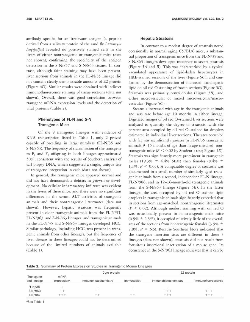
Table 2. Summary of Protein Expression Studies in Transgenic Mouse Lineages
Transgeneand lineage
mRNAexpressiona
Core protein E2 protein
Immunohistochemistry Immunoblot Immunohistochemistry Immunofluorescence
FL-N/35 1 2 2 2 2S-N/863 11 2 2 111 111S-N/857 111 11 11 111 111
aSee Table 1.
358 LERAT ET AL. GASTROENTEROLOGY Vol. 122, No. 2

Figure 5. Hepatic steatosis in FL-N/35 lineage mice. (A) Nontransgenic male, age 12 months, H&E-stained paraffin section showing normal liverhistology (103 objective). (B ) Transgenic male, age 13 months, H&E-stained paraffin section (103 objective). Note marked steatosis presentdiffusely in zones 2 and 3. (C) Higher magnification of the pericentral region of the same liver as shown in B (403 objective) showing mixedmacrovesicular and microvesicular characteristics of the steatosis. (D) Transgenic male, age 11 months, oil red O–stained frozen section (103objective) showing darkly stained lipid droplets in zones 2 and 3. (E) Quantitative analysis of digitized images of oil red O–stained frozen liversections. For each liver, the mean percent area occupied by oil red O–stained lipid droplets was calculated for 3–5 randomly selected fields.Results shown represent the means for transgenic (shaded bars) and nontransgenic (open bars) animals in 3 different lineages: FL-N/35,FL-N/963 (no nontransgenic animals), and S-N/863. For the FL-N/35 lineage, 15 transgenic animals aged 9–15 months (mean age, 12.1months; 67% males) were compared with 13 nontransgenic animals of comparable age and sex (mean age, 11.3 months; 69% males) from thecolony (P , 0.05 by 2-sided t test). Results for the FL-N/963 lineage were restricted to 4 transgenic animals, 3 females and 1 male (mean age,12.3 months). For the S-N/863 lineage, 9 transgenic animals aged 13–16 months (mean age, 14.6 months; 67% males) were compared with6 comparably aged nontransgenic littermates (mean age, 14.7 months; 100% males; P , 0.02 by t test). Error bars indicate SDs.
February 2002 LIVER INJURY IN HEPATITIS C TRANSGENIC MICE 359

related to the expression of one or more of the structuralproteins.
Although centrilobular microvesicular steatosis canresult from mitochondrial injury and predispose to in-creased lipid peroxidation,35 there were no significantdifferences in the basal level of lipid peroxides in non-transgenic animals (51.1 6 4.5 SEM pmol/mg proteinover the age of 8 months, n 5 24) and in similarly agedFL-N/35 transgenic (55.6 6 8.9 pmol/mg, n 5 15) orS-N/863 transgenic animals (38.9 6 8.9 pmol/mg,n 5 15).
Hepatic Fibrosis
Fibrosis is a characteristic pathologic feature ofchronic hepatitis C.18,35 However, there was no fibrosis inthese mouse livers in the absence of cancer. HCC tissuesfrom transgenic animals contained fibrosis evident intrichrome stains (data not shown), as well as prominentactivation of stellate cells detectable by immunostainingwith a monoclonal antibody specific for a-smooth muscleactin36 (data not shown). Cells staining specifically fora-smooth muscle actin were present only occasionally innon-tumor tissue from these animals, or in transgenicanimals without tumors.
Hepatocellular Proliferation and Apoptosis
Because in vitro studies suggest that HCV pro-teins may alter cell signaling and regulate apoptoticpathways,23–26 we compared transgenic mice from bothlineages with their age-matched nontransgenic litter-mates for evidence of increased or decreased numbers ofapoptotic hepatocytes. No consistent differences wereapparent. We also compared basal hepatocyte prolifera-tion rates in 9–11-month-old transgenic and nontrans-genic mice by assessing the frequency of hepatocytesincorporating BrdU over a 5-hour period before necropsy(Figure 6). Although the highest numbers of proliferat-ing cells were observed in transgenic males from theFL-N/35 lineage (Figure 6A, 3.1 6 1.3 cells per 403field compared with 0.82 6 0.7 cells in nontransgenicmales from the lineage), this difference did not achievestatistical significance. Somewhat higher numbers of pro-liferating cells were also present in transgenic mice com-pared with nontransgenic mice in the S-N/863 lineage(Figure 6B), but this difference also did not achievestatistical significance.
Hepatocellular Carcinogenesis
Hepatic adenomas or HCC ranging from a fewmillimeters to more than 2 cm in diameter were presentat necropsy in 5 of 37 FL-N/35 transgenic mice and in 1
of 42 S-N/863 transgenic mice older than 13 months(Table 3). The morphology of the HCCs was variable,with some showing well-differentiated hepatic cordswith proliferating hepatocytes and others containingmarkedly thickened trabeculae or acini of dysplastichepatocytes with prominent sinusoidal spaces (Figure 7).Multiple tumors were identified in 2 transgenic animals.One liver with a typical carcinoma contained a separatemass with morphology consistent with focal nodularhyperplasia. Focal or diffuse steatosis was sometimesdisproportionately present in the adenomas or HCCs(Figure 7A and C ), but there was no apparent correlationbetween the presence or severity of steatosis and theoccurrence of HCC.
All of the tumors identified in transgenic animalsoccurred in males. Adenomas or HCC were present in 5of 17 male FL-N/35 transgenic mice older than 13months, a frequency that was significantly greater thanin age-matched female FL-N/35 transgenic animals (0 of20; P , 0.02 by Fisher exact test) or in nontransgenicmale mice (0 of 24; P , 0.01). In contrast, HCC wasfound in only 1 older transgenic male from the S-N/863lineage (Table 3). The frequency of tumors in older maleS-N/863 transgenic animals (1 of 24) was significantlyless than that in comparably aged males in the FL-N/35
Figure 6. Numbers of hepatocytes showing evidence of DNA synthe-sis as indicated by BrdU incorporation in 10 randomly selected 403fields of liver sections from transgenic and nontransgenic animals. (A)Numbers of hepatocyte nuclei showing BrdU incorporation in 8 FL-N/35 transgenic animals (mean age, 10.0 months) compared with anequal number of age-matched nontransgenic littermates (P 5 0.19).(B) Numbers of hepatocyte nuclei showing BrdU incorporation in 7S-N/863 transgenic animals (mean age, 10.6 months) compared with4 nontransgenic littermates (mean age, 10.7 months; P 5 0.12). Ineach panel, the mean numbers of positive cells are shown separatelyfor transgenic (shaded bars) and nontransgenic (open bars) litter-mates with values shown for individual male (F) and female (E) mice.Error bars represent SDs.
360 LERAT ET AL. GASTROENTEROLOGY Vol. 122, No. 2
lineage (P , 0.05), and not statistically different fromthe absence of adenomas or liver cancer in age-matchednontransgenic males. Because the structural proteins ofHCV were expressed at a higher level in the S-N/863than the FL-N/35 lineage (Figure 4), the greater fre-quency of cancer in comparably aged mice in the FL-N/35 lineage suggests that the nonstructural proteins of
HCV may contribute to hepatic carcinogenesis evenwhen they are expressed at levels below those detectableby immunohistochemistry. However, a relatively smallnumber of S-N/863 animals were available for necropsyat ages .18 months, and further observation is needed todetermine the risk of cancer in very old mice in thislineage.
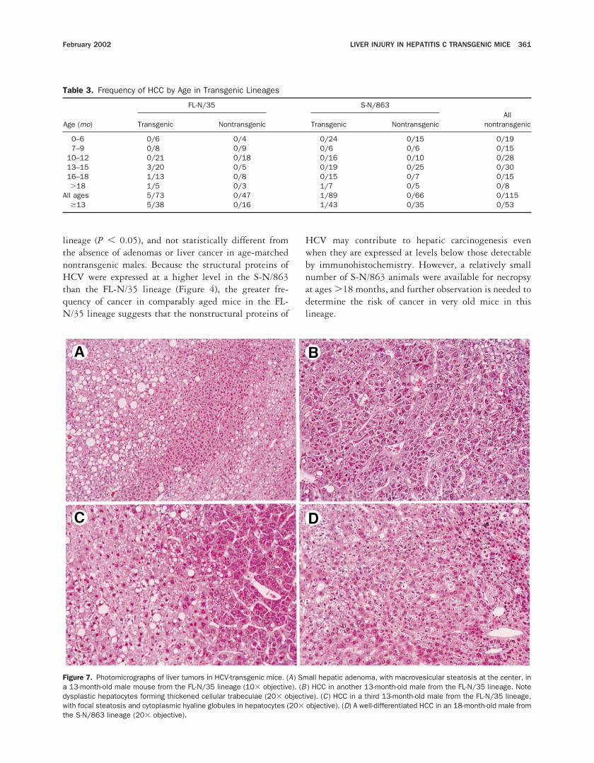
Figure 7. Photomicrographs of liver tumors in HCV-transgenic mice. (A) Small hepatic adenoma, with macrovesicular steatosis at the center, ina 13-month-old male mouse from the FL-N/35 lineage (103 objective). (B ) HCC in another 13-month-old male from the FL-N/35 lineage. Notedysplastic hepatocytes forming thickened cellular trabeculae (203 objective). (C) HCC in a third 13-month-old male from the FL-N/35 lineage,with focal steatosis and cytoplasmic hyaline globules in hepatocytes (203 objective). (D) A well-differentiated HCC in an 18-month-old male fromthe S-N/863 lineage (203 objective).
Table 3. Frequency of HCC by Age in Transgenic Lineages
Age (mo)
FL-N/35 S-N/863All
nontransgenicTransgenic Nontransgenic Transgenic Nontransgenic
0–6 0/6 0/4 0/24 0/15 0/197–9 0/8 0/9 0/6 0/6 0/15
10–12 0/21 0/18 0/16 0/10 0/2813–15 3/20 0/5 0/19 0/25 0/3016–18 1/13 0/8 0/15 0/7 0/15.18 1/5 0/3 1/7 0/5 0/8
All ages 5/73 0/47 1/89 0/66 0/115$13 5/38 0/16 1/43 0/35 0/53
February 2002 LIVER INJURY IN HEPATITIS C TRANSGENIC MICE 361

We examined tumors from the FL-N/35 lineage forevidence of transgene mRNA and antigen expression andfound results similar to those obtained with normaltransgenic tissue. Specific HCV mRNA was detectableby RT-PCR, and results of Northern analyses of thesetissues were repeatedly negative (Figure 2). Neither thecore nor the E2 antigens were present in amounts de-tectable by immunohistochemical techniques.
HCC was not restricted to the FL-N/35 and S-N/863lineages but also occurred in isolated transgenic animalsfrom other lineages (Table 1). The morphologies of thesetumors were similar to those described previously, andall occurred in older male animals (15–17 months old)from lineages with active transcription of the transgene.HCC was not observed in mice from transcriptionallyinactive lineages or in nontransgenic animals, includinga total of 115 nontransgenic littermates from the FL-N/35 and S-N/863 lineages (Table 3). These nontrans-genic littermates were caged with transgenic animals andreceived an identical diet.
DiscussionThe HCV-transgenic mice that we have derived
constitutively express low levels of RNA encoding thecomplete viral polyprotein, or higher levels of the RNAsegment encoding only the structural proteins of thevirus, under control of a liver-specific promoter. Proteinexpression was confirmed directly only in the mice ex-pressing the structural region of the polyprotein andwas insufficient for detection even with sensitive im-munohistochemical techniques in mice with the full-length reading frame as the transgene. Nonetheless, theseFL transgenic animals showed distinctive phenotypes,strongly suggesting the presence of a biologically rele-vant level of HCV protein expression. Both types oftransgenic mice showed no evidence of inflammation,indicating that they do not mount a significant cellularimmune response to expressed viral proteins. Thus, thepathologic findings in these animals, including steatosisin mice of the FL-N/35, FL-N/963, and S-N/863 lin-eages and the development of liver cancer in older maleFL-N/35 mice, arise as a direct result of the expression ofviral proteins. These findings suggest that similar non-immune mechanisms may contribute to the developmentof steatosis and liver cancer in humans with chronichepatitis C.
These HCV-transgenic mice are significantly differentfrom those described previously by others. AlthoughMoriya et al.37,38 also observed steatosis and HCC intransgenic mice, these animals expressed only the coreprotein, and not the other structural or nonstructural
proteins encoded by the FL-N and S-N transgenes. Thecore protein interacts with the envelope proteins39 andpossibly also one or more nonstructural proteins, andsuch interactions between viral proteins could signifi-cantly modulate their effects in vivo. This makes itimportant to study the activities of HCV proteins withinthe context of the entire viral polyprotein rather than inisolation. In addition, the mice described by Moriya etal.37,38 appear to express core protein in relatively largeamounts because it was readily detectable in immuno-blots. This contrasts with the absence of detectable ex-pression of HCV proteins in the FL-N/35 lineage and thefact that RNA transcripts could be detected in thislineage only by RT-PCR (Figure 3). RT-PCR is rou-tinely required to detect the low levels of HCV RNAtranscripts in most patients. Furthermore, viral proteinsare not present at amounts detectable in many patientswith chronic HCV infection. Although results have var-ied in different studies, 25%–75% of patients withchronic hepatitis C do not have levels of core antigendetectable in liver tissue even with sensitive assays usingmonoclonal antibodies.17,40–42 Thus, not only does theFL-N/35 lineage express RNA encoding the full com-plement of HCV proteins, it is also likely to express theviral proteins at levels mimicking the situation in manyinfected humans.
Despite the absence of detectable levels of proteinexpression, liver cancer developed as early as 13 monthsof age in FL-N/35 mice. In contrast, liver cancers did notoccur until 16 months in the core-transgenic mice de-scribed by Moriya et al.38 However, steatosis was prom-inent by age 6 months in the transgenic mice studied byMoriya et al.37 and was frequently present in females,whereas steatosis was rarely present before age 10 monthsin FL-N/35 animals and was infrequently seen in fe-males. This latter difference may reflect the higher levelof core protein expression in the mice studied by Moriyaet al., but it also suggests the absence of a direct linkbetween steatosis and carcinogenesis. It further suggeststhat HCV proteins other than core may promote thedevelopment of cancer. This conclusion is reinforced bythe low rate of cancer in 13–18-month-old S-N/863lineage animals compared with that observed in theFL-N/35 lineage (Table 1). Dysregulation of cell growthhas been associated with the expression of core in cul-tured cells,24–26,43 but potential roles in carcinogenesishave also been suggested for the NS3 and NS5A pro-teins.27,44 These proteins are encoded by the FL-N/35transgene, and the higher rate of cancer in this lineagesuggests that even very low-level expression of theseproteins may contribute to hepatocellular carcinogenesis.
362 LERAT ET AL. GASTROENTEROLOGY Vol. 122, No. 2

Further observation of the S-N/863 lineage is needed todetermine whether these transgenic mice are at increasedrisk for liver cancer at ages greater than 16–18 months.
Both the mice described by Moriya et al.37,38 and thosedescribed here were developed within a predominantlyC57BL/6 genetic background. This may be important tothe disease observed in these animals; 2 other groups ofinvestigators, Kawamura et al.45 and Pasquinelli et al.,46
found no pathology in transgenic mice expressing thecore protein of HCV in different genetic backgrounds.Others have noted that normal aging C57BL/6 mice areat risk for the development of mild steatosis,47 a phe-nomenon we confirmed in our animals (Figure 5E ). Thusit is possible that HCV gene expression may act toexacerbate an underlying metabolic abnormality inC57BL/6 mice that is not present in other inbred strainsof mice. If genetic background is proven to be an im-portant factor in determining disease expression in HCV-transgenic mice, it may provide useful clues to themechanisms underlying the variable risk of disease inpersons with chronic HCV infection.
Although it is not clear which HCV protein(s) pro-mote steatosis in the FL-N/35 mice, its occurrence in theS-N/863 lineage indicates the involvement of one ormore structural proteins. This is consistent with theoccurrence of steatosis in transgenic animals that expressonly high levels of the core protein.38 Core proteinphysically associates with cytoplasmic lipid dropletsthrough an interaction with apolipoprotein A2, and thismay alter normal lipid transport leading to steatosis.22,48
However, steatosis was at least as prominent in theFL-N/35 lineage as the S-N/863 lineage (Figure 5E ),despite the absence of a detectable abundance of HCVstructural proteins in the former (Figure 4D). This raisesthe possibility that very low-level expression of one of thenonstructural proteins encoded by the transgene in theFL-N/35 or FL-N/963 animals may have contributed tosteatosis. NS5A has recently been found to interact withapolipoprotein A1 and to colocalize with the core proteinon cytoplasmic lipid droplets (Stephanie T. Shi andMichael M. C. Lai, personal communication, April2001). Thus it is possible that this interaction furtherpromotes the accumulation of intracellular lipids in-duced by core.
Alternatively, core protein is capable of interactingwith the cytoplasmic domain of several members of thetumor necrosis factor receptor family.25,43 The conse-quences of this interaction are controversial but couldinclude constitutive activation of proapoptotic cell sig-naling, leading to a mitochondrial permeability transi-tion and subsequent oxidative stress.49,50 However, we
did not find increased rates of hepatocellular apoptosis inour mice. Yet a third possibility is that the core proteincould interact directly with mitochondria.38 We recentlyfound that the expression of core protein leads to demon-strable mitochondrial injury with attendant increases inthe abundance of reactive oxygen species in several typesof cultured cells.51 The centrilobular (zone 3) micro-vesicular steatosis observed in these mice could reflectsuch mitochondrial injury. As reported here, we found noincreases in the basal levels of lipid peroxides in the liversof mice from either the FL-N/35 or S-N/863 lineages.However, we have documented elevated levels of intra-hepatic lipid peroxidation products in transgenic S-N/863 mice after low-dose carbon tetrachloride exposure.51
These results suggest that these transgenic mice are atincreased risk of tissue damage in the face of oxidativestress. The accumulation of reactive oxygen species re-sulting from mitochondrial dysfunction could contributeto carcinogenesis by promoting chromosomal DNAdamage and altering the regulation of apoptosis.35 Inaddition to the core protein,52 the generation of intra-cellular reactive oxygen species has been linked recentlyto expression of the NS5A protein, providing yet anotherpotential explanation for the increase in hepatic carcino-genesis we observed with the inclusion of the HCVnonstructural proteins among those encoded by thetransgene in the FL-N/35 animals.
In addition to these potential nonimmune mecha-nisms of liver injury, chronic inflammation associatedwith the cellular immune response to HCV infection islikely to play an important role in the development ofHCC in humans. Inflammation would be expected toexacerbate oxidative stress induced by the expression ofviral proteins in an already vulnerable liver, potentiallyaccelerating the progression to hepatocellular carcino-genesis. Nonetheless, the results described here showthat the intrahepatic expression of HCV proteins directlypromotes the development of steatosis and significantlyenhances the risk of liver cancer in the absence of inflam-mation, and provide novel evidence for the involvementof the nonstructural proteins of HCV in these pathoge-netic processes.
References1. Choo Q-L, Kuo G, Weiner AJ, Overby LR, Bradley DW, Houghton M.
Isolation of a cDNA clone derived from a blood-borne non-A, non-Bviral hepatitis genome. Science 1989;244:359–362.
2. Alter MJ, Margolis HS, Krawczynski K, Judson FN, Mares A,Alexander WJ, Hu PY, Miller JK, Gerber MA, Sampliner RE, MeeksEL, Beach MJ. The natural history of community-acquired hepati-tis C in the United States. N Engl J Med 1992;327:1899–1905.
3. Seeff LB, Buskell-Bales Z, Wright EC, Durako SJ, Alter HJ, Iber FL,Hollinger FB, Gitnick G, Knodell RG, Perrillo RP, Stevens CE,Hollingsworth CG, NHLBI Study Group. Long-term mortality after
February 2002 LIVER INJURY IN HEPATITIS C TRANSGENIC MICE 363

transfusion-associated non-A, non-B hepatitis. N Engl J Med1992;327:1906–1911.
4. Seeff LB. Natural history of hepatitis C. Hepatology 1997;26:21S–28S.
5. Di Bisceglie AM, Simpson LH, Lotze MT, Hoofnagle JH. Develop-ment of hepatocellular carcinoma among patients with chronicliver disease due to hepatitis C viral infection. J Clin Gastroen-terol 1994;19:222–226.
6. Kiyosawa K, Furuta S. Hepatitis C virus and hepatocellular carci-noma. In: Reesink HW, ed. Hepatitis C virus. Basel, Switzerland:Karger, 1994:98–120.
7. Kiyosawa K, Sodeyama T, Tanaka E, Gibo Y, Yoshizawa K, Na-kano Y, Furata S, Akahane Y, Nishioka K, Purcell RH, Alter HJ. In-terrelationship of blood transfusion, non-A, non-B hepatitis andhepatocellular carcinoma: analysis by detection of antibody tohepatitis C virus. Hepatology 1990;12:671–675.
8. Alter MJ, Mast EE, Moyer LA, Margolis HS. Hepatitis C. Infect DisClin North Am 1998;12:13–26.
9. McHutchison JG, Gordon SC, Schiff ER, Shiffman ML, Lee WM,Rustgi VK, Goodman ZD, Ling MH, Cort S, Albrecht JK. Interferonalfa-2b alone or in combination with ribavirin as initial treatmentfor chronic hepatitis C. Hepatitis Interventional Therapy Group.N Engl J Med 1998;339:1485–1492.
10. Schalm SW, Hansen BE, Chemello L, Bellobuono A, Brouwer JT,Weiland O, Cavalletto L, Schvarcz R, Ideo G, Alberti A. Ribavirinenhances the efficacy but not the adverse effects of interferon inchronic hepatitis C—meta-analysis of individual patient data fromEuropean centers. J Hepatol 1997;26:961–966.
11. Lemon SM, Chisari FV, Lai MM, Nishioka K, Mishiro S, JohnsonL. The Nineteenth United States-Japan Joint Hepatitis PanelMeeting. Hepatology 1998;28:881–887.
12. Koziel MJ, Dudley D, Wong JT, Dienstag J, Houghton M, RalstonR, Walker BD. Intrahepatic cytotoxic T lymphocytes specific forhepatitis C virus in persons with chronic hepatitis. J Immunol1992;149:3339–3344.
13. Cerny A, Chisari FV. Pathogenesis of chronic hepatitis C: immu-nological features of hepatic injury and viral persistence. Hepa-tology 1999;30:595–601.
14. Nakamoto Y, Guidotti LG, Kuhlen CV, Fowler P, Chisari FV. Im-mune pathogenesis of hepatocellular carcinoma. J Exp Med1998;188:341–350.
15. Rubbia–Brandt L, Quadri R, Abid K, Giostra E, Male PJ, Mentha G,Spahr L, Zarski JP, Borisch B, Hadengue A, Negro F. Hepatocytesteatosis is a cytopathic effect of hepatitis C virus genotype 3.J Hepatol 2000;33:106–115.
16. Scheuer PJ, Davies SE, Dhillon AP. Histopathological aspects ofviral hepatitis. J Viral Hepat 1996;3:277–283.
17. Fujie H, Yotsuyanagi H, Moriya K, Shintani Y, Tsutsumi T,Takayama T, Makuuchi M, Matsuura Y, Miyamura T, Kimura S,Koike K. Steatosis and intrahepatic hepatitis C virus in chronichepatitis. J Med Virol 1999;59:141–145.
18. Goodman ZD, Ishak KG. Histopathology of hepatitis C virus in-fection. Semin Liver Dis 1995;15:70–81.
19. Fischer HP, Willsch E, Bierhoff E, Pfeifer U. Histopathologic find-ings in chronic hepatitis C. J Hepatol 1996;24:35–42.
20. Schiff ER. Hepatitis C and alcohol. Hepatology 1997;26:39S–42S.
21. Thomas DL, Astemborski J, Rai RM, Anania FA, Schaeffer M,Galai N, Nolt K, Nelson KE, Strathdee SA, Johnson L, Laeyen-decker O, Boitnott J, Wilson LE, Vlahov D. The natural history ofhepatitis C virus infection: host, viral, and environmental factors.JAMA 2000;284:450–456.
22. Barba G, Harper F, Harada T, Kohara M, Goulinet S, Matsuura Y,Eder G, Schaff Z, Chapman MJ, Miyamura T, Brechot C. HepatitisC virus core protein shows a cytoplasmic localization and asso-ciates to cellular lipid storage droplets. Proc Natl Acad Sci U S A1997;94:1200–1205.
23. Ray RB, Lagging LM, Meyer K, Ray R. Hepatitis C virus coreprotein cooperates with ras and transforms primary rat embryofibroblasts to tumorigenic phenotype. J Virol 1996;70:4438–4443.
24. Ray RB, Meyer K, Ray R. Suppression of apoptotic cell death byhepatitis C virus core protein. Virology 1996;226:176–182.
25. Zhu N, Khoshnan A, Schneider R, Matsumoto M, Dennert G,Ware C, Lai MM. Hepatitis C virus core protein binds to thecytoplasmic domain of tumor necrosis factor (TNF) receptor 1 andenhances TNF-induced apoptosis. J Virol 1998;72:3691–3697.
26. Honda M, Kaneko S, Shimazaki T, Matsushita E, Kobayashi K,Ping LH, Zhang HC, Lemon SM. Hepatitis C virus core proteininduces apoptosis and impairs cell-cycle regulation in stablytransformed Chinese hamster ovary cells. Hepatology 2000;31:1351–1359.
27. Sakamuro D, Furukawa T, Takegami T. Hepatitis C virus nonstruc-tural protein NS3 transforms NIH 3T3 cells. J Virol 1995;69:3893–3896.
28. Alter HJ, Holland PV, Purcell RH, Popper H. Transmissible agentin non-A, non-B hepatitis. Lancet 1978;1:459–463.
29. Tabor E, Gerety RJ, Drucker JA, Seeff LB, Hoofnagle JH, JacksonDR, April M, Barker LF, Pineda-Tamondong G. Transmission ofnon-A, non-B hepatitis from man to chimpanzee. Lancet 1978;1:463–466.
30. McPherson CE, Shim E-Y, Friedman DS, Zaret KS. An active,tissue-specific enhancer and bound transcription factors existingin a precisely positioned nucleosomal array. Cell 1993;75:387–398.
31. Hayashi N, Higashi H, Kaminaka K, Sugimoto H, Esumi M, Kom-atsu K, Hayashi K, Sugitani M, Suzuki K, Tadao O, Mizuno K,Shikata T. Molecular cloning and heterogeneity of the humanhepatitis C virus (HCV) genome. J Hepatol 1993;17:S94–S107.
32. Beard MR, Abell G, Honda M, Carroll A, Gartland M, Clarke B,Suzuki K, Lanford R, Sangar DV, Lemon SM. An infectious mo-lecular clone of a Japanese genotype 1b hepatitis C virus. Hepa-tology 1999;30:316–324.
33. Lemon SM, Lerat H, Weinman SA, Honda M. A transgenic mousemodel of steatosis and hepatocellular carcinoma associated withchronic hepatitis C virus infection in humans. Trans Am ClinClimatol Assoc 2000;111:146–156.
34. Whitby K, Garson JA. A single tube two compartment reversetranscription polymerase chain reaction system for ultrasensitivequantitative detection of hepatitis C virus RNA. J Virol Methods1997;66:15–18.
35. Lemasters JJ, Nieminen AL. Mitochondrial oxygen radical forma-tion during reductive and oxidative stress to intact hepatocytes.Biosci Rep 1997;17:281–291.
36. Friedman SL. Molecular regulation of hepatic fibrosis, an inte-grated cellular response to tissue injury. J Biol Chem 2000;275:2247–2250.
37. Moriya K, Yotsuyanagi H, Shintani Y, Fujie H, Ishibashi K, Mat-suura Y, Miyamura T, Koike K. Hepatitis C virus core proteininduces hepatic steatosis in transgenic mice. J Gen Virol 1997;78:1527–1531.
38. Moriya K, Fujie H, Shintani Y, Yotsuyanagi H, Tsutsumi T, Ishi-bashi K, Matsuura Y, Kimura S, Miyamura T, Koike K. The coreprotein of hepatitis C virus induces hepatocellular carcinoma intransgenic mice. Nat Med 1998;4:1065–1067.
39. Lo SY, Selby MJ, Ou JH. Interaction between hepatitis C viruscore protein and E1 envelope protein. J Virol 1996;70:5177–5182.
40. Yap SH, Willems M, Van den Oord J, Habets W, Middeldorp JM,Hellings JA, Nevens F, Moshage H, Desmet V, Fevery J. Detectionof hepatitis C virus antigen by immuno-histochemical staining: ahistological marker of hepatitis C virus infection. J Hepatol 1994;20:275–281.
41. Gonzalez-Peralta RP, Fang JW, Davis GL, Gish R, Tsukiyama-
364 LERAT ET AL. GASTROENTEROLOGY Vol. 122, No. 2

Kohara K, Kohara M, Mondelli MU, Lesniewski R, Phillips MI,Mizokami M. Optimization for the detection of hepatitis C virusantigens in the liver. J Hepatol 1994;20:143–147.
42. Hiramatsu N, Hayashi N, Haruna Y, Kasahara A, Fusamoto H,Mori C, Fuke I, Okayama H, Kamada T. Immunohistochemicaldetection of hepatitis C virus-infected hepatocytes in chronic liverdisease with monoclonal antibodies to core, envelope and NS3regions of the hepatitis C virus genome. Hepatology 1992;16:306–311.
43. Chen CM, You LR, Hwang LH, Lee YH. Direct interaction ofhepatitis C virus core protein with the cellular lymphotoxin-betareceptor modulates the signal pathway of the lymphotoxin–betareceptor. J Virol 1997;71:9417–9426.
44. Gale MJ, Kwieciszewski B, Dossett M, Nakao H, Katze MG.Antiapoptotic and oncogenic potentials of hepatitis C virus arelinked to interferon resistance by viral repression of the PKRprotein kinase. J Virol 1999;73:6506–6516.
45. Kawamura T, Furusaka A, Koziel MJ, Chung RT, Wang TC,Schmidt EV , Liang TJ. Transgenic expression of hepatitis C virusstructural proteins in the mouse. Hepatology 1997;25:1014–1021.
46. Pasquinelli C, Shoenberger JM, Chung J, Chang KM, Guidotti LG,Selby M, Berger K, Lesniewski R, Houghton M, Chisari FV. Hep-atitis C virus core and E2 protein expression in transgenic mice.Hepatology 1997;25:719–727.
47. Blake WL, Ulrich RG, Marotti KR, Melchior GW. The developmentof fatty liver is accelerated in transgenic mice expressing cyno-molgus monkey cholesteryl ester transfer protein. Biochem Bio-phys Res Commun 1994;205:1257–1263.
48. Sabile A, Perlemuter G, Bono F, Kohara K, Demaugre F, KoharaM, Matsuura Y, Miyamura T, Brechot C, Barba G. Hepatitis C viruscore protein binds to apolipoprotein AII and its secretion ismodulated by fibrates. Hepatology 1999;30:1064–1076.
49. Bradham CA, Qian T, Streetz K, Trautwein C, Brenner DA, Lemas-ters JJ. The mitochondrial permeability transition is required fortumor necrosis factor alpha-mediated apoptosis and cytochromec release. Mol Cell Biol 1998;18:6353–6364.
50. Lemasters JJ. The mitochondrial permeability transition: frombiochemical curiosity to pathophysiological mechanism. Gastro-enterology 1998;115:783–786.
51. Okuda M, Li K, Beard MR, Showalter LA, Scholle F, Lemon SM,Weinman SA. Mitochondrial injury, oxidative stress and antioxi-dant gene expression are induced by hepatitis C virus coreprotein. Gastroenterology 2002;122:366–375.
52. Gong G, Waris G, Tanveer R, Siddiqui A. Human hepatitis C virusNS5A protein alters intracellular calcium levels, induces oxidativestress, and activates STAT-3 and NF-kappa B. Proc Natl Acad SciU S A 2001;98:9599–9604.
Received May 14, 2001. Accepted October 8, 2001.Address requests for reprints to: Stanley M. Lemon, M.D., Depart-
ment of Microbiology and Immunology, The University of Texas Medi-cal Branch, 301 University Boulevard, Galveston, Texas 77555-1019.e-mail: [email protected]; fax: (409) 772-3757.
Dr. Lerat’s present address: Institut de Genetique Moleculaire,CNRS, Montpellier, France. Dr. Gosert’s present address: Institute forMedical Microbiology, University of Basel, Basel, Switzerland.
Supported in part by the National Institute of Allergy and InfectiousDiseases (U19-AI40035). H.L. was the recipient of a McLaughlin Fel-lowship from the University of Texas Medical Branch.
The authors thank Annette Martin for critically reviewing the manu-script, Brian West for assistance in the interpretation of histopathol-ogy, Elbert Whorton for assistance with statistical analysis, and Li-HuaPing and Genevieve Inchauspe for generously providing HCV-specificantibodies.
Drs. Lerat and Honda contributed equally to this work.
February 2002 LIVER INJURY IN HEPATITIS C TRANSGENIC MICE 365
Copyright © 2022 FDOKUMEN


![[Hepatic steatosis, visceral fat and metabolic alterations in apparently healthy overweight/obese individuals]](https://static.fdokumen.com/doc/165x107/6324f8237fd2bfd0cb03375f/hepatic-steatosis-visceral-fat-and-metabolic-alterations-in-apparently-healthy.jpg)