Recognition of HLA-A2-restricted mammaglobin-A-derived epitopes
Upload
independentCategory
view
1download
0
JOURNAL OF VIROLOGY,0022-538X/01/$04.0010 DOI: 10.1128/JVI.75.7.3164–3174.2001
Apr. 2001, p. 3164–3174 Vol. 75, No. 7
Copyright © 2001, American Society for Microbiology. All Rights Reserved.
Identification of T-Cell Epitopes in Nonstructural Proteins ofFoot-and-Mouth Disease Virus
ESTHER BLANCO,1 MERCEDES GARCIA-BRIONES,1,2 ARANTZA SANZ-PARRA,1 PAULA GOMES,3
ELIANDRE DE OLIVEIRA,3 MARI LUZ VALERO,3 DAVID ANDREU,3
VICTORIA LEY,1 AND FRANCISCO SOBRINO1,2*
Centro de Investigacion en Sanidad Animal, INIA, Valdeolmos, 28130 Madrid,1 Centro de BiologıaMolecular “Severo Ochoa” (CSIC-UAM), Cantoblanco, 28049 Madrid,2 and Departament
de Quımica Organica, Universitat de Barcelona, 08028 Barcelona,3 Spain
Received 3 July 2000/Accepted 27 December 2000
Porcine T-cell recognition of foot-and-mouth disease virus (FMDV) nonstructural proteins (NSP) was testedusing in vitro lymphoproliferative responses. Lymphocytes were obtained from outbred pigs experimentallyinfected with FMDV. Of the different NSP, polypeptides 3A, 3B, and 3C gave the highest stimulations in the invitro assays. The use of overlapping synthetic peptides allowed the identification of amino acid regions withinthese proteins that were efficiently recognized by the lymphocytes. The sequences of some of these antigenicpeptides were highly conserved among different FMDV serotypes. They elicited major histocompatibility com-plex-restricted responses with lymphocytes from pigs infected with either a type C virus or reinfected with a het-erologous FMDV. A tandem peptide containing the T-cell peptide 3A[21–35] and the B-cell antigenic site VP1[137–156] also efficiently stimulated lymphocytes from infected animals in vitro. Furthermore, this tandem pep-tide elicited significant levels of serotype-specific antiviral activity, a result consistent with the induction of anti-FMDV antibodies. Thus, inclusion in the peptide formulation of a T-cell epitope derived from the NSP 3A pos-sessing the capacity to induce T helper activity can allow cooperative induction of anti-FMDV antibodies by B cells.
The identification and characterization of T-cell epitopesis important for understanding protective immunity againstpathogens mediated by CD81 lymphocytes as well as CD41
lymphocyte activities (3). Recognition of T-cell epitopes bylymphocytes from different species and individuals is restrictedby the polymorphism of the major histocompatibility complex(MHC) molecules, which are responsible for the presentationof foreign antigens by antigen-presenting cells (25). Therefore,the identification of T-cell epitopes capable of inducing aneffective response, while being widely recognized by MHCalleles frequent in natural populations of host species, is aproblem for the development of new vaccines, particularlythose based on synthetic peptides (41).
Foot-and-mouth disease virus (FMDV) is a picornavirusthat produces a highly contagious disease of cloven-hoofedfarm animals (36). The FMDV particle contains a positive-strand RNA molecule of about 8,500 nucleotides, enclosedwithin an icosahedral capsid comprising 60 copies each of fourvirus proteins VP1 to VP4 (reviewed in reference 4). Thegenome encodes a unique polyprotein from which the differentviral polypeptides are cleaved by viral proteases (46), includingnine different mature nonstructural proteins (NSP). Each ofthese NSP, as well as some of the precursor polypeptides, areinvolved in functions that are relevant to the virus life cycle ininfected cells (reviewed in reference 37). FMDV shows a highgenetic and antigenic variability, which is reflected in the sevenserotypes and the numerous variants described to date (re-viewed in reference 22). FMD control is mainly implementedby using chemically inactivated whole virus vaccines (reviewed
in reference 5). Viral infection and immunization with conven-tional vaccines usually elicit high levels of circulating neutral-izing antibodies, which correlate with protection against thehomologous and antigenically related viruses (54). However,chemically inactivated vaccines have a number of disadvan-tages. Among these are the requirement for a cold chain topreserve capsid stability, the need for periodic re vaccinationwith virus strains antigenically similar to the circulating viruses,and the risk of virus release during vaccine production (5).These limitations have led to the search of alternative, safeimmunogens.
The antigenic structure of the virus recognized by B lym-phocytes has been characterized in detail (reviewed in refer-ences 10 and 30), from which the main B-cell epitopes are seento be located in defined structural motives exposed on thesurface of the capsid (2). A region located in the G-H loop, atpositions 140 to 160 of capsid protein VP1, has been identifiedas the main continuous viral epitope recognized by host Blymphocytes to produce neutralizing antibodies (6, 9). Peptidesspanning VP1 residues 140 to 160 retain reactivity with neu-tralizing monoclonal antibodies (MAbs) and induce neutraliz-ing antibodies when used as immunogens (6, 9, 10, 30). How-ever, VP1, either purified from virions or expressed in differentsystems, has been shown to be a poor immunogen in terms ofproduction of neutralizing antibodies and protection, probablydue to an unproper exposure of site A (9, 22). The B-cell siteA has been widely used as an immunogenic peptide (reviewedin reference 9). DiMarchi et al. (21) reported protectionagainst virus challenge infection in cattle immunized with apeptide in which the VP1 residues 140 to 160 were colinearlysynthesized with those corresponding to VP1 residues 200 to213. However, further results involving larger number of ani-mals have shown that these peptides afford limited protection
* Corresponding author. Mailing address: CBMSO, Cantoblanco28049, Madrid, Spain. Phone: 34-91-6202300. Fax: 34-91-6202247. E-mail: [email protected].
3164
in natural hosts (51). One of the limiting factors of peptidevaccines may be the absence of T-cell epitopes capable ofinducing the T-cell help required in cooperation with immuneB lymphocytes for the production of specific antibody (15, 41,48). The induction of anti-FMDV antibodies is T cell depen-dent (18). In recent years, several T-cell epitopes recognized bycattle and swine lymphocytes have been identified in theFMDV capsid proteins (7, 17, 39, 55). Inclusion of one of theseT-cell epitopes identified in VP1 residues 21 to 40 in a tandempeptide with the B-cell site A has been shown to overcomeindividual nonresponsiveness of cattle to peptide A (17). How-ever, the recognition of this and other T-cell epitopes identi-fied in FMDV capsid proteins is significantly restricted by theMHC polymorphism of the host species (24, 26, 39), and theirpotential to improve synthetic vaccines is, therefore, limited.Yet T-cell epitopes relevant to the induction of a protectiveresponse have also been described in the NSP of several vi-ruses (13, 27). Consequently, an extension of analysis of T-cellepitopes to FMDV NSP offers the possibility to broaden therepertoire of viral epitopes recognized by host immune de-fenses. In addition, the low degree of amino acid variation inNSP among different FMDV serotypes should enable the iden-tification of T-cell epitopes recognizable in a heterotypic man-ner, an important requirement for inclusion in a syntheticvaccine against this virus.
We therefore analyzed the heterotypic lymphoproliferativeresponses against different NSP, using lymphocytes from FMDV-infected pigs. For the NSP 3A, 3B, and 3C, which consistentlyinduced higher responses, overlapping synthetic peptides wereemployed to identify MHC class II-restricted T-cell sites. Atandem peptide including the B-cell antigenic site VP1[137–156] colinearly synthesized with one of this T-cell peptides,corresponding to 3A residues 21 to 35, was capable of elicitingsignificant levels of serotype-specific antiviral activity, a findingconsistent with the induction of anti-FMDV antibodies.
MATERIALS AND METHODS
Experimental infections. Ten Landrance 3 Large White pigs, 3 to 4 monthsold (obtained from different litters), were inoculated by intradermal injection of105 PFU of a type C FMDV isolate (C-S8) into the coronary matrix of the foot.All of the animals were free of previous FMD contact, as confirmed by theabsence of detectable anti-FMDV antibodies in the serum. At 120 days postin-fection (p.i.), all pigs were reinfected with 105 PFU of a type O FMDV isolate(O-BFS). In all cases, infections and reinfections resulted in fever and lesion(vesicle) development from day 2 p.i. One animal inoculated with phosphate-buffered saline (PBS) was used as a negative control.
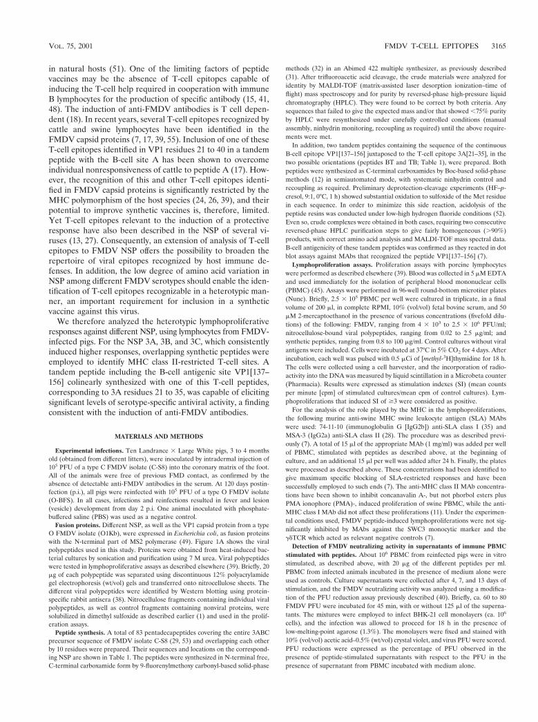
Fusion proteins. Different NSP, as well as the VP1 capsid protein from a typeO FMDV isolate (O1Kb), were expressed in Escherichia coli, as fusion proteinswith the N-terminal part of MS2 polymerase (49). Figure 1A shows the viralpolypeptides used in this study. Proteins were obtained from heat-induced bac-terial cultures by sonication and purification using 7 M urea. Viral polypeptideswere tested in lymphoproliferative assays as described elsewhere (39). Briefly, 20mg of each polypeptide was separated using discontinuous 12% polyacrylamidegel electrophoresis (wt/vol) gels and transferred onto nitrocellulose sheets. Thedifferent viral polypeptides were identified by Western blotting using protein-specific rabbit antisera (38). Nitrocellulose fragments containing individual viralpolypeptides, as well as control fragments containing nonviral proteins, weresolubilized in dimethyl sulfoxide as described earlier (1) and used in the prolif-eration assays.
Peptide synthesis. A total of 83 pentadecapeptides covering the entire 3ABCprecursor sequence of FMDV isolate C-S8 (29, 53) and overlapping each otherby 10 residues were prepared. Their sequences and locations on the correspond-ing NSP are shown in Table 1. The peptides were synthesized in N-terminal free,C-terminal carboxamide form by 9-fluorenylmethoxy carbonyl-based solid-phase
methods (32) in an Abimed 422 multiple synthesizer, as previously described(31). After trifluoroacetic acid cleavage, the crude materials were analyzed foridentity by MALDI-TOF (matrix-assisted laser desorption ionization–time offlight) mass spectroscopy and for purity by reversed-phase high-pressure liquidchromatography (HPLC). They were found to be correct by both criteria. Anysequences that failed to give the expected mass and/or that showed ,75% purityby HPLC were resynthesized under carefully controlled conditions (manualassembly, ninhydrin monitoring, recoupling as required) until the above require-ments were met.
In addition, two tandem peptides containing the sequence of the continuousB-cell epitope VP1[137–156] juxtaposed to the T-cell epitope 3A[21–35], in thetwo possible orientations (peptides BT and TB; Table 1), were prepared. Bothpeptides were synthesized as C-terminal carboxamides by Boc-based solid-phasemethods (12) in semiautomated mode, with systematic ninhydrin control andrecoupling as required. Preliminary deprotection-cleavage experiments (HF–p-cresol, 9:1, 0°C, 1 h) showed substantial oxidation to sulfoxide of the Met residuein each sequence. In order to minimize this side reaction, acidolysis of thepeptide resins was conducted under low-high hydrogen fluoride conditions (52).Even so, crude complexes were obtained in both cases, requiring two consecutivereversed-phase HPLC purification steps to give fairly homogeneous (.90%)products, with correct amino acid analysis and MALDI-TOF mass spectral data.B-cell antigenicity of these tandem peptides was confirmed as they reacted in dotblot assays against MAbs that recognized the peptide VP1[137–156] (7).
Lymphoproliferation assays. Proliferation assays with porcine lymphocyteswere performed as described elsewhere (39). Blood was collected in 5 mM EDTAand used immediately for the isolation of peripheral blood mononuclear cells(PBMC) (45). Assays were performed in 96-well round-bottom microtiter plates(Nunc). Briefly, 2.5 3 105 PBMC per well were cultured in triplicate, in a finalvolume of 200 ml, in complete RPMI, 10% (vol/vol) fetal bovine serum, and 50mM 2-mercaptoethanol in the presence of various concentrations (fivefold dilu-tions) of the following: FMDV, ranging from 4 3 103 to 2.5 3 106 PFU/ml;nitrocellulose-bound viral polypeptides, ranging from 0.02 to 2.5 mg/ml; andsynthetic peptides, ranging from 0.8 to 100 mg/ml. Control cultures without viralantigens were included. Cells were incubated at 37°C in 5% CO2 for 4 days. Afterincubation, each well was pulsed with 0.5 mCi of [methyl-3H]thymidine for 18 h.The cells were collected using a cell harvester, and the incorporation of radio-activity into the DNA was measured by liquid scintillation in a Microbeta counter(Pharmacia). Results were expressed as stimulation indexes (SI) (mean countsper minute [cpm] of stimulated cultures/mean cpm of control cultures). Lym-phoproliferations that induced SI of $3 were considered as positive.
For the analysis of the role played by the MHC in the lymphoproliferations,the following murine anti-swine MHC swine leukocyte antigen (SLA) MAbswere used: 74-11-10 (immunoglobulin G [IgG2b]) anti-SLA class I (35) andMSA-3 (IgG2a) anti-SLA class II (28). The procedure was as described previ-ously (7). A total of 15 ml of the appropriate MAb (1 mg/ml) was added per wellof PBMC, stimulated with peptides as described above, at the beginning ofculture, and an additional 15 ml per well was added after 24 h. Finally, the plateswere processed as described above. These concentrations had been identified togive maximum specific blocking of SLA-restricted responses and have beensuccessfully employed to such ends (7). The anti-MHC class II MAb concentra-tions have been shown to inhibit concanavalin A-, but not phorbol esters plusPMA ionophore (PMA)-, induced proliferation of swine PBMC, while the anti-MHC class I MAb did not affect these proliferations (11). Under the experimen-tal conditions used, FMDV peptide-induced lymphoproliferations were not sig-nificantly inhibited by MAbs against the SWC3 monocytic marker and thegdTCR which acted as relevant negative controls (7).
Detection of FMDV neutralizing activity in supernatants of immune PBMCstimulated with peptides. About 106 PBMC from reinfected pigs were in vitrostimulated, as described above, with 20 mg of the different peptides per ml.PBMC from infected animals incubated in the presence of medium alone wereused as controls. Culture supernatants were collected after 4, 7, and 13 days ofstimulation, and the FMDV neutralizing activity was analyzed using a modifica-tion of the PFU reduction assay previously described (40). Briefly, ca. 60 to 80FMDV PFU were incubated for 45 min, with or without 125 ml of the superna-tants. The mixtures were employed to infect BHK-21 cell monolayers (ca. 106
cells), and the infection was allowed to proceed for 18 h in the presence oflow-melting-point agarose (1.3%). The monolayers were fixed and stained with10% (vol/vol) acetic acid–0.5% (wt/vol) crystal violet, and virus PFU were scored.PFU reductions were expressed as the percentage of PFU observed in thepresence of peptide-stimulated supernatants with respect to the PFU in thepresence of supernatant from PBMC incubated with medium alone.
VOL. 75, 2001 FMDV T-CELL EPITOPES 3165

FIG. 1. Heterotypic lymphoproliferative response to viral polypeptides by FMDV-infected pigs. Fusion polypeptides from type O isolate (O1Kb)were used to in vitro stimulate PBMC from pigs infected with type C isolate (C-S8), as described in Materials and Methods. (A) Schematic rep-resentation of FMDV genomic RNA showing the location in the viral polyprotein of the fusion polypeptides used in this study. (B) Time coursestudy of the lymphoproliferative response to FMDV polypeptides by lymphocytes from infected pigs 1 and 2. Assays were performed with lympho-cytes obtained at 0, 4, 10, 21, 38, 50, 63, 70, 81, and 93 d.p.i. In those d.p.i. (i.e., from 10 to 80) in which positive SI were observed, peak responseswere obtained with a nitrocellulose-bound protein concentration of 2.5 mg/ml, with the following exceptions in which the highest stimulation wasobtained at a concentration of 0.5 mg/ml: VP1, day 10 (pigs 1 and 2); 3ABC, day 50 and 63 (pig 1) and days 63, 70, and 80 (pig 2); 3C, day 63 (pig2); and 3D, day 63 (pig 1). (C) Peak lymphoproliferative response to FMDV polypeptides by the four pigs (animals 1 to 4) analyzed. The dottedline represent the value (SI $3), above which responses were considered positive. The d.p.i. values, followed by the nitrocellulose-bound proteinconcentration (in micrograms per milliliter) corresponding to each of the responses, are shown above each bar (i.e., “70/2.5” indicates an SI ob-tained at 70 d.p.i. with 2.5 mg of nitrocellulose-bound protein per ml). In all cases, The results are expressed as SI as described in Materials and Meth-ods. The standard deviations of these values never exceeded 15% of the mean. A control animal was inoculated with PBS. The background cpmvalues (obtained with lymphocytes incubated with medium alone) were 502 (pig 1), 293 (pig 2), 1,140 (pig 3), 971 (pig 4), and 508 (control pig 5).
3166 BLANCO ET AL. J. VIROL.

RESULTS
Lymphoproliferative responses to FMDV NSP. In order tostudy the contribution of the NSP to the T-cell response elic-ited by FMDV in swine, type O polypeptides expressed in E.coli (Fig. 1A) were used to stimulate in vitro PBMC from fouroutbred pigs (animals 1 to 4) infected with type C isolate(C-S8). The experimental approach was designed to identifyregions conserved among different FMDV serotypes, whichwere capable of inducing heterotypic T-cell responses in lym-phocytes from domestic pigs. Lymphoproliferative responsesagainst the different polypeptides were monitored using PBMC
obtained from 0 to 100 days p.i. (d.p.i.). The magnitude of theresponses observed showed animal-to-animal variation, but aconsistent trend was noted. Positive lymphoproliferations (SI$3) against NSP and VP1 capsid protein (also included in thisanalysis) were detected from day 38 p.i. until day 70 p.i., andthe higher SI were observed at between 38 and 50 d.p.i. Nostimulations were observed with PBMC obtained from theanimals prior to infection (day 0 p.i.) nor from a control animalinoculated with PBS (pig 5). Figure 1B shows the results foundwith cells from the highest-responder pigs 1 and 2, which wererepresentative of the results obtained with cells from the four
TABLE 1. Synthetic peptides used in this study
Peptidea No. Amino acid sequenceb Residuesc Peptidea No. Amino acid sequenceb Residuesc
3A 1 ISIPSQKSVLYFLIE 1–52 QKSVLYFLIEKGQHE 6–203 YFLIEKGQHEAAIEF 11–254 KGQHEAAIEFFEGMV 16–305 AAIEFFEGMVHDSIK 21–356 FEGMVHDSIKEELRP 26–407 HDSIKEELRPLIQQT 31–458 EELRPLIQQTSFVKR 36–509 LIQQTSFVKRAFKRL 41–5510 SFVKRAFKRLKENFE 46–6011 AFKRLKENFEIVALC 51–6512 KENFEIVALCLTLLA 56–7013 IVALCLTLLANIVIM 61–7514 LTLLANIVIMIRETH 66–8015 NIVIMIRETHKRQKM 71–8516 IRETHKRQKMVDDAV 76–9017 KRQKMVDDAVNEYIE 81–9518 VDDAVNEYIEKANIT 86–10019 NEYIEKANITTDDQT 91–10520 KANITTDDQTLDEAE 96–11021 TTDDQTLDEAEKNPLE 101–11522 LDEAEKNPLETSGAS 106–12023 KNPLETSGASTVGFR 111–12524 TSGASTVGFRERTLP 116–13025 TVGFRERTLPGQKACD 121–13526 ERTLPGQKACDDVNS 126–14027 GQKACDDVNSEPAQP 131–14528 DDVNSEPAQPTEEQP 136–15029 EPAQPTEEQPQAEGP 141–15530 TEEQPQAEGPYAGPL 146–153
3B 1 QAEGPYAGPLERQRP 1–122 YAGPLERQRP LKVRA 3–173 ERQRP LKVRAKLPRQ 8–224 LKVRAKLPRQE 13–235 GPYAGPMERQKPLKV 24–386 PMERQKPLKVKARAP 29–437 KPLKVKARAPVVKEG 34–488 KARAPVVKEGPYEGP 39–539 VVKEGPYEGPVKKPV 44–5810 PYEGPVKKPVALKVK 49–6311 VKKPVALKVKAKNLI 54–6812 ALKVKAKNLIVTE 59–73
a Peptides were 15 amino acids in size and overlapped by 10 amino acids. The viral NSP corresponding to each set of peptides (3A, 3B, and 3C) is indicated.b Peptide amino acid sequence as in reference (51).c Amino acid positions spanned by each peptide in the corresponding protein. The carboxy-terminal residue of each NSP is underlined. Peptide 3A-30 included the
six amino-terminal residues of 3B. Peptide 3B-1 included the three carboxy-terminal residues of 3A. The sequences corresponding to VP1 and 3A, including in thetandem peptides, are shown.
d Tandem peptides, in which the VP1 and 3A residues are indicated, were colinearly synthesized in the two possible orientations (BT and TB). The 3A sequencesare in italics.
e That is, VP1 [137–156].f That is, 3A [21–35].
3C 1 SGAPPTDLQKMVMGN 1–152 TDLQKMVMGNTKPVE 6–203 MVMGNTKPVELILDG 11–254 TKPVELILDGKTVAI 16–305 LILDGKTVAICCATG 21–356 KTVAICCATGVFGTA 26–407 CCATGVFGTAYLVPR 31–458 VFGTAYLVPRHLFAE 36–509 YLVPRHLFAEKYDKI 41–5510 HLFAEKYDKIMLDGR 46–6011 YDKIMLDGRALTDS 51–6512 MLDGRALTDSDYRVF 56–7013 ALTDSDYRVFEFEIK 61–7514 DYRVFEFEIKVKGQD 66–8015 EFEIKVKGQDMLSDA 71–8516 VKGQDMLSDAALMVL 76–9017 MLSDAALMVLHRGNR 81–9518 ALMVLHRGNRVRDIT 86–10019 HRGNRVRDITKHFRD 91–10520 VRDITKHFRDVARMK 96–11021 KHFRDVARMKKGTPV 101–11522 VARMKKGTPVVGVIN 106–12023 KGTPVVGVINNADVG 111–12524 VGVINNADVGRLIFS 116–13025 NADVGRLIFSGEALT 121–13526 RLIFSGEALTYKDIV 126–14027 GEALTYKDIVVCMDG 131–14528 YKDIVVCMDGDTMPG 136–15029 VCMDGDTMPGLFAYR 141–15530 DTMPGLFAYRAATKA 146–16031 LFAYRAATKAGYCGG 151–16532 AATKAGYCGGAVLAK 156–17033 GYCGGAVLAKDGADT 161–17534 AVLAKDGADTFIVGT 166–18035 DGADTFIVGTHSAGG 171–18536 FIVGTHSAGGNGVGY 176–19037 HSAGGNGVGYCSCVS 181–19538 NGVGYCSCVSRSML 186–20039 YCSCVSRSMLLKMKA 191–20540 RSMLLKMKAHIDPEP 196–21041 KMKAHIDPEPHHE 201–215
BT3A-5d TASARGDLAHLTTTHARH
LPAAIEFFEGMVHDSIK137–156e
T3A-5Bd AAIEFFEGMVHDSIKTASARGDLAHLTTTHARHLP
21–35 f
VOL. 75, 2001 FMDV T-CELL EPITOPES 3167

animals analyzed. In general, the responses were dose depen-dent, and the higher SI were obtained with nitrocellulose-bound protein at from 0.5 to 2.5 mg/ml. Figure 1C shows thepeak responses found in each of the animals analyzed. Poly-peptides 3AB and 3C were recognized by the lymphocytesfrom all the animals analyzed. Proteins 2C, 3D, and VP1 wererecognized by cells from only two of the four pigs. These re-sults suggest the presence within the 3ABC polypeptide regionof T-cell epitopes conserved among FMDV serotypes, whichare consistently recognized by lymphocytes from domestic pigs.
Identification of T-cell epitopes in 3A, 3B, and 3C. In orderto characterize further the T-cell epitopes located in the 3ABCregion, a set of overlapping peptides (15-mer), covering pro-teins 3A, 3B, and 3C of the C-S8 virus, was synthesized. Thesepeptides were employed to stimulate in vitro lymphocytes frominfected pigs (see Materials and Methods and Table 1 fordetails). For this purpose, six additional pigs (animals 6 to 11)were experimentally infected with C-S8 virus, and their PBMCwere isolated on days 14 and 28 p.i. None of the peptidesinduced positive SI in PBMC obtained from these animalsbefore infection (data not shown). In general, the responsesagainst individual peptides were already detectable at day14 p.i., being more consistent at day 28 p.i. They were dosedependent, and the highest values were obtained with peptideconcentrations ranging from 4 to 100 mg/ml (Table 2). Amongthe 83 overlapping peptides tested, 45 did not induce signifi-cant proliferations in any of the pigs analyzed. The peak re-
sponses against the 19 peptides that induced positive SI in atleast four of the six animals are indicated in Table 2. Themagnitude of the response against individual peptides corre-lated with the SI observed against the whole virus, but thepattern of peptide recognition was different in each of theanimals studied. However, three individual peptides consis-tently stimulated PBMC from the six pigs analyzed: 3A-5[21–35], 3C-12[56–70], and 3C-27[131–145] (see Table 2).
For studies on the T-cell antigenic specificity elicited uponchallenge, animals 7 to 11 were reinoculated 4 months after theinitial infection with a heterologous type O FMDV virus (O-BFS). The five pigs developed an acute episode of the disease,and PBMC obtained at days 10, 21, and 35 postreinfection(p.r.i.) were used to perform lymphoproliferative assays inwhich the 3ABC peptides were used as stimulator antigens. Ingeneral, the responses against peptides were detected at day21 p.r.i., being more consistent at day 35 p.r.i. The responseswere dose dependent, and the highest values were obtainedwith peptide concentrations (4 to 100 mg/ml) similar to thosefound with cells after the first infection (Table 3). In thisexperiment, positive SI were also induced with peptide con-centrations lower than those effective with cells from the ini-tially infected animals (0.8 mg/ml; data not shown). The efficacyof the heterologous restimulation showed individual variationamong the animals. A clear boost of individual peptide re-sponses was observed for lymphocytes from animal 8. Lympho-cytes from animals 9, 10, and 11 showed an increased, albeit of
TABLE 2. Peptides corresponding to 3A, 3B, and 3C that weresignificantly recognized by lymphocytes from FMDV-infected pigs
Peptidea No. Amino acidresiduesb
SIc for pig:
6 7 8 9 10 11
3A 3 11–25 ND 5.3* 14.7 8.1* 18.2 2.5*5 21–35 16.4 7.3* 20.2 3.2* 4.2 4.6†6 26–40 7.6* 5.8 18.5 3.6* 14* 2.4†9 41–55 7.3 9.7 2.8 4† 6.6 4.8*25 121–135 ND 7.7 8.1 2.9 3.5 6.2†
3B 4 13–23 2.5 10.8 3.5 5.6* 5.2 2.2*8 39–53 3.3 11.6 2.1 7.3† 5.5 4.3*
3C 11 51–65 ND 17.2 12.6 6.7 10.4 2.712 56–70 6.1* 18.6 22.4 6 4.5 5.413 61–75 7.1 7.8 19.7* 6.2* 2.8 7.1*19 91–105 3† 5.5† 15.2* 6.6* 7.4 2.9*20 96–110 2† 18.1* 2.5* 4.5* 3.6 3.925 121–135 9.8 16.6 14 4.3 7.7 2.5†27 131–145 10.8* 27.4 17.3 7.3 3.2 9.2*28 136–150 ND 14.8 10.6 3.2 4 2.7*33 161–175 ND 15.6 4.5 3.9 6 2.9*34 166–180 ND 16.6 18.2 4.1 15 4.4†39 191–205 ND 5.4 2.5 7 3.5 4.2†40 196–210 2.2 20.9 3.7 6.8 7.3 7.3
C-S8d 24.3 24.7 ND 10.7 19 ND
a Peptides that induced an SI of .3 in at least four of the six pigs analyzed. Thecorresponding protein to which the peptides belong is indicated.
b Amino acid residues spanned by each of the peptides in the 3A, 3B, or 3CNSP.
c That is, the highest SI induced by the different antigens in PBMC isolated atday 28 p.i. Unless indicated otherwise, optimal responses were obtained with apeptide concentration of 100 mg/ml. ND, not done. *, SI was obtained with apeptide concentration of 20 mg/ml; †, SI was obtained with a peptide concentra-tion of 4 mg/ml.
d SI induced by C-S8 virus. The optimal responses shown were obtained witha viral concentration of 5 3 105 PFU/ml.
TABLE 3. Peptides corresponding to 3A, 3B, and 3C, that weresignificantly recognized by lymphocytes from FMDV-reinfected pigs
Peptidea No. Amino acidresiduesb
SIc for pig:
7 8 9 10 11
3A 3d 11–25 3.2 20 4.2 28.6* 2.1*5d 21–35 5.7 63.4 5.9* 10.6 3.3†6d 26–40 2.6* 40.2 5* 8.5* 2.9*9d 41–55 4.1* 13.4* 7† 6.4 1.9†17 81–95 4.5 29.7* 4.4 19.3 1.4*
3B 2 3–17 5.5 10.1 8.4 3.5* 1.1*4d 13–23 2.1 9.6 8 8.1 2.6*
3C 12d 56–70 4.1 93.4 17.3 5.2 9.213d 61–75 4.1 98.8* 13.1† 5.2 9.2*20d 96–110 2.7 99.5 1.8* 5.4 8.725d 121–135 3.1 42.9* 3.5 7.1 ND26 126–140 15.9 10.5* 4.9 4.9* ND30 146–160 7.6 102.2 ND 6.5 ND32 156–170 6.7 63.9 ND 5.8 ND34d 166–180 10.2 42 2.8 33.2 10.9†40d 196–210 8.6 10.8 8.9 4.2 11.5
C-S8e 107.5 307.9 26 56.2 ND
O-BFSe 217.3 112.4 11 52.6 ND
a Peptides that induced an SI of . 3 in at least three of the five pigs analyzed.b Amino acid residues spanned by each peptides in the 3A, 3B, or 3C NSP.c That is, the highest SI induced by the different antigens in PBMC isolated at
day 35 p.r.i. Unless indicated otherwise, optimal responses were obtained with apeptide concentration of 100 mg/ml. ND, not done. *, SI obtained with a peptideconcentration of 20 mg/ml; †, SI obtained with a peptide concentration of 4mg/ml.
d Peptides that induced positive SI in at least four of the six initially infectedpigs (see Table 2).
e SI induced by C-S8 or O-BFS viruses. The optimal responses shown wereobtained with a viral concentration of 5 3 105 PFU/ml.
3168 BLANCO ET AL. J. VIROL.

lower magnitude, in most of the SI, and the responses ofPBMC from animal 7 were lower than those observed after thefirst infection. Table 3 summarizes the SI obtained with thepeptides that induced positive responses in at least three of thefive animals analyzed.
Lymphocytes from reinfected pigs responded to 11 of the 19peptides identified as antigenic after analyses with cells ob-tained following the first infection (Table 3). Peptides 3A-3, -5,-6, and -9, peptide 3B-4, and peptides 3C-12, -13, -20, -25, -34,and -40, previously identified as good stimulators (see Table 2),also induced positive responses in at least three of the fivereinfected pigs. In addition, the following new peptides in-duced proliferation in lymphocytes taken from the majority ofthe reinfected animals analyzed: 3A-17; 3B-2; and 3C-26, -30,and -32. Again, several individual peptides induced lympho-proliferative responses by cells from the five animals uponheterologous reinfection (Table 3): 3A-5[21–35], 3C-12[56–70], 3C-13[61–75], 3C-34[166–180], and 3C-40[196–210]. Rep-resentative dose responses induced by some of these peptidesin PBMC from infected and reinfected animals are shown inFig. 2.
SLA restriction of the anti-peptide response. Informationabout the SLA restriction of the detected lymphoproliferativeresponses was obtained using MAbs against SLA class I andclass II. These were used to inhibit in vitro the lymphoprolif-erations induced in cells from reinfected pigs. The peptides3A-3[11–25], 3A-5[21–35], 3C-34[166–180], and 3C-40[196–210] were selected for this study because they induced consis-tent responses in cells from most of the analyzed animals, afterboth the first infection and the heterologous re infection (Ta-bles 2 and 3). Results showed the highest inhibition of peptide-induced lymphoproliferation (.75%) when the anti-SLA classII MAb was used. This is consistent with lymphoproliferationsinduced by peptides being dependent on CD41 T -helper lym-phocyte activities (Table 4). The anti-SLA class I MAb wasalso inhibitory, but the magnitude of inhibition was generallylower than that observed with the anti-SLA class II antibody.
Antigenicity of tandem peptides including B- and T-cell epi-topes. The potential of the T-cell epitopes identified in NSP toimprove vaccine formulation of FMDV synthetic peptides re-quires that T-cell epitopes remain immunostimulatory when incombination with B-cell epitopes. In order to investigate this
FIG. 2. Effect of peptide dose on the proliferative response of PBMC obtained from pigs 8, 9, and 10 at days 28 p.i. and 35 p.r.i. The valuescorresponding to 0 on the x axis indicate the background of the assay (PBMC incubated with medium alone).
VOL. 75, 2001 FMDV T-CELL EPITOPES 3169

point, the immunostimulatory potential of the T-cell peptide3A-5[21–35] was further analyzed by using tandem peptides inwhich this epitope was colinearly synthesized with the B-cellsite VP1[137–156]. Two possible orientations were used: pep-tides BT and TB (Table 1). With lymphocytes from pigs 7, 9,10, and 11, the tandem epitopes induced lymphoproliferativeresponses in vitro upon infection with C-S8 virus, as shown inFig. 3A. The responses were dose dependent, and the highestSI was obtained with a peptide concentration of 20 mg/ml.Positive SI were found with cells from all four animals stimu-lated with peptide T3A-5B, whereas only cells from animals 7and 11 responded to peptide BT3A-5. The SI of the latter cellswere lower than those induced by peptide T3A-5B. In this ex-periment, peptide 3A-5[21–35] was also recognized with an SIlower than those found using peptide T3A-5B.
Upon reinfection with the heterologous O-BFS virus, lym-phocytes from pigs 7, 8, and 10 responded to T3A-5B, againwith SI higher than those induced by peptide 3A-5[21–35]alone (Fig. 3B). Thus, tandem peptide T3A-5B stimulates invitro lymphocytes from both infected and reinfected animals.
It was then interesting to address whether the incorporationof 3A-5[21–35] into the tandem peptide could result in thestimulation of T cells to provide the necessary immunologicalhelp for antigen-stimulated B lymphocytes. To this end, thestimulation of T cells by peptide T3A-5B was analyzed in termsof cooperation with the induction of an antiviral humoral re-sponse. Lymphocytes from reinfected pig 8 were stimulated invitro with peptides T3A-5B, T, or B. At different times post-stimulation, the presence of anti-FMDV activity in the super-natants was measured using a plaque reduction assay. Signifi-cant PFU reductions were detected using supernatants fromcultures stimulated with peptide T3A-5B from the fourth day,reaching a maximum with day 7 supernatants (ca90%) (Fig. 4).The PFU reductions were higher than those obtained using
supernatants from cultures stimulated by peptides T and Bseparately. In addition, the antiviral activity was restricted tothe homologous virus C-S8, since no plaque reduction wasdetected when a type O FMDV was used in the assay (Fig. 4).Thus, a serotype-specific anti-FMDV activity, consistent withthe induction of anti-FMDV neutralizing antibodies, was de-tected upon stimulation of immune lymphocytes with peptideT3A-5B.
DISCUSSION
In the present studies the antigenic specificity of the porcine(an important natural host of this virus) T-cell response againstFMDV NSP was analyzed. The aim was the identification ofT-cell epitopes in NSP widely recognized by domestic pigs, the
TABLE 4. Inhibition by MAb to porcine class I and class II ofthe proliferative response to 3A and 3C peptides
Peptidea MAbb
Inhibition in pigc:
9 8
Dcpm % Dcpm %
3A-3[11–25] None 8,615 6 980 4,101 6 615Anti-class I 2,249 6 337 73.8 2,740 6 411 33.1Anti-class II 2,850 6 484 66.9 905 6 108 77.9
3A-5[21–35] None 9,599 6 1151 7,853 6 471Anti-class I 4,007 6 640 58.2 3,807 6 609 51.2Anti-class II 1,938 6 193 79.8 1,525 6 213 59.9
3A-34[166–180] None 17,389 6 869 9,808 6 689Anti-class I 5,692 6 1024 67.2 3,757 6 676 61.6Anti-class II 2,701 6 243 84.4 2,484 6 298 74.6
3C-40[196–210] None 7,360, 6 1030 5,292 6 793Anti-class I 2,854, 6 342 61.2 4,626 6 277 12.5Anti-class II 1,145 6 171 84.4 2,210 6 198 58.2
a The characteristics of the peptides are described in Table 1. Peptides wereused at a concentration of 20 mg/ml. None, lymphocytes cultured in the absenceof MAb.
b A total of 15 ml (1 mg/ml) of MAb anti-SLA class I (74-11-10, Ig-G2b) orclass II (MSA-3, IgG2a) was added per well at the beginning of culture. After24 h, the cultures were supplemented with another 15 ml of MAb.
c Lymphocytes from pigs 8 and 9, obtained at day 35 p.r.i. were used in theseexperiments.
FIG. 3. Lymphoproliferative responses to peptides 3A-5[21–35],BT3A-5, and T3A-5B. The data shown correspond to peak responses oflymphocytes obtained at days 28 p.i. with C-S8 virus (A) and 21 p.r.i.with the heterologous O-BFS virus (B). The standard deviations areindicated. The peptide concentrations giving the SI shown were 20mg/ml, except for pig 9 (in panel A) in which the peak response wasobtained with 100 mg/ml. The background cpm levels were 405 (pig 7),450 (pig 9), 525 (pig 10), and 345 (pig 11) (A) and 302 (pig 7), 214 (pig8), and 258 (pig 10) (B).
3170 BLANCO ET AL. J. VIROL.

sequences of which were conserved among different FMDVserotypes. Animals were selected from different litters to en-sure a representative sample of outbred pigs and to bring por-cine MHC polymorphism in the T-cell epitope recognition intothe study. FMDV NSP are absent, or incorporated in verysmall amounts, in the virus particle (34). Therefore, the anal-yses used animals experimentally infected with a type C FMDVisolate.
T-cell antigenicity of the NSP was first analyzed in terms ofthe capacity of different polypeptides to stimulate in vitro Tcells from the infected animals. A heterotypic lymphoprolif-erative response against NSP in cells from FMDV-infectedpigs was tested. Lymphoproliferations were detected from day38 to day 70 p.i. Although the SI showed animal-to-animalvariations, polypeptides 3AB and 3C induced specific and con-sistent responses in lymphocytes from all animals analyzed.Polypeptides 2B and 3D and capsid protein VP1 were notrecognized by all the animals (Fig. 1C). This finding contrastswith the recognition of type O FMDV 3D as an immunodom-inant T-cell determinant by cattle lymphocytes, as recentlyreported (16). In addition to differences between cattle andswine in terms of antigenic recognition patterns, the lowerantigenicity of 3D for porcine lymphocytes may also reflectdifferences between FMDV type C and type O. The low het-erotypic recognition of VP1 is consistent with the sequencedivergence that exists between type O and type C FMDV.
Overlapping synthetic peptides allowed a detailed analysisof the T-cell epitopes recognized in proteins 3A, 3B, and 3C.Despite animal to animal variation, 19 peptides were stimu-lated in vitro lymphocytes from, at least, four of the six infected
pigs analyzed (Table 2). The responsive peptides define T-cellregions efficiently recognized by swine lymphocytes at the fol-lowing amino acid positions: 3A[11–55] and -[121–135], 3B[13–23] and -[39–53], and 3C[51–75],-[91– 110], -[121–150], -[161–180], and -[191–210]. Eleven of these peptides were alsorecognized by lymphocytes from at least three of the five het-erotypically reinfected pigs analyzed (Table 3). Five new pep-tides, which did not induced in vitro responses in the initiallyinfected animals, became responsive upon the induction of asecondary response (Table 3). The efficacy of the heterologousrestimulation was uneven among the five animals studied. Aclear boost of individual peptide responses was only observedfor lymphocytes from animal 8. Although the number of ani-mals used for this type of study was not sufficient for statisticaldemonstration, we think that the data are consistent with theidentification of a number of peptide epitopes in the nonstruc-tural polypeptides 3A, 3B, and 3C of FMDV, which are capa-ble of stimulating porcine T cells following infection with vi-ruses of different serotypes, and that some of them may boostprimed lymphocytes.
SLA class II MAb efficiently inhibited the lymphoprolifera-tive response. This demonstrated the involvement of CD41 Thlymphocytes in the recognition of some of the stimulatorypeptides (Table 4). Anti-class I MAb gave lower inhibitions, inagreement with the previous reports on porcine lymphoprolif-eration against whole virus (14, 45) and FMDV capsid proteinpeptides (7). These inhibitions might relate to the high pro-portion of CD81 CD41 double-positive cells found in swine(58), dominated by memory Th lymphocytes (50, 59). Cer-tainly, it must also be considered that the animals were in-fected with FMDV and, therefore, that stimulation of CD81
cells through class I presentation could have occurred (14).The implication of CD81 lymphocytes in protective responsesto FMDV remains largely unknown and thus its characteriza-tion was not pursued here. Attempts to obtain porcine T-cellclones, a highly valuable tool for understanding the antigenicspecificity and function of T cells, have had very limited suc-cess. Thus, as a first step in the characterization of the immuneresponses induced by the T-cell epitopes thus for identified,our attention was focused on the potential of these peptides tostimulate CD41 cells to provide T help to immune B lympho-cytes. In this way, we attempted to identify T-cell epitopes thatcould enhance the induction of anti-FMDV antibodies by pep-tide vacines. For practical reasons, such T-cell epitopes shouldbe conserved among different FMDV serotypes. Peptides 3A-3[11–25], 3A-5[21–35], 3C-25[121–135], and 3C-34[166–180]do indeed show amino acid sequences completely conservedamong serotypes A, O, and C (Table 5). These 3A and 3Cpeptides are frequently and efficiently recognized by porcinelymphocytes in both a homotypic and heterotypic manner.Consequently, they constitute potential candidates for inclu-sion in a peptide vaccine formulation.
The colinear expression of T-helper and B-cell epitopes inpeptide vaccines can result in the enhancement of antibodyresponses (8, 17). The design of functional combinations ofT-cell and B-cell epitopes is rather empirical (19, 47) andshould maintain the immunostimulatory ability of the B- andT-cell components. T-cell peptide 3A-5[21–35] consistentlystimulated lymphocytes from all initially infected and rein-fected animals analyzed in this study, as well as lymphocytes
FIG. 4. FMDV PFU reduction by supernatants of immune PBMCstimulated in vitro with viral peptides. The data correspond to PBMCfrom pig 8 (obtained at day 35 p.r.i.) assayed at different days of in vitrostimulation with 20 mg of the indicated peptides per ml. The results areexpressed as the percent inhibition in the PFU recovered from cellsstimulated with each peptide with respect to the PFU observed with acontrol supernatant incubated with medium alone. The assay was per-formed using two FMDV isolates of different serotypes: C-S8 (type C)and O-BFS (type O). The standard deviations are indicated.
VOL. 75, 2001 FMDV T-CELL EPITOPES 3171

from four additional FMDV-infected domestic pigs (unpub-lished results). Therefore, peptide 3A-5[21–35] was employedto explore the potential of tandem peptides including the im-munodominant B-cell site VP1[137–156], with either the BT orthe TB orientation, to stimulate lymphocytes from infectedanimals. We first analyzed whether these tandem peptides re-tained the capacity to induce lymphoproliferation of immunelymphocytes. Each of the tandem peptides was recognized byimmune lymphocytes, with T3A-5B being the most efficientstimulator of lymphocytes from both initially infected and re-infected animals (Table 3). Different factors may mediate thehigher stimulations observed with tandem peptide T3A-5B:among these factors is the influence of the different flankingsequences on the binding to MHC molecules that could modifylymphocyte activation (56) or on the susceptibility of the T-cellepitopes to proteases in antigen processing (20).
The induction of FMDV neutralizing antibodies by immunePBMC in response to in vitro viral stimulation has been pre-viously reported as an indicator of T-cell–B-cell cooperation(40). Interestingly, a significant FMDV PFU reduction activity(up to 90%) was detected in supernatants of PBMC upon
incubation with peptide T3A-5B under conditions in which theinhibitions induced by supernatants of PBMC stimulated withpeptide A[21–35] or 3A-5[21–35] did not exceed 20%. Thisreduction was serotype specific, since it was not observed witha type O virus (Fig. 4). These results are consistent with theinduction of anti-FMDV antibodies in the peptide-stimulatedcultures, and new experiments are being conducted to furthercharacterize the antiviral activity induced.
Inclusion of FMDV T-cell epitopes may enhance the immu-nogenicity of subunit and peptide vaccines. According to themodel of intermolecular-intrastructural help (33, 43), B cellscan be activated by T cells resulting from the stimulation byepitopes derived from a different protein, provided that bothproteins are associated in a common complex. Whether 3ABCand viral structural proteins, VP1 in particular, may becomeassociated in FMDV-infected cells remains to be studied. Onthe other hand, the stimulation of T cells by viral peptides mayparticipate not only in providing T-cell help to B lymphocytesbut also in eliciting a synergistic response against the virusinfection. T cells secrete cytokines such as gamma interferonand interleukins that might play an important role in the pro-
TABLE 5. Alignment of the amino acid sequences corresponding to NSP 3A and 3C
Protein Virus Sequencea
z z z z z 503A C-S8 ISIPSQKSVLYFLIEKGQHEAAIEFFEGMVHDSIKEELRPLIQQTSFVKRO-BFSA12
z z z z z 100C-S8 AFKRLKENFEIVALCLTLLANIVIMIRETHKRQKMVDDAVNEYIEKANITO-BFS RA12 R
z z z z z 150C-S8 TDDQTLDEAEKNPLETSGASTVGFRERTLPGQKARDDVNSEPAQPTEEQPO-BFS K S C VA12 T T R CN R A
z 153C-S8 QAEO-BFSA12
z z z z z 503C C-S8 SGAPPTDLQKMVMGNTKPVELILDGKTVAICCATGVFGTAYLVPRHLFAE
O-BFSA12
z z z z z 100C-S8 KYDKIMLDGRALTDSDYRVFEFEIKVKRGQDMLSDAALMVLHRGNRVRDIO-BFS V MA12 M
z z z z z 150C-S8 TKHFRDVARMKKGTPVVGVINNADVGRLIFSGEALTYKDIVVCMDGDTMPO-BFS TA12 T V
z z z z z 200C-S8 GLFAYKAATKAGYCGGAVLAKDGADTFIVGTHSAGGNGVGYCSCVSRSMLO-BFS RA12 S K
z z 214C-S8 LKMKAHIDPEPHHEO-BFSA12 R V Q
a Amino acid substitutions in FMDV of serotypes O (O-BFS) and A (A12) with respect to the C-S8 sequence are indicated. Sequences were obtained from thefollowing Swiss-Prot accession numbers: Q9QCE3 (C-S8), POLG_FMDVA(A12), and POLG_FMDVO (O-BFS). Overdots indicate 10-amino-acid segments; numbersin italics indicate the cumulative amino acid count.
3172 BLANCO ET AL. J. VIROL.

tection against the virus (23, 57). Consistent with this hypoth-esis are the results described recently (46) that show that im-munization with a recombinant adenovirus expressing the P1polypeptide elicits partial protection against FMDV in pigsand cattle in the absence of antiviral antibody response. Toexplore these possibilities, experiments are in progress to as-sess the immunogenicity in pigs of combinations of the B-cellsite VP1[140–160] and the T-cell peptides identified here.
ACKNOWLEDGMENTS
We thank C. Sanchez for excellent work in the animals experiments;J. I. Nunez and M. A. Jimenez for experimental help and support; andJ. Domınguez, A. Ezquerra, F. Alonso, A. Canals, and K. McCulloughfor advice and constructive discussions. We also thank E. Beck forproviding plasmids expressing FMDV NSP.
Work at CISA-INIA and CBMSO was supported by CICYT (grantBIO99-0833-02-01), by the EU grants FAIR CT97-3665 and CT96-1317, and by the Fundacion Ramon Areces. Work in Barcelona wassupported by DGES (grant PB97-0873), by the EU (grant FAIR CT97-3577), and by Generalitat de Catalunya (CERBA). P.G. thanks theFundaca#o Calouste Gulbenkian (Lisbon, Portugal) for her Ph.D. grantand the University of Porto (Porto, Portugal) for a temporary leavefrom teaching duties.
REFERENCES
1. Abou-Zeid, C., E. Filley, J. Steele, and G. A. W. Rook. 1987. A simple methodfor using antigens separated by polyacrylamide gel electrophoresis to stim-ulate lymphocytes in vitro after converting bands cut from Western blots intoantigen-bearing particles. J. Immunol. Methods 98:5–10.
2. Acharya, R., E. Fry, D. Stuart, D. Rowlands, and F. Brown. 1989. Three-dimensional structure of foot-and-mouth disease virus at 2.9 3 resolution.Nature 337:709–716.
3. Ahmed, R., and C. A. Biron. 1999. Immunity to viruses, p. 1295–1334. InW. E. Paul (ed.), Fundamental immunology. Lippincott-Raven Publishers,New York, N.Y.
4. Bachrach, H. L. 1977. Foot-and-mouth disease virus, properties, molecularbiology and immunogenicity, p. 3–32. In J. A. Romberger (ed.), Virology inagriculture. Beltsville Symposia in Agricultural Research. I. Allanheld, Mont-clair, N.J.
5. Barteling, S. J., and J. Vreeswijk. 1991. Development in foot-and-mouthdisease vaccines. Vaccine 9:75–88.
6. Bittle, J. L., R. A. Houghten, H. Alexander, T. M. Shinnick, J. G. Sutcliffe,and R. A. Lerner. 1982. Protection against foot-and-mouth disease by im-munization with a chemically synthesized peptide predicted from the viralnucleotide sequence. Nature 298:30–33.
7. Blanco, E., K. McCullough, A. Summerfield, J. Fiorini., D. Andreu, C. Chiva,E. Borras, P. Barnett, and F. Sobrino. 2000. Interspecies MHC-restricted Thcell epitope on foot-and-mouth disease virus capsid protein VP4. J. Virol.74:4902–4907.
8. Borras-Cuesta, F., A. Petit-Camurdan, and Y. Fedon. 1987. Engineering ofimmunogenic peptide by co-linear synthesis of determinants recognized by Band T cells. Eur. J. Immunol. 17:1213–1215.
9. Brown, F. 1992. New approaches to vaccination against foot-and-mouthdisease. Vaccine 10:1022–1026.
10. Brown, F. 1995. Antibody recognition and neutralization of foot-and-mouthdisease virus. Semin. Virol. 6:243–248.
11. Bullido, R., N. Domenech, B. Alvarez, F. Alonso, M. Babın, A. Ezquerra, E.Ortuno, and J. Domınguez. 1997. Characterization of five monoclonal anti-bodies specific for swine class II major histocompatibility antigens and cross-reactivity studies with leukocytes of domestic animals. Dev. Comp. Immunol.21:311–322.
12. Carreno, C., X. Roig, J. J. Cairo, J. A. Camarero, M. G. Mateu, E. Domingo,E. Giralt, and D. Andreu. 1992. Studies on antigenic variability of C strainsof foot-and-mouth disease virus by means of synthetic peptides and mono-clonal antibodies. Int. J. Peptide Protein Res. 39:41–47.
13. Celis, E., J. Larson, Jr., L. Otvos, and W. H. Wunner. 1990. Identification ofa rabies virus T cell epitope on the basis of its similarity with a hepatitis Bsurface antigen peptide presented to T cells by the same MHC molecule(HLA-DPw4). J. Immunol. 145:305–310.
14. Childerstone, A. J., J. Cedillo-Baron, R. Foster-Cuevas, and M. E. Park-house. 1999. Demonstration of bovine CD81 T-cell responses to foot-and-mouth disease virus. J. Gen. Virol. 80:663–669.
15. Collen, T. 1994. Foot-and-mouth disease (aphthovirus): viral T cell epitopes,p. 173–197. In B. M. L. Goddeevis and I. Morrison (ed.), Cell mediatedimmunity in ruminants. CRC Press, Inc., Boca Raton, Fla.
16. Collen, T., J. Baron, A. Childerstone, A. Corteyn, T. R. Doel, M. Flint, M.
Garcıa-Valcarcel, M. E. Parkhouse, and M. D. Ryan. 1998. Heterotypicrecognition of recombinant FMDV proteins by bovine T-cells: the polymer-ase (P3Dpol) as an immunodominant T-cell immunogen. Virus Res. 56:125–133.
17. Collen, T., R. DiMarchi, and T. R. Doel. 1991. A T cell epitope in VP1 offoot-and-mouth disease virus is immunodominant for vaccinated cattle.J. Immunol. 146:749–755.
18. Collen, T., L. Pullen, and T. R. Doel. 1989. T cell-dependent induction ofantibody against foot-and-mouth disease virus in a mouse model. J. Gen.Virol. 70:395–403.
19. Cox, J. H., J. Ivanyi, D. B. Young, J. R. Lamb, A. D. Syred, and M. J. Francis.1988. Orientation of epitopes influences the immunogenicity of syntheticpeptide dimers. Eur. J. Immunol. 18:2015–2019.
20. Del Val, M., H. J. Schlicht, T. Ruppert, M. J. Reddehase, and U. H. Koszi-nowski. 1991. Efficient processing of an antigenic sequence for presentationby MHC class I molecules depends on its neighbouring residues in theprotein. Cell 66:1145–1153.
21. DiMarchi, R., G. Brooke, C. Gale, V. Cracknell, T. Doel, and N. Mowat.1986. Protection of cattle against foot-and-mouth disease by a syntheticpeptide. Science 232:639–641.
22. Domingo, E., M. G. Mateu, M. A. Martınez, J. Dopazo, A. Moya, and F.Sobrino. 1990. Genetic variability and antigenic diversity of foot-and-mouthdisease virus, p. 233– 266. In E. Kurstak, R. G. Marusyk, S. A. Murphy, andM. H. V. Van-Regenmortel (ed.), Applied virology research, vol. 2. Virusvariation and epidemiology. Plenum Publishing Corp., New York, N.Y.
23. Emile, D., M. Peuchmaur, M. C. Maillot, M. C. Crevon, N. Brouse, J. F.Delfraissy, J. Dromont, and P. Galanaud. 1990. Production of interleukins inhuman immunodeficiency virus 1 in replicating lymph nodes. J. Clin. Inves-tig. 86:148–159.
24. Garcıa-Briones, M. M., G. Russell, E. Carrillo, E. L. Palma, O. Taboga, C.Tami, F. Sobrino, and E. J. Glass. 2000. Association of bovine DRB3 alleleswith the immune response to foot-and-mouth disease virus peptides andprotection against viral challenge. Vaccine 19:1167–1171.
25. Germain, R. N. 1999. Antigen processing and presentation, p. 287–340. InW. E. Paul (ed.), Fundamental immunology. Lippincott-Raven Publishers,New York, N.Y.
26. Glass, E. J., and P. Millar. 1994. Induction of effective cross-reactive immu-nity by FMDV peptides is critically dependent upon specific MHC-peptide-Tcell interactions. Immunology 82:1–8.
27. Hacket, C. J., D. Horowitz, M. Wysocka, and S. B. Dillon. 1992. Influenzavirus infection elicits class II major histocompatibility complex-restricted Tcells specific for an epitope identified in the NS1 non-structural protin.J. Gen. Virol. 73:1339–1343.
28. Hammerberg, C., and G. G. Schurig. 1986. Characterization of monoclonalantibodies directed against swine leukocytes. Vet. Immunol. Immunopathol.11:107–121.
29. Martınez-Salas, E., J. Ortın, and E. Domingo. 1985. Sequence of the viralreplicase gene from foot-and-mouth disease virus C1-Santa Pau (C-S8).Gene 35:55–61.
30. Mateu, M. G. 1995. Antibody recognition of picornaviruses and escape fromneutralization: a structural view. Virus Res. 38:1–24.
31. Mateu, M. G., M. L. Valero, D. Andreu, and E. Domingo. 1996. Systematicreplacement of amino acid residues within an Arg-Gly-Asp-containing loopof foot-and-mouth disease virus and effect on cell recognition. J. Biol. Chem.271:12814–12819.
32. Merrifield, R. B. 1963. Solid phase synthesis. I. The synthesis of a tetrapep-tide. J. Am. Chem. Soc. 85:2149–2154.
33. Milich, D. R., A. McLachlan, G. B. Thornton, and J. L. Hughes. 1987.Antibody production to the nucleocapsid and envelope of the hepatitis Bvirus primed by a single synthetic T-cell site. Nature 329:547–549.
34. Newman, J. F. E., P. G. Piatti, B. M. Gorman, T. G. Burrage, M. D. Ryan, M.Flint, and F. Brown. 1994. Foot-and-mouth disease virus particles containreplicase protein 3D. Proc. Natl. Acad. Sci. USA 91:733–737.
35. Pescovitz, M. D., J. R. Thistlethwaite, Jr., H. Auchincloss, Jr., S. T. Ildstad,T. G. Sharp, R. Terrill, and D. H. Sachs. 1984. Effect of class II antigenmatching on renal allograft survival in miniature swine. J. Exp. Med. 160:1495–1508.
36. Pereira, H. G. 1981. Foot-and-mouth disease, p. 333–363. In E. P. J. Gibbs(ed.), Virus disease of food animals. Academic Press, Inc., New York, N.Y.
37. Porter, A. G. 1993. Picornavirus nonstructural proteins: emerging roles invirus replication and inhibition of host cell functions. J. Virol. 67:6917–6921.
38. Rodrıguez, A., J. Dopazo., J. C. Saiz, and F. Sobrino. 1994. Immunogenicityof nonstructural proteins of foot-and-mouth disease virus: differences be-tween infected and vaccinated swine. Arch. Virol. 136:123–131.
39. Rodrıguez, A., J. C. Saiz, I. S. Novella, D. Andreu, and F. Sobrino. 1994.Antigenic specificity of porcine T cell response against foot-and-mouth dis-ease virus structural proteins: identification of T helper epitopes in VP1.Virology 205:24–33.
40. Rodrıguez, A., J. C. Saiz, and F. Sobrino. 1995. In vitro synthesis of foot-and-mouth disease virus specific antibodies by porcine leukocytes. Arch.Virol. 140:1645–1652.
41. Rowlands, D. J. 1994. Experimental determination of immunogenic sites, p.
VOL. 75, 2001 FMDV T-CELL EPITOPES 3173

137–168. In B. H. Nicholson (ed.), Synthetic vaccines. Blackwell ScientificPublications, Oxford, England.
42. Rueckert, R. R. 1985. Picornaviruses and their replication, p. 705–738. InB. N. Fields, D. P. Knipe, R. M. Chanoc, J. L. Melnick, B. Roizmann, andR. E. Shope (ed.), Fields virology. Raven Press, New York, N.Y.
43. Russell, S. M., and F. Y. Liew. 1979. T cells primed by influenza virioninternal components can cooperate in the antibody response to haemagglu-tinin. Nature 280:147– 148.
44. Ryan, M. D., G. J. Belsham, and A. M. Q. King. 1989. Specificity of enzyme-substrate interactions in foot-and-mouth disease virus polyprotein process-ing. Virology 173:35–45.
45. Saiz, J. C., A. Rodrıguez, M. Gonzalez, F. Alonso, and F. Sobrino. 1992.Heterotypic lymphoproliferative response in pigs vaccinated with foot-and-mouth disease virus. Involvement of isolated capsid proteins. J. Gen. Virol.73:2601–2607.
46. Sanz-Parra, A., M. A. Jimenez-Clavero, M. M. Garcıa-Briones, E. Blanco, F.Sobrino, and V. Ley. 1999. Recombinant viruses expressing the foot-and-mouth disease virus capsid precursor polypeptide (P1) induce cellular butnot humoral antiviral immunity and partial protection in pigs. Virology259:129–134.
47. Saw, D. M., C. M. Stanley, C. D. Partidos, and M. Steward. 1993. Influenceof the T-helper epitope on the titer and affinity of antibodies to B-cellepitopes after coimmunization. Mol. Immunol. 30:961–968.
48. Sobrino, F., E. Blanco, M. M. Garcıa-Briones, and V. Ley. 1999. Syntheticpeptide vaccines: foot-and-mouth disease virus as a model. Dev. Biol. Stand.101:39–43.
49. Strebel, K., E. Beck, K. Strohmaier, and H. Schaller. 1986. Characterizationof foot-and-mouth disease virus gene products with antisera against bacte-rially synthesized proteins. J. Virol. 57:983–991.
50. Summerfield, A., H.-J. Rziha, and A. Saalmuller. 1996. Functional charac-terization of porcine CD41 CD81 extrathymic T lymphocytes. Cell. Immu-nol. 168:291–296.
51. Taboga, O., C. Tami, E. Carrillo, J. I. Nunez, A. Rodrıguez, J. C. Saiz, E.Blanco, M. L. Valero, X. Roig, J. A. Camarero, D. Andreu, M. G. Mateu, E.Giralt, E. Domingo, F. Sobrino, and E. L. Palma. 1997. A large-scale eval-uation of peptide vaccines against foot-and-mouth disease: lack of solidprotection in cattle and isolation of escape mutants. J. Virol. 71:2606–2614.
52. Tam, J. P., W. F. Heath, and R. B. Merrifield. 1983. SN2 deprotection ofsynthetic peptides with a low concentration of HF in dimethyl sulfide: evi-dence and application in peptide synthesis. J. Am. Chem. Soc. 105:6442–6455.
53. Toja, M., C. Escarmıs, and E. Domingo. 1999. Genomic nucleotide sequenceof a foot-and-mouth disease virus clone and its persistent derivatives. Impli-cations for the evolution of viral quasispecies during a persistent infection.Virus Res. 64:161–171.
54. Van Bekkum, J. G. 1969. Correlation between serum antibody level andprotection against challenge with FMD, p. 38–41. In Session of ResearchGroup of the Standing Technical Committee of the European Commissionfor the Control of FMD, Brescia, Italy. Food and Agriculture Organization,Rome, Italy.
55. Van Lierop, M. J., J. P. Wagenaar, J. M. van Noort, and E. J. Hensen. 1995.Sequences derived from the highly antigenic VP1 region 140 to 160 offoot-and-mouth disease virus do not prime for a bovine T-cell responseagainst intact virus. J. Virol. 69:4511–4514.
56. Vignali, D. A., and J. L. Strominger. 1994. Amino acid residues that flankcore peptide epitopes and the extracellular domains of CD4 modulate dif-ferential signaling through the T cell receptor. J. Exp. Med. 179:1945–1956.
57. Zinkernagel, R. M. 1996. Immunology taught by viruses. Science 271:173–178.
58. Zuckermann, F. A., and R. J. Husmann. 1996. Functional and phenotypicanalysis of porcine peripheral blood CD4/CD8 double positive T cells. Im-munology 87:500–512.
59. Zuckermann, F. A. 1999. Extrathymic CD4/CD8 double positive T cells. Vet.Immunol. Immunopathol. 72:55–66.
3174 BLANCO ET AL. J. VIROL.
Copyright © 2022 FDOKUMEN