Particle conformation regulates antibody access to a conserved GII.4 norovirus blockade epitope

2006, 80(16):7816. DOI: 10.1128/JVI.00532-06. J. Virol.
GreenWobus, Stephanie M. Karst, Herbert W. Virgin and Kim Y.Victor G. Prikhodko, Larissa B. Thackray, Christiane E. Stanislav V. Sosnovtsev, Gaël Belliot, Kyeong-OK Chang, Polyprotein in Infected Cellsof the Murine Norovirus Nonstructural Cleavage Map and Proteolytic Processing
http://jvi.asm.org/content/80/16/7816Updated information and services can be found at:
These include:
SUPPLEMENTAL MATERIAL Supplemental material
REFERENCEShttp://jvi.asm.org/content/80/16/7816#ref-list-1at:
This article cites 51 articles, 32 of which can be accessed free
CONTENT ALERTS more»articles cite this article),
Receive: RSS Feeds, eTOCs, free email alerts (when new
http://journals.asm.org/site/misc/reprints.xhtmlInformation about commercial reprint orders: http://journals.asm.org/site/subscriptions/To subscribe to to another ASM Journal go to:
on Decem
ber 17, 2013 by guesthttp://jvi.asm
.org/D
ownloaded from
on D
ecember 17, 2013 by guest
http://jvi.asm.org/
Dow
nloaded from
JOURNAL OF VIROLOGY, Aug. 2006, p. 7816–7831 Vol. 80, No. 160022-538X/06/$08.00�0 doi:10.1128/JVI.00532-06Copyright © 2006, American Society for Microbiology. All Rights Reserved.
Cleavage Map and Proteolytic Processing of the Murine NorovirusNonstructural Polyprotein in Infected Cells†
Stanislav V. Sosnovtsev,1* Gael Belliot,1‡ Kyeong-OK Chang,1§ Victor G. Prikhodko,1Larissa B. Thackray,2 Christiane E. Wobus,2 Stephanie M. Karst,2
Herbert W. Virgin,2 and Kim Y. Green1
Laboratory of Infectious Diseases, National Institute of Allergy and Infectious Diseases, National Institutes of Health, Bethesda,Maryland,1 and Department of Pathology and Immunology, Washington University School of Medicine, St. Louis, Missouri2
Received 14 March 2006/Accepted 23 May 2006
Murine norovirus (MNV) is presently the only member of the genus Norovirus in the Caliciviridae that canbe propagated in cell culture. The goal of this study was to elucidate the proteolytic processing strategy of MNVduring an authentic replication cycle in cells. A proteolytic cleavage map of the ORF1 polyprotein wasgenerated, and the virus-encoded 3C-like (3CL) proteinase (Pro) mediated cleavage at five dipeptide cleavagesites, 341E/G342, Q705/N706, 870E/G871, 994E/A995, and 1177Q/G1178, that defined the borders of six proteins withthe gene order p38.3 (Nterm)-p39.6 (NTPase)-p18.6-p14.3 (VPg)-p19.2 (Pro)-p57.5 (Pol). Bacterially expressedMNV 3CL Pro was sufficient to mediate trans cleavage of the ORF1 polyprotein containing the mutagenized Prosequence into products identical to those observed during cotranslational processing of the authentic ORF1polyprotein in vitro and to those observed in MNV-infected cells. Immunoprecipitation and Western blotanalysis of proteins produced in virus-infected cells demonstrated efficient cleavage of the proteinase-poly-merase precursor. Evidence for additional processing of the Nterm protein in MNV-infected cells by caspase3 was obtained, and Nterm sequences 118DRPD121 and 128DAMD131 were mapped as caspase 3 cleavage sitesby site-directed mutagenesis. The availability of the MNV nonstructural polyprotein cleavage map in concertwith a permissive cell culture system should facilitate studies of norovirus replication.
Noroviruses, members of the family Caliciviridae, are themajor etiologic agents of nonbacterial epidemic gastroenteritis(13, 27, 30, 46, 48). The lack of a cell culture system for humanpathogens has necessitated a recombinant DNA-based ap-proach for the classification of circulating strains, the genera-tion of diagnostic tests, and the elucidation of a proposed virusreplication strategy. The human noroviruses segregate intothree major genogroups (designated GI, GII, and GIV), withmultiple genetic clusters, or genotypes, defined within eachgenogroup (2, 18, 52). The 7.6- to 7.7-kb RNA genome of thenoroviruses is organized into three separate open readingframes (ORFs) (ORFs 1, 2, and 3) (17, 23). ORF1 encodes alarge nonstructural polyprotein that is processed by the virus-encoded 3C-like (3CL) proteinase (Pro) into the mature non-structural proteins (17, 23, 25). ORF2 encodes the major cap-sid protein, VP1, and ORF3 encodes a minor structuralprotein, VP2 (11, 17, 23).
In in vitro experiments, the human norovirus 3CL Pro rec-ognized five cleavage sites within ORF1 with various efficien-cies to release six mature cleavage products with the geneorder Nterm-NTPase-p20/p22-VPg-Pro-polymerase (Pol) (4,
5, 15, 25, 26, 39). In addition, several intermediate precursorproducts were observed in in vitro studies, but their presenceduring norovirus replication could not be confirmed in theabsence of a cell culture system (4, 25).
The recent identification of murine norovirus (MNV) andthe discovery that it can be propagated in a murine macro-phage-like cell line (RAW264.7 [RAW]) provided the first cellculture system to study the molecular mechanisms of norovirusreplication (20, 51). The MNV (murine norovirus 1 [MNV-1])RNA genome is 7,382 nucleotides (nt) in length, with thecoding region flanked by a short, 5-nt sequence at the 5� endand a 75-nt sequence at the 3� end followed by a poly(A) tail(20). The MNV genome is organized into three ORFs with apredicted gene order identical to that of human noroviruses.However, MNV is sufficiently divergent in its sequence to war-rant its segregation into a new norovirus genogroup, desig-nated GV (20, 52). Northern blot analysis of MNV-infectedcells showed the presence of two major positive-strand RNAspecies corresponding to the genomic and subgenomic RNA,which is characteristic of calicivirus positive-strand RNA rep-lication (51).
The goal of our present work was to define the cleavage mapof the MNV nonstructural polyprotein and examine proteo-lytic processing of norovirus proteins during an authentic in-fection in cell culture. We demonstrate that the MNV 3CL Procan mediate the cleavage of the MNV nonstructural polypro-tein in vitro to release six mature nonstructural proteins iden-tical in size to their counterparts in MNV-infected cells. Adecrease in the size of the N-terminal protein was observedover time in virus-infected cells, and this processing was tem-porally associated with the induction of apoptosis. A compar-
* Corresponding author. Mailing address: 50 South Drive MSC8007,Building 50, Room 6316, Bethesda, MD 20892-8007. Phone: (301)594-1666. Fax: (301) 480-5031. E-mail: [email protected].
† Supplemental material for this article may be found at http://jvi.asm.org/.
‡ Present address: Laboratoire de Virologie, Centre National deReference des Virus Enteriques, UFR Medecine-CHU de Dijon, 7boulevard Jeanne d’Arc, 21079 Dijon CEDEX, France.
§ Present address: Department of Diagnostic Medicine/Pathobiol-ogy, Kansas State University, Manhattan, KS 66506.
7816
on Decem
ber 17, 2013 by guesthttp://jvi.asm
.org/D
ownloaded from
ison of MNV processing with that observed in vitro for humannoroviruses suggests that the study of MNV replication maylead to new insights into the conserved, as well as unique,features of replication among the members of this diversecalicivirus genus.
MATERIALS AND METHODS
Cells and virus. The murine macrophage-like cell line RAW264.7 was ob-tained from the ATCC (Manassas, VA) and maintained in Dulbecco’s modifiedEagle’s medium containing penicillin (250 units/ml) and streptomycin (250 �g/ml) and supplemented with 5% heat-inactivated fetal bovine serum. A plaque-purified isolate of MNV-1, designated MNV-1.CW1P3, was characterized pre-viously (51) and was used as the source of virus for this study. Propagation andplaque titration assays of MNV-1 in RAW cells were carried out as describedpreviously (51).
Lysates of MNV-1-infected cells were prepared by infecting (or mock infect-ing) RAW cell monolayers (5 � 107 cells) with MNV-1 at a multiplicity ofinfection (MOI) of 2 to 4 and incubating them at 37°C. At different timespostinfection (p.i.), cells were washed with phosphate-buffered saline (PBS),scraped into fresh PBS, and pelleted by low-speed centrifugation. After centrif-ugation, cells were resuspended in PBS (300 �l), freeze-thawed three times, andsubjected to sonication on ice (Virsonic 600; VirTis, Gardiner, NY). The proteinconcentration of each cell lysate was measured using the Coomassie Plus proteinassay reagent (Pierce, Rockford, IL), and the lysate samples were stored at�70°C.
For radiolabeling of virus-specific proteins, RAW cell monolayers (108 cells)were mock infected or infected with MNV-1 at an MOI of 2 to 4 and incubatedat 37°C. At 12 and 16 h p.i., cells were washed with Met- and Cys-free growthmedium and incubated in the same medium for 30 min. The cells were thenmetabolically labeled in the presence of 2 mCi of [35S]methionine (�1,000Ci/mmol, Promix [35S] in vitro cell labeling mix; Amersham Biosciences,Piscataway, NJ) in 10 ml of the methionine- and cysteine-free medium for anadditional 3 h. The adherent and floating cells were collected by centrifugation,lysed in 2 ml of radioimmunoprecipitation assay buffer (37), and used in 300-�laliquots for immunoprecipitation analysis.
Plasmid construction. Standard recombinant DNA methods were used forplasmid constructions (37). A full-length cDNA clone of the MNV-1.CW1 RNAgenome was constructed as follows. One microgram of purified virus RNA wasused as the template for cDNA synthesis using the SuperScript III first-strandsynthesis kit and the oligo(dT) primer (Invitrogen, Carlsbad, CA) as specified bythe manufacturer. The entire genome was PCR amplified using Pfu Turbo DNApolymerase (Stratagene, La Jolla, CA) and primers 5�-gactagttaatacgactcactataGTGAAATGAGGATGGC-3�, containing an SpeI restriction site (underlined),T7 bacteriophage RNA polymerase promoter (boldface type), and the first 16 nt(uppercase type) of the virus genome, and 5�-ataagaatgcggccgctttttttttttttttttttttGAAATGCATCTAACTACC-3�, which contained a NotI restriction site (un-derlined), a poly(T21) sequence, and the last 18 nt (uppercase type) of thegenome. The PCR amplification parameters were as follows: 5 cycles of 1 min at94°C, 1 min at 65°C, and 3 min at 72°C; 5 cycles of 1 min at 94°C, 1 min at 60°C,and 3 min at 72°C; and 22 cycles of 1 min at 94°C, 1 min at 55°C minus0.2°C/cycle, and 3 min plus 30 s/cycle at 72°C. After digestion with SpeI and NotI,the purified fragments were ligated into SpeI-NotI-linearized p�Lac/T7-SPORT1 (4). The resulting clone, designated p20.3, contained the full-lengthcDNA sequence of the MNV-1 genome downstream of the T7 bacteriophageRNA polymerase promoter. Sequence analysis confirmed that the cloned ge-nome corresponded to the consensus sequence of MNV.1.CW1 (GenBank ac-cession number DQ285629), with the exception of a 3�-end C residue immedi-ately upstream of the poly(A) tract that was engineered in the reverse primersequence.
A cDNA clone of the MNV-1 ORF1, in which the first two AUG codons nearthe 5� end were abolished, was constructed. Forward primer 5�-GTGAATTCTAGAAGGCAACGCCATCTTCTGCGCCC-3� (corresponding to the first 35 nu-cleotides of the MNV-1 genome and containing mutations indicated in boldfacetype) and reverse primer 5�-CAAACAGTATTTCACCTGGGGTGTTTCGAGGC-3� (complementary to nt 5265 to 5296 of the virus genome) were used toamplify a cDNA fragment that was cloned into the pCR-XL-TOPO vector usingthe TOPO XL PCR cloning kit (Invitrogen). The selected clone, p�NORF1,contained the entire MNV ORF1 and the first 241 nt of ORF2 cloned down-stream from the vector T7 promoter sequence.
Selected regions of the MNV-1 genome were PCR amplified from plasmidp20.3 as a template and cloned into the bacterial expression vector pET-28a or
pET-24a (Novagen, San Diego, CA) or into the mammalian expression vectorpCI (Promega, Madison, WI). (Amplified MNV-1 sequences as well as cloningvectors and their restriction sites used for cloning are listed in Table S1 of thesupplemental material.) The pET-based constructs contained cloned ORF1 se-quences fused to an N- or C-terminal His6 tag to facilitate protein purificationusing immobilized-metal affinity chromatography (IMAC). The pCI-based ex-pression plasmids contained genes of the individual virus proteins with engi-neered initiation and termination codons. Primers used in the construction andsequence analyses of the clones listed in Table S1 (see the supplemental mate-rial) are available upon request.
To analyze the processing of the C-terminal part of the ORF1 polyprotein, theORF1 sequence beginning at nt 2565 through the 3� end of the polymerase genewas subcloned into the bacterial expression vector pET-28a in two steps. First,the intermediate construct, plasmid pMBX, was obtained as follows. The2,031-bp BamHI-XhoI fragment from plasmid p20.3 was subcloned into theBamHI-XhoI-linearized pET-28a vector. The resulting plasmid contained anMNV-1 ORF1 sequence (nt 2565 to 4596) that was fused to the vector sequenceencoding a His6 tag under the control of the T7 promoter and that was locateddownstream from the bacterial ribosome-binding site. Second, to extend thepolymerase sequence encoded in pMBX, the 501-bp XhoI fragment from plas-mid pETMN-F (see Table S1 in the supplemental material) was ligated into theXhoI-linearized pMBX vector. Bacterial clones selected after transformationwere screened by restriction enzyme digestion and by sequence analysis for thedesired orientation of insertion. The resulting clone, pMBN, contained theORF1 sequence beginning at nt 2565 through the 3� end of the polymerase gene,which was fused to the vector sequence encoding a His6 tag at the C terminus ofthe recombinant protein.
To analyze processing by virus Pro at the junction of the NTPase and p18protein sequences, the corresponding region of the ORF1 polyprotein (aminoacids [aa] 683 to 744) was fused in frame at its C terminus to the Pro sequenceand expressed in bacteria. In order to clone the NTPase-p18 junction sequenceupstream of Pro, a DNA fragment containing the Pro sequence was amplifiedfrom plasmid p20.3 using sense primer 5�-tataaatattaagcttGCCCCAGTCTCCATCTGGTCCCGTG-3� and antisense primer 5�-taaaattatttctcgagtgcggccgcttactagtggtggtggtggtggtgCTGGAACTCCAGAGCCTCAAG-3�. The sequence of thefirst oligonucleotide contained 25 nt corresponding to the beginning of the Progene (nt 2988 to 3012) (uppercase type) and included a HindIII site (under-lined). The second oligonucleotide corresponded to the sequence complemen-tary to the end of the Pro gene (nt 3516 to 3536) (uppercase type) and includedtwo engineered termination codons (boldface type) and NotI and XhoI sites(underlined). To amplify the DNA fragment corresponding to the NTPase-p18junction sequence, the following pair of primers was employed: 5�-aaatattataggatccaGGCGCCACTGCTCTTTGCGCG-3� (sense) and 5�-tattaatattaagcttGGTGCACCCCATTGCTTTGCACCC-3� (antisense). The sequence of the first oli-gonucleotide contained 21 nt corresponding to nt 2052 to 2072 of the MNVgenome (uppercase type) and included a BamHI site (underlined). The secondoligonucleotide contained the sequence complementary to nt 2214 to 2237 (up-percase type) and a HindIII site (underlined). The purified PCR fragments weretreated with HindIII and XhoI and BamHI and HindIII, respectively, and ligatedinto BamHI-XhoI-linearized pET-28b. The resulting plasmid, designatedpF2R2Pro, was verified by sequence analysis.
Site-directed mutagenesis. Site-directed mutagenesis was performed using theQuikChange XL site-directed mutagenesis kit (Stratagene, La Jolla, CA) accord-ing to the manufacturer’s protocol. (Sequences of the sense primers employed inPCR mutagenesis are given in Table S2 in the supplemental material.) Thecorresponding antisense primers had the precise reverse and complementarysequences. The selected clones were verified by sequence analysis.
Bacterial expression of recombinant proteins, purification, and production ofregion-specific antiserum. Expression of recombinant proteins in bacterial cellswas performed using the Escherichia coli BL21(DE3) cell expression system(Novagen), and proteins were purified by the IMAC method using Ni-nitrilotri-acetic (NTA) agarose (QIAGEN, Valencia, CA) as described previously (4, 42).The purified proteins expressed for the generation of the region-specific antiserawere dialyzed twice against a 1,000� volume of PBS and used for immunizationof guinea pigs as described previously (45). The soluble recombinant Pro (pro-duced for use in the in vitro trans cleavage assay) was dialyzed against a solutioncontaining 10% glycerol, 50 mM NaCl, 1 mM dithiothreitol, and 20 mM Tris-HCl(pH 8.0); aliquoted; and stored at �70°C.
In vitro translation assay. In vitro synthesis of 35S-radiolabeled proteins wasaccomplished using coupled transcription and translation (TNT) systems, theTNT T7 Coupled Reticulocyte Lysate system or the TNT T7 Coupled WheatGerm Extract system (Promega). The 50-�l reaction mixtures contained 1 to 5
VOL. 80, 2006 CLEAVAGE MAP OF THE MNV NONSTRUCTURAL POLYPROTEIN 7817
on Decem
ber 17, 2013 by guesthttp://jvi.asm
.org/D
ownloaded from

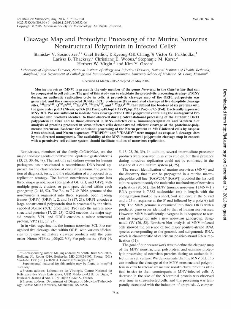
FIG. 1. Expression of MNV polyprotein fragments in E. coli and specificities of the corresponding antisera. (A) Schematic diagram showingthe organization of the MNV genome, putative cleavage products of the virus ORF1 polyprotein, and location of cDNA fragments expressed inE. coli. Predicted dipeptide cleavage sites are indicated with arrows, and the calculated molecular weights (MW) (in thousands) of the expected
7818 SOSNOVTSEV ET AL. J. VIROL.
on Decem
ber 17, 2013 by guesthttp://jvi.asm
.org/D
ownloaded from

�g of the desired plasmid DNAs. Radiolabeling was performed in the presenceof [35S]methionine (�1,000 Ci/mmol; Amersham Biosciences).
Immunoprecipitation and Western blot analysis. Immunoprecipitation assaysof radiolabeled virus proteins from infected cell lysates or in vitro translationmixtures were carried out using region-specific antisera as described previously(44, 45).
Western blot analysis was performed according to standard techniques (37).Briefly, each cell lysate sample (10 �g) was solubilized in sodium dodecyl sulfate(SDS)-polyacrylamide gel electrophoresis (PAGE) sample buffer containing2.5% �-mercaptoethanol, heated at 95°C for 5 min, and subjected to SDS-PAGEseparation in a 4 to 20% Tris-glycine gel (Cambrex, Rockland, ME). The re-solved proteins were transferred onto a nitrocellulose membrane (Invitrogen) byelectroblotting, and the membrane was incubated with region-specific antiserum.The binding of the primary antibodies was detected using goat anti-guinea pigimmunoglobulins conjugated with horseradish peroxidase (Kirkegaard & PerryLaboratories, Gaithersburg, MD) followed by development with SuperSignalWest Pico chemiluminescent substrate (Pierce) according to the manufactur-er’s protocol. Detection of the His6-tagged proteins expressed in bacteria wasperformed using Penta-His antibody (QIAGEN) and goat anti-mouse immu-noglobulins conjugated with horseradish peroxidase (Pierce) as a secondaryantibody.
Nucleotide and protein sequencing. Nucleotide sequence analysis of the con-structed plasmid DNAs was performed by using the BigDye Terminator v3.1Cycle Sequencing Ready Reaction kit and an ABI 3100 automated sequencer(Applied Biosystems, Foster City, CA).
Direct N-terminal sequence analyses of radiolabeled virus-specific proteinsand proteins expressed in bacteria were conducted as described previously(42, 44).
Caspase cleavage. Caspase cleavage assays were carried out using mouserecombinant caspase 3 (EMD Biosciences, La Jolla, CA). Briefly, 1 unit ofcaspase 3 was added to radiolabeled Nterm protein in buffer containing 50 mMHEPES (pH 7.4) and 10 mM dithiothreitol, and the reaction mixture was incu-bated at 37°C for 1 to 3 h.
RESULTS
Immunoprecipitation analysis of MNV-1 ORF1 polyproteincleavage products with region-specific antisera. The ORF1nonstructural polyproteins of human norovirus strains Southamp-ton virus (SHV), Norwalk virus (NV), Camberwell virus (CV),and MD-145 virus (MDV) have been reported to undergo pro-teolytic processing at five cleavage sites that would yield six ma-ture cleavage products designated Nterm, NTPase, p18 (p22),VPg, Pro, and Pol (4, 5, 15, 25, 26, 39, 40). In addition, severalprecursor proteins that represent intermediate forms of polypro-tein processing have been described previously (4, 5, 15, 25, 39,40). The MNV-1 ORF1 polyprotein is 1,687 aa in length andshows 59 to 64% similarity with the corresponding polyproteins ofSHV, NV, CV, and MDV.
An alignment of the MNV-1 polyprotein with these norovi-ruses identified five potential cleavage sites, 341E/G342, 705Q/N706, 870E/G871, 994E/A995, and 1177Q/G1178 (Fig. 1A and seebelow), which would yield six mature proteins with calculatedmolecular masses of 38.3, 39.6, 18.6, 14.3, 19.2, and 57.5 kDa.In order to confirm this predicted cleavage map, a full-length
cDNA clone (p20.3) of the MNV-1 genome was constructed,and the ORF1 encoded in this clone was expressed in an invitro-coupled TNT system. Several proteins with masses rang-ing from approximately 18 to 110 kDa were observed (Fig. 1B,lane 1). To facilitate the identification of the protein products,we generated a panel of region-specific antisera to selectedparts of the MNV-1 polyprotein within the predicted borders.The full-length sequences of the predicted mature MNV-1VPg, Pro, and ProPol proteins were successfully expressed inbacteria, whereas expression of the predicted mature Nterm,NTPase, and p18.6 proteins failed. Consequently, discretesmaller regions from the Nterm, NTPase, and p18.6 proteinswere selected and expressed in bacteria (Fig. 1A; see Table S1in the supplemental material) as His6-tagged fusion proteinsusing the pET-28a plasmid (Novagen). The recombinant pro-teins were purified from the insoluble fractions of the bacterialcells by the IMAC purification procedure using Ni-NTA aga-rose columns (QIAGEN) to at least 90% homogeneity (Fig.1A). Guinea pigs were immunized with each protein, and theresulting antisera were shown to be antigen specific by West-ern blot analysis, with the exception of serum raised againstNTPase (data not shown). The recombinant protein derivedfrom the cells expressing p�NTPase was not immunogenic(data not shown).
The ORF1 region-specific antisera were used to localize thecoding sequences for individual nonstructural proteins in theMNV-1 ORF1 sequence (Fig. 1B). The anti-Nterm serum,raised against the extreme N-terminal region of ORF1, recog-nized three major proteins (two migrating as a 45-kDa doubletand one at 39 kDa) (Fig. 1B, lane 3). Translation of the ORF1following elimination of the first two AUGs (constructp�NORF1) led to the disappearance of the 45-kDa doubletband and efficient production of the 39-kDa (�NNterm) pro-tein (Fig. 1B, lane 12), indicating that the three proteins thatimmunoprecipitated with the anti-Nterm serum were productsof initiation at three different AUGs. The predicted C-terminalborder of these products would define Nterm proteins withmolecular masses of 33.2, 38, and 38.3 kDa. The observeddiscrepancy between the predicted and observed molecularmasses of these proteins could be explained by either abnormalmobility in SDS-PAGE or the utilization of an unexpectedcleavage site to release the C terminus of the Nterm protein.Other predicted individual proteins precipitated by region-specific sera were an 18-kDa protein with anti-p18 (Fig. 1B,lane 5), a 19-kDa protein (Pro) with anti-Pro and anti-ProPol(Fig. 1B, lanes 9 and 11), and a 57-kDa protein (Pol) withanti-ProPol (Fig. 1B, lane 11).
Larger proteins representing ORF1 processing intermedi-
cleavage products are shown. The cDNA fragments engineered for expression in E. coli are depicted as dark gray bars. Fusion proteinscorresponding to the selected ORF1 regions were purified using Ni-NTA agarose. For lanes 1 to 6, a sample of each region-specific protein(identified above the lanes) was analyzed by SDS-PAGE followed by Coomassie blue staining. (B) Immunoprecipitation of the MNV ORF1nonstructural proteins synthesized in a TNT reaction from the genomic full-length clone p20.3 using a panel of region-specific antisera.Radiolabeled MNV ORF1 polyprotein cleavage products (lane 1) were immunoprecipitated with either pre- or postimmunization sera raisedagainst Nterm protein (lanes 2 and 3), p18 protein (lanes 4 and 5), VPg protein (lanes 6 and 7), Pro protein (lanes 8 and 9), and ProPol protein(lanes 10 and 11) and subjected to SDS-PAGE in a 12% Tris-glycine gel followed by autoradiography of the dried gel. Lane 12, TNT productsderived from the MNV ORF1 clone pDNORF1 in which the first two AUG codons were abolished. Proteins that were not identified in this studyand that likely represented nonspecific products in the TNT reaction are indicated with asterisks.
VOL. 80, 2006 CLEAVAGE MAP OF THE MNV NONSTRUCTURAL POLYPROTEIN 7819
on Decem
ber 17, 2013 by guesthttp://jvi.asm
.org/D
ownloaded from
ates were precipitated with region-specific sera. The antisera tothe p18, VPg, Pro, and ProPol regions precipitated a 110-kDaprotein from the in vitro translation mixture derived fromp20.3 (Fig. 1B, lanes 5, 7, 9, and 11). Three sera, anti-VPg,anti-Pro, and anti-ProPol, precipitated a protein of approxi-mately 90 kDa, while anti-p18 serum did not recognize thisprotein but precipitated one protein with a molecular mass of18 kDa. Anti-Pro and anti-ProPol sera both recognized a 77-kDa protein, whereas anti-p18 and anti-VPg sera did not.Thus, the 110-kDa protein was likely a precursor to the 18- and90-kDa proteins and represented the p18.6-VPg-Pro-Pol pro-tein complex. Furthermore, the 90-kDa protein correspondedto the VPg-Pro-Pol precursor, and the 77-kDa protein corre-sponded to the ProPol precursor. A 14-kDa band correspond-ing to the mature VPg protein was not observed among trans-lational products precipitated with the anti-VPg serum, afinding which was likely due to the absence of methionineresidues in this protein sequence.
All four sera (anti-p18, anti-VPg, anti-Pro, and anti-ProPol)precipitated 55-, 45 (migrating as a doublet)-, 39-, and 35-kDaproteins (Fig. 1B, lanes 5, 7, 9, and 11). The 35- and 55-kDaproteins were detected only as faint bands in products precip-itated by the anti-Nterm serum (serum specific to the N ter-minus of the ORF1 polyprotein), and the observed mass ofthese proteins was consistent with those of p18-VPg and p18-VPg-Pro, respectively. A coprecipitation of the three forms ofthe Nterm protein (45-kDa doublet and 39 kDa) was observedunder these experimental conditions with the anti-p18, anti-VPg, anti-Pro, and anti-ProPol sera. This observation sug-gests possible interactions of the Nterm protein with thep18.6, VPg, Pro, and Pol proteins. These interactions wouldalso be consistent with the detection of the p18-VPg-Pro-Pol(110-kDa) protein among the products precipitated by theanti-A serum. The possibility of such interactions among thenonstructural proteins has been reported recently for felinecalicivirus (FCV) (19).
None of the region-specific sera recognized the 40-kDa pro-tein from the p20.3 translational mixture. The identity of the40-kDa protein as the NTPase was determined by mutagenesisand microsequencing experiments (see below).
Mapping of VPg, 3CL Pro, and polymerase. Cleavage of thecalicivirus nonstructural polyprotein has been associated with a3CL proteinase encoded in the C-terminal part of the ORF1polyprotein (25, 34, 45, 49). Comparison of the C-terminal partof the MNV-1 ORF1 polyprotein amino acid sequence withthose of CV, MDV, NV, and SHV revealed the presence of a1131GDCG1134 motif in the MNV-1 polyprotein (see Fig. S1 inthe supplemental material) that included a cysteine residuethat was previously shown to be part of the catalytic site (32,41). Substitution of the Cys1133 with Ala in a full-lengthgenomic MNV-1 clone (p20.3Prom) (see Table S2 in the sup-plemental material) abolished autocatalytic processing of theORF1-encoded polyprotein (see Fig. S1 in the supplementalmaterial), thus confirming the identity of the MNV-1 enzymeas a cysteine 3CL Pro.
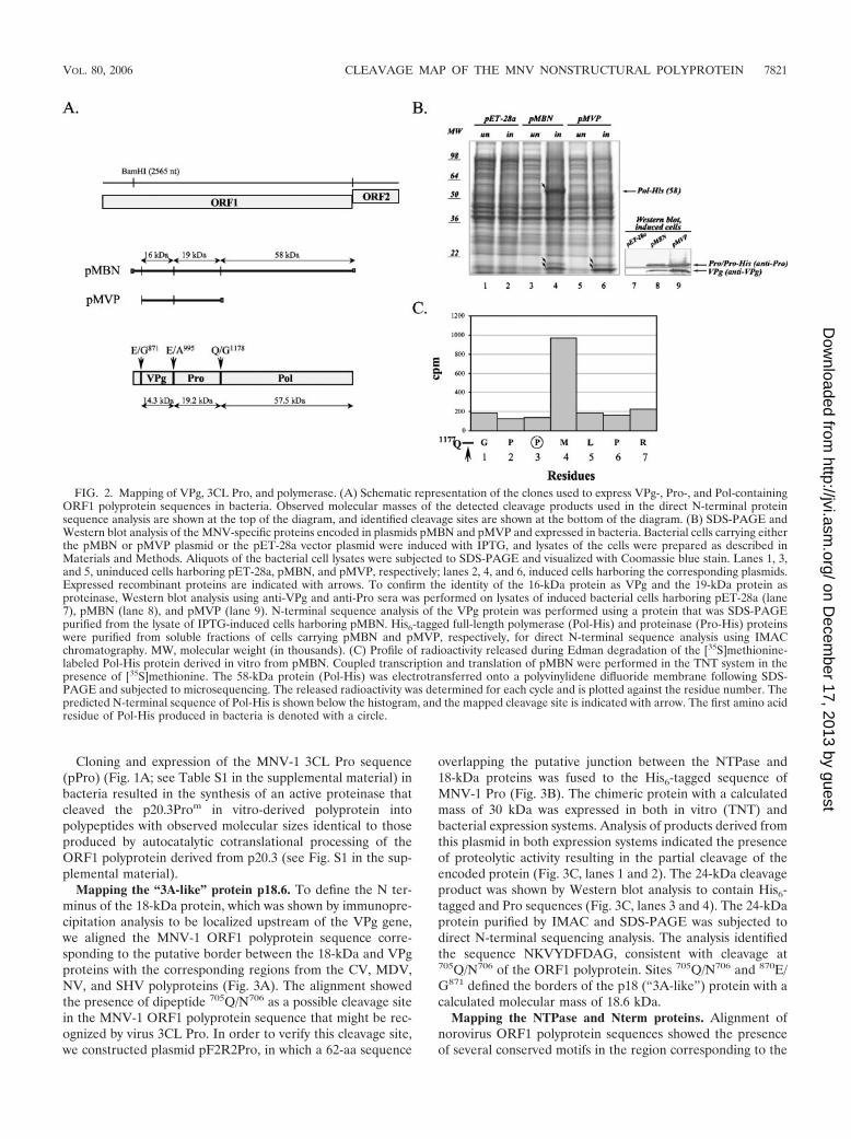
To verify the cleavage sites bordering the MNV-1 Pro se-quence, the ORF1 sequence localized between the BamHI(2,565 nt) restriction site and the ORF1 3� end was subclonedto generate clone pMBN. The protein sequence encoded inpMBN overlapped the entire VPg, Pro, and Pol and contained
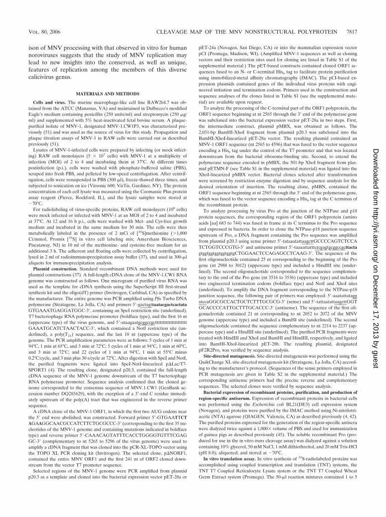
a partial sequence of p18 that should yield an expressed pro-tein of 97.3 kDa (Fig. 2A). However, comparative SDS-PAGEanalysis of the proteins expressed by bacteria carrying thepMBN plasmid (Fig. 2B, lane 4) with those from the pET-28acontrol (Fig. 2B, lane 2) showed the presence of three uniqueproteins of 58, 19, and 16 kDa in the lysate of pMBN-trans-formed IPTG (isopropyl-�-D-thiogalactopyranoside)-inducedcells. Western blot analysis of the corresponding cell lysateswith region-specific antisera showed that the 16-kDa proteinband was specifically recognized by anti-VPg serum and thatthe 19-kDa band was recognized by anti-Pro serum (Fig. 2B,lane 8). N-terminal sequence analysis of the 16-kDa proteinidentified the sequence GKKGKNKK, which was consistentwith the utilization of the cleavage site at position 870E/G871 torelease the N terminus of VPg (Fig. 2A).
To identify the 58-kDa protein, we purified the expressedprotein from the soluble fraction of the bacterial cells using theIMAC procedure (data not shown) and subjected it to N-terminal sequence analysis. The identified sequence, PMLPRPSG, corresponded to aa 1180 to 1187 of the ORF1 polypro-tein and was located two residues downstream from the 1177Q/G1178 cleavage site that was predicted to release the Nterminus of the polymerase (Fig. 2C). Norovirus 3CL Pro hasbeen reported to recognize cleavage sites that contain glu-tamine or glutamic acid residues in the P1 position (4, 5, 15, 25,26, 39, 40). The observation of an unusual cleavage productwith a Pro residue in the P1 and P1� positions suggested thepresence of contaminating protease activity in the bacterial celllysates. However, the use of a protease inhibitor cocktail dur-ing the protein purification procedure did not prevent thisaberrant processing (data not shown). To establish whether thesame N terminus is generated in a eukaryotic expression sys-tem, we expressed the pMBN sequence in a TNT T7 system.Microsequencing performed on the in vitro-synthesized 58-kDa protein radiolabeled with [35S]methionine demonstrated apeak of radioactivity for residue 4 (Fig. 2C), which was con-sistent with cleavage of the ORF1 polyprotein at the 1177Q/G1178 site (Fig. 2C). Furthermore, mutagenesis of the Gln1177
residue to Gly in the p20.3 clone abolished expression of the58-kDa protein (data not shown).
We cloned and expressed the sequence of the ORF1polyprotein bordered by the newly determined 870E/G871 and1177Q/G1178 cleavage sites (pMVP) (see Table S1 in the sup-plemental material) in bacteria (Fig. 2A). Two bands corre-sponding to proteins with molecular masses of approximately16 and 19 to 20 kDa (Fig. 2B, lane 6) comigrated with theanalogous proteins from pMBN-expressing cells and were rec-ognized by anti-VPg and anti-Pro sera (Fig. 2B, lane 9). A His6
tag sequence introduced at the C terminus of the pMVP-cloned sequence facilitated the purification of the 19- to 20-kDa protein from the soluble fraction of the bacterial cells.Direct sequence analysis of the purified protein identified thesequence APVSI at its N terminus, consistent with the pres-ence of a cleavage site at position 994E/A995 that would definethe border between the VPg and Pro proteins in the ORF1polyprotein. Cleavage site 994E/A995 together with 1177Q/G1178
defined the boundaries of MNV-1 Pro with a calculated mo-lecular mass of 19.2 kDa, while sites 870E/G871 and 994E/A995
defined the boundaries of MNV-1 VPg with a calculated mo-lecular mass of 14.3 kDa (Fig. 2A).
7820 SOSNOVTSEV ET AL. J. VIROL.
on Decem
ber 17, 2013 by guesthttp://jvi.asm
.org/D
ownloaded from
Cloning and expression of the MNV-1 3CL Pro sequence(pPro) (Fig. 1A; see Table S1 in the supplemental material) inbacteria resulted in the synthesis of an active proteinase thatcleaved the p20.3Prom in vitro-derived polyprotein intopolypeptides with observed molecular sizes identical to thoseproduced by autocatalytic cotranslational processing of theORF1 polyprotein derived from p20.3 (see Fig. S1 in the sup-plemental material).
Mapping the “3A-like” protein p18.6. To define the N ter-minus of the 18-kDa protein, which was shown by immunopre-cipitation analysis to be localized upstream of the VPg gene,we aligned the MNV-1 ORF1 polyprotein sequence corre-sponding to the putative border between the 18-kDa and VPgproteins with the corresponding regions from the CV, MDV,NV, and SHV polyproteins (Fig. 3A). The alignment showedthe presence of dipeptide 705Q/N706 as a possible cleavage sitein the MNV-1 ORF1 polyprotein sequence that might be rec-ognized by virus 3CL Pro. In order to verify this cleavage site,we constructed plasmid pF2R2Pro, in which a 62-aa sequence
overlapping the putative junction between the NTPase and18-kDa proteins was fused to the His6-tagged sequence ofMNV-1 Pro (Fig. 3B). The chimeric protein with a calculatedmass of 30 kDa was expressed in both in vitro (TNT) andbacterial expression systems. Analysis of products derived fromthis plasmid in both expression systems indicated the presenceof proteolytic activity resulting in the partial cleavage of theencoded protein (Fig. 3C, lanes 1 and 2). The 24-kDa cleavageproduct was shown by Western blot analysis to contain His6-tagged and Pro sequences (Fig. 3C, lanes 3 and 4). The 24-kDaprotein purified by IMAC and SDS-PAGE was subjected todirect N-terminal sequencing analysis. The analysis identifiedthe sequence NKVYDFDAG, consistent with cleavage at705Q/N706 of the ORF1 polyprotein. Sites 705Q/N706 and 870E/G871 defined the borders of the p18 (“3A-like”) protein with acalculated molecular mass of 18.6 kDa.
Mapping the NTPase and Nterm proteins. Alignment ofnorovirus ORF1 polyprotein sequences showed the presenceof several conserved motifs in the region corresponding to the
FIG. 2. Mapping of VPg, 3CL Pro, and polymerase. (A) Schematic representation of the clones used to express VPg-, Pro-, and Pol-containingORF1 polyprotein sequences in bacteria. Observed molecular masses of the detected cleavage products used in the direct N-terminal proteinsequence analysis are shown at the top of the diagram, and identified cleavage sites are shown at the bottom of the diagram. (B) SDS-PAGE andWestern blot analysis of the MNV-specific proteins encoded in plasmids pMBN and pMVP and expressed in bacteria. Bacterial cells carrying eitherthe pMBN or pMVP plasmid or the pET-28a vector plasmid were induced with IPTG, and lysates of the cells were prepared as described inMaterials and Methods. Aliquots of the bacterial cell lysates were subjected to SDS-PAGE and visualized with Coomassie blue stain. Lanes 1, 3,and 5, uninduced cells harboring pET-28a, pMBN, and pMVP, respectively; lanes 2, 4, and 6, induced cells harboring the corresponding plasmids.Expressed recombinant proteins are indicated with arrows. To confirm the identity of the 16-kDa protein as VPg and the 19-kDa protein asproteinase, Western blot analysis using anti-VPg and anti-Pro sera was performed on lysates of induced bacterial cells harboring pET-28a (lane7), pMBN (lane 8), and pMVP (lane 9). N-terminal sequence analysis of the VPg protein was performed using a protein that was SDS-PAGEpurified from the lysate of IPTG-induced cells harboring pMBN. His6-tagged full-length polymerase (Pol-His) and proteinase (Pro-His) proteinswere purified from soluble fractions of cells carrying pMBN and pMVP, respectively, for direct N-terminal sequence analysis using IMACchromatography. MW, molecular weight (in thousands). (C) Profile of radioactivity released during Edman degradation of the [35S]methionine-labeled Pol-His protein derived in vitro from pMBN. Coupled transcription and translation of pMBN were performed in the TNT system in thepresence of [35S]methionine. The 58-kDa protein (Pol-His) was electrotransferred onto a polyvinylidene difluoride membrane following SDS-PAGE and subjected to microsequencing. The released radioactivity was determined for each cycle and is plotted against the residue number. Thepredicted N-terminal sequence of Pol-His is shown below the histogram, and the mapped cleavage site is indicated with arrow. The first amino acidresidue of Pol-His produced in bacteria is denoted with a circle.
VOL. 80, 2006 CLEAVAGE MAP OF THE MNV NONSTRUCTURAL POLYPROTEIN 7821
on Decem
ber 17, 2013 by guesthttp://jvi.asm
.org/D
ownloaded from

border of Nterm and NTPase protein sequences, including aQG dipeptide identified as the cleavage site for SHV andMDV (4, 5, 15, 25, 40). The analogous site in the MNV-1ORF1 polyprotein was an EG dipeptide in positions 341 and342 (Fig. 4A). To investigate whether this site is utilized byvirus Pro, we attempted to express protein sequences of vari-ous lengths that overlapped the Nterm-NTPase border andthat were fused to the Pro sequence.
The expression of larger Nterm and NTPase sequences inbacteria failed (data not shown). Moreover, bacterial expres-sion of chimeric proteins containing truncated Nterm-NTPasejunction sequences fused to Pro did not result in proteolyticprocessing (data not shown), indicating that the recognition ofthis site by virus Pro might be dependent on the conformationof the adjacent protein sequence. To confirm cleavage at the341E/G342 site, the Glu341 residue was replaced with a Glyresidue using site-directed mutagenesis of the p20.3 full-lengthclone. SDS-PAGE analysis of the proteins expressed from the
resulting clone, p20.3m341, in an in vitro translation systemshowed the disappearance of the 40-and 45-kDa protein bandsand the appearance of two unique higher-molecular-mass pro-teins (Fig. 4B, compare lanes 1 and 3). These two larger pro-teins likely represented unprocessed precursors of the Ntermand NTPase proteins, suggesting that cleavage had been abol-ished at this site and that initiation had occurred at two dif-ferent AUG codons. It should be noted that products of ap-proximately 40 kDa were sometimes observed in the analysis ofp20.3m341 (Fig. 4B, lane 3), which might indicate processing atan alternative cleavage site. Additional support for the utiliza-tion of the 341E/G342 cleavage site was obtained from proteinmicrosequence analysis. The in vitro [35S]methionine-labeled40-kDa protein was resolved by SDS-PAGE in a 20-cm-longpolyacrylamide gel, excised from the gel, and subjected toEdman degradation analysis. The profile of radioactivity re-leased during microsequencing revealed a peak representing aradiolabeled methionine in position 19, which was consistent
FIG. 3. Analysis of the cleavage site between NTPase and p18. (A) Alignment of the amino acid sequence of the putative junction of the MNVNTPase and p18 proteins with those of CV, MDV, NV, and SHV. Numbers correspond to the ORF1 polyprotein sequence of each virus, andasterisks indicate conserved amino acids. The cleavage site in this region, identified previously for human norovirus strains, is indicated with anarrow. (B) Schematic representation of the pF2R2Pro plasmid that was engineered to contain the MNV-1 NTPase-p18 junction and an active 3CLPro. The chimeric protein was fused to the vector sequence (gray box) at its N terminus, and the entire VPg and p18 C-terminal sequences weredeleted. (C) SDS-PAGE analysis of proteins encoded in pF2R2Pro when expressed in vitro and in E. coli. Lane 1, in vitro TNT translation productsderived from pF2R2Pro (autoradiography); lane 2, IMAC-purified proteins isolated from the insoluble fraction of the induced bacterial cellscarrying pF2R2Pro stained with Coomassie blue (Cblue); lanes 3 and 4, Western blot analysis of the same protein sample using PentaHis antibody(QIAGEN) and anti-Pro serum, respectively. The two major pF2R2Pro proteins were expressed in both in vitro translation reactions and bacteria.The N-terminal sequence of the 24-kDa protein (NKVYDFDAG) is shown.
7822 SOSNOVTSEV ET AL. J. VIROL.
on Decem
ber 17, 2013 by guesthttp://jvi.asm
.org/D
ownloaded from
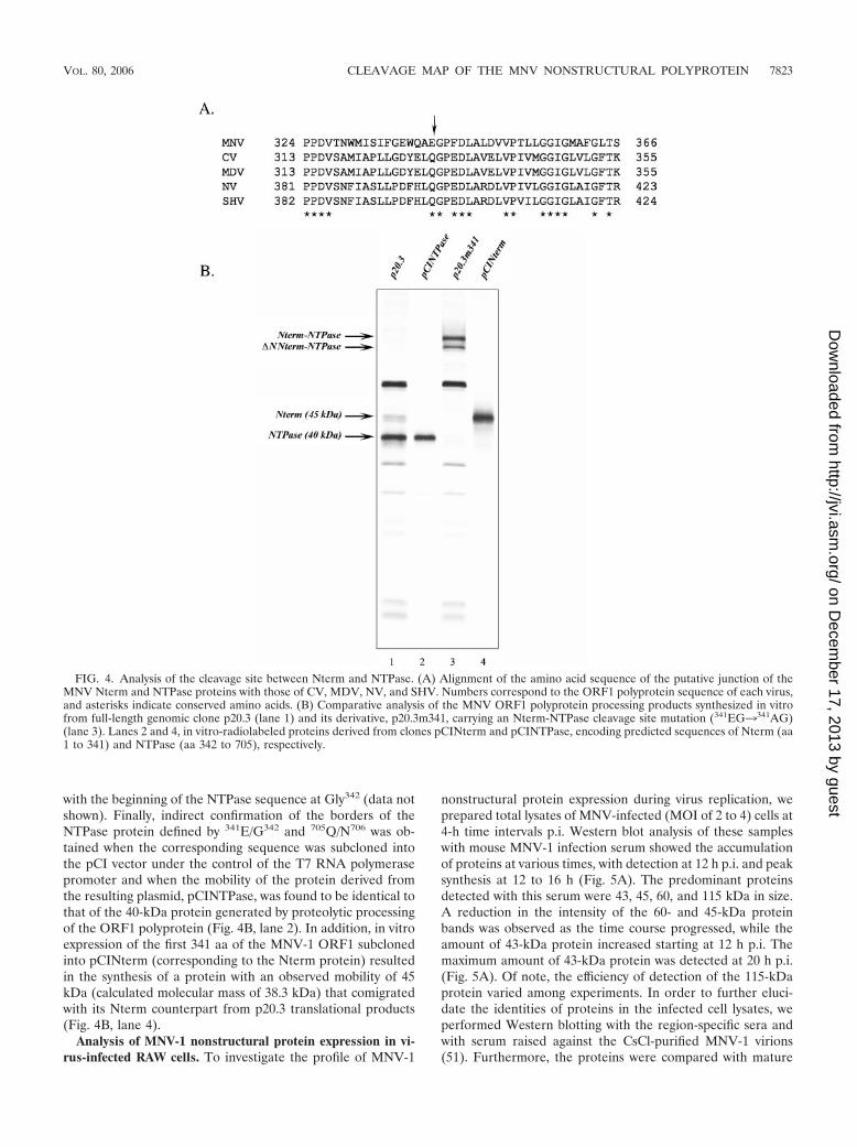
with the beginning of the NTPase sequence at Gly342 (data notshown). Finally, indirect confirmation of the borders of theNTPase protein defined by 341E/G342 and 705Q/N706 was ob-tained when the corresponding sequence was subcloned intothe pCI vector under the control of the T7 RNA polymerasepromoter and when the mobility of the protein derived fromthe resulting plasmid, pCINTPase, was found to be identical tothat of the 40-kDa protein generated by proteolytic processingof the ORF1 polyprotein (Fig. 4B, lane 2). In addition, in vitroexpression of the first 341 aa of the MNV-1 ORF1 subclonedinto pCINterm (corresponding to the Nterm protein) resultedin the synthesis of a protein with an observed mobility of 45kDa (calculated molecular mass of 38.3 kDa) that comigratedwith its Nterm counterpart from p20.3 translational products(Fig. 4B, lane 4).
Analysis of MNV-1 nonstructural protein expression in vi-rus-infected RAW cells. To investigate the profile of MNV-1
nonstructural protein expression during virus replication, weprepared total lysates of MNV-infected (MOI of 2 to 4) cells at4-h time intervals p.i. Western blot analysis of these sampleswith mouse MNV-1 infection serum showed the accumulationof proteins at various times, with detection at 12 h p.i. and peaksynthesis at 12 to 16 h (Fig. 5A). The predominant proteinsdetected with this serum were 43, 45, 60, and 115 kDa in size.A reduction in the intensity of the 60- and 45-kDa proteinbands was observed as the time course progressed, while theamount of 43-kDa protein increased starting at 12 h p.i. Themaximum amount of 43-kDa protein was detected at 20 h p.i.(Fig. 5A). Of note, the efficiency of detection of the 115-kDaprotein varied among experiments. In order to further eluci-date the identities of proteins in the infected cell lysates, weperformed Western blotting with the region-specific sera andwith serum raised against the CsCl-purified MNV-1 virions(51). Furthermore, the proteins were compared with mature
FIG. 4. Analysis of the cleavage site between Nterm and NTPase. (A) Alignment of the amino acid sequence of the putative junction of theMNV Nterm and NTPase proteins with those of CV, MDV, NV, and SHV. Numbers correspond to the ORF1 polyprotein sequence of each virus,and asterisks indicate conserved amino acids. (B) Comparative analysis of the MNV ORF1 polyprotein processing products synthesized in vitrofrom full-length genomic clone p20.3 (lane 1) and its derivative, p20.3m341, carrying an Nterm-NTPase cleavage site mutation (341EG3341AG)(lane 3). Lanes 2 and 4, in vitro-radiolabeled proteins derived from clones pCINterm and pCINTPase, encoding predicted sequences of Nterm (aa1 to 341) and NTPase (aa 342 to 705), respectively.
VOL. 80, 2006 CLEAVAGE MAP OF THE MNV NONSTRUCTURAL POLYPROTEIN 7823
on Decem
ber 17, 2013 by guesthttp://jvi.asm
.org/D
ownloaded from
7824
on Decem
ber 17, 2013 by guesthttp://jvi.asm
.org/D
ownloaded from

nonstructural proteins that had been cloned into a pCI vectorand expressed in the TNT system. The approximately 60-kDaband was identified as the virus capsid protein VP1, because itwas recognized by serum raised against purified MNV-1 virions(51) (Fig. 5B, lanes 3 to 7), and its mobility was similar to thatof VP1 derived by in vitro translation from a plasmid(pCIORF2) carrying the entire ORF2 of the MNV-1 genomecloned under the control of the T7 RNA polymerase promoter(Fig. 5B, lane 8). The similarity in the observed mobilities ofthese proteins indicates that the MNV-1 capsid protein doesnot undergo proteolytic processing during its maturation. Ofinterest, the anti-MNV-1 virion serum recognized also a 120-kDa protein that was consistent in size with a dimeric form ofthe MNV-1 capsid protein (Fig. 5B, lanes 4 and 5). Westernblot detection of capsid protein dimers has previously beendescribed for FCV and rabbit hemorrhagic disease virus(RHDV) (24, 44).
As shown in Fig. 5C, using anti-Nterm serum, we identified
proteins with observed masses of 43, 45, and 115 kDa in MNV-1-infected cells as Nterm-related proteins. The mobility of the45-kDa protein in cells corresponded to that of the Ntermprotein expressed from pCINterm in the TNT system, indicat-ing that the 45-kDa protein was the full-length Nterm protein(Fig. 5C, lanes 4 to 8, and D, lanes 1 to 4). The detected changein the relative amounts of the 45- and 43-kDa proteins in theNterm Western blot was similar to that observed with theinfection serum and suggested a modification of the Ntermprotein over time. The presence of the 115-kDa protein couldreflect either the existence of a larger precursor that wouldinclude sequences of p18 and possibly VPg or the existence ofmultimeric forms of Nterm or an Nterm-NTPase complex.However, the probing of cell lysates with anti-p18 serum re-sulted in the detection of only the p18 protein (Fig. 5G, lanes4 to 7). The characterization of the 115-kDa complex willrequire further study.
The predominant form of the virus polymerase recognized
FIG. 5. Expression of MNV-1 proteins in infected cells. (A to C and E to H) Virus proteins in MNV-1-infected cells collected at different timepoints following infection were analyzed. Ten micrograms of each cell lysate prepared from collected cells was resolved on a 4 to 20%polyacrylamide gel by SDS-PAGE and transferred electrophoretically onto a nitrocellulose membrane. The membrane was probed with serumcollected from a mouse that had undergone infection with MNV-1 (A). The same lysates from the MNV-infected cells were probed withregion-specific antisera: anti-capsid (B), anti-Nterm (C), anti-ProPol (E), anti-Pro (F), anti-p18 (G), and anti-VPg (H) sera. To compare themobilities of the virus proteins synthesized in infected cells and in vitro, TNT products derived from pCIORF2, pCIPol, pCIPro, and pCINtermwere analyzed concurrently with the corresponding cell lysate samples probed with anti-capsid (B, lane 8), anti-Nterm (C, lane 8), and anti-ProPol(E, lane 8) sera. (D) Comparison of MNV Nterm proteins expressed in vitro and in infected cells. Immunoprecipitation (IP) of the radiolabeledNterm protein synthesized in the in vitro system from pCINterm and p20.3 (lanes 1 and 3, respectively) and in infected cells (lane 2) is shown. Lane4, the MNV ORF1 nonstructural proteins derived from p20.3 in vitro. (I) Comparative immunoprecipitation analysis of p18-, VPg-, and Pro-relatedproteins derived by in vitro cleavage of the ORF1 polyprotein or synthesized in MNV-infected cells. The radiolabeled p18-related proteins wereprecipitated using anti-p18 serum from the ORF1 (lane 1) and pCIP18 (lane 3) translation mixture or from MNV-infected cells (lane 2). Theradiolabeled VPg-related proteins were precipitated using anti-VPg serum from the ORF1 translation mixture (lane 4) or from MNV-infected cells(lane 5). The radiolabeled Pro-related proteins were precipitated using anti-Pro serum from the ORF1 (lane 6) and pCIPro (lane 8) translationmixture or from MNV-infected cells (lane 7). (J) Expression of the p18 protein in in vitro systems. Lanes 1 and 2, immunoprecipitation of the p18protein derived from pCIP18 using the TNT T7 Coupled Reticulocyte Lysate system (RR) or the TNT T7 Coupled Wheat Germ Extract system(WGE) (Promega), respectively; lane 3, the MNV ORF1 nonstructural proteins derived from p20.3 using the TNT T7 Coupled Reticulocyte Lysatesystem. Modified and free forms of the p18 protein are indicated with arrows. MW, molecular weight (in thousands).
VOL. 80, 2006 CLEAVAGE MAP OF THE MNV NONSTRUCTURAL POLYPROTEIN 7825
on Decem
ber 17, 2013 by guesthttp://jvi.asm
.org/D
ownloaded from
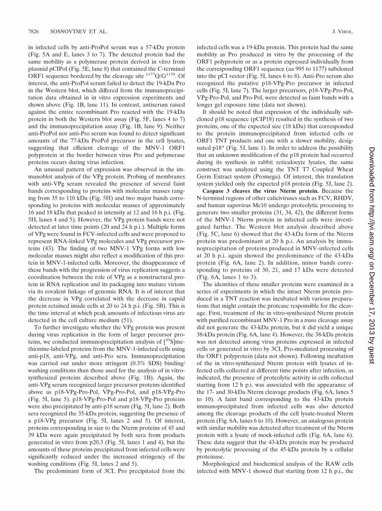
in infected cells by anti-ProPol serum was a 57-kDa protein(Fig. 5A and E, lanes 3 to 7). The detected protein had thesame mobility as a polymerase protein derived in vitro fromplasmid pCIPol (Fig. 5E, lane 8) that contained the C-terminalORF1 sequence bordered by the cleavage site 1177Q/G1178. Ofinterest, the anti-ProPol serum failed to detect the 19-kDa Proin the Western blot, which differed from the immunoprecipi-tation data obtained in in vitro expression experiments andshown above (Fig. 1B, lane 11). In contrast, antiserum raisedagainst the entire recombinant Pro reacted with the 19-kDaprotein in both the Western blot assay (Fig. 5F, lanes 4 to 7)and the immunoprecipitation assay (Fig. 1B, lane 9). Neitheranti-ProPol nor anti-Pro serum was found to detect significantamounts of the 77-kDa ProPol precursor in the cell lysates,suggesting that efficient cleavage of the MNV-1 ORF1polyprotein at the border between virus Pro and polymeraseproteins occurs during virus infection.
An unusual pattern of expression was observed in the im-munoblot analysis of the VPg protein. Probing of membraneswith anti-VPg serum revealed the presence of several faintbands corresponding to proteins with molecular masses rang-ing from 35 to 110 kDa (Fig. 5H) and two major bands corre-sponding to proteins with molecular masses of approximately16 and 18 kDa that peaked in intensity at 12 and 16 h p.i. (Fig.5H, lanes 4 and 5). However, the VPg protein bands were notdetected at later time points (20 and 24 h p.i.). Multiple formsof VPg were found in FCV-infected cells and were proposed torepresent RNA-linked VPg molecules and VPg precursor pro-teins (43). The finding of two MNV-1 VPg forms with lowmolecular masses might also reflect a modification of this pro-tein in MNV-1-infected cells. Moreover, the disappearance ofthese bands with the progression of virus replication suggests acoordination between the role of VPg as a nonstructural pro-tein in RNA replication and its packaging into mature virionsvia its covalent linkage of genomic RNA. It is of interest thatthe decrease in VPg correlated with the decrease in capsidprotein retained inside cells at 20 to 24 h p.i. (Fig. 5B). This isthe time interval at which peak amounts of infectious virus aredetected in the cell culture medium (51).
To further investigate whether the VPg protein was presentduring virus replication in the form of larger precursor pro-teins, we conducted immunoprecipitation analysis of [35S]me-thionine-labeled proteins from the MNV-1-infected cells usinganti-p18, anti-VPg, and anti-Pro sera. Immunoprecipitationwas carried out under more stringent (0.3% SDS) binding/washing conditions than those used for the analysis of in vitro-synthesized proteins described above (Fig. 1B). Again, theanti-VPg serum recognized larger precursor proteins identifiedabove as p18-VPg-Pro-Pol, VPg-Pro-Pol, and p18-VPg-Pro(Fig. 5I, lane 5). p18-VPg-Pro-Pol and p18-VPg-Pro proteinswere also precipitated by anti-p18 serum (Fig. 5I, lane 2). Bothsera recognized the 35-kDa protein, suggesting the presence ofa p18-VPg precursor (Fig. 5I, lanes 2 and 5). Of interest,proteins corresponding in size to the Nterm proteins of 45 and39 kDa were again precipitated by both sera from productsgenerated in vitro from p20.3 (Fig. 5I, lanes 1 and 4), but theamounts of these proteins precipitated from infected cells weresignificantly reduced under the increased stringency of thewashing conditions (Fig. 5I, lanes 2 and 5).
The predominant form of 3CL Pro precipitated from the
infected cells was a 19-kDa protein. This protein had the samemobility as Pro produced in vitro by the processing of theORF1 polyprotein or as a protein expressed individually fromthe corresponding ORF1 sequence (aa 995 to 1177) subclonedinto the pCI vector (Fig. 5I, lanes 6 to 8). Anti-Pro serum alsorecognized the putative p18-VPg-Pro precursor in infectedcells (Fig. 5I, lane 7). The larger precursors, p18-VPg-Pro-Pol,VPg-Pro-Pol, and Pro-Pol, were detected as faint bands with alonger gel exposure time (data not shown).
It should be noted that expression of the individually sub-cloned p18 sequence (pCIP18) resulted in the synthesis of twoproteins, one of the expected size (18 kDa) that correspondedto the protein immunoprecipitated from infected cells orORF1 TNT products and one with a slower mobility, desig-nated p18* (Fig. 5J, lane 1). In order to address the possibilitythat an unknown modification of the p18 protein had occurredduring its synthesis in rabbit reticulocyte lysates, the sameconstruct was analyzed using the TNT T7 Coupled WheatGerm Extract system (Promega). Of interest, this translationsystem yielded only the expected p18 protein (Fig. 5J, lane 2).
Caspase 3 cleaves the virus Nterm protein. Because theN-terminal regions of other caliciviruses such as FCV, RHDV,and human sapovirus Mc10 undergo proteolytic processing togenerate two smaller proteins (31, 34, 42), the different formsof the MNV-1 Nterm protein in infected cells were investi-gated further. The Western blot analysis described above(Fig. 5C, lane 6) showed that the 43-kDa form of the Ntermprotein was predominant at 20 h p.i. An analysis by immu-noprecipitation of proteins produced in MNV-infected cellsat 20 h p.i. again showed the predominance of the 43-kDaprotein (Fig. 6A, lane 2). In addition, minor bands corre-sponding to proteins of 30, 21, and 17 kDa were detected(Fig. 6A, lanes 1 to 3).
The identities of these smaller proteins were examined in aseries of experiments in which the intact Nterm protein pro-duced in a TNT reaction was incubated with various prepara-tions that might contain the protease responsible for the cleav-age. First, treatment of the in vitro-synthesized Nterm proteinwith purified recombinant MNV-1 Pro in a trans cleavage assaydid not generate the 43-kDa protein, but it did yield a unique38-kDa protein (Fig. 6A, lane 4). However, the 38-kDa proteinwas not detected among virus proteins expressed in infectedcells or generated in vitro by 3CL Pro-mediated processing ofthe ORF1 polyprotein (data not shown). Following incubationof the in vitro-synthesized Nterm protein with lysates of in-fected cells collected at different time points after infection, asindicated, the presence of proteolytic activity in cells collectedstarting from 12 h p.i. was associated with the appearance ofthe 17- and 30-kDa Nterm cleavage products (Fig. 6A, lanes 5to 10). A faint band corresponding to the 43-kDa proteinimmunoprecipitated from infected cells was also detectedamong the cleavage products of the cell lysate-treated Ntermprotein (Fig. 6A, lanes 6 to 10). However, an analogous proteinwith similar mobility was detected after treatment of the Ntermprotein with a lysate of mock-infected cells (Fig. 6A, lane 6).These data suggest that the 43-kDa protein may be producedby proteolytic processing of the 45-kDa protein by a cellularproteinase.
Morphological and biochemical analysis of the RAW cellsinfected with MNV-1 showed that starting from 12 h p.i., the
7826 SOSNOVTSEV ET AL. J. VIROL.
on Decem
ber 17, 2013 by guesthttp://jvi.asm
.org/D
ownloaded from

infected cells exhibited a number of changes characteristic ofapoptosis, including activation of caspases 3, 8, and 9 (S. V.Sosnovtsev, unpublished data). To test whether active caspasesare responsible for the cleavage of the MNV-1 Nterm protein,we incubated in vitro-synthesized Nterm protein with recom-binant human caspases 3, 8, and 9 and mouse caspase 3. Incu-bation with caspases 8 and 9 did not produce any cleavage
products (data not shown), while incubation with both human(data not shown) and mouse caspase 3 resulted in the detec-tion of 30-, 28-, 18-, and 17-kDa proteins (Fig. 6A, lane 11). Ofinterest, the 28-kDa protein was also present among Ntermcleavage products generated by the incubation of this proteinwith the infected cell lysate. It comigrated with one of theproteins observed in the control sample, and it was detected
FIG. 6. Detection and mapping of the MNV Nterm protein cleavage by caspase 3. (A) Comparison of cleavage products of the MNV Ntermprotein in virus-infected cells with those generated by cleavage with the virus Pro protein and caspase 3 in vitro. Lanes 1 and 2, immunoprecipi-tation of the radiolabeled Nterm protein from the virus-infected cells using pre- and postimmunization anti-Nterm serum, respectively; lane 4,products of incubation of the radiolabeled Nterm protein derived by TNT translation from pCINterm (Nterm TNT) (2 �l of the translationalmixture) with recombinant virus proteinase (HisPro) (2 �g, 3 h, 37°C), respectively (the 38-kDa cleavage product is indicated with an arrow); lanes6 to 10, products of incubation of the Nterm TNT with lysates (10 �l) of the cell collected at different times of infection; lane 11, incubation ofthe Nterm TNT with one unit of mouse recombinant caspase 3 (Cas3). Untreated Nterm TNT (incubated 3 h at 37°C) was included as a control(lanes 3 and 5). Asterisks denote cleavage products of the Nterm protein precipitated from infected cells that had mobilities similar to thosegenerated by caspase 3 cleavage. MW, molecular weight (in thousands). (B) Schematic representation of the Nterm clones pCINtermG103,pCINtermG121, and pCINtermG131 derived from pCINterm by site-directed mutagenesis of the caspase 3 putative cleavage sites 100DMSD103,118DRPD121, and 128DAMD131, respectively. Predicted caspase 3 cleavage products of the mutant Nterm proteins are depicted as gray boxes, andtheir molecular masses are shown above the diagram. (C) SDS-PAGE analysis of cleavage products obtained after incubation (3 h at 37°C) ofcaspase 3 (Cas3) with TNT-radiolabeled Nterm (lane 4) or with its mutant versions, NtermG103 (lane 5), NtermG121 (lane 6), and NtermG131 (lane7). TNT mixture without plasmid (lane 1), TNT-derived Nterm protein (lane 2), and the same protein after 3 h of incubation in assay buffer (lane3) were included as controls.
VOL. 80, 2006 CLEAVAGE MAP OF THE MNV NONSTRUCTURAL POLYPROTEIN 7827
on Decem
ber 17, 2013 by guesthttp://jvi.asm
.org/D
ownloaded from
only after prolonged SDS-PAGE separation of these proteins(data not shown). It should be noted that caspase 3 treatmentof the in vitro-synthesized Nterm protein did not generate the43-kDa protein observed in infected cells. The mechanismresponsible for the production of this protein will require fur-ther studies.
Examination of the Nterm amino acid sequence for a puta-tive caspase 3 cleavage site (DXXD) showed the presence ofthree sequences that could be processed by this proteinase:100DMSD103, 118DRPD121, and 128DAMD131 (Fig. 6B). Toinvestigate whether these sites are recognized by caspase 3, wereplaced Asp residues in the P1 positions of these cleavagesites with Gly using site-directed mutagenesis and plasmidpCINterm as the template. The mutagenized Nterm proteinswere expressed in the TNT system and incubated with activerecombinant mouse caspase 3. SDS-PAGE analysis of thecleavage products of the protein derived from pCINtermG103
showed a profile similar to that observed for the authenticNterm protein, suggesting that the 100DMSD103 site is notrecognized by caspase 3 (Fig. 6C, lane 5). In contrast, mutagen-esis of 118DRPD121 and 128DAMD131 sites resulted in changesin the cleavage profile (Fig. 6C, lanes 6 and 7). The D1213G121
mutation led to the disappearance of the 30-kDa protein bandand the generation of a new 20-kDa protein band (Fig. 6C,lane 6). The D1313G131 substitution resulted in the disappear-ance of the 28-kDa protein band (Fig. 6C, lane 7). The absenceof the 20-kDa and the presence of 30-kDa protein bands incleavage products of the Nterm protein suggested more effi-cient recognition by caspase 3 at the 118DRPD121 site. Ofinterest, the molecular masses of the caspase 3 cleavage prod-ucts with observed mobilities of 18, 28, and 30 kDa werepredicted to be 13.6, 23.7, and 24.7 kDa, respectively. Thediscrepancies in the predicted and observed protein SDS-PAGE mobilities were consistent with the unusual mobility ofthe full-length Nterm protein observed.
DISCUSSION
The replication strategy of viruses in the family Caliciviridaeincludes proteolytic processing of a large polyprotein by thevirus-encoded 3CL Pro protein to release several nonstructuralproteins. The 3CL Pro of MNV-1 maps to amino acids 995 to1177 of the polyprotein and processes the polyprotein at fivecleavage sites, 341E/G342, 705Q/N706, 870E/G871, 994E/A995, and1177Q/G1178, to release six proteins with the following geneorder: p38.3 (Nterm)-p39.6 (NTPase)-p18.6-p14.3 (VPg)-p19.2(Pro)-p57.5 (Pol). Despite the divergence of the ORF1polyprotein amino acid sequences, the location of the cleavagesites as well as a sequence preference for glutamic acid orglutamine residues in the P1 position were found to be wellconserved among noroviruses. In contrast, some sequence vari-ation in the P1� position of the cleavage sites is tolerated. Acomparison of the MNV-1 ORF1 sequence with those of otherMNV strains in GenBank (accession numbers DQ223041,DQ223042, and DQ223043) showed variation in the P1� posi-tion at the sites 705Q/N706 and 870E/G871 (N7063S706 andG8713S871).
Our studies confirmed that the processing events identifiedin in vitro studies of MNV correlated with those observed ininfected cells, with the exception of the Nterm protein. The
observed mass (45 kDa) of the MNV-1 Nterm protein detectedin cells early in infection was identical to that of the full-lengthNterm protein derived by cleavage of ORF1 in vitro or by invitro expression of the first 341 aa of ORF1 (pCIM1-341).Moreover, the size of the Nterm protein in infected cells wasconsistent with the utilization of the first AUG of ORF1 forinitiation of translation. As the infection proceeded, smallerforms (43, 30, 21, and 17 kDa) of the Nterm protein accumu-lated, suggesting that the protein was undergoing additionalproteolytic processing. Previously, proteolytic processing of thenorovirus Nterm protein had been observed only in studies ofthe Camberwell GII human norovirus strain in which theORF1 polyprotein was expressed in transfected cells (40). Itwas previously suggested that virus proteinase might cleave thisprotein at a predicted 194E/S195 cleavage site in a mechanismthat required cellular factors, because in vitro processing ofNterm in TNT reactions had not been detected in this study orother studies (15, 25, 40). In contrast, 3CL Pro-mediated cleav-age was readily observed in vitro for the Nterm region proteinsof other caliciviruses, including the 16- and 23-kDa proteinsof RHDV (Lagovirus), the 5.6- and 32-kDa proteins of FCV(Vesivirus), and the 11- and 28-kDa proteins of human sapo-virus Mc10 (Sapovirus) (34, 42, 50). In addition, processedforms of the Nterm-equivalent proteins were detected in FCV(p32)- and RHDV (p16 and p23)-infected cells (21, 42). Theimportance of this cleavage event for the FCV replication cyclewas demonstrated when the ablation of the cleavage site be-tween the p5.6 and 32-kDa proteins in an infectious cDNAclone of FCV was shown to prevent virus recovery (42). In thisstudy, an alignment of norovirus Nterm sequences with thoseof other caliciviruses failed to reveal the presence of a con-served 3CL cleavage site (data not shown). In addition, an invitro trans cleavage assay did not implicate MNV 3CL Pro asthe proteinase responsible for the production of the 43-, 30-,21-, and 17-kDa proteins detected in the MNV-infected cells.The latter three proteins were likely produced by the cleavageof the MNV Nterm protein by the cellular proteinase caspase3, which was activated at a late stage in infection when cellswere undergoing apoptotic changes (Sosnovtsev, unpublished).These products could also be generated by incubation ofMNV-infected cell lysates or recombinant caspase 3 withNterm protein synthesized in vitro. It is not clear whether thiscaspase-specific cleavage late in infection is essential forMNV-1 replication, since RNA replication, protein synthesis,and the release of infectious virus can be detected earlier (51).However, two mapped caspase 3 cleavage sites, 118DRPD121
and 128DAMD131, were found to be conserved as DXXDcaspase 3 recognition sites in the ORF1 sequences of theMNV strains available from GenBank (accession numbersDQ223041, DQ223042, and DQ223043). The caspase-specificcleavage of the MNV-1 Nterm protein might reflect proteolyticdegradation associated with MNV-induced apoptosis of RAWcells, as described previously for the FCV capsid protein dur-ing infection (1). Similarly, the 45- to 43-kDa conversion of theNterm protein that was detected using both Western blottingand immunoprecipitation analysis might represent additionalproteolytic cleavage by an unknown cellular proteinase re-leased (or activated) due to ongoing virus-induced intracellularchanges. In addition, it remains possible that active virus rep-
7828 SOSNOVTSEV ET AL. J. VIROL.
on Decem
ber 17, 2013 by guesthttp://jvi.asm
.org/D
ownloaded from

lication or a cellular factor might modulate substrate recogni-tion and activity of MNV 3CL Pro.
The Nterm protein, together with the next two proteins(NTPase and p18) in the gene order of the MNV ORF1, maybe involved the formation of the virus replication complex (14,19, 50). Despite the presence of significant divergence (up to58% for Nterm, 34% for NTPase, and 65% for p18 amongnorovirus proteins), all three MNV proteins contain conservedregions of hydrophobic amino acids consistent with those of amembrane-spanning region (35, 36). These predicted regionsare located in the C-terminal parts of the Nterm (aa 284 to315) and p18 (aa 106 to 125) proteins and in the N terminus ofthe NTPase protein (aa 1 to 28). The identification of theputative membrane interaction domains suggests a membrane-anchoring function for these proteins during the formation ofnorovirus replication complexes. In agreement with this hy-pothesis, the FCV equivalents of the norovirus Nterm, NTPase,and p18 proteins were found to be associated with the virusmembranous replication complexes (14). Targeting of the in-tracellular membranes may be mediated by the presence ofadditional signals in the sequences of these proteins. The NVNterm 301- to 398-aa sequence was sufficient to target thetransport of the chimeric enhanced green fluorescent proteinto Golgi complex-associated structures (9). Moreover, Ettayebiand Hardy previously reported that the NV p48 (Nterm) pro-tein interacts with the SNARE regulator VAP-A and sug-gested that this protein could play a regulatory role in intra-cellular vesicle transport, likely affecting post-Golgi networktrafficking (8). The assembly of replication complexes might beassisted by protein-protein interactions, because Kaiser et al.previously showed that p32, the FCV analog of the norovirusNterm protein, efficiently interacts with most of the virus non-structural proteins, including p39 (NTPase), p30 (p18-like),and p76 (ProPol) (19).
The function of the calicivirus 3A-like protein (p18 forMNV-1) is unknown. Most of the p18 protein precipitatedfrom MNV-infected cells was in the form of the mature proteinitself. However, larger proteins, likely intermediate precursorforms, were also detected. The p18-associated precursors in-cluded p18-VPg, p18-VPg-Pro, and p18-VPg-Pro-Pol. A pos-sible function for one or more of these precursors might in-volve an interaction with cellular membranes and other virusnonstructural proteins to position the VPg for its proposedrole in replication as a primer for RNA synthesis (28).
The 14.3-kDa MNV-1 VPg protein is a 123-aa protein map-ping between the p18 and 3CL proteinase. The VPg sequenceis relatively conserved among caliciviruses; the MNV-1 VPgsequence shows 60 to 65% similarity to that of other norovi-ruses and 31 to 42% similarity to vesiviruses, sapovirus, andlagoviruses. The calicivirus VPg protein is covalently linked tothe 5� end of the viral RNA and is associated with the initiationof translation (6, 7, 12, 16, 38). Direct interaction of VPg witheukaryotic cell translation initiation factors such as eukaryoticinitiation factor 3 and eukaryotic initiation factor 4E has beendemonstrated for NV and FCV proteins (6, 12).
The two major cleaved forms of VPg observed in MNV-infected cells are likely free (16-kDa) and nucleotidylated (18-kDa) forms of the protein. Two FCV VPg proteins, 13 and 15kDa, were detected in FCV-infected cells, and the 15-kDaform was radiolabeled when cells were incubated with
[32P]orthophosphate (43; Sosnovtsev, unpublished). Similar tothe FCV VPg protein, the MNV-1 VPg protein was also de-tected in infected cells as part of larger precursor proteins(p18-VPg, p18-VPg-Pro, and p18-VPg-Pro-Pol) (see above).In addition to the precursors containing the p18 sequence, theVPg sequence was associated with a possible VPg-Pro-Pol in-termediate. Of interest, this precursor has not been observedin in vitro studies of ORF1 polyprotein processing of othernoroviruses (4, 5, 15, 26); however, it was expressed in mam-malian cells transfected with ORF1 cDNA that contained amutagenized cleavage site between VPg and Pro (39). Thesynthesis of this precursor during infection was also reportedfor FCV (42). Besides the proposed function of presentingVPg during virus RNA replication, the VPg precursors mightplay a regulatory role in RNA translation. Due to the bindingof translational factors, the VPg precursors might facilitate theconcentration of these factors next to the replication sites,thereby enhancing translation (12). Alternatively, the precur-sors might compete with VPg-linked RNA for the translationalmachinery providing a regulatory function during virus proteinexpression, replication, and packaging.
The final two proteins encoded in norovirus ORF1, 3CL Proand polymerase, are the most genetically conserved and well-characterized calicivirus nonstructural proteins. The MNV-1Pro and polymerase proteins share 71 to 73% amino acidsequence similarity with those of NV, SHV, CV, and MDV.Studies of vesiviruses showed that polymerase and Pro wereproduced in infected cells in the form of the ProPol precursorprotein (29, 33, 45). The uncleaved 72-kDa ProPol proteinwas also detected in lagovirus (RHDV)-infected hepatocytes,along with comparable amounts of cleaved 58-kDa polymeraseand 15-kDa Pro (21). Analysis of the bacterially expressedFCV ProPol demonstrated that the protein was an active pro-teinase and polymerase (22, 47).
Most of the data concerning expression of the norovirusproteinase and polymerase proteins have relied on in vitrostudies of norovirus ORF1 polyprotein processing that havesuggested an inefficient cleavage of ProPol into the matureproteinase and polymerase proteins. The cleavage was ob-served only under conditions of overexpression in bacteria (5,26), during transient expression of the ORF1 sequence in cellculture (39), or when an ORF1 in vitro translational mixturecontaining active proteinase was incubated for an increasedamount of time (4). Of interest, both forms of the noroviruspolymerase (ProPol and Pol) were active when expressed inbacteria, suggesting possible differences in their functions androles in virus replication (3, 10). In our study, detection of the19- and 57-kDa proteins (corresponding to the mature Pro andPol proteins, respectively) beginning early in infection (8 to12 h p.i.) indicated efficient cleavage at the border of theMNV-1 Pro and Pol proteins. No more than a faint bandcorresponding to ProPol was observed among the immunopre-cipitated proteins, suggesting that the ProPol form of Pro andPol might play a transient role in MNV-1 replication. Addi-tional pulse-chase labeling studies of MNV-1 protein synthesisare needed to further characterize the precursor-product rela-tionship of the polymerase-related proteins. Elucidation of thefunctional differences of the two forms of virus polymeraseshould benefit from the development of a reverse geneticssystem for MNV.
VOL. 80, 2006 CLEAVAGE MAP OF THE MNV NONSTRUCTURAL POLYPROTEIN 7829
on Decem
ber 17, 2013 by guesthttp://jvi.asm
.org/D
ownloaded from

Calicivirus proteinase and polymerase share not only se-quence homology but also significant structural homology withthe corresponding picornavirus proteins. The norovirus pro-teinase was found to have a folding similar to that of picorna-virus chymotrypsin-like cysteine proteinases. The crystal struc-ture of the enzyme (Chiba norovirus) determined at 2.8-Åresolution showed a protein composed of two N- and C-ter-minal domains with the active site located in the cleft betweenthem (32). The active-site residues included conserved His30,Glu54, and Cys139, where His30 and Cys139 were recognizedas catalytic residues and the function of Glu54 was described asbeing nonessential (32). Of interest, His30 and Cys139 are alsoconserved in the sequence of MNV-1 Pro; however, Glu54 isreplaced with Asp54. The MNV-1 Pro sequence has a numberof conserved amino acid residues found to be a part of thesubstrate binding site of the norovirus Pro protein (32); hence,the specificity of Pro for scissile bond recognition was found tobe similar to that previously described for other noroviruses.The MNV-1 Pro protein recognized cleavage sites with glu-tamic acid and glutamine residues in the P1 position and ala-nine and glycine in the P1� position. In addition, the cleavagesite established as the border of NTPase and p18 has a glu-tamic acid-asparagine dipeptide reminiscent of the FCV cleav-age site between analogous proteins.
The elucidation of the norovirus processing strategy has reliedon in vitro expression systems for the mapping of the Pro cleavagesites in the absence of a permissive cell culture system. This studyis the first to compare the cleavage maps of the norovirus ORF1polyprotein generated in an in vitro expression system and ininfected cells. We found that the recombinant DNA-based ex-pression systems, in general, properly identified the cleavageevents that occur in cells. However, additional cleavage eventsoccurred in cells, indicating that the physiologic processing of theMNV polyprotein involves both autocatalysis and host protein-mediated proteolysis.
In summary, five cleavage sites recognized by virus 3CLproteinase were identified in the murine norovirus ORF1polyprotein. A comparison of these cleavage sites with thoseavailable for other caliciviruses showed similarities in proteo-lytic processing strategies that are conserved across the familyCaliciviridae, and a unified system of nomenclature couldprove useful. We propose that the murine norovirus proteinsbe numbered beginning at the N terminus of ORF1 as follows:NS1-2 (Nterm), NS3 (NTPase), NS4 (3A-like), NS5 (VPg),NS6 (Pro), and NS7 (Pol).
ACKNOWLEDGMENTS
We thank Mark Garfield of LMS, NIAID, NIH, and Dave McCourtof Midwest Analytical, Inc., for their assistance with the protein se-quence analysis. We thank Tanaji Mitra for his dedicated technicalsupport. We extend our appreciation to Albert Z. Kapikian and RobertH. Purcell, LID, NIAID, NIH, for continuing support.
REFERENCES
1. Al-Molawi, N., V. A. Beardmore, M. J. Carter, G. E. Kass, and L. O. Roberts.2003. Caspase-mediated cleavage of the feline calicivirus capsid protein.J Gen. Virol. 84:1237–1244.
2. Ando, T., J. S. Noel, and R. L. Fankhauser. 2000. Genetic classification of“Norwalk-like viruses.” J. Infect. Dis. 181(Suppl. 2):S336–S348.
3. Belliot, G., S. V. Sosnovtsev, K. O. Chang, V. Babu, U. Uche, J. J. Arnold,C. E. Cameron, and K. Y. Green. 2005. Norovirus proteinase-polymerase andpolymerase are both active forms of RNA-dependent RNA polymerase.J. Virol. 79:2393–2403.
4. Belliot, G., S. V. Sosnovtsev, T. Mitra, C. Hammer, M. Garfield, and K. Y.Green. 2003. In vitro proteolytic processing of the MD145 norovirus ORF1nonstructural polyprotein yields stable precursors and products similar tothose detected in calicivirus-infected cells. J. Virol. 77:10957–10974.
5. Blakeney, S. J., A. Cahill, and P. A. Reilly. 2003. Processing of Norwalk virusnonstructural proteins by a 3C-like cysteine proteinase. Virology 308:216–224.
6. Daughenbaugh, K. F., C. S. Fraser, J. W. Hershey, and M. E. Hardy. 2003.The genome-linked protein VPg of the Norwalk virus binds eIF3, suggestingits role in translation initiation complex recruitment. EMBO J. 22:2852–2859.
7. Dunham, D. M., X. Jiang, T. Berke, A. W. Smith, and D. O. Matson. 1998.Genomic mapping of a calicivirus VPg. Arch. Virol. 143:2421–2430.
8. Ettayebi, K., and M. E. Hardy. 2003. Norwalk virus nonstructural proteinp48 forms a complex with the SNARE regulator VAP-A and prevents cellsurface expression of vesicular stomatitis virus G protein. J. Virol. 77:11790–11797.
9. Fernandez-Vega, V., S. V. Sosnovtsev, G. Belliot, A. D. King, T. Mitra, A.Gorbalenya, and K. Y. Green. 2004. Norwalk virus N-terminal nonstructuralprotein is associated with disassembly of the Golgi complex in transfectedcells. J. Virol. 78:4827–4837.
10. Fukushi, S., S. Kojima, R. Takai, F. B. Hoshino, T. Oka, N. Takeda, K.Katayama, and T. Kageyama. 2004. Poly(A)- and primer-independent RNApolymerase of Norovirus. J. Virol. 78:3889–3896.
11. Glass, P. J., L. J. White, J. M. Ball, I. Leparc-Goffart, M. E. Hardy, andM. K. Estes. 2000. Norwalk virus open reading frame 3 encodes a minorstructural protein. J. Virol. 74:6581–6591.
12. Goodfellow, I., Y. Chaudhry, I. Gioldasi, A. Gerondopoulos, A. Natoni, L.Labrie, J. F. Laliberte, and L. Roberts. 2005. Calicivirus translation initiationrequires an interaction between VPg and eIF 4 E. EMBO Rep. 6:968–972.
13. Green, K. Y., R. M. Chanock, and A. Z. Kapikian. 2001. Human caliciviruses,p. 841–874. In D. M. Knipe, P. M. Howley, D. E. Griffin, R. A. Lamb, M. A.Martin, B. Roizman, and S. E. Straus (ed.), Fields virology, 4th ed., vol. 1.Lippincott Williams & Wilkins, Philadelphia, Pa.
14. Green, K. Y., A. Mory, M. H. Fogg, A. Weisberg, G. Belliot, M. Wagner, T.Mitra, E. Ehrenfeld, C. E. Cameron, and S. V. Sosnovtsev. 2002. Isolation ofenzymatically active replication complexes from feline calicivirus-infectedcells. J. Virol. 76:8582–8595.
15. Hardy, M. E., T. J. Crone, J. E. Brower, and K. Ettayebi. 2002. Substratespecificity of the Norwalk virus 3C-like proteinase. Virus Res. 89:29–39.
16. Herbert, T. P., I. Brierley, and T. D. Brown. 1997. Identification of a proteinlinked to the genomic and subgenomic mRNAs of feline calicivirus and itsrole in translation. J. Gen. Virol. 78:1033–1040.
17. Jiang, X., M. Wang, K. Wang, and M. K. Estes. 1993. Sequence and genomicorganization of Norwalk virus. Virology 195:51–61.
18. Kageyama, T., M. Shinohara, K. Uchida, S. Fukushi, F. B. Hoshino, S.Kojima, R. Takai, T. Oka, N. Takeda, and K. Katayama. 2004. Coexistenceof multiple genotypes, including newly identified genotypes, in outbreaks ofgastroenteritis due to Norovirus in Japan. J. Clin. Microbiol. 42:2988–2995.
19. Kaiser, W. J., Y. Chaudhry, S. V. Sosnovtsev, and I. G. Goodfellow. 2006.Analysis of protein-protein interactions in the feline calicivirus replicationcomplex. J. Gen. Virol. 87:363–368.
20. Karst, S. M., C. E. Wobus, M. Lay, J. Davidson, and H. W. Virgin IV. 2003.STAT1-dependent innate immunity to a Norwalk-like virus. Science 299:1575–1578.
21. Konig, M., H. J. Thiel, and G. Meyers. 1998. Detection of viral proteins afterinfection of cultured hepatocytes with rabbit hemorrhagic disease virus.J. Virol. 72:4492–4497.
22. Kuyumcu-Martinez, M., G. Belliot, S. V. Sosnovtsev, K. O. Chang, K. Y.Green, and R. E. Lloyd. 2004. Calicivirus 3C-like proteinase inhibits cellulartranslation by cleavage of poly(A)-binding protein. J. Virol. 78:8172–8182.
23. Lambden, P. R., E. O. Caul, C. R. Ashley, and I. N. Clarke. 1993. Sequenceand genome organization of a human small round-structured (Norwalk-like)virus. Science 259:516–519.
24. Laurent, S., J.-F. Vautherot, M.-F. Madelaine, G. Le Gall, and D. Rasschaert.1994. Recombinant rabbit hemorrhagic disease virus capsid protein expressed inbaculovirus self-assembles into viruslike particles and induces protection. J. Vi-rol. 68:6794–6798.
25. Liu, B., I. N. Clarke, and P. R. Lambden. 1996. Polyprotein processing inSouthampton virus: identification of 3C-like protease cleavage sites by invitro mutagenesis. J. Virol. 70:2605–2610.
26. Liu, B. L., G. J. Viljoen, I. N. Clarke, and P. R. Lambden. 1999. Identificationof further proteolytic cleavage sites in the Southampton calicivirus polypro-tein by expression of the viral protease in E. coli. J. Gen. Virol. 80:291–296.
27. Lopman, B., H. Vennema, E. Kohli, P. Pothier, A. Sanchez, A. Negredo, J.Buesa, E. Schreier, M. Reacher, D. Brown, J. Gray, M. Iturriza, C. Galli-more, B. Bottiger, K. O. Hedlund, M. Torven, C. H. von Bonsdorff, L.Maunula, M. Poljsak-Prijatelj, J. Zimsek, G. Reuter, G. Szucs, B. Melegh, L.Svennson, Y. van Duijnhoven, and M. Koopmans. 2004. Increase in viralgastroenteritis outbreaks in Europe and epidemic spread of new norovirusvariant. Lancet 363:682–688.
28. Machin, A., J. M. Martin Alonso, and F. Parra. 2001. Identification of the amino
7830 SOSNOVTSEV ET AL. J. VIROL.
on Decem
ber 17, 2013 by guesthttp://jvi.asm
.org/D
ownloaded from

acid residue involved in rabbit hemorrhagic disease virus VPg uridylylation.J. Biol. Chem. 276:27787–27792.
29. Martin-Alonso, J. M., D. E. Skilling, L. Gonzalez-Molleda, G. del Barrio, A.Machin, N. K. Keefer, D. O. Matson, P. L. Iversen, A. W. Smith, and F.Parra. 2005. Isolation and characterization of a new Vesivirus from rabbits.Virology 337:373–383.
30. Mead, P. S., L. Slutsker, V. Dietz, L. F. McCaig, J. S. Bresee, C. Shapiro,P. M. Griffin, and R. V. Tauxe. 1999. Food-related illness and death in theUnited States. Emerg. Infect. Dis. 5:607–625.
31. Meyers, G., C. Wirblich, H. J. Thiel, and J. O. Thumfart. 2000. Rabbithemorrhagic disease virus: genome organization and polyprotein processingof a calicivirus studied after transient expression of cDNA constructs. Vi-rology 276:349–363.
32. Nakamura, K., Y. Someya, T. Kumasaka, G. Ueno, M. Yamamoto, T. Sato,N. Takeda, T. Miyamura, and N. Tanaka. 2005. A norovirus protease struc-ture provides insights into active and substrate binding site integrity. J. Virol.79:13685–13693.
33. Oehmig, A., M. Buttner, F. Weiland, W. Werz, K. Bergemann, and E. Pfaff.2003. Identification of a calicivirus isolate of unknown origin. J. Gen. Virol.84:2837–2845.
34. Oka, T., K. Katayama, S. Ogawa, G. S. Hansman, T. Kageyama, H. Ushi-jima, T. Miyamura, and N. Takeda. 2005. Proteolytic processing of sapovirusORF1 polyprotein. J. Virol. 79:7283–7290.
35. Persson, B., and P. Argos. 1996. Topology prediction of membrane proteins.Protein Sci. 5:363–371.
36. Rost, B., and C. Sander. 1993. Prediction of protein secondary structure atbetter than 70% accuracy. J. Mol. Biol. 232:584–599.
37. Sambrook, J., E. F. Fritsch, and T. Maniatis. 1989. Molecular cloning: alaboratory manual, 2nd ed. Cold Spring Harbor Laboratory Press, ColdSpring Harbor, N.Y.
38. Schaffer, F. L., D. W. Ehresmann, M. K. Fretz, and M. I. Soergel. 1980. Aprotein, VPg, covalently linked to 36S calicivirus RNA. J. Gen. Virol. 47:215–220.
39. Seah, E. L., J. A. Marshall, and P. J. Wright. 1999. Open reading frame 1 ofthe Norwalk-like virus Camberwell: completion of sequence and expressionin mammalian cells. J. Virol. 73:10531–10535.
40. Seah, E. L., J. A. Marshall, and P. J. Wright. 2003. trans activity of thenorovirus Camberwell proteinase and cleavage of the N-terminal proteinencoded by ORF1. J. Virol. 77:7150–7155.
41. Someya, Y., N. Takeda, and T. Miyamura. 2002. Identification of active-siteamino acid residues in the Chiba virus 3C-like protease. J. Virol. 76:5949–5958.
42. Sosnovtsev, S. V., M. Garfield, and K. Y. Green. 2002. Processing map andessential cleavage sites of the nonstructural polyprotein encoded by ORF1 ofthe feline calicivirus genome. J. Virol. 76:7060–7072.
43. Sosnovtsev, S. V., and K. Y. Green. 2000. Identification and genomic map-ping of the ORF3 and VPg proteins in feline calicivirus virions. Virology277:193–203.
44. Sosnovtsev, S. V., S. A. Sosnovtseva, and K. Y. Green. 1998. Cleavage of thefeline calicivirus capsid precursor is mediated by a virus-encoded proteinase.J. Virol. 72:3051–3059.
45. Sosnovtseva, S. A., S. V. Sosnovtsev, and K. Y. Green. 1999. Mapping of thefeline calicivirus proteinase responsible for autocatalytic processing of thenonstructural polyprotein and identification of a stable proteinase-polymer-ase precursor protein. J. Virol. 73:6626–6633.
46. Vipond, I. B., E. O. Caul, D. Hirst, B. Carmen, A. Curry, B. A. Lopman, P.Pead, M. A. Pickett, P. R. Lambden, and I. N. Clarke. 2004. Nationalepidemic of Lordsdale Norovirus in the UK. J. Clin. Virol. 30:243–247.
47. Wei, L., J. S. Huhn, A. Mory, H. B. Pathak, S. V. Sosnovtsev, K. Y. Green,and C. E. Cameron. 2001. Proteinase-polymerase precursor as the activeform of feline calicivirus RNA-dependent RNA polymerase. J. Virol. 75:1211–1219.
48. Widdowson, M. A., S. S. Monroe, and R. I. Glass. 2005. Are norovirusesemerging? Emerg. Infect. Dis. 11:735–737.
49. Wirblich, C., M. Sibilia, M. B. Boniotti, C. Rossi, H. J. Thiel, and G. Meyers.1995. 3C-like protease of rabbit hemorrhagic disease virus: identification ofcleavage sites in the ORF1 polyprotein and analysis of cleavage specificity.J. Virol. 69:7159–7168.
50. Wirblich, C., H. J. Thiel, and G. Meyers. 1996. Genetic map of the calicivirusrabbit hemorrhagic disease virus as deduced from in vitro translation studies.J. Virol. 70:7974–7983.
51. Wobus, C. E., S. M. Karst, L. B. Thackray, K. O. Chang, S. V. Sosnovtsev,G. Belliot, A. Krug, J. M. Mackenzie, K. Y. Green, and H. W. Virgin. 2004.Replication of Norovirus in cell culture reveals a tropism for dendritic cellsand macrophages. PLoS Biol. 2:e432.
52. Zheng, D. P., T. Ando, R. L. Fankhauser, R. S. Beard, R. I. Glass, and S. S.Monroe. 2005. Norovirus classification and proposed strain nomenclature.Virology 346:312–323.
VOL. 80, 2006 CLEAVAGE MAP OF THE MNV NONSTRUCTURAL POLYPROTEIN 7831
on Decem
ber 17, 2013 by guesthttp://jvi.asm
.org/D
ownloaded from
Copyright © 2022 FDOKUMEN