Coronaviral Polyprotein Nsp7-10: Proteolytic Processing and ...
236
Coronaviral Polyprotein Nsp7-10: Proteolytic Processing and Dynamic Interactions within the Transcriptase/Replicase Complex Vorgelegt von Sven Falke, Hamburg, 2013 Dissertation eingereicht im Fachbereich Chemie, Universität Hamburg zur Erlangung des akademischen Grades Doctor rerum naturalium (Dr. rer. nat.)
-
Upload
khangminh22 -
Category
Documents
-
view
13 -
download
0
Transcript of Coronaviral Polyprotein Nsp7-10: Proteolytic Processing and ...

Coronaviral Polyprotein Nsp7-10:
Proteolytic Processing and Dynamic Interactions within
the Transcriptase/Replicase Complex
Vorgelegt von
Sven Falke,
Hamburg, 2013
Dissertation
eingereicht im Fachbereich Chemie, Universität Hamburg
zur Erlangung des akademischen Grades Doctor rerum naturalium
(Dr. rer. nat.)


II
Die vorliegende Arbeit wurde im Zeitraum von Januar 2010 bis Oktober 2013 in der
Arbeitsgruppe von Dr. Lars Redecke im Laboratorium für Strukturbiologie von Infektion und
Entzündung der Universitäten Lübeck und Hamburg und am Institut für Biochemie und
Molekularbiologie des Fachbereichs Chemie der Universität Hamburg durchgeführt.
1. Gutachter: Prof. C. Betzel
2. Gutachter: Prof. R. Bredehorst
Tag der Disputation: 06.12.2012

III

IV
Für mich und alle die an mich geglaubt haben
„Strike 2…“

V
Table of contents
1. Introduction ....................................................................................................................................................... 1
1.1. Coronaviruses ............................................................................................................................................. 1
1.2. Polyproteins: pp1a and pp1ab ..................................................................................................................... 4
1.3. The coronaviral main proteases (EC 3.4.22.69) ......................................................................................... 6
1.4. Mature Nsps and their interaction partners ................................................................................................. 9
1.5. RNA polymerase activity – basic knowledge about Nsp7, Nsp8 and Nsp12 (and Nsp9) ....................... 11
1.6. RNA capping – interaction of Nsp10 with Nsp14 and Nsp16 ................................................................. 13
1.7. Advanced methods in structural biology: Structure, function and oligomerization ................................. 15
1.8. Aims of this project ................................................................................................................................... 16
2. Material and methods ...................................................................................................................................... 18
2.1. Material and devices ................................................................................................................................. 18
2.1.1. Devices ............................................................................................................................................ 18
2.1.2. Plastic consumables ........................................................................................................................ 18
2.1.3. Plasmids and oligonucleotides ........................................................................................................ 19
2.2. Methods ..................................................................................................................................................... 23
2.2.1. Agarose gel electrophoresis ............................................................................................................ 23
2.2.2. Polymerase chain reaction (PCR) ................................................................................................... 23
2.2.3. Mutagenesis PCR ............................................................................................................................ 24
2.2.4. Transformation of E. coli ................................................................................................................ 24
2.2.5. Plasmid isolation ............................................................................................................................. 24
2.2.6. Heterologous gene overexpression in E. coli .................................................................................. 24
2.2.7. Gene expression and in vivo crystallization experiments in Sf9 insect cells .................................. 25
2.2.8. Cell lysis and chromatography ........................................................................................................ 25
2.2.9. Protein quantification ...................................................................................................................... 27
2.2.10. SDS polyacrylamide gel electrophoresis (SDS-PAGE).................................................................. 27
2.2.11. Native gel electrophoresis ............................................................................................................... 28
2.2.12. Western Blot .................................................................................................................................... 28
2.2.13. Quantification of free thiol groups .................................................................................................. 29
2.2.14. Quantification of enzymatic activity of Mpro .................................................................................. 29
2.2.15. Protein processing by Mpro .............................................................................................................. 29
2.2.16. T7 transcription, labelling of RNA and filter retention RNA-binding assay .................................. 30
2.2.17. RNA-dependent RNA polymerase (RdRp) activity assay .............................................................. 31
2.2.18. STD-NMR spectroscopy ................................................................................................................. 31
2.2.19. Surface plasmon resonance (SPR) spectroscopy ............................................................................ 32
2.2.20. Circular dichroism (CD) spectroscopy ........................................................................................... 33
2.2.21. Dynamic light scattering ................................................................................................................. 33
2.2.22. Small-angle X-ray scattering (SAXS) ............................................................................................. 34
2.2.23. Crystallization, data collection and model building........................................................................ 35
2.2.24. Mass spectrometry (MS) ................................................................................................................. 36

VI
2.2.25. In silico docking/modelling ............................................................................................................. 38
3. Results and discussion ..................................................................................................................................... 40
3.1. Sequence analysis and cloning ................................................................................................................. 40
3.2. HCoV-229E and SARS-CoV pp710 ........................................................................................................ 42
3.2.1. Gene overexpression in E. coli and polyprotein purification ......................................................... 42
3.2.2. Variable oligomerization of pp710 ................................................................................................. 45
3.2.3. Structural characterization of pp710 ............................................................................................... 52
3.2.4. Trials to crystallize pp710 ............................................................................................................... 71
3.2.5. Insights into the function of pp710 ................................................................................................. 73
3.3. SARS-CoV Mpro (Nsp5): Structural properties and monomer-dimer equilibrium ................................... 76
3.4. Proteolytic processing of polyprotein Nsp7-10 by SARS-CoV Mpro ....................................................... 79
3.4.1. Interactions of mature Nsps ............................................................................................................ 84
3.4.2. Investigation of pp710 processing and cleavage product interactions by native MS ..................... 86
3.5. Interaction of Mpro with substrates lacking glutamine in P1 position ....................................................... 91
3.5.1. Full-length polyproteins .................................................................................................................. 91
3.5.2. Full-length polyproteins with Q/N substitution in P1 position ..................................................... 100
3.5.3. Inhibition of Mpro by peptides derived from polyprotein recognition sites .................................. 104
3.6. Interaction of Mpro with flavonoid derivatives ........................................................................................ 115
3.7. Co-crystallization trials with SARS-CoV Mpro ....................................................................................... 116
3.8. Purification and oligomerization of SARS-CoV pp79 ........................................................................... 118
3.8.1. Dimerization of SARS-CoV pp79 ................................................................................................ 118
3.8.2. Mutation of a GXXXG motif does not alter dimerization of SARS-CoV pp79 and pp710 ......... 120
3.9. Polyprotein Nsp7-8 (pp78) ..................................................................................................................... 123
3.9.1. Oligomerization of HCoV-229E and SARS-CoV pp78 ............................................................... 123
3.9.2. SARS-CoV pp78 binds tRNA with a low µM affinity ................................................................. 126
3.10. Structural/functional comparison of pre-processed polyproteins of SARS-CoV and HCoV-229E ...... 127
3.10.1. Conservation of the Nsps’ secondary structure during maturation ............................................... 129
3.10.2. RNA-dependent RNA polymerase (RdRp) activity assay ............................................................ 132
3.10.3. Identification of binding epitopes of nucleoside derivatives by STD-NMR spectroscopy .......... 134
3.11. Interaction of Nsp7 and Nsp8 ................................................................................................................. 138
3.11.1. Oligomerization of SARS-CoV Nsp7 and Nsp8 ........................................................................... 138
3.11.2. Nsp(7+8) in comparison to its precursor pp78 by SAXS ............................................................. 143
3.11.3. SARS-CoV Nsp7 forms a dimer in solution ................................................................................. 150
3.11.4. C-terminal domain of HCoV-229E Nsp8 ..................................................................................... 153
3.12. HCoV-229E Nsp9 ................................................................................................................................... 159
3.12.1. DLS and SEC do not suggest a tight interaction of Nsp9 with Nsp(7+8) .................................... 162
3.13. HCoV-229E Nsp10 ................................................................................................................................. 163
3.13.1. Purification, folding and oligomeric state ..................................................................................... 163
3.13.2. Crystallization of HCoV-229E Nsp10 .......................................................................................... 166
3.13.3. Structural characterization in solution .......................................................................................... 172
3.13.4. Impact of Nsp10 on the interaction with HCoV-229E Nsp16 ...................................................... 175
VII
4. Conclusion ..................................................................................................................................................... 179
5. Summary ....................................................................................................................................................... 181
6. Zusammenfassung ......................................................................................................................................... 183
7. References ..................................................................................................................................................... 186
8. Appendix ....................................................................................................................................................... 201
9. Risk and safety statements ............................................................................................................................ 207
9.1. Chemicals (GHS classification) .............................................................................................................. 207
9.2. Commercial crystallization solutions ..................................................................................................... 211
9.3. GHS and risk symbols and information about hazard-, risk-, safety- and precaution-statements ......... 212
10. Poster presentations and publications ..................................................................................................... 216
11. Acknowledgement .................................................................................................................................. 218
12. Curriculum vitae ..................................................................................................................................... 219

VIII
Abbreviations
2D two dimensional
Å Ångström (unit, 10-10 m)
aa amino acid(s)
AHT anhydrotetracycline
AMP adenosine monophosphate
AmpR ampicillin resistance
ANSI American National Standards Institute
approx. approximately
APS ammonium persulfate
aqua dest/dH2O distilled water
FTIR Fourier transformation infrared spectroscopy
AUC analytical ultracentrifugation
BCIP 5-brom-4-chlor-3-indoyl phosphate
BCoV Bovine coronavirus
BLAST Basic Local Alignment Search Tool
bp base pair(s)
BSA bovine serum albumin
c concentration
°C degree Celsius
CD circular dichroism
cDNA complementary DNA
CIAP calf intestinal alkaline phosphatase
CID collision-induced dissociation
cmc critical micelle concentration
CmR chloramphenicol resistance
CoV coronavirus
d day(s)
D diffusion coefficient
DDM n-dodecyl β-D-maltoside
DEPC diethyl pyrocarbonate
DESY Deutsches Elektronen Synchrotron (German electron synchrotron)
DLS dynamic light scattering
DMF dimethylformamide
DMSO dimethylsulfoxide
DNA deoxyribonucleic acid
dNTPs 2’-deoxynucleoside-5’-triphosphate
ds double strand (DNA/RNA)
DTT dithiothreitol
E. Escherichia
EDC 1-ethyl-3-(3-dimethylaminopropyl)carbodiimide

IX
EDTA ethylene diamine tetraacetic acid
EGCG epi-gallocatechin gallate
EMBL European Molecular Biology Laboratory
ER endoplasmic reticulum
ESI electrospray ionization
et al. et alii
EtBr ethidium bromide
EtOH ethanol
f.c. final concentration
FCoV feline coronavirus
FRET fluorescence resonance energy transfer/Förster resonance energy transfer
g gram (unit)
g relative centrifugal force (rcf) as multiples of the gravitational acceleration on earth
GCB granada crystallization box
GCG gallocatechin gallate
gRNA genomic RNA
GST glutathione S-transferase
h hour
HCoV human coronavirus
HEPES 4-(2-hydroxyethyl)-1-piperazineethanesulfonic acid
HOAc acetic acid
Hz Hertz (unit)
I intensity
IBV infectious bronchitis virus
IPTG isopropyl-β-D-thiogalactopyranoside
k kilo (multiplied by 103)
K Boltzmann constant
K Kelvin (unit)
KD dissociation constant
KanR kanamycin resistance
kb kilobase(s)
kDa kilodalton
l litre (unit)
LB Luria Bertani
M mega (multiplied by 106)
M molar
m milli- (multiplied by 10-3)
max maximum
Mb mega base pair(s)
MCS multiple cloning site
MERS(-CoV) middle-east respiratory syndrome (coronavirus)

X
MES 2-(N-morpholino)ethanesulfonic acid
MHV murine hepatitis virus
min minutes
MPD 2-methyl-2,4-pentanediol
Mpro main protease
MS mass spectrometry
MW molecular weight (g/mol or Da)
MWCO molecular weight cut off
n nano- (multiplied by 10-9)
NA Avogadro’s constant
NaOAc sodium acetate
NBT nitro blue tetrazolium chloride
NCBI National Centre for Biotechnology Information
n.d. not determined
NMR nuclear magnetic resonance
no. number
NSD normalized spatial discrepancy
Nsp non-structural protein
NTA nitrilotriacetic acid
OD optical density (e.g. OD600, at a wavelength of 600 nm)
ORF open reading frame
p pico- (multiplied by 10-12)
pp polyprotein
PAA polyacrylamide
PAGE polyacrylamide gel electrophoresis
PBS phosphate buffered saline
PCR polymerase chain reaction
PCT pre-crystallization test
PDB protein Data Bank
PEG polyethylene glycol
Pfu Pyrococcus furiosus (polymerase)
PMSF phenylmethylsulfonyl fluoride
pNA para-nitroaniline
Q quadrupole
Rg radius of gyration
Rh hydrodynamic radius
RdRp RNA-dependent RNA polymerase
RMSD root-mean-square deviation
RNA ribonucleic acid
rpm revolutions per minute
RU response unit

XI
s scattering vector (SAXS; depending on angle and wavelength of scattered waves)
s second(s)
S Svedberg (unit)
SARS severe acute respiratory syndrome
SAXS small angle X-ray scattering
SDS sodium dodecyl sulfate
SEC size-exclusion chromatography
sec. secondary
sgRNA subgenomic RNA
SIB Swiss Institute of Bioinformatics
SLS static light scattering
spec. species
SPR surface plasmon resonance
ss single strand (DNA/RNA)
STD saturation transfer difference
struc. structure
T temperature [K]
t time [s]
TAE Tris-acetate-EDTA
Taq Thermus aquaticus (polymerase)
TB Tris-borate
TCEP Tris(2-carboxyethyl)phosphine
TetR tetracycline resistance
TE Tris-EDTA
TEMED N,N,N’,N’-tetramethylethylenediamine
term. terminus
TEV (protease) tobacco etch virus (protease)
TFE 2,2,2-trifluorethanol
theor. theoretical
TLC thin layer chromatography
Tm melting temperature
TMD transmembrane domain
TOF time of flight
Tris Tris(hydroxymethyl)aminomethane
tRNA transfer ribonucleic acid
U unit (enzyme activity)
UTR untranslated region
UV ultra violet
V volt
v/v volume per volume
vis visible

XII
vol volume
w/v weight per volume
wt wild type
XFEL X-ray free electron laser
X-gal 5-bromo-4-chloro-3-indolyl-β-D-galactopyranoside
Y2H yeast-two-hybrid
ZIGE Zone-interference gel-electrophoresis
β-ME β-mercaptoethanol
β-OG n-octyl-β-D-glucopyranoside
μ micro- (multiplied by 10-6)
Abbreviations of nucleotides
A adenine
C cytosine
G guanine
T thymine
U uridine
Abbreviations of amino acids
A Ala alanine
C Cys cysteine
D Asp aspartate
E Glu glutamate
F Phe phenylalanine
G Gly glycine
H His histdine
I Ile isoleucine
K Lys lysine
L Leu leucine
M Met methionine
N Asn asparagine
P Pro proline
Q Gln glutamine
R Arg arginine
S Ser serine
T Thr threonine
V Val valine
W Trp tryptophan
Y Tyr tyrosine

1. Introduction
1
1. Introduction
1.1. Coronaviruses
The majority of known viruses possesses an RNA genome including most plant viruses, some
bacteriophages and many human and animal viruses, e.g. within the order of Nidovirales. The taxon
Nidovirales is characterized by an envelope and (+)-ssRNA as well as a set of nested (latin: nidus =
nest) subgenomic mRNAs. The taxon is commonly subdivided into the subfamilies Arteriviridae
(Arterivirus, equine arteritis virus), Roniviridae (Okavirus, gill-associated virus) and Coronaviridae
[1]. Members of the Nidovirales have a chymotrypsin-like protease in common owing a catalytic dyad
consisting of cysteine and histidine. For the phylogeny of the Nidovirales a conserved order of
homologous replicase domains and transcriptional/(post)-translational strategies are the most
important criteria, beside the overall polycistronic genome organization [2]. A unique genetic marker
used in identification of Nidovirales is a uridylate-specific endonuclease (NendoU) that allows the
discrimination from all other RNA viruses [3].
In 1965, the first coronaviruses were identified and subsequently termed human coronavirus
OC43 (HCoV-OC43) and human coronavirus 229E (HCoV-229E), responsible for relatively mild
respiratory diseases [4]. Coronaviruses, i.e. the subfamily Coronaviridae, gained a much higher public
and scientific attention in early 2003, due to the epidemic outbreak of a respiratory disease, which
likely had its zoonotic origin in southern China in late 2002 (fig. 1). It spread throughout the whole
northern hemisphere in an area spanning 29 countries. Due to the high pathogenicity and the relatively
high fatality rate of approximately 10% the World Health Organization (WHO) called the virus severe
acute respiratory syndrome coronavirus (SARS-CoV). Among elderly people the case-to-fatality rate
reached even 50% [5]. Overall, the symptoms are flu-like with dry non-productive dyspnea,
lymphopenia, infiltrate on chest radiography, myalgia and fever exceeding 38 °C [5; 6]. From
November 2002 to February 2003 some cases of atypical pneumonia appeared, reported in the
beginning of 2003 by the Chinese ministry of Health, which was initially thought to be caused by
Chlamydia pneumoniae. Most probably a Chinese doctor from Guangdong, who treated patients there,
spread the disease while he was resident in a hotel in Hong Kong. The search for a closely related
virus originating from bats in animals identified one in palm civets (fig. 1A) and racoon dogs that are
gourmet food in this region [7]. The close contact to animals could have promoted the virus to quickly
jump to human beings. Recently, the on-going emergence of a novel Betacoronavirus, called MERS-
CoV (middle-east respiratory syndrome coronavirus) was a severe reminder that coronaviruses are still
a major global health threat, requiring perspectives for anti-viral drug development and medicinal
preparedness in the future. So far 34 out of 64 cases of MERS-CoV have been confirmed fatal [8].
Most cases are related to a period of residence in the Kingdom of Saudi Arabia. The emergence and its
consequences are carefully investigated at the moment. The outbreak of SARS-CoV promoted the

1. Introduction
2
discovery of HCoV-NL63 isolated from young hospitalized patients [9]. Beyond human
coronaviruses, highly economically relevant animal coronaviruses like bovine coronavirus (BCoV),
infectious bronchitis virus (IBV), transmissible gastroenteritis virus (TGEV), and the sporadically
arising feline infectious peritonitis virus strain of feline coronavirus (FCoV) causing a fatal systemic
disease [10] should not be unrecognized. All coronaviruses known to date are grouped as either
Alpha-, Beta-, Gamma- or Deltacoronavirus (or genetic group 1-4), whereas SARS-CoV was
commonly considered a basal outgroup of the Betacoronaviruses, as determined for instance by the
sequence of its RdRp Nsp12 [11] (fig. 1C), originating from bat species. HCoV-229E is a very
distantly related Alphacoronavirus which is, together with HCoV-OC43, estimated to cause 10-35% of
human common cold [12] with only a few cases of severe pneumonia commonly affecting
immunocompromised patients [13].
A B C
D
A B C
D
Figure 1. (A) Palm civet are available on Chinese markets (Zhou Guoqiang/ZUMA PRESS; Nature vol. 424,
2003), a putative source of pandemic SARS-CoV; (B) Coloured electron micrograph of a SARS-CoV virion (R.
Kightley/SPL; Nature vol. 424, 2003); (C) Initial phylogenetic tree [11] according to the RdRp Nsp12. SARS-
CoV was characterized as an outgroup of the Betacoronavirus phylum (Group 2); (D) SARS-CoV infections
world-wide as reported up to the beginning of July 2003 (WHO; Nature vol. 424, 2003). More than 50% of all
cases were reported within China.

1. Introduction
3
Structurally, Coronaviridae are enveloped and contain remarkably large (+)-ssRNA genomes
with a 3'-poly(A) tail and a 5'-methylguanosin cap structure [14]. The MHV-A59 (murine hepatitis
virus, strain A59) genome consists of 31357 nt. MHV is frequently used as a model organism,
primarily because it is maintainable at a low biological safety level. All coronaviruses posses
structured cis-acting untranslated regions at both ends, essentially involved in viral (continuous and
discontinuous) replication [3]. The 5’-UTR (and 3’-UTR) strongly varies in length, whereas even
distantly related coronaviruses own a defined pattern of at least five stem loops [15]. It is presumed
that the 3’-UTR is similarly heavily involved in replication regulation and carries a hyper variable
region (HVR). Interestingly, it contains a highly conserved octanucleotide motif in Betacoronaviruses
which is assumed to be essential, while the hypervariable region in total was reported to be not
essential but to modulate the pathogenicity [16] [see figure 6 for a model of the (-)ssRNA replication
initiation]. A few protein interaction partners like the poly(A)-binding protein [17] have been
identified. During membrane-associated replication in the cytosol of a host cell minus-strand RNA is
formed along with a set of subgenomic (sg) mRNAs. They own a common 3’ and 5’ sequence and are
synthesized by discontinuous replication within 75-90 minutes post infection [18]; their formation is
regulated by transcriptional regulatory sequences (TRS). The common 5’ end results from base pairing
with a six nucleotide leader core sequence. The addition of the leader requires discontinuous
replication and a recombination involving nascent (-)-RNA and a leader sequence copy [19]. (-)-RNA
acts as a template for those mono- or bicistronic sgRNAs. SARS-CoV provides eight sg mRNAs that
give rise to structural and accessory proteins that are discussed below, when translated [20]. Viral
proteins involved in transcription and replication are historically called non-structural proteins as they
do not participate in structural integrity and in surface determination of the virions. Beside those
"replicase proteins" encoded by the two alternative open reading frames ORF1a/ORF1ab,
approximately one third of the genome is covered by four major structural proteins (spike (S),
envelope (E), membrane (M), nucleoprotein (N)) and interspacing accessory proteins close to the 3'
terminus (for review see [11]). In contrast to the other three, the N-protein is located inside the
envelope and forms the nucleocapsid along with genomic RNA (gRNA). The preference to
specifically incorporate gRNA into nascent particles is accomplished by highly variable cis-acting
packaging signals requiring a defined RNA secondary structure [21]. The nucleocapsid interacts with
the C-terminus of the M protein, as demonstrated for TGEV, to enlarge the nucleocapsid stability [22].
The S protein likely contributes to the infection specificity and to the individual course of the disease,
supported by the recently revealed unique interaction of the spike protein (N-terminal receptor binding
domain) of the novel MERS-CoV with DPP4 (CD26) at molecular level [23]. The trimeric surface
exposed spike protein interacts with a host cell receptor and undergoes a conformational change [24]
to facilitate receptor mediated endocytosis that favours a low pH value [25] – angiotensin converting
enzyme (ACE2) is the receptor of SARS-CoV. Remarkably, NL63 which is an Alphacoronavirus
binds to the zinc peptidase ACE2 as well, whereas other Alphacoronaviruses commonly bind to the

1. Introduction
4
aminopeptidase CD13. Mechanistic details of the cell entry remain poorly understood. The C-terminal
half of the S protein is the membrane anchor. Visually, the spike protein molecules on the surface give
rise to the typical corona-like shape (fig.1B); sun corona) of the particles. Additionally, there are
accessory proteins, e.g. the ORF6 protein that is discussed below. Some of them have regulatory
properties, like influencing the virulence/interferon response [26; 27]. Moreover, 3a and 7a are known
to induce apoptosis [28; 29]. At least one transmembrane domain has been suggested for some of the
accessory proteins, namely for 3b, 6, 7a and 7b (for review see [11]).
1.2. Polyproteins: pp1a and pp1ab
The coronaviral genome typically encodes 16 non-structural proteins (Nsp), i.e. Nsp1-Nsp16 that
were suggested to be involved in the formation of a large hypothetical transcriptase/replicase complex
(RTC). As an exception infectious bronchitis virus (IBV) encodes only 15 Nsps. Most probably, the
RTC involves host-cell factors as well and is localized at specialized virus-induced lipid bilayer
structures in the cytosol, derived from late endosomes or the ER [30].
When (+)ssRNA is released into the cytosol of a host cell, subsequent translation is obligate to
obtain the polymerase and other non-structural proteins required for genome replication – most
probably ordinary eukaryotic 5’-7-methylguanosine cap-mediated translation. However, the primary
translation products of coronaviral ORF1a and ORF1ab are the large polyproteins 1a (pp1a; Nsp1 to
Nsp11, ≈ 4300 amino acids) and, as a result of a (-1) ribosomal frameshift (RFS), polyprotein 1ab
(pp1ab; ≈ 7100 amino acids), respectively. The ribosomal frameshift “site” consists of a “slippery”
sequence (7 nt) and a short spacer that separates it from an individual downstream frameshift-
stimulating pseudoknot [31; 32]. There is still a discussion about the existence of Nsp11 as a protein
which is encoded in this region. Due to the low frequency of ribosomal frameshifts the amount of pp1a
exceeds that of pp1ab by a factor of three to five. The polyproteins contain several transmembrane
domains, e.g. six are predicted for Nsp6, which allow anchoring in ER-derived membranes and
protection of the replicase structures [33]. Very recently, mature Nsp3, Nsp4 and Nsp6 were reported
to cooperatively induce the formation of specialized double lipid layer vesicles required for the
transcription and replication machinery [34]. The membrane spanning Nsp4 and Nsp3 (interacting
with Nsp2) had already been suggested before to modify and “specialize” a membrane for replication,
studying equine arteritis virus [35] and murine coronavirus Nsp4 [36].
The polyprotein 1ab fragment typically comprising Nsp5 to Nsp16 is sequentially processed by
the chymotrypsin-like main protease (Nsp5; Mpro, 3CL protease), which releases itself by N-terminal
autocleavage. Previously, Nsp1 to Nsp4 are sequentially cleaved by the proteolytic activity of the
multi-domain protein Nsp3, with contains one papain-like protease activity in SARS-CoV [37] and

1. Introduction
5
two (PLP1 and PLP2) in other coronaviruses [20]. The discovery and investigation of the SARS-CoV
unique domain (SUD) of Nsp3 that is not encoded by other coronaviruses [38; 39] further promotes
the search for unique characteristics [including oligomerization, as discussed below] of Nsps in a
certain coronavirus, which could, for instance, explain the high pathogenicity of selected species.
The current model of sequential polyprotein processing consequently results in a certain amount
of various polyproteins released in the cytosol of the infected host-cell. The sequence specificity of the
main protease is known and results in remarkably different turn-over rates of the respective cleavage
site [40]. In a few studies so far pre-polyproteins of different sizes derived from pp1a/1ab were
detected in vivo. Two large processed products (approximately 240 and 290 kDa in size) of the
ORF1a-derived polyprotein of murine hepatitis virus (MHV) were successfully detected [41; 42]. In
1998, several cleavage sites of MHV were predicted and verified as well and a 22 kDa protein
fragment of polyprotein 1a (ORF1a) was observed in MHV infected cells using a specific antibody
[43]. Furthermore, pulse-chase experiments revealed the existence of initially formed pre-processed
fragments of the MHV polyprotein 1a (p28, p72, p250, and p150) that are further processed to form
the MHV main protease, p65, p210, p40, p27, and p15. P210 and p40 are predicted to originate from
p250. A protein with an approximated mass of 150 kDa may correspond to the fragment Nsp4-
Nsp10/11. Interestingly, a fragment of approximately 72 kDa (p72) appears to be unique for the strain
JHM of MHV [44; 45]. Similarly, a monoclonal antibody allowed the in vivo identification of an
HCoV-229E ORF1b-derived processing product of 41 kDa that displayed punctate, perinuclear
distribution [46]. All these observations already strongly suggest a potential relevance of native pre-
processed polyproteins.

1. Introduction
6
Figure 2. Genome and replicase proteome of coronaviruses. (A) Scheme of the genome of murine hepatitis virus
(MHV) strain A59, representing a typical arrangement of open reading frames, and (B) of the translated replicase
polyprotein 1ab. (C) Predicted membrane topology explaining the localization of the main protease (Mpro)
flanked by Nsp4 and Nsp6 in the “pre-cleavage” state [47].
According to a proposed model based on sequence analysis [47] the full-length polyproteins
1a/1ab are anchored by six flanking transmembrane domains (TMD) of Nsp6, along with 4 TMDs of
Nsp4 and membrane-spanning Nsp3 (fig. 2) This specific topology of membrane association realizes
that both ends of the main protease are exposed to the cytosol, suggesting a cis-acting autoproteolytic
release of Mpro, followed by trans-acting cleavage. Interestingly, mutations of Arg4, Glu290 and
Arg298 which are critical to form the native dimer of the protease are still capable of N-terminal
autocatalytic cleavage [48], even though the dimeric state was seen to be essential for catalytic activity
in vitro (see section 1.3).
1.3. The coronaviral main proteases (EC 3.4.22.69)
Native SARS Mpro forms a catalytically active dimer under a variety of in vitro conditions [49-
51]. Structurally, it was shown that the dimeric state is essential for catalytic activity, and MD-
simulations indicated that, due to an asymmetric fold of both protomers, the two active centres within
a dimer cannot be active at the same time [52]. High-resolution structures of the Mpro dimer at various
pH-values and binding to diverse ligands are available and elucidated the catalytic mechanism and
inhibitory potential, e.g. see [53-55]. In addition, the monomeric state was crystallized after
stabilization by the mutation R298A [56]. A “super-active” octameric SARS-CoV Mpro (pdb code
3IWM), controversially discussed regarding its physiological relevance, represents the third
aggregation state that was observed in an Mpro crystal [57]. The KD-value of the dimer formation was

1. Introduction
7
experimentally determined with highly varying results between 190 nM and 227 µM [58; 59].
However, different complementary approaches indicated a tendency for the low µM range [60]. The
concentration-dependent dimerization may be hypothesized to provide a mechanism to regulate the
polyprotein maturation during infection.
The SARS-CoV Mpro enzyme owns a catalytic dyad (C145 and H41) similar to other coronavirus
main proteases, reflecting the typical catalytic cycle of a cysteine protease [61]. In contrast to serine
proteases that harbour a catalytic serine, the cysteine thiol group is more acidic and the thiolate moiety
generated in the first step is consequently a stronger nucleophile. Experimentally approximated pKa-
values for C145 and H41 imply that the free “resting” enzyme prefers the neutral state rather that the
zwitterions state [62]. An oxyanion hole is formed after nucleophilic addition, targeting the carbonyl
carbon atom of the peptide bond. The optimal pH range of Mpro is between pH 7.3 and pH 8.5. This
was explained by a pH-dependent cooperative movement of several side chains hindering the
substrate-binding and resulting in a collapsed oxyanion hole at pH 6.0 [53]. A single protease
protomer consists of the domains I-III (residues 8-101/102-184/201-303), dimerizing via the five -
helix containing domain III. Domains I and II form an anti-parallel -barrel, the active centre is
located at its interface. This chymotrypsin-like fold is also present in other viral proteases [63].
Nonetheless, the cysteine of a catalytic dyad (instead of serine in chymotrypsin), domain III that is
unique to viruses and the essential function of the main protease render it as a prominent drug target.
The highly similar substrate specificity of different CoV Mpro homologues and the high level of
sequence identity resulted in much stronger efforts to design Mpro-specific inhibitors compared to that
on papain-like proteases. Aligning HCoV-229E Mpro (pdb code 1P9S) and SARS-CoV Mpro (pdb code
1Q2W) results in a low RMSD of 0.889 Å as determined by the PyMOL Molecular Graphics System
(version 0.99; Schrödinger, LLC). A glutamine residue is the ubiquitously conserved and essential
amino acid in P1 position of the recognition/cleavage site(s) of a substrate, i.e. coronaviral
polyproteins. Glutamine in P1 position is common even for proteases of other RNA virus taxa (fig. 3).
A similar catalytic activity for histidine at P1 position was indicated by a single study that uses
tetrapeptide substrates [64]. In P1’ position, a small hydrophobic amino acid is preferred, while only
minor preferences have been observed at the other flanking positions, e.g. Leu in P2 position [40; 65]
as determined by modified peptide substrates. It has to be mentioned that several (artificial) substrates
varying in length and modification were applied for these investigations, which impairs an accurate
comparison of the results.

1. Introduction
8
PGQLH/Q/EF/YD/ENV (norwalk virus)
PGQL/FV/L/TAEEV71 (enterovirus)
PGQFLAECVA16 (coxsackievirus)
GA/SQLV/T/KASSARS-CoV Mpro
P2‘P1‘P1P2P3P4P5protease
PGQLH/Q/EF/YD/ENV (norwalk virus)
PGQL/FV/L/TAEEV71 (enterovirus)
PGQFLAECVA16 (coxsackievirus)
GA/SQLV/T/KASSARS-CoV Mpro
P2‘P1‘P1P2P3P4P5protease
Figure 3. Alignment of consensus recognition sites of main proteases in the picornavirus-like protease super
cluster (adopted from [66]). A strictly conserved glutamine residue in P1 position and a preference for L or F in
P2 position is obvious for structurally distantly related proteases in the large picornavirus taxon.
Numerous chemically diverse inhibitors of viral main proteases (3C/3CL protease) have already
been reported. Quercetin, epigallocatechin gallate (EGCG), and gallocatechin gallate (GCG), all
representing flavan derivatives from the secondary plant metabolism, displayed IC50-values in the
moderately low µM range with SARS-CoV main protease expressed in P. pastoris [67]. The galloyl
ester moiety of EGCG and GCG is critical for the inhibition magnitude. Tannic acid consisting of
multiple galloyl moieties that form a polyester with a glucose ring was reported to inhibit with an IC50
value of 3 µM as well [68] and is associated with health benefits [69]. Flavonoids are well known
inhibitors of a variety of enzymes [70] and more than 4000 of the typically antioxidant flavonoid
compounds were identified in nature [71]. Interestingly, a group of related geranylated plant flavan
derivatives also displayed inhibitory activity against the papain-like protease of SARS-CoV [37].
The modified tripeptide Z-Val-Leu-Ala(pyrrolidone-3-yl)-2-benzothiazole that displayed a Ki-
value of 4.1 nM recently served as a starting point to design effective dipeptide lead compounds
covering an epitope that comprises the positions S1’ [binding of the benzothiazole moiety] to S3 [72;
73]. The high conservation of the S1 subsite and strong similarities of the S2 and S4 sites, as recently
also revealed for the novel MERS-CoV, suggest that many characterized inhibitors, e.g. the peptide-
like broad-spectrum inhibitor N3, may be applicable for related newly emerging coronaviruses in the
future [74]. A potent antiviral activity against IBV in chicken embryos was already confirmed for N3
[75]. It undergoes a Michael addition reaction, forming a covalent bond between the catalytic cysteine
and a vinyl group of the inhibitor. In contrast, another inhibitor AG7088 displays specificity for
rhinovirus main proteases and does not inhibit coronaviral main proteases, while analogue compounds
can inhibit them [76; 77]. A synthesized macrocyclic inhibitor was proven to inhibit not only the 3CL
protease of coronavirus, but also the homologue 3CL proteases of norovirus and enterovirus (CVB3
Nancy strain) with similar IC50-values in the low µM range. Macrocyclic compounds were associated
with beneficial drug-like characteristics, enhanced protease stability and cellular permeability.
Furthermore, the binding affinity is enhanced due to the minimization of entropy loss upon binding of
such cyclic compounds to its target enzyme [66]. Peptides derived from the C-terminus of the cleavage
site ending with a glutamine and modified by an aldehyde group were successfully applied to co-

1. Introduction
9
crystallization trials, revealing the formation of a reversible thiohemiacetal involving the catalytic
sulfhydryl group. Interestingly, the property to accommodate a large aspartate residue well in the
hydrophobic S2 pocket was observed as well [78]. This is not obvious when analyzing the P2
preference of the protease and a previous crystal structure suggested that such an aspartate residue of a
modified peptide inhibitor would face out of the pocket [53]. In summary, a large number of
“warheads” has been developed to act as an electrophile attacked by the active site cysteine. Inhibition
experiments used epoxides [79], α,β-unsaturated esters [76], nitriles [80], aldehydes [78], benzotriazol
esters [81] and others, inhibiting the protease in a wide range of IC50/Ki-values.
1.4. Mature Nsps and their interaction partners
The knowledge about structural and functional properties of individual mature CoV Nsps
significantly increased within the recent years. A high-resolution structure of TGEV Nsp1 was, for
example, solved by X-ray crystallography [82]. This protein is known to suppress eukaryotic
translation at different stages [83]. Likewise, Nsp2 of avian infectious bronchitis virus (IBV) was
successfully crystallized, even though its function is completely unknown so far [84]. Fragments of the
multi-domain Nsp3 were structurally characterized as well, the FCoV X domain [85] and the SARS-
unique domain (SUD) that is binding oligonucleotides forming G-quadruplexes and is discussed to
participate in the host cell's response to the viral infection [38; 39]. The identities of many non-
structural proteins are widely well established, figure 4 provides a systematic genome/proteome
overview. In terms of anti-coronavirus drug development natural compounds capable of inhibiting the
helicase Nsp13 were identified in a FRET-based assay [86] comprising an approach to target
coronaviruses independent of the proteases.

1. Introduction
10
Figure 4. Schematic organization of the coronavirus genome and function of some of the derived mature Nsps
(adopted from Dr. R. Ponnusamy, dissertation, 2009). [$: Cys/his binding site that may allow metal-ion binding;
*: predicted transmembrane domain(s)]
Up to now it is highly difficult to elucidate and describe the complexity of the protein-protein-
interaction network that involves the mature non-structural proteins as well as most probably their
polyprotein precursors as a putative “core” structure of a dynamic RTC. Other coronaviral proteins or,
most importantly, a set of host-cell proteins are suggested to by additionally involved. Nevertheless,
some efforts were spend on proteome-wide screening of coronaviruses for putative interactions
applying in vivo two-hybrid methods [87; 88], essentially summarized in figure 5. Previously, some
interaction partners of Nsp2, namely Nsp3, Nsp6, Nsp8, Nsp16 and ORF 3a were verified
independently by co-immune precipitation (CoIP) [89; 90]. Only for ORF9b itself, already 16
interaction partners within the SARS proteome were identified using Y2H methods, while only four of
them were confirmed by CoIP [87]. CoIP was also applied to verify the interaction of the hexameric
endoribonuclease Nsp15 [91] with the retinoblastoma tumor suppressor protein which can affect
coronavirus infections [92]. In general, a powerful complementary technique to confirm interactions
might be the in vivo investigation of co-localization of the involved molecules by fluorescence
microscopy, presuming that the proteins are labelled. Taken together, although a few specific and
reasonable Nsp-Nsp-interactions have been detected and discovered in more detail (see discussion
below) most of the putative interactions remain enigmatic or non-specific.

1. Introduction
11
Figure 5. Simplified schematic part of the SARS-CoV interactome as investigated by Y2H techniques (updated
and modified from [87]). Nsps are coloured in blue, accessory proteins in green. Interactions with host cell
proteins are depicted in yellow. The question marks were placed substitutionally for many incomplete
“interaction pathways” in this diagram. The interaction of Nsp7 and Nsp8 (Nsp(7+8); red line) is introduced in
the next sections. Putative interaction partners of Nsp1 and Nsp15 are not further resolved, even though some
were initially suggested.
1.5. RNA polymerase activity – basic knowledge about Nsp7, Nsp8 and
Nsp12 (and Nsp9)
The RNA-dependent RNA polymerase (RdRp) Nsp12 might act as one of the key components of
the replicase machinery, displaying primer-dependent and primer-independent polymerase activity,
while Mn2+ is strictly crucial [93]. Gene over-expression of N-terminally his-tagged full-length Nsp12
(> 100 kDa; 932 aa) was successfully established in E. coli, even though a high-resolution structure is
not yet available. Nsp12 displayed a preference for homopolymeric pyrimidine RNA templates
without adding an oligonucleotide primer. It is further suggested to be a promising drug target (e.g.
targeted by ribavirin), although there is a lack of suitable compounds targeting Nsp12 to treat
coronavirus infections [93].
A GST pull-down experiment indicated that Nsp12 interacts with Nsp8 [94] a second non-
canonical RdRp that additionally interacts with Nsp7. The fold of the Nsp8 C-terminal domain is
similar to the catalytic palm-subdomain of other RdRps [95]. A tight interaction of the predominantly
a-helical Nsp7 with the approximately “golf-club”-shaped Nsp8 was demonstrated for different
coronaviruses. High-resolution X-ray structures of a hexadecameric Nsp7/Nsp8 complex from SARS-
CoV as well as of a 2:1- Nsp7/Nsp8 interaction in FCoV have been obtained. For the FCoV complex,
the ability to synthesize RNA of up to template length (67 nt) as a non-canonical RNA polymerase
was revealed [96], assuming that Nsp7 represents more than just a small structural element to stabilize
a certain state of Nsp8. In 2006, a bivalent cation-dependent primase activity was already detected for

1. Introduction
12
SARS-CoV Nsp8 and the corresponding Nsp(7+8) complex, 5’-(G/U)CC-3’ was shown to be a
sufficient template for synthesis of a complementary oligonucleotide with low fidelity [94].
Recently, the RdRp activity of the SARS-CoV Nsp(7+8) complex was shown not only to
elongate RNA primer, but also to synthesize RNA de novo [97].The polymerase activity and
oligomerization seem to depend on the position of the purification tag within Nsp8, indicating that an
authentic end or a “mimic” of an incomplete proteolytic processing is critical for the regulation of the
Nsp8 polymerase activity. Structurally, the shape of a hexadecameric (8:8) Nsp7-Nsp8 interaction
state forming a hollow cylinder core was in discussion to act as a processivity “clamp” binding
dsRNA, based on the corresponding structure solved by X-ray crystallography [98]. This suggestion is
supported by the dimensions of the central channel as well as an accumulation of positively charged
residues around it which was identified to be critical for RNA binding in vitro [98]. Despite the high-
resolution structures that are available up to now, the detailed mechanisms of RNA-binding and of the
specific catalytic activity of the Nsp(7+8) complex remain at least partly enigmatic. For Nsp8 the
common motifs “A” and “C” that are usually indicative for RdRp activity are missing [94]. Referring
to the traditional nomenclature of polymerases, the motifs A-D within the palm subdomain of RNA
polymerases are highly conserved, even among viruses [95]. On the other hand, a (D/E)-X-(D/E) motif
that is additionally well-known for DNA-dependent RNA polymerases is conserved at position 50-52
as well as at position 161-163. A replacement of either D161 or D163 by alanine resulted only in
minor effects on the activity of SARS-CoV Nsp8, in contrast to the significant effects reported for the
mutations D50A, D52A and K58A [97]. Considering a major importance of the residues 50 and 52 for
the catalytic activity, a proper substrate-accessible orientation is not obvious, since the residues point
in opposite directions of a helix, e.g. in FCoV [96]. In theory, this could be overcome by either
conformational flexibility in this region or a dimerization mode to complement such an active centre
involving residues of two protomers. Another high-resolution structure of Nsp7 interacting with an N-
terminally truncated isoform of Nsp8 suggested a mechanism for the truncated Nsp8 to interfuse into a
larger complex to regulate RNA replication [99]. In contrast to Nsp8 the structure of SARS-CoV Nsp7
without its interaction partner was revealed by NMR spectroscopy, indicating remarkable differences
of length and orientation of the helices at different pH values [100], which has not yet been associated
to a physiological function.
The hypothetical RTC Nsp(7+8) element described above might temporarily associate with
postulated oligomers of other Nsps, e.g. Nsp9 [101], and/or of host-cell factors that remain to be
identified. Nsp9 is an RNA- and DNA-binding protein lacking nucleotide sequence specificity. KD-
values in the µM range were determined for SARS-CoV and HCoV-229E Nsp9 [102-104]. The fold of
Nsp9 from HCoV-229E is related to the OB fold (oligonucleotide/oligosaccharide-binding module,
which is common for several proteins that bind single-stranded nucleic acids, e.g. in bacteria [105] and

1. Introduction
13
in viruses [106]. It was shown that SARS-CoV Nsp9 dimerizes in vivo involving a loop with a
GXXXG motif. The Nsp9 dimerization was reported to be essential for efficient viral growth [104].
For the polyprotein pp78, an essential unknown function was reported before cleavage of the
covalent bond between Nsp7 and Nsp8 by Mpro [107] that crucially requires the correct order of Nsps
encoded in the coronaviral genome. Exchanging the position of MHV Nsp7 with that of Nsp8 resulted
in a mutant that was not viable. In contrast, pp910 that carries a mutation that prevents cleavage
resulted in a viable phenotype, indication that this specific processing event is not important for viral
replication [107]. Recently, an enzymatic function was revealed for the large pre-processed
polyprotein comprising Nsp7-Nsp10 (pp710) for the first time. Pp710 of HCoV-229E was shown to
have primase activity [96]. Additionally, a functional RTC likely contains further protein interaction
with Nsp8. A model of negative strand RNA replication initiation mediated by Nsp8 was postulated by
[108] (fig. 6). The accessory protein ORF6 was described as an interacting partner of Nsp8 in yeast
two-hybrid experiments, co-immunoprecipitation and co-localization in vivo. Therefore, the ORF6
protein is suggested to be a replication-related link between an Nsp and an accessory protein to
modulate virulence and accelerate the replication [26; 27; 109]. Even though some of the accessory
proteins are poorly understood, the ORF 3b protein could be another link to the non-structural proteins
encoded by ORF1a, as an interaction with Nsp8 was suggested by a proteome-wide mammalian two-
hybrid screen [88].
Figure 6. Schematic model of the negative strand RNA replication initiation and elongation according to [108]
(modified), precisely regulated by RNA secondary structure and Nsps: In an early stage, a „core unit“ of the
replicase complex including Nsp8 is binding to the 3’-UTR of the viral genome close the poly(A) tail and a
stem loop structure (BSL). This event results in the formation of a pseudo knot (PKS1) of a basal part of the
BSL hairpin S2, while the original base pairing of BSL in this area is disrupted. The new PKS1 structure
allows binding of additional (viral-)proteins and allows initiation of negative strand synthesis.
1.6. RNA capping – interaction of Nsp10 with Nsp14 and Nsp16
For sufficient export of mRNA from the nucleus, initiation of translation, and stability a three to
four step capping mechanism of the 5’-end is highly conserved in eukaryotes. A similar modification
1. Introduction
14
of coronaviral mRNA was first identified analyzing MHV [110]. For SARS-CoV a conserved 2’-O-
methyltransferase (Nsp16) was identified, initially by sequence analysis, allowing methylation of the
ribose of a terminal adenosine to complete the RNA cap. However, it was shown that the transferase
activity requires a rigid interaction with Nsp10 to allow Nsp16 to bind m7GpppA-RNA and the co-
factor SAH, i.e. demethylated S-adenosyl methionine. Furthermore, the unspecific and relatively weak
binding of Nsp10 to various nucleic acids [111] fits the suggestion that Nsp10 can enhance the RNA
affinity and prolongs the respective binding groove of Nsp16. In addition, the dependence of the
Nsp16 activity on bivalent cations, preferably Mg2+, likely provides another regulatory mechanism that
is not usual for methyltransferases in general [112]. A core structure of SARS-CoV Nsp10 (amino acid
42-120) was shown to be sufficient for activating Nsp16, which is significantly lager than required to
obtain the entire interaction affinity. In contrast, short peptides derived from Nsp10 were reported to
have an inhibitory effect [113] This effect was shown for GGASCCLYCRCH (position 69-80) and
FGGASCCLYCRCHIDHPNPKGFCDLKGKY (position 68-96), which are both examples for
replication-inhibitory compounds against SARS-CoV derived from a full length Nsp.
The Nsp10/Nsp16 interaction was initially identified by a yeast-two-hybrid screening and verified in
detail by X-ray crystallography of the SARS-CoV complex including SAH [112; 114]. In contrast to
Nsp16, another interaction partner of Nsp10, which is Nsp14, owns both guanine-N7-
methyltransferase (for RNA capping) and exoribonuclease (ExoN) activity. The feature of both
activities in close proximity within one Nsp is unique to coronaviruses [115]. The DEDD-motif of
Nsp14 is highly conserved among the ExoN superfamily. Nsp14 is capable of hydrolyzing ssRNA and
dsRNA to produce fragments of 10 nt or less and was therefore suggested to participate in minus-
strand discontinuous transcription and recombination. It was further demonstrated that SARS-CoV
Nsp14 ExoN activity is enhanced with Mg2+ as a cofactor, which is preferred compared to Mn2+ or
Zn2+ [116].
Recently, a homology model of Nsp14 was published combined with site-directed mutagenesis of
key residues of Nsp14; inter alia a conserved DXGXPXA motif (amino acid 331-338) to bind the co-
factor SAM was revealed [115], whereas high-resolution data is not available. Again, in the presence
of Nsp10 the ExoN activity was shown to be increased (20-fold at equimolar ratio), while it did not
affect the transferase activity of Nsp14. The Nsp10 mutants G69A, H80A and D82A displayed a
significantly reduced affinity towards Nsp14 and resulted in a strong decrease of Nsp14 ExoN activity
compared to the wild type complex [117].
A mutagenesis screening for essentially conserved residues within SARS-CoV Nsp10 [118]
revealed a stable core that is resistant to mutations. However, different mutations in the periphery of
both Zn-binding sites resulted in non viable phenotypes. Overall, an essential function of Nsp10 in

1. Introduction
15
RNA synthesis was observed in vivo. Nsp10 is suggested to be a common mediator, e.g. associated
with RNA mismatch excision, while an enzymatic function is still unknown. By regulating Nsp16,
Nsp10 is also indicated to be involved in host cell immune evasion processes [119]. In addition to the
interaction with Nsp16 and Nsp14, some regulatory host cell interaction partners of SARS-CoV Nsp10
have been suggested as well, including the transcription factors BTF3, ATF5, NADH4L and
cytochrome oxidase II [120].
1.7. Advanced methods in structural biology:
Structure, function and oligomerization
The application of X-ray crystallography, which is well established sine many years to obtain
high-resolution structures from biomolecules, critically requires the growth of a crystal of sufficient
size and quality. Within the recent years, significant improvements have been made in the field of X-
ray crystallography, particularly by applying highly brilliant X-rays from 3rd generation synchrotrons
or from the recently commissioned pulsed X-ray free-electron lasers (FEL). In 2012, a high-resolution
protein structure was solved for the first time by exposing small crystals (10 µm3 average volume)
spontaneously grown within living Sf9 insect cells with FEL pulses. A suspension of hundreds of
thousands of TbCatB crystals that have so far been considered useless for crystallographic analysis due
to their small size was sprayed into the laser beam using a liquid jet and successfully produced high-
resolution diffraction data that can be combined into a three-dimensional dataset [121; 122]. Thus, the
extreme energy within the ultrashort femtosecond pulses of an FEL enables the diffraction data
collection from protein crystals down to a nanometre size, without any indication for radiation damage
[123]. This new approach is named serial femtosecond crystallography (SFX) [124]. Crystal growth in
vivo has the advantage that large amounts of tiny crystals, as required for FELs, can be easily
produced by enlarging the cell number. Therefore, in vivo crystal growth suits very well for FEL-
based SFX. In addition, microfocus beamlines at 3rd generation synchrotrons are already able to
produce diffraction data from crystals in the low µm size range.
Beside the crystallographic techniques also solution scattering methods (e.g. small-angle X-ray
scattering, SAXS) have been benefited from third-generation synchrotron sources, e.g. PETRA III
(DESY, Hamburg). The possibility to study a protein, including its shape, flexibility and variability
(e.g. depending on pH, temperature or a ligand) in solution in a wide range of masses (1 to 100 nm
particle diameter) represents the general potential of SAXS. Usually, the dependence of the intensity
of scattered hard X-rays from the scattering angle is recorded. Technical improvements including the
high brilliance of PETRA III and the beam focussing allow obtaining improved structural details of a
molecule’s shape and reduce the experimental time and sample consume significantly. SAXS
techniques are additionally applicable for the characterization of advanced materials like

1. Introduction
16
nanocomposites. In this context, metal-coating on the surface of large tobacco mosaic virus (TMV)
particles that may serve as a template for nanowire formation was successfully analyzed by SAXS
[125]. Moreover, SAXS provided insights into the 480 kDa dodecameric DnaB/C “helicase loader”
complex of E. coli [126] as well as in sugar-induced dynamic conformational changes in the tertiary
structure of the botulinum toxin complex [127].
A complementary method to study the oligomeric state of biomolecules represents native mass
spectrometry that recently gained more attention in protein biochemistry. Mass spectrometry in
general was always an essential technique, particularly in organic chemistry. The discovery of
electrospray ionization was honoured with the chemistry Nobel Prize in 2002 (J. Fenn & K. Tanaka);
the Nobel lecture was entitled “Electrospray Wings for Molecular Elephants”. At low speed, ESI
actually represents a structure-conserving method for ionization and transfer into the gas phase that
allowed the determination of the oligomeric composition of a considerably huge TMV particle at high
mass accuracy. Remarkably, the ionization did not alter the infectivity of these particles [128]. To
highlight the impact of mass spectrometry for biochemistry it should be mentioned that it is
indispensable to analyze labelling of proteins (e.g. H-D-exchange), small tightly bound ligands, and
simple cross-linking reactions to obtain structural information. Various chemical reagents are available
to covalently link primary amino groups (e.g. by forming a Schiff base or an amide bond) as well as
thiol groups or carboxy groups, allowing to analyze the oligomeric state and eventually identify the
reaction site within a biomolecule. Recently, ion-mobility mass spectrometry established a new
dimension of ion separation in mass spectrometry. Determining the drift-speed of an ion in a gas-filled
chamber of the spectrometer ions with similar m/z-ratio in complex protein/peptide mixtures can be
separated. The drift time strongly depends on the shape and the collision cross section Ω of a molecule
(for review see [129]). Recently, the tertiary structure of the tetradecameric GroEL (around 800 kDa)
was studied by a combination of surface induced dissociation (SID) – alternatively to collision-
induced dissociation (CID) – and ion-mobility mass spectrometry, revealing heptameric building
blocks [130].
Within this thesis, the advanced and optimized techniques for structural biology, e.g. SAXS and
crystallographic methods, were combined with well-established conventional methods, e.g. saturation
transfer difference NMR spectroscopy, surface plasmon resonance (SPR) spectroscopy, and mass
spectrometry, to provide broad structural and functional insights into the putative coronaviral RTC,
focussing on Nsp7-10 and its precursor pp710.
1.8. Aims of this project
Different coronaviruses are still emerging or expected to potentially emerge in the future. As
reported for SARS-CoV and MERS-CoV, the highly pathogenic ones provide or will provide a major

1. Introduction
17
world-wide health thread particularly to mammals including humans. Although coronaviral main
proteases have been successfully exploited as first drug targets, these compounds did not reach the
market so far. Thus, the current lack of therapies to treat coronaviral infections promotes the need to
provide new perspectives and additional potential drug targets. Different mature non-structural
proteins (Nsps) as well as their pre-processed polyprotein precursors are considered to constitute the
essential viral RTC complex. However, to target the assembly of the RTC for therapeutic purpose
strongly requires a basic functional and structural knowledge about the involved proteins, their
oligomerization states, and their specific interactions.
In this study, polyprotein Nsp7-10 (pp710) is selected for functional and structural investigation,
since its Nsp domains are suggested to be involved in the RTC formation. Investigating the pp710
processing by SARS-CoV main protease in terms of protease specificity, oligomerization of the
intermediate products, and interaction of the partly and fully processed proteins will result in new
insights about the polyprotein maturation. This finally promotes understanding of the architecture of
the large hypothetical coronaviral RTC. For instance it was speculated about the impact of Nsp10 on
Nsp7, Nsp8 and Nsp9 and the authentic oligomeric state of Nsp10, e.g. [131]. Nsp7-10 comprises the
C-terminal part of pp1a which is assumed to be cleaved off from the large transmembrane Nsp6 by
Nsp5 with a high turn over rate and is therefore considered physiologically relevant. Deleting either
Nsp7 or Nsp8 or Nsp9 or Nsp10 in MHV-A59 has been revealed to be lethal [107]. This
project largely addresses a comparative investigation of Alpha- (HCoV-229E) and a highly pathogenic
Betacoronavirus (SARS-CoV). Purification strategies that were developed to purify mature Nsps and
polyproteins are assumed to be at least partly applicable to other coronaviruses that may emerge in the
future. Along with the structural aspects, potential functions of pre-processed polyproteins during
proteolytic Nsp maturation are poorly understood or even unknown. Remarkably, different oligomeric
complexes of Nsp7 interacting with Nsp8 were revealed by investigating different coronaviruses.
Those crystallographic high-resolution structures are worth to be further investigated in solution
applying e. g. SAXS and mass spectrometry. In the course of this thesis, the oligomeric states of
mature Nsps remain to be compared to the respective precursor of different coronaviruses to
understand the composition of RTC building blocks. Basic knowledge about structural details of
mature Nsps compared to their precursors, particularly involving coronaviruses that are only distantly
related to SARS-CoV, is expected to provide perspectives to interfere with the
function/oligomerization of an Nsp by anti-viral compounds in the future. For instance Nsp10 is
essentially involved in different protein interactions and might be a valuable target aimed to be
characterized. Furthermore, starting points to inhibit polyprotein processing and a detailed
understanding of the structural basis of inhibition by substrate-like inhibitors and its consequences are
highly appreciated. Particularly, the understanding of the monomer-dimer equilibrium of the main
protease (Nsp5) that might depend on the presence of a substrate remains incomplete [56; 132].

2. Material and methods
18
2. Material and methods
2.1. Material and devices
2.1.1. Devices
Table 1. Selected devices
centrifuge centrifuge 5804R/5810R/5415R/5424 (Eppendorf, Germany) centrifuge Minispin® Plus (Eppendorf, Germany) Optima TL ultracentrifuge (Beckman Coulter, USA)
thermomixer Thermomixer comfort (Eppendorf, Germany)
spectrophotometer GeneQuant 1300 (GE Healthcare, UK) Nanodrop 2000c (Thermo Scientific, Peqlab, Germany) GENios microplate reader (Tecan, Schweiz)
incubator 37 °C incubator Kelvitron® T (Thermo scientific, USA) 4 °C incubator (Rubarth, Germany) 20 °C incubator (Rubarth, Germany)
(orbital) shaker
IRC-1-U (Adolf Kühner AG, Switzerland) Innova® 43/43R (New Brunswick Scientific, USA) Innova® 4330 (New Brunswick Scientific, USA) GFL 3017 (GFL, Germany)
sonifier Branson Sonifier 250/450 (Emerson Electric Co, USA) balance
TE3102S (Sartorius AG, Germany) LP224S-0CE (Sartorius AG, Germany)
pH meter SevenEASY (Mettler Toledo, USA)
FPLC ÄKTA Purifier P-901 (GE Healthcare, UK) with fraction collector and UV detector
micropipette Micropipette Research (Eppendorf, Germany) microwave microwave MR-6450 (Hitachi, Japan) freezer (-20 °C) Liebherr premium (Liebherr, Germany)
SPR spectroscopy Biacore T-1000 (Biacore, Germany)
thermocycler Mastercycler® gradient, Mastercycler® personal (Eppendorf, Germany)
hot-plate magnetic stirrer VMS-A (VWR, USA) MR 3001 (Heidolph, Germnay)
CD spectrometer J-815 (Jasco, UK)
electrophoresis power supply EV 231 (Peqlab, Germany) Power PAC 200 (Bio-Rad, Germany)
UV-light source CrystalLIGHT 100 (Nabitec, Germany)
pipetting robot Honeybee 961 (Genomic Solutions, USA) Lissy (Zinsser, Germany)
DLS instrument SpectroSIZE 300 (Xtal-Concepts) Spectro Light 500 (Xtal-Concepts)
acryl amide gel chamber SE275 (Hoefer)
western blot transfer chamber TE 22 Mini Tank Transfer Unit (GE-Healthcare)
agarose gel chamber SE260 Mighty Small II Deluxe Mini electrophoresis unit (Hoefer)
crystal plate incubator RUMED 3001 (Rubarth, Germany) incubators
crystal imaging CrystalScore (Diversified Scientific Inc., USA) microscope SZX12 with camera DP10 (both Olympus, Japan)
2.1.2. Plastic consumables
Reaction tubes and pipette tips were obtained from Sarstedt (Germany) and Eppendorf
(Germany)

2. Material and methods
19
2.1.3. Plasmids and oligonucleotides
Table 2 and 3-6 list the plasmids and oligonucleotides used for cloning in the course of this
project. The commercial primers T7/T7 terminator, IBA fw/rev, pFastBac fw/rev were usually used
for sequencing as described later. Table 7 contains the desoxyoligonucleotides to obtain the
corresponding ssRNA by in vitro T7 transcription. Strains of Escherichia coli used to maintain
plasmids and to synthesize the proteins of interest are described in table 8 and were cultivated in either
of the two media from table 9.
Table 2. Plasmid table
plasmid size features supplier
pRSETA 2897 bp ampR, enterokinase cleavage site, N-term. (his)6-tag Invitrogen
pGEX-6p-1 4984 bp ampR, PrescissionTM protease site, N-term. GST-tag GE Healthcare
pET-11d 5677 bp ampR Agilent
pASK-IBA 3plus 3247 bp ampR, C-term. Strep-tag IBA
pASK-IBA 33plus 3250 bp ampR, C-term. (his)6-tag IBA
pASK-IBA 5plus 3260 bp ampR, N-term. Strep-tag IBA
pASK-IBA 35plus 3263 bp ampR, N-term. (his)6-tag IBA
pASK-IBA 16 3335 bp ampR, TEV protease cleavage site, ompA signal
peptide, N-term. strep tag, IBA
pFastBac1 4775 bp ampR Invitrogen
pFastBacHTb 4856 bp ampR, (his)6-tag Invitrogen
Table 3. Primer table I (HCoV-229E), cloning into pRSETA
name primer (5’ → 3’) length
SF_Ensp7_2_fw GCGGATCCTCCATCTTGCAGTCTAAATTGACTGATCTTAAGTG 43
SF_Ensp7_rev GCGAATTCGCATACCAACAAAAGAAGATG 29
SF_Ensp8_fw GCGGATCCCTTATTTTGAGAACGACTCCATT 31
SF_Ensp8_re GCGAATTCATCTTGCCCGGCATTATTTCATT 31
SF_Ensp9_fw GCGGATCCTTTGGCCTCTGATTTTGACTTGT 31
SF_Ensp9_re GCGAATTCCTAAACAGCAAAAGAACAATGTGT 32
SF_Ensp10_fw GCGGATCCAGGAGAGGTGCTGTTTTGGGTTAC 32
SF_Ensp10_rev GCGAATTCGATCTTAATGGTGATGATGGTGATG 33
SFpK16_N8C_E_fw ATGGTAGGTCTCAGCGCCGACACTATCCTTAATATGGCACGT 42
SFpK16_N8C_E_re ATGGTAGGTCTCATATCACTGCAATTTAACGACACGTTCACA 42
Table 4. Primer table II (SARS-CoV), cloning into pASK IBA vectors
name primer (5’ → 3’) length
3+Nsp7 F ATGGTAGGTCTCAAATGTCTAAAATGTCTGACGTAAAGTGCA 42
3+Nsp8 F ATGGTAGGTCTCAAATGGCTATTGCTTCAGAATTTAGTTCTTT 43
3+Nsp8 R ATGGTAGGTCTCAGCGCTCTGTAGTTTAACAGCTGAGTTGGC 42
3+Nsp9 R ATGGTAGGTCTCAGCGCTCTGAAGACGTACTGTAGCAGCTA 41

2. Material and methods
20
3+Nsp10 R ATGGTAGGTCTCAGCGCTCTGCATCAAGGGTTCGCGGAG 39
5+Nsp7 F ATGGTAGGTCTCAGCGCCTCTAAAATGTCTGACGTAAAGTGCA 43
5+Nsp8 F ATGGTAGGTCTCAGCGCCGCTATTGCTTCAGAATTTAGTTCTTT 44
5+Nsp8 R ATGGTAGGTCTCATATCACTGTAGTTTAACAGCTGAGTTGGC 42
5+Nsp9 R ATGGTAGGTCTCATATCACTGAAGACGTACTGTAGCAGCTA 41
5+Nsp10 R ATGGTAGGTCTCATATCACTGCATCAAGGGTTCGCGGAG 39
Table 5. Primer table III, cloning into pFastBac HTB/ pFastBac1
name primer (5’ → 3’) length
SFpB1_N8E_fw gcggatccATGAGTGTTGCATCTTCTTTTGTTGGTA 36
SFpB1_N9E_re gcgaattcTTATTTGCCAGCTTGCAATCTCACAGT 35
SFpB1_N10E_re gcggatccTTGGTCACAACTACAGCCATAACC 32
SFpB1_N7SA_fw_2 gcggatccATGGTACAGTCTAAAATGTCTGACGTAAAG 38
SFpB1_N10SA_re gcggatccTTGGTCACAACTACAGCCATAACC 32
Table 6. Mutagenesis primer for either an HCoV-229E (blue) or a SARS-CoV polyprotein gene template
(green)
name primer (5’ → 3’) length mutation*
78_E_QN_f CGACTCCATTTTGAATAGTGTTGCATCTTCTTTTGTTGGTATGCC 45 Q83N
78_E_QN_r GGCATACCAACAAAAGAAGATGCAACACTATTCAAAATGGAGTCG 45
910_E_QN_f GTGCCACTGTGAGATTGAATGCTGGCAAACAGACTGAG 38 Q387N
910_E_QN_r CTCAGTCTGTTTGCCAGCATTCAATCTCACAGTGGCAC 38
78_E_QA_f GAGAACGACTCCATTTTGGCAAGTGTTGCATCTTCTTTTGTTGG 44 Q83A
78_E_QA_r CCAACAAAAGAAGATGCAACACTTGCCAAAATGGAGTCGTTCTC 44
89_E_QA_f GTGAACGTGTCGTTAAATTGGCGAACAATGAAATAATGCCGGGC 44 Q278A
89_E_QA_r GCCCGGCATTATTTCATTGTTCGCCAATTTAACGACACGTTCAC 44
910_E_QA_f GGTGCCACTGTGAGATTGGCAGCTGGCAAACAGACTGAG 39 Q387A
910_E_QA_r CTCAGTCTGTTTGCCAGCTGCCAATCTCACAGTGGCACC 39
10His_E_QA_f GTGACCGGACTGCTATCGCACATCACCATCATCACC 36 Q525A
10His_E_QA_r GGTGATGATGGTGATGTGCGATAGCAGTCCGGTCAC 36
710_E_C347S_f CAGTTGAATTGGAACCACCTAGCAGATTTGTTATAGACAC 40 C347S
710_E_C347S_r GTGTCTATAACAAATCTGCTAGGTGGTTCCAATTCAACTG 40
710_E_G378E_f GAGAGGTGCTGTTTTGGAATACATTGGTGCCACTGTG 37 G378E
710_E_G378E_r CACAGTGGCACCAATGTATTCCAAAACAGCACCTCTC 37
8_E_T123C_f CTTTCCGTTATCCCTGCTTGTTCTGCAGCCAGGCTCGTC 39 T206C
8_E_T123C_r GACGAGCCTGGCTGCAGAACAAGCAGGGATAACGGAAAG 39
710_S_G381E_f CTTAAACAACCTAAATAGAGAAATGGTGCTGGGCAGTTTAGC 42 G381E
710_S_G381E_r GCTAAACTGCCCAGCACCATTTCTCTATTTAGGTTGTTTAAG 42
78_S_QN_f CTCGATAACCGTGCTACTCTTAATGCTATTGCTTCAGAATTTAG 44 Q84N
78_S_QN_r CTAAATTCTGAAGCAATAGCATTAAGAGTAGCACGGTTATCGAG 44
V11E L14R_f CGTAAAGTGCACATCTGAGGTACTGCGCTCGGTTCTTCAACAAC 44 V11E/
2. Material and methods
21
V11E L14R_r GTTGTTGAAGAACCGAGCGCAGTACCTCAGATGTGCACTTTACG 44 L14R
5_S_R298A_f CACCATTTGATGTTGTTGCACAATGCTCTGGTGTTAC 37 R298A
(Nsp5) 5_S_R298A_r GTAACACCAGAGCATTGTGCAACAACATCAAATGGTG 37
*except for the SARS-CoV Mpro mutation R298A, the amino acid position corresponds to the sequence of native
pp710.
Table 7. Oligonucleotides of the SARS-CoV 3’-UTR applied to T7-transcription
name sequence (5’ → 3’) length
3’-UTR
ΔHVR
TTTTTTTTTTTTTGTCATTCTCCTAAGAAGCTATTAAAATCACATGGCTATGTGAGATTAAAGT
TAACTAAACCTACTTGTGCTGTTTAGTTACGAGAATTCATTCCTATAGTGAGTCGTATTACAT 127
3’-UTR
ΔHVR-SH
AATTTTTAATACATTAATGCGGGTAAAAATATCTTAGATGTCTGTTTCGACTATGGTCTTAAGC
TGCATTCCATTAATCTCTTTTTCTAGGATAGTTCTTTAATACCTATAGTGAGTCGTATTACAT 127
2.1.4. E. coli culture and common solutions
Table 8. Strains of E. coli
strain genotype resistance/ selection
marker supplier
BL21 StarTM
(DE3) F-ompT hsdSB(rB
-, mB-) gal dcm rne 131 (DE3) - Invitrogen
BL21 AI F-ompT gal dcm Ion hsdSB(rB-mB
-) araB::T7RNAP-tetA - Invitrogen
OrigamiTM
(DE3)
Δ(ara-leu)7697 ΔlacX74 ΔphoA PvuII phoR araD139 ahpC
galE galK rpsLF‘[lac+lacIq pro] (DE3)gor522::Tn10 trxB
(KanR, StrR, TetR)
kanamycine, tetracycline,
streptamycine Novagen
DH5α TM F- end A1 glnV44 thi-1 recA1 relA1 gyrA96 deoR nupG
Φ80dlacZΔM15 Δ(lacZYA-argF)U169, hsdR17(rK-mK
+), λ- nalidixic acid Invitrogen
XL10-Gold
endA1 glnV44 recA1 thi-1 gyrA96 relA1 lac Hte Δ(mcrA) 183
Δ(mcrCB-hsdSMR-mrr)173 tetR F‘[proAB lacIqZΔM15
Tn10(TetR Amy CmR)
nalidixic acid, tetracycline,
chloramphenicol Stratagene
XL1-Blue endA1 gyrA96(nalR) thi-1 recA1 relA1 lac glnV44 F‘[ ::Tn10
proAB+lacIq Δ(lacZ)M15] hsdR17(rK-mK
+) nalidixic acid, tetracycline, Stratagene
Table 9. Media for cultivation of E. coli
medium composition
LB medium [133] 10 g tryptone, 10 g NaCl, 5 g yeast extract
(for solid LB medium 1.5% (w/v) agar agar was added)
2YT Medium 16 g l-1 tryptone, 5 g l-1 NaCl, 10 g l-1 yeast extract
2. Material and methods
22
Table 10. Common solutions, electrophoresis and affinity chromatography
solution composition
Tris-acetate-EDTA (1×TAE)
(Agarose gel electrode buffer)
80 mM Tris
40 mM acetic acid
2 mM EDTA
Tris-borate-EDTA (1×TBE)
(native PAGE, agarose gel electrode
buffer)
89 mM Tris base
89 mM boric acid
2 mM EDTA
coomassie staining solution
0.25% (w/v) coomassie brillant blue G-250
25% (v/v) 2-propanol
10% (v/v) acetic acid
coomassie destaining solution 20% (v/v) acetic acid
SDS-PAGE electrode buffer
250 mM Tris
1.9 M glycine
1% (w/v) SDS
SDS-PAGE sample buffer (2×)
3.55 ml H2O
1.25 ml 0.5 M Tris-HCl
2.5 ml glycerol
2.0 ml 10% (w/v) SDS
0.2 ml 0.5% (w/v) bromophenol blue
20 mM DTT
stacking gel buffer 0.5 M Tris-HCl, pH 6.5
separating gel buffer 1.5 M Tris-HCl, pH 8.5
lysis buffer* 50 mM NaH2PO4, pH 7.9 / 50 mM Tris-HCl pH 7.9
300 mM NaCl (15 mM imidazole for his-tag-based purification)
Strep-Tactin resin elution buffer
100 mM Tris-HCl, pH 8.0
260 mM NaCl
1 mM EDTA
2.5 mM desthiobiotin
Ni-NTA resin elution buffer* lysis buffer supplemented with imidazole
GST resin elution buffer* PBS-lysis buffer containing 200 mM NaCl in total,
supplemented with reduced glutathione
* common buffer composition; changes of pH, ionic strength and additives are stated elsewhere below.
Staining of PAGE gels with AgNO3, providing a higher sensitivity was carried out according to
[134]. Aqueous solutions in contact with RNA were treated with 1% (v/v) DEPC prior to use.

2. Material and methods
23
2.2. Methods
2.2.1. Agarose gel electrophoresis
After boiling, 0.8-2% agarose in TAE buffer was poured into a chamber (Peqlab) and allowed to
cool down. The gel was overlaid with TAE buffer. A Power Pac 300 power supply (Biorad) was
connected to the chamber and typically adjusted to 95 V (approx. 70 mA) for nucleic acid separation
(loading dye obtained from Thermo Scientific; method described by Adkins and Burmeister [135] and
subsequent detection by either ethidium bromide (Carl Roth) or SYBR Gold (Invitrogen).
2.2.2. Polymerase chain reaction (PCR)
The components listed below were typically mixed in a reaction tube for a PCR [136] and placed
in a thermocycler.
DNA template 15 - 100 ng
polymerase buffer (10×) (Thermo Scientific) 5 µl
dNTPs, 2 mM each 4 µl
primer forward, 10 pmol µl-1 1 µl
primer revers, 10 pmol µl-1 1 µl
pfu / taq polymerase (2.5 U µl-1 / 1 U µl-1) (Thermo Scientific) 1 µl
MgCl2 (2.5 mM; for Mg2+-free taq polymerase buffer) 2 µl
dH2O ad 50 µl
Basic PCR program:
I 95 °C 90 s (initial denaturation)
II 95 °C 40 s (denaturation)
III 55 °C 50 s (annealing)
IV 72 °C (68 °C) 60 s per 900 nt (elongation)
(steps II-IV were repeated 30 times followed by 72 °C for 6 min. and storage at 4 °C)
To clone a gene of interest into a vector, the PCR product was purified by agarose gel
electrophoresis and cleaved with the desired enzymes (e.g. BamHI and EcoRI) in a suitable buffer as
recommended. Restriction enzymes were obtained from Thermo Scientific and NEB. A 1.5-fold
excess of purified digestion product over plasmid was incubated with T4 ligase according to the
instructions of Thermo scientific. Up to 5 µl of a 20 µl reaction containing 20-50 ng vector and 1 U of
T4 ligase were used for transformation of E. coli. After analytical PCR/digestion, the sequence of the
gene of interest obtained from a single clone was verified by a sequencing reaction according to
Sanger [137] by GATC Biotech AG (Konstanz).

2. Material and methods
24
2.2.3. Mutagenesis PCR
Forward and reverse primer designed for mutagenesis "whole-plasmid" PCRs are pair-wise
reverse and complement to each other. The reaction and PCR program (20 cycles, primer annealing at
55 °C) were set up according to the instructions of the Quick Change II – Site-directed mutagenesis kit
(Invitrogen) that was used. Afterwards, the reaction mixture was supplemented with 10 U of DpnI and
incubated for another 1 h at 37 °C to digest the non-mutated methylated template. Up to 5 µl of PCR
product were used for a transformation of chemically competent E. coli XL1 blue cells. The
sequence/mutation of the corresponding gene was verified by a sequencing reaction with isolated
plasmid (GATC Biotech AG).
2.2.4. Transformation of E. coli
Chemically competent cells of various E. coli strains, prepared according to [138; 139] and stored
at -80 °C, were thawed on ice. Plasmid DNA (40-80 ng) was added to the cell suspension and
incubated on ice for 20 minutes. Subsequently, the cells were exposed to 42 °C for 45 s and cooled
down on ice again, before they were spread on LB agar (1.8% (w/v) agar) with the suitable antibiotic
and allowed to incubate over night at 37 °C.
2.2.5. Plasmid isolation
Plasmids were isolated from E. coli cells by using the peqGOLD Plasmid Miniprep Kit 1
(PEQLAB) according to the manufacturer’s instructions.
2.2.6. Heterologous gene overexpression in E. coli
Frequently, the initial pre-culture of E. coli was inoculated with cells in the logarithmic growth
phase that had been stored at -80 °C in the presence of 30% (v/v) glycerol. A pre-culture of E. coli
cells of a strain optimized for gene overexpression was grown over night in either LB- or 2YT
medium in the presence of the required antibiotic(s), usually 100 mg l-1 ampicillin. Fresh 2YT medium
(1.2 l per 5 l Erlenmeyer flask) was inoculated with the pre-culture. The cells were grown at 37 °C
until a specific optical density determined by a spectrophotometer was obtained. For promoter
induction, either L(+)-arabinose (20% stock solution in dH2O), IPTG (1 M in dH2O) or AHT (2 g l-1 in
DMF) were added. After gene expression for several hours, the cells were finally harvested by
centrifugation and stored at -20 °C after removing the supernatant (table 30 is a summary of the
specific optimized conditions, see appendix).
The SARS main protease (Mpro, Nsp5) gene cloned into pGEX-6p-1 was kindly provided by Prof.
R. Hilgenfeld (University of Lübeck). Gene expression and purification of the SARS main protease
was performed as previously described by [78].

2. Material and methods
25
2.2.7. Gene expression and in vivo crystallization experiments in Sf9 insect cells
Full-length pp710 of SARS-CoV was cloned into pFastBac1 using the BamH1 and EcoRI
restriction sites. E. coli DH10 bac cells were transformed with the desired plasmid after verification of
the sequence. The cells were additionally allowed to gently shake at 37 °C for 4 h for double
homologous recombination. Cells harvested from an overnight culture of a single clone in the presence
of gentamycine, tetracycline and kanamycine were lysed by the method of alkaline lysis (Birnboim
and Doly, 1979) to finally purify the bacmid by subsequent precipitation using ethanol. The dried
DNA was resuspended in dH2O and short-term stored at 4 °C. Recombination in E. coli DH10 bac
cells was verified by PCR using the isolated bacmid as a template and pUC/M13 primer binding to the
gene flanking regions. For transfection, 1 µg of bacmid in 100 µl Ex-Cell Medium was gently mixed
with 2 µl ESCORT IV reagent and incubated at room temperature for 20 min. This mixture was drop
wise added to 2 ml Ex-CellMedium containing 1.6 . 106 Sf9 insect cells in a 6-well plate (greiner bio-
one) followed by incubation for 5 h at 27 °C. The medium was replaced by fresh medium containing
penicillin and streptomycin. After additional incubation for 4 days at 27 °C the medium was aspired
yielding the P1 virus stock. The P2 and P3 virus stocks were created by supplementing the same
amount of cells with 400 µl of the P1 stock, followed by incubation at 27 °C. After 4 days the P2 virus
stock was harvested and used to access the P3 virus stock in the same way. Finally, the P3 virus stock
was used to screen the infected cells for in vivo grown crystals. To obtain this, 1.6 . 106 Sf9 insect cells
were supplemented with different volumes of the P3 virus stock (2 µl, 10 µl, 20 µl, 100 µl, 200 µl) in
a 6-well plate (1 well comprises the negative control) and incubated at room temperature and at 27 °C
for up to 10 days with daily analyzes by light microscopy.
2.2.8. Cell lysis and chromatography
2.2.8.1. Affinity chromatography
For the purification of oligo-histidine-tagged proteins (HCoV-229E pp710 and genes cloned
into pRSETA or pASK IBA 33plus/35plus), the harvested cells were lysed in an appropriate buffer by
sonication ([140]; Branson Sonifier 250). Subsequently, the lysate was centrifuged for 45 min (10000-
15000×g) at 4 °C. The supernatant was treated with benzonase (SIGMA) (ideally in a lysis buffer with
< 300 mM NaCl) at room temperature for 1 h, if an increase in viscosity or protein-bound nucleic
acids were expected. The supernatant was applied to a column containing Ni-NTA resin and incubated
for 5 min, while slowly shaking. The resin was washed with additional 8-10 CVs of wash buffer
before eluting the target protein from the resin by stepwise increasing the imidazole concentration to
up to 400 mM within 12 CVs. All purification steps were verified by SDS-PAGE. For benzonase
treatment after purification by affinity chromatography, 1 mM MgCl was added in a dialysis tube.

2. Material and methods
26
Proteins with N-or C-terminal strep-tag were purified using the high affinity to Strep-Tactin
resin (IBA). An additional SA linker (SAWSHPQFEK) is provided by the vector pASK IBA 3plus (C-
terminal tag) and MASWSHPQFEKGA comprises the N-terminal tag encoded by pASK IBA 5plus.
Cells were disrupted by sonication as described above. The soluble protein fraction was allowed to
pass very slowly through a column containing equilibrated resin. The resin was subsequently washed
with ten CVs of lysis buffer. Bound protein molecules were finally eluted with five CVs of Strep-
Tactin resin elution buffer (2 ml fractions each) containing 2.5 mM D-desthiobiotin.
Pp78 of HCoV-229E was recombinantly produced fused to an N-terminal GST-tag. After
sonication and centrifugation the soluble cell lysate was incubated with GST resin (QIAGEN)
carefully shaking in a column for 20 min at 6 °C. Subsequently, the resin was washed with eight CVs.
The protein was eluted by a stepwise increase of reduced L-glutathione (γ-glutamyl-cysteinyl-glycine;
GSH) to up to 30 mM. For lysis and affinity chromatography elution of pp78-GST fusionprotein, PBS
[141] with a NaCl concentration increased to 200 mM was used.
All kinds of affinity chromatography resin were maintained according to the instructions of the
manufacturer. The GST-tag of HCoV-229E pp78 and the his6-tag of SARS-CoV Mpro were removed
particularly for crystallization by prescission protease (GE-Healthcare) at a ratio of 20:1 in the
presence of 2 mM DTT either for 30 h at 6 °C or over night at 20 °C.
2.2.8.2. Size-exclusion chromatography (SEC)
An ÄKTA FPLC purification system (ÄKTA Purifier P-901; GE Healthcare, UK) connected to a
UV detector and a fraction collector was provided and maintained at 16 °C. The device including the
column was previously rinsed with water and buffer and separation parameters were optimized
controlled by the accompanying UNICORN software, which also supported the chromatogram
evaluation. Applied columns obtained from GE Healthcare are listed below.
Hi Load 16/60 Superdex 75
Hi Load 16/60 Superdex 200
Superose 12 10/300 GL
Lysozyme (hen egg, 14 kDa; SIGMA), thaumatin (Thaumatococcus daniellii; 22 kDa; SIGMA),
ovalbumin (hen egg; 44 kDa), conalbumin (chicken egg white; 75 kDa), aldolase (rabbit muscle; 158
kDa), ferritin (440 kDa; horse spleen), thyroglobulin (bovine thyroid; 669 kDa) and Blue Dextran
2000 (all GE Healthcare) were used for calibration of SEC columns. To change the buffer composition
of a protein solution in between of purification/experimental steps the protein was either dialysed to
2. Material and methods
27
introduce/remove compounds via a dialysis membrane with a sufficient MWCO or applied to an ultra
filtration membrane (AMICON; 3 kDa/10 kDa/30 kDa/50 kDa MWCO) used in a centrifuge
according to the company’s instructions.
2.2.9. Protein quantification
The protein concentration in solution was usually determined by measuring the specific
absorbance of ultra violet light at a wavelength of 280 nm according to the law of Lambert and Beer –
see equation 1.
A280 nm = ε d c (1)
The theoretical molar extinction coefficient ε [M-1 cm-1] was calculated by ProtParam ([142];
Expasy, SIB); d represents the path length passed by the UV-light (cuvette diameter) and c the protein
concentration of interest.
Table 11. List of theoretical molar extinction coefficients, assuming all thiol groups are reduced. All
extinction coefficients applied in the course of this project were calculated by ProtParam ([142]; SIB)
HCoV-229E ε [M-1 cm-1] SARS-CoV ε [M-1 cm-1]
pp710 (C-terminal his6-tag) 58330 pp710 (strep-tag) 58330
pp78 (N-terminal his6-tag, long linker) 32430 pp710 (his-tag) 52830
pp78 (without GST-tag) 25440 pp79 (strep-tag) 45380
Nsp7 8480 pp78 (strep-tag) 32430
Nsp8 16960 pp89 (N-term. strep-tag) 39880
Nsp9 15930 Nsp9 12950
Nsp10 16960 Mpro (C-terminal his6-tag) 32890
2.2.10. SDS polyacrylamide gel electrophoresis (SDS-PAGE)
SDS-PAGE is a simple method to determine size and purity of a protein sample under denaturing
conditions. It is based on the electrophoretic mobility of proteins that are negatively charged by SDS
(Laemmli, 1970). Discontinuous PAGE gels consist of a narrow stacking gel (4% acryl amide, Tris
pH 6.8) to focus the sample molecules and a separation gel Tris-buffered at pH 8.8. Protein samples
were mixed with 2× concentrated sample buffer and incubated at 95 °C for 5 min for denaturation.
After pouring and polymerization of the gel due to addition of APS and TEMED, the gel was
vertically placed in a Mighty Small II gel chamber (Hoefer Inc, USA) and connected to an EV 231
power supply (Peqlab, Germany) to adjust the electric field. Electrophoresis was terminated as soon as
bromophenol blue reached the low end of the gel. Typically the gel was stained for at least one hour in
coomassie staining solution and subsequently destained in 20% (v/v) acetic acid until a sufficient
contrast was obtained. The gels were documented by a colour scanner (Epson). Alternatively, proteins

2. Material and methods
28
were either stained by AgNO3 [134] or prepared for western blotting. To approximate the mass of a
protein the molecular weight markers #26610, #26628, #26632, #26634 (all Thermo Scientific) were
applied.
2.2.11. Native gel electrophoresis
2.2.11.1. Clear/blue native PAGE
Pre-cast polyacrylamide gels (3-12% and 4-16% gradient gels) for blue or clear native PAGE
were obtained from SERVA (Heidelberg) and used according to the instructions in the manual
applying a mighty small II PAGE chamber (Hoefer, USA). Furthermore, a standard protein marker to
approximate the molecular weight, sample buffer as well as anode and cathode buffer was purchased
from SERVA.
2.2.11.2. Isoelectric focussing/non-equilibrium pH-gradient gel electrophoresis
(NEPHGE)
Pre-cast ampholyt polyacrylamide gels obtained from SERVA (pH range 3.5-9.5) were used for
isoelectric focussing (or NEPHGE) according to the instructions of SERVA using SERVA
anode/cathode buffer, 2× IEF sample buffer, SERVA Violet 17 for staining and SERVA IEF Marker
3-10 Liquid Mix.
2.2.11.3. Zone-interference gel electrophoresis (ZIGE)
The principle to investigate protein-ligand interactions by ZIGE was described by Abrahams
[143] and performed accordingly. A 1% (w/v) agarose gel was prepared with Tris-borate buffer
supplemented with 100 mM NaCl and 400 µM MgCl2. Electrophoresis of pp78 with the horizontal gel
was performed in a Peqlab 40-1410 chamber strictly on ice. Electrophoretic separation was terminated
after approximately 2 h at 200 V. To visualize RNA after electrophoresis, the gel was fixed in 3.5%
(w/v) α-sulfosalicilic acid and 10% (w/v) trichloroacetic acid. The protein was stained by Coomassie
Brillant Blue G-250.
2.2.12. Western blot
Proteins were separated by SDS-PAGE prior to electrophoretic transfer to a PVDF membrane
that was activated with methanol [144]. Membrane and acrylamide gel on two layers of Whatman
paper covered with sponges were put into a wet-blot chamber filled with CAPS-buffer (10 mM CAPS,
pH 11) for the transfer for 3 h at 6 °C and 50 V voltage. Afterwards, the membrane was washed twice
with TBS (750 mM NaCl, 1 M Tris-HCl, pH 7.5). Unspecific binding to the membrane was blocked
by incubation with blocking buffer (0.5% (w/v) BSA in TBS) for 30 min before the membrane was
washed again with TBS. The membrane was incubated with mouse anti-tetra-histidine IgG1
(QIAGEN) in blocking buffer for 2 h at room temperature. After washing with TBST (5× TBS buffer,

2. Material and methods
29
0.05% (v/v) Tween 20) and TBS the membrane was incubated with the secondary goat anti-mouse
antibody linked with APC for 45 min. The membrane was washed again with TBS and TBST and
transferred to reaction buffer supplemented with 50 g l-1 NBT (dissolved in 70% DMF) and 20 g l-1
BCIP. At a sufficient level of staining the membrane was rinsed with deionized water to stop the
reaction and finally documented by a colour scanner.
2.2.13. Quantification of free thiol groups
Determination of free thiol groups in proteins by Elman’s reagent (5, 5’-dithiobis-(2-nitrobenzoic
acid, DTNB) was carried out in transparent flat bottom 96-well microtiter plates (Greiner). Proteins
were adjusted to a concentration of 40 µM in 20 mM Bis-Tris-propane supplemented with 300 mM
NaCl and adjusted to pH 7.5. 50 µl of the protein solution were incubated with an equal volume of 1.5
mM DTNB. The absorbance of the solution at 405 nm was monitored by a GENios
spectrophotometer/ELISA reader (TECAN) and XFLUOR4 software. An extinction coefficient of
14150 M-1 cm-1 was used for data evaluation. The assay was additionally calibrated using reduced L-
glutathione.
2.2.14. Quantification of enzymatic activity of Mpro
The proteolytic activity of SARS-CoV Mpro was determined in flat-bottom 96-well microtiter
plates (Greiner) using a total volume of 100 µl per well with a final concentration of 400 µM
TSAVLQ-pNA (purity 98.5%; GenScript, NJ, USA) and 2-20 µM Mpro purified by Ni-NTA affinity
chromatography. Again, the absorbance of the solution was monitored at 405 nm using a GENios
spectrophotometer (TECAN). Catalysis was promoted by adding 1 mM TCEP.
2.2.15. Protein processing by Mpro
Prior to polyprotein/tag cleavage by Mpro, imidazole (if present) was removed by dialysis since it
inhibits the proteolytic activity. If not stated otherwise the substrate in its specific buffer was mixed
with SARS-CoV Mpro at a molar ratio of 10:1 in the presence of 4 mM DTT at pH 7.5-8. Proteolytic
cleavage was performed at either 20 °C or 6 °C at least over night. The processing reaction was
verified by SDS-PAGE. Afterwards the protease was typically removed by incubating the solution
with equilibrated Ni-NTA resin slowly shaking for 20 min. The resin was separated either by filtration
or centrifugation at 400×g. The resin was washed with buffer twice to optimize the yield of cleaved
protein.

2. Material and methods
30
2.2.16. T7 transcription, labelling of RNA and filter retention RNA-binding assay
A conserved 105 nt fragment of the SARS-CoV 3’-UTR (see also [108]) lacking a hypervariable
region (3’-UTRΔHVR) and a shuffled RNA fragment comprising the same nucleotides, but a
completely different sequence (3’-UTRΔHVR-SH) was produced by in vitro T7 transcription.
Initially, a T7 promoter primer was hybridized with the complementary part of the ssDNA template
for both fragments. The hybridization mixture (5 µM ssDNA template, 10 µM T7 primer in 1×
hybridization buffer [0.2 M Tris-HCl, pH 8.0, 0.5 M NaCl, 10 mM EDTA], dH2O ad 20 µl) was
heated to 95 °C for 2 min and slowly cooled down to RT. The T7 transcription was conducted for 2 h
at 37 °C. The reaction setup is described below and the products were verified by denaturing PAGE
(10% (w/v) acrylamide/bisacrylamide (19:1) in 1×TBE containing 8 M urea). Therefore samples were
mixed with an equal amount of RNA sample buffer (see below). The desired product was eluted from
a denaturing PAGE gel. RNA in the gel was detected by UV shadowing on a TLC plate containing
fluorescent manganese activated zinc silicate. Elution of the RNA from the gel by diffusion was
performed by incubation of the excised gel piece in 0.3 M sodium acetate, pH 5.4 two times for 1 h at
37 °C. RNA in the supernatant was precipitated using 2-propanol.
T7 transcription
wt (3’-UTRΔHVR) shuffled (3’-UTRΔHVR-SH)
hybridization reaction 2.5 µl 5 µl
3 × transcription buffer 16.6 µl 16.6 µl
100 μM MgCl2 15 µl 20 µl
NTP-Mix [25 mM] 5 µl 5 µl
T7-RNA polymerase [150 U/μL] 2.5 µl 2.5 µl
dH2O ad 50 µl ad 50 µl
RNA sample buffer (2×)
80% (v/v) 8 M urea
10% (v/v) glycerin
10% (v/v) 0.5 M EDTA, pH 8.0
For radioactive labelling, purified RNA molecules were dephosphorylated by alkaline
phosphatase (FastAP, Thermo Scientific) for 30 min at 37 °C according to the instructions of the
supplier in a total reaction volume of 40 µl. Subsequently, the reaction was terminated at 75 °C for
5 min. The RNA was phosphorylated again by T4 polynucleotide kinase (T4-PNK) and γ-[32P]-ATP
(10 μCi μl-1) as a substrate. Two units T4-PNK were incubated with 0.4 µM RNA and 3 µl γ-[32P]-
ATP in 1× PNK buffer A in a total reaction volume of 50 µl.

2. Material and methods
31
Various concentrations of pp710 were incubated with a constant amount of both RNA fragments.
The solution was drawn through a nitro cellulose membrane (Carl Roth), while the protein binds to the
membrane. Unbound nucleic acid should flow through, whereas the RNA molecules that bind to the
protein remain on the filter detected using a phosphor imager (BioRad, München)
2.2.17. RNA-dependent RNA polymerase (RdRp) activity assay
The assay addressing RdRp activity was performed in cooperation with Y. Xiao (University of
Lübeck, Germany). For a single reaction 9 µl of the mixture described below was mixed with 1 µl of
protein solution. The solution was incubated at 30 °C for 1 h, terminated by the addition of EDTA. If
the α-phosphate group (of one kind of nucleotide) carries the radioactive label, reaction products of
various sizes are presumed to carry the radioactive phosphor; free diphosphate is a side-product.
Radioactively labeled reaction products were analyzed and separated by denaturing PAGE (17.5%
acrylamide and 7 M urea) in TBE buffer (89 mM Tris, 89 mM boric acid, 2 mM EDTA, pH 8.0). The
gel was screened and scanned with a phosphor imager (Typhoon; GE Healthcare).
Tris, pH 8.0 5µl (25 mM f.c.)
KCl 2 µl (2 M stock solution)
MgCl2 50 µl (10 mM stock solution)
ATP/CTP/UTP 1 µl each
α-32P-GTP 1 µl
RNAsin® (Promega) 2 µl
RNA template* 10 µl (2 µM)
dH2O ad 110 µl
*FCoV [strain FIPV WSU-79/1146] 3’-UTR, stemloop1; 67 nucleotides:
5′-GGCAACCCGAUGUCUAAAACUUGUCUUUCCGAGGAAUUACGGGUCAUCGCGCUGCC
UACUCUUGUAC-3′ (supplied by Riboxx)
2.2.18. STD-NMR spectroscopy
Saturation transfer difference (STD) NMR spectroscopy was performed in cooperation with Dr.
Miriam Kötzler (University of Hamburg), who performed the data processing in addition to the
experiments, in the research group of Prof. Bernd Meyer. The development and optimization of the
method widely refers to B. Meyer and Co-workers. Applying a Bruker Avance 700 MHz NMR
spectrometer (Bruker BioSpin, USA) equipped with a TXI triple resonance cryoprobe head
incorporating gradients in the z-axis, saturation transfer difference (STD) NMR spectra were recorded
at 300 K. Each 160 µL samples in a 3 mm sample tubes contained 20 µM protein and 2.0 mM ligand
if not indicated otherwise. Deuterium oxide buffered with 30 mM phosphate and containing 2 mM

2. Material and methods
32
on
offD k
kK
magnesium chloride, 0.01% sodium azide and 200 mM NaCl adjusted to pH 7.9 or 160 mM sodium
chloride at pH 7.3 for polyproteins and for SARS-CoV main protease, respectively, was used as
solvent. Polyproteins under investigation are SARS-CoV pp710 (N-terminal strep-tag) and SARS-
CoV pp78 (N-terminal his6-tag) as well as HCoV-229E pp710 (C-terminal his6-tag) and HCoV-229E
pp78, after cleaving the N-terminal GST-tag off the fusion protein.
Data was acquired using a Bruker standard sequence (stddiff.3) incorporating T1 relaxation filter
sequence with a spin-lock duration of 10 ms to suppress residual protein resonances. On resonance
irradiation was applied at 0 ppm for all ligands and off resonance irradiation at 40 ppm. Saturation was
achieved by a cascade of Gaussian pulses with duration of 50 ms (field strength 100 Hz) to give a total
saturation time of 2 s. STD NMR spectra and reference spectra were acquired with 32,768 data points
and a total of 256 transients. FIDs were multiplied with an exponential function (line broadening 1 Hz)
before Fourier transformation. All ligands were assigned using 1D proton and carbon and 2D
correlation NMR spectroscopy as necessary. To determine the size of STD effects, resonances in the
STD spectrum were integrated with respect to the reference spectrum. Therefore, an absolute STD
effect of 100% results if the resonances in both spectra show the same intensity. As different
relaxation time constants may bias the epitope, all longitudinal relaxation times were determined by
means of an inversion recovery experiment and absolute STD effects were divided by the T1 of the
respective proton. These corrected absolute STD effects are then normalized to the largest STD effect
in the molecule to yield the relative STD effects in order to map the ligand epitope. All ligands were
also measured in the absence of protein in order to ensure that the observed saturation is not biased by
other effects than saturation transfer from the protein.
2.2.19. Surface plasmon resonance (SPR) spectroscopy
SPR spectroscopy was performed with a Biacore T1000 device in the laboratory of Prof. I.
Kursula (Helmholtz Centre for Infection Research, Hamburg). A CM5 sensor chip (carboxy-methyl
surface, Biacore) was activated by an NHS-EDC solution (molar ratio 1:1) at a flow rate of 5 µl min-1.
At the same speed a target protein was immobilized after diluting it with 50 mM sodium acetate buffer
(pH 4.0) to 1 µM final concentration. Remaining free binding sites were saturated with ethanolamine
for 7 min at 5 µl min-1. The ligand of interest was typically passing the immobilized protein at a speed
of 20 µl min-1. The maximum resonance was determined for various ligand concentrations and plotted
according to the one-site-binding model. The KD-value depends on association and dissociation rate as
follows:
(2)

2. Material and methods
33
cd
MRWMRE
10
2.2.20. Circular dichroism (CD) spectroscopy
CD spectroscopy is a common method to obtain insights into a protein’s secondary structure
content and to a lower extend into the tertiary structure. It allows structural comparison of protein
variants or proteins obtained from different expression systems. CD spectroscopy is very useful for
characterizing small chiral molecules and macromolecules like DNA or RNA as well. The CD
spectrometer (J-815, Jasco, UK) equipped with a peltier element was calibrated according to the
supplier’s instructions. The peltier element allowed precise investigations concerning the thermal
stability of a certain protein fold. The ellipticity of the sample was typically measured in a 1 mm
quartz cuvette in a wavelength interval ranging from 185-260 nm (far-UV spectrum) or in a 1 cm
cuvette for wavelength values from 260-320 nm (near-UV spectrum). The baseline recorded for the
corresponding buffer was subtracted. The ellipticity θ is defined as the difference in absorbance of
clockwise and counter clockwise circular polarized light.
(3)
The ellipticity that was determined was converted to MRE (equation (4); mean residue ellipticity,
[deg cm2 dmol-1], according to [145]) according to the following equation (MRW: mean amino acid
residue weight, c: protein concentration, d: path length through the sample): For the entire recorded
spectrum the total absorbance of the sample including all salts and additives should not exceed 3 AU.
Standard curves determined by [146] served as analytical references, e. g. landmarks of the ellipticity
minima indicating α-helical structure.
(4)
To investigate the folding stability of a protein the melting temperature (Tm) was determined
three times individually. A Boltzmann function was fit to the experimentally determined dependency
of the ellipticity on the temperature. The Tm value of each protein is the global maximum of dθ/dT, i.e.
the temperature of the highest loss of ellipticity according to the Boltzmann function. If not stated
otherwise Tm was determined at a wavelength of 220 nm.
2.2.21. Dynamic light scattering
To investigate the dispersity and the numerical hydrodynamic radius (and its time-dependent
change) of macromolecules in solution a spectroLIGHT 300/500 DLS instrument (nabitec/Xtal
concepts) was applied. This allows to use either at least 12 µl in a quartz cuvette or at least 2 µl of
sample in situ, e.g. in a terazaki 96 well plate overlaid with paraffin oil to reduce evaporation. The
sample was irradiated by a red light class 3b laser (λ = 690 nm; laser power 10-50 mW) to detect
)(4
10ln180RL AA

2. Material and methods
34
3
43.2 hAtheoW RNM
3
1
4
3
A
Stheoh N
hVMR
isotropic scattering at an angle of 90 degree. In preparation, the samples were centrifuged for 30 min.
at 16,100 × g, if not stated otherwise, and the desired volume was drawn from slightly below the
surface. The Rh is experimentally determined by its dependence on viscosity η and the diffusion
coefficient D0 (Stokes-Einstein equation).
(5)
A theoretical hydrodynamic radius of nearly globular particles may be estimated as follows. [ρ:
Density of the protein; NA: Avogadro constant; h: Hydration volume (0.35 g H2O (g protein)-1); VS:
Specific particle volume (commonly set to 0.73 cm3 g-1)]
(6)
Isolating the molecular mass M the equation is rearranged as follows.
(7)
2.2.22. Small-angle X-ray scattering (SAXS)
A monodisperse solution containing purified protein was applied to SAXS at the EMBL
BioSAXS beamlines X33 of the DORIS storage ring (Hamburg, Germany) and P12 of the PETRA III
3rd generation synchrotron source. SAXS aimed to obtain low-resolution structural data of randomly
oriented particles in solution. The scattering data was recorded by a 2D Photon counting Pilatus 1M-W
pixel X-ray detector (beamline X33) or a 2D photon counting Pilatus 2M pixel X-ray detector
(beamline P12) respectively. The sample was positioned 2.7 m away from the detector at DORIS and
3.1 m at PETRA III. Typically 20-100 µl sample solution each (with stepwise increasing protein
concentration) was exposed to the beam with a wavelength of 1.54 Å (beamline X33) or 0.124 Å
(beamline P12) at 10 °C. The scattering profiles recorded in the momentum transfer range 0.01 < s ≤
6 nm-1 were initially plotted and evaluated using Primus [147] (e.g. containing AutoRg, SASPLOT).
To determine the radius of gyration (Rg) the Guinier approximation was used, presuming that the
scattering intensity at very small angles (s < 1.3 Rg-1) is given by I(s) = I(0) exp[−(sRg)
2/3] [148],
derived from
06 D
KTRh

2. Material and methods
35
3
²²1)(ln
sRsI
g and
sin4
s (8/9)
Linear Guinier plots indicated monodispersity. The pair-distribution function was calculated by
GNOM [149]. Ab initio models were calculated and superimposed using DAMMIN/DAMMIF [150]
and the automated mode of DAMAVER [151]. CRYSOL [152] allows comparison/superposition of
the scattering data to known high-resolution structures. Crysol is aware of the hydration shell and
calculates a spherically averaged scattering pattern using multipole expansion of the scattering
amplitudes. Varying the background, hydration shell contrast and relative amount of displaced solvent,
the χ-value is minimized. This value comprises a quality measure of the deviation of the fitted function
and the scattering data. SASREF/GASBOR [153; 154] and MIXTURE [147] were applied both for
rigid-body modelling in addition.
Bovine serum albumin (66 kDa; in 50 mM HEPES, pH 7.5) and maltose binding protein served
as a molecular weight reference.
The following equation that represents a suitable approximation for nearly globular particles was
used to compare the radius of gyration (Rg) and the hydrodynamic radius (Rh) as determined by DLS.
(10)
2.2.23. Crystallization, data collection and model building
In the beginning, to adjust the protein concentration that is considered to be favourable for
crystallization, a pre-crystallization test (Hampton) was performed according to the instructions. Initial
screenings to obtain promising crystallization conditions was carried out applying the pipetting robot
Honeybee 961 (Genomic solutions, UK) combined with either a 2-well MRC plate (NUNC) or a
Nextal Qia1 plate (Qiagen, Germany). Usually, 300 nl of protein solution were mixed with 300 nl of
precipitant solution and equilibrated against 50 µl reservoir solution (sitting drop, vapour diffusion) at
a specific constant temperature (at first 20 °C). Sets of crystallization solutions that were screened
comprise JCSG+, Classic suite, Cryos and ComPAS (all Qiagen) as well as PACT premier, Morpheus
and Stura footprint screen (Molecular Dimensions). Promising conditions were refined by either
hanging/sitting drop vapour diffusion experiments, counter diffusion in a GCB or crystallization under
oil in a 96-well terazaki plate. For tiny crystals microseeding was applied. Differentiation between
protein and salt crystals was performed by analysing protein solution drops with the CrystalScore
device (Diversified Scientific Inc, USA) according to the manufacturer’s specifications to detect
specific fluorescence of the protein crystals.
3
5gh RR
2. Material and methods
36
For HCoV-229E Nsp10 a single crystal was cryoprotected by a transfer into the reservoir solution
containing 1% MPD and 16% glycerol in a nylon loop and exposed to an X-ray beam (wavelength
0.81 Å) at the consortium beamline X13 of the DORIS storage ring (DESY, Hamburg). Diffraction
data was collected to up to 1.8 Å applying a MARCCD detector. MOSFLM [155] and SCALA [156]
were used for indexing and data reduction. Initial phases were obtained by molecular replacement
using PHASER [157]. SARS-CoV Nsp10 (pdb code 2FYG) was used as a search model. Refinement
of the electron density towards the diffraction data after phasing was performed using REFMAC
V5.2.0019 [158]. Model building was further conducted by consecutive rounds of manual
optimization in COOT [159; 160] and restraint refinement using REFMAC. The data processing
software was similarly applied to process SARS-CoV Mpro diffraction data.
Synchrotron radiation sources and crystallography beamlines
P14 Beamline Petra III, DESY (Hamburg)
Synchrotron source: PETRA III storage ring
Wavelength: 1.69 Å
Focal Spot: 10x10 µm2
Detector: PILATUS 6M
X13 Consortium Beamline, HASYLAB/DESY (Hamburg)
Synchrotronsource: DORIS III storage ring
Wavelength: 0.81 Å
Focal spot: 2 mm x 0.4 mm
Detector: MARCCD 165 mm
2.2.24. Mass spectrometry (MS)
2.2.24.1. MS-based protein identification
Digestion by trypsin and mass spectrometry data collection was performed in cooperation with
the research group of Prof. H. Schlüter (UKE, Hamburg). Coomassie-stained protein samples were
excised from an SDS-PAGE gel and destained. If not stated otherwise, disulfides were reduced by
adding DTT dissolved in NaHCO3 and modified by iodoacetamide. Digestion by trypsin was typically
conducted over night by adding trypsin in trypsin-resuspension buffer (sequencing grade; Promega) to
the sample. Peptides were extracted from the gel by adding 50% (v/v) ACN/0.1% TFA. Peptide
samples were desalted by reversed phase chromatography. In addition to an ESI-Q-TOF instrument
(see also 2.2.24.2) an ESI ion trap (LC/MSD Trap XCT Ultra II) instrument was used to detect
peptides that were identified and listed using the mascot search engine (Matrix Science).

2. Material and methods
37
2.2.24.2. Analytical covalent cross-linking
In-gel digestion of protein samples by trypsin to investigate cross-linking reactions and mass
spectrometry data collection was done in cooperation with the research group of Prof. H. Schlüter
(UKE, Hamburg), particularly Marcel Kwiatkowski. Peptides were analyzed by a nanoUPLC device
coupled to an ESI-Q-TOF system (QTOF Premier, Micromass/Waters, UK). Peptides eluted from the
precolumn (nanoACQUITY UPLC column, C18, 180 μm × 200 mm, Waters, UK; solvent A:
0.1% FA; solvent B: 99.9% ACN, 0.1% FA; flow rate: 5 µl min-1) towards a separation column
(nanoACQUITY UPLC column, C18, 75 μm × 150 mm, Waters, UK; flow rate: 300 nl min-1).
Full scan spectra were recorded and analyzed in DDA modus (m/z range: 300-1,800; scan rate: 0.6 s;
inter-scan-delay 0.05 s) Two most intense peaks of the full-scan spectrum were selected for
fragmentation (fragmentation m/z range: 100-1,800; scan rate: 3.95 s; inter-scan-delay 0.05 s).
Selected m/z values were blocked for 0.5 s after fragmentation by the DDA algorithm. Cross-linking
reagents are listed in table 12 below. In case of BS2G (bis[sulfosuccinimidyl] glutarate) a mixture of
unmodified BS2G-d0 and a heavy isotope (BS2G-d4; two deuterium labels at carbon atom 2 and 4
each) carrying four deuterium labels was used in one reaction as a “mass-label” to detect cross-linking
sites. Detection of both isotopes improves the credibility of a putative cross-linking site. BS2G is an
N-hydroxy-succinimide ester forming an amide bond with primary amino groups, glutaraldehyde links
amino groups as well, while EDC (1-ethyl-3-[3-dimethylaminopropyl]carbodiimide hydrochloride)
requires one carboxy group and a slightly acidic buffer, MES pH 6.5 was used, to complete the two-
step reaction. A 10 µM SARS-CoV pp710 solution at pH 7.9 was mixed with an equal volume of EDC
(freshly dissolved in 500 mM MES, pH 6.5; final concentration 1 g l-1). The reaction was quenched by
adding DTT to a final concentration of 5 mM. The cross-linking reaction with BS2G was performed as
specified by the protocol of the supplier Thermo scientific. Reaction products were separated by SDS-
PAGE. Some products were cut from an SDS-PAGE gel for trypsin digestion followed by mass
spectrometry. Additionally, a negative control without cross-linking reagent was included in mass
spectrometry experiments. For cross-linking by EDC, the obtained spectra were analyzed by XQuest
[161], BS2G linked peptides were identified by the XQuest/XProphet software package [161; 162] in
the iontag mode. For XQuest/XProphet data evaluation, peptides of 3-50 amino acids in length with up
to 4 missed trypsin cleavage sites were taken into account, presuming a minimum charge of 2 for
cross-linked peptides. Fixed mass modifications are 57.0215 Da and 15.9949 Da. All mass tolerance
values were set to default parameters.

2. Material and methods
38
Table 12. Cross-linking compounds
cross-linking reagent cross-linking site spacer arm length mass / cross-link mass difference [g mol-1]
glutaraldehyde (AppliChem)
primary amino groups 5 Å 100.12 / 64.08
EDC-HCl* (AppliChem)
links pr. amino gr. with carboxy group
0 Å 191.7 / -18.02
BS2G-d0/d4
(Thermo Scientific) primary amino groups 7.7 Å
530.35 (534.37) / 96.02 (100.04) [monolink: 114.03 (118.05)]
* 1-ethyl-3-[3-dimethylaminopropyl]carbodiimide hydrochloride 1 bis[sulfosuccinimidyl] glutarate
2.2.24.3. Native mass spectrometry
Native mass spectrometry experiments were performed by Dr. Charlotte Uetrecht (European
XFEL GmbH, Hamburg; Uppsala University, Sweden). Aiming to analyze protein-protein interactions
by native mass spectrometry including collision-induced dissociation (CID), the proteins of interest
were applied in a buffer containing 350 mM ammonium acetate adjusted to pH 7.9. Ammonium
acetate provides a valuable buffer capacity in a wide pH range. Samples were supplemented with up to
1 mM DTT to reduce intermolecular disulfide bonds and stabilize the activity of SARS-CoV Mpro, if a
cleavage reaction is investigated. Mpro wt and Mpro R298A were applied in a buffer containing 250 mM
ammonium acetate adjusted to pH 7.4. Gold-coated pointed glass capillaries were prepared by a
micropipette puller (Sutter Instruments) and filled with the sample typically containing 5-10 µM
protein for subsequent low-speed electrospray ionization at an ESI-TOF LCT (Waters, UK) or ESI-Q-
TOF instrument (Q-TOF II; Micromass/Waters) [163]. Q-TOF experiments (in MS/MS mode)
allowed analysing the composition of protein complexes by collision-induced dissociation in a xenon
gas collision chamber, essentially as previously described [164]. Xenon at a pressure of 1-2 . 10−2 mbar
was used as collision gas. Caesium iodide allowed calibration of the spectra. The capillary voltage was
in the range 1,300-1,500 V, the sample cone ranged from 120 to 200 V. Spectra were processed by
Massign [165] starting with linearization of the m/z-axis and including simulation of component
spectra and mass assignment.
2.2.25. In silico docking/modelling
2.2.25.1. Online server
The I-Tasser homology modelling server was used to calculate/obtain homology models
(http://zhanglab.ccmb.med.umich.edu/I-TASSER) [166]. The FTmap server provided grid-based
small-molecule docking (protein “hot spot” mapping) applying a fast Fourier transform correlation
algorithm (http://ftmap.bu.edu/) [167]. The electrostatic surface potential of HCoV-229E Nsp10 was
calculated by the PBEQ-Solver (http://www.charmm-gui.org/input/pbeqsolver) [168]. Additionally,
SITEHOUND-web [169] was used for ligand binding site prediction (http://sitehound.sanchezlab.org).

2. Material and methods
39
2.2.25.2. In silico modelling of the interaction of Nsp10 with Nsp16
SARS-CoV Nsp10 interacting with SARS-CoV Nsp16 (pdb code 2XYQ) was used as a template
for in silico modelling of the interface of the homologue proteins by Sybyl-X 1.2 (SYBYL, Tripos
International, St. Louis, USA). SARS-CoV Nsp10 was superposed (“fit monomer” option) with
HCoV-229E Nsp10 and subsequently replaced, while SARS-CoV Nsp16 was similarly superposed by
a homology model [I-Tasser modelling server 166, RMSD 2.7 ± 2.0 Å; C-score: 1.73, TM-score: 0.96;
57% sequence identity] to obtain the homologue complex of HCoV-229E by the command “merge”.
All four combinations including the hypothetical chimeric complexes involving a SARS-CoV and an
HCoV-229E protein were modeled and comparatively analyzed for putative non-covalent interactions
at the interface. Evaluation was supported by the PISA server (EMBL-EBI). A quantitative surface
charge distribution was calculated using the charge model according to Gasteiger/Hückel. Images
were created by PyMOL Molecular Graphics System (Schrödinger).
2.2.25.3. Docking
The subprogram Flexidock™ implemented in Sybyl-X 1.2 was used for docking of modified
peptide substrates to the surface of SARS-CoV Mpro. Water was removed; charges were calculated
according to Gasteiger-Hückel. The maximum number of iterations was 200,000. The peptides were
prepositioned at the active site taking the approximate position of the glutamine residue of a ligand in
an Mpro crystal structure [78] into consideration.

3. Results and discussion
40
3. Results and discussion The aim of this study is to obtain new insights into the structure and the function of pp710, i.e.
Nsp7-10 of two different distantly related coronaviruses and its proteolytic processing into mature
Nsps. SARS-CoV emerging in the beginning of 2003 is a highly pathogenic Betacoronavirus like the
currently emerging MERS-CoV, while the Alphacoronavirus HCoV-229E that is usually not lethal
upon infection was chosen to be the second source of pp710. As a pre-requirement to perform this
study, the pp710 genes as well as smaller processing fragments were successfully cloned and
overexpressed to allow protein purification. The large number of completely sequenced coronavirus
genomes already available in databases provides a basic understanding of the genome structure as
described in the introduction section. A fundamental biochemical and biophysical characterization of
pp710 of HCoV-229E as well as SARS-CoV, both comprising around 58 kDa of the C-terminus of the
associated polyprotein pp1a, but lacking the Nsp11 peptide, has been performed. Due to a high degree
of structural similarity, pp710 of HCoV-229E also served as a template/substrate for SARS-CoV main
protease Nsp5 to study the proteolytic processing reaction and to further purify mature Nsps, which
allowed for instance the structural characterization of HCoV-229E Nsp10 by SAXS and X-ray
crystallography.
3.1. Sequence analysis and cloning
Comparing pp710 of HCoV-229E and SARS-CoV the amino acid sequence identity is 47% (fig.
7; table 13). The percentage of identical residues within the individual Nsp domains ranges between
52.6% for Nsp10 and 38.6% for Nsp7. HCoV-229E Nsp8, Nsp9 and Nsp10 are slightly shorter than
their homologues in SARS-CoV. Interestingly, SARS-CoV Nsp10 (Betacoronavirus) shares a higher
sequence identity with Nsp10 of the Gammacoronavirus IBV than with that of other
Betacoronaviruses, e.g. MHV and BCoV, as well as with that of the distantly related HCoV-229E
(Alphacoronavirus). It highlights that the virus phylogeny strongly depends on the applied marker
sequence. Table 13 lists characteristics of HCoV-229E and SARS-CoV pp710 as well as processing
products and mature Nsps that represent targets proteins of this study. Sequences were obtained from
the NCBI nucleotide database and the Uniprot database.
Referring to the aims of the project, the gene of polyprotein Nsp7-10 (pp710) of HCoV-229E
cloned into pET11d and the cDNA of the SARS-CoV replicon including all 16 non-structural proteins
(both kindly provided by Prof. R. Hilgenfeld, University of Lübeck) were used as a template for PCR
to clone the pp710 gene of both viruses as well as fragments of it (tab. 13). into plasmids suitable for
heterologous overexpression in bacteria. The gene fragments were amplified, cleaved with the
appropriate restriction enzymes to generate “sticky ends” and ligated into the cleaved vector as
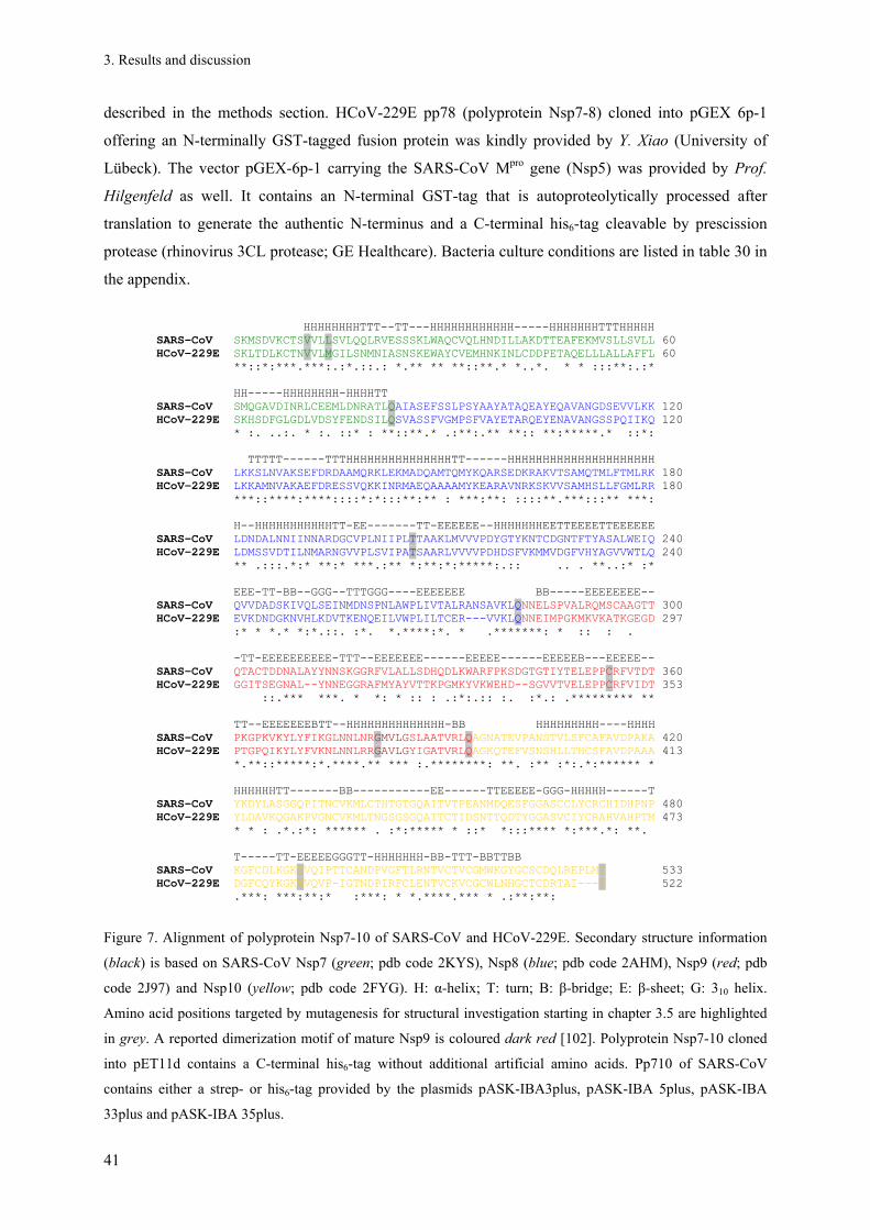
3. Results and discussion
41
described in the methods section. HCoV-229E pp78 (polyprotein Nsp7-8) cloned into pGEX 6p-1
offering an N-terminally GST-tagged fusion protein was kindly provided by Y. Xiao (University of
Lübeck). The vector pGEX-6p-1 carrying the SARS-CoV Mpro gene (Nsp5) was provided by Prof.
Hilgenfeld as well. It contains an N-terminal GST-tag that is autoproteolytically processed after
translation to generate the authentic N-terminus and a C-terminal his6-tag cleavable by prescission
protease (rhinovirus 3CL protease; GE Healthcare). Bacteria culture conditions are listed in table 30 in
the appendix.
HHHHHHHHTTT--TT---HHHHHHHHHHHH-----HHHHHHHTTTHHHHH
SARS-CoV SKMSDVKCTSVVLLSVLQQLRVESSSKLWAQCVQLHNDILLAKDTTEAFEKMVSLLSVLL 60 HCoV-229E SKLTDLKCTNVVLMGILSNMNIASNSKEWAYCVEMHNKINLCDDPETAQELLLALLAFFL 60 **::*:***.***:.:*.::.: *.** ** **::**.* *..*. * * :::**:.:* HH-----HHHHHHHH-HHHHTT SARS-CoV SMQGAVDINRLCEEMLDNRATLQAIASEFSSLPSYAAYATAQEAYEQAVANGDSEVVLKK 120 HCoV-229E SKHSDFGLGDLVDSYFENDSILQSVASSFVGMPSFVAYETARQEYENAVANGSSPQIIKQ 120 * :. ..:. * :. ::* : **::**.* .:**:.** **:: **:*****.* ::*: TTTTT------TTTHHHHHHHHHHHHHHHTT------HHHHHHHHHHHHHHHHHHHHH SARS-CoV LKKSLNVAKSEFDRDAAMQRKLEKMADQAMTQMYKQARSEDKRAKVTSAMQTMLFTMLRK 180 HCoV-229E LKKAMNVAKAEFDRESSVQKKINRMAEQAAAAMYKEARAVNRKSKVVSAMHSLLFGMLRR 180 ***::****:****::::*:*:::**:** : ***:**: ::::**.***:::** ***: H--HHHHHHHHHHHTT-EE-------TT-EEEEEE--HHHHHHHEETTEEEETTEEEEEE SARS-CoV LDNDALNNIINNARDGCVPLNIIPLTTAAKLMVVVPDYGTYKNTCDGNTFTYASALWEIQ 240 HCoV-229E LDMSSVDTILNMARNGVVPLSVIPATSAARLVVVVPDHDSFVKMMVDGFVHYAGVVWTLQ 240 ** .:::.*:* **:* ***.:** *:**:*:*****:.:: .. . **..:* :* EEE-TT-BB--GGG--TTTGGG----EEEEEEE BB-----EEEEEEEE--
SARS-CoV QVVDADSKIVQLSEINMDNSPNLAWPLIVTALRANSAVKLQNNELSPVALRQMSCAAGTT 300 HCoV-229E EVKDNDGKNVHLKDVTKENQEILVWPLILTCER---VVKLQNNEIMPGKMKVKATKGEGD 297
:* * *.* *:*.::. :*. *.****:*. * .*******: * :: : . -TT-EEEEEEEEEE-TTT--EEEEEEE------EEEEE------EEEEEB---EEEEE-- SARS-CoV QTACTDDNALAYYNNSKGGRFVLALLSDHQDLKWARFPKSDGTGTIYTELEPPCRFVTDT 360 HCoV-229E GGITSEGNAL--YNNEGGRAFMYAYVTTKPGMKYVKWEHD--SGVVTVELEPPCRFVIDT 353 ::.*** ***. * *: * :: : .:*:.:: :. :*.: .********* ** TT--EEEEEEEBTT--HHHHHHHHHHHHHH-BB HHHHHHHHH----HHHH SARS-CoV PKGPKVKYLYFIKGLNNLNRGMVLGSLAATVRLQAGNATEVPANSTVLSFCAFAVDPAKA 420 HCoV-229E PTGPQIKYLYFVKNLNNLRRGAVLGYIGATVRLQAGKQTEFVSNSHLLTHCSFAVDPAAA 413 *.**::*****:*.****.** *** :.********: **. :** :*:.*:****** *
HHHHHHTT-------BB-----------EE------TTEEEEE-GGG-HHHHH------T SARS-CoV YKDYLASGGQPITNCVKMLCTHTGTGQAITVTPEANMDQESFGGASCCLYCRCHIDHPNP 480 HCoV-229E YLDAVKQGAKPVGNCVKMLTNGSGSGQAITCTIDSNTTQDTYGGASVCIYCRAHVAHPTM 473 * * : .*.:*: ****** . :*:***** * ::* *:::**** *:***.*: **. T-----TT-EEEEEGGGTT-HHHHHHH-BB-TTT-BBTTBB SARS-CoV KGFCDLKGKYVQIPTTCANDPVGFTLRNTVCTVCGMWKGYGCSCDQLREPLMQ 533 HCoV-229E DGFCQYKGKWVQVP-IGTNDPIRFCLENTVCKVCGCWLNHGCTCDRTAI---Q 522 .***: ***:**:* :***: * *.****.*** * .:**:**:
Figure 7. Alignment of polyprotein Nsp7-10 of SARS-CoV and HCoV-229E. Secondary structure information
(black) is based on SARS-CoV Nsp7 (green; pdb code 2KYS), Nsp8 (blue; pdb code 2AHM), Nsp9 (red; pdb
code 2J97) and Nsp10 (yellow; pdb code 2FYG). H: α-helix; T: turn; B: β-bridge; E: β-sheet; G: 310 helix.
Amino acid positions targeted by mutagenesis for structural investigation starting in chapter 3.5 are highlighted
in grey. A reported dimerization motif of mature Nsp9 is coloured dark red [102]. Polyprotein Nsp7-10 cloned
into pET11d contains a C-terminal his6-tag without additional artificial amino acids. Pp710 of SARS-CoV
contains either a strep- or his6-tag provided by the plasmids pASK-IBA3plus, pASK-IBA 5plus, pASK-IBA
33plus and pASK-IBA 35plus.

3. Results and discussion
42
Table 13. Overview of target proteins of HCoV-229E (blue) and SARS-CoV (black) obtained by gene
cloning and recombinant bacterial expression
*calculated by ProtParam ([142]; SIB)
3.2. HCoV-229E and SARS-CoV pp710
3.2.1. Gene overexpression in E. coli and polyprotein purification
C-terminally his6-tagged HCoV-229E pp710 was successfully purified to more than 90% purity
after Ni-NTA affinity chromatography as judged by SDS-PAGE (fig. 8A). Compared to a one-step
elution with 400 mM imidazole the purity was improved by applying a stepwise increase of imidazole
(15-400 mM) for protein elution. The identity of the protein was verified by ESI-MS/MS after trypsin
digestion, applying soluble protein directly from an affinity chromatography elution fraction and
additionally a single coomassie-stained band that was cut out of an SDS-PAGE gel (data not shown).
To further increase the purity and to analyze the aggregation state of HCoV-229E pp710 in
solution, a Hi-Load 16/60 Superdex 200 size-exclusion chromatography column was applied (fig. 8B).
Depending on the buffer conditions a maximum of four peaks corresponding to different oligomeric
states were resolved. The peak identity of one SEC experiment (fig. 8B, dark blue chromatogram) was
exemplarily verified by reducing SDS-PAGE (fig. 8C). SEC indicated the formation of dimeric and
protein length
[amino acids]
molecular
mass [Da]
theor.
pI*
sequence identity with SARS-CoV
[identical residues/total length (%
of identity)]
pp1a (HCoV-229E; Nsp1-11) 4085 n. d. n. d. n. d.
pp710 (HCoV-229E) 522 57310.9 8.03 246/522 (47.1%)
pp78 (HCoV-229E) 278 30923.7 n.d. 125/278 (45.0%)
Nsp7 (HCoV-229E) 83 9300.6 4.38 32/83 (38.6%)
Nsp8 (HCoV-229E) 195 21623.1 9.32 93/195 (47.7%)
Nsp9 (HCoV-229E) 109 12045.8 9.24 50/109 (45.9%)
Nsp10 (HCoV-229E) 135 14395.3 7.66 71/135 (52.6%)
Nsp11 (HCoV-229E)
[SFDNSYLNESGALVPLD] 17 1840.9 3.49 n.d.
pp1a (SARS-CoV) 4382 n. d. n. d. -
pp710 (SARS-CoV) 533 58324.9 7.33 -
pp79 (SARS-CoV) 394 43584.0 7.99 -
pp78 (SARS-CoV) 281 31182.9 n.d. -
Nsp7 (SARS-CoV) 83 9267.8 5.18 -
Nsp8 (SARS-CoV) 198 21915.1 6.58 -
Nsp9 (SARS-CoV) 113 12401.1 9.1 -
Nsp10 (SARS-CoV) 139 14843.9 6.3 -

3. Results and discussion
43
higher oligomeric states in solution. The dimeric state is indicated by approx. the mol weight
characterizing peak III at VE = 66.2 ml (kav = 0.30) obtained under non-reducing conditions. Analyzing
protein from the dimer peak by SDS-PAGE showed a predominant protein band corresponding to the
MW of a pp710 monomer, indicating the major presence of a non-covalent dimer in solution.
However, if the SEC is performed with reducing conditions (yellow), peak III is slightly reduced,
confirming also the presence of disulfide-linked dimers. The comparison of SEC chromatograms
obtained by applying different protein concentrations (fig. 8B, dark blue and red chromatogram) and
buffer compositions (fig. 8B, green and red chromatogram) revealed that the proportion of oligomeric
states is obviously concentration- and buffer-dependent. A peak corresponding to monomeric protein
elutes at VE = 77.7 ml (kav = 0.45), which increased in absorption under reducing conditions, by high
ionic strength and at low protein concentration (fig. 8). Isoelectric focussing of HCoV-229E pp710
revealed a band at pH 6.0 (data not shown), lower than pH 7.4 as calculated by ProtParam for pp710
with his6-tag ([142], SIB).
Figure 8. Purification of HCoV-229E pp710. (A) The success of the purification procedure is visualized by non-
reducing SDS-PAGE and coomassie staining [SN: supernatant after disrupting the harvested E. coli cells and
centrifugation; E: protein eluted from the Ni-NTA affinity resin; M: molecular weight marker]. A small amount
of covalent dimer was identified (116 kDa) next to the monomeric pp710 (58 kDa). (B) The aggregation state of
pp710 depending on the buffer conditions was analyzed by SEC (HiLoad 16/60 Superdex 200). The molecular
weight calibration of the column by standard proteins is shown. For each experiment, 5 ml of protein solution
were loaded onto the column. Dark blue: 300 mM NaCl, 30 mM NaH2PO4, pH 7.9; cyan: 300 mM NaCl 30 mM
Tris pH 7.9; dark green: 120 mM NaCl, 20 mM NaH2PO4 pH 7.9; yellow: 300 mM NaCl, 30 mM NaH2PO4 pH
7.9 3 mM DTT; red: 400 mM NaCl, 5% (v/v) glycerol, 40 mM NaH2PO4 pH 7.9. (C) The fractions
corresponding to the absorption peaks I-IV of one experiment (dark blue chromatogram) were analyzed by
reducing SDS-PAGE [pp: pp710 SEC column input; I-IV: SEC peaks].
66
45

3. Results and discussion
44
Applying a similar one-step purification strategy, four variants of SARS-CoV pp710 were
successfully purified by affinity chromatography, i.e. an N-terminal and a C-terminal his6-tag variant
using a Ni-NTA resin column (SDS-PAGE gel, fig. 9A) as well as an N-terminal and a C-terminal
strep-tag variant using Strep-Tactin resin. Solubility and purity of the differently tagged SARS-CoV
proteins turned out to be highly similar to HCoV-229E pp710. Calibrated SEC revealed the existence
of different oligomeric states in solution, including a monomeric and a dimeric state (fig. 9B), as
previously observed for the homologue HCoV-229E pp710 (fig. 8). However, the SEC analysis of
pp710 from both coronaviruses (fig. 8B, 9B) consistently showed that the pp710 monomer is the
predominant state in solution, compared to other oligomers.
Interestingly, supplementing the E. coli expression culture of SARS-CoV pp710 with 500 µM
Zn2+ resulted in a significantly reduced amount of oligomers larger than a dimer (fig. 9B, dark blue
chromatogram). The effect of Zn2+ may originate from two highly conserved Zn2+-binding sites within
Nsp10. Investigating the isolated Nsp10 domain of MHV, it was previously shown that Zn2+
significantly altered the oligomerization behaviour by partly de-aggregation, as determined by
dynamic light scattering techniques [170].
Figure 9. (A) Purified SARS-CoV pp710 visualized by reducing SDS-PAGE and coomassie staining. (B)
Calibrated SEC (Hi-Load 16/60 Superdex 200) reveals two well defined peaks (II and III) conserved in all
chromatograms, corresponding to a dimeric and monomeric state in solution, respectively. The light blue
chromatogram was obtained in 280 mM NaCl and 30 mM NaH2PO4, pH 7.9. Exchanging sodium phosphate by
30 mM Tris resulted in the blue chromatogram. A significantly reduced amount of higher oligomers was
observed after supplementing the E. coli expression culture with 500 µM Zn2+ and adding 2 mM DTT to the
purified protein (dark blue).

3. Results and discussion
45
In summary, SEC revealed a co-existence of different oligomeric states of both purified pp710
homologues in solution which is subsequently investigated in more detail using native PAGE, DLS
and SAXS techniques.
3.2.2. Variable oligomerization of pp710
3.2.2.1. Blue native PAGE & Elman’s reagent
Addressing to investigate the number, the approximate size and the quantity of oligomeric states
of pp710 in solution, blue native PAGE was performed. During this electrophoresis technique the
proteins are additionally negatively charged on the surface by interaction with a trace amount of
coomassie stain. Nevertheless, the native state of a protein remains widely conserved [171], allowing
studying protein-protein interactions with low sample consumption. Analysing both homologues, a
comparable number and estimated quantity of four oligomeric states up to a putative octamer were
consistently identified for HCoV-229E and SARS-CoV pp710 by native blue PAGE under non-
reducing and even under mildly reducing conditions (fig. 10A/C). The monomeric state of both
homologue proteins appears to be the most abundant one.
However, for HCoV-229E pp710, a significant amount of oligomers with a molecular weight
above 400 kDa was observed after incubation of the sample purified by affinity chromatography for 7
days (fig. 10A). Probing the time-dependent oxidation of cysteine side chains, i.e. the formation of
disulfide bonds, by Elman’s reagent (5,5'-dithiobis -2-nitrobenzoic acid, DTNB) under non-reducing
conditions revealed a continuous oxidation down to around two reduced thiol groups per molecule
after 18 days (fig. 10B). Consequently, the high molecular weight oligomers of HCoV-229E are
mainly a result of covalent disulfide bonds formation due to aerobic incubation of pp710 in solution.
This is further supported by the observation that reducing conditions partly disassemble these
oligomers, resulting in a significant increase of the amount of monomeric and dimeric states in
solution (fig. 10A). The remaining high molecular weight fraction is stable in reducing conditions,
indicating non-covalent interactions. Blue native PAGE of large HCoV-229E pp710 oligomers
separated by SEC (fig. 8B, peak I, dark blue chromatogram) showed a disassembly into smaller states
up to a monomer after 7 days incubation (not shown), attributed to the non-covalently linked
oligomers. Remarkably, two thiol groups of HCoV-229E remained reduced for at least 18 days at 6 °C
under oxidizing conditions (fig. 10B). One of them might be C281 within Nsp8 that is buried
according to the high resolution structure (pdb code 2AHM) [98]. The second cysteine residue could
not be attributed so far.

3. Results and discussion
46
Figure 10. Investigation of the pp710 oligomerization. (A) Blue native PAGE of HCoV-229E pp710: Lane 1-4:
Pp710 purified by affinity chromatography and incubated for 7 days at 6 °C; the buffer composition is displayed
above the gel. The pH was consistently adjusted to pH 7.9. (B) Stepwise oxidation of thiol groups of HCoV-
229E pp710 photometrically determined by DTNB up to 18 days after cell disruption. *approximately 3 h after
cell lysis in elution buffer. (C) Blue native PAGE of SARS-CoV pp710: Lane 5: Dimer after incubation at 6 °C
for 7 days; lane 6: Monomer, after 7 days; lane 7: Monomer, after 7 days supplemented with 2 mM DTT. The
protein buffer contains 280 mM NaCl and 18 mM sodium phosphate, pH 7.9.
For freshly prepared SARS-CoV pp710, the monomeric and the dimeric state were isolated by
SEC (fig. 9B, light blue chromatogram, peak III and II). The homogeneity of both samples was
verified by dynamic light scattering (table 14, monomer and dimer sample). Incubating monomeric
SARS-CoV pp710 for 7 days, oligomeric states appear on the gel, including a dimer, tetramer and
octamer (fig. 10C, lane 6). Since these oligomers are stable at mild reducing conditions (fig. 10C, lane
7), a non-covalent interaction is suggested. The purified pp710 dimer also tends to form these
oligomers after one week, even a slight disassembling into the monomeric state has been detected (fig.
10C, lane 5). Interestingly, high molecular weight oligomers as observed for HCoV-229E pp710 did
not form to the same extend during incubation for 7 days (fig 10C, lane 5 and 6), which could be
attributed to an altered distribution of cysteine residues in SARS-CoV pp710 (fig. 7).
Overall, the native PAGE experiments indicated an equilibrium of monomer and non-covalent
dimer, as well as to a lesser extend higher oligomers for both pp710 homologues in solution.
Moreover, disulfide bridged high molecular weight oligomers seem to represent artefacts that are
formed by oxidation during storage under aerobic conditions.

3. Results and discussion
47
3.2.2.2. Dynamic light scattering (DLS)
To further characterize the stability of the isolated SARS-CoV pp710 monomer and dimer in
solution and to analyze the influence of buffer conditions on the aggregation behaviour of monomeric
pp710 and its high molecular weight oligomers, dynamic light scattering techniques were applied.
This method is based on the scattering of a red-light laser on protein molecules that is focussed on the
sample. The fluctuation of the scattering intensity due to Brownian motion of the molecules in solution
is correlated with the hydrodynamic radius (Rh) of a particle, allowing the molecular weight estimation
in homogenous solutions. Pure monomer and dimer fractions separated by SEC were investigated in
comparison to a mixture of oligomeric states obtained directly after affinity chromatography.
Monomeric SARS-CoV pp710 isolated by SEC (fig. 9B, peak III) is characterized by a
hydrodynamic radius of Rh = 3.8 ± 0.1 nm, while an Rh = 6.5 ± 1.1 nm was determined for pure
dimeric pp710 (table 14). For the mixture of oligomeric states a hydrodynamic radius of 11.9 ± 3.8 nm
was determined. To access the stability of monomeric SARS-CoV pp710, the solution was monitored
by DLS for up to 7 days. After 12 hours the hydrodynamic radius was stable (Rh = 3.8 ± 0.3 nm). Even
after two days of incubation, only a marginally increased radius of Rh = 4.1 ± 0.1 nm was detected,
indicating the formation of a small amount of pp710 dimers. Within one week, the hydrodynamic
radius of monomeric pp710 increased up to 6.9 ± 1.1 nm, highly similar to that determined for a pure
dimeric state, confirming the formation of a slow equilibrium as observed by blue native PAGE (fig.
10C). However, the latter method additionally showed protein bands corresponding to higher
oligomeric states after 7 days of incubation.
The presence of 1 mM DTT during incubation of the monomeric pp710 fraction slightly
decreased the hydrodynamic radius after 7 days, supporting the coexistence of non-covalent and
disulphide linked dimers. In contrast, large concentrations of reducing agents (10 mM β-ME, 30 mM
TCEP) significantly increased the aggregation of SARS-CoV pp710. On the other hand, EDTA,
MgSO4, CaCl2 or ZnCl2 as well as 10% glycerol, 0.5% β-OG or 2 mM CHAPS did not affect the
dimerization of pp710 over 7 days. Only 1% DDM slightly reduced the hydrodynamic radius to Rh =
5.1 ± 0.1 nm, attributed to the partial disassembling of non-covalent dimers. Interestingly, the presence
of 1 mM MnCl2 induced the formation of large oligomers, suggesting specific manganese binding sites
in pp710 of SARS-CoV. This is consistent with the report of a manganese-dependency of the RNA-
dependant RNA polymerase activity of mature SARS-CoV Nsp8 [94].

3. Results and discussion
48
Table 14. Summary of SARS-CoV pp710 (N-terminal strep tag) DLS experiments. The basic protein
buffer included 280 mM NaCl and 30 mM NaH2PO4 at pH 7.9.
sample additive radius countrate
peak 1 peak 2 peak 3
monomer untreated 3.8 ± 0.1 nm - - 57.2 ± 1.3 kHz
dimer untreated 6.5 ± 1.1 nm - - 45.3 ± 13.3 kHz
oligomer* untreated 11.9 ± 3.8 nm - - 400 ± 22.4 kHz
monomer
(after one
week)
untreated 6.9 ± 1.1 nm - - 213.0 ± 10.5 kHz
1 mM DTT 4.7 ± 0.2 nm - - 57.9 ± 1.2 kHz
10 mM β-ME 16.2 ± 1.1 nm - - 1110.5 ± 34.6 kHz
30 mM TCEP 17.1 ± 0.7 nm 87.1 ± 8.1 nm - 1477.7 ± 85.8 kHz
1% DDM 5.1 ± 0.1 nm - - 398.8 ± 22.2 kHz
0.5% β-OG 7.2 ± 0.7 nm - - 175.2 ± 7.8 kHz
10% glycerol 6.8 ± 0.2 nm - - 108.7 ± 2.8 kHz
2 mM CHAPS 6.4 ± 0.7 nm - - 133.6 ± 9.1 kHz
1 mM ZnCl2 6.0 ± 0.2 nm - - 113.3 ± 2.4 kHz
1 mM MnCl2 11.4 ± 1.0 nm 78.7 ± 8.7 nm - 652.1 ± 39.9 kHz
1 mM MgSO4 7.0 ± 1.4 nm - - 160.3 ± 8.9 kHz
1 mM CaCl2 7.0 ± 0.3 nm 76.8 ± 12.2 nm - 148.6 ± 16.6 kHz
1 mM EDTA 7.0 ± 0.3 nm - - 146.1 ± 5.2 kHz
mixture of oligomeric
states (after 30 min)
500 mM NaCl 7.8 ± 1.2 nm 17.3 ± 2.2 nm - 689.5 ± 24.9 kHz
1 mM DTT 9.8 ± 2.0 nm 21.5 ± 4.8 nm - 659.4 ± 27.8 kHz
10 mM β-ME 11.1 ± 1.0 nm 20.2 ± 3.6 nm - 935.4 ± 37.6 kHz
30 mM TCEP 100.9 ± 13.4 nm - - 3506.9 ± 358.7 kHz
0.5% β-OG 21.0 ± 1.9 nm - - 645.6 ± 27.8 kHz
2 mM CHAPS 18.3 ± 1.3 nm - - 783.5 ± 28.7 kHz
1 mM MgSO4 19.2 ± 3.7 nm - - 932.7 ± 46.5 kHz
1 mM CaCl2 35.1 ± 6.7 nm - - 1872.9 ± 102.4 kHz
1 mM EDTA 16.4 ± 4.5 nm - - 864.2 ± 34.1 kHz
mixture of oligomeric
states
(after one
week)
500 mM NaCl 17.6 ± 3.0 nm - - 826.1 ± 28.1 kHz
1 mM DTT 16.3 ± 2.6 nm 43.1 ± 7.2 nm - 2317.9 ± 102.5 kHz
10 mM β-ME 20.7 ± 2.7 nm 37.3 ± 10.7 nm - 4300.1 ± 152.2 kHz
30 mM TCEP 23.6 ± 3.2 nm 82.6 ± 19.8 nm - 1892.7 ± 116.3 kHz
0.5% DDM 7.5 ± 0.5 nm 20.7 ± 4.6 nm 143.8 ± 38.1 nm 725.4 ± 27.0 kHz
0.5% β-OG 17.6 ± 3.1 nm 33.4 ± 7.2 nm - 3038.8 ± 113.4 kHz
10% glycerol 27.2 ± 4.6 nm - - 1131.2 ± 53.5 kHz
2 mM CHAPS 38.1 ± 4.4 nm - - 2652.7 ± 112.6 kHz
1 mM ZnCl2 71.3 ± 13.7 nm - - 1826.9 ± 104.7 kHz
1 mM MnCl2 38.7 ± 7.4 nm - - 2983.6 ± 130.5 kHz
1 mM MgSO4 18.8 ± 4.7 nm - - 2203.7 ± 81.7 kHz
1 mM CaCl2 30.6 ± 4.1 nm - - 2799.8 ± 114.3 kHz
1 mM EDTA 25.9 ± 3.7 nm - - 2402.9 ± 89.3 kHz
*mixture of oligomeric states of SARS-CoV pp710 after affinity chromatography purification
3.2.2.3. Native ESI-Q-TOF mass spectrometry
The previous investigations indicated the presence of a continuum of variable oligomeric states
for pp710 in solution with a relatively high abundance of the monomeric and the dimeric state. A
semi-quantitative method providing a very high mass accuracy is native mass spectrometry, which is
applied to exactly determine the size and ratio of the different oligomeric states of SARS-CoV pp710
in ammonium acetate buffer at pH 7.9. Ammonium acetate is volatile in a vacuum and therefore a
suitable salt for mass spectrometry. An ESI-Q-TOF instrument uses electrospray ionization (ESI) of
the proteins that pass a quadrupole (Q) component on the way to the time-of-flight (TOF) mass
detector. Mass spectrometry experiments were performed in cooperation with Dr. C. Uetrecht at the
University of Utrecht.

3. Results and discussion
49
Native ESI-Q-TOF applying 5 µM SARS-CoV pp710, purified by affinity chromatography,
detected the coexistence of a series of different oligomers in solution, up to a state in the range of a
decamer characterized by approximately 630 kDa (fig. 11A/B). In agreement with SEC experiments
and blue native PAGE the monomeric state with a calculated mass of 59.695 kDa for pp710 with N-
terminal strep tag is most abundant. The relative content of the putative monomer (≈ 60 kDa), dimer
(≈ 120 kDa) and trimer (≈ 180 kDa) is determined to 22.9%, 19.8% and 10.7% of the total intensity at
the TOF detector, respectively (fig. 11A). Therefore, with an increase in molecular weight up to a
tetramer, the relative fraction of the respective state is indicated to decrease. Comparing panel A and B
of figure 11, a small mass variation of monomeric pp710 probably due to a ligand or truncation of the
termini is indicated at an increased collision voltage of 100 V. Two further masses assigned to the
spectrum in panel B are 119,537 ± 23 Da and 179,273 ± 30 Da well fitting dimeric and trimeric
SARS-CoV pp710, respectively.

3. Results and discussion
50
Figure 11. Oligomerization of SARS-CoV pp710 investigated by native ESI-Q-TOF MS. (A) Pp710 mass
spectrum with different ion series after linearization, smoothing and background subtraction with the raw data
(dark blue). The calculated mass of pp710 with N-terminal strep tag is 59.695 kDa. The expected oligomeric
species were simulated by Massign and presented in the same spectrum (different colours). Different oligomeric
states were detected in solution, i.e. a putative monomer (60,034 Da), dimer (120,154 Da), trimer (187,504 Da)
and other states up to a putative decamer (632,038 Da). (B) The collision voltage is increased to 100 V for
further fragmentation of oligomers/protein complexes (light blue). The persisting oligomers could be covalently
linked. Simulated individual mass species that approximately fit the monomeric state are coloured and
superposed with original data.
The results obtained for both pp710 homologues by SEC, blue native PAGE, DLS and native
mass spectrometry essentially agree that the pp710 monomer, representing the most abundant state, is
in equilibrium with a prominent dimer and with larger oligomeric states. The oligomerization process,
triggered partly by non-covalent interactions, but also partly by disulphide formation, can be artificial
or of relevance for the native function of pp710 in vivo. Larger oligomers detected by either method

3. Results and discussion
51
are slightly different in quantity and their maximum size, which might be related to individual
limitations of detection and quantification by the respective method, e.g. incomplete ionization in mass
spectrometry or different charge states of a protein in blue native PAGE.
However, oligomers of pp710 might be essential for their function. Extended oligomers of
HCoV-229E Nsp9 were already suggested to be involved in RNA binding [101], probably involving a
similar mechanism as known for single stranded DNA binding protein (SSB) of E. coli [172]. Other
proteins that bind nucleic acids, e.g. the Trp RNA-binding attenuation protein (TRAP) and DNA
replication associated proteins, like the processivity factor PCNA [173], form large ring structures
directly associated with their function [174]. Orb2, a member of the RNA-binding CPEB family
(cytoplasmic polyadenylation element-binding proteins), is even capable of forming amyloid fibers
that are involved in long-term memory [175]. However, these proteins are suggested to be functionally
only distantly related to pp710. Poliovirus RNA-dependent RNA polymerase (Mahoney type 1
poliovirus polymerase), which seems to be closely related to the putative function of Nsp8, forms a
small oligomer as observed by X-ray crystallography that is reported to be essential for efficient RNA-
binding and formation of the active centre [176]. Although high molecular weight oligomers have
been detected for both pp710 homologues in this study, the applied techniques did not provide
sufficient indications for the formation of ordered fibril-shaped or ring-shaped aggregates under in
vitro conditions that may correlate with a coordinative “structural” function.
Different hetero-oligomeric states have been reported for complexes of mature Nsp7 and Nsp8 by
high-resolution crystal structures. For SARS-CoV Nsp7 and Nsp8, a putative hexadecameric state was
described that is assembled of eight subunits of Nsp7 and Nsp8 each [98], whereas FCoV Nsp7 and
Nsp8 form a 2:1 heterotrimeric complex [96]. Both structures involve a specific Nsp7 dimer interface.
Although HCoV-229E and SARS-CoV pp710 unlikely form a significant hexadecamer-like state in
solution, as previously shown, mentioned complexes reveal that specific non-covalent Nsp-Nsp
interactions exist, which could be also involved in the assembly of pp710 oligomers Moreover, these
examples additionally highlight that a comparison of different Nsps/polyproteins of distantly related
coronaviruses is highly important. Mature Nsp9 of HCoV-229E forms a disulphide-linked dimeric
state in solution that is reported to have a higher affinity to nucleic acid [101], indicating that at least
the detected covalently linked dimers of both pp710 homologues could be of functional relevance or
of regulatory impact. In this context, it was for example observed that DNA-binding specificity for the
tumour repressor protein p53 is dependent on the reduced state of cysteines [177]. Despite the
unfavourable redox potential in the cytosol, disulfide formation is also critical for the function of NF-
κB, PaX, Fos, OxyR and eukaryotic transcription factors (for review see [178]). Additionally, non-
covalent oligomerization beyond a dimeric state was reported for mature HCoV-229E Nsp9, triggered
by the presence of sulfate ions [101]. A comparable specific oligomerization effect was not detected

3. Results and discussion
52
for SARS-CoV and HCoV-229E pp710 up to a concentration of 1 mM MgSO4. This could be due to
sterically hindered sulphate binding sites within the polyprotein.
The oligomerization of pp710 that is under investigation in this study may result from a
combination of different (independent) homo-dimerization or homo-oligomerization modes, e.g. via
Nsp-Nsp homointeractions which would favour higher oligomers in the polyprotein state that are
probably irrelevant and decreased in size after cleavage/maturation, which is subsequently investigated
in more detail However, intermolecular interactions of the individual Nsp domains within the
polyprotein oligomers appear at least weakened under the chosen experimental conditions compared to
that of mature Nsps. For the complex of SARS-CoV Nsp7 and Nsp8 [98] or for HCoV-229E Nsp9
[101], the monomeric state is not the most abundant one under a wide range of conditions, in contrast
to both pp710 homologues.
3.2.3. Structural characterization of pp710
3.2.3.1. Secondary structure content
To compare the secondary structure composition of Alphacoronavirus pp710 (HCoV-229E) and
Betacoronavirus pp710 (SARS-CoV), circular dichroism (CD) spectroscopy was applied. Far-UV CD-
spectra, i.e. the ellipticity of the sample in the wavelength range from 190 nm to 260 nm, were
recorded (fig. 12A). The applied buffer contained 250 mM NaF and 20 mM NaH2PO4 at pH 7.9.
Sodium chloride was substituted by sodium fluoride to reduce the absorbance of circular polarized
light by the buffer during the experiment. Comparing HCoV-229E and SARS-CoV pp710, almost
superimposable ellipticity curves (fig. 12A) indicate a highly similar secondary structure composition
for both pp710 homologues.
Two distinct minima at a wavelength of 207 nm and 222 nm are characteristic for a predominant
α-helical secondary structure content. This reflects the secondary structure of mature Nsp7 and Nsp8,
as revealed by known high-resolution structures [96; 98]. Small fractions of β-sheet and random coil
structure have also been deduced as well from the spectra in figure 12A, which is in agreement with a
high β-sheet content of HCoV-229E Nsp9 [101] and SARS-CoV Nsp9 [102]. The far-UV spectrum of
HCoV-229E is not affected by sodium acetate buffer (pH 5.0), by replacement of NaF by NaCl, or by
a four-fold molar excess of ZnCl2 (not shown).
For an initial comparison of the tertiary structure, near-UV spectra ranging from a wavelength of
250 to 320 nm were recorded (fig. 12B), representing a structural “fingerprint” of the protein. The
increased absolute ellipticity of HCoV-229E pp710 around 260-270 nm may correspond to additional
disulfide bonds. The peak positions in this area are sensitive towards the local orientation of
phenylalanine residues (255-260 nm) and tyrosine (260-275 nm). In addition, an ellipticity maximum

3. Results and discussion
53
at approximately 290 nm corresponding to the absorbance of tryptophan is highly conserved for all
analyzed pp710 samples of HCoV-229E and SARS-CoV pp710, including a purified monomer and a
mixture of oligomeric states respectively. This consistency indicates consistent characteristics of the
tertiary structure, even for different oligomeric states of pp710. Beside differences in the tertiary
structure of both pp710 homologues indicated by the near-UV spectrum, the secondary structure is
highly similar. Moreover, a mixture of different secondary structure elements with a dominating
fraction of α-helical structure initially well reflects the sum of the secondary structure composition of
the four mature Nsp domains.
Figure 12. (A) Far-UV CD spectra of SARS-CoV pp710 (blue) and HCoV-229E pp710 (red). (B) Near-UV
"fingerprint" spectra (right panel): HCoV-229E pp710 (red) purified by affinity chromatography and treated with
benzonase in 280 mM NaCl 30 mM NaH2PO4 pH7.9. Monomeric SARS-CoV pp710 purified by SEC (dark
blue), a mixture of oligomeric states purified by affinity chromatography (mid blue) and the same mixture
supplemented with 1 mM DTT (cyan) are shown for comparison.
3.2.3.2. Tertiary and quaternary structure of pp710
High resolution X-ray crystallography provides essential insights into protein molecules;
however it cannot be ruled out that there are structural artefacts due to flexibility, diverse
oligomerization, a high protein concentration or non-physiological compounds in the solutions during
crystallization. Therefore, a combination with “in-solution methods” like small-angle X-ray scattering
(SAXS) is highly beneficial and indispensable in order to analyze particularly partly flexible multi-
domain proteins. In this project SAXS aims at analyzing and comparing the tertiary structure of
HCoV-229E and SARS-CoV pp710, which should be interpreted by known high-resolution protein
structures of individual domains. Monomeric and dimeric states of both pp710 homologues will be
compared in more structural detail, having in mind that their secondary structure is widely conserved
(fig. 12). In preparation for SAXS experiments, the monodispersity of the samples was verified by
A B

3. Results and discussion
54
DLS (see also chapter 3.2.2, table 15). The SAXS sample with the lowest concentration of both
proteins was directly taken from SEC without modifications of the buffer or adjustment of the
concentration; the concentration of the others was adjusted by ultra filtration. SARS-CoV pp710
scattering data was collected at DORIS beamline X33. The scattering pattern of monomeric and
dimeric HCoV-229E separated by SEC were recorded at two different synchrotron radiation sources,
since the advanced 3rd generation synchrotron source PETRAIII beamline P12 (DESY, Germany)
became recently available in the course of this study. The scattering intensity was investigated up to a
scattering vector s of 6 nm-1.
Initially, it was observed that purification and SAXS data collection in the presence of 2 mM
DTT reduced the determined radius of gyration (Rg) as a measure of size (and shape) of HCoV-229E
pp710 by 5-10% (fig. 13A). This effect again demonstrates the existence of a minor fraction of
covalently linked oligomers under non-reducing conditions, as previously observed by SEC and blue
native PAGE (see section 3.2.1/3.2.2). In polydisperse solutions the determined Rg refers to an average
value of all present particles. Moreover, it was observed that the radius of gyration Rg and the particle
volume determined for SARS-CoV and HCoV-229E pp710 monomer and dimer samples are clearly
concentration-dependent under non-reducing conditions and in the presence of 2 mM DTT (fig. 13A).
To get rid of this property of pp710 during data evaluation, the scattering data was extrapolated to
infinite dilution. It is concluded that the data of HCoV-229E recorded at beamline P12 in an early
stage of operation and beamline X33 is highly similar (fig. 13B). While SAXS data is frequently
visualized by the plot of the scattering intensity I versus scattering vector s (see figure 14 for
comparison), figure 13B displays the alternative Kratky plot. In this type of data presentation, the
scattering intensity I multiplied by the square of the scattering vector s, which directly depends on the
scattering angle (see methods section), is plotted against the scattering vector s. For highly flexible
particles or unfolded proteins the data points do not converge to the scattering vector axis at higher
scattering angles, instead the product Is2 is continuously increasing. Therefore, the plot allows a
flexibility prediction, where in this case the bending of the graph deduced from the raw data is
indicative for a multi-domain protein with a relatively low overall flexibility and a rather
compact/rigid particle, particularly for the dimer. It is conceivable that high local flexibility within the
monomer of pp710 of both virus species is additionally stabilized within the dimer, which could
theoretically be a physiological relevance in vivo.
The approximated molecular weight after extrapolation of the scattering data to infinite dilution
based on the porod volume well matches the value expected for a monomer and dimer of HCoV-229E
and SARS-CoV, respectively. SAXS results are summarized in table 15.

3. Results and discussion
55
Figure 13. SAXS investigation of HCoV-229E and SARS-CoV pp710. (A) Concentration-dependent equilibrium
of oligomeric states of HCoV-229E pp710 as investigated by SAXS. Monomeric and dimeric pp710 isolated by
SEC were analyzed in the presence and absence of 2 mM DTT, as shown in the legend. (B): Kratky plot (Is2
versus s) comparing dimer (dark blue) and monomer (blue) samples of HCoV-229E investigated at PETRA III
P12 and dimer (purple) and monomer (red) samples applied to beamline DORIS X33. SARS-CoV pp710
monomer (DORIS X33; orange) is shown for comparison, displaying slight intensity differences between 0.2
and 0.4 nm-1 that affect the radius of gyration Rg.
Table 15. Summary of size properties of pp710 of HCoV-229E and SARS-CoV under reducing conditions
virus protein Rh
[nm]1 Rg [nm]2
Rh from
Rg [nm]3
Dmax [nm] NSD4
SARS-CoV5 pp710 monomer 4.6 ± 0.2 3.99 ± 0.027
5.2 13.1 0.74 ± 0.05
pp710 dimer 6.5 ± 1.1 5.42 ± 0.01 7.0 18.0 1.29 ± 0.08
HCoV-229E6 pp710 monomer 4.8 ± 0.1 3.43 ± 0.10 4.4 10.5 0.72 ± 0.10
pp710 dimer 6.5 ± 1.1 4.07 ± 0.08 5.3 14.3 0.74 ± 0.10
1 hydrodynamic radius determined by DLS 2 radius of gyration taken from the distance-distribution function (P(R)-function) of the molecule determined by
GNOM and verified by AutoRg implemented in PRIMUS; commonly the raw scattering data was extrapolated
to infinite dilution, if a concentration-dependency of the Rg was observed.
3 (approximation of Rh, see method section)
4 normalized spatial discrepancy, calculated by the “automated mode” of DAMAVER involving at least 10 ab
initio models calculated by DAMMIF. NSD is a quantitative measure of similarity of particles including all its
components. For identical models the NSD is equal to zero [179]. 5 280 mM NaCl, 30 mM NaH2PO4, pH 7.9, 2 mM DTT, proteins contains an N-terminal strep-tag 6 300 mM NaCl, 30 mM NaH2PO4, pH 7.9, 2 mM DTT, protein contains a C-terminal his-tag 7 reduced to 3.66 nm for protein originating from an expression culture supplemented with 500 µM Zn2+ and in
the presence of 3 mM DTT instead of 2 mM DTT.
3
5gh RR
3. Results and discussion
56
Characteristic parameters of the size and shape of both pp710 homologues in comparison to other
related proteins and to SEC experiments are listed and discussed later (chapter 3.10, table 24 and 25).
However, an increased radius of gyration and maximum diameter of monomeric and dimeric SARS-
CoV pp710 compared to its homologue obviously indicates a slightly different geometry, i.e. a more
elongated shape (tab. 15). Nevertheless, the approximated molecular weight of monomeric SARS-
CoV and HCoV-229E pp710 is in the expected range, as further verified by an approach reported
recently [180]. To apply this approach, as an example, the scattering pattern of SARS-CoV pp710
monomer was extrapolated to I(0), i.e. obtaining the scattering intensity at an angle of s = 0 by the fit
graph of the modelling software DAMMIF. This intensity was used to calculate the variable Vc
according to [180], resulting in 483.9 Å2 and QR = 6397.6 Å3. In a plot of I multiplied by s versus s,
the area that is enclosed by the s-axis and the graph was determined by integration. For integration, the
data was cut to a maximum scattering vector of 0.6 Å-1. This integral depends on the molecular weight
of pp710 via a suggested power-law-relationship for protein samples. Using additional constants
experimentally determined by Rambo & Tainer [180], the calculated mass for putative monomeric
SARS-CoV pp710 was determined to 52 kDa (theoretical MWpp710 ≈ 59 kDa). This method is
applicable for flexible/unfolded proteins as well, since in contrast to a Kratky plot (I multiplied by s2
plotted against s), the applied plot I versus s is converging at higher angles anyway, as required for the
calculation of the porod invariant Q. However, in this case the approach sufficiently proofed the
existence of a monomeric state of SARS-CoV pp710 in solution, providing a valuable basis to model
the shape of pp710.

3. Results and discussion
57
Figure 14. Small-angle X-ray scattering data of pp710 used for rigid-body modelling (left panel: SARS-CoV;
right panel: HCoV-229E). The optimized fit graph of the model generated by SASREF based on high-resolution
structures with modelling restrains described in the text is shown for comparison [pdb codes 2AHM (SARS-CoV
Nsp7, Nsp8), 2J97 (HCoV-229E Nsp9)/2UW7 (SARS-CoV Nsp9), 2FYG (SARS-CoV Nsp10)]. These
SASREF graphs demonstrate that the scattering data is well interpreted by the high-resolution structures even
with the restrain of a covalent connection of the Nsp subunits. It is therefore assumed that the overall fold and
shape of the mature Nsps is already preformed within the pp710 molecules. The χ- value calculated by SASREF
is a quality measure of the similarity of scattering data and generated model. A low χ-value indicates a good data
fit. For HCoV-229E pp710 monomer the χ-value is 1.38, for SARS-CoV pp710 monomer the χ-value is 0.97.
[The value I in this figure is a measure of the relative scattering intensity; s is the scattering vector depending on
the X-ray scattering angle 2θ and the constant wavelength.]
To calculate a low-resolution surface structure of both states the angle-dependent scattering
intensity was extrapolated to a theoretical concentration of zero. The ab initio model of HCoV-229E
pp710 monomer and dimer was created by DAMMIF, resulting in a set of well superimposable
models. This is demonstrated by a low normalized spatial discrepancy (NSD; tab. 15), in other words a
highly similar relative positions of the “dummy spheres” in different models. An exemplary ab initio
model is displayed in figure 15 (grey spheres). A requirement for ab initio modelling is the distance
distribution function (P(R)-function) obtained by indirect Fourier transform calculated by GNOM. The
distance distribution function represents the abundance of distances between two random points in the
molecule and is therefore characteristic for the shape of the molecule. The molecular weight of HCoV-
229E pp710 dimer based on the calculated scattering intensity for s = 0 was approximated to 118 kDa
(monomer expected: 59 kDa). Assuming P2 symmetry, the ab initio model for this HCoV-229E pp710
dimer is elongated with a thickened globular core and moderately curved lengthening (fig. 15). The
maximum diameter is more than twice as high as the minimum diameter. Independent rigid-body
modelling well reflects that shape (fig. 15). A rigid-body model aims to interpret scattering data by
known structures of domains/fragments of the protein, for instance to additionally propose inter-
domain contacts and the dimer interface of HCoV-229E pp710. The software SASREF optimizes the
orientation of subunits in cycles of translation/rotation to optimally fit the SAXS scattering pattern (fit
function of monomer/dimer, fig. 14), indicated by a low χ-value in the desired range of angles. For

3. Results and discussion
58
rigid-body modelling (fig. 15) of the dimeric state using the subunits Nsp7, Nsp8, Nsp9 and Nsp10,
the dimer and monomer scattering data were equally taken into account by SASREF to additionally
verify the model. It is considered that dimerization does not significantly influence the tertiary
structure of both protomers. Therefore, one protomer of the dimer model fits the scattering data of the
monomer. As a restrain for rigid-body modelling by SASREF the sequential covalent linkage of the
Nsps in the polyprotein was used, i.e. the distance of C-terminus to N-terminus linkage was set to 5Ǻ.
It has to be carefully remarked that rigid high-resolution structures were used for modelling [pdb
codes 2AHM (SARS-CoV Nsp7, Nsp8), 2J97 (HCoV-229E Nsp9)/2UW7 (SARS-CoV Nsp9), 2FYG
(SARS-CoV Nsp10)]. Moreover, while high-resolution structures of all four Nsps from SARS-CoV
are available, only the Nsp9 domain of HCoV-229E pp710 was reported at high resolution so far
[101].
Figure 15. Rigid-body and ab initio model (grey spheres) of HCoV-229E pp710 dimer owing a C-terminal his-
tag. The experimental scattering pattern recorded in 30 mM NaH2PO4 pH 7.9 supplemented with 300 mM NaCl
and 3 mM DTT and the optimized theoretical scattering data calculated by SASREF is shown in figure 14. For
rigid-body modelling, the high-resolution structures of SARS-CoV Nsp7 (green), SARS-CoV Nsp8 (blue),
HCoV-229E Nsp9 (pink) and SARS-CoV Nsp10 (yellow) were used.
In the rigid-body model of dimeric HCoV-229E pp710, the Nsp8 "domain" of both protomers is
located at the dimerization interface, whereas the C-terminal thickened globular domain of the Nsp8
domains are oriented towards each other. Nsp9 is located close to the interface in the dimer model as
well, although the model suggests either a minor or no contribution to the binding affinity due to
sterically impaired dimerization. The verification or disproof of an Nsp9-Nsp9 interface within the
pp710 dimer is of specific interest, since mature SARS-CoV and HCoV-229E Nsp9 is known to
consistently form a tight dimer via a GXXXG motif [101; 102]. Nsp10 is predicted to be "loosely"
attached to the quaternary structure with either a minor or no contribution to inter-domain interactions
at all. Since rigid structures were used for modelling, some additional flexibility for Nsp8 has to be

3. Results and discussion
59
considered, because different isoforms are already known [98]. Moreover, 42 residues of chain E and
54 residues of chain F at the N-terminus of Nsp8 that are not defined by the high-resolution structure
of the bended Nsp8 chains within the Nsp7-Nsp8-hexadecamer used for modelling have to be taken
into account. Consequently, Nsp7 is assumed to be able to be "bended down" to occupy a cavity in the
rigid HCoV-229E pp710 model close to the C-terminal domain of Nsp8. This unblocks a suitable
volume for the additional N-terminal residues of Nsp8 that are missing in the high-resolution structure.
This bending of the N-terminus would additionally enable Nsp7 to occupy the same binding site
reported for mature SARS-CoV Nsp7 and Nsp8 within the Nsp(7+8) high-resolution structure
mentioned before [98]. Although the SARS-CoV pp710 ab initio model is more elongated than that of
HCoV-229E, the bending is similarly conceivable. The difference of the models is actually supposed
to originate from the structure of the Nsp8 N-terminus. It has to be considered that rigid high-
resolution structures of mature Nsps incompletely reflect the potentially different tertiary structure
within a polyprotein precursor due to the spatial restrain of the covalent linkage. However, according
to the structure of the other Nsp8 chains in the high resolution structure of SARS-CoV, the Nsp8 N-
terminus can at least adopt either a straight helical or a partly flexible conformation. More details are
further discussed in this study in the context of the investigation of polyprotein Nsp7-8 (pp78, chapter
3.11).
As already indicated by SEC, SARS-CoV pp710 behaves similar in terms of oligomerization
compared to HCoV-229E pp710. The obtained scattering data of SARS-CoV pp710 was also used as a
template for ab initio modelling by DAMMIF/DAMAVER (fig. 14) and rigid-body modelling by
SASREF (fig. 15), respectively.
The ab initio model of the SARS-CoV pp710 dimer is similarly elongated with a slightly higher
maximum diameter, reflecting the small difference in the hydrodynamic radius of both homologue
polyproteins determined by DLS. A comparative rigid-body modelling approach was performed by
GASBOR that is capable of indicating cavities and internal voids in a molecule by using “dummy C-
atoms”, thus this approach might be favourable for multi-domain-proteins. However, it resulted in
similar models compared to DAMMIF models (not shown). Fig. 16 displays the result of the rigid-
body modelling, suggesting a very similar orientation of the four Nsp domains compared to HCoV-
229E pp710, particularly for the dimer interface. Full-length SARS-CoV Nsp8 with stretched N-
terminus well interpreted the slightly different shape of the “elongated termini” of the SARS-CoV
pp710 ab initio model. However, some flexibility of Nsp8 to transfer covalently linked Nsp7 into an
alternative position than calculated in the rigid model, close to the globular C-terminus of Nsp8 is
suggested for SARS-CoV pp710 as well. The hypothesis of an Nsp7-Nsp8 interaction within pp710 is
based on the well-characterized tight interaction of Nsp7 and Nsp8 in the mature state, after the
polyprotein was processed by a viral protease [96; 98]. Furthermore, it is not excluded that not all

3. Results and discussion
60
Nsp7 domains in their respective pp710 molecule are interacting with one putative Nsp8 interface,
which could result in minor shape heterogeneity of Nsp8 within pp710.
Figure 16. Rigid-body and ab initio model (grey spheres) of SARS-CoV pp710 dimer (N-terminal strep-tag).
The SAXS pattern was recorded in 30 mM NaH2PO4, pH 7.9, supplemented with 280 mM NaCl and 3 mM DTT
investigated by SAXS. Referring to the scattering data in figure 14, rigid-body model and ab initio model (grey
spheres) are depicted in different orientations. For rigid-body modelling, the high-resolution structures of SARS-
CoV Nsp7 (green), SARS-CoV Nsp8 (blue), SARS-CoV Nsp9 (pink) and SARS-CoV Nsp10 (yellow) were
used.
3.2.3.3. Cross-linking and mass spectrometry for tertiary structure mapping of pp710
To investigate the oligomerization and particularly inter-domain and inter-molecular binding-
epitopes of pp710 in solution in more detail, covalent cross-linking was combined with mass
spectrometry. This approach allows the identification of surface areas in close contact at a low
concentration under almost physiological conditions. This data is required to further verify the
previously proposed models of pp710 based on SAXS measurements. Interaction sites are covalently
linked by a reagent with typically two reactive groups targeting a specific functional group on the
protein surface. After this reaction, tryptic peptides of the products are analyzed by mass spectrometry
to detect additional masses arising from the cross-linking reaction. Beside the reactive group the
reagents significantly differ in size that is critical for the distance of protein side chains that can be
linked. EDC (1-ethyl-3-[3-dimethylaminopropyl]carbodiimide) and BS2G (bis[sulfosuccinimidyl]
glutarate) were selected in this study. EDC is a zero-length cross-linker, therefore, a successful link of
two amino acids requires a distance close to 0 Å, while BS2G contains a “spacer arm” length of 7.7 Å
(see table 12). Cross-linking with BS2G was performed with an equimolar mixture of non-deuterated
and four times deuterated BS2G (BS2G-d4) to eventually verify the results by detecting reaction
products containing both isotopes with the expected mass difference. For this cross-linking approach,
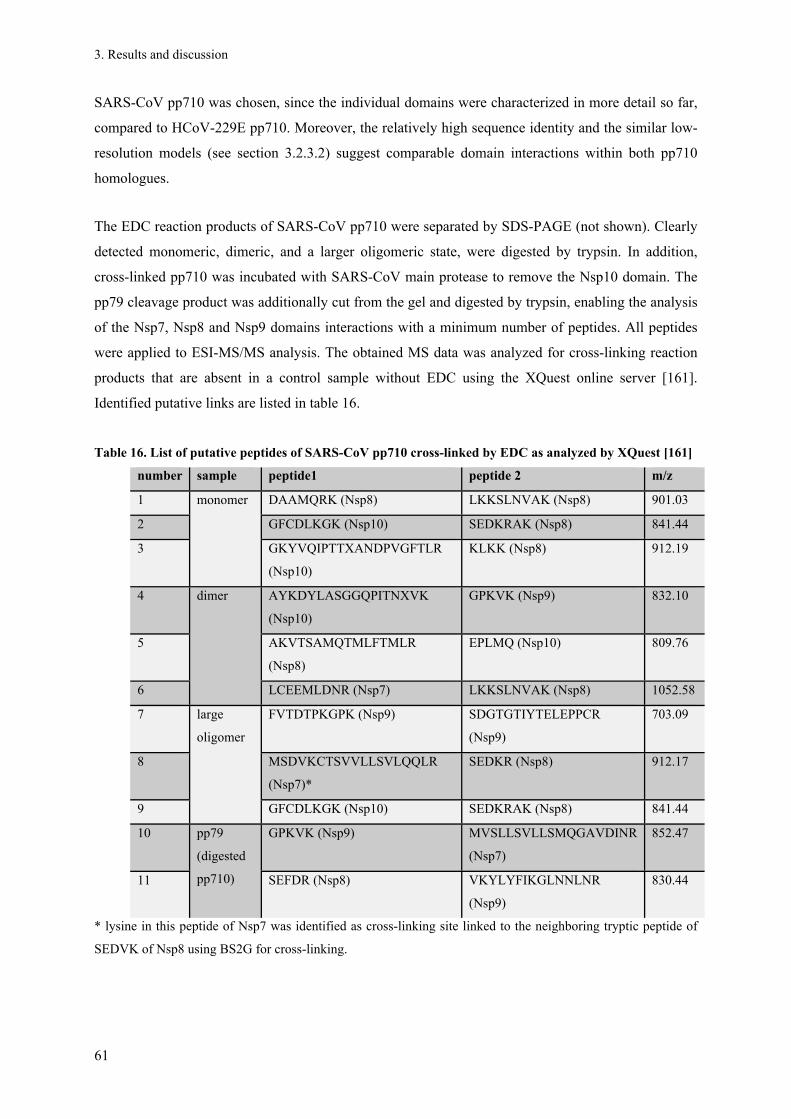
3. Results and discussion
61
SARS-CoV pp710 was chosen, since the individual domains were characterized in more detail so far,
compared to HCoV-229E pp710. Moreover, the relatively high sequence identity and the similar low-
resolution models (see section 3.2.3.2) suggest comparable domain interactions within both pp710
homologues.
The EDC reaction products of SARS-CoV pp710 were separated by SDS-PAGE (not shown). Clearly
detected monomeric, dimeric, and a larger oligomeric state, were digested by trypsin. In addition,
cross-linked pp710 was incubated with SARS-CoV main protease to remove the Nsp10 domain. The
pp79 cleavage product was additionally cut from the gel and digested by trypsin, enabling the analysis
of the Nsp7, Nsp8 and Nsp9 domains interactions with a minimum number of peptides. All peptides
were applied to ESI-MS/MS analysis. The obtained MS data was analyzed for cross-linking reaction
products that are absent in a control sample without EDC using the XQuest online server [161].
Identified putative links are listed in table 16.
Table 16. List of putative peptides of SARS-CoV pp710 cross-linked by EDC as analyzed by XQuest [161]
number sample peptide1 peptide 2 m/z
1 monomer
DAAMQRK (Nsp8) LKKSLNVAK (Nsp8) 901.03
2 GFCDLKGK (Nsp10) SEDKRAK (Nsp8) 841.44
3 GKYVQIPTTXANDPVGFTLR
(Nsp10)
KLKK (Nsp8) 912.19
4 dimer
AYKDYLASGGQPITNXVK
(Nsp10)
GPKVK (Nsp9) 832.10
5 AKVTSAMQTMLFTMLR
(Nsp8)
EPLMQ (Nsp10) 809.76
6 LCEEMLDNR (Nsp7) LKKSLNVAK (Nsp8) 1052.58
7 large
oligomer
FVTDTPKGPK (Nsp9) SDGTGTIYTELEPPCR
(Nsp9)
703.09
8 MSDVKCTSVVLLSVLQQLR
(Nsp7)*
SEDKR (Nsp8) 912.17
9 GFCDLKGK (Nsp10) SEDKRAK (Nsp8) 841.44
10 pp79
(digested
pp710)
GPKVK (Nsp9) MVSLLSVLLSMQGAVDINR
(Nsp7)
852.47
11 SEFDR (Nsp8) VKYLYFIKGLNNLNR
(Nsp9)
830.44
* lysine in this peptide of Nsp7 was identified as cross-linking site linked to the neighboring tryptic peptide of
SEDVK of Nsp8 using BS2G for cross-linking.

3. Results and discussion
62
Figure 17. Two putative cross-linking sites within SARS-CoV pp710 (cross-link number 8 and 4, according to
tab. 16; the acidic amino acid and the lysine of the linked peptides are highlighted as sticks). (A) Nsp7 (green)
and Nsp8 (blue, pdb code 2AHM chain H; Nsp7 was manually positioned considering the covalent connection).
This Nsp7-Nsp8 cross-link can reasonably be related to an epitope revealed at high-resolution for mature Nsp7
that binds close to the C-terminus of Nsp8 (pdb code 2AHM), considering that the putative flexible N-terminus
of Nsp7 is oriented towards the stretched -helical N-terminus of Nsp8. Compared to the manual positioning of
Nsp7 and Nsp8 shown here, the N-terminus of Nsp8 is considerably bended (based on SAXS data discussed in
chapter 3.11). (B) SARS-CoV Nsp9 (magenta; pdb code 1UW7) and Nsp10 (yellow; pdb code 2FYG).
Similarly, N-terminally strep-tagged SARS-CoV pp710 was cross-linked by BS2G (with and
without deuterium “label”) as displayed in fig. 18. SARS-CoV polyprotein Nsp7-8 (pp78; see section
3.9) was included and cross-linked as well to compare its oligomerization to SARS-CoV pp710. While
non-linked “monomeric” pp78 visualized by coomassie-stained SDS-PAGE almost completely
disappeared during the reaction in the presence of 25 mM BS2, pp710 formed a significantly lower
fraction of covalent oligomers in this reaction. Based on this observation, a different oligomerization
of pp78 and pp710 is hypothesized, i.e. different dissociation constants. However, the presence of the
“domains” Nsp9 and Nsp10 may influence the formation of a comparably small fraction of dimer and
a larger fraction of putative trimer of pp710 compared to pp78. The indicated specific trimeric state –
as at least a small amount is detectable by mass spectrometry –could be mediated by trimerization of
Nsp10, as observed within a controversially discussed dodecameric high-resolution structure ([131;
118; 181]). Nevertheless, this is highly speculative since a similar oligomeric state of Nsp10 has never
been observed in solution and Nsp7, Nsp8 and Nsp9 could contribute as well to form higher
oligomeric states, including intermolecular disulfide bonds.
A B

3. Results and discussion
63
5 mM 25 mM
0.25 0.5 1 2 time [h] - 0.25 0.5 1 2
100
30
kDa
IV III II
I
M
5 mM 25 mM
100
30
kDa
V IV III
II
I
- 0.25 0.5 1 2 0.25 0.5 1 2 MMM
5 mM 25 mM
0.25 0.5 1 2 time [h] - 0.25 0.5 1 2
100
30
kDa
IV III II
I
M
5 mM 25 mM
100
30
kDa
V IV III
II
I
- 0.25 0.5 1 2 0.25 0.5 1 2 MMM
Figure 18. Two different total concentrations of an equimolar mixture of BS2G-d0/d4 were incubated with 5 µM
SARS-CoV pp710 (A) and 5 µM SARS-CoV pp78 (B) for different time periods up to 2 h. Both proteins harbor
an N-terminal strep tag. Reaction products were visualized by SDS-PAGE and coomassie staining. Oligomeric
states are continuously labeled with numbers I-V, where I is the monomer.
All oligomeric states identified by SDS-PAGE were cut from the gel and applied to tryptic in-gel
digestion. Trypsin cleaves N-terminally of lysine and arginine residues. The tryptic peptides were
separated according to their mass to charge ratio by an ESI-Q-TOF instrument with significantly
advanced separation capacity. The XQuest/XProphet software package [161; 162] was applied to
search MS/MS spectra of suitable masses for fragment ions of peptides cross-linked by BS2G-d0
and/or deuterated BS2G-d4. Therefore, the identification of cross-linking sites is significantly
improved compared to the EDC-based cross-linking investigated by an ESI-ion trap instrument
(LC/MSD Trap XCT Ultra II). Oxidation and carbamido methyl modifications of amino acid side
chains were taken into consideration for all spectra. MS/MS spectra of putative BS2G cross-link
events are summarized in the appendix. The basic method including the beneficial use of isotope
labels for verification of the results has proven to be powerful in modelling particularly of large multi-
domain proteins[182; 183], optionally combined with homology modelling and different “low-
resolution” methods summarized by Venselaar et al. [184] and native mass spectrometry [185].
Initially, to establish the method two putative intra-domain cross-linking sites within SARS-CoV
pp710 indicated by XQuest/XProphet are described (fig. 19). Subsequently, 5 additional putative
interdomain cross-linking sites indentified in the sample of monomeric pp710 and 1 intra- or
interdomain cross-linking site of Nsp8 in the sample of presumably trimeric pp710 (oligomeric state
III in fig. 18A), as cut from an SDS-PAGE, are presented in the following paragraphs. These cross-
links were selected according to the highest fragment ion coverage in the MS/MS spectra. Spectra of
all putative BS2G cross-linking sites are displayed in the appendix in figure 87A and figure 87B.
These figures include two additional cross-linking sites not presented in the main text in the sample of
larger oligomers (oligomeric state IV in fig. 18A), which are hardly interpretable, since different
perhaps unspecific oligomerization epitopes are expected in such oligomers that are not related to the
A B

3. Results and discussion
64
SAXS models presented in chapter 3.2.3.2. A control sample of SARS-CoV pp710, free of BS2G was
included as a negative control.
At first, an intramolecular covalent link of K84 and K86 of SARS-CoV Nsp9 was identified (fig.
19, upper panel). Both isotopes of the cross-linking product were identified by high fragment-ion
coverage of the sequence. Additionally, this result is strongly supported by the close proximity of
these residues in the corresponding high-resolution structure of mature Nsp9 (fig. 19, upper panel). A
putative intramolecular link of K72 and K79 of Nsp8 by BS2G-d0 can be explained by the
predominantly helical structure of the N-terminal half of mature Nsp8, which enables the residues to
localize in close contact – remarkably on the same side of the helix. Nevertheless, loop segments
within the Nsp8 N-terminus might have a flexible random structure that allows the helical N-terminus
to adopt a “bended” conformation as revealed in Nsp8 high-resolution structures [96; 98]. The
alternative conformation of Nsp8 in the same high-resolution structures is a prolonged stretched α-
helical N-terminus as observed for SARS-CoV [98] and feline CoV (FCoV) [96]. According to the
geometry of the BS2G molecule and the orientation of K72 and K79 in the “stretched” conformation
of the α-helix (see fig. 19, light blue), a cross-link might be sterically impaired. However, the cross-
linking reaction between K72 and K79 could have been allowed by the bended conformation, similar
to the Nsp8 structure depicted in cyan (fig. 19), even though the number of detected fragment ions is
rather low compared to the intrapeptide Nsp9 cross-link described before. However, both examples
further indicate that the method in principle is sufficient to obtain structural information.

3. Results and discussion
65
Figure 19. Intra-domain cross-linking sites of SARS-CoV pp710. The left panel displays the MS/MS spectra.
Fragment ions that were identified in a respective MS/MS spectrum with the default mass tolerance are labelled
either green (common mass) or red (mass containing the cross-linker). The lysine residues labelled red below the
spectra were linked by BS2G. The covalent link of K84 and K86 (upper panel) was identified by high fragment-
ion coverage of the sequence; both isotopes BS2G-d0 and -d4 of the peptide were identified, the BS2Gd0-link is
shown (precursor mass: 726.4146 Da, charge 2+, isotope mass difference: 4.0237 Da). This link is strongly
supported by the high-resolution structure of SARS-CoV Nsp9 [102] (upper right panel) and at least further
reveals that the method is capable of identifying surface areas in close proximity. Similarly, the link between
K72 and K79 within Nsp8 by BS2G-d0 in the monomer sample can be well explained. Assuming a prolonged
stretched -helical N-terminus of Nsp8 the flexible lysine residues are facing in the same direction (SARS-CoV
Nsp8, pdb code 2AHM, chain H in light blue), separated by two helix windings. The “bended” N-terminal
conformation of Nsp8 suggests a more probable close position of both lysines separated by a short turn motif
(FCoV Nsp8, pdb code 3UB0, chain A in cyan). Peaks fitting the putative cross-link site are labelled. A similar
cross-linked peptide of Nsp8 created by alternative trypsin cleavage was identified by XQuest/XProphet and is
depicted in the enframed box.

3. Results and discussion
66
Further cross-linking sites suggested by mass spectrometry indicate the close proximity of the
elongated helical subdomain of Nsp8 with that of Nsp9, i.e. Nsp8 K46 linked with Nsp9 K77 and
Nsp8 K39 connected to Nsp9 K89. Due to the structure of BS2G, a distance of around 8 Å between
those sites is expected, whereas EDC presented earlier requires a much lower distance. Assuming that
one suggested link could be “false positive” in the spectra, the other Nsp8-Nsp9-link is a valuable
validation. An additional cross-verification is presented by the detected peptide SEFDR (Nsp8) that is
assumed to be linked by EDC with VKYLYFIKGLNNLNR (Nsp9). In parallel, a BS2G-mediated link
was identified for KLKKSLNVAKSEFDRDAAMQ and YLYFIKGLNNLNR, notably both peptides
overlap with the previously mentioned EDC linked Nsp8 and Nsp9 peptides. Consequently, according
to the rigid-body model of both homologues of pp710, Nsp9 seemed to be further translated towards
the cross-linking lysine residues of Nsp8 (red arrow, fig. 20), as it is well visualized for HCoV-229E
pp710 as well. Therefore, Nsp9 might even displace Nsp10 that is considered to be “loosely” attached
to Nsp9, particularly as no specific interaction of mature Nsp10 with the other three mature Nsps is
indicated, as investigated and discussed in chapter 3.4.1. A translation of Nsp9 is furthermore
supported by the shape of the ab initio model, providing sufficient space for Nsp9, and the N-terminus
would still be in sufficiently close proximity to the C-terminus of Nsp8 (fig. 20, compare left and right
panel). In parallel, the N-terminal helical domain of Nsp8 is assumed to bend in the opposite direction
(green arrow, fig 20). For comparison to the rigid-body model of HCoV-229E pp710 the ab initio
models of SARS-CoV pp710 and HCoV-229E pp710 are shown (fig. 20, left panel). Nsp8-Nsp9
cross-link events together with three cross-links of Nsp8 with Nsp7 or the strep tag fused to the N-
terminus of Nsp7 are summarized in figure 20, respectively. All five cross-linking sites were identified
in the sample of monomeric pp710. Consequently, these links have to be intramolecular.

3. Results and discussion
67
Figure 20. Based on the high sequence identity of 47.1% and similar ab initio models shown in the left panel, the
structure of SARS-CoV and HCoV-229E pp710 is interpreted taking SAXS and mass spectrometry data into
consideration. Cross-linking sites of SARS-CoV pp710 connecting Nsp8 (blue) and Nsp9 (pink) are visualized in
the rigid-body model of monomeric HCoV-229E pp710 (right panel, solid black lines, involved residues are
coloured red). The arrows indicate the suggested direction of subdomain translation based on the Nsp8-Nsp9
cross-link sites identified by MS. Some termini of Nsp domains are labelled for clarity. Three cross-linking sites
involving the N-terminus or the N-terminal strep-tag of Nsp7 (Nsp7N; Nsp7 is coloured green) and the
elongated helical domain of Nsp8 are listed above the rigid-body model and are visualized in fig. 21. The dashed
line represents an EDC cross-linking site connecting Nsp7 and Nsp8, as previously depicted in figure 17A, in
good agreement with the putative BS2G cross-linking sites. Nsp10 is coloured yellow.
The mass spectrometry-based cross-linking analysis of pp710 indicated that the highly elongated
N-terminus of Nsp8 represents an uncertain aspect within the rigid-body models of both pp710
homologues. According to the ab initio models of pp710 (fig. 20, left panel), the structure of the N-
terminus of Nsp8 appears to be slightly different, comparing both coronavirus species. As previously
mentioned, a high-resolution structure contained one isoform of Nsp8 with stretched N-terminal α-
helical domain and another one with a specific degree of bending of the N-terminus, allowed by short
flexible loop structures in between of helical structure [96; 98]. Crystal contacts identified by CryCo
5.1 analysis [186] are suggested to stabilize the elongated N-terminus in the crystal. Considering this

3. Results and discussion
68
putative flexibility, the identified Nsp8-Nsp8-link in the pp710 dimer sample (KLKKSLNVAKSEFD
linked to KLDNDALNNIINNAR) would be well explained by a relative close proximity of both Nsp8
termini, due to tight bending. Nsp7 is expected to bind to Nsp8 close to the Nsp8 C-terminus, which
indeed results in a close proximity of both Nsp8 termini within pp710. Consequently, the Nsp8-Nsp8-
link is suggested to be an intra-molecular link, rather than associated with the dimer interface
involving two different Nsp8 molecules. Considering that the Nsp8 N-terminus is at least partly
bending towards Nsp9 and Nsp7 (fig. 20, right panel, rigid-body model), Nsp8 could most likely not
reach the elongated termini of both pp710 ab initio models (fig. 20), and therefore not span the entire
molecule according to the maximum diameter determined by SAXS. Thus, mass spectrometry data
questions the position of Nsp10 relative to Nsp8, since a minor translation of Nsp10 would allow the
required bending of the Nsp8 N-terminus and the maximum diameter of the ab initio model would be
increased by Nsp10 at the same time, corresponding to the ab initio model (fig. 21). To enable the
hypothesized “bending” of the Nsp8 N-terminus, SARS-CoV Nsp8 (chain H, extracted from pdb code
2AHM) was cut into 5 fragments (amino acids 1-34 (fragment 1), 35-73 (fragment 2), 74-113
(fragment 3), 114-124 (fragment 4), 125-195 (fragment 5, C-terminus)) to design the final concluding
structural model (fig. 21). These Nsp8 fragments were designed according to reported putative sites of
flexibility within the molecule [96; 98], and personal communication with Y. Xiao (University of
Lübeck). Additionally, a loop comprising residues 99-101 could be flexible as well to potentially
increase maximum diameter of the proposed model, which is slightly lower than determined by SAXS.
As observed for the complex of mature Nsp7 and Nsp8, Nsp7 is expected to be located close to
the globular Nsp8 C-terminus, involving hydrophobic residues in the N-terminal helix 1 and helix 3 of
Nsp7 [98; 96]. The cross-linking sites connecting Nsp8 and Nsp7 or the strep tag N-terminally linked
to Nsp7 are in good agreement with this model: Nsp8 K46 linked to Nsp7 K7 and a lysine within the
strep-tag linked to K72 and K40 of Nsp8, respectively. The two different cross-links of the strep-tag
could be explained by its flexibility.
There is a valuable agreement comparing the results obtained by either cross-linking reagent with
each other. For instance, using EDC XQuest suggested a link connecting
MSDVKCTSVVLLSVLQQLR (Nsp7) and SEDKR (Nsp8). A cross-link suggested by BS2G is
KMSDVKCTSVVLLSVLQQL linked to MADQAMTQMYKQAR which is located next to the N-
terminus of SEDKR in Nsp8, further securizing the orientation of the Nsp7 N-terminus towards a
defined fragment of the corresponding Nsp8 helix. This is in good agreement with the interaction of
the N-terminal helix 1 of Nsp7 with a central part of the Nsp8 chain in the complex of the two mature
Nsps, allowing to suggest a very similar orientation of Nsp7 helix 1 within pp710. Therefore, a
conserved orientation of helix 3 is hypothesized as well, which also contributes to the interaction of
mature Nsp7 and Nsp8 [98]. Finally, a summarizing rigid-body model of SARS-CoV pp710 involving

3. Results and discussion
69
known high-resolution structures of SARS-CoV and five individual fragments of Nsp8 considered all
putative BS2G cross-linking events of Nsp7 with Nsp8 (orange spheres in fig. 21) as well as of Nsp8
with Nsp9 (red spheres) of SARS-CoV, as displayed in Fig. 21. The residues that were linked
according to mass spectrometry are located in close proximity, mainly by the rearrangement of the
structure of Nsp8. All four Nsp domains of SARS-CoV pp710 are presumed to have flexible termini
according to the high-resolution structures. The flexibility allows an adjustment of the position of the
cleavage sites within pp710 to guarantee that the main protease cleavage site to process pp710 is well
surface-exposed, as already suggested by the model in fig. 21.

3. Results and discussion
70
Figure 21. Optimized structural model of monomeric SARS-CoV pp710, taking the identified cross-linking sites
of SARS-CoV pp710 into consideration. Lysine residues involved in two cross-links connecting Nsp9 and Nsp8
are labelled by red spheres (Nsp8 K46 – Nsp9 K77; Nsp8 K39 – Nsp9 K86), three sites linking Nsp7 and Nsp8
(see fig. 20) are labelled in orange (Nsp7 K7 – Nsp8 K46; Nsp7-tag – Nsp8 K72; Nsp7-tag – Nsp8 K40). Nsp7,
Nsp9, Nsp10 and five fragments of SARS-CoV Nsp8 (comprising chain H, extracted from pdb code 2AHM)
were used for modelling. The fragments of Nsp8 individually used to model the bended conformation of the full-
length Nsp8 domain within pp710 are: amino acids 1-34 (fragment 1), 35-73 (fragment 2) 74-113 (fragment 3),
114-124 (fragment 4), 125-195 (fragment 5, C-terminus). It may additionally be assumed that a loop comprising
residues 99-101 is flexible as well to potentially increase the maximum diameter of the model, which is slightly
lower than determined by SAXS. Fragments of Nsp8 were manually positioned according to the ab initio model.
The ab initio model of SARS-CoV is shown again for comparison. All covalent Nsp-Nsp contacts within pp710
are sufficiently surface-exposed to allow cleavage by the coronavirus main protease.

3. Results and discussion
71
This concluding model of SARS-CoV pp710 that is drawn here allows a valuable prediction
concerning the overall structure of pp710 with slight differences between pp710 of an
Alphacoronavirus compared to a Betacoronavirus (fig. 20). It also highlights the hypothetic previously
unknown interface of mature Nsp8 and Nsp9, although there are contradictions in the literature
concerning the strength of this interaction [101; 102]. Moreover, it is strongly suggested that Nsp7 in
the polyprotein state is already capable of occupying a binding site with Nsp8 close to its C-terminus,
as reported for the complex of mature Nsp7 and Nsp8 [96; 98]. Finally it was observed that pp710 of
SARS-CoV has a significantly higher radius of gyration and a higher maximum diameter than the
HCoV-229E homologue. However, except these differences, the low-resolution surface structures of
HCoV-229E and SARS-CoV clearly share significant similarities (fig. 20).
3.2.4. Trials to crystallize pp710
Knowing that pp710 displayed variable oligomerization in a slow equilibrium and that it further
represents a multi-domain protein that is assumed to be locally flexible, it was not considered to be
easy to crystallize the full-length pp710 proteins. Both homologues were applied to a conventional
robot-assisted crystallization condition screening. Applying a solution with predominantly monomeric
or dimeric pp710 of either SARS-CoV or HCoV-229E to approximately 2000 different crystallization
conditions each resulted in a few promising observations (like phase separation) that were worth to be
refined. However, refinement failed to result in crystals suitable for a diffraction analysis up to now.
One exemplary crystallization condition is displayed in figure 22. Refinement approaches included
microseeding of very tiny crystals and “crystalline precipitation” as well as counter diffusion
experiments. Moreover, smaller fragments of pp710, e.g. pp79 (polyprotein Nsp7-9) and pp78
(polyprotein Nsp7-8) (described in chapter 3.9) as well as and some of the mutated variants of pp710
(described in chapter 3.5) were included in crystallization trials. Limited proteolysis using a serial
dilution of trypsin, chymotrypsin, papain and subtilisin did not reveal a stable domain of more than
approximately 10-12 kDa in size and was therefore not considered to be beneficial for crystallization
of a rigid pp710 fragment. Similarly, covalent cross-linking trials did not result in homogeneous
solutions that are favourable for crystallization trials.

3. Results and discussion
72
Figure 22. Crystallization of HCoV-229E pp710. A 300 nl drop of 40 µM HCoV-229E pp710 in Tris buffer
containing 260 mM NaCl was mixed with an equal amount of 0.3 M magnesium formate, 0.1 M Bis-Tris pH 5.5
and sealed in a 96-well Nextal plate (Qiagen). Crystallization success was investigated and documented after
three weeks under visible light (left image) and excited by UV-light (right image). The drop contained
monomeric protein purified by SEC supplemented with 2 mM DTT. For orientation, the white arrow points at
the same spheroid “drop” in both images. Thin two-dimensional crystals are encircled in the left panel. On the
right image, a putative crystal with dimensions in the low µm range that is fluorescent is highlighted.
It was recently observed and reported by members of our research group that heterogeneous gene
overexpression in baculovirus-infected Sf9 insect cells can yield small crystals (10 µm3 average
volume) grown in vivo within the living cells. As a first proof-of-principle, glycosylated cathepsin B
of the parasite T. brucei crystallized within the late ER was isolated and successfully applied to the
novel technique of serial femtosecond X-ray crystallography at a free-electron laser to solve the
protein structure [122]. Based on this promising approach, in vivo crystallization of SARS-CoV pp710
was investigated following the established protocol, representing the very first trial for directed
crystallization of a polyprotein within insect cells. The corresponding full-length gene was integrated
by double homologous recombination into a bacmid that is controlled by a polyhedron promoter to
express the pp710 gene and release the protein in the cytosol of Sf9 insect cells. The insect cells
showed the typical morphology of infected cells using a light microscope, crystalline structures within
the cells incubated at either 27 °C or room temperature were not observed. Although various different
approaches to obtain crystals of pp710 were applied, high resolution structural data could not be
obtained within this study.

3. Results and discussion
73
3.2.5. Insights into the function of pp710
3.2.5.1. Native gel electrophoresis: Interaction of pp710 with nucleic acid
Native PAGE is useful to initially detect a protein’s ability to interact with nucleic acid.
However, this method does not allow a reliable KD-value calculation. Particularly for very weak
protein-nucleic acid interactions an electrophoretic mobility shift is hardly quantitative. Nevertheless,
an interaction of HCoV-229E pp710 with random ssDNA (fig. 23A), random dsDNA (fig. 23B, upper
panel) as well as tRNA (fig. 23 B, lower panel) was qualitatively verified for the full-length pp710 of
HCoV-229E in a concentration-dependent manner. Even though RNA is the physiologically relevant
ligand of coronaviral Nsps, those three different types of nucleic acid provide structural diversity and
the opportunity to compare the results with previous studies about the interaction of Nsps with
different kinds of nucleic acid. In the presence of protein, a putative protein-nucleic acid complex that
is reduced in electrophoretic mobility became visible (fig. 23A) and more abundant (fig. 23B) with
increasing protein concentrations. Since the stain used for the detection in the assay is specific for
nucleic acid, the electrophoretic mobility shift is due to the interaction of the different nucleic acids
with pp710.
These results indicated that pp710, the precursor of four mature Nsps, is already capable of
binding nucleic acid, supporting that pp710 could have an impact on the replication of the virus.
Interactions of mature Nsp domains with nucleic acid in the µM range were previously observed in
vitro, for instance for mature SARS-CoV Nsp10 with dsDNA, dsRNA and ssRNA [111], Nsp9 with
ssDNA and ssRNA [101; 187] and HCoV-229E Nsp8 with tRNA and other RNA fragments [103]. In
particular, an interaction of processed MHV Nsp10 with tRNA was demonstrated by using an
electrophoresis technique with horizontal agarose gels, called zone-interference gel electrophoresis
[170] and revealed a low µM dissociation constant.

3. Results and discussion
74
Figure 23. Native PAGE under non-reducing conditions demonstrating an interaction of HCoV-229E pp710 with
a random ssDNA, dsDNA and yeast tRNA (Roche). Pp710 was initially treated with benzonase and applied to
the native PAGE in sodium phosphate buffer at pH 7.9 supplemented with 100 mM NaCl and 2 mM MgCl2. (A)
Interaction of pp710 with ssDNA. “pp”, 300 pmol polyprotein; “+”, 300 pmol HCoV-229E pp710 mixed with 50
pmol ssDNA (5’-GCGAATTCCTAAACAGCAAAAGAACAATGTGT-3’). Other lanes contain 10 to 100 pmol
pure ssDNA. (B) Interaction of pp710 with 60 pmol dsDNA (5’-GCATTTGTCGACCAT-3’) and 20 pmol
tRNA. Double stranded DNA was heated to 80 °C for three minutes and cooled down at room temperature. The
amount of pp710 ranges between 60 and 300 pmol in both gels. A nucleic acid-free and a protein-free control
was included as well (“-dsDNA/-tRNA” and “-Pp710”). To detect dsDNA, gels were stained with EtBr.
Detection of ssDNA and tRNA was accomplished by incubating the gels with SybrGOLD diluted in
electrophoresis buffer.
3.2.5.2. Pp710 interacts with RNA derived from the 3'-UTR
Considering that SARS-CoV Nsp8 is known to specifically bind genomic RNA in an early stage
of replication [108], this investigation aims to analyze the protein-RNA interaction of the precursor
SARS-CoV pp710. It is assumed that a filter retention assay, usually applied to reveal protein-nucleic
acid interactions in the range of µM dissociation constants, is suitable to detect pp710-RNA
interactions, obtaining further insights into a putative function of pp710.
A structurally conserved fragment of the SARS-CoV genomic 3’-untranslated region (3’-
UTRdeltaHVR, 105 nt, see methods for reference) and a shuffled sequence with identical nucleotide
composition (3’-UTRdeltaHVR-SH) were produced by T7 polymerase in vitro transcription. RNA
transcription products were separated on a denaturing acrylamide gel (fig. 24A/B). These fragments
represented the ligands applied to the assay. The secondary structure of the 3’-UTR fragment, lacking
a hyper variable region (HVR), is shown in figure 24C, as predicted by the software mfold [188]. The
RNA fragments were terminally labelled with 32P-phosphate to enable a sensitive and specific
detection. Various concentrations of SARS-CoV pp710 were incubated with a constant amount of both
RNA fragments. The solution was drawn through a membrane, while only the protein bound to the
A B

3. Results and discussion
75
membrane. Unbound nucleic acid were not retained, whereas labeled RNA molecules that interacted
with the protein remained on the filter and were subsequently detected and quantified using a
phosphor imager. A positive control, i.e. an RNA aptamer binding to streptavidin in the nM range, was
included for comparison (fig. 24C).
In spite of a relatively high background due to unspecific binding of RNA on the membrane, a
concentration-dependent interaction is strongly suggested for both fragments (fig. 24D) by plotting the
detected radioactivity against the protein concentration (not shown). The detected RNA-interaction of
pp710 additionally indicated that the polyprotein precursor represents a functional component of the
RTC. However, due to the interaction with the shuffled sequence it appears that pp710 does not only
specifically recognize the sequence and secondary structure of the putative Nsp8 binding site within
the 3’-UTR of the SARS-CoV genome. This is in good agreement with the observation that even
mature Nsp8 does not seem to be highly sequence-specific with a long recognition site [103], but
clearly prefers 5′-(G/U)CC-3′ in a template to initiate the synthesis of a short complementary fragment
[94]. Furthermore, mature coronaviral Nsp9 and Nsp10 were not reported to harbour a sequence-
dependency for nucleic acid interaction [111; 187] and likely also contribute to the interaction of
pp710 with nucleic acid. However, an interaction of Nsp9 with hairpin SL2 of the 3’-UTR was
demonstrated [104]. This interaction as well as the FCoV Nsp8 RNA polymerase activity significantly
depends on the environmental pH, preferring approximately pH 7-7.5 in both interactions [96]. This is
reflected by the binding buffer at pH 7.6 used in this experiment. Facing the problem that in a filter
retention assay a small amount of RNA is unspecifically binding to the membrane and that Nsp9 and
Nsp10 may contribute to sequence independent interaction of pp710 with nucleic acid, it was decided
to continue by investigating a specific catalytic activity, i.e. polymerase activity, of pp710 and its
processing products. However it is summarized, that pp710 harbours at least one accessible binding
site to interact with DNA and RNA. Moreover, SARS-CoV and HCoV-229E pp710 appear to have
either no or a weak overall sequence specificity in binding to nucleic acid.

3. Results and discussion
76
Figure 24. Interaction of SARS-CoV pp710 with RNA. (A) In vitro transcription products of two different DNA
templates derived from the SARS-CoV 3’UTR were separated on a denaturing urea acrylamide gel. (B) The
desired RNA fragments, i.e. the authentic RNA fragment and a shuffled variant, isolated by gel extraction
showed high homogeneity. (C) Prediction of the secondary structure of the 3’-UTR lacking a HVR using mfold
[188]. (D) Phosphorimager screen of the filter-retention assay involving both RNA fragments and N-terminally
strep-tagged SARS-CoV pp710. 10% of the total amount of RNA per sample in the respective lane is spotted
directly on the membrane for control. An RNA aptamer provided by S. Kruspe (University Hamburg) was
applied as positive controls.
3.3. SARS-CoV Mpro (Nsp5): Structural properties and monomer-dimer
equilibrium
During replication in a host cell, coronaviral pp710 is cleaved by the viral main protease to release
mature Nsps. Pp710 itself is considered to be an intermediate product during processing of a large
fragment of pp1a (Nsp4-Nsp16). To analyze the time-course of the processing pathway of pp710 in
the presence of the SARS-CoV main protease (Mpro) and eventually purify cleavage products of pp710
for structural characterization, the protease had to be purified and initially characterized.
SARS-CoV Mpro was heterologously expressed as previously described [78] and purified by Ni-
NTA affinity chromatography and size-exclusion chromatography (fig. 25A). The presence of the C-
terminal his-tag was verified by Western blot. Isoelectric focusing (IEF) determined the pI to 6.6 (fig.
25A). The N-terminal GST-tag to optimize the protein solubility was cleaved off in vivo by N-terminal
autocleavage of the protease, comprising a first opportunity of activity verification by analysing the
protein size by SDS-PAGE. The gene of the protease mutant R298A was generated by mutagenesis
PCR, the protein was purified applying the same protocol. Amino acid R298 is known to be critical for

3. Results and discussion
77
the dimerization of Mpro [56]. Thus, the mutant is interesting to analyze the monomer-dimer
equilibrium and protein interactions with the main protease in more detail. The mutant is still capable
of GST-tag autocleavage in E. coli cells, even though the dimeric main protease is known to be the
catalytically active state [48; 49]. In agreement with an expected predominantly monomeric state of
the mutant, the hydrodynamic radius determined by DLS peaks at Rh = 3.5 ± 0.3 nm, whereas an Rh =
4.2 ± 0.1 nm was determined for the dimeric wild type of SARS Mpro applying 90 µM protein each.
The melting temperatures of Mpro wild type and the mutant R298A determined by CD spectroscopy
are significantly different in 150 mM NaCl, 15 mM NaH2PO4, pH 7.6. For the wild type enzyme the
most significant change in absolute ellipticity monitored at 220 nm was detected at Tm = 55.8 ± 1.5 °C.
For the mutant a Tm = 53.1 ± 0.7 °C was detected. These values correspond to the turning point of an
optimized Boltzmann function calculated by Origin 8.5, fitting the raw data. The increased thermal
stability of the wild type is expected to be a result of the dimerization, perhaps with a minor
contribution of a local structural rearrangement due to the mutation [56]. In analytical size-exclusion
chromatography, 90 µM Mpro R298A were applied to the Superose 12 10/300 GL SEC column in the
absence of a substrate. In spite of the mutation, a minor fraction of the mutated protease was observed
to be still dimeric. In contrast Mpro wild type displayed no detectable amount of monomer and a small
peak corresponding to a large oligomer beside the dimeric state (see also fig. 44). SAXS
measurements further supported the predominant monomeric state of Mpro R298A, in contrast to the
protease wild type. The altered pair-distribution function (P(R)-function (for explanation see chapter
3.2.5) indicated a different geometry and size for both molecules (fig. 25B). The three domains of the
high-resolution structure of the monomeric mutant are well superimposable with the ab initio model,
indicating that the correct overall fold is obtained (fig. 25C; DAMMIF), based on the P(R)-function of
Mpro R298A (fig. 25 B). The low χ-value of the high-resolution structure (Rg = 23.34) of 0.98
(determined by CRYSOL) compared to the SAXS scattering pattern additionally demonstrates that
both structures are well superimposable. Mass spectrometry that ensures a high mass resolution and
accuracy did not detect a specific large oligomeric state which was previously reported by [189].
Nevertheless, detection of predominantly dimeric Mpro wt and predominantly monomeric mutant (fig.
25D) comprised a valuable test system for the method, qualifying it for analysing cleavage reactions of
SARS-CoV Mpro (chapter 3.4). The activity of Mpro R289A is reduced by 92% compared to the wild
type enzyme, as determined by monitoring the cleavage of the chromogenic substrate TSAVLQ-pNA
at a wavelength of 405 nm. The loss of activity was already reported [56]. However, there are
indications that certain protease substrates significantly alter the monomer-dimer equilibrium of Mpro
[56; 132] demonstrated by AUC. An octameric state of the wild type main protease according to [189]
has not been observed by the applied methods. A minor amount of octamer created by domain-
swapping could artificially arise from either the termini of the protein or the experimental conditions.
However, the octamer may theoretically be considered as a specific stencil, perhaps underrepresented
in a host cell and promoting efficient polyprotein cleavage.

3. Results and discussion
78
Figure 25. (A) SDS-PAGE analysis of purified SARS-CoV Mpro (Nsp5) and its mutant R298A after Ni-NTA
affinity chromatography and size-exclusion chromatography. Western blot (WB) analysis of Mpro wild type
verifies the presence of the his6-tag by incubating the blot membrane with a primary goat anti-tetra-his antibody
(QIAGEN) and a secondary anti-goat antibody conjugated with alkaline phosphatase. Isoelectric focusing (IEF)
reveals a pI of 6.6. IEF Marker 3-10 (“liquid mix”, SERVA) was used as a standard. The gel was stained with
SERVA violet. (B) The difference between the dimeric state of Mpro wild type (blue) and purified monomeric
R298A mutant (red) is obvious from the P(R)-functions calculated from recorded SAXS datasets by GNOM.
The maximum of the P(R)-function is shifted to a lower radius R and the maximum diameter for the mutant is
slightly reduced. (C) The ab initio model of the R298A mutant fits the superposed high-resolution structure of
the Mpro monomer (pdb code 3M3T, [56]). (D) Native ESI-Q-TOF mass spectrometry investigating the mass and
oligomeric states of Mpro in solution. The wild type is predominantly dimeric (MW = 69.8 kDa) and the mutant
predominantly monomeric (MW = 34.8 kDa) in solution. Collision-induced dissociation of the dimer at 130 V
collision voltage in the mass spectrometer resulted in the detection of the monomer subunit. The verified dimeric
state of the wild type main protease is detectable by native ESI-Q-TOF MS.

3. Results and discussion
79
3.4. Proteolytic processing of polyprotein Nsp7-10 by SARS-CoV Mpro
Purified SARS-CoV Mpro was incubated with the substrate pp710 of SARS-CoV and also of
HCoV-229E, which is presumable cleavable by SARS-CoV Mpro with the same specificity, due to the
highly conserved protease structure. In vivo, it was recently shown that a chimeric exchange of main
proteases within different Betacoronavirus species resulted in only marginally decreased fitness of the
virus [190]. The aim of this investigation is to semi-quantitatively analyze the specificity of the
cleavage reaction with the full-length substrate pp710 comprising four Nsp domains. A generalized
scheme of cleavage sites is presented in fig. 26A. The following protease cleavage experiments
depicted in figure 26B, 27A and 28 were performed in sodium phosphate buffer pH 7.9 supplemented
with 3 mM DTT and 280 mM NaCl. Using a simple SDS-PAGE approach, the overall order and time
course of proteolytic cleavage of HCoV-229E and SARS-CoV pp710 by SARS-CoV Mpro was
determined to be similar at a protease to substrate ratio of 1:10. An SDS-PAGE gel of the cleavage
reaction involving SARS-CoV pp710 monitored for up to 44 h is displayed in figure 26B, while the
first 20 min of the reaction with HCoV-229E pp710 as a substrate is depicted in figure 27A.
There was no indication that the ratio of Mpro and pp710 substrate alters the specificity or the
cleavage rate of either pp710 homologue. Several intermediate processing products of pp710 were
detected to be remarkably stable at the applied experimental conditions. Finally mature Nsp7, Nsp8,
Nsp9 and Nsp10 were released (fig. 26, fig. 27).

3. Results and discussion
80
pp710pp79
pp89/Mpro
pp78
Nsp8Nsp10Nsp9Nsp7
0 0.2 0.5 1 2.5 20 30 44 M kDa
6050
30
201510
100200
incubation time [h] (6 °C) B
A
polyprotein Nsp7-10 (pp710)
Nsp7 Nsp8 Nsp9 Nsp10Nsp1-6
polyprotein 1a
Mpro
Nsp11
Mpro Mpro Mpro Mpro
polyprotein Nsp7-10 (pp710)
Nsp7 Nsp8 Nsp9 Nsp10Nsp1-6
polyprotein 1a
Mpro
Nsp11
Mpro Mpro Mpro Mpro
Figure 26. Proteolytic processing of SARS-CoV pp710. (A) Schematic overview of pp710, the main protease
substrate under investigation. (B) Time course of SARS-CoV pp710 cleavage. A 10-fold molar excess of SARS-
CoV pp710 was incubated with Mpro and analyzed by SDS-PAGE at different time points up to a total incubation
time of 44 h. For proteolytic processing, monomeric pp710 was purified by SEC. For the processing
intermediates pp78 and pp89, an increased stability was indicated.
Noteworthy, SARS-CoV pp78 (Nsp7-Nsp8; connected by the cleavage site RATLQ↓AIASE) as
well as its homologue from HCoV-229E appears to be substantially more stable than SARS-CoV
pp910 (Nsp9-Nsp10; TVRLQ↓AGNAT) towards proteolytic cleavage (fig. 26 B). Pp78 and pp89 were
still detected after 44 h of incubation. Therefore, the turnover-rate of the third cleavage site in between
of Nsp8 and Nsp9 (AVKLQ↓NNELS) appears to be low as well. Particularly, HCoV-229E pp910
(polyprotein Nsp9-10, 26 kDa) and pp78 that are released upon cleavage of the site in between Nsp8
and Nsp9 within HCoV-229E pp710 are hardly detectable.

3. Results and discussion
81
Figure 27. Time course of HCoV-229E pp710 processing by SARS-CoV pp710 Mpro. (A) Pp710 was incubated
with SARS-CoV Mpro at a molar ratio of 10:1 at 20 °C. Samples were analyzed after different time points up to
20 min by SDS-PAGE. The cleavage products that were assigned to the detected protein bands are displayed. (B)
Proteolytic processing of a 20-fold molar amount of HCoV-229E pp78 prepared with N- and C-terminal tag by
SARS-CoV Mpro for up to 60 min. The cleavage products that were assigned to the detected protein bands are
schematically displayed.
To investigate the cleavage sites flanking Nsp7 and Nsp8, including the Nsp8-Nsp9 cleavage site,
pp78 of HCoV-229E was cloned with an N-terminal his6-tag connected by a long linker (≈ 4 kDa) that
contains the native Mpro cleavage site located between the membrane protein Nsp6 and Nsp7. The
amino acids SILQ (P4-P1) of the cleavage site were introduced by the forward primer. The C-terminus
of pp78 contained 9 N-terminal amino acids of Nsp9 and 24 C-terminal amino acids encoded by the
vector pRSETA up to its “TAG” stop codon. Analysing the cleavage of pp78 by Mpro by SDS-PAGE
(fig. 27B) in the presence of 2 mM DTT revealed that the amount of pp78 cleaved off from N-terminal
tag and C-terminal tag, i.e. N-terminus of Nsp9 is very low. The specific activity of Mpro cleaving the
site which links Nsp7 and Nsp8 seems to be remarkably higher. It appears that the cleavage site
linking Nsp7 and Nsp8 as well as the site linking Nsp7 and the N-terminal tag are cleaved first.
Consequently, after cleaving the covalent bond linking Nsp7 and Nsp8, the majority of Nsp7 and Nsp8
could still be covalently linked to the “membrane anchor” Nsp6 in vivo or more probable to Nsp9
during processing of the polyprotein. However, this is in contradiction to the Nsp6-Nsp7 cleavage site
sequence (HCoV-229E) that is remarkably close to the consensus sequence of SARS-CoV Mpro as
calculated by NetCorona 1.0 [40] (table 17). The relatively high stability of the cleavage site between
SARS-CoV Nsp7 and Nsp8 (fig. 26B) compared to the less stable site in HCoV-229E should be
further analyzed by (semi-)quantitative methods applying different oligomeric states.

3. Results and discussion
82
1´ 10´ 30´ 90´ 3 h 6 h 21 h 46 h
pp710 pp79
pp78
Nsp70
10
20
30
40
0 200 400 600 800 1000 1200 1400
time [min]
inte
nsi
ty (
rela
tive
un
its)
Pp71
Pp79
Pp78
A B
pp710
pp79
pp78
Figure 28. (A) Western blot analysis of SARS-CoV pp710 processing. (A) A ten-fold excess of SARS-CoV
pp710 was incubated with SARS-CoV Mpro at 20 °C for up to 46 h. Western blot after different time periods
revealed a defined specificity of proteolytic cleavage. The primary antibody detected the N-terminal strep-tag of
pp710. NBT and BCIP were used for detection. (B) The intensity of the signals on the membrane was quantified
using ImageJ; the result is displayed for an incubation time of up to 21 h: pp710 in blue, pp79 in yellow, pp78 in
red. The cleavage site connecting Nsp9 and Nsp10 is clearly preferred compared to the site that links Nsp8 and
Nsp9. After 6 h the remaining amount of pp710 and pp79 was highly similar, while both species were
completely processed after 21 h. Pp78 showed maximum concentration after 3 h of incubation, while most
intensive signals for pp79 were detected after approximately 1.5 h.
For an additional semiquantitative comparison of cleavage products a western blot with SARS-
CoV pp710 after affinity chromatography was performed, assuming to achieve increased
quantification accuracy. A tenfold excess of SARS-CoV pp710 with N-terminal strep-tag was mixed
with SARS-CoV Mpro and incubated at 20 °C with 3 mM DTT to further analyze the order and the
time-course of the reaction (fig. 28). Western blot analysis revealed a defined specificity of cleavage.
After 46 h of incubation, all intermediate cleavage products have been processed. The cleavage site
connecting Nsp9 and Nsp10 is clearly preferred for proteolytic processing compared to the site that
links Nsp8 and Nsp9 as well as Nsp7 and Nsp8 (fig. 28), in agreement with previous SDS-PAGE
analysis (fig. 26B, 27A). After 6 h the remaining amounts of pp710 and pp79 are very similar after
21 h both species are gone. Pp78 peaks later than pp79 after approximately 3 h. Consequently the
cleavage at the C-terminus of Nsp8 is again not preferred (see fig. 27A, for comparison). The reduced
cleavage preference of this site can be explained by the large asparagine residue in P1’ position (table
17). This effect was previously observed using short peptide substrates [65] and is supported by the
rapid release of Nsp10 compared to free pp910 (fig. 27A). According to [191], a residue with a
volume smaller than approximately 50 Å3 in P1’ position is strongly preferred by Mpro. Similar to the
semi-quantitative analysis by SDS-PAGE (fig. 26B and 27A) the online-server NetCorona 1.0 [40]
(http://www.cbs.dtu.dk/services/NetCorona/) calculated that the cleavage site connecting SARS-CoV
Nsp8 and Nsp9 is not perfectly matching the consensus sequence – indicated by a low score – and
considered to be cleaved slowly, even though the score is still higher than for the cleavage site in

3. Results and discussion
83
between of Nsp7 and Nsp8. However, there are partly huge differences of the NetCorona 1.0
“cleavage score”, particularly of the Nsp6-Nsp7 “cleavage-score”, comparing different coronaviruses,
e.g. HCoV-229E and SARS-CoV (tab. 17). This observation may reflect that the NetCorona “cleavage
score” can hardly predict the actual turnover rate (tab. 17).
Table 17. Alignment of Mpro recognition/cleavage sites*: Cleavage sites between Nsp7-10 are highly
similar comparing HCoV-229E and SARS-CoV. An unusual NNE motif in P1’-P3’ position (blue), an
alternative cleavage site (green) and some species-specific variations in P2’ position are labelled (orange).
cleavage site HCoV-229E SARS-CoV MERS-CoV MHV1
Nsp4 - Nsp5 GSTLQ↓AGLRK SAVLQ↓SGFRK SGVLQ↓SGLVK TSFLQ↓SGIVK
Nsp5 - Nsp6 GVNLQ↓SGKTT GVTFQ↓GKFKK GVVMQ↓SGVRK GVKLQ↓SKRTR
Nsp6 - Nsp7 VSTVQ↓SKLTD(0.905) VATVQ↓SKMSD(0.783) VAAMQ↓SKLTD VSQIQ↓SRLTD
Nsp7 - Nsp8 DSILQ↓SVASS(0.897) RATLQ↓AIASE(0.838) PSVLQ↓ATLSE LQALQ↓SEFVN
Nsp8 - Nsp9 VVKLQ↓NNEIM(0.849) AVKLQ↓NNELS(0.860) AVKLQ↓NNEIK TVVLQ↓NNELM
Nsp9 - Nsp10 TVRLQ↓AGKQT(0.880) TVRLQ↓AGNAT(0.904) TVRLQ↓AGSNT TVRLQ↓AGTAT
Nsp10 - Nsp11 RTAIQ↓SFDNS(0.801) EPLMQ↓SADAS(0.650) DSLRQ↓AALPQ GSQFQ↓SKDTN
Nsp12 - Nsp13 STVLQ↓AAGLC HTVLQ↓AVGAC PTTLQ↓AVGSC SAVMQ↓SVGAC
Nsp13 - Nsp14 MTDLQ↓SESSC VATLQ↓AENVT SSPLQ↓SYACW NPRLQ↓CTTNL
Nsp14 - Nsp15 EVNLQ↓GLENI FTRLQ↓SLENV FTKVQ↓GLENI FTRLQ↓SLENV
Nsp15 - Nsp16 YPQLQ↓SAEWK YPKLQ↓ASQAW YPRLQ↓ASADW YPRLQ↓AAADW
* some cleavage sites were analyzed by NetCorona 1.0 [40]; “cleavage-score” [0;1] is specified in brackets 1 murine hepatitis virus
Moreover, the western blot indicates that Nsp10 was cleaved off faster than Nsp7. Although the
release of Nsp7 is already detectable in the beginning of the reaction, the amount quantified by using
ImageJ (http://rsbweb.nih.gov/ij/) is at least slightly reduced compared to pp79, which is released after
cleaving off Nsp10 from pp710 (fig. 28). Notably, the maximum intensity detected for pp78 was
higher than for pp79, which is another indication for the stability of the Nsp7-Nsp8 cleavage site of
SARS-CoV, even though the number of data points in the time-course of cleavage was limited in this
experiment.
In SARS-CoV the Nsp7-Nsp8 cleavage site contains a threonine in P3 position instead of an
isoleucine in HCoV-229E, but this position was generally observed to have a minor influence on
kcat/Km [192]. Nonetheless, this putative effect on the cleavage reaction is probably too small to see it
by SDS-PAGE or Western Blot for a full-length polyprotein. Interestingly, the kcat/Km-values listed by
Grum-Tokars et al. [193] for 11-mer peptides spanning the Mpro cleavage sites (P6-P5’) derived from
SARS-CoV pp1ab indicate only a slightly lower catalytic efficiency at the cleavage site connecting
Nsp7 and Nsp8 compared to the slowly cleaved site linking Nsp8 and Nsp9. A difference of 3% of the
absolute kcat/Km-value of the rapidly cleaved N-terminal autocleavage site, i.e. the site connecting

3. Results and discussion
84
Nsp4 and the protease, of the SARS-CoV Mpro was determined. This report also aimed at
standardizing conditions for an Mpro activity assay, since the use of substrates of different length
affects the kinetics of the protease [193].
A potential effect of the oligomeric state of pp710 on cleavage by Mpro remains enigmatic. Even
though the dimeric state of pp710 was enriched in the Western Blot experiment after SEC and larger
oligomers were cleavable similarly well with a comparably high stability of the cleavage site linking
Nsp8 and Nsp9. Using HCoV-229E pp710 obtained from the SEC peak containing larger oligomers
the ratio of pp79 and pp810 seems to be slightly altered during cleavage compared to HCoV-229E
pp710 after affinity chromatography (SDS-PAGE; figure 45). Even though these isolated oligomers
tend to readjust an equilibrium in solution including smaller oligomers as observed by blue native
PAGE (data not shown). Applying an authentic mixture of oligomeric states of pp710 to SAXS results
in the calculation of an elongated average ab initio model, suggesting that some higher oligomers
might be even more elongated than the dimer (data not shown). Therefore, the ratio of surface to
volume would be increased to favour a rapid cleavage with a similar specificity compared to
monomeric pp710. A cleavage site with an alternative recognition site in close proximity like
confirmed for pp78 cleavage in MHV [107; 194] (table 17; green) is irrelevant for either HCoV-229E
or SARS-CoV according to the amino acid sequence.
Since none of the SDS-PAGE gels to investigate the reaction of either HCoV-229E or SARS-
CoV pp710 displayed a significant amount of pp910, due to rapid cleavage of the respective site (fig.
27A), the following model is promoted. Nsp10 needs to be released quickly from other proteins of
pp1a to allow interactions, e. g. with either Nsp14 or Nsp16 for a time-efficient RNA-capping after
infection. On the other hand, Nsp10 needs to be part of pp1a to enable a higher protein concentration,
compared to Nsps that are not part of pp1a. As mentioned above the transcription level of pp1a
exceeds the level of pp1ab by a factor of three to five, therefore a high level of Nsp10 could efficiently
interact with Nsp14 and Nsp16 at the same time as speculated by [117], considering that their
interfaces on the surface of Nsp10 are overlapping. In conclusion, the remarkable stability of the
cleavage site connecting Nsp8 and Nsp9 as well as the persistence of pp78 for some hours at the
applied experimental in vitro conditions promote the hypothesis that intermediate partly processed
polyproteins may act as essential building blocks of the coronaviral RTC.
3.4.1. Interactions of mature Nsps
Next, the cleavage products obtained after pp710 processing have been investigated to identify
non-covalent interactions among each other. Insights into Nsp-Nsp interactions will additionally verify
possibilities to purify individual Nsps out of this cleavage mixture. Processed SARS-CoV pp710
obtained after incubation for 60 h with SARS-CoV Mpro (fig. 26 B; SDS-PAGE) was applied to Strep-
Tactin resin to obtain indications for protein-protein interactions by a "pull-down" approach (fig. 30C).

3. Results and discussion
85
Only N-terminally strep-tagged Nsp7 could bind specifically and efficiently to the resin. Thus, the
detection of approximately half of the loaded untagged Nsp8 molecules in the elution fraction should
be due to the interaction with bound Nsp7. A minor amount of Nsp9 co-eluted with Nsp7 and Nsp8 as
well, while the amount of eluted Nsp10 is not significant.
The interaction of mature Nsps was further verified for HCoV-229E pp710 by size-exclusion
chromatography analysis (figure 29A). As shown by SDS-PAGE analysis of peak C of the SEC
chromatogram (fig. 29B), Nsp8 also co-localized with Nsp7 and a minor amount of Nsp9 of HCoV-
229E. An interaction of Nsp8 with Nsp7 is suggested (fig. 29B), as already reported for SARS-CoV
[98] and FCoV [96] in high resolution structures. The dimeric his6-tagged SARS-CoV main protease
(Nsp5; ≈ 68 kDa) was also part of this peak, but not assumed to interact with mature Nsps in this
mixture, identifying the protease a valuable additional mass standard. Nsp10 and a major amount of
Nsp9 are co-localizing in peak D. Peaks comprising the orange chromatogram (fig. 29A; SEC buffer:
250 mM NaCl, 30 mM NaH2PO4, pH 7.9) were similarly observed, if the chromatography was
performed at the same concentration of the protein mixture, but with a lower ionic strength (140 mM
NaCl, 30 mM NaH2PO4 pH 7.5) that is considered to be closer to physiological conditions (data not
shown). These results suggests that, under the applied conditions, there is either no or only a very
weak interaction of Nsp10 with Nsp7 and Nsp8, consistently for SARS-CoV and HCoV-229E. After
incubation of the processed Nsps of HCoV-229E with Ni-NTA resin prior to SEC to remove his-
tagged Mpro, Nsp10 interacted with the Ni-NTA resin, allowing the purification of HCoV-229E Nsp9
by SEC (fig. 29A, peak H). Comparing the blue and orange chromatogram (with and without further
incubation with Ni-NTA resin after cleavage) peak H containing Nsp9 is remarkably shifted compared
to peak D, which contains co-localizing Nsp9 and Nsp10. The position of SEC peak H at lower protein
concentration (VE = 81 ml) was indicative for a shift to the position characteristic for the monomeric
state of Nsp9. Beside the well-known dimer, the monomer might appear at relatively low
concentrations. The blue chromatogram was obtained with a buffer at pH 7.5 containing 50 mM Tris
and 140 mM NaCl.

3. Results and discussion
86
Figure 29. Interaction of cleavage products of HCoV-229E pp710. (A) Co-localization of HCoV-229E Nsp7,
Nsp8, and Nsp9 indicated by SEC (HiLoad 16/60, Superdex 75). Purified monomeric HCoV-229E pp710 (58
kDa) is shown as an additional mass calibration (green chromatogram, peak A). With SARS-CoV Mpro
completely processed HCoV-229E pp710 was initially incubated with Ni-NTA resin to remove the his-tagged
protease and subsequently analyzed by SEC (blue), or directly analyzed by SEC in the presence of Mpro
(orange). (B) Non-reducing SDS-PAGE gel investigating the protein composition of the SEC elution peaks B, C
and D. Peak H was assigned to Nsp9, peak F was assigned to Nsp7 and Nsp8.
For comparison, completely cleaved SARS-CoV pp710 was applied to SEC as well (fig. 30B).
According to the mass calibration, monomeric Nsp10 elutes at a volume comparable to HCoV-229E
Nsp10, also co-eluting with Nsp9. The elution volume of SARS-CoV Nsp8 is comparable as well,
reflecting a molecular weight of roughly 60 kDa. Altogether, different common structural
characteristics of the corresponding part of the RTC are indicated.
A B

3. Results and discussion
87
Figure 30. Interaction of cleavage products of SARS-CoV pp710. (A) The mixture of final cleavage products
was applied to Strep-Tactin resin. Since Nsp7 contained an N-terminal strep tag, bound Nsp7 could "capture"
interaction partners. D: SARS-CoV pp710 cleaved by Mpro after 60 h incubation; FT: “Flow-through”, not
interacting with the resin; E: Elution by 2 mM desthiobiotin; M: Molecular weight marker. (B) Cleavage
products of SARS-CoV pp710 after incubation with SARS-CoV Mpro separated by SEC (HiLoad 16/60 Superdex
200). Two peaks were detected with an average elution volume of 78.7 ml and 92.3 ml, respectively, both
corresponding to masses below 100 kDa. The absorbance was monitored at 280 nm (dark blue) and 260 nm
(light blue). The protein was applied in 40 mM sodium phosphate buffer (pH 7.9) supplemented with 250 mM
NaCl. (C) The composition of peaks A and B was revealed by reducing SDS-PAGE.
3.4.2. Investigation of pp710 processing and cleavage product interactions by native
mass spectrometry
After analyzing the time course of pp710 maturation and the interactions of the cleavage
intermediates and products by SDS-PAGE, western blot, and SEC techniques, the reaction was
investigated by native mass spectrometry. This approach provides a higher mass accuracy, mass
species resolution and allows mass determination at relatively low protein concentrations. After
mixing 4 µM SARS-CoV pp710 with 0.4 µM SARS-CoV Mpro, masses corresponding to free
monomeric Nsp10 (14.98 kDa) and, to a lower amount, remarkably monomeric Nsp9 (12.40 kDa)
were detected, already after 80 s of incubation (fig. 31). Therefore, Nsp9 and Nsp10 do not seem to
tightly interact with either Nsp7 or Nsp8 in the frame of experimental conditions. Nsp7 and Nsp8

3. Results and discussion
88
which are known to interact with each other as well as a main protease dimer are likely overlapping
with masses roughly around 60 kDa, which remains to be verified.
The mass of monomeric Nsp10 was slightly higher than expected, due to additional cation
binding that occupied, for example, the Zn2+-binding sites. An intensity quantification of the ion peaks
indicated that the amount of monomeric Nsp10 is significantly higher than the combined amount of
monomeric and dimeric Nsp9 after 80 (appendix, fig. 86). During the course of the reaction, the
relative intensity increase of monomeric Nsp9 is significantly lower than that of monomeric Nsp10
over more that 3 h. However, this could correlate with another homo- or heterooligomeric state of
Nsp9, e.g. an interaction with pp78 or pp89 that would result in a continuous mass shift during the
reaction. Furthermore, a 24 kDa mass species that fits an Nsp9 dimer increased in later stages of the
reaction (appendix, fig. 86), particularly significant after 18 days. Monomeric Nsp9 is not necessarily
in contradiction to an essential dimerization of Nsp9 in vivo [104]. In fact, it represents a further
indication for a concentration-dependent monomeric state that is predominant at relatively low protein
concentrations [102] in an early stage of the processing reaction. Applying isolated HCoV-229E Nsp9
to mass spectrometry in the same buffer, a monomeric state was detected as well (see chapter 3.12).
Moreover, in contrast to the previous DLS experiments (chapter 3.3), highly sensitive mass
spectrometry detected also small amounts of monomeric SARS-CoV Mpro at a concentration of 0.4 µM
(34.87 kDa; fig. 31).
Interestingly, in addition to a mass of 120 kDa at the beginning of the processing reaction (fig.
31) considered to represent a non-covalent dimeric state of SARS-CoV pp710 (1 mM DTT was added
to the reaction mixture), no other oligomers with a mass above 120 kDa was detected during the
reaction. Most particles co-localize at around 60 kDa. This mass presumably comprises a huge variety
of different interaction states involving Nsp8 and Nsp7. Free monomeric Nsp7 was not detected. The
shape of peaks putatively corresponding to monomeric Mpro altered during the reaction, suggesting an
overlay with other mass states at 3,000-3,600 m/z-value, perhaps around 40 kDa (fig. 31).
Considering that the concentration of Nsps at ER-derived membranes during virus replication
is relatively high in an advanced infection stage [47], the impact of concentration-dependent
oligomerization of Nsps is not reflected by the mass spectrometry experiments. However, it is worth to
remember that the detection of main protease activity in HCoV-229E by a specific antiserum indicated
a very low abundance [195]. To judge the regulatory impact of concentration-dependent
oligomerization of Nsps in vivo in different infection stages, including the phenomenon of domain-
swapping, it would be highly supporting to understand the absolute local protein concentration profile
around the “replicase membrane structures” in more detail in the future.

3. Results and discussion
89
18 d
9 min 45 s
3 h 21 min
1 min 20 s
4 min 45 s
12404 Da
14976 Da
38430 Da
*
*
*
59850 Da
69800 Da
34870 Da
59040 Da
120 kDa*
Figure 31. Native ESI-TOF MS analysis of proteolytic processing of SARS-CoV pp710. The time-course of
mass changes during the cleavage reaction is revealed in a specific range of m/z-values. Pp710 in 350 mM
ammonium acetate at pH 7.9 and SARS-CoV Mpro in 250 mM ammonium acetate adjusted to pH 7.4 were mixed
(4 µM:0.4 µM final concentration) and analyzed by an LCT2 instrument (Waters) for relevant masses related to
intermediates or mature cleavage products of pp710. The activity of the protease at these conditions was
previously confirmed by SDS-PAGE (not shown). Monomeric (58 kDa) and dimeric pp710 (120 kDa) were
present at the start of the reaction. An increasing amount of monomeric Nsp10 (asterisk, 14,976 Da, calculated
by ProtParam, SIB [142]: 14,400 Da1) and a small amount of monomeric Nsp9 (12.4 kDa, calculated:
12,400 Da) were detected. See appendix for the development of the relative fraction of mass species (fig. 86).
In a similar mass spectrometry experiment the cleavage of HCoV-229E pp710 by SARS-CoV
Mpro was monitored by ESI-TOF, consistently revealing the release of mature monomeric Nsp9 and
Nsp10 (fig. 32). A mass around 31 kDa is indicative for monomeric pp78 or a homodimer of Nsp7 and
Nsp8 significantly increasing during the reaction, although a marginal amount of free Nsp7, below 1%
of the total intensity of detected masses, was also observed. Remaining pp710 (58598 Da) and
monomeric/dimeric main protease were detected and assigned in figure 32 as well. A mass of 33.9
kDa may be indicative for monomeric pp89, which is reduced in a later stage of the reaction. For one
ion series, the mass of 42.9 kDa (42.8 kDa after 10 min 15 s) was assigned, increasing to around 8% of
the total intensity (m/z 500-11,000) after 5 min 20 s in accordance with a heterotrimeric interaction of
Nsp7, Nsp8 and Nsp9, characterized by an expected molecular weight of 42,969 Da. Alternatively,
this mass species could be assigned to monomeric pp79 or an interaction of mature Nsp7 with pp89,
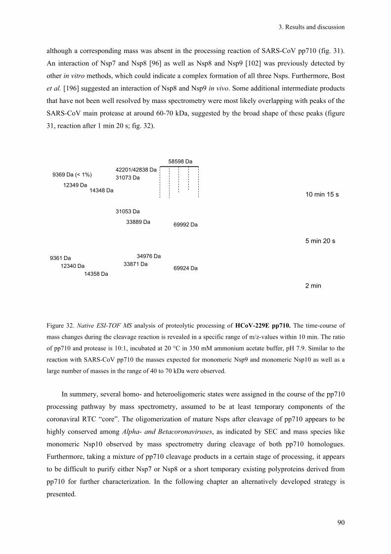
3. Results and discussion
90
although a corresponding mass was absent in the processing reaction of SARS-CoV pp710 (fig. 31).
An interaction of Nsp7 and Nsp8 [96] as well as Nsp8 and Nsp9 [102] was previously detected by
other in vitro methods, which could indicate a complex formation of all three Nsps. Furthermore, Bost
et al. [196] suggested an interaction of Nsp8 and Nsp9 in vivo. Some additional intermediate products
that have not been well resolved by mass spectrometry were most likely overlapping with peaks of the
SARS-CoV main protease at around 60-70 kDa, suggested by the broad shape of these peaks (figure
31, reaction after 1 min 20 s; fig. 32).
2 min
10 min 15 s
5 min 20 s
9361 Da
12340 Da
14358 Da69924 Da
69992 Da
58598 Da
34976 Da
33871 Da
31073 Da9369 Da (< 1%)
14348 Da12349 Da
42201/42838 Da
33889 Da
31053 Da
Figure 32. Native ESI-TOF MS analysis of proteolytic processing of HCoV-229E pp710. The time-course of
mass changes during the cleavage reaction is revealed in a specific range of m/z-values within 10 min. The ratio
of pp710 and protease is 10:1, incubated at 20 °C in 350 mM ammonium acetate buffer, pH 7.9. Similar to the
reaction with SARS-CoV pp710 the masses expected for monomeric Nsp9 and monomeric Nsp10 as well as a
large number of masses in the range of 40 to 70 kDa were observed.
In summery, several homo- and heterooligomeric states were assigned in the course of the pp710
processing pathway by mass spectrometry, assumed to be at least temporary components of the
coronaviral RTC “core”. The oligomerization of mature Nsps after cleavage of pp710 appears to be
highly conserved among Alpha- and Betacoronaviruses, as indicated by SEC and mass species like
monomeric Nsp10 observed by mass spectrometry during cleavage of both pp710 homologues.
Furthermore, taking a mixture of pp710 cleavage products in a certain stage of processing, it appears
to be difficult to purify either Nsp7 or Nsp8 or a short temporary existing polyproteins derived from
pp710 for further characterization. In the following chapter an alternatively developed strategy is
presented.

3. Results and discussion
91
3.5. Interaction of Mpro with substrates lacking glutamine in P1 position
3.5.1. Full-length polyproteins
As an additional strategy to purify individual polyproteins (e.g. polyprotein Nsp7-9 (pp79)) and
mature Nsps originating from purified pp710 of HCoV-229E it was aimed to selectively block a
protease cleavage site by site-directed mutagenesis. The glutamine residue in P1 position at selected
main protease cleavage sites was exchanged by either alanine or asparagine which completely
abolished the cleavage activity as proven by SDS-PAGE (fig. 33 C; column input).
0
2
4
6
8
10
1 10 100 1000
radius [nm]
rela
tive
inte
nsity
Pp79QA
Pp79QA+Mpro 0h
Pp79QA+Mpro 18h
Pp79QA+Mpro 24h
(RH=18.8 nm)
(RH=11.9/4.5 nm)(RH=6.6 nm)
(RH=5.1 nm)
A B
C D
45
35
kDa
116
18.4
14.4
0
200
400
600
800
1000
1200
20 40 60 80 100volume [ml]
A28
0 nm
[m
AU
]
Mpro
Pp710QA Verdau + Mpro
Pp710QA Verdau - Mpro II
4.5 (phyc) 6.5 6.9/6.8 9.5 (cytc)
Pp79
Nsp5
Nsp10
* *1
2
3
M I II III input
I III
0
2
4
6
8
10
1 10 100 1000
radius [nm]
rela
tive
inte
nsity
Pp79QA
Pp79QA+Mpro 0h
Pp79QA+Mpro 18h
Pp79QA+Mpro 24h
(RH=18.8 nm)
(RH=11.9/4.5 nm)(RH=6.6 nm)
(RH=5.1 nm)
A B
C D
45
35
kDa
116
18.4
14.4
0
200
400
600
800
1000
1200
20 40 60 80 100volume [ml]
A28
0 nm
[m
AU
]
Mpro
Pp710QA Verdau + Mpro
Pp710QA Verdau - Mpro II
4.5 (phyc) 6.5 6.9/6.8 9.5 (cytc)
Pp79
Nsp5
Nsp10
* *1
2
3
M I II III input
I III
Figure 33. Investigation of the interaction of pp79 Q83A/Q278A with SARS-CoV main protease (A) Specific
de-aggregation of HCoV-229E pp79 Q83A/Q278A by addition of SARS-CoV Mpro was detected by DLS
measurements. Pp79, monitored before adding Mpro (Rh = 19.4 ± 1.2 nm/18.8 ± 2.2 nm; dark green), 15 min after
adding an equimolar ratio (1:1) of protease (Rh = 11.9 ± 2.4 nm/4.5 + 0.5 nm; green), as well as the same
equimolar sample after 18 h (Rh = 6.6 ± 1.0 nm; light green) and after 24 h of incubation (Rh = 5.1 ± 0.28 nm;
yellow). (B) Size distribution profile of processed pp710 Q83A/Q278A obtained from SEC (Hi Load 16/60
Superdex 200) after incubation with an equimolar amount of Mpro (purple) and the supernatant after an
additional incubation with Ni-NTA resin binding the his6-tagged protease (blue). Purified dimeric Mpro is shown
for comparison (red). (C) SDS-PAGE investigating the composition of the elution fractions referring to the
purple chromatogram in panel B. Notably, a minor amount of pp79 is aggregating (panel C, peak I). (D)
NEPHGE; lane1: Freshly prepared processed pp710 Q83A/A278A (pp79 + Mpro + Nsp10), lane 2: Pp79/Mpro,
left edge of the SEC peak II (panel B), lane 3: Pp79/Mpro right edge of the SEC peak II (panel B); NEPHGE
marker (SERVA), phyc: Phytochrome C, cytc: Cytochrome C. All three lanes show one protein band at pI = 6.5
assumed to contain pp79 Q83A/Q278A and main protease.

3. Results and discussion
92
Exchanging either Q83 and/or Q278 in HCoV-229E pp710 by alanine resulted in increased
aggregation of the mutated polyprotein as evident by dynamic light scattering (DLS) and size-
exclusion chromatography, which likely results from an enhanced local hydrophobicity ("hydrophobic
patch", fig. 33A and table 24). The two mutations Q83A and Q278A target the P1 position of the
cleavage sites connecting Nsp7 and Nsp8 as well as Nsp8 and Nsp9, respectively, which are
consequently not cleaved anymore. In spite of large aggregates, the double mutant containing both
glutamine/alanine substitutions (Q83A/Q278A) was completely cleaved by a catalytic amount of
SARS-CoV Mpro, resulting in the release of mature Nsp10 and intact pp79 (fig. 33C). Applying size-
exclusion chromatography (SEC) to the protein solution after cleavage, pp79 and Nsp10 were
successfully separated from each other (fig. 33B, purple chromatogram). This result further indicated
that Nsp10 did not interact with Nsp7-9 in the polyprotein state. For the mutated pp79 Q83A/Q278A
isolated from peak I a hydrodynamic radius of Rh = 18.8 ± 2.2 nm was determined by DLS (fig. 33A,
dark green graph), indicating large aggregation. Adding an equal molar amount of SARS-CoV Mpro to
the pp79 aggregates, the hydrodynamic radius stepwise decreased down to a monodisperse solution
(Rh = 5.1 ± 0.3 nm) after 24 h of incubation at 6 °C (fig. 33A, yellow graph). The observation of
aggregate dissociation is highly indicative for a direct interaction of both proteins, most probably
shielding the increased hydrophobicity at the mutated cleavage site of the polyprotein, although the
protease is not capable of cleaving anymore. Thus, it was subsequently investigated, if new insights
into the putative interaction of pp79 and Mpro could be obtained allowing insights into a state of Mpro
binding to the cleavage site.
Indeed, SEC revealed a co-localization of both proteins (fig. 33B purple chromatogram; fig. 33C,
peak II). To further probe the interaction, pp79 and his6-tagged protease obtained from SEC peak II
were incubated with Ni-NTA resin. After separating the resin by centrifugation, the supernatant
contained almost no main protease and additionally only a reduced amount pp79; while an increased
amount of aggregated pp79 was detected by SEC analysis (fig. 33B, blue chromatogram). This agrees
with the postulated interaction of pp79 and Mpro, since free pp79 is presumed to be unable to bind to
the Ni2+ ions on the resin. Nevertheless, SEC revealed only a marginal amount of larger
heterooligomeric states of the putative interaction (fig. 33 B/C) which could be dimeric pp79 binding
two protease dimers at one mutated site, where a protease dimer could theoretically bind another
substrate. Such interactions might be sterically impaired or, regarding the dimeric protease, result from
the observation of an asymmetric fold, i.e. only one active centre within the dimer is able to catalyse a
reaction at the same time [52].
In SEC, the pp79-Mpro-complex is characterized by a retention volume of 78.4 ml (16/60 HiLoad
Superdex 200; 41.0 ml void volume), main protease peaks at 80.5 ml (fig. 33B purple chromatogram).

3. Results and discussion
93
Purified pp79 with main protease as well as a mixture containing SARS-CoV Mpro and HCoV-229E
pp710 (Q83A/Q278A; molar ratio 1:1) were further applied to NEPHGE. Similar to isoelectric
focussing (IEF), this native pH-gradient electrophoresis technique is particularly suitable for moderate
pI values in the range between 6 and 11). Similar to the IEF result (chapter 3.13, fig. 77A) two bands
(pH 6.8 and 6.9) arise from Nsp10 (fig. 33D, yellow asterisk), whereas main protease and pp79
Q83A/Q278A again seem to co-localize at around pH 6.5 (fig. 33 D, red asterisk), as previously
observed.
To quantify the binding affinity of SARS-CoV Mpro to the double mutant pp79 Q83A/Q278A,
surface plasmon resonance spectroscopy was performed, allowing the KD-value determination.
HCoV-229E pp79 (49 fmol) was immobilized by EDC/NHS coupling to the dextran layer
functionalized CM5-chip. Mpro was injected at concentrations ranging from 0-400 µM at a flow rate of
20 µl min-1 and the association time of the protein was limited to 240 s. The binding equilibrium was
reached within this time even at low protease concentrations (fig. 34A). The maximum RU value was
plotted against the ligand concentration and the fit of the data points resulted in a dissociation constant
of KD = 11 ± 0.8 µM, according to the one-site-binding model.
Figure 34. Interaction of HCoV-229E pp79 Q83A/Q278A and SARS-CoV Mpro analyzed by surface plasmon
resonance spectroscopy. (A) Sensorgram of the injected Mpro and (B) the dependence of the maximum resonance
on the concentration of SARS-CoV Mpro as determined by SPR spectroscopy. The fit resulted in a dissociation
constant in the low µM-range according to the one-site-binding model, proving the suggested interaction.
Insights into size and geometry of the pp79/Mpro interaction were obtained by SAXS analysis The
complex isolated by SEC after mixing mutated HCoV-229E pp710 with SARS-CoV Mpro (molar ratio
1:1) was analyzed in the presence of 400 mM and 100 mM NaCl, respectively, combined with
ultracentrifugation at either 280,000 g or 541,000 g for 30 min. Results are listed in table 18.
Apparently, the smallest Rg value was determined at low ionic strength after centrifugation at
541,000 g. Shape and volume of the particle fit the expected parameters of monomeric pp79
(molecular mass 43 kDa) (table 18). Considering an average protein density of 1.22 g cm-3 [197] a

3. Results and discussion
94
particle volume of 65 nm3 results in a calculated molecular mass of 48 kDa. Since pp79 contained two
putative protease binding sites at the mutated polyprotein cleavage sites connecting Nsp7 and Nsp8 as
well as Nsp8 and Nsp9 and since either monomeric or dimeric protease could bind to the two cleavage
sites, rigid body modelling based on the scattering data was applied to determine the preferred binding
mode. It was specified that only one of the cleavage sites is occupied by protease. Consequently, four
different complexes were modelled (table 19). A low -value of the rigid-body fit function calculated
by SASREF and a low penalty indicate high similarity to the raw scattering data. Scattering vectors up
to s ≤ 0.24 Å-1 were taken into consideration for three independent rounds of SASREF modelling with
default penalty score specifications. The high-resolution structures included for modelling as well as
the obtained quality measures are specified in table 19. According to rigid-body modelling, it is
statistically preferred that one of the mutated cleavage sites of monomeric pp79 is occupied by
monomeric Mpro (tab. 19) – for both cleavage sites the -value is in a similar range still considering
that there might be mixtures of oligomeric interaction states.
Table 18. Pp79 and Mpro: SAXS result summery of Rg, Dmax and particle volume depending on sample
preparation conditions.
Condition concentration [g l-1]* Rg [nm] Dmax [nm] V [nm³]
280,000 g, 400 mM NaCl 2.3 5.1 15.4 212
280,000 g, 100 mM NaCl 0.5; 1.0; 1.9 4.1 14.2 175
541,000 g, 400 mM NaCl 0.6; 1.2; 2.3 3.5 12.3 97
541,000 g, 100 mM NaCl 0.5; 1.0; 1.9 2.9 10.1 65
* scattering data obtained for different protein concentrations was merged, lacking a concentration-dependent
aggregation or particle repulsion.
Table 19. Pp79 and Mpro: Samples centrifuged at 541,000 g supplemented with 400 mM NaCl further
analyzed by SASREF [input high-resolution structures: pdb code 2AHM (SARS-CoV Nsp7/Nsp8), 2J97
(HCoV-229E Nsp9), 2C3S (SARS-CoV Mpro)]; all putative complexes were modelled considering P1
symmetry.]
Mpro monomeric Mpro * dimeric Mpro
binding site Nsp7/8 Nsp8/9 Nsp7/8 Nsp8/9
-value 0.82 ± 0.03 0.83 ± 0.02 1.08 ± 0.12 1.04 ± 0.12
penalty 0.06 ± 0.09 0.20 ± 0.12 0.27 ± 0.16 0.17 ± 0.08
* one protomer of the dimer
The interaction mode of polyprotein and protease appeared to be worth for further investigation
to enlighten the process of processing of RTC building blocks in more detail. The single mutant
HCoV-229E pp710 Q278A containing a C-terminal his6-tag was generated, purified and cleaved by
incubation with an equimolar amount of SARS-CoV Mpro. Since the mutation is located at the

3. Results and discussion
95
cleavage site connecting Nsp8 and Nsp9, an interaction of protease and pp89 Q278A is expected,
along with the cleavage products of mature Nsp7 and Nsp10. Applying HCoV-229E pp710 Q278A in
the presence of an equimolar amount of Mpro to SEC analysis in a revealed the expected co-elution of
Mpro and pp89QA, which have a similar molecular weight around 34 kDa. The identity of both proteins
in the SDS-PAGE gel (fig. 35B) was verified by ESI-Q-TOF MS after trypsin digestion. Interestingly,
mature Nsp7 (9.3 kDa) also co-eluted and therefore interacted with pp89QA (fig. 35B, peak B),
presuming that Nsp7 did not interact with Mpro, which is rather unlikely. Furthermore, Nsp10 was
separated by SEC (fig. 35B, peak C), as previously observed after cleaving HCoV-229E pp79
Q83A/Q278A (fig. 33).
Since the processing rate of pp89 by SARS-CoV Mpro was previously demonstrated to be very
slow (chapter 3.4), the putative interaction of pp89 and Nsp7 could represent a major intermediate
building block of the RTC, presumably even in vivo. Thus, Nsp7 is suggested to be capable of
interacting not only with mature Nsp8 [96; 98], but also with larger pre-processed polyproteins (e.g.
pp89 and pp810), even though a direct interaction of Nsp7 and Nsp9 was not excluded so far. The
abundance of pp810 (Nsp8-10) is considered to be rather low due to the rapid cleavage of the Nsp9-
Nsp10 cleavage site.
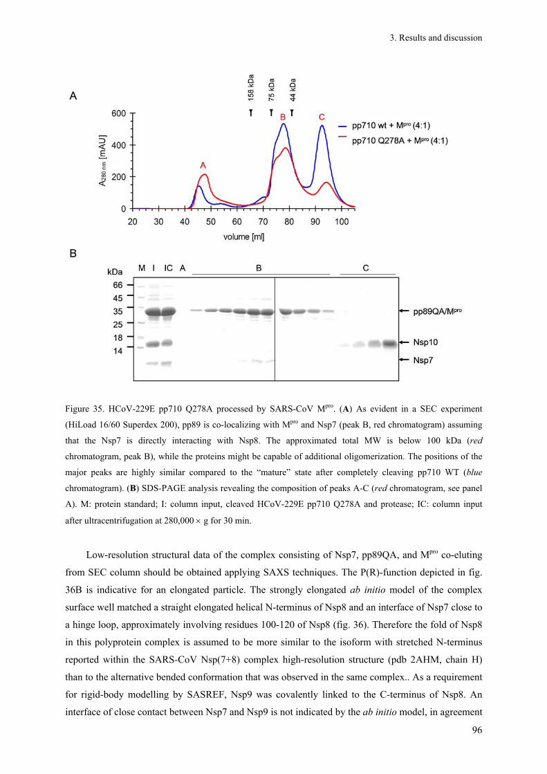
3. Results and discussion
96
Figure 35. HCoV-229E pp710 Q278A processed by SARS-CoV Mpro. (A) As evident in a SEC experiment
(HiLoad 16/60 Superdex 200), pp89 is co-localizing with Mpro and Nsp7 (peak B, red chromatogram) assuming
that the Nsp7 is directly interacting with Nsp8. The approximated total MW is below 100 kDa (red
chromatogram, peak B), while the proteins might be capable of additional oligomerization. The positions of the
major peaks are highly similar compared to the “mature” state after completely cleaving pp710 WT (blue
chromatogram). (B) SDS-PAGE analysis revealing the composition of peaks A-C (red chromatogram, see panel
A). M: protein standard; I: column input, cleaved HCoV-229E pp710 Q278A and protease; IC: column input
after ultracentrifugation at 280,000 g for 30 min.
Low-resolution structural data of the complex consisting of Nsp7, pp89QA, and Mpro co-eluting
from SEC column should be obtained applying SAXS techniques. The P(R)-function depicted in fig.
36B is indicative for an elongated particle. The strongly elongated ab initio model of the complex
surface well matched a straight elongated helical N-terminus of Nsp8 and an interface of Nsp7 close to
a hinge loop, approximately involving residues 100-120 of Nsp8 (fig. 36). Therefore the fold of Nsp8
in this polyprotein complex is assumed to be more similar to the isoform with stretched N-terminus
reported within the SARS-CoV Nsp(7+8) complex high-resolution structure (pdb 2AHM, chain H)
than to the alternative bended conformation that was observed in the same complex.. As a requirement
for rigid-body modelling by SASREF, Nsp9 was covalently linked to the C-terminus of Nsp8. An
interface of close contact between Nsp7 and Nsp9 is not indicated by the ab initio model, in agreement

3. Results and discussion
97
with a SEC analysis of completely processed wild type HCoV-229E pp710, which showed that a
strong interaction between Nsp7 and Nsp9 is absent (chapter 3.4.1/3.4.2). Therefore, a previous yeast-
2-hybrid approach that displayed an initial indication of a direct Nsp7-Nsp9 interaction in one of two
conceivable fusion protein orientations [87] is not supported at the applied experimental conditions.
Figure 36. SAXS investigation of pp89 interaction with Mpro and Nsp7. (A) SASREF rigid-body model of the
single mutant pp89 Q278A in complex with Nsp7 and main protease, consisting of SARS-CoV Nsp7 (blue;
extracted from pdb code 2AHM) and Nsp8 (green; chain H, pdb code 2AHM), HCoV-229E Nsp9 (pink; pdb
code 2J97), and dimeric main protease (grey, pdb code 1UJ1). Modelling restrains were defined that Nsp8 and
Nsp9 are covalently linked by C- to N-terminus (distance 5 Å) and that main protease interacts with the protease
recognition site (P5-P5’) connecting Nsp8 and Nsp9 within the polyprotein. (B) The maximum diameter of the
complex is 12 nm, according to the distance distribution function (P(R)-function) (C) Ten ab initio models
generated by DAMMIF are characterized by a mean normalized spatial discrepancy value of. 0.91 ± 0.04,
calculated by DAMAVER. The average model is shown. The calculated particle volume of 135 nm3
corresponded to an approximated molecular weight of 82 kDa [MWNsp7+8+9 = 43 kDa, MWprotease = 34 kDa]. The
ab initio and the rigid-body model without Mpro are well superimposable. The remaining volume of the ab initio
model can be explained by a certain fraction of main protease molecules at the expected position.
Moreover, an average rigid-body model of pp89-Nsp7-Mpro complex was calculated that fits well
to the ab initio model previously described (fig. 36C). Additional particle volume in the ab initio
model that is not occupied by Nsp7, Nsp8 or Nsp9 in the rigid-body model is interpretable by the main
protease, partly filling the putative binding site at the mutation spot. The volume-based molecular
weight determination approximately reflected an equimolar ratio of all molecules within the complex.
However, a mixture of free cleavage site, binding sites occupied by a dimeric protease molecule or
heterodimers containing monomeric main protease cannot be excluded. This might be explained by the
slow catalytic efficiency of the protease, cleaving at a site with a large asparagine residue in P1
position [192] which may result in a higher dissociation constant of both interaction partners, as
investigated in chapter 3.5.3. Beside the monomeric state, the Nsp7 and Nsp8 interface within the ab
initio model of the pp89-Nsp7-Mpro complex is in good agreement with that observed in a mature

3. Results and discussion
98
Nsp7-Nsp8 complex of SARS-CoV [98], although the individual Nsp structures were independently
fitted into the SAXS model. Furthermore, the SAXS model provided an indication for an at least weak
interaction of Nsp8 and Nsp9 in the pre-processed state, as discussed for the mature Nsps [101]
(chapter 3.12.1).
The proteolytic processing of the HCoV-229E pp710 Q278A mutant with SARS-CoV Mpro wt
was additionally investigated applying native ESI-TOF MS to verify suggested protein-protein
interactions as well as the masses of the corresponding complexes (fig. 37A). Around 40% of the total
intensity at the detector after 1 min of incubation resulted from a mass of around 70 kDa (69,866 ± 47
Da) that increased during the next 3 h and would fit the mass of dimeric protease. At the same time, a
4% fraction (with reference to the total intensity in the displayed m/z range) of an approximately 600
Da lower mass at 69,313 Da appeared with a relatively high standard deviation and in close proximity
to other m/z species that may correspond to a 1 to 1 interaction of main protease and mutated pp89. A
mass of 34,974 Da for monomeric Mpro was determined by native ESI-Q-TOF MS/MS spectra and
33,969 Da is the expected mass of pp89. In addition, two masses above 100 kDa were detected.
Particularly the mass of 139.8 kDa which is too high for representing a pp710 dimer, well fits a dimer
of pp89 interacting with a dimer of protease, where the mass of 105 kDa could represent a monomer
of pp89 interacting with dimeric protease. The relative fraction of 34,915 ± 6 Da corresponding to
monomeric Mpro significantly decreased during incubation (fig. 37A), probably due to a concentration-
dependent interaction with a polyprotein substrate in an advanced stage of cleavage or a substrate-
induced alteration of the monomer-dimer equilibrium of the main protease. For comparison, the
proteolytic processing of pp710 Q278A was also performed with the predominantly monomeric main
protease mutant R298A to probe the requirement of the dimeric protease wild type for the formation of
a specific interaction state. In fact, the two mass species above 100 kDa observed in the presence of wt
Mpro were not detected in the reaction with the R298A mutant (fig 37B, see also chapter 3.3). Thereby,
it is indicated that these mass species (105 kDa, 139.8 kDa) involve and require the native dimeric
state of the wild type. In agreement with SEC (chapter 3.4.1), only a small amount of free mature
Nsp7 at a mass of 9,337 Da was detected by mass spectrometry after a reaction time of more than 3 h
in the presence of the wild type protease (fig. 37A), indicating that mature Nsp7 partly interacted with
pp89. The determined mass of 43,013 ± 78 Da after 3 h 24 min (fig. 37A) agrees with the expected
mass of pp89 interacting with Nsp7. Even though, the whole putative complex of monomeric Nsp7
interacting with monomeric pp89 binding to main protease was not observed among the assigned
masses, it was possible to identify masses indicative for fragments of the expected complex with two
interaction partners, i.e. pp89 and Nsp7 as well as pp89 and protease.

3. Results and discussion
99
Figure 37. Native ESI-TOF MS investing the interaction of Mpro wt or Mpro R298A with HCoV-229E pp710
Q278A. (A) MS spectra of pp710 Q278A incubated with an equimolar ratio of Mpro for 1 min and for 204 min at
20 °C, respectively. (B) Pp710 Q278A was mixed with an equal molar amount of mutant Mpro R298A and
investigated by native ESI-TOF MS at similar time points. The displayed masses were assigned according to a
simulation performed with Massign.
Trying to understand the composition of detected mass species (fig. 37 A/B) it has to be
considered that nucleic acids present in the protein preparation from E. coli cell lysis could bind
particularly to pp89. Applying SARS-CoV pp89 containing a C-terminal strep-tag, separately purified
by Strep-Tactin affinity chromatography and in the absence of Nsp7, to SEC, the absorbance ratio at
260 nm/280 nm of a putative monomeric state is strongly increased and higher than in a fraction of
larger oligomers (data not shown). This observation suggests that nucleic acid is binding and de-

3. Results and discussion
100
aggregating pp89. However, prior to the mass spectrometry experiments described in this chapter a
probably high content of nucleic acid of the samples was strongly reduced by incubation of the protein
with benzonase prior to the affinity chromatography.
The formation of specific protease-polyprotein substrate complexes after mutation of the protease
recognition site was revealed by DLS and additionally verified by SEC and MS experiments.
3.5.2. Full-length polyproteins with Q/N substitution in P1 position
For comparison, a different amino acid exchange in P1 position within a different polyprotein
substrate on the way to form the RTC is the focus of the forthcoming section. Assuming an increased
authenticity and affinity of the protease-polyprotein interaction, an exchange of glutamine in P1
position of the respective cleavage site by asparagine was favoured in forthcoming experiments to
further enlighten and understand this interaction described in the beginning of this chapter. Moreover,
an exchange by asparagine indeed reduced the additional aggregation of the respective polyprotein
compared to the exchange by alanine presented earlier.
In contrast to the pp79 double mutant that was described at the beginning of this chapter (fig.
33), pp78 Q83N contains only one mutated cleavage site for stable binding of Mpro. Using SDS-PAGE
it was verified that the Q→N mutation likewise completely inhibited the proteolytic processing at the
cleavage site connecting Nsp7 and Nsp8 (fig 38). SEC analysis of HCoV-229E pp710 Q83N
processed by SARS-CoV Mpro displayed a significant co-localization of mutated pp78 and Mpro within
a symmetric peak (fig. 38A, light blue chromatogram) which is again indicative for a major fraction of
a relatively small heterooligomeric interaction state of pp78 and Mpro. The composition of the peaks A,
D and E (fig. 38A, blue chromatogram) was investigated by SDS-PAGE (fig. 38B), as for the peaks A,
B, C and E of the green chromatogram (fig. 38A) investigated by SDS-PAGE, shown in figure 38B
(lower panel). The green chromatogram (fig. 38A) of incompletely processed pp710 Q83N reveals co-
localization of the main protease with pp78 Q83N and to a lower extend with pp79 Q83N;
polyproteins are present in excess. However, it is remarkable that pp79 and pp78 peak separately,
indicating that the formation of a heterodimer of pp78 and pp79 is not preferred. Nsp9 and Nsp10 peak
separately (fig. 38, peak E in all chromatograms) and are therefore not interacting with either pp78 or
Mpro for the chosen experimental conditions.
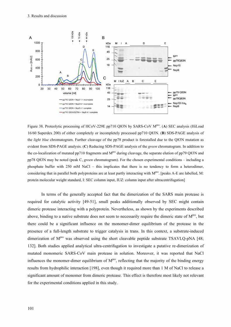
3. Results and discussion
101
Figure 38. Proteolytic processing of HCoV-229E pp710 Q83N by SARS-CoV Mpro. (A) SEC analysis (HiLoad
16/60 Superdex 200) of either completely or incompletely processed pp710 Q83N. (B) SDS-PAGE analysis of
the light blue chromatogram. Further cleavage of the pp78 product is forestalled due to the Q83N mutation as
evident from SDS-PAGE analysis. (C) Reducing SDS-PAGE analysis of the green chromatogram. In addition to
the co-localization of mutated pp710 fragments and Mpro during cleavage, the separate elution of pp79 Q83N and
pp78 Q83N may be noted (peak C, green chromatogram). For the chosen experimental conditions – including a
phosphate buffer with 250 mM NaCl – this implicates that there is no tendency to form a heterodimer,
considering that in parallel both polyproteins are at least partly interacting with Mpro. [peaks A-E are labelled, M:
protein molecular weight standard, I: SEC column input, IUZ: column input after ultracentrifugation]
In terms of the generally accepted fact that the dimerization of the SARS main protease is
required for catalytic activity [49-51], small peaks additionally observed by SEC might contain
dimeric protease interacting with a polyprotein. Nevertheless, as shown by the experiments described
above, binding to a native substrate does not seem to necessarily require the dimeric state of Mpro, but
there could be a significant influence on the monomer-dimer equilibrium of the protease in the
presence of a full-length substrate to trigger catalysis in trans. In this context, a substrate-induced
dimerization of Mpro was observed using the short cleavable peptide substrate TSAVLQ-pNA [48;
132]. Both studies applied analytical ultra-centrifugation to investigate a putative re-dimerization of
mutated monomeric SARS-CoV main protease in solution. Moreover, it was reported that NaCl
influences the monomer-dimer equilibrium of Mpro, reflecting that the majority of the binding energy
results from hydrophilic interaction [198], even though it required more than 1 M of NaCl to release a
significant amount of monomer from dimeric protease. This effect is therefore most likely not relevant
for the experimental conditions applied in this study.

3. Results and discussion
102
Figure 39. Native ESI-TOF MS investing the interaction of SARS-CoV Mpro with HCoV-229E pp710 Q83N
incubated at 20 °C and at equimolar ratio for 75 s and for 14 min 46 s, respectively. Peaks corresponding to
52.4 – 69.1 kDa that are not well resolved indicate the interaction of the SARS-CoV Mpro with pp78 Q83N
(MWexpected = 65.7 kDa for a heterodimer). Other depicted masses fit a monomeric and dimeric state of SARS-
CoV Mpro.
Aiming a high mass accuracy to investigate protein-protein interactions ESI-TOF was applied
again to investigate the proteolytic processing of the herein described HCoV-229E pp710 Q83N
mutant (fig. 39). Beside monomeric and dimeric protease (34,891 Da/69,849 Da), different mass
species in close proximity to each other on the m/z-axis corresponding to masses ranging from 52.4 to
69.1 kDa are observed during the reaction that would be in good agreement with a heterodimer of
pp78 Q83N (expected MW = 30.9 kDa) with authentic ends and SARS-CoV main protease (expected
MWmonomer = 34.9 kDa). However, for this polyprotein the additional peaks could at least partly
contain dimeric pp78 (expected MW = 61.8 kDa), even though, considering the absence of the
protease-polyprotein complex, particularly a monomeric state of pp78 would be expected to be more
abundant (see chapter 3.9.2 for comparison).
To confirm the complex formation of SARS-CoV Mpro with the pp78 Q83N and to investigate
the ability of the main protease to dimerize during interaction, native PAGE was applied. This
experiment addressed if pre-dominantly monomeric Mpro, like the mutant R298A, is able to interact
with a mutated cleavage site in a monomeric state or if partly dimerization induced by the substrate
occurs. N-terminally GST-tagged pp78 Q83N was purified and the tag was removed by precision
protease as described in the method section (fig. 40 A). Pp78 Q83N was pre-incubated with main

3. Results and discussion
103
protease for 2 h at 22 °C, while the protease cleaved off six amino acids of the C-terminus of Nsp6
together with four additional residues that remain after removing the N-terminal GST-tag. A native
polyacrylamide gel was loaded with SARS-CoV Mpro wt, the R298A mutant and HCoV-229E pp78
Q83N as well as mixtures of the protease and pp78 Q83N (fig. 40B). Since the pI of the protease was
determined to be around 6.6 (fig. 25), both variants of the Mpro migrated slowly towards the anode at
pH 6.5 and stopped at the increased pH value of 8.0 in the lower part of the gel. The electrophoretic
mobility of pp78 Q83N was significantly altered in the presence of main protease. Interestingly, the
extend of the shift was even more significant in the presence of the Mpro mutant R298A (fig. 40A). A
small piece (approx. 15 mm2) of the lowest band in the lane containing a 1:1 molar ratio of pp78
Q83N and Mpro R298A was carefully excised. After in-gel digestion with trypsin peptides were
applied to an iontrap ESI-MS/MS instrument. Pp78Q83N as well as Mpro were both identified with
adequate sequence coverage. Consequently, both the gel shift and the co-localization within the same
protein band clearly demonstrated a significant interaction of pp78 Q83N with Mpro, even more
pronounced with the monomeric state of the protease.
The complex of Mpro wt and pp78 Q83N at a 1:1 molar ratio was applied to SAXS; an elongated
particle (Rg = 2.96 ± 0.01 nm) that is well interpretable by a 1:1 heterodimeric interaction was revealed
(fig. 40C). Nevertheless, it is not ruled out that there is some remaining homodimer of either protein
inside that fits the approximated molecular weight similarly well. The maximum diameter of 9.6 nm is
close to the respective value of monomeric pp78 (Dmax = 8.7 nm) indicating that at low concentrations
and after extrapolation to infinite dilution pp78 is monomeric in this mixture. Nonetheless, pp78 tends
to form an increased amount of dimer at higher concentrations as indicated by an increasing radius of
gyration. The sample concentrations ranged from 0.8 to 6.5 g l-1 (not shown).
Mass spectrometry spectra displayed in figure 39 (see also fig. 37) basically agree with the
existence of a putative 1:1 interaction of mutated pp78/pp89 and protease, perhaps in addition to a
minor amount of additional heterooligomers, corresponding to peaks indicated in the respective
figures.

3. Results and discussion
104
Figure 40. Investigation of the interaction of HCoV-229E pp78 Q83N with SARS-CoV Mpro and its mutant
R298A. (A) SDS-PAGE gel showing pure pp78 Q83N (B) Native electrophoretic mobility shift assay: The upper
gel layer contains 4% polyacrylamide (PAA) at pH 6.5; middle layer: 10% PAA pH 6.5; low separating layer:
12% PAA, pH 8.0. Electrophoresis was performed in TBE buffer supplemented with 100 mM NaCl. The
electrophoretic mobility shift upon the addition of Mpro R298A is highlighted by dashed lines. The molar ratio of
protease and pp78 Q83N is depicted above. Pp78 Q83N was pre-incubated with main protease for 2 h at 22 °C
before electrophoresis (C) Average ab initio model of a particle in solution representing a 1:1 pp78/Mpro-mixture
considerably able to interact at equimolar ratio obtained by SAXS. The average NSD-value of 10 models is
1.68 ± 0.06, with a radius of gyration of Rg = 2.96 nm ± 0.01 and a maximum diameter of 9.6 nm. All samples
were supplemented with 3 mM DTT to avoid radiation damage, which did not alter the results compared to non-
reducing conditions.
3.5.3. Inhibition of Mpro by peptides derived from polyprotein recognition sites
After obtaining fundamental insights into the interaction of “uncleavable” polyproteins with Mpro,
short peptides derived from the main protease cleavage sites were analyzed in terms of interaction and
inhibition of Mpro. In silico analysis using FlexidockTM as well as in vitro characterization using SPR
spectroscopy and native mass spectrometry was performed to quantify the binding affinity of the
peptides. Initially, pentapeptides containing either Q, N or A in P1 position of the protease cleavage
site were analyzed in silico concerning the impact of this residue (fig. 41) on the affinity, compared to
peptides carrying a serine or asparagine residue in P1’ position, respectively (see table 20 for
overview).

3. Results and discussion
105
3.5.3.1. In silico peptide docking
Pentapeptides representing native and modified cleavage-recognition sites of SARS-CoV Mpro
(pdb code 3SND) were used for in silico docking to its surface as described in the method section. The
calculated binding energy for all ligands is listed in table 20. Substitution of the essential glutamine in
P1 position of the peptide by asparagine resulted in the formation of a stabilizing hydrogen bond with
G143 (2.43 Å) of the enzyme, while a relatively weak hydrogen bond with N142 of the enzyme (3.39
Å) was observed for a native glutamine in P1 position, according to the docking approach (fig 42). In
contrast, alanine substitution in P1 position did not result in an alternative hydrogen bond to strengthen
the peptide-enzyme interaction. Therefore, according to reduced hydrophobicity and the increased
binding affinity, peptides containing asparagine in P1 position, considerably close to a native
substrate, were favoured to investigate the inhibition of the protease. It was predicted that the peptide
KLQNN is also capable of interacting with HCoV-229E Mpro in a similar way (fig. 42, HCoV-229E
Mpro in green).

3. Results and discussion
106
Figure 41. Pentapeptides representing native and modified cleavage-recognition sites of SARS-CoV Mpro (pdb
code 3SND) were used for docking using FlexidockTM. The calculated binding energies were listed in table 20.
Substitution of the essential glutamine in P1 position by asparagine results in a putative stabilizing hydrogen
bond with G143 (2.43 Å) of the enzyme, which did not form in case of an alanine substitution.

3. Results and discussion
107
3.14 Å
2.39 Å
2.9 Å
3.14 Å
2.15 Å
Q192
T190G 143
C 145
H41
P1 - Q
3.39 Å
P3-KLQNN-P2‘
N 142 (rotated)
P3 - K
P2‘ - N
3.14 Å
2.39 Å
2.9 Å
3.14 Å
2.15 Å
Q192
T190G 143
C 145
H41
P1 - Q
3.39 Å
P3-KLQNN-P2‘
N 142 (rotated)
P3 - K
P2‘ - N
Figure 42. The interaction of P3-KLQNN-P2’ and related peptides with Mpro is mainly stabilized by hydrogen
bonds. The image displays hydrogen bonds of SARS-CoV Mpro (pdb code 3SND; green) and the peptide
superposed with HCoV-229E Mpro (pdb code 2ZU2, pink). [N142 of SARS-CoV Mpro was rotated around the
Cα-C and the C-Cγ bond to reduce the distance of the hydrogen bond with the P1-glutamine]
Table 20. Overview of pentapeptides docked to the active site of SARS-CoV Mpro and binding energies
calculated by in silico docking.
peptide
(location in the pp710 sequence) binding energy
P3- KLQNN -P2' (Nsp8-Nsp9) -1222.9 kcal mol-1
P3- KLANN -P2' (Nsp8-Nsp9) -1032.7 kcal mol-1
P3- ILQSV -P2' (Nsp7-Nsp8) -1088.6 kcal mol-1
P3- ILASV -P2' (Nsp7-Nsp8) -964.3 kcal mol-1
P3- ILNSV -P2' (Nsp7-Nsp8) -996.6 kcal mol-1
The calculated binding energy of -1223 kcal mol-1 for the ligand P3-KLQNN-P2' is primarily
based on hydrogen bonds between the ligand’s lysine and Mpro T190 and Q192 as well as ligand’s
serine/asparagine with Mpro G143 (fig. 42). Replacing glutamine by alanine destroys a hydrogen bond
involving SARS-CoV Mpro N142 that specifically positioned the substrate into the active centre. This
is reflected by a loss in binding energy of almost 200 kcal mol-1. Substitution of glutamine by
asparagine established a comparable hydrogen bond with G143, therefore limiting the loss in binding
energy (table 20). The substrate stabilization due to P1-glutamine is supported by the high-resolution
structure of SARS-CoV Mpro lacking the catalytic histidine in complex with a cleavable substrate (pdb
code 2Q6G; [75]). In contrast to a small serine of the ligand in this protease high-resolution structure,

3. Results and discussion
108
the asparagine in position P1' of P3- KLQNN -P2' is facing out of the pocket with its amide group.
This agrees with the observation that, apart substitutions with proline and acidic amino acids that are
disallowed due to electrostatic repulsion, all other amino acids in P1 position allowed a certain degree
of cleavage [40; 65]. However, in the absence of suitable reactive groups, potential inhibitors of this
type do not yet allow to target the catalytic cysteine by a covalent bond, e.g. by a thiohemiacetal
involved in inhibition by aldehydes. The performed docking approaches provided insights into the
potential to target coronaviral main proteases by nearly native substrates involving the S1’ and S2’
site, in addition to the other subsites already targeted by the comparable peptide aldehyde inhibitors
[78].
The proposed binding affinity and the inhibitory effect of the peptides remained to be verified in
vitro, which may additionally contribute to the understanding of the specificity of the polyprotein
maturation process. Therefore, a number of peptides were synthesized, according to table 21.
Peptide V comprised the N-terminal autocleavage site of the main protease, which was included as a
control peptide that is known to be cleaved with a high catalytic efficiency [192].
Table 21. Synthetic peptides under investigation, derived from pp1a of HCoV-229E. Peptides were
acetylated at the N-terminus and unmodified at the C-terminus (purity > 95%, obtained from Genescript).
peptide molecular weight [Da]
I TVRLNAGKQ (Nsp9-Nsp10) 1027.2
II RTAINSFDN (Nsp10-Nsp11) 1078.1
III DSILNSVAS (Nsp7-Nsp8) 946.0
IV VVKLNNNEI (Nsp8-Nsp9) 1083.2
V SAVLQSGFRK (Nsp4-Nsp5) 1134.2
The secondary structure of each peptide was investigated by CD spectroscopy in the far-UV
wavelength range (192-260 nm) in buffer, water and in the presence of 2,2,2-trifluorethanol (TFE)
(fig. 43A), since the structure of a cleavage site loop could potentially influence polyprotein cleavage.
The spectra of all peptides corresponding to the different cleavage sites displayed a random coil “fold”
characterized by a strong ellipticity minimum below 200 nm and no positive ellipticity in the entire
spectrum. Only the CD spectrum of peptide IV displayed a significantly different shape and intensity
of the minimum, probably corresponding to a β-turn. This peptide displayed a low binding affinity to
the main protease in SPR spectroscopy (fig. 44A) and the respective cleavage site between Nsp8 and
Nsp9 is cleaved relatively slow (chapter 3.4), which could be influenced by the local secondary
structure. Moreover, even 18% (v/v) TFE failed to induce another secondary structure of the peptides I
and III. Therefore, although a limited number of cleavage-site-derived peptides were tested, the
potential inhibition by the peptides and the cleavage of the corresponding native sites may not be
influenced by the secondary structure, at least for peptides I-III. A significant effect of α-helix and β-

3. Results and discussion
109
sheet propensities of a SARS-CoV main protease substrate was reported previously, at some amino
acid positions β-sheet propensities increase the turnover rate [191; 192; 199]. However, it has to be
mentioned that the structural properties within a full-length polyprotein, including additional non-
covalent interactions, might influence the structure of the cleavage site loop in a different way
compared to the isolated peptides.
-20
-16
-12
-8
-4
0
190 210 230 250 270
wavelength [nm]
MR
E [1
03 d
eg
cm
2 dm
ol-1
]
P I, Nsp9-Nsp10
P II, Nsp10-Nsp11
P III, Nsp7-Nsp8
P IV, Nsp8-Nsp9
P V, Nsp4-Nsp5
P III, 18% TFE
-80
-40
0
40
80
250 270 290 310
wavelength [nm]
MR
E [d
eg
cm
2 dm
ol-1
]
main protease+ P I+ P II+ P III+ P IV
A B
-20
-16
-12
-8
-4
0
190 210 230 250 270
wavelength [nm]
MR
E [1
03 d
eg
cm
2 dm
ol-1
]
P I, Nsp9-Nsp10
P II, Nsp10-Nsp11
P III, Nsp7-Nsp8
P IV, Nsp8-Nsp9
P V, Nsp4-Nsp5
P III, 18% TFE
-80
-40
0
40
80
250 270 290 310
wavelength [nm]
MR
E [d
eg
cm
2 dm
ol-1
]
main protease+ P I+ P II+ P III+ P IV
-20
-16
-12
-8
-4
0
190 210 230 250 270
wavelength [nm]
MR
E [1
03 d
eg
cm
2 dm
ol-1
]
P I, Nsp9-Nsp10
P II, Nsp10-Nsp11
P III, Nsp7-Nsp8
P IV, Nsp8-Nsp9
P V, Nsp4-Nsp5
P III, 18% TFE
-80
-40
0
40
80
250 270 290 310
wavelength [nm]
MR
E [d
eg
cm
2 dm
ol-1
]
main protease+ P I+ P II+ P III+ P IV
A B
Figure 43. (A) Far-UV CD spectra of synthetic peptides derived from Mpro cleavage sites of HCoV-229E pp1a,
as listed in table 21. Measurements have been performed in 50 mM NaCl and 8 mM sodium phosphate buffer,
pH 7.6. Peptides I-III and V displayed a comparable spectrum characteristic for a random coil structure, while
peptide IV was significantly different at lower wavelengths, (B) Near-UV spectra of Mpro incubated with a 10-
fold molar excess of the respective peptide confirmed that the (tertiary) structure of Mpro, particularly the
aromatic amino acids, was not affected upon peptide binding.
KD = 13 µM ± 5 µM
DSILNSVAS
RU
ma
x
KD = 181 µM ± 52 µM
VVKLNNNEI
RU
ma
x
concentration [µM]concentration [µM]
KD = 13 µM ± 5 µM
DSILNSVAS
RU
ma
x
KD = 181 µM ± 52 µM
VVKLNNNEI
RU
ma
x
concentration [µM]concentration [µM]
Figure 44. SPR spectroscopy experiment investigating the interaction of SARS-CoV Mpro with two inhibitory
peptides. The dependence of the maximum resonance on the concentration of the respective peptide is plotted.
To verify that binding of the peptides did not trigger a significant effect on the tertiary structure
of the protease, near-UV CD-spectra of 150 µM SARS-CoV Mpro were additionally recorded in the
presence of a 7-fold molar excess of each peptide (fig. 43B). Beside the intensity of the maximum
ellipticity caused by the aromatic residues, the peaks were not significantly shifted (e. g. by

3. Results and discussion
110
quenching), indicating a conserved tertiary structure upon binding. This observation was basically
confirmed by SAXS measurements. The Rg of the Mpro was only marginally reduced in the presence of
the peptides (data not shown). The C-terminal his-tag was cleaved off prior to the experiment resulting
in authentic termini of the protease. Moreover, it was not possible to significantly alter either the
monomer-dimer equilibrium or the pI of Mpro in the presence of a 10-fold molar excess of peptide
(data not shown).
Inspired by the observation of Wu et al. [132] that the presence of an excess of the artificial
substrate TSAVLQ-pNA can trigger the dimerization of the monomeric Mpro mutant R298A the
peptide VVKLNNNEI (IV, table 21), representing the cleavage site connecting HCoV-229E Nsp8 and
Nsp9, was applied to analytical size-exclusion chromatography. According to SEC (fig. 45), the
amount of dimeric mutated main protease was slightly increased in the presence of a 5-fold molar
excess of peptide, similar to the effect of a 2-fold molar excess of TSAVLQ-pNA. However, Mpro
R298A appeared to be still partly monomeric – for the chosen experimental conditions.
Figure 45. Dimerization of SARS-CoV Mpro. Mpro wt applied to a Superose 12 10/300 GL SEC column
consistently displayed a dimeric state in a concentrations range from 12 to 150 µM (different shades of blue;
100µl each). In contrast, Mpro R298A displayed a predominantly monomeric state (yellow). Incubating the
mutant with a 6-fold molar excess of peptide VVKLNNNEI for 1 h at 4 °C resulted in the purple chromatogram,
while a comparable incubation of Mpro with a two-fold molar excess of the synthetic cleavable peptide
TSAVLQ-pNA yielded a similar chromatogram (red). [Chromatography buffer: 40 mM NaH2PO4, 250 mM
NaCl, pH 7.8.]

3. Results and discussion
111
Figure 46. (A) Processing of native HCoV-229E pp710 by SARS-CoV Mpro was inhibited by four different
peptides as demonstrated by SDS-PAGE. Pp710 was cleaved by Mpro in the presence of a 9-fold molar excess of
peptide I to IV and 1 mM DTT up to 24 h. As a control, the same reaction was performed in the absence of
peptides (-peptide). (B) The amount of all four mature Nsps was reduced after 48 h incubation at 6 °C (red arrow
points at Nsp8). The lane “-p” represents an inhibitor-free control. The inhibitory effect of each peptide is
obvious at this ratio, e.g. by the strongly reduced amount of mature Nsp8 (21 kDa, red arrow)
The impact of peptide I-IV on the proteolytic processing pathway of HCoV-229E pp710 was
analyzed by SDS-PAGE (fig. 46), consistently revealing a remarkable inhibition of SARS-CoV Mpro,
indicated by the reduced release of mature Nsps. Particularly the release of Nsp8 (21 kDa) and Nsp9
(12 kDa) was obviously reduced (fig. 46, red arrow). The extend of inhibition at a 9-fold excess is in a
similar range for all peptides, comprising a starting point for further optimized protease inhibitors. The
affinity of the peptide inhibitors described in this study is still in the range of a native substrate.
Applying TSAVLQ-pNA as an artificial substrate for a colorimetric quantification of the Mpro activity
in the presence of the peptides, no significant inhibitory effect was observed. This could be attributed
to the fact that the expected dissociation constants of the substrate-like peptides were not increased
compared to that of the substrate in the assay, which was reported to be in the low µM range for
mature SARS-CoV Mpro [200].
Furthermore, no inhibition was observed supplementing the protease with either a similar excess
of Onchocerca volvulus onchocystatin (OV7; Dr. R. Eberle, University Hamburg) or with the peptide
pEQW that was identified as a broad spectrum protease inhibitor in snake venom (Dr. A. Munawar,
University Hamburg). These conclusions were drawn based on SDS-PAGE analysis of the cleavage
reaction.
The specific binding affinity of the peptides I, III and IV towards SARS-CoV Mpro was
determined applying surface plasmon resonance spectroscopy. Remarkably, an approximately 10-fold
lower binding affinity was revealed for peptide IV containing an asparagine in P1' position (fig. 44B)
compared to peptide III (DSILNSVAS (Nsp7-Nsp8) (fig 44A). The determined KD-value of peptide I
A B

3. Results and discussion
112
(TVRLNAGKQ; KD = 16 µM; RUmax = 7 RU; not shown) was in the same range as that of peptide III.
This observation differed from the binding energy calculated for shorter peptides (5 amino acids),
indicating that the docking of ILQSV may not well reflect the native conditions, resulting in an
underestimated binding energy, even though binding energy does not necessarily correlate with
catalytic activity in general. A significant increase in binding energy by flanking residues of ILQSV is
rather improbable. A weak specificity in position P4 [193] occupied by serine and valine, respectively,
is not expected to have a significant impact on the binding energy in this case, comparing the docking
and the SPR spectroscopy approach. However, the reduced affinity of peptide IV correlated well with
the observation that the cleavage site connecting Nsp8 and Nsp9 is poorly cleaved (see fig. 26B, 27A).
Nevertheless, all KD-values were in the µM range and SDS-PAGE displayed a similar inhibitory effect
of all four peptides (fig. 46).
For comparison and further validation, the binding affinity of peptides I-IV to the SARS-CoV
Mpro was additionally analyzed by native mass spectrometry in cooperation with Dr. C. Uetrecht at the
University of Utrecht (fig. 47). The affinity of peptide IV was almost too low to be detectable. The
displayed spectra allowed a relative comparison of the peptide affinity, largely reflecting the order of
KD-values obtained by SPR spectroscopy. Peptide DSILNSVAS displayed the lowest KD-value,
significantly lower than the peptide IV with asparagine in P1’ position [VVKLNNNEI (Nsp8-Nsp9)].
Differing from SPR spectroscopy, the peptide TVRLNAGKQ (Nsp9-Nsp10) seemed to bind
significantly weaker than peptide III [DSILNSVAS (Nsp7-Nsp8)] in mass spectrometry experiments,
instead of exhibiting a comparable affinity (fig.46, SPR spectroscopy). This could be related to the ESI
process in mass spectrometry. Remarkably, an interaction of monomeric and dimeric protease with
peptide molecules is indicated.
Interestingly, the affinity of RTAINSFDN (Nsp10-Nsp11) was significantly higher than the
affinity of peptide IV. Considering that this affinity correlates with a moderately high turn-over rate at
the Nsp10-Nsp11 cleavage site (see also table 16), it might be important for the viral replication to
cleave off Nsp11. Nevertheless, the short Nsp11 peptide is not conserved in different coronavirus
genomes and a high-resolution structure of SARS-CoV Nsp10 attached to Nsp11 did not reveal any
ordered structure of Nsp11 [131]. Furthermore, there is no indication yet that Nsp11 could interfere
with an interaction of Nsp10 and another protein.

3. Results and discussion
113
Figure 47. Native ESI-TOF MS experiment (LCT device, Waters) to quantify the affinity of substrate-like
inhibitory peptides to SARS-CoV Mpro: Peptide and enzyme were incubated for 30 min at 20 °C in the presence
of 1 mM DTT and 250 mM ammonium acetate. A peak corresponding to dimeric main protease without peptide
("Mpro") is shown for every peptide and concentration ratio, together with a shifted peak originating from a main
protease dimer-peptide complex ("Mpro + peptide"). The intensity of the latter peak relative to the other Mpro peak
allows a relative comparison of the affinity of the different peptides. Increased intensity of the “Mpro + peptide”
peak indicates an increased affinity. Notably, the binding mode is not suggested to be cooperative and,
presuming that the signal at around 4220 m/z as part of the respective ion series is not an artefact, both active
sites within a Mpro dimer can be occupied at the same time ("Mpro + 2× peptide peak"). For all peptides, the
relative differences of the binding affinities were verified by other m/z-states, including the ion series
corresponding to monomeric main protease binding to one peptide. (A) peptide I (TVRLNAGKQ); (B) peptide
II (RTAINSFDN); (C) peptide III (DSILNSVAS); (D) comparison of peptides I-III.
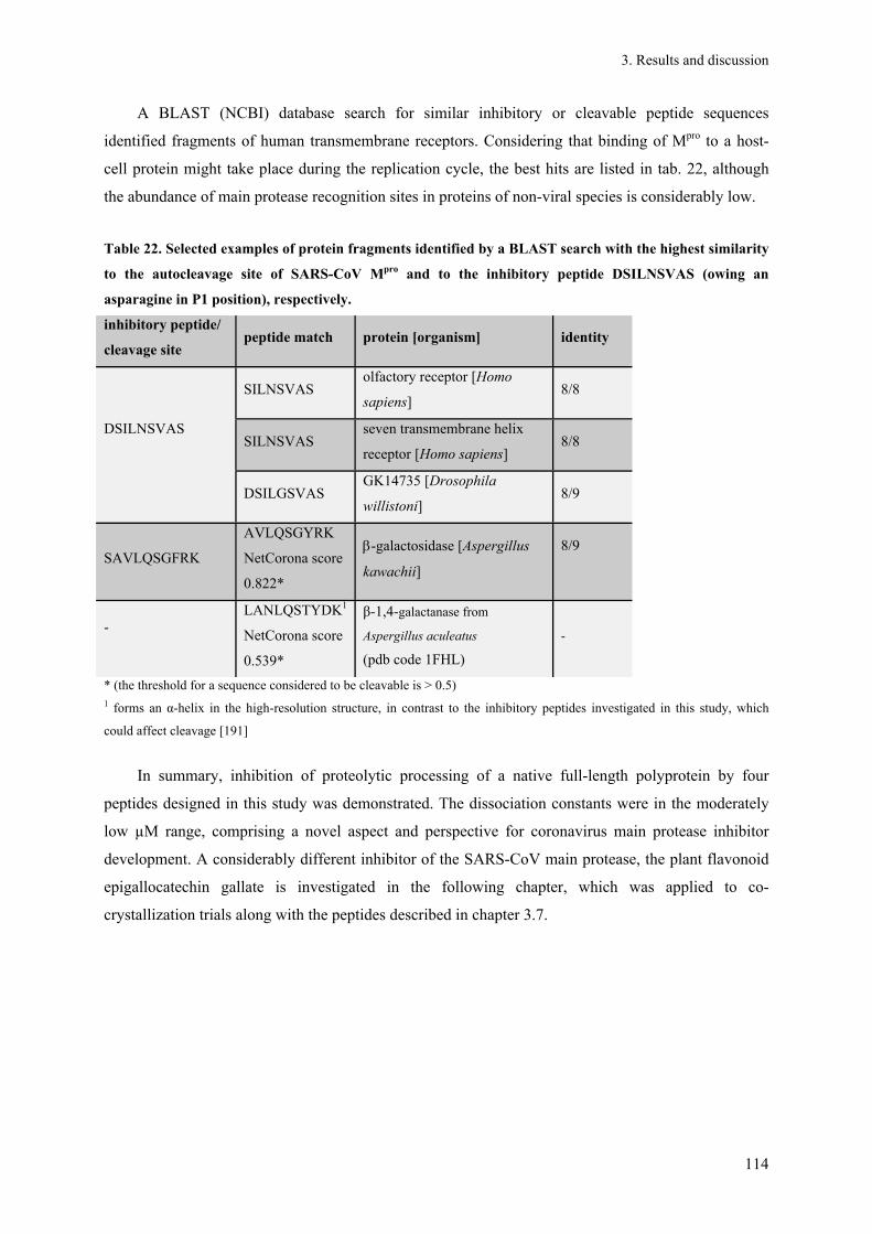
3. Results and discussion
114
A BLAST (NCBI) database search for similar inhibitory or cleavable peptide sequences
identified fragments of human transmembrane receptors. Considering that binding of Mpro to a host-
cell protein might take place during the replication cycle, the best hits are listed in tab. 22, although
the abundance of main protease recognition sites in proteins of non-viral species is considerably low.
Table 22. Selected examples of protein fragments identified by a BLAST search with the highest similarity
to the autocleavage site of SARS-CoV Mpro and to the inhibitory peptide DSILNSVAS (owing an
asparagine in P1 position), respectively.
inhibitory peptide/
cleavage site peptide match protein [organism] identity
DSILNSVAS
SILNSVAS olfactory receptor [Homo
sapiens] 8/8
SILNSVAS seven transmembrane helix
receptor [Homo sapiens] 8/8
DSILGSVAS GK14735 [Drosophila
willistoni] 8/9
SAVLQSGFRK
AVLQSGYRK
NetCorona score
0.822*
-galactosidase [Aspergillus
kawachii]
8/9
-
LANLQSTYDK1
NetCorona score
0.539*
β-1,4-galactanase from
Aspergillus aculeatus
(pdb code 1FHL)
-
* (the threshold for a sequence considered to be cleavable is > 0.5) 1 forms an α-helix in the high-resolution structure, in contrast to the inhibitory peptides investigated in this study, which
could affect cleavage [191]
In summary, inhibition of proteolytic processing of a native full-length polyprotein by four
peptides designed in this study was demonstrated. The dissociation constants were in the moderately
low µM range, comprising a novel aspect and perspective for coronavirus main protease inhibitor
development. A considerably different inhibitor of the SARS-CoV main protease, the plant flavonoid
epigallocatechin gallate is investigated in the following chapter, which was applied to co-
crystallization trials along with the peptides described in chapter 3.7.

3. Results and discussion
115
3.6. Interaction of Mpro with flavonoid derivatives
The inhibition of SARS-CoV Mpro by various plant flavonoids was already reported several times
[68; 201] (see also introduction, part 1.3). Nevertheless, besides in silico predictions of the binding
epitopes, there is still a lack of experimental structural data. Therefore, STD-NMR spectroscopy in
parallel to co-crystallization trials was performed to characterize the corresponding binding epitope.
STD-NMR spectroscopy allows determining the epitope of a small ligand binding to a comparably
large protein. Upon an electromagnetic radio-frequency excitation pulse to the protein the saturation of
the nucleus spins will spread to a bound ligand, indicating a close proximity of the respective part of
the ligand to the protein.
Figure 48. (A) Inhibition of SARS-CoV Mpro by epigallocatechin gallate was verified photometrically using the
chromogenic substrate TSAVLQ-pNA (C-terminal p-nitroanilide, MW = 755.8 Da). Triplicate measurements
have been performed with main protease (green) and main protease mixed with EGCG at a molar ratio of 1:8
(red). (B) The interaction of Mpro with epigallocatechin gallate was investigated by STD-NMR spectroscopy.
The protein was freshly prepared and supplemented with a 100-fold molar excess of ligand. The ligand epitope
was investigated immediately to prevent a significant amount of keto-enol tautomerism. The NMR sample was
supplemented with 2 mM deuterated DTT. The relative STD effect is indicated by a 3-level colour code, where a
high relative STD effect indicated close proximity to the protein surface. The highest STD effect was set to
100%.
Initially, the inhibition of SARS-CoV Mpro by epigallocatechin gallate (EGCG) was verified
photometrically in three independent experiments. An 8-fold molar amount of EGCG almost
completely abolished the catalytic activity of Mpro (fig. 48A, red data points). The ligand epitope of
EGCG was investigated by STD NMR spectroscopy, indicating that the oxane ring is located very
close (and oriented parallel) to the protease surface. Therefore, the geometry of the benzooxane moiety
is considered to be critical for the level of interaction. Aiming at correlating structure and inhibitory
effect of flavonoids, the binding mode of the diverging quercetin-3-β-galactoside was predicted in
A B

3. Results and discussion
116
silico [202]. Despite the different sterical properties of the galactoside moiety compared to the
aromatic ring, the orientation of the ligand appears to be similar compared to the binding epitope of
EGCG. Moreover, even though the area around the poly-hydroxylated six-rings is not perfectly
covered by STD-NMR spectroscopy-relevant hydrogen atoms, the experimentally determined epitope
essentially agrees with the epitope of the related gallocatechin gallate as predicted in silico [67]
Since other phenolic plant compounds were proven to harbour potential to inhibit SARS-CoV
Mpro [203; 204], it is worth to further investigate more and particularly larger compounds of the diverse
secondary metabolism of higher plants. A docking approach with amentoflavone which is a
biflavonoid indicated a significantly different epitope, occupying more space in the active centre cleft.
The IC50-value of this compound is already as low as 8 µM [201]. Another flavonoid, tannic acid that
has a huge inhibitory potential (IC50 = 3 µM) [68] consists of five galloylester moieties surrounding a
glucose ring. The structure of tannic acid suggests that specific binding to more than one active centre
might be simultaneously possible, which could result in a chain-like oligomerization of Mpro. At least
different epitopes within this tannic acid molecule are conceivable. However, investigations by DLS
did not reveal a specific effect of tannic acid on the oligomerization of Mpro, but indicated that tannic
acid induced a continuous unspecific oligomerization of the protease already at equimolar amounts,
preventing further crystallization trials. Native ESI-TOF mass spectrometry did not reveal an altered
oligomerization of the protease.
3.7. Co-crystallization trials with SARS-CoV Mpro
To analyze the binding epitope of epigallocatechin gallate (EGCG) in addition to the
characterization by STD-NMR spectroscopy in more detail, co-crystallization trials were set up.
SARS-CoV main protease mixed with a 2-fold molar amount of EGCG formed a dimer in a
monodisperse solution, as verified by DLS (data not shown). Even though crystallization of SARS-
CoV main protease was successful (fig. 49, particularly panel I), co-crystallization trials with EGCG
were not successful, i.e. screening 300 conditions containing various PEGs as well as applying
published conditions [78] and the counter diffusion technique (fig. 49I/J). Even a low excess of EGCG
significantly impaired the formation of protein crystals. An optimization of the co-crystallization
conditions by applying a continuous gradient of PEG 6000 precipitant solution in a Granada
Crystallization Box® (GCB) did not result in the formation of diffraction quality crystals (fig. 49J).
Soaking of main protease crystals to a final concentration of 1 mM EGCG in the drop worked fine and
did not significantly affect the diffraction quality of the Mpro crystals. Additionally, SARS-CoV Mpro
crystals were soaked with the substrate-like peptides listed in table 21to obtain high-resolution
structures of the respective complex (see fig. 49 for a summary of experimental conditions).
Nevertheless, all diffraction data sets that were collected and processed so far did not reveal

3. Results and discussion
117
sufficient electron density of the respective ligand in one of the active centres of the Mpro dimer. The
highest resolution obtained for an Mpro crystal diffraction dataset was 1.6 Å. Datasets were
successfully indexed revealing either a P1, C121 or P21212 space group.
Figure 49. SARS-CoV Mpro co-crystallization trials. A-G: Vapour-diffusion experiment in a linbro 24-well plate;
if not stated otherwise 10 g l-1 Mpro in 1 mM DTT, 20 mM NaCl and 30 mM Tris pH 7.5 were mixed with an
equal volume of reservoir solution, 2 µl drops were equilibrated against 1ml reservoir precipitant solution each.
(A) Reservoir: 8% PEG 6,000, 0.1 M MES pH 6.0, 3% DMSO, 1% MPD, 1 mM EDTA. Cube-shaped crystal
soaked with 1 mM EGCG over night, diffracted at beamline X13 (DORIS) to a resolution of up to 2.2 Å. (B) 8
g l-1 Mpro mixed with an 8-fold amount of peptide III; reservoir: 5.5% PEG 6000, 0.1 M MES pH 6.0, 2% MPD;
two different crystal morphologies. (C) 8 g l-1 Mpro mixed with an 7-fold amount of peptide II; reservoir: 5%
PEG 6000, 0.1 M MES pH 6.0, 2% MPD, 1 mM EDTA, 2 mM DTT. (D) Main protease crystal soaked with a
final concentration of 500 µM EGCG for 5 h, diffracted at beamline X13 (DORIS) up to a resolution of 2.8 Å;
reservoir: 6% PEG 6,000, 0.1 M MES pH 6.0, 3% DMSO. (E) Brick-shaped crystal (approx. 350 µm in length)
diffracted to a resolution of up to 1.6 Å at beamline P14 (PETRA III). (F) 6 g l-1 Mpro 200 mM NaCl in the
protein buffer; reservoir 0.8 M sodium malonate, 0.1 M MES pH 6.0, 2% 2-propanol. (G) 7 g l-1 Mpro; 200 mM
NaCl in the protein buffer; reservoir 0.8 M sodium malonate, 0.1 M MES pH 6.0, 0.5% 2-propanol mixed with a
10-fold amount of peptide III. (H) 6 g l-1 Mpro; 100 mM NaCl in the protein buffer; reservoir 0.8 M sodium
malonate, 0.1 M MES pH 6.0, 1% 2-propanol; mixed with a 10-fold amount of peptide II. (I) 180 µM purified
monodisperse Mpro (180 µM) was drawn into a capillary that was punched into a Granada Crystallization Box®
(GCB) sealed with agarose. The displayed GCB was filled with 10% PEG 6000, 0.1 M MES pH 6.0, 2% MPD,
2% DMSO and 1 mM DTT and incubated to allow crystallization by counter-diffusion. (J) Aiming co-
crystallization a two-fold molar excess of EGCG was mixed with 180 µM purified monodisperse Mpro and drawn
into a another capillary that was set up in a GCB essentially as described for panel I.

3. Results and discussion
118
3.8. Purification and oligomerization of SARS-CoV pp79
3.8.1. Dimerization of SARS-CoV pp79
As previously presented, HCoV-229E pp79 Q83A/Q278A formed a highly aggregated state in
the absence of main protease that prevented any structural characterization; presumably due to locally
increased hydrophobicity at the mutation sites (section 3.5). Therefore, the wild type SARS-CoV pp79
gene was separately cloned into pASK-IBA plasmids by BsaI restriction and recombinantly expressed
to enable a structural comparison of pp79 with its precursor SARS-CoV pp710 and its maturation
product pp78 that is formed by cleaving of Nsp9. SARS-CoV pp79 fused with an N-terminal strep-tag
was purified by affinity chromatography (fig. 50A). The expected secondary structure content of the
protein was verified by CD spectroscopy (data not shown). Along with significant amounts of high
molecular weight oligomers, a monomeric and a dimeric state of SARS-CoV pp79 were detected in
solution and separated by SEC (fig. 50B). Therefore, SARS-CoV pp79 and SARS-CoV pp710
consistently shared these states, as previously observed (fig. 9B). The abundance of the pp79
monomer is only slightly increased compared to the dimer in 280 mM NaCl, 30 mM NaH2PO4, pH 7.9
(fig. 50B). The monodispersity of the monomeric state was additionally verified by DLS. A long-term
DLS experiment (> 140 h) demonstrated a remarkably good stability of the monomeric state (Rh =
4.5 ± 0.1 nm) that was incubated for 5 d at 6 °C prior to the DLS experiment (fig. 50C). After 40 h, the
hydrodynamic radius started to slowly increase, indicating the formation of dimers and subsequently
higher oligomers that continuously increased in size. Therefore, a reformation of the dimer was
observed, although this was most likely attributed to an unspecific effect of storage than to the
establishment of an equilibrium, as the hydrodynamic radius continuously increased. An explanation
for the significantly increased stability of the pp79 monomer compared to the monomeric state of
SARS-CoV pp710 (see chapters 3.2.2.2 and 3.2.2.1) could be the absence of 13 cysteine residues
located in SARS-CoV Nsp10, which might be responsible to form aggregates via intermolecular
disulfide bonds, as observed for purified HCoV-229E Nsp10 (see chapter 3.13).
To obtain a low-resolution structural model of pp79, particularly for comparison of the
monomeric and the dimeric state of pp79 with that of pp710, SAXS measurements were performed.
Monodisperse monomeric and dimeric pp79 containing an N-terminal strep-tag isolated by SEC were
exposed to synchrotron radiation at DORIS storage ring beamline X33. Based on the scattering data
recorded for different concentrations (1 - 4.5 g l-1) and after extrapolation to infinite dilution, ab initio
low-resolution models were calculated (fig. 51) to predict the dimerization mode. Significant
parameters of monomeric and dimeric pp79 as determined by SAXS, including a quality estimate of
the ab initio models are presented in table 25.

3. Results and discussion
119
Figure 50. (A) N-terminally strep-tagged SARS-CoV pp79 purified by Strep-Tactin affinity chromatography is
visualized by reducing SDS-PAGE. (B) Size-exclusion chromatography (HiLoad 16/60 Superdex 200) of the
purified protein revealed a co-existence of different oligomeric states with the monomer that eluted at a volume
of 78 ml in solution, corresponding to 54 kDa (expected MWpp79 monomer = 44 kDa). (C) The monomeric state of
pp79 isolated by SEC was (Rh = 4.5 ± 0.1 nm, grey line) was investigated by long-term DLS measurements. A 2
µl drop (60 µM protein in 280 mM NaCl, 40 mM NaH2PO4, pH 7.9) in a 96-well Terazaki plate (NUNC)
covered by paraffin oil was applied to record the DLS data and store the protein. After around 40 h of incubation
at 20 °C, the detected hydrodynamic radius indicated the formation of a significant amount of dimer, followed
by subsequent strong aggregation. The particle abundance is colour-coded in the displayed heat map, ranging
from blue (low) to dark red (high) abundance.
The obtained ab initio models of both states reasonably allowed predicting the dimerization site
of two rigid monomers forming an elongated “V”-shaped dimer as displayed in fig. 51. As previously
observed for pp710, it is well-conceivable that the dimerization of pp79 was mediated by Nsp8,
orienting its C-terminal towards the dimerization interface (rigid-body model not shown). Moreover,
assuming that Nsp10 does not provide additional non-covalent interactions with other domains within
either SARS-CoV or HCoV-229E pp710, as investigated and discussed in chapter 3.4.1, it was
reasonable to suggest a conserved dimer interface in pp79 and pp710, if Nsp10 does not sterically
mask the interface in pp710. However, this appeared to be unlikely, based on the previously evaluated
model of pp710 presented in chapter 3.2.3.

3. Results and discussion
120
90°
120 Å
Figure 51. Prediction of the dimerization of SARS-CoV pp79 investigated by SAXS technique. Two copies of
the average ab initio model of monomeric SARS-CoV pp79 are shown in blue and blue-grey, respectively. The
predicted rigid assembly of the two monomers well interpreted the experimental ab initio shape of the pp79
dimer (grey). The dimerization involved the thickened end of the longitudinal model axis of the monomer to
form a smooth “V”-like dimer shape.
3.8.2. Mutation of a GXXXG motif does not alter dimerization of SARS-CoV pp79 and
pp710
SARS-CoV Nsp9 was reported to form a dimer via a 100GXXXG104 loop motif by a rigid “helix-
groove” packing allowed by the small glycine residues located at the C-terminal helix of Nsp9 [102;
104]. The GX3GX2(G/A/S) motif is highly conserved in coronaviral Nsp9 and transmembrane helices
of various proteins “packing” with similar motifs [205], e.g. the amyloid beta protein precursor [206].
Exchanging one of the two glycins by glutamate resulted in the formation of monomeric Nsp9
according to multi-angle light scattering tests [104]. The impact of this loop structure on the
dimerization of SARS-CoV pp79 and pp710 was investigated by replacing G100 of the Nsp9 domain
in both proteins by glutamate. The mutants were purified by Strep-Tactin affinity chromatography.
The chromatogram obtained from SEC experiments demonstrated that the freshly prepared SARS-
CoV pp710 G381E and pp79 G381E still formed a similar amount of dimer compared to the wild type
protein (fig. 50A). Dimeric pp79 G381E isolated by SEC was applied to SAXS analysis for
comparison with the wild type protein (fig. 50B). The superimposable scattering data at comparable
protein concentrations strongly indicated that the intact GXXXG loop, which is crucial for

3. Results and discussion
121
dimerization of mature Nsp9 [104], is not relevant for dimer formation of SARS-CoV pp79, as for the
SARS-CoV pp710 dimer formation as investigated by SAXS (chapter 3.2.3.2) as well. Furthermore,
this result implied that also a mature Nsp9 monomer can not bind to pp79 via the GXXXG interface.
Searching the sequence of coronaviral replicase polyproteins for another conserved GXXXG motif,
which could be indicative for another interaction partner, had no result. However, such a motif was
identified in the spike protein transmembrane domain, unique to SARS-CoV and stabilizing the trimer
formation of the spike protein [207].
In addition to the GXXXG-dependent dimerization of Nsp9, β-sheet 5 (residues 63-69) of SARS-
CoV Nsp9 was also reported to trigger dimerization, as indicated by a high-resolution crystal structure
[101]. Nevertheless, mutation of the GXXXG motif introducing either glutamate or aspartate instead
of glycine resulted in severe growth defects of the virus and was reported to be critical even for the
formation of the RTC complex of SARS-CoV and IBV [104; 208], highlighting the importance of this
motive in vivo. Therefore, it has to be assumed that this helix-helix dimerization is either much weaker
in pp79 than another dimerization that was observed by SAXS or that the helix-helix dimer mediated
by GXXXG is entirely formed by uncovering this interface after cleaving of Nsp9 by the main
protease. The latter explanation is suggested to be more likely, since the mutation introduced into the
GXXXG motif did not have any effect on the quaternary structure of pp79. SARS-CoV pp79 as well
as SARS-CoV pp710 is not significantly impaired in forming higher oligomers. Therefore, larger
oligomers appear to be formed by other protein-protein interfaces as well. Consequently, proteolytic
processing between Nsp8 and Nsp9 in the precursor state is assumed to be essential for viral
replication to allow the “helix-helix” dimerization of mature Nsp9. This suggestion is in agreement
with the in vivo observation by Deming et al. [107] that disruption of the Nsp8-Nsp9 cleavage site by
replacing P1-glutamine by alanine in MHV is lethal, because the Nsp9 dimerization is essential in vivo
[104]. This phenotype was similarly observed for the mutation in P1 position in between of Nsp7 and
Nsp8 – taking both alternative cleavage sites of MHV at this position (see table 17) into consideration
– no syncytium formation or subgenomic mRNA was detected after implanting the mutated RNA by
electroporation.
Interestingly, a comparable exchange of the essential P1 glutamine by alanine between either
Nsp7 and Nsp8 or Nsp8 and Nsp9 of HCoV-229E pp710 and pp79 consistently induced aggregation
as revealed in this study, well explained by locally increased hydrophobicity. Therefore, it is highly
suggested to substitute the P1-glutamine by asparagine in future in vivo experiments to keep the local
charge close to native, as it was investigated in chapter 3.5.

3. Results and discussion
122
Figure 52. (A) Particle size distribution of pp79 G381E obtained from SEC. Absorbance was monitored at 280
nm (dark blue) and 260 nm (light blue). Freshly prepared pp79 G381E was supplemented with 1 mM TCEP to
disassemble disulphide-linked covalent dimers. The SEC column was equilibrated with 200 mM NaCl, 30 mM
sodium phosphate, pH 7.9. Standard protein masses are indicated. (B) Monomeric and dimeric state of pp79
G381E were investigated by SAXS. The scattering pattern (I(s) versus s) of dimeric pp79 wt (dark blue) and
pp79 G381E mutant (cyan) are well superimposable, indicating no change in size or shape of the molecules.
At similar buffer conditions, the addition of an equimolar amount of ssDNA
(5’-GATTAGTTACCTGAC-3’) and a final MgCl2 concentration of 1 mM to SARS-CoV pp79
resulted in a slightly altered SAXS scattering curve compared to that of the pp79 monomer in solution
(fig. 52B, red and purple pattern). While the progression at low angles was highly similar to that of
monomeric Nsp9, increased scattering intensity has been detected at higher angles, indicating a
slightly changed tertiary structure of pp79 in the presence of ssDNA, although the overall size
corresponded to a monomeric molecule. However, the binding affinity of ssDNA could be
significantly weakened by the relatively high ionic strength of the applied buffer conditions (200 mM
NaCl). The alteration of the tertiary structure in the presence of nucleic acid is in agreement with
conformational changes of Nsp8 upon RNA-binding already suggested by preliminary FRET results
[96].
A B

3. Results and discussion
123
3.9. Polyprotein Nsp7-8 (pp78)
3.9.1. Oligomerization of HCoV-229E and SARS-CoV pp78
For structural comparison with pp79 and pp710, both homologues of pp78 were separately
cloned and successfully purified from an E. coli cell lysate by affinity chromatography, exploiting
either the interaction of an N- or C-terminal strep-tag fused to SARS-CoV pp78 with Strep-Tactin
resin or of an N-terminal GST-tag fused to HCoV-229E pp78 with glutathione resin (fig. 53A, lane
1/2). The GST-tag of HCoV-229E pp78 was removed by prescission protease cleavage. Remaining
impurities and the free GST-tag were separated by incubating the solution with glutathione resin again
(fig. 53C). Purification of GST-tagged HCoV-229E pp78 from E. coli BL21 AI cells commonly
resulted in a minor double-band of the target protein as verified by mass spectrometry. This effect was
attributed to the artificial cleavage of some GST molecules from the fusion protein already in the E.
coli cells.
Analytical SEC (HiLoad 16/60 Superdex 200) performed to initially identify the oligomeric
states of both pp78 homologues in solution consistently revealed a predominant mixture of monomer
and dimer of pp78 for both virus species (fig. 53A/B). For HCoV-229E, the monomeric state of pp78
was clearly preferred compared to the dimeric state at two different buffer conditions (fig. 53B, peak
II), contrary to the ratio of SARS-CoV pp710 as previously revealed by SEC (fig. 9). Both states were
additionally verified by native mass spectrometry (data not shown). High molecular weight oligomers
have not been detected. A slightly smaller fragment of HCoV-229E pp78 observed by SDS-PAGE
could falsify the ratio of monomer and dimer observed by SEC. In contrast, SARS-CoV pp78 with N-
terminal strep-tag showed a prevalence for the dimer in solution, and significant amounts of large
oligomers have additionally been detected (fig. 53B, peak II). Moreover, the oligomerization of
SARS-CoV pp78 was significantly affected by the position of the purification tag, since pp78 with C-
terminal strep-tag predominantly formed large aggregates, together with only small and not well
separated fractions of dimeric and monomeric protein. The co-existence of a monomeric and a dimeric
state of SARS-CoV pp78 with N-terminal strep-tag was verified by native blue PAGE as well (fig.
53C). The ratio of absorbance at wavelengths of 280 nm and 260 nm was constant for all peaks of both
homologue proteins (fig. 53A/B), indicating that there is no significant enrichment of nucleic acid
carried over from E. coli cells.

3. Results and discussion
124
Figure 53. Purification and characterization of pp78 homologues in solution. (A) SDS-PAGE analysis (left) of
SARS-CoV pp78 purification by affinity chromatography, exploiting either an N-terminal (lane 1) or C-terminal
strep-tag (lane 2). The corresponding SEC chromatograms display a putative monomeric and dimeric state along
with larger oligomers that are pre-dominant for C-terminally tagged protein. (B) The purity of HCoV-229E pp78
was verified by SDS-PAGE applied to SEC (HiLoad 16/60 Superdex 200) revealing two major oligomeric states
(peak I and II), assumed to be dimer and monomer. (C) N-terminally strep-tagged SARS-CoV pp78 was
investigated by blue native PAGE (right panel). SARS-CoV pp710 and a ten-fold amount of pp710 incubated
with Mpro for 30 min at 20 °C are shown for comparison. Monomeric pp710 roughly co-localizes with dimeric
pp78. The existence of monomeric and dimeric states of pp78 was confirmed.
For comparison of folding stability, the melting temperature of HCoV-229E and SARS-CoV
pp78 with N-terminal strep-tag was examined by CD spectroscopy in a temperature interval up to
80 °C, estimating the conservation of the overall sum of non-covalent interactions stabilizing the
pp78-fold of an Alpha- and a Betacoronavirus. The thermal stability which is characterized by the
turning point of an optimised Boltzmann fit (Tm, “melting point”), determined based on the ellipticity
monitored at a wavelength of 220 nm, was identical within the error range for both homologue
proteins determined at identical buffer conditions. For HCoV-229E pp78, a Tm of 61.8 ± 1.4 °C was
obtained, while the Tm of SARS-CoV pp78 accounted to 61.3 ± 0.7 °C. For an initial comparison of
the low-resolution structure, monomeric and dimeric state of SARS-CoV and HCoV-229E pp78
observed at higher concentration were isolated by SEC and applied to SAXS experiments. Protein
concentrations ranged from 1 up to 4.5 g l-1. SAXS results for the monomeric and the dimeric state of
both pp78 homologues are presented in table 23 and 25, verifying the expected molecular weight of

3. Results and discussion
125
monomer and dimer. Low resolution structural models of the proteins calculated based on the
scattering data using DAMMIF are discussed in detail in chapter 3.10.
Table 23. Summary of structural properties of pp78 obtained by SAXS and DLS
virus protein Rh [nm] Rg [nm] Dmax [nm]
SARS-CoV pp78 monomer 3.3 ± 0.3 3.20 ± 0.01 10.0
pp78 dimer 5.3 ± 0.1 3.88 ± 0.01 13.0
HCoV-229E
pp78 monomer
(GST-tag cleaved) 3.4 ± 0.4 2.65 ± 0.11 8.7
pp78 dimer
(GST-tag cleaved) 4.5 ± 0.2 3.37 ± 0.11 11.4
Exploiting the high mass accuracy of native mass spectrometry, minor amounts of tri- and
tetrameric SARS-CoV pp78 were detected in ammonium acetate buffer at pH 7.9, co-existing with
predominant mono- and dimeric states (fig. 54), as basically observed by SEC analysis (fig. 53 A).
Larger oligomers were almost absent in the m/z spectra. The oligomeric states were specifically
fragmented by CID, resulting in the detection of a mass of 32,354 Da. This species is in good
agreement with the monomer mass detected at lower collision voltages (fig. 54, approx. 3,000-3,300
m/z) and the calculated mass of pp78 of 31.16 kDa + 1.48 kDa accounting to the strep-tag provided by
the pASK-IBA5plus vector.
In conclusion, highly abundant mono- and dimeric states were identified for HCoV-229E and
SARS-CoV pp78, even though the ratio is altered comparing the two different homologues of pp78.
For SARS-CoV pp78 the aggregation is altered by the tag position. Moreover, Rg and maximum
diameter of HCoV-229E pp78 are significantly lower compared to the homologue protein, as observed
similarly for HCoV-229E and SARS-CoV pp710 (chapter 3.2.3.2), strongly indicating an altered
tertiary structure. Most probably the effect is related to the tertiary structure of Nsp8, since structural
variation of Nsp8 due to flexibility was observed in the three high-resolution structures of a mature
coronavirus Nsp(7+8) complex [96; 98; 99].

3. Results and discussion
126
Figure 54. N-terminally strep-tagged pp78 of SARS-CoV investigated by native ESI-Q-TOF. A co-existence of
the monomer with a defined dimeric, trimeric and tetrameric state was revealed in solution and additionally
fragmented by CID. The abundance of the specific states significantly decreased with enhanced oligomerization.
Moreover, increasing the collision voltage resulted in a reduced abundance of pp78 oligomers. Finally, at 150 V
collision voltage, only monomeric pp78 remained (32,292 Da/32,382 Da), corresponding to peaks below 3,000
m/z-value that are already visible at lower voltage (32,354 Da at 125 V).
3.9.2. SARS-CoV pp78 binds tRNA with a low µM affinity
Using conventional native PAGE techniques to verify the interaction of HCoV-229E pp710 with
nucleic acids (chapter 3.2.7), a relatively low amount of “shifted” protein-nucleic acid complexes was
observed, indicating rather weak interactions. However, such weak interactions with nucleic acids can
be detected and quantified applying the method of zone-interference gel electrophoresis (ZIGE).
According to [143], ZIGE is particularly valuable for low affinity interactions in the µM range,
because the protein is in contact with the ligand for a longer time during electrophoresis. Considering
that mature Nsp8 is reported to comprise RNA-dependent RNA polymerase activity [94; 97], tRNA
was used as an easily accessible random RNA pool for interaction tests. As described in the same
report, the KD-value is determined to represent the negative reciprocal of the slope of the linear
function that is displayed in fig. 55A, depending on the migration distance of the protein in the
presence of various ligand concentrations. This approximation revealed an interaction of SARS-CoV
pp78 with tRNA in the low micromolar range in the presence of low quantities of Mg2+. Similar KD-
values were previously reported for mature Nsp8 of HCoV-229E and SARS-CoV [103]. This implies
that the affinity of Nsp8 towards tRNA, as well as probably for different other random RNA
fragments, remains highly conserved in the pre-processed pp78 state.
3. Results and discussion
127
45 µ
M tR
NA
1 : 2
1 : 4
1 : 8
1 : 6
4
1 : 3
2
Mp
ro+
45
µM
tRN
A
pp78
20
µM
pp78
40
µM
tRN
A22
.5 µ
M
y = -0.7366 + 10.671
R2 = 0.9747
KD= 1.4 µM
A B
zone
slot
sample slot
d exp
45 µ
M tR
NA
1 : 2
1 : 4
1 : 8
1 : 6
4
1 : 3
2
Mp
ro+
45
µM
tRN
A
pp78
20
µM
pp78
40
µM
tRN
A22
.5 µ
M
y = -0.7366 + 10.671
R2 = 0.9747
KD= 1.4 µM
A B
zone
slot
sample slot
d exp
Figure 55. Zone-interference gel electrophoresis. (A) 10 µl of 20 µM SARS pp78 (N-terminal his6-tag) were
applied to the sample slots of an agarose gel (0.8% (w/v) agarose in TB buffer supplemented with 100 µM
MgCl2 and 100 mM NaCl). A serial dilution of yeast tRNA (Roche) in dH2O (treated with 1% (v/v) DEPC) was
applied to the zone slots (100 µl each; concentration in red). The voltage of the electric field was adjusted to 100
V for 90 min, the chamber was properly cooled on ice and the gel was stained with coomassie, detecting proteins
afterwards. (B) The KD-value was calculated according to [143]: The migration distance of the protein with
tRNA (dexp) was plotted against dexp - dM (dM: migration distance of the protein in the absence of the ligand)
multiplied by the concentration of tRNA, whereas the slope of the displayed function represents -KD-1. SARS-
CoV Mpro was used as a negative control and did not display an electrophoretic mobility shift at the applied
experimental conditions.
3.10. Structural/functional comparison of pre-processed polyproteins of
SARS-CoV and HCoV-229E
Summarizing the oligomerization characteristics of pp710, pp79 and pp78 presented within this
thesis, a mixture of predominantly monomeric and dimeric states of the polyproteins was consistently
identified in solution, along with additional less abundant higher oligomeric states. The associated
molecular weights of the different protein species were approximated based on the retention volumes
on the calibrated HiLoad 16/60 Superdex 200 SEC column (table 24). It was indicated that pp710,
pp79 and pp78 consistently formed a non-covalent dimeric state. The corresponding structural
parameters of the investigated polyprotein monomers and dimers revealed by DLS and SAXS
experiments are summarized in table 25. Overestimation of the molecular weight by SEC and DLS for
some protein species was most likely caused by the elongated shapes of the molecules, as demonstrate
by SAXS data. Noteworthy, the maximum diameter of monomeric pp78 is relatively close to the value
determined for monomeric pp710, promoting the suggestion that Nsp8 is the decisive factor for the
elongated geometry of the polyproteins.

3. Results and discussion
128
Interestingly, results obtained from SEC analysis (fig. 8/9), blue native PAGE (fig. 10), and
cross-linking experiments with BS2G (fig. 18) consistently strengthened the suggestion that the
equilibrium position of SARS-CoV pp710 is close to the monomeric state in solution, while C-
terminal truncation by proteolytic processing that results in the formation of pp79 and subsequently of
pp78 continuously shifts the equilibrium position to the dimeric state. Nevertheless, all three pre-
processed polyproteins proteins additionally formed moderately higher oligomers, up to a tetrameric
state for SARS-CoV pp78, as detected by mass spectrometry.
Table 24. Molecular weight approximation of polyprotein species based on calibrated SEC
virus absorbance peak retention
volume [ml] calculated mass [kDa]1 oligomeric state
SARS-CoV
pp710 peak I 65.1 159 dimer
pp710 peak II 76.2 63 monomer
pp710 peak I (30 mM HEPES buffer,
expression culture : + 0.5 mM Zn2+) 65 160 dimer
pp710 peak II (30 mM HEPES buffer,
expression culture: + 0.5 mM Zn2+) 76.1 63 monomer
pp710 G381E peak I 65.5 154 dimer
pp710 G381E peak II 74.7 71 monomer
pp79 peak I 69.6 109 dimer
pp79 peak II 78 54 monomer
pp78 peak I 72.2 88 dimer
pp78 peak II 81.5 40 monomer
HCoV-229E
pp710 “aggregate” * 47.9 > 600 -
pp710 peak I 66 148 dimer
pp710 peak II 77.5 56 monomer
pp710 Q83A peak I* 47.1 > 600 -
pp78 peak I (GST-tag cleaved) 71.4 94 dimer
pp78 peak II (GST-tag cleaved) 80.2 45 monomer
*not considered to be a defined oligomeric state 1Thaumatin (Thaumatococcus daniellii; 22 kDa) (Sigma), ovalbumin (hen egg; 44 kDa), conalbumin (chicken
egg white; 75 kDa), aldolase (rabbit muscle; 158 kDa), thyroglobulin (bovine thyroid; 669 kDa) (all GE
Healthcare) were used for column calibration.

3. Results and discussion
129
Table 25. Summary of structural properties obtained by SAXS and DLS
virus protein Rh [nm]1 Rg [nm]2 Rh from Rg
[nm]3 Dmax [nm] NSD4
SARS-CoV5
pp78 monomer 3.3 ± 0.3 3.20 ± 0.01 4.1 10.0 0.98 ± 0.08
pp78 dimer 5.3 ± 0.1 3.88 ± 0.01 5.0 13.0 0.80 ± 0.05
pp79 monomer 4.5 ± 0.1 3.82 ± 0.02 4.9 13.1 0.65 ± 0.04
pp79 dimer 5.8 ± 0.1 4.76 ± 0.02 6.1 17.9 0.58 ± 0.03
pp710 monomer 4.6 ± 0.2 3.99 ± 0.027 5.2 13.1 0.74 ± 0.05
pp710 dimer 6.5 ± 1.1 5.42 ± 0.01 7.0 18.0 1.29 ± 0.08
HCoV-229E6
pp78 monomer
(GST-tag cleaved) 3.4 ± 0.4 2.65 ± 0.11 3.4 8.7 1.43 ± 0.06
pp78 dimer
(GST-tag cleaved) 4.5 ± 0.2 3.37 ± 0.11 4.3 11.4 1.45 ± 0.06
pp710 monomer
(C-terminal his6-tag) 4.8 ± 0.1 3.43 ± 0.10 4.4 10.5 0.72 ± 0.10
pp710 dimer
(C-terminal his6-tag) 6.5 ± 1.1 4.07 ± 0.08 5.3 14.3 0.74 ± 0.10
1 hydrodynamic radius, determined by DLS 2 radius of gyration, taken from the P(R)-function determined by GNOM and verified by AutoRg implemented in
PRIMUS; commonly the raw scattering data was extrapolated to infinite dilution, if a concentration-dependency
of the Rg was observed.
3 (approximation of Rh, see method section)
4 normalized spatial discrepancy, calculated by the “automated mode” of DAMAVER involving at least 10 ab
initio models calculated by DAMMIF. NSD is a quantitative measure of similarity of particles including all its
components. For identical models the NSD is equal to zero [179]. 5 280 mM NaCl, 30 mM NaH2PO4, pH 7.9, 2 mM DTT, all proteins owned an N-terminal strep-tag 6 300 mM NaCl, 30 mM NaH2PO4, pH 7.9, 2 mM DTT 7 reduced to 3.66 nm for protein originating from an expression culture supplemented with 500 µM Zn2+ and in
the presence of 3 mM DTT.
3.10.1. Conservation of the Nsps’ secondary structure during maturation
During proteolytic pp710 maturation, the intermediate products pp78 and pp79 shared similar
oligomerization characteristics, although the equilibrium position appeared to be different. Thus, the
structural conservation of the fold of the Nsp subdomains within the polyproteins during maturation
remained to be investigated. High-resolution structures of all mature Nsps assembling pp710 have
already been reported for both coronaviruses [101; 102], except for Nsp7, Nsp8 and Nsp10 of HCoV-
3
5gh RR

3. Results and discussion
130
229E, representing a valuable comparison source for the recorded CD data. For Nsp9, the high
resolution structures of HCoV-229E (pdb code 2J97) and SARS-CoV (pdb code 1QZ8) are highly
similar, as determined by an average RMSD value of 1.483 Å (PyMOL Molecular Graphics System,
Schrödinger) for the backbone atoms and expected due to the high sequence identity of 45.9%. Since
the latter value is in a similar range also for Nsp7, Nsp8 and Nsp10 of both viruses (see table 13), at
least basically homologue structures were assumed for the other three Nsps. In this context, CD-
spectra of pp710 revealed indeed a comparable secondary structure composition of the SARS-CoV
and HCoV-229E homologues in this study (chapter 3.2.3.1).
Figure 56. Investigation of the secondary structure content of selected polyproteins in comparison to individual
mature Nsps. (A) Purification of the proteins of interest was verified by reducing SDS-PAGE. To purify HCoV-
229E Nsp8, the phosphate buffer used during cells lysis was supplemented with 10% glycerol and 0.01% β-OG,
while it was supplemented with 5% glycerol for purification of N-terminally strep-tagged pp89 of SARS-CoV.
Authentic termini of HCoV-229E Nsp8 (21.6 kDa) and Nsp9 (12.0 kDa) as well as pp910 (26.4 kDa, the
cleavage site was mutated in P1-position Q → N) were generated by Mpro cleavage. HCoV-229E pp78 contained
six amino acids of Nsp6 at the N-terminus in addition to four amino acids upstream related to the cloning
process and the prescission protease cleavage site. (B) Far-UV CD spectra of the samples described in (A),
measured in sodium phosphate buffer supplemented with 200 mM NaF.
The CD spectrum of SARS-CoV pp89 indicated a mixed α-helical and β-sheet secondary
structure that approximately fits the merged spectra obtained for individual mature Nsp8 and Nsp9
(both HCoV-229E). Considering that HCoV-229E Nsp10 (see chapter 3.13) contains a slightly
increased content of α-helix than Nsp9, which is β-sheet-enriched, these secondary structures have
also been well preformed in pp910, since its α-helix-content was higher than that of Nsp9, with a
similar percentage of b-sheet structure, according to the CD spectrum. Consequently, no indications
for significant differences in the secondary structure composition have been obtained for the pre- and
A B

3. Results and discussion
131
post-cleavage states of the proteins investigated in this study, while the SAXS results already indicate
at least common characteristics of the tertiary structure of pp710 at low resolution (chapter
3.2.3.2/3.2.3.3).
SARS-CoV pp79 SARS-CoV pp710 HCoV-229E pp710
190
210
220
230
260
190
210
220
230
260
260
200195
220
240
280
30 40 50 60 70 30 40 50 60 70 30 40 50 60 70 75
wa
vele
ngth
[nm
]
temperature [°C] temperature [°C] temperature [°C]
wa
vele
ngth
[nm
]
wa
vele
ngth
[nm
]
-30
60
0
mdeg
A B C
SARS-CoV pp79 SARS-CoV pp710 HCoV-229E pp710
190
210
220
230
260
190
210
220
230
260
260
200195
220
240
280
30 40 50 60 70 30 40 50 60 70 30 40 50 60 70 75
wa
vele
ngth
[nm
]
temperature [°C] temperature [°C] temperature [°C]
wa
vele
ngth
[nm
]
wa
vele
ngth
[nm
]
-30
60
0
mdeg
SARS-CoV pp79 SARS-CoV pp710 HCoV-229E pp710
190
210
220
230
260
190
210
220
230
260
260
200195
220
240
280
30 40 50 60 70 30 40 50 60 70 30 40 50 60 70 75
wa
vele
ngth
[nm
]
temperature [°C] temperature [°C] temperature [°C]
wa
vele
ngth
[nm
]
wa
vele
ngth
[nm
]
-30
60
0
mdeg
A B C
Figure 57. In comparative melting experiments the ellipticity of three proteins was continuously recorded by a
CD spectrometer in phosphate buffer supplemented with 300 mM NaF, pH 7.9. The ellipticity is colour-coded,
light green-blue comprises an ellipticity of zero, according to the displayed scale on the left. At a wavelength of
220 nm the melting temperature of (A) SARS-CoV pp79 is 50.5 °C, (B) for SARS-CoV pp710 Tm = 52.5 °C,
and (C) Tm = 61.0 °C for HCoV-229E pp710. Beside this difference the ellipticity spectrum at room temperature
is highly similar for all three proteins, indicating a similarly high -rich fold.
Comparing the thermal stability of pp79 and pp710 by CD spectroscopy, results in the colour-
coded interval CD spectra with an additional temperature axis displayed in figure 57. Complete far-
UV CD spectra were recorded in temperature intervals of 1 °C at a continuous scanning speed of
100 nm min-1 without time delay. The determined thermal stability of SARS-CoV pp79 (Tm = 50.5 °C)
was slightly decreased compared to that of corresponding pp710 (Tm = 52.5 °C), reflected by the
temperature corresponding to the most significant loss of ellipticity at a wavelength of 220 nm. The
difference might be explained by a decreased number of unspecific intermolecular disulfide bonds
formed involving cysteine residues of SARS-CoV Nsp10 that is absent in pp79. For HCoV-229E
pp710, a significantly increased stability (Tm = 61.0 °C) compared to its homologue was detected. A
slightly altered monomer-dimer equilibrium could have an effect on the stability, together with the
different amount of cysteine residues in pp710 of SARS-CoV (21 cysteines) and HCoV-229E (17
cysteines) that could form intra- or intermolecular disulfide bonds. Remarkably, in all three spectra the
ellipticity crossed the value of zero ellipticity (x-axis) at almost the same wavelength at 20 °C,
indicating a similar ratio of α-helix and β-sheet. The cooperative continuous unfolding reaction
represents another interesting consistent characteristic. There was no obvious step-wise domain
unfolding, suggesting that the stability of one Nsp domain affects the others. Since β-sheet-enriched
Nsp9, which was observed to be significantly stabilized (see fig. 77C), showed a comparably low
mean residue ellipticity that was partly overlaid by the denaturation of α-helix structure during the

3. Results and discussion
132
thermal unfolding experiment of pp710, almost preventing to follow the impact of the Nsp9 structure
on the stability of other Nsp domains. In contrast to the cooperative continuous unfolding mechanism
observed for coronaviral polyproteins in this study, immunoglobulin G, for example, that also
represents a large multi-domain protein is reported to follow a typical unfolding reaction in two
defined transition stages [209]. Despite recent progress in analyzing near UV spectra, in establishing
protocols to analyze thermodynamic values by CD spectroscopy [210] and in deconvoluting secondary
structure fractions, e.g. by using the convex constraint algorithm [211], the calculation and separation
of the relative secondary structure fractions of different domains of proteins still lacks some accuracy.
3.10.2. RNA-dependent RNA polymerase (RdRp) activity assay
To probe a putative polymerase activity of different pp710 intermediate cleavage products,
polyproteins that are supposed to have a functional impact on the RTC, a sensitive RdRp assay was
carried out with strong support by Yibei Xiao (University of Lübeck). Using ATP that was
radioactively labelled with -32P-phosphate hydrolytic activity of SARS-CoV pp79 (fig. 58A, lane 4),
HCoV-229E pp78 (lane 7) and digested HCoV-229E pp710 (lane 9) was demonstrated. This enzyme
activity was indicated by the detection of radioactively labelled diphosphate released from the labelled
nucleotide. A negative control without protein was included (lane 6). To reveal polymerase activity,
the analyzed proteins were incubated with α-32P-labeled GTP, resulting in the incorporation of the
sensitive label into products of the polymerase reaction. The overall efficiency of catalysis and
synthesis of a certain fragment length probably depends on the secondary structure of the RNA
template, therefore an authentic 67 nt stem loop structure was selected with a sequence derived from
FCoV. Polymerase activity was successfully demonstrated for SARS-CoV pp79 (fig. 58B, lane 4).
Moreover, his6-taged HCoV-229E pp78 was capable of synthesizing polymerase products of various
size (fig. 58B, lane 7), whereas HCoV-229E pp710 displayed only weak RdRp activity with a
preference for producing short RNA fragments (fig. 58B, lane 8). The concentration of some
polymerase products might be below the detection limit. Interestingly, HCoV-229E pp78 (with
cleaved GST-tag, but GPLG left at the N-terminus) displayed polymerase activity without significant
product size specificity, while SARS-CoV pp78 (N-terminal his6-tag) synthesized predominantly small
RNA fragments (4-6 nt). The RdRp activity of HCoV-229E pp710 before and after proteolytic
processing by SARS-CoV Mpro was weak, but detectable in comparison to the negative control (lane 6
in each panel).
Overall, considering the product fragment length, it additionally has to be considered that RdRp
activity might be influenced by the storage conditions, the ionic strength of the buffer, as well as by
the tag length and position. The potential impact of the protein’s oligomeric state is most interesting to
identify a regulatory effect in vivo/in vitro of the concentration-dependent oligomerization observed
for the two homologues of pp710 (fig. 8/9). Having in mind that pp710 was predominantly monomeric

3. Results and discussion
133
under various conditions (fig. 8/9), this state could be correlated with the synthesis of small RNA
fragments, in agreement with the primase activity postulated for HCoV-229E pp710 [96]. De novo
RNA synthesis of different fragment sizes by SARS-CoV pp79 was revealed for the first time in this
study. The pp79 preparation purified by SEC was assumed to be enriched in monomeric and dimeric
pp79 as verified by DLS prior to the experiment (not shown). However, the conditions of the
polymerase assay may further affect the oligomeric state of the protein. Based on the obtained results,
pp79 is considered to represent a functional building block of the RTC. Consequently, it is indicated
that neither a hetero-hexadecameric or similar oligomeric state nor an N- or C-terminal cleavage of
Nsp7 or Nsp9, respectively, is required for RdRp activity of Nsp8.
1 2 3 4 5 6 7 8 9
1 2 3 4 5 6 7 8 9
1 2 3 4 5 6 7 8 91 2 3 4 5 6 7 8 9
1 2 3 4 5 6 7 8 91234 56 789 diphosphate
γ32P-ATP
sho
rt f
ragm
en
ts (
5-1
0 n
t)la
rge
fra
gme
nts
(u
p to
67
nt)
larg
e f
ragm
en
ts (
up
to 6
7 n
t)
5-6 nt
A B C
Figure 58. Products of the RdRp assay, performed as described in the method section, were separated on a
denaturing PAGE gel. Separation of the RdRp activity assay products using -32P-labeled ATP (panel A) and α-32P-labeled GTP (panel B and C) is depicted. SARS-CoV pp79 (panel B, lane 4) owing an N-terminal strep-tag
displays polymerase activity synthesizing even larger fragments up to almost template size (panel A and B, lane
4). SARS-CoV pp78 owing an N-terminal his6-tag displays polymerase activity as well (panel C, lane 2) even
though for the chosen experimental conditions the synthesis of a short oligoribonucleotide is specifically
preferred. Lane 6 in every panel comprises the negative control without protein.

3. Results and discussion
134
3.10.3. Identification of binding epitopes of nucleoside derivatives by saturation-
transfer-difference (STD) NMR spectroscopy
STD-NMR spectroscopy was performed in cooperation with Dr. Miriam Kötzler (University of
Hamburg), who further processed the experimental data. STD-NMR spectroscopy essentially allows
determining the epitope of a small ligand binding to a comparably large protein. A high level of
saturation-transfer-difference at a hydrogen atom of the bound ligand upon a resonance-saturating
pulse selectively targeting the protein, reflects a close proximity of the respective part of the ligand to
the protein surface. On the other hand, a low relative STD-effect indicates a larger distance of the
hydrogen atom to the protein surface, facing away from the protein. In the last chapter, RNA-
dependent RNA polymerase activity (EC 2.7.7.48) was successfully demonstrated for different
polyproteins (fig. 58), as previously shown for mature Nsp8 of SARS-CoV [94; 97]. To identify
binding epitopes and binding modes of nucleoside derivates at the surface of polyproteins, the
interaction with ATP, AMP, uridine, dGDP and NAD was investigated applying STD-NMR
spectroscopy. For all ligands displayed herein (fig. 59/60), a well-defined specific ligand epitope,
highlighting the importance of specific functional groups, is presented.

3. Results and discussion
135
Figure 59. Identification of binding epitopes of AMP (left) and ,γ-methylen-ATP (right) for coronaviral
polyproteins by STD-NMR spectroscopy. The epitopes are visualized by colour-coded relative STD effects at
three different levels at the respective hydrogen atom that is not exchanging with deuterium from the solvent.
The highest absolute saturation transfer difference in a molecule is set to 100%. The similarities of the ,γ-
methylen-ATP binding mode with either HCoV-229E pp78 or pp710 suggest that there is no influence by either
Nsp9 or Nsp10.
Belonging to the enzyme class of transferases, RNA-dependent RNA polymerases are presumed
to hydrolyse the ,γ-phosphorester bond of a nucleoside triphosphate like ATP. Diphosphate is
released and the RNA is elongated by AMP forming another phosphorester bond involving the 3’-
hydroxy group of the terminal ribose moiety. Investigating the ligand epitope of AMP binding to
pp78, the highest saturation transfer difference is observed at the C6-ring of the purine moiety. This
observation is consistent for pp78 of HCoV-229E and of SARS-CoV. As soon as a triphosphate ester
is attached to the nucleoside (e.g. ,γ-methylen-ATP), forming a nucleotide substrate, a remarkably
different epitope including the phosphate moiety that turned into close proximity to the protein surface
is indicated. This effect was quantified by the two additional hydrogen atoms at the methylen group of
,γ-methylen-ATP and equals 100% relative STD effect for all three proteins. A specific recognition
and additional interaction around the sugar moiety is hardly visible for pp78 and pp710 HCoV-229E.
For SARS-CoV pp78 the STD effect around the ribose moiety is increased, particularly at the 4’ and
5’ carbon atom (72% and 79%, respectively). This ligand orientation would overall well match the
specific recognition of the triphosphate moiety of an RNA polymerase [212] or at least ATP-

3. Results and discussion
136
hydrolysis activity for HCoV-229E and SARS-CoV pp78 as well as for HCoV-229E pp710. Notably,
the epitope determined in the presence of pp78 is not significantly different in the presence of pp710,
demonstrating that Nsp9 and Nsp10 did not alter the epitope by changing the structure of the pp78
binding site or by providing an alternative binding site, e.g. recognizing the nucleobase moiety.
Investigating uridine that lacks a phosphate ester, the epitope of the interaction with SARS-CoV pp78
was different compared to adenosine with one phosphate group (AMP) due to a high saturation around
the ribose – particularly at the 4’ and 5’ carbon atom. In protein samples mixed with uridine a varying
degree of its hydrolysis was observed referring to the depicted reaction (fig. 60). Therefore, it was not
possible to perform a reliable epitope-determination of uridine with either purified HCoV-229E pp710
or HCoV-229E pp78. It is unknown whether this could be an intrinsic activity of the polyproteins
under certain experimental conditions.
Nicotinamide adenine dinucleotide (NAD) which is a common co-factor in redox reactions, is
binding to pp710 and pp78 in a specific orientation as well (fig. 60). Similar to the binding epitope
determined for AMP a high STD effect is detected at the adenine base. Even though, in agreement
with the expected function, there was no significant interaction distributed throughout the whole
molecule and no expectation of a redox activity, the adenosine group is sterically able to bind to the
polyproteins. In contrast, the nicotinic acid 6-ring which is similarly connected to the other ribose
moiety by an N-glycosidic bond clearly seems to be specifically averted from the protein surface.
Nevertheless, there is a minor increase of saturation transfer difference at the ribose moieties of NAD
binding to HCoV-229E pp710 in comparison to HCoV-229E pp78. Applying
desoxyguanosindiphosphate (dGDP), which is not expected to be a suitable substrate for an RNA
polymerase, again at a molar ratio of 100:1 with HCoV-229E pp78, no significant STD was displayed.
Therefore a specific binding of dGDP was not indicated.

3. Results and discussion
137
Figure 60. Identification of binding epitopes of NAD (left) and uridine (right) for coronaviral polyproteins by
STD-NMR spectroscopy. The relative saturation transfer difference was assigned by the colour-coding as
introduced in fig. 59. The highest absolute saturation transfer difference in a molecule is set to 100%. * Due to
hydrolysis of the N-glycosidic bond in uridine, as evident from the NMR spectra, it was not possible to perform
a reliable epitope-determination of uridine with either purified HCoV-229E pp710 or HCoV-229E pp78; the
reaction is depicted.
For comparison, particularly to the epitope determined for ,γ-methylen-ATP, the binding site of
ATP and UTP in two RNA polymerase high-resolution structures, respectively, is shown in figure 61.
Although in detail both binding sites seem to disagree with the ligand epitopes determined by NMR
spectroscopy, particularly the ATP epitope of SARS-CoV pp78 shares some aspects with the UTP
ligand epitope in panel A and B, e.g. the close proximity of the ,γ-phosphorester groups to the protein
surface. At the binding site of the HCV RNA polymerase. Specifically the -phosphate of UTP is
stabilized by a hydrogen bond with F224, which fits the observed high relative STD-effect at the
methylen group of ,γ-methylen-ATP well. The high relative STD effect at this position is conserved
for both coronavirus species as well as the low STD-effect around the nucleobase in the presence of a
- and γ-phosphate group.
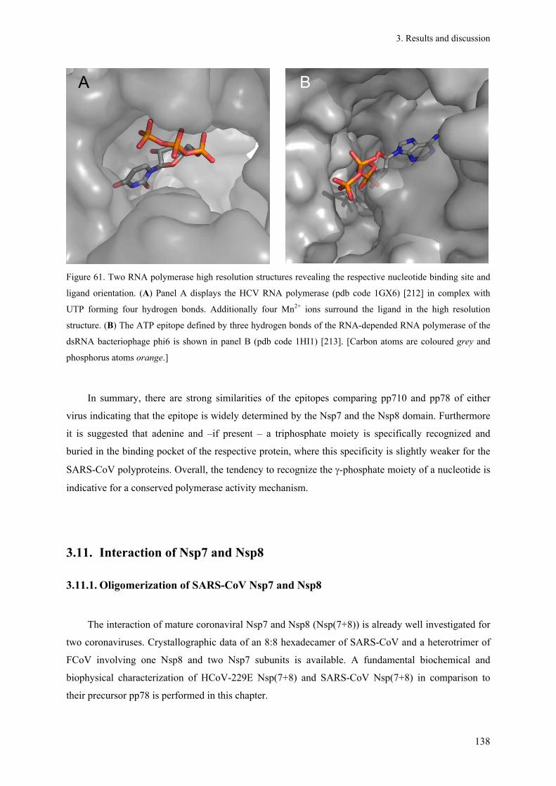
3. Results and discussion
138
A B
Figure 61. Two RNA polymerase high resolution structures revealing the respective nucleotide binding site and
ligand orientation. (A) Panel A displays the HCV RNA polymerase (pdb code 1GX6) [212] in complex with
UTP forming four hydrogen bonds. Additionally four Mn2+ ions surround the ligand in the high resolution
structure. (B) The ATP epitope defined by three hydrogen bonds of the RNA-depended RNA polymerase of the
dsRNA bacteriophage phi6 is shown in panel B (pdb code 1HI1) [213]. [Carbon atoms are coloured grey and
phosphorus atoms orange.]
In summary, there are strong similarities of the epitopes comparing pp710 and pp78 of either
virus indicating that the epitope is widely determined by the Nsp7 and the Nsp8 domain. Furthermore
it is suggested that adenine and –if present – a triphosphate moiety is specifically recognized and
buried in the binding pocket of the respective protein, where this specificity is slightly weaker for the
SARS-CoV polyproteins. Overall, the tendency to recognize the -phosphate moiety of a nucleotide is
indicative for a conserved polymerase activity mechanism.
3.11. Interaction of Nsp7 and Nsp8
3.11.1. Oligomerization of SARS-CoV Nsp7 and Nsp8
The interaction of mature coronaviral Nsp7 and Nsp8 (Nsp(7+8)) is already well investigated for
two coronaviruses. Crystallographic data of an 8:8 hexadecamer of SARS-CoV and a heterotrimer of
FCoV involving one Nsp8 and two Nsp7 subunits is available. A fundamental biochemical and
biophysical characterization of HCoV-229E Nsp(7+8) and SARS-CoV Nsp(7+8) in comparison to
their precursor pp78 is performed in this chapter.

3. Results and discussion
139
The putative complex of mature SARS-CoV and HCoV-229E Nsp7 (10 kDa, tab. 13) and Nsp8
(21 kDa) was formed by cleaving pp78 with SARS-CoV main protease. In analytical calibrated SEC
(HiLoad 16/60 Superdex 200) SARS-CoV Nsp7 and Nsp8 co-localized and peaked at a retention
volume corresponding to a molecular weight of 62 kDa (fig. 62A). This corresponds to an interaction
ratio of 2:2 at the applied experimental conditions. At a retention volume of around 85 ml, the
absorbance ratio A260 nm/A280nm significantly increased, likely originating from remaining nucleic acid
carried over from E. coli cells. For further experiments, SARS-CoV Mpro was removed from the
cleavage mixture by incubating the solution with Ni-NTA resin. Figure 62B shows the purified
Nsp(7+8) complex of SARS-CoV (lane II) and HCoV-229E (lane III) verified by SDS-PAGE.
Analytical SEC (Superose 12 10/300 GL) revealed that the applied buffer had a slight influence on the
oligomerization of SARS-CoV Nsp(7+8) (fig. 62C; see fig. 62A for comparison). However, a state
indicative for a 2:2-interaction of Nsp7 and Nsp8 corresponding to roughly 60-70 kDa was most
abundant, together with higher oligomeric states in the range of 100-180 kDa, as demonstrated by
clear native PAGE (fig. 62D). The protein was carefully excised from the gel and further analyzed by
mass spectrometry (ESI ion trap device) after trypsin digestion. Nsp7 and Nsp8 were identified with a
sequence coverage of 84% and 57%, respectively. A coomassie-stained putative complex with a
similar molecular weight was obtained by blue native PAGE as well (not shown). These results
strongly indicate the existence of a highly abundant 2:2 interaction state of Nsp7 and Nsp8 in solution,
clearly smaller in size than the hexadecameric state previously observed in a high-resolution crystal
structure [98] which might exist in addition. The proposed 2:2 interaction of Nsp7 and Nsp8 is in
agreement with the oligomerization of the homologue proteins of the Alphacoronavirus HCoV-229E
as previously indicated by SEC (fig. 29).

3. Results and discussion
140
450
272
146
67
45
kDa M Nsp
(7+
8)
1 2 3 4 5 6 7 8 9
Nsp5
Nsp8
Nsp7
4535 25
116
14
kDa
I II III
35
25
18
14
kDa
A
B C D
A
B
A B
35 k
Da
70 k
Da
44 k
Da
75 k
Da
158
kDa
14 k
Da
158
kDa
450
272
146
67
45
kDa M Nsp
(7+
8)
1 2 3 4 5 6 7 8 9
Nsp5
Nsp8
Nsp7
4535 25
116
14
kDa
I II III
35
25
18
14
kDa
A
B C D
A
B
A B
35 k
Da
70 k
Da
44 k
Da
75 k
Da
158
kDa
14 k
Da
158
kDa
Figure 62. Interaction of mature Nsp(7+8). (A) SARS-CoV pp78 containing an N-terminal strep-tag was
processed by SARS-CoV Mpro at a molar ratio of 4:1 and subsequently applied to SEC (Hi-Load 16/60 Superdex
200). Nsp7 and Nsp8 co-eluted and peaked slightly earlier than Mpro (purple chromatogram, A280 nm), as evident
from SDS-PAGE analysis. The red chromatogram displays the absorbance of the same sample at 220 nm.
Processing and analysing pp78 containing a C-terminal strep-tag fused to Nsp8 by SEC resulted in the blue
chromatogram, while buffer and sample preparation were identical. (B) Mpro was removed by subsequent
incubation with equilibrated Ni-NTA resin and centrifugation to obtain the pure Nsp(7+8) complex [lane I:
SARS-CoV pp78, lane II: SARS-CoV Nsp(7+8), lane III: HCoV-229E Nsp(7+8). (C) The Nsp(7+8)-complex
containing N-terminally strep-tagged Nsp7 was analyzed by SEC (Superose 12 10/300 GL) in HEPES buffer
(dark blue) and in 350 mM ammonium acetate adjusted to pH 7.9, respectively (light pink). Besides a small
protein fraction directly eluting from the column (MW > 300 kDa, “exclusion volume“), the estimated MW of
all additional states is predicted to be smaller than 200 kDa. (D) A defined oligomeric state of Nsp(7+8)
characterized by a molecular weight of 60 to 70 kDa is revealed by clear-native PAGE, corresponding to a
heterotetramer.
To verify interaction states of SARS-CoV Nsp(7+8) at a high mass accuracy, native mass
spectrometry was applied. The spectra displayed in fig. 63 support the previous observations,
demonstrating a high abundance of a mass in agreement with a heterotetrameric 2:2-interaction
(65,011 Da). This mass species was fragmented by collision-induced dissociation, yielding monomeric
Nsp7 and Nsp8 (fig. 63C). However, smaller fractions of a heterodimer (32,547 Da) and a putative

3. Results and discussion
141
4:4-interaction (heterooctamer; 131,802 Da) requiring a second dimerization of the 2:2-
heterotetrameric state were also detected.
Figure 63. An equimolar mixture of Nsp7 and Nsp8 was obtained by completely cleaving SARS-CoV pp78 with
N-terminal strep-tag by Mpro. The protease was removed prior to the experiment by Ni-NTA resin. (A) The m/z-
spectrum at 10 V collision voltage. (B) Referring to panel A, individual mass species of the spectrum above are
shown that were simulated by Massign. Nsp7 and Nsp8 were analyzed by an ESI-Q-TOF instrument revealing
different oligomeric states; more than 30% of the total intensity in the displayed m/z range (around 65 kDa) fits a
2:2-interaction of both proteins along with a lower amount of a 4:4-interaction state indicative for a further
dimerization mode. This observation nicely agrees with SEC experiments (fig. 62). The broadness of the peaks
indicates a minor mass variation, perhaps due to small ligands carried over from E. coli or ions from the buffer.
Another oligomeric state fits an Nsp8 dimer (green in the panel B), most probably overlaying with a marginal
amount of a 2:1-(Nsp7:Nsp8)-complex fragment (purple). (C) Stepwise CID released a dimeric Nsp8 “core” and

3. Results and discussion
142
free Nsp8 (21,900 ± 36 Da), in parallel free Nsp7 (10,608 ± 12 Da) is enriching by increasing the voltage (lower
panel). The depicted purple spectrum was obtained at 120V collision voltage.
Due to the high sequence identity of Nsp7 and Nsp8 of distantly related coronaviruses (see also
tab. 13), a 2:2-interaction state as proposed for the SARS-CoV Nsp7 and Nsp8 complex might be
conceivable for other closely-related Betacoronaviruses as well, e.g. MERS-CoV, along with other
oligomeric states. As shown above, mass spectrometry demonstrated the formation of small amounts
of these additional oligomeric states, suggesting a model of a condition-dependent transition and
balance of oligomers similar to the previous observations for polyprotein pp710 (e.g. chapter 3.2).
This could also include specific oligomeric states with a molecular mass of more than 200 kDa that
were not clearly detected by the methods applied in this study, but have been previously reported in a
crystalline environment [98]. For mass spectrometry ammonium acetate buffer had to be applied, that
altered the SEC chromatogram of the Nsp(7+8) complex compared to HEPES buffer (fig. 62C). A
second dominating peak appeared at lower elution volumes, corresponding to a state larger than a 2:2
interaction. Thus, slight variations in the oligomerization pathway are assumed to depend on the buffer
conditions required for the applied technique.
However, an impact of the purification tag position on the formation of oligomeric states could
not be excluded. In chemical cross-linking experiments performed by te Velthuis et al. [97], C-
terminal his-tagged Nsp8 formed an octameric state, while an N-terminal his-tag hindered the
formation of higher order oligomers. Thus, the formation of a hexadecameric state of Nsp(7+8) could
depend on correct N-terminal processing of Nsp8. Nevertheless, even the variation of the strep-tag
position in pp78 did not result in any indication for a significant amount of a hexadecameric
interaction state in this study. It has to be mentioned that chemical cross-linking as performed by te
Velthuis et al. might have the disadvantage that it is difficult to distinguish between continuums of
different oligomeric states, e.g. a mixture of dimer, tetramer and octamer and/or even larger (specific)
oligomers. Additionally, cross-linking depends on the location of suitable amino acid residues at the
interaction interface. Up to now, it has not been sufficiently proven by applying different methods at
conditions close to physiological that octameric Nsp8 or hexadecameric Nsp(7+8) represent exclusive
aggregation states of these proteins in solution, even though comparative SEC at a relatively low mass
resolution was performed previously [97] indicating a hexadecameric interaction of SARS-CoV Nsp7
and Nsp8.
According to the present results it is suggested that a mixture of different oligomeric states is
formed in solution, with at least a preference for a 2:2 interaction for the respective complex of HCoV-
229E and SARS-CoV. This interaction state was independently observed in SEC experiments for
SARS-CoV Nsp7 and Nsp8 after cleavage of pp78 containing different tags as well as authentic
termini (chapter 3.4.1). This hypothesis agrees well with a significant amount of dimeric pp78
detected in solution (see chapter 3.9) and allows the conclusion that even distantly related

3. Results and discussion
143
coronaviruses share the capability to form different small (hetero-)oligomeric states of mature Nsp7
and Nsp8, also displaying a similar oligomerization compared to the precursor pp78. To further
investigate whether the domain-interactions within monomeric and dimeric states of pp78 are similar
to that within heterotetrameric states of mature Nsp7 and Nsp8 in solution, SAXS technique was
subsequently applied.
3.11.2. Nsp(7+8) in comparison to its precursor pp78 by SAXS
To address the question of the interaction of Nsp7 and Nsp8 in the precursor state pp78, SAXS
experiments were performed as described. SAXS particularly aimed at enlightening the geometry of
the N-terminus of Nsp8 within pp78, which was shown to fold into a long flexible α-helix in a high-
resolution crystal structure of SARS-CoV Nsp(7+8) [98]. The fold of the N-terminal domain of Nsp8
in pp78 might be influenced by non-covalent interactions with Nsp7 at interfaces different to the
covalent link of both proteins. Consequently, the aim was to predict the binding epitope(s) of SARS-
CoV Nsp7 and Nsp8 in the consecutive precursor state. Monomeric and dimeric pp78 of SARS-CoV
and HCoV-229E were purified by SEC as described in fig. 53 (chapter 3.9.1). The monodispersity of
the sample was verified by DLS, revealing a hydrodynamic radius of 3.3 ± 0.3 nm for monomeric
SARS-CoV pp78 and 5.3 ± 0.1 nm for dimeric pp78 of SARS-CoV. In comparison, the hydrodynamic
radius of monomeric HCoV-229E pp78 is 3.4 ± 0.4 nm and 4.5 ± 0.2 nm for dimeric HCoV-229E
pp78. Results of SAXS measurements at protein concentrations of 1 - 4.5 g l-1 resulted in Rg and Dmax
values specified in table 25. A comparison of the distance-distribution functions of SARS-CoV pp78
monomer and dimer is shown in figure 64A, demonstrating the increased maximum diameter of the
dimer (yellow curve, Dmax = 13 nm) compared to that of the monomer (red curve, Dmax = 10 nm).
Furthermore, the incubation of monomeric SARS-CoV pp78 with SARS Mpro (ratio 10:1)
demonstrated that the distance-distribution function continuously changed during proteolytic
processing of the pp78 precursor. After 5 min (dark blue curve) the maximum diameter of the
molecules increased by 2 nm, while an additional increase up to 13 nm was observed after 15 min of
incubation (light blue). Ab initio models of dimeric pp78 of SARS-CoV and HCoV-229E (see also
chapter 3.10) were calculated based on the distance-distribution functions (fig. 64B/C). Both dimeric
pp78 molecules shared an elongated shape and a similar overall geometry, even though radius of
gyration and maximum diameter of HCoV-229E pp78 are slightly smaller (Dmax = 11.4 nm, Rg =
3.37 nm) compared to SARS-CoV pp78 (Dmax = 13 nm, Rg = 3.88 nm).

3. Results and discussion
144
0
0,4
0,8
1,2
0 4 8 12 16
R [nm]
P(R
), r
elat
ive
units
pp78 monomer
pp78 dimer
pp710 dimer
pp78+ Nsp5 5 min
pp78 + Nsp5 15 min
A B C
0
0,4
0,8
1,2
0 4 8 12 16
R [nm]
P(R
), r
elat
ive
units
pp78 monomer
pp78 dimer
pp710 dimer
pp78+ Nsp5 5 min
pp78 + Nsp5 15 min
0
0,4
0,8
1,2
0 4 8 12 16
R [nm]
P(R
), r
elat
ive
units
pp78 monomer
pp78 dimer
pp710 dimer
pp78+ Nsp5 5 min
pp78 + Nsp5 15 min
A B C
Figure 64. SAXS analysis of pp78. (A) P(R)-functions of SARS-CoV pp78 before and during incubation with
SARS-CoV Mpro. The maximum diameter corresponds to the point of intersection with the radius axis. (B) Ab
initio model of dimeric SARS-CoV pp78 with a maximum diameter of 13 nm; (C) Ab initio model of HCoV-
229E pp78 shown for comparison (Dmax = 11.4 nm).
Evaluating the scattering pattern of monomeric HCoV-229E pp78 resulted in a valuable rigid-
body model using the high-resolution structures of Nsp7 (green, pdb code 1YSY) and Nsp8 (blue,
chain A and H, extracted from pdb code 2AHM) (fig. 65A). 68 cycles of refinement have been
performed (penalty 0.4442, best chi value of the scattering fit: 1.0656, considering scattering vector
values 0.01 < s ≤ 0.28 Å-1). The restrain that Nsp7 and Nsp8 are linked C-terminus to N-terminus was
set for rigid-body modelling and the rigid “stretched” isoform of SARS-CoV Nsp8 was used as a
template, together with Nsp7. However, this rigid Nsp8 structure seemed to be insufficient to interpret
the ab initio model (calculated by DAMMIF) of monomeric pp78 correctly (fig. 65C), as already
discussed concerning full-length HCoV-229E pp710 (chapter 3.2.3.2/3.2.3.3). Since Nsp7 is reported
to occupy a known interaction epitope at Nsp8 in the complex of mature Nsp7 and Nsp8 [98], a
comparable interaction could be expected for pp78. The epitope shared by SARS-CoV Nsp7 and Nsp8
(chain A and E) involves four hydrogen bonds (Nsp7 Q19/Nsp8 T84, Nsp7 S61/Nsp8 P116, Nsp7
S57/Nsp8 I120, Nsp7 S54/Nsp8 I120). Assuming a specific degree of flexibility in the N-terminal
domain of Nsp8, this epitope is also conceivable for HCoV-229E pp78. To take the flexibility of Nsp8
into account, Nsp8 was divided into four individual fragments that were used as templates for
improved rigid-body modelling by SASREF (fig. 65B). The fragments of Nsp8 were designed
according to reported sites of flexibility based on high-resolution structures [96; 98] and personal
communication with Y. Xiao (University of Lübeck).
The resulting rigid-body model fitted the corresponding ab initio model even better, notably with
a significant bending of two fragments of the Nsp8 N-terminus (fig. 65C). A manual repositioning of
Nsp7 according to free volume within the ab initio model of the “scattering envelope” of SARS-CoV
pp78 not defined by Nsp8 was performed. An ab initio model of monomeric pp78 of HCoV-229E is
displayed for comparison (fig. 65D). For SARS-CoV pp78, the position of Nsp7 obtained by rigid-

3. Results and discussion
145
body modelling (fig. 65C, green) was changed according to the known interface of mature Nsp7 and
Nsp8, resulting in the red Nsp7 molecule. This optimized rigid-body model fitted the ab initio model
equally well (fig. 65C). To still allow the covalent bond between Nsp7 and Nsp8, a higher degree of
bending of the N-terminal domain of Nsp8 is suggested. The resulting ab initio model of SARS-CoV
pp78 shares common characteristics in terms of shape and tertiary structure with HCoV-229E pp78,
for which the ab initio model was additionally well interpreted by an independent homology model
that was calculated using the I-Tasser modelling server based of sequence homology (fig. 65E).
Fig. 65F displays the rigid body and the ab initio model of dimeric HCoV-229E pp78 based on
the scattering data in solution. The “bended” SARS-CoV Nsp8-chain (blue) taken from the Nsp(7+8)
complex (pdb code 2AHM) is truncated at the N-terminus, because this part of Nsp8 was not defined
by the electron density [98]; therefore the native Nsp8 N-terminus is assumed to allow Nsp7 to be
located even closer to the C-terminus of Nsp8 as observed for the complex of mature SARS-CoV
Nsp7 and Nsp8. The interface of the HCoV-229E pp78 dimer is predicted to involve the C-terminal
domain of Nsp8, as previously observed for the pp710 dimer of HCoV-229E (chapter 3.2.3.2/3.2.3.3).
According to the rigid-body model the dimerization of HCoV-229E pp78, based on the proposed
symmetry and shape, as calculated by SASREF, is not expected to involve intermolecular Nsp7-Nsp7
interactions at the dimerization interface, although one pp78 molecule could rotate around the
longitudinal axis to get the Nsp7 domains in the pp78 dimer in contact with each other. Moreover, this
Nsp7-Nsp7 dimerization at the dimer interface of pp78 would provide an additional stabilization of the
pp78 dimer. Furthermore, dimerization of mature SARS-CoV Nsp7 was previously indicated in
solution (chapter 3.11.3) and agrees with a dimerization of mature FCoV Nsp7 in solution detected by
covalent cross-linking [96].
The dimerization of mature HCoV-229E Nsp(7+8) appeared to be mediated by the C-terminus of
Nsp8 as well, forming a largely elongated molecule as evident from the ab initio model (fig. 65G).
Comparison of HCoV-229E pp78 and the non-covalent complex of mature Nsp7 and Nsp8 (fig. 65,
F/G) revealed that the complex is further elongated upon processing by main protease, well explaining
the increase of the maximum diameter observed in a SAXS experiment that followed the cleavage
reaction (fig. 65A). However, in summary, the C-terminal globular domain of Nsp8 still appeared to
be essential for dimerization even after complete maturation of Nsp7 and Nsp8.

3. Results and discussion
146
Figure 65. Comparison of DAMMIF ab initio models of SARS-CoV and HCoV-229E pp78 as well as Nsp(7+8)
of HCoV-229E obtained by SAXS and interpretation by high-resolution structures. (A) Rigid-body model based
on scattering data of monomeric HCoV-229E pp78 using the rigid full-length SARS-CoV Nsp8 structure taken
from the Nsp(7+8) complex (pdb code 2AHM, chain H) and Nsp7 (pdb code 1YSY) with the restrain of a
covalent connection. These prerequisites did not allow a sufficient interpretation of the data that suggested a
translation of Nsp7 (black arrow). (B) Nsp8 was segmented into four individual fragments to simulate the sites
of flexibility: amino acids 1-73 (fragment 1, blue-green), 74-113 (fragment 2, grey), 114-124 (fragment 3, blue),
125-195 (fragment 4; purple). (C)/(D) Low resolution ab initio structural models of the pp78 homologues of
SARS-CoV and HCoV-229E are presented for comparison. The rigid-body model of SARS-CoV pp78
calculated using four Nsp8 fragments that were independently positioned by SASREF is fitted into the
corresponding ab initio model in panel C. The Nsp7 molecule (green) was manually positioned close to the Nsp8
C-terminus, where the calculated low-resolution envelope provided sufficient space (red). (E) Two possibilities
to interpret the ab initio model of monomeric HCoV-229E pp78 by a homology model obtained from the I-
Tasser modelling server (RMSD: 13.2 ± 4.1 Å, C-score: -3.01, TM-score: 0.37 ± 0.13). Superposition II was
favoured because Nsp7 (green) can occupy a part of the ab initio model close to the Nsp8 (blue) C-terminus. (F)
Rigid-body model of dimeric HCoV-229E pp78. Truncated and bended Nsp8 was used as a template (blue, chain
E, pdb code 2AHM), together with Nsp 7 (green, pdb code 1YSY). Cleavage sites connecting Nsp7 and Nsp8
are labelled by red arrows. (G) Rigid-body model of the mature Nsp(7+8) dimer of HCoV-229E obtained by
SASREF (88 cycles of refinement, penalty 0.0001, best χ-value of the scattering fit: 0.90532, considering
scattering vector values 0.01 < s ≤ 0.24 Å-1). The averaged low resolution ab initio model of mature HCoV-229E
Nsp(7+8) presented for comparison displayed an increased maximum diameter (Dmax = 14.5 nm) compared to
dimeric HCoV-229E pp78 (Dmax = 11.4 nm). The mean NSD of 10 DAMMIF models is 1.124 ± 0.044.

3. Results and discussion
147
To compare the stability of the fold of HCoV-229E pp78 with the mature complex consisting of
Nsp7 and Nsp8 CD-spectroscopy was applied, having in mind that previously significantly different
low-resolution structures were presented (fig. 65). At 30 °C, a far-UV CD spectrum with two minima
characteristic for a high α-helix content is observed, reflecting the expected fold of Nsp7 and Nsp8
[96; 98]. However, the similar temperature stability of HCoV-229E pp78 in the pre-cleavage state
compared to Nsp(7+8) in the post-cleavage state (fig. 66) indicated that the overall secondary structure
and the sum of inter-domain interactions are not significantly altered. The slight difference could
originate from differences in the aggregation behaviour, the different purification protocols or
theoretically a minor difference of the Nsp7-Nsp8 interaction site in the polyprotein state.
Assuming that pp78, pp79 and pp710 have different equilibrium positions of oligomeric states,
this could have impact on the melting temperature as well as an altered structure of the protomers. For
example, as cross-linking with BS2G suggested, SARS-CoV pp710 is monomeric at a higher
percentage compared to SARS-CoV pp78. This would correlate with the higher thermal stability of
SARS-CoV pp78. Applying purified monomeric and dimeric SARS-CoV pp710 to CD spectroscopy
there was no significant difference, even though higher oligomers appeared to be marginally more
stable (data not shown). This observation might already be close to the resolution limit of relatively
similar melting temperatures. Nonetheless, for instance for the L-tryptophan biosynthesis regulator
(TRAP) [174] tight specific oligomerization and ligand-binding was correlated with significantly
higher melting temperatures determined by CD spectroscopy. Additionally, this was observed for the
reduced thermal stability of Mpro R298A (Tm = 2.7 °C; Gdissociation = 4.2 kcal mol-1 (PISA,EMBL-
EBI, calculated for pdb code 1Q2W)) compared to the wild type known to form a relatively tight
dimer [53; 60].

3. Results and discussion
148
HCoV-229E pp78 HCoV-229E Nsp(7+8)
190
210
220
230
260
190
210
220
230
260
30 40 50 60 70 30 40 50 60 70
wa
vele
ngt
h [n
m]
temperature [°C] temperature [°C]
wa
vele
ngt
h [n
m]
wa
vele
ngt
h [n
m]
190
210
220
230
260-30
60
0
mdeg
A B
HCoV-229E pp78 HCoV-229E Nsp(7+8)
190
210
220
230
260
190
210
220
230
260
30 40 50 60 70 30 40 50 60 70
wa
vele
ngt
h [n
m]
temperature [°C] temperature [°C]
wa
vele
ngt
h [n
m]
wa
vele
ngt
h [n
m]
190
210
220
230
260-30
60
0
mdeg
A B
Figure 66. Investigation of the thermal stability of HCoV-229E pp78 (A) and Nsp7+8 (B) applying CD
spectroscopy. Temperature interval scanning experiments were performed at temperatures ranging from 30 °C to
70 °C. The typical shape of the spectrum at 30 °C involving two minima characteristic for high α-helix content is
conserved. The highest loss of absolute ellipticity of the minima is observed in a similar range close to 60 °C,
even though the stability of the mature Nsp(7+8) complex determined at 220 nm is a little bit higher. The
original two minima observed at around room temperature tend to fuse to form one resulting flat minimum above
65 °C, after the collapse of most of the helix structure.
To obtain indication for putative ligand binding sites at the C-terminus of SARS-CoV Nsp8,
small-molecule docking was applied (fig. 67). The FT map server [167] identifies putative ligand
binding sites on the surface of a protein by screening small organic molecules of a default library in
terms of ligand binding energy. Applying this algorithm to SARS-CoV Nsp8 (pdb code 2AHM, chain
H), FTmap identified one “hot-spot” for ligand binding at a helix in the almost globular C-terminal
domain of the protein (red circle). This observation could not only be indicative for RNA/nucleotide
binding, but also for an interaction with small regulatory ligands in vivo. This C-terminal domain was
previously considered to be a favourable protein binding site and probably part of the Nsp8
dimerization interface (fig. 65F/G). Interestingly, the C-terminal domain of Nsp8 exhibits structural
similarities with hnRNP (pdb code 2UP1), i.e. a human single strand nucleic acid-binding protein [94].
Therefore, the C-terminal domain could be involved in catalysis or at least RNA binding, which has
been reported for the hexadecameric complex of mature SARS Nsp(7+8). This is supported by the
suggestion that an N-terminally truncated isoform of Nsp8 is able to preserve nucleic acid binding and
exhibits a regulatory effect on the hexadecameric Nsp(7+8) complex. However, as already mentioned,
such an oligomeric state was not detected in the course of this project.
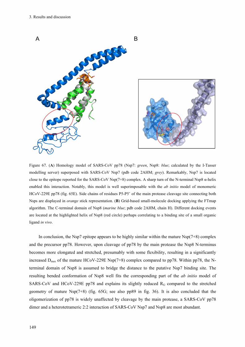
3. Results and discussion
149
Figure 67. (A) Homology model of SARS-CoV pp78 (Nsp7: green, Nsp8: blue; calculated by the I-Tasser
modelling server) superposed with SARS-CoV Nsp7 (pdb code 2AHM; grey). Remarkably, Nsp7 is located
close to the epitope reported for the SARS-CoV Nsp(7+8) complex. A sharp turn of the N-terminal Nsp8 α-helix
enabled this interaction. Notably, this model is well superimposable with the ab initio model of monomeric
HCoV-229E pp78 (fig. 65E). Side chains of residues P5-P5’ of the main protease cleavage site connecting both
Nsps are displayed in orange stick representation. (B) Grid-based small-molecule docking applying the FTmap
algorithm. The C-terminal domain of Nsp8 (marine blue; pdb code 2AHM, chain H). Different docking events
are located at the highlighted helix of Nsp8 (red circle) perhaps correlating to a binding site of a small organic
ligand in vivo.
In conclusion, the Nsp7 epitope appears to be highly similar within the mature Nsp(7+8) complex
and the precursor pp78. However, upon cleavage of pp78 by the main protease the Nsp8 N-terminus
becomes more elongated and stretched, presumably with some flexibility, resulting in a significantly
increased Dmax of the mature HCoV-229E Nsp(7+8) complex compared to pp78. Within pp78, the N-
terminal domain of Nsp8 is assumed to bridge the distance to the putative Nsp7 binding site. The
resulting bended conformation of Nsp8 well fits the corresponding part of the ab initio model of
SARS-CoV and HCoV-229E pp78 and explains its slightly reduced RG compared to the stretched
geometry of mature Nsp(7+8) (fig. 65G; see also pp89 in fig. 36). It is also concluded that the
oligomerization of pp78 is widely unaffected by cleavage by the main protease, a SARS-CoV pp78
dimer and a heterotetrameric 2:2 interaction of SARS-CoV Nsp7 and Nsp8 are most abundant.
A B

3. Results and discussion
150
3.11.3. SARS-CoV Nsp7 forms a dimer in solution
To investigate the impact of a hydrophobic patch located within the N-terminal α-helix of Nsp7
on the interaction with Nsp8 the double mutant Nsp7 V11E/L14R of SARS-CoV pp78 was designed.
The side chains of these two residues point in different orientations (pdb code 2AHM) [98; 100]. Parts
of the sequence 11VVLL14 of SARS-CoV Nsp7 are surface exposed, while intramolecular hydrophobic
interactions are also formed within the Nsp(7+8) high-resolution structure [98]. Putative
intermolecular hydrophobic interaction of the helices of two Nsp7 molecules could also influence the
oligomerization of pp78. Introducing large polar residues may alter the peripheral hydrophobicity of
the potential binding epitope, resulting in an altered hydrophobic interaction involving the N-terminal
helix of Nsp7. Furthermore, V11 is known to hydrophobically interact with S20 of another Nsp7
molecule in the SARS-CoV Nsp(7+8) complex, stabilizing an Nsp7 dimer, while each Nsp7 molecule
is additionally interacting with one Nsp8 molecule to form a building block of the Nsp(7+8)
hexadecamer [98].
The obtained double mutant V11E/L14R of SARS-CoV pp78 was purified by affinity
chromatography to homogeneity (fig. 68B). Interestingly, the protein yield of the double mutant
obtained from E. coli cells was significantly increased compared to the wild type protein, which could
correlate with improved protein solubility due to locally reduced surface hydrophobicity. After
cleavage of the precursor pp78 by SARS-CoV Mpro, calibrated SEC showed two main peaks (fig. 68,
dark green chromatogram). As shown by non-reducing SDS-PAGE analysis mature Nsp7 V11E/L14R
was successfully separated from mature Nsp8 (fig. 68B). The approximated molecular weight of
mutated mature Nsp7 optimally fits a dimeric state (VE = 91.5 ml → 17.2 kDa). A minor amount of
remaining pp78 co-localized with mature Nsp8 at a retention volume between 75-80 ml, indicative for
an interaction of pp78 and Nsp8 (fig. 68A, see red chromatogram compared to peak A). Applying
fractions of peak A to SAXS, elongated particles at a molecular weight below 70 kDa were
reconstructed (data not shown). The mutated precursor pp78 still dimerized in solution
(experimentally calculated MWpp78 = 62 kDa) in a concentration-dependent manner (fig. 68A).
Interestingly, additional larger oligomers previously observed by SEC with pp78 wild type (fig. 53A)
were absent. The observation that a major amount of SARS-CoV Nsp7 V11E/L14R was separated
from Nsp8 by SEC is an indication that the corresponding conserved hydrophobic patch within α-helix
1 has a minor contribution to the interaction. In spite of the mutation, CD spectroscopy displayed a
conserved α-helix-rich fold of Nsp7 (fig. 71), in good agreement with known high-resolution
structures [98; 100].

3. Results and discussion
151
Mpro
Pp78
Nsp8
Nsp8CNsp7
Pp78 I A B N8C M
A
B
kDa
45
35
25
14
A B
Mpro
Pp78
Nsp8
Nsp8CNsp7
Pp78 I A B N8C M
A
B
kDa
45
35
25
14
Mpro
Pp78
Nsp8
Nsp8CNsp7
Pp78 I A B N8C M
A
B
kDa
45
35
25
14
A B
Figure 68. Proteolytic cleavage of mutated SARS-CoV pp78 by SARS Mpro. (A) Different stages of the
processing reaction of pp78 V11E/L14R were applied to SEC (HiLoad 16/60 Superdex 200). The excess of pp78
towards Mpro was 7-fold. Red chromatogram: 6 mg pp78 V11E/L14R, brown: 1 mg pp78 V11E/L14R light
green: 60% of pp78 V11E/L14R cleaved by Mpro, dark green: 90% cleaved, as judged by SDS-PAGE. All
samples were buffered in 30 mM NaH2PO4, pH 7.6, supplemented with 250 mM NaCl. (B) Individual fractions
referring to the dark green chromatogram were analyzed by SDS-PAGE. Pp78: Purified pp78 V11E/L14R after
affinity chromatography; I: Column input (pp78 DM after Mpro cleavage); A, B: Fractions of respective SEC
peaks; Nsp8C: C-terminal domain of Nsp8 as a molecular weight standard for 12.8 kDa.

3. Results and discussion
152
Figure 69. (A) SAXS investigation revealed an elongated dimer of Nsp7 V11E/L14R: Scattering data at protein
concentrations of up to 2.8 g l-1 did not indicate a concentration-dependency of the particle size. Optimized fit
function of the rigid-body model (red) created by SASREF considering P2 symmetry, i.e. a dimer, well fits the
experimental scattering data (blue). The rigid-body model of Nsp7 is depicted on the right (template: FCoV
Nsp7 extracted from pdb code 3UB0). The model is characterized by a chi-value of 1.1972 after 58 cycles
(penalty 0.0016; default penalty scores and weight) using a maximum scattering vector s of 0.1826 nm-1 (1707
data points). (B) The Nsp7 dimer (green) is stabilized by hydrogen bonds of Q18 (orange spheres). The Nsp7-
Nsp7 and Nsp7-Nsp8 (blue) interaction within the Nsp(7+8) hexadecameric complex (pdb code 2AHM) is
visualized. Nsp7 Q18 is labeled by orange spheres; V11 and L14 are displayed as red spheres.
A
B
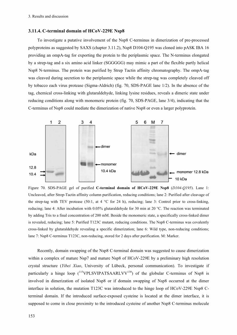
3. Results and discussion
153
3.11.4. C-terminal domain of HCoV-229E Nsp8
To investigate a putative involvement of the Nsp8 C-terminus in dimerization of pre-processed
polyproteins as suggested by SAXS (chapter 3.11.2), Nsp8 D104-Q195 was cloned into pASK IBA 16
providing an ompA-tag for exporting the protein to the periplasmic space. The N-terminus elongated
by a strep-tag and a six amino acid linker (SGGGGG) may mimic a part of the flexible partly helical
Nsp8 N-terminus. The protein was purified by Strep Tactin affinity chromatography. The ompA-tag
was cleaved during secretion to the periplasmic space while the strep-tag was completely cleaved off
by tobacco each virus protease (Sigma-Aldrich) (fig. 70, SDS-PAGE lane 1/2). In the absence of the
tag, chemical cross-linking with glutaraldehyde, linking lysine residues, reveals a dimeric state under
reducing conditions along with monomeric protein (fig. 70, SDS-PAGE, lane 3/4), indicating that the
C-terminus of Nsp8 could mediate the dimerization of native Nsp8 or even a larger polyprotein.
Figure 70. SDS-PAGE gel of purified C-terminal domain of HCoV-229E Nsp8 (D104-Q195). Lane 1:
Uncleaved, after Strep-Tactin affinity column purification, reducing conditions; lane 2: Purified after cleavage of
the strep-tag with TEV protease (50:1, at 4 °C for 24 h), reducing; lane 3: Control prior to cross-linking,
reducing; lane 4: After incubation with 0.05% glutaraldehyde for 30 min at 20 °C. The reaction was terminated
by adding Tris to a final concentration of 200 mM. Beside the monomeric state, a specifically cross-linked dimer
is revealed, reducing; lane 5: Purified T123C mutant, reducing conditions. The Nsp8 C-terminus was covalently
cross-linked by glutaraldehyde revealing a specific dimerization; lane 6: Wild type, non-reducing conditions;
lane 7: Nsp8 C-terminus T123C, non-reducing, stored for 2 days after purification. M: Marker.
Recently, domain swapping of the Nsp8 C-terminal domain was suggested to cause dimerization
within a complex of mature Nsp7 and mature Nsp8 of HCoV-229E by a preliminary high resolution
crystal structure (Yibei Xiao, University of Lübeck, personal communication). To investigate if
particularly a hinge loop (115VPLSVIPATSAARLVV130) of the globular C-terminus of Nsp8 is
involved in dimerization of isolated Nsp8 or if domain swapping of Nsp8 occurred at the dimer
interface in solution, the mutation T123C was introduced to the hinge loop of HCoV-229E Nsp8 C-
terminal domain. If the introduced surface-exposed cysteine is located at the dimer interface, it is
supposed to come in close proximity to the introduced cysteine of another Nsp8 C-terminus molecule

3. Results and discussion
154
during dimerization, resulting in the formation of a disulfide bond under non-reducing conditions.
Wild type Nsp8 D104-Q195 contains only one native cysteine residue that is not exposed to the
surface. Since secretion into the periplasmic space should favour the formation of disulfide bonds
within the E. coli cells, mutated protein Nsp8 was cloned as an ompA-tag fusion protein. The protein
was purified by Strep-Tactin affinity chromatography (fig.70B, lane 5). To verify that the truncated
and mutated variant of Nsp8 is well folded, CD spectroscopy was performed in comparison to other
Nsps (fig. 71). The spectra demonstrated that the C-terminal domain of Nsp8 was rich in β-sheet and
random coil structure, while full-length Nsp8 was largely composed of α-helical structure due to its N-
terminus. A fraction of random coil is in agreement high-resolution structures of the Nsp(7+8)
complex [96; 98], even though a higher content of β-sheet would be expected.
The α-helix-rich fold of HCoV-229E Nsp8 was indicated by two predominant minima of the
ellipticity around 208 nm and 222 nm that were absent in the spectra of Nsp8 C-terminus wt and Nsp8
C-terminus T123C.
-10
-5
0
5
10
15
195 210 225 240 255
wavelength [nm]
MR
E [1
04 d
eg c
m2 d
mol
-1]
Nsp8 C-terminus HCoV-229E
Nsp8 HCoV-229E
Nsp8 C-terminus T123C HCoV-229E
Nsp7 V11E/L14R SARS-CoV
Nsp(7+8) HCoV-229E
Figure 71. Far-UV CD-spectra ranging from 260 to 195 nm wavelength, collected in sodium phosphate buffer.
The spectrum of Nsp7 V11E/L14R (green) is characteristic for an -helix-rich protein, confirming its proper
native fold. The complex of full-length mature Nsp(7+8) of HCoV-229E (wine red) and full-length Nsp8 of
HCoV-229E (dark blue) showed a comparable -helix-rich fold. Wild type Nsp8 C-terminal domain (grey-blue)
and its T123C mutant (cyan) showed a relatively low absolute ellipticity and a single minimum around 204 nm
and 201 nm, respectively, indicating a fold dominated by random coil and -sheet fold, but a very low α-helix
content, in agreement with reported crystal structures [98], even though folding artefacts due to the truncation or
mutation could not be not excluded.
To probe the close proximity of the introduced cysteine residues within a dimer of the Nsp8 C-
terminal domain, non-reducing SDS-PAGE of the purified T123C mutant of Nsp8 D104-Q195 was
performed immediately after purification as well as after two days incubation under oxidising
conditions (fig. 70B, lane 6 and 7). In contrast to the wild type protein, the mutant formed a covalent
dimer during incubation, representing a first indication that the additionally introduced cysteine

3. Results and discussion
155
residue formed a specific link with the cysteine of another molecule of the Nsp8 C-terminal domain.
Additional covalent oligomers of mutant or wild type were absent in non-reducing SDS-PAGE.
The indicated covalent dimerization was further analyzed by analytical size-exclusion
chromatography (HiLoad 16/60, Superdex 200, GE Healthcare). A single protein peak was observed
for pure Strep-tagged Nsp8 C-terminus wild type at an elution volume of 90.9 ml, corresponding to a
molecular weight of 29.4 kDa that represents a dimeric state of the protein (fig. 72A). Under identical
non-reducing conditions, a comparable sample of the corresponding T123C mutant eluted slightly
earlier than the wild type at a volume of 88.7 ml from the SEC column, corresponding to a molecular
weight of 33.6 kDa. This indicated the presence of trimeric protein, consisting of a dimer covalently
linked at the mutated site that interacted non-covalently with further molecules. After removing the
strep-tag under reducing conditions and further incubating the mutant for 24 h at 6 °C, SEC displayed
a similar dimeric state (VE = 92.5 ml). The elution is slightly shifted due to the removal of the tag
compared to the strep tagged wild type. Significant amounts of higher oligomeric states have not been
detected for all samples (fig. 72A).
Mutated, strep-tagged C-terminal Nsp8 (fig. 72A light blue chromatogram) was additionally
investigated by SAXS technique, resulting in Rg = 2.57 ± 0.01 and Dmax = 9 nm. Based on the
scattering data in solution, a low-resolution ab initio model was calculated considering P2 symmetry
for a dimeric state, which is depicted in fig. 72B. As already indicated by SEC, the approximated
molecular weight of the Nsp8 C-terminus T123C mutant of 30 kDa, based on the ab initio model
calculation by DAMMIF, appeared to be slightly increased compared to that expected for a dimer
(MWdimer = 26 kDa). In conclusion, this truncated Nsp8 "model system" to investigate dimerization of
Nsp8 could be either altered in oligomerization due to the truncation at the N-terminus or display an
artificial folding in the absence of the N-terminus in E. coli, as indicated by CD spectroscopy.
The oligomerization might also be a matter of the concentration, considering the hypothesized
involvement of domain-swapping. Moreover, as observed by non-reducing SDS-PAGE the relative
fraction of covalently linked dimers of the Nsp8 C-terminus T123C which were presumed to be
indicative for the potential dimer interface, is rather low (fig. 70 lane 7). Finally, it still has to be
considered that the introduced surface-exposed cysteine residue could result in an unspecifically
formed dimer. However, the dimeric state of mutated Nsp8 C-terminus appeared to be predominant in
solution. Moreover, the calculated ab initio model is well superimposable with the corresponding
fragment of the ab initio model of full-length pp78 (fig. 72B, see also fig. 65F for comparison),
indicating a relevance of this putative dimeric Nsp8 C-terminus molecule in the dimerization of pp78.
To verify the identity of the disulfide bond observed by non-reducing SDS-PAGE of the Nsp8 C-
terminus T123C mutant, the covalent dimer (fig. 70, lane 7) was cut from the gel and digested by
trypsin for ESI-Q-TOF mass spectrometry (fig. 73). At sufficient fragment ion coverage, mass

3. Results and discussion
156
spectrometry detected the expected peptide containing a disulfide bond formed by C123 of two
different protomers within the dimer. For comparison, dimeric pp710 with the same cysteine mutation
(HCoV-229E pp710 T206C) was purified, stored under aerobic conditions, and applied to non-
reducing SDS-PAGE one week after cell lysis. A corresponding dimer band was detected and
subsequently applied to mass spectrometry, revealing the existence of the C206 intermolecular
disulfide bond that linked two identical peptides of different pp710 molecules as well (fig. 73).
Figure 72. Dimerization of HCoV-229E Nsp8. (A) Analytical SEC (HiLoad 16/60 Superdex 200) of Nsp8 C-
terminus wt and T123C mutant in 200 mM NaCl and 40 mM Na2PO4, pH 7.6, indicative for a dimeric state of
the T123C mutant under reducing conditions. Moreover, the strep-tagged C-terminal domain of Nsp8 wt forms a
dimer in solution. The putative dimeric state is not affected by the N-terminal strep-tag of the protein. (B) An ab
initio model calculated based on the SAXS data obtained for HCoV-229E Nsp8 C-terminus T123C mutant was
superposed with a dimer of Nsp8 fragments (green/purple) obtained from a preliminary high resolution structure
of HCoV-229E Nsp(7+8) heterotetramer (Yibei Xiao, University of Lübeck, personal communication,
unpublished data). Both Nsp7 subunits and the N-terminus of one Nsp8 protomer were removed for clarity. The
T123 mutation site is highlighted. The Nsp8 C-terminus T123C mutant is characterized in solution by Rg = 2.27
± 0.04 nm and Dmax = 9 nm. The mean NSD of 10 ab initio models was 1.143 ± 0.266. A single ab initio model
of pp78 V11E/L14R is displayed for comparison (left inset). The ab initio model of the mutated Nsp8 C-
terminus fits well to a domain highlighted in red. Including the stretched N-terminus of Nsp8 and Nsp7 of the
preliminary heterotetrameric high-resolution structure, this structure is well superimposable with the ab initio
model of HCoV-229E Nsp(7+8) (enframed image on the right; Nsp8 in blue, Nsp7 is coloured green).

3. Results and discussion
157
In summary, the obtained results suggested that C-terminal Nsp8 T123C as well as HCoV-229E
pp710 T206C (T123 in Nsp8 and T206 in pp710 represent identical residues) covalently dimerized in
a comparable way via the introduced cysteine residue. Consequently, the dimerization of Nsp8
appeared to be conserved during maturation of HCoV-229E pp710. Moreover, considering the ab
initio models of pp710 this dimerization site was suggested to be an essential intermolecular
interaction within the investigated dimer of HCoV-229E pp710 (fig. 15).
Figure 73. The dimeric state of HCoV-229E Nsp8 C-terminal domain T123C (left) and HCoV-229E pp710
T206C as purified by SDS-PAGE, digested by trypsin and applied to ESI mass spectrometry. As evident from
the MS/MS spectra depicted above both peptide samples contain a dimer of the mutated peptide containing a
cysteine. Therefore, both mutated proteins are capable of dimerizing via a disulfide involving the introduced
cysteine of both protomers. Both spectra show the same fragment ion coverage of the b- and y- ion series, i.e.
fragment ions corresponding to the sequence NGVVPLS are labelled in both spectra. The coverage and the
expected disulfide-bonded product in both spectra are depicted above the left spectrum.
The previous chapters contain fundamental insights into the interaction states of Nsp7 and Nsp8
in comparison to the precursor pp78. Overall, assuming that the dimerization interface is conserved for
the pre-processed pp78, Nsp7-9 and Nsp7-10, the putative dimerization via intermolecular Nsp8-Nsp8
interactions could be a common regulatory mechanism to mask a ligand-binding site upon
dimerization. In this context, an increasing concentration of polyproteins translated during replication
that results in a concentration-dependent increase of the dimeric state might influence a signalling
pathway. This process could further induce termination or down-regulation of RNA polymerase
activity. Concentration-dependent oligomerization, as it was observed in SAXS experiments with
pp78, is a common characteristic among coronaviral replicase proteins [102; 51]. For instance, after a
few translation events in an early stage of replication pp710 is predominantly monomeric, while the
dimeric state is subsequently continuously increasing which may display a significant regulatory
potential to influence the RTC composition. Particularly, having in mind that the Nsp8-C-terminus
that was suggested to be involved in dimerization is indicated to bind small organic ligands (fig. 66B),

3. Results and discussion
158
where the respective binding sites might be masked or altered in the dimeric state of Nsp8.
Additionally, a monomer-dimer equilibrium was suggested for mature Nsp9 [102], where the
monomeric state is favoured at concentrations below approximately 1.5 g l-1, as further verified by
native mass spectrometry data in this study. In contrast to those examples, the monomer-dimer
equilibrium of SARS-CoV Mpro, which is required in an early stage of replication to release different
mature replicase Nsps, shifts to the active dimeric state at comparably low concentrations, supported
by SEC results presented previously (fig. 44) and by reports demonstrating a tight dimerization [60].
The observation that all coronaviral main proteases (3CL proteases) require dimerization to be
catalytically active [199; 214; 215] is in contrast to the related picornavirus 3C protease that does not
require dimerization. However, the molecular basis for the different activity is not understood so far.
In terms of catalytic activity the surface region around D50 and D52 of Nsp8 was predicted to be
critical for RdRp activity, associated with RNA binding [97]. However, this region was not identified
as a binding site for small molecules of the default library of FTmap in the docking approach (fig.
66B), which may allow to deduce a nucleotide binding site. This could be explained by the suggested
requirement of a conformational change of Nsp8 to become catalytically active [96]. However, SAXS
data did not indicate an obvious tertiary structure modification within Nsp8 upon dimerization. Parts
of the helical N-terminus of Nsp8 are predicted to be flexible or disordered, as indicated by the online
server fold index. However, this is particularly hard to interpret by the SAXS technique due to the
limited resolution. Therefore high-resolution data is required or alternatively a method to probe
changes in the tertiary structure upon nucleic acid binding, e.g. fluorescence resonance energy transfer
measurements. Beyond that, the structural variability, i.e. the fold and the oligomerization, of mature
Nsp8 in solution is not well understood. Assuming that the α-helix-rich N-terminus retains its
secondary structure as supported by CD-spectroscopy, different tertiary structures of the elongated
helical N-terminus of Nsp8 have been considered, including a stretched variant [98] visualized in
figure 65B and a variant with a bended N-terminus by approximately 90° [98]. Moreover, a tightly
bended conformation that enables the covalently linked Nsp7 to interact with Nsp8 relatively close to
its globular C-terminus was proposed in this study. The low resolution shape of the mature Nsp(7+8)
of HCoV-229E as well as of pp89 indicated a preferably straight and stretched geometry of
approximately the first 100 N-terminal amino acids of Nsp8. Thus, the geometry of the Nsp8 N-
terminus proposed for pp78 requires the already reported flexibility between segments of the extended
α-helix [96; 98] and perhaps some flexibility of more than 40 amino acids at the N-terminus that are
not defined in the structure of the “bended” Nsp8 chains of the hexadecameric SARS-CoV Nsp(7+8)
complex (pdb code 2AHM). Nsp7 can keep the binding epitope known for the mature Nsp7-Nsp8
interaction. After proteolytic processing, the maximum diameter of the mature Nsp(7+8)-complex
increased, indicative for a small change in the tertiary structure of the Nsp8 N-terminus into the more
stretched variant. Major structural rearrangements during processing of pp78 were not predicted.

3. Results and discussion
159
This model of Nsp(7+8) interaction was independently supported by SAXS and mass
spectrometry data of pp710 that resulted in the proposal of a comparable Nsp7-Nsp8 interface also in
pp710. Consequently, the Nsp(7+8) interface appears to be well conserved in various polyprotein
precursor proteins. The pre-formation of the Nsp(7+8) epitope might be required in an early stage of
the nascent polyprotein to stabilize both proteins and gain efficient enzymatic activity already at the
polyprotein stage. This suggestion could explain the observation that exchanging the order of the Nsp7
and Nsp8 gene fragments of ORF1a resulted in a non-viable phenotype of MHV [107].
The slightly increased values of Rh and Rg determined in chapter 3.2.3 for SARS-CoV pp710
compared to its homologue of HCoV-229E (see table 25) can also be well explained by an altered,
probably taxon-related conformation of the Nsp8 N-terminus. However, it is not excluded that during
cleavage of pp78 an essential interaction epitope is uncovered that does not significantly effect the
binding of Nsp7 to the Nsp8 C-terminus. A better understanding of the regulatory impact of the Nsp8
geometry requires a fundamental knowledge about Nsp8 interaction partners and the corresponding
binding epitopes that are unknown so far. The dimerization of Nsp8 via its C-terminal domain appears
to be equally possible for the Nsp8 domain in the state of pp78 during proteolytic maturation.
The folding of the strongly N-terminally truncated and dimeric variant of Nsp8 might be
influenced by the absence of Nsp7, since all high-resolution crystal structures that are available so far
for comparison of the Nsp8 structure in solution contain Nsp7 [96; 98; 99]. Even though a slightly
altered folding of Nsp8 due to the truncation is not excluded, the dimerization of the Nsp8 C-terminal
domain in solution was demonstrated by SEC and covalent cross-linking. Moreover, the formation of a
disulfide bond of C123 with C123 of another Nsp8 molecule was verified further supporting the
hypothesized interface. It was further observed that a mutant of SARS-CoV Nsp7 is dimeric and
additionally Nsp7 is in close contact to the dimer interface of dimeric SARS-CoV and HCoV-299E
pp78 according to the SAXS data. Therefore, dimerization of Nsp7 even within the polyprotein state is
conceivable, but remains to be verified by a high-resolution structure of pp78.
3.12. HCoV-229E Nsp9
The structures of HCoV-229E Nsp9 and its C69S mutant, as well as of SARS-CoV Nsp9 were
previously solved by X-ray crystallography [101; 102]. The C69S mutant was investigated to verify
the impact of an intermolecular disulfide-bond within the wild type molecule on the oligomerization of
Nsp9.
To analyze the dimerization of HCoV-229E Nsp9 in solution and to investigate its additional
oligomerization, SAXS and native MS experiments have been performed. HCoV-229E Nsp9 wild type
and the Nsp9 C69S mutant were obtained by proteolytic processing of pure HCoV-229E pp710 wild

3. Results and discussion
160
type and pp710 C347S mutant, respectively, with SARS-CoV Mpro. The pp710 C347S mutant was
purified followed the established protocol for wild type pp710, yielding similar amounts of protein.
After SEC chromatography of the processed protein mixture (HiLoad 16/60 Superdex 200), mature
Nsp10 co-localized with mature Nsp9 wt or with the Nsp9 mutant, respectively, in the elution
fractions (fig. 74A; see also chapter 3.4.1). To isolate Nsp9 wt/mutant, mature Nsp10 was precipitated
by directly adding 10% of the total volume of 10 mM ZnCl2 to 100 µM protein in solution. A final
concentration of 20 mM CoCl2 had a similar effect. After centrifugation, a pure and monodisperse
solution of Nsp9 was obtained (fig. 74A). HCoV-229E Nsp9 C69S obtained by this method formed
crystals in a hanging drop vapour diffusion experiment with a reservoir solution containing 2 M
ammonium sulphate and either 0.1 M Bis-Tris pH 5.5 or 0.2 M NaCl and 0.1 M sodium cacodylate
pH 6.5 which is close to conditions that were already reported [101].
Alternatively, to purify Nsp9, Nsp10 was removed by incubating the mixture of mature Nsp9 and
Nsp10 with Ni-NTA resin that weakly binds Nsp10, although it did not contain a his6-tag. The HCoV-
229E Nsp9 wt gene was additionally cloned into pRSETA vector and separately expressed in E. coli
cells, resulting in approximately 10-20 mg pure protein from one litre of E. coli culture.
The secondary structure composition of HCoV-229E Nsp9 wt determined by CD spectroscopy
well reflected that of the high-resolution structure, characterized by a high random coil and β-sheet
content (data not shown). Investigating Nsp9 C69S by SAXS (fig. 74B) demonstrated that this mutant
formed a dimer in solution in a buffer containing 250 mM NaCl and 20 mM NaH2PO4 pH 7.9, as
discussed before, while higher oligomers were absent. Since the only native cysteine of wild type
Nsp9 was substituted by serine in this mutant, the dimer was formed by non-covalent interactions. In
agreement with SAXS data and with the previous investigation of polyprotein cleavage by native mass
spectrometry (fig. 31, 32), native ESI-Q-TOF spectra of isolated Nsp9 wt detected dimeric Nsp9
(expected mass: 24,092 Da, observed mass: 24,113 Da/24,164 Da). In addition, mass spectrometry at a
protein concentration of 5 µM revealed a similar amount of monomeric Nsp9 (expected mass: 12,046
Da, observed mass: 12,069 ± 17 Da) (fig. 75). Two slightly different masses of the dimer (ΔMW ≈ 50
Da) were suggested to result from binding of additional ions to Nsp9, e.g. one Ca2+ or two Mg2+ ions
per Nsp9 molecule.

3. Results and discussion
161
Figure 74. (A) Reducing SDS-PAGE of Nsp10 and Nsp9 co-localizing in an elution fraction after SEC of the
processing mixture of pp710. After adding ZnCl2, Nsp10 precipitated, resulting in pure Nsp9 in solution. The
Nsp9 C69S mutant was isolated following the same protocol. (B) SAXS data of Nsp9 C69S was collected at a
concentration of 120 µM protein in 250 mM NaCl, 20 mM NaH2PO4 pH 7.9. The scattering plot was interpreted
by rigid-body modelling using P2 symmetry (dimer) with SASREF for a momentum transfer of up to 0.2 nm-1.
The rigid high-resolution structure input was HCoV-229E Nsp9 wt (pdb code 2J97). The approximated
molecular weight of 20 kDa well fitted the expected dimer as analyzed by SASREF (100 cycles; χ-value:
1.12678; penalty: 0.2315).
In the following section, a putative interaction of purified HCoV-229E Nsp9 C69S, lacking the
only cysteine residue, with the complex of Nsp7 and Nsp8 is investigated by DLS.
Figure 75. Native ESI-Q-TOF spectra of HCoV-229E Nsp9 wt at two different collision conditions (blue/red).
Three components of the red Nsp9 spectrum, representing a monomeric and a dimeric state as well as a dimer
that binds additional ions, have been simulated. 5 µM protein dissolved in 350 mM ammonium acetate, pH 7.9
was injected.
A B

3. Results and discussion
162
3.12.1. DLS and SEC do not suggest a tight interaction of Nsp9 with Nsp(7+8)
In vivo Nsp9 is most abundant in the perinuclear region of MHV-A59-infected DBT cells and co-
localizing with other components of the RTC including Nsp8, referred to as p1a-22 by the authors
[196]. Aiming to investigate weather HCoV-229E Nsp9 influences the oligomeric state of the complex
of HCoV-229E Nsp7 and Nsp8, DLS was applied. Nsp9 C69S and Nsp(7+8) were applied to DLS
separately as well as an equimolar mixture of all three Nsps assuming that Nsp7 and Nsp8 interact
with an equimolar complex stochiometry as previously suggested by SEC (fig. 29B). However SEC
indicated only a partly co-localization of Nsp9 with Nsp(7+8), indicating a rather weak interaction in
the frame of experimental conditions. For DLS measurements a 2 µl drop in a Terazaki 96-well plate
covered with paraffin oil was prepared for every sample.
Table 26. Hydrodynamic radii determined by DLS
Figure 76. Half-logarithmic size-distribution plot obtained by DLS experiments to investigate the effect of
HCoV-229E Nsp9 on the complex formed by mature Nsp7 and Nsp8 (Nsp(7+8)). For comparison, a
hydrodynamic radius of 2.3 ± 0.2 nm was determined for isolated HCoV-229E Nsp9 wt (3 g l-1) in a regular
quartz cuvette with 15 µl sample volume under identical conditions. Due to the substitution of Nsp9 C69 by
serine a disulfide bond with either Nsp7 or Nsp8 was excluded. The protein buffer consisted of 250 mM NaCl,
30 mM NaH2PO4, pH 7.6. Samples were pre-incubated for 1 h at 20 °C.
The determined hydrodynamic radii (table 26) were not indicative for an interaction of HCoV-
229E Nsp9 with the Nsp(7+8) complex. There was a minor decrease in hydrodynamic radius upon
addition of Nsp9, likely representing an average signal of the components (fig. 76). Therefore, it was
not possible to detect larger hetero-oligomers consisting of all three Nsps. This widely agrees with
different SEC experiments in this study (fig. 29, 30, 38) showing that not more than a marginal
amount of Nsp9 co-localizes with Nsp8. Previously, a reduction of the hydrodynamic radius of HCoV-
229E Nsp(7+8) in the presence of HCoV-229E Nsp9 was observed by Dr. R. Ponnusamy using DLS
Nsp9 C69S Nsp7+8 Nsp[(7+8)+9
1.9 ± 0.2 nm 5.9 ± 0.9 nm 4.5 ± 0.3 nm

3. Results and discussion
163
(University of Lübeck, unpublished). Moreover, the interaction of Nsp8 and Nsp9 of SARS-CoV was
qualitatively previously indicated by sedimentation equilibrium experiments [102], promoting the
DLS experiment in this study. However, a complex of all three Nsps might require additional proteins
or ligands in vivo. High-resolution structures of such a large complex would be highly beneficial, even
though it might not be easy to obtain a crystal to collect diffraction data, if dissociation constants are
rather low. An indirect large interaction state of Nsp7, Nsp8 and Nsp9 might functionally require Nsp9
to coordinate the binding of (long) RNA fragments to allow most efficient RNA polymerase activity
of Nsp(7+8). A model deduced from variable oligomeric states of Nsp9 is a complex of an elongated
Nsp9 oligomer wrapping around ssRNA, most probably mediated by the previously described OB fold
[101].
3.13. HCoV-229E Nsp10
3.13.1. Purification, folding and oligomeric state
Nsp10 is a regulatory protein for which several interaction partners of the host and virus
proteome were identified, some of them have been well characterized [112; 114; 115; 120]. The
crystal structure of Nsp10 of the Betacoronavirus SARS-CoV was already reported [111], but there is
still no atomic scale structural information of Nsp10 of an Alphacoronavirus, e.g. HCoV-229E,
available for comparison.
For isolation of Nsp10 with authentic termini, HCoV-229E pp710 Q278A (chapter 3.5.1) was
processed using Mpro. After separation of Nsp10 by SEC the purity was verified by SDS-PAGE (fig.
77A) and the identity of the target protein was additionally verified by ESI-MS/MS after digestion by
trypsin. This strategy is particularly valuable since an approach to purify Nsp10 after cloning the
HCoV-229E Nsp10 gene into the expression vector pRSETA resulted in a relatively poor protein yield
due to significantly reduced expression levels compared to the entire pp710.
Isoelectric focussing of pure Nsp10 revealed two major bands around pI = 7 (fig. 77B) under
non-reducing conditions, which could be explained by the presence of different oligomeric states due
to unspecific intermolecular disulfide bonds or two alternative folding states with a different surface
charge. The homogeneity under reducing conditions (2 mM DTT) was confirmed by dynamic light
scattering (DLS) (RH = 2.1 nm ± 0.2 nm) and analytical SEC (fig. 77C), indicating a monomeric state
in solution, according to a calculated molecular weight of 13.9 kDa in SEC. However, under non-
reducing conditions additional oligomerization was observed, likely correlating with unspecific
oxidation of cysteine thiol groups, since HCoV-229E Nsp10 contains 12 cysteine residues. The
hydrodynamic radius observed by DLS after storing the protein for 1 week under aerobic conditions
significantly increased to 3.13 ± 0.41 nm, corresponding to an approximately tetrameric state
assuming a roughly globular shape of the protein. To verify the fold of monomeric Nsp10 CD

3. Results and discussion
164
spectroscopy was applied, revealing a mixture of α-helix and β-sheet secondary structures, together
with a random coil fraction. At least a part of the Nsp10 fold showed a remarkable stability up to a
temperature of 70 °C (fig. 77D). Moreover, a tolerance to EDTA treatment demonstrated that the
secondary structure stability does not significantly depend on the occupancy of two conserved Zn2+-
binding sites that have been identified by sequence comparison.
Figure 77. (A) Purification and characterization of HCoV-229E Nsp10. (A) Reducing SDS-PAGE analysis
revealed pure Nsp10 after pp710 cleavage and isolation by SEC (theor. MWmonomer = 14.4 kDa). (B) Isoelectric
focussing of pure Nsp10 under non-reducing conditions revealed two major charge states in a pH-range between
6.7 and 7.2. (C) SEC (HiLoad 16/60 Superdex 200) applying Nsp10: The protein eluted in one single peak
characterized by VE = 94.1 ml (kav = 0.66) corresponding to a calculated molecular weight of 13.9 kDa. (D)
Thermal stability of Nsp10 analyzed by CD spectroscopy. Nsp10 is characterized by a mixture of α-helix and β-
sheet structure. A significant β-sheet percentage of Nsp10 is stable at 70 °C, while most α-helix structure is lost
at temperatures above 50 °C.
It is well established that SARS-CoV Nsp10 unspecifically binds to ss/dsDNA and ss/dsRNA
[111]. Thus, SEC (Superose 12 10/300 GL column) was applied to probe an interaction of HCoV-
229E with nucleic acids. As depicted in fig. 78, the retention volume of Nsp10 shifted upon the
A B C
D
29E Nsp9 HCoV-229E Nsp10
50 60 70 30 40 50 60 70
temperature [°C]
wa
vele
ngth
[nm
]
190
210
220
230
260
-30
60
0
mdeg
29E Nsp9 HCoV-229E Nsp10
50 60 70 30 40 50 60 70
temperature [°C]
wa
vele
ngth
[nm
]
190
210
220
230
260
-30
60
0
mdeg

3. Results and discussion
165
addition of ssDNA to lower values, indicating an increase in particle size and therefore an interaction.
As expected based on the sequence identity of 52.6% to SARS-CoV Nsp10 (fig. 7/79), there was no
indication for altered functional properties of Nsp10 of an Alphacoronavirus, which is further
supported by the structural data presented in the subsequent paragraphs.
0
100
200
300
400
10 11 12 13 14 15 16 17 18
volume [ml]
A [
mA
U]
A(280 nm) ssDNA
A(280 nm) Nsp10
A(280 nm) Nsp10 + ssDNA
A(260 nm) Nsp10 + ssDNA
Figure 78. SEC analysis of nucleic acid interaction with HCoV-229E Nsp10 under non-reducing conditions.
Supplementing Nsp10 with a 10-fold molar amount of a random 15-mer of ssDNA
(5’-GATTAGTTACCTGAC-3’) resulted in a significantly lower retention volume of both interaction partners
monitored at a wavelength of 280 nm and 260 nm (purple), compared to the individual components (red/blue).
This clearly indicates an interaction between Nsp10 and nucleic acids.
Aligning the sequences of Nsp10 from different coronavirus taxa (fig. 79) revealed a high degree
of conservation, for instance highlighting the importance of the two Zn2+-binding sites revealed for
SARS-CoV Nsp10 [111]. However, the conservation of the C-terminal part was detected to be
particularly low (fig. 79). Among the amino acids that were identified in a mutagenesis approach to
influence the interaction of SARS-CoV Nsp10 with Nsp14 [117], amino acid position 82 displays the
highest variability among the Coronaviridae with a preference for an acidic amino acid. Whereas
residues Cys42, Gly70 and His80 that were crucial for the Nsp14 (ExoN) interaction of SARS-CoV
Nsp10 are conserved, HCoV-229E Nsp10 harbours an alanine residue at position 82 instead of an
aspartate. Nevertheless, the lack of structural data of Nsp14 and of the Nsp10/Nsp14 complex
prevented a prediction whether an altered Nsp14 structure of HCoV-229E might compensate the
amino acid exchange in terms of binding energy.
3. Results and discussion
166
4 4 Z Z sec. struc. HHHHHHHHH----HHHHHHHHHHTT-------BB-----------EE------TTEEEEE-GGG-HHHHH SARS-CoV 1 ~AGNATEVPANSTVLSFCAFAVDPAKAYKDYLASGGQPITNCVKMLCTHTGTGQAITVTPEANMDQESFGGASCCLYCRC HCoV-229E 1 ~AGKQTEFVSNSHLLTHCSFAVDPAAAYLDAVKQGAKPVGNCVKMLTNGSGSGQAITCTIDSNTTQDTYGGASVCIYCRA Feline CoV 1 ~AGKPTEHPSNSGLLTLCAFAPDPAKAYVDAVKRGMQPVTNCVKMLSNGAGNGMAITNGVESNTQQDSYGGASVCIYCRC Turkey CoV 1 SKGHETEEVDAVGILSLCSFAVDPADTYCKYVAAGNQPLGNCVKMLTVHNGSGFAITSKPSPTPDQDSYGGASVCLYCRA MHV JHM 1 ~AGTATEYASNSAILSLCAFSVDPKKTYLDYIQQGGVPVTNCVKMLCDHAGTGMAITIKPEATTNQDSYGGASVCIYCRS HCoV-NL63 1 ~AGKQTELAVNSGLLTACAFSVDPATTYLEAVKHGAKPVSNCIKMLSNGAGNGQAITTSVDANTNQDSYGGASICLYCRA Avian IBV 1 SKGHETEEVDAVGILSLCSFAVDPADTYCKYVAAGNQPLGNCVKMLTVHNGSGFAITSKPSPTPDQDSYGGASVCLYCRA Porcine TGEV 1 ~AGKPTEHPSNSSLLTLCAFAPDPAKAYVDAVKRGMQPVNNCVKMLSNGAGNGMAVTNGVEANTQQDSYGGASVCIYCRC Bat CoV HKU5 1 ~AGSNTEFAINSSVLSAVTFSVDPGKAYLDFVNAGGAPLTNCVKMLTPKTGTGIAVSVKPEANADQDTYGGASVCLYCRA Bovine CoV 1 ~AGTATEYASNSSILSLCAFSVDPKKTYLDFIQQGGTPIANCVKMLCDHAGTGMAITVKPDATTSQDSYGGASVCIYCRA MERS-CoV 1 ~AGSNTEFASNSSVLSLVNFTVDPQKAYLDFVNAGGAPLTNCVKMLTPKTGTGIAISVKPESTADQETYGGASVCLYCRA consensus 1 .* .** ....*....*..**...*.... *..*..**.***. . *.*.*........ .*...****.*.***. 4 4Z Z Z Z Z Z sec. struc. ------ TT-----TT- EEEEEGGGTT-HHHHHHH-BB-TTT-BBTTBB SARS-CoV 80 HIDHPN....PKGFCDLKGKYVQIPTTCANDPVGFTLRNTVCTVCGMWKGYGCSCDQLREPLMQ~~ HCoV-229E 80 HVAHPT....MDGFCQYKGKWVQVPIG.TNDPIRFCLENTVCKVCGCWLNHGCTCDRTAIQ~~~~~ Feline CoV 80 HVEHPA....IDGLCRYKGKFVQVPTG.TQDPIRFCIENEVCVVCGCWLNNGCMCDRTSMQ~~~~~ Turkey CoV 81 HIAHPGGAGNLDGRCQFKGSFVQIPTT.EKDPVGFCLRNKVCTVCQCWIGHGCQCDAIRQQKPSVQ MHV JHM 80 RVEHPD....VDGLCKLRGKFVQVPLG.IKDPVSYVLTHDVCQVCGFWRDGSCSCVGTGSQFQ~~~ HCoV-NL63 80 HVPHPS....MDGYCKFKGKCVQVPIG.CLDPIRFCLENNVCNVCGCWLGHGCACDRTTIQ~~~~~ Avian IBV 81 HIAHPGSVGNLDGRCQFKGSFVQIPTT.EKDPVGFCLRNKVCTVCQCWIGYGCQCDSLRQPKSSVQ Porcine TGEV 80 HVEHPA....IDGLCRYKGKFVQIPTG.TQDPIRFCIENEVCVVCGCWLNNGCMCDRTSMQ~~~~~ Bat CoV HKU5 80 HIEHPD....VTGVCKFKGKFVQVPLH.IRDPVGFCLQNTPCNVCQFWIGHGCNCDALRGTTIPQ~ Bovine CoV 80 RVEHPD....VDGLCKLRGKFVQVPVG.IKDPVSYVLTHDVCQVCGFWRDGSCSCVSTDTTVQ~~~ MERS-CoV 80 HIEHPD....VSGVCKYKGKFVQIPAQCVRDPVGFCLSNTPCNVCQYWIGYGCNCDSLRQ~~~~~~ consensus 81 ...** ..*.*...*..**.*.. .**..... . .* **. * . .*.*. .
Figure 79. Amino acid sequence alignment of Alpha-, Beta- and Gammacoronavirus Nsp10 created by Clustal
Omega (EMBL-EBI) and visualized by Boxshade V 3.2 (SIB). SARS-CoV and HCoV-229E Nsp10 share a
sequence identity of 52.6%. An insertion of four predominantly small amino acids is found in turkey CoV and
avian IBV. 4: residue involved in the interaction with Nsp14 [117]; Z: residue involved in zinc binding; H: α-
helix; T: turn; B: β-bridge; E: β-sheet; G: 310 helix; secondary structure according to SARS-CoV Nsp10 (pdb
code 2FYG).
3.13.2. Crystallization of HCoV-229E Nsp10
Aiming at obtaining high-resolution structural data of HCoV-229E Nsp10, the concentration of
pure Nsp10 was increased to 520 µM using an AMICON ultra filtration unit. Initial robot-assisted
crystallization trials including approximately 500 different crystallization conditions were performed
using 96-well MRC2 crystallization plates (Molecular Dimensions) and the sitting drop vapour
diffusion method. A promising crystallization condition obtained within the first screening consisted
of a precipitant composition of 1.26 M (NH4)2SO4, 0.2 M Li4SO4, 0.1 M Tris pH 8.5. After further
optimization of this initial crystallization condition, final crystals were obtained by mixing 1.5 µl of
protein solution with an equal volume of precipitant solution containing 0.9 M (NH4)2SO4, 0.2 M
Li2SO4, 2 mM DTT, 0.1 M Tris pH 7.5. Crystals of polyhedral-bipyramidal shape grew using a
hanging drop setup with 1000 µl of reservoir solution at 293 K to a final size of approximately
150×150×150 µm3 after three days. Using different ratios of (NH4)2SO4 and Li2SO4 in the reservoir
solution, additional crystals of similar quality and size were obtained. The successfully grown Nsp10
crystals are depicted in figure 80A-D. Without supplementing the SEC buffer (280 mM NaCl, 2 mM

3. Results and discussion
167
DTT, 25 mM HEPES pH 7.6) for the final purification of Nsp10 with 500 µM ZnCl2 it was not
possible to obtain crystals of similar size and quality, although around 500 different crystallization
conditions were initially screened again.
Figure 80. Polyhedral-bipyramidal shaped crystals of HCoV-229E Nsp10 obtained from hanging drop vapor
diffusion experiments applying the following reservoir buffer conditions, diffracted to 1.85 Å at DORIS
beamline X13: (A) 1.6 M (NH4)2SO4, 0.1 M Tris pH 8.0; (B) 1.2 M (NH4)2SO4, 0.1 M Tris pH 7.5, 2 mM DTT;
(C) 1.25 M (NH4)2SO4, 0.2 M LiSO4 0.1 M Tris pH 7.5; diffracted to 1.77 Ǻ at PETRA III beamline P14; (D)
0.5 M (NH4)2SO4, 1 M LiSO4, 0.1 M sodium citrate.

3. Results and discussion
168
Table 27. X-ray data collection and refinement statistics for HCoV-229E Nsp10. (Numbers in parentheses
refer to the highest resolution shell.)
space group P61 a (Å) 72.98 b (Å) 72.98 c (Å) 50.67 VM (ų/Da) 2.6 solvent content (%) 52.66 completeness of data (%) 100 (100) no. of total reflections 74,272 (10796) no. of unique reflections 9,778 (1421) average I/σ intensity 16.7 (5.8) resolution (Å) 26.83-2.05 (2.15-2.05) redundancy 7.6 (7.6) Rmerge (%) 10.6 (34.7) no. of reflections used in refinement 9,310 no. of reflections used in Rfree 455 Rcrystal (%) 17.5 Rfree (%) 22.1 average B-factor (Ų) main-chain atoms 15.227 side chain atoms 19.188 root mean square deviation bonds (Å) 0.0185 bond angles (°) 1.6534 residues in regions of the Ramachandran plot (%) most favoured 95 (114 aa) allowed 5 (6 aa) disallowed 0 generally allowed 0
Optimized Nsp10 crystals displayed a polyhedral bi-pyramidal-like shape (fig. 80). Diffraction
data was collected to up to 1.8 Å at DORIS storage ring (consortium beamline X33, DESY, Hamburg)
Diffraction data was indexed considering space group P61 and successfully processed. The crystal
contains one molecule per asymmetric unit and a symmetry-related hexamer. Phasing was performed
by molecular replacement using the structure of SARS-CoV Nsp10 (pdb code 2FYG) as a search
model. Diffraction data up to 2.05 Å resolution was used for modelling and refinement.
The refined HCoV-229E Nsp10 structure shares the overall fold of SARS-CoV Nsp10 (pdb code
2FYG), as revealed by an RMSD of 0.97 Å for the backbone atoms (fig. 81A/B). Two predominant N-
terminal α-helices up to residue 33 are connected by a short loop structure. Another α-helix of similar
size as in SARS-CoV Nsp10 is located at the C-terminus (residues 106-114), flanked by the two
highly conserved Zn-binding sites. A small “core” of three anti-parallel β-sheets is conserved as well.
The third strand is connected by a loop structure comprising 20 residues. The Zn2+-binding sites
involve identical residues in the high-resolution structures of HCoV-229E (fig. 81) and SARS-CoV

3. Results and discussion
169
Nsp10 (pdb code 2FYG) [111]. Both binding sites are occupied by a zinc ion in the HCoV-229E
Nsp10 structure and well-defined in the electron density map. The Cys4 binding site is reported to be
grouped as a distant gap-knuckle type binding site, while the motif including C74, C77, C90, and H83
was previously identified as unique novel type [111]. The latter type of binding site is still unique to
coronaviruses, since the DALI online server (Holm and Sander, 1998) is unable to identify a similar
structure (Z-score > 2.0). It is not clear yet, weather or why particularly coronaviruses contain a large
number of unique folds, as also revealed for Nsp7 [98], for the SARS-CoV nucleocapsid protein [216]
and others.
Binding of a glycerol molecule as observed in the high resolution structure of SARS-CoV Nsp10
[111] was not observed. However, electron density was identified that was well interpreted by a
calcium ion coordinated by water molecules in close proximity to residue A71 and to a substantial
surface area of predominantly positive charge. There is no indication that this ion is involved in a
regulatory mechanism, even though bivalent cations potentially alter the fold of RNAs and/or improve
the affinity of RNA towards proteins [94; 217].

3. Results and discussion
170
Figure 81. (A) Cartoon and stick visualization of the high-resolution structure of HCoV-229E Nsp10. Two
conserved Zn2+-binding sites were occupied, the involved amino acids are highlighted. An additional Ca2+-
binding site was identified. (B) Superposed structures of SARS-CoV Nsp10 (pdb code 2FYG; light grey blue)
and HCoV-229E Nsp10 (marine blue). An RMSD value for the backbone atoms of 0.97 Å was determined by
NOC vers. 3; http://noch.sourceforge.net/. (C) Surface charge of HCoV-229E Nsp10 as analyzed by PBEQ-
Solver [168] displays a rim of predominantly positive charge (blue), as similarly reported for SARS-CoV Nsp10
[111]. Negative charge is coloured in red. The loop containing D87 sticking out of the protein results in
additional negative charge (black arrow) in contrast to positive charge provided by K87 in the homologue loop
of SARS-CoV Nsp10.
Analyzing the electrostatic surface potential of HCoV-229E Nsp10, a belt of positive charge
involving K93 and R78 reported for SARS-CoV Nsp10 [111] is similarly present (fig. 81C),
supporting the hypothesis that the homologue proteins share the same functional characteristics. For
SARS-CoV Nsp10 residues forming this area remain conserved among most, but not all
coronaviruses. Avian IBV and turkey CoV, for example, share a hydrophobic valine in position 11
(fig. 79). The charged residues K93 and R78 are conserved, with the exception that a few
coronaviruses have an arginine in position 93. The positively charged belt is supposed to be associated
with the RNA-binding function of SARS-CoV Nsp10 [111], It was reported that the mutation Q65E

3. Results and discussion
171
disrupts the patch of positive charge [111]. The Nsp10 Q65E mutant in MHV resulted in the
observation of a temperature-sensitive phenotype and the conclusion that Nsp10 even seems to be a
critical factor in the regulation of main protease activity, implicating the regulation of polyprotein
maturation [118].
In contrast, a surface loop containing residue D87 that sticks out of the protein provides
additional negative charge that is not conserved (fig. 81C). The homologue loop of SARS-CoV Nsp10
contains a lysine residue at this position. Different interaction partners of Nsp10 have been suggested,
without structural knowledge of the putative interface. Charge differences at the protein surface could
have an impact on the interaction partner, depending on the coronavirus species. For example, the
interaction with the cellular oxido-reductase machine was investigated by transfecting human embryo
lung fibroblasts by the Nsp10 gene, resulting in a depolarized inner mitochondrial membrane [120].
However, a preferred binding site for small organic molecules is not indicated in close proximity to
the described loop, as investigated by small molecule docking using the FTmap server [167].
To map and analyze hypothetical small molecule binding sites on the surface HCoV-229E Nsp10
and to identify differences compared to SARS-CoV Nsp10, the FTmap and also the SiteHound online
server were applied. These algorithms map cavities and surface substructures that could potentially be
occupied by a ligand. Comparing the optimised shape and distribution of all ligands modelled by
SiteHound, significantly different results have indeed been observed for the two Nsp10 homologues
(fig. 82, left panel). However, having a closer look at the few specific binding sites with defined
molecules identified by small-molecule-docking using FTmap indicates strong similarities. One “cross
cluster” including small molecules (acetone, benzene, isobutanol, benzaldehyde and others) that are
predicted by FTmap to bind at a specific HCoV-229E Nsp10 site (fig. 82, orange circle) involves two
hydrogen bonds of G94 and K95 for binding, in close proximity to the Ca2+-binding site observed in
the high-resolution structure (fig. 81). Remarkably, the glycerol binding site identified by Joseph et al.
[111] co-localizes with one docking cluster of small molecules on the surface of SARS-CoV Nsp10
(pink carbon atoms), highlighting the plausibility of this binding site. At the corresponding position on
the surface of HCoV-229E Nsp10 another comparable docking cluster was identified (yellow carbon
atoms), pointing to a similar binding cavity. The enframed “backside” of the protein harbours a
similarly large binding cavity in both proteins, containing more than one docking cluster, although the
geometry (a more elongated deep cleft is observed in HCoV-229E, green circle) and composition of
the clusters is slightly different. In conclusion, several candidate binding sites of small molecules have
been predicted on the surface of HCoV-229E Nsp10, most of them remain conserved for SARS-CoV
Nsp10, promoting further experiments to develop larger high affinity ligands. Such compounds could,
for instance, interfere with the reported interaction of Nsp10 with Nsp14 or Nsp16. This is of specific
interest in terms of compound development to treat coronaviral infections.

3. Results and discussion
172
Figure 82. Ligand binding site prediction based on the surface topology of Nsp10 using SiteHound (left panel)
and grid-based docking of small molecules performed by FTmap (right panel). The SiteHound server was used
with the implemented carbon probe to identify potential binding sites of drug-like molecules. The predicted
geometry of ligands resulting from all “docking clusters” is depicted with solid surface in green. FTmap was
applied using the entire default library of ligands. An area of altered surface charge is encircled purple. The
electrostatic potential was calculated by PyMOL Molecular Graphics System. The insets on the right show the
surface on the “backside” of the protein (180° rotation).
3.13.3. Structural characterization in solution
To verify the tertiary structure of HCoV-229E Nsp10 and its monomeric state in solution
independent of the crystal environment and to investigate the impact of Zn2+ on the Nsp10 structure,
SAXS experiments were performed (fig. 83).

3. Results and discussion
173
A B
C D
20 Å
A B
C D
20 Å
Figure 83. Structure of HCoV-229E Nsp10 in solution determined by small-angle X-ray scattering. (A)
Scattering pattern I(s) plotted against s [nm-1] of HCoV-229E Nsp10 in 400 mM NaCl, 1 mM EDTA, 5%
glycerol, 40 mM NaH2PO4, pH 7.9 (dark blue), and 280 mM NaCl, 2 mM DTT, 25 mM HEPES pH 7.6
supplemented with 500 µM ZnCl2 (light blue), respectively. The calculated CRYSOL graph (red) corresponds to
the theoretical scattering of the high-resolution structure of SARS-CoV Nsp10 (pdb code 2FYG). (B) Pair-
distribution function of Nsp10 resulting from a Fourier transform by GNOM in the absence (dark blue) and
presence (light blue) of Zn2+. (C) Kratky plot (Is2 plotted against s) of HCoV-229E Nsp10 with the same colour
code. (D) Superposition of monomeric HCoV-229E Nsp10 crystal structure and a single ab initio model of the
zinc-containing samples (DAMMIF). Superposition and comparison of ten individual DAMMIF models was
done using DAMMAVER resulting in a mean NSD of 0.810 (NSD variation: 0.039).
The scattering pattern at small angles recorded for Nsp10 in the presence and absence of zinc
ions appeared to be highly similar. A slightly elongated shape characterized by an Rg of 1.81 ±
0.02 nm and a maximum diameter of Dmax = 6.4 nm is indicated in the presence of Zn2+, while an Rg of
1.95 ± 0.08 nm and a Dmax = 6.3 nm were obtained in the absence of zinc ions. Therefore, although a
slight difference in the P(R)-function was detected, the occupied state of the Zn-binding sites did not
quantitatively trigger oligomerization under reducing conditions. This represents another strong
indication for a functional authentic monomeric state of the protein in the range of concentrations
applied in this experiment. However, a high-resolution crystal structure of SARS-CoV Nsp10 (pdb
code 2G9T) displayed a dodecameric state characterized by an envelope diameter and Rg that is
approximately three times higher than obtained for the monomers (table 28). Even though a

3. Results and discussion
174
dodecameric state was sterically excluded for the complex consisting of Nsp10 and Nsp16, a
physiological relevance of a “free” Nsp10 dodecamer was not excluded [181]. Nonetheless,
considering that pp910 cleavage is dispensable for the viability of murine hepatitis virus [107],
dodecamer-formation would consequently need to allow the formation of a pp910 oligomer of similar
size at least for MHV.
To compare the shape of HCoV-229E Nsp10 in solution with the high-resolution structures of
SARS-CoV and HCoV-229E Nsp10, respectively, Crysol [152] was used to determine the
discrepancy. A chi value of ≈ 1 for the comparison with both monomeric structures (SARS-CoV, pdb
code 2FYG; HCoV-229E, this study) consistently indicates high structural similarity with the solution
state (table 28). A slightly increased experimentally determined Rg value might result from additional
flexibility in solution. For example, the exposed loop region containing the residues 85 to 90
(85TMDGFC90) of HCoV-229E Nsp10 displays significantly increased B-values and high local RMSD
values (> 3Å) compared to the SARS-CoV homologue and was therefore considered to be flexible.
The solvent accessibility of this loop is significantly altered in the crystal environment, according to
the online server CryCo 5.2 [186]. Moreover, particularly close to the N- and C-terminus of the Nsp10
structure, according to the superposition with a DAMMIF model, some additional volume was
calculated by DAMMIF, additionally indicating increased flexibility in solution. This was also
indicated for larger regions throughout the molecule by the characteristic uncommon Kratky plot of
the scattering pattern [I(s)s2] (fig. 83C) that is not clearly converging towards the radius axis at higher
angles. According to CryCo 5.2 the N-terminal helix is largely kept in place by crystal contacts, which
is similarly observed in the crystal structure of SARS-CoV Nsp10 [111]. This might, for example,
explain a slight increase in flexibility in solution observed by SAXS. A certain degree of structural
variability to adapt to different interaction partners is consequently not excluded.
Table 28. Comparison of SAXS data and different rigid Nsp10 high-resolution structures by an optimized
CRYSOL fit
Nsp10 (HCoV-229E; SAXS)
Nsp10 (HCoV-229E)
2FYG (SARS-CoV)
2G9T (SARS-CoV)*
Rg [nm] 1.81 1.67 1.71 5.22 solution scattering fit: χ-value
- 0.999 0.969 4.59
envelope diameter [nm] 6.4 (= Dmax) 5.1 5.8 1.7
Rh 2.1 nm ± 0.2 nm (+ 2 mM DTT); calc. estimation1: 1.8 nm - -
oligomeric state monomer monomer monomer dodecamer
* the protein contains the Nsp11 peptide at the C-terminus 1 according to [218]

3. Results and discussion
175
3.13.4. Impact of Nsp10 on the interaction with HCoV-229E Nsp16
For activation of the 2’-O-methyltransferase Nsp16 in SARS-CoV the interaction with Nsp10
was shown to be required. The respective complex of the SARS-CoV Nsps was solved at atomic
resolution [112]. In order to investigate a homologue interaction between both Nsps for HCoV-229E,
the corresponding interface was examined in silico.
The structure of free SARS-CoV Nsp10 (pdb entry 2FYG) with a solvent-exposed Nsp16
interface does not significantly differ from that within the Nsp10/Nsp16 complex of SARS-CoV (pdb
code 2XYQ). Since Nsp16 binding does not induce significant structural changes in Nsp10, an in
silico investigation of the same interaction in HCoV-229E using the solved crystal structure of free
HCoV-229E Nsp10 is validated. The high-resolution-structure of SARS-CoV Nsp10 interacting with
SARS-CoV Nsp16 (pdb code 2XYQ) was used as a template to model the equivalent interaction for
the HCoV-229E Nsps. Using Sybyl-X 1.2, SARS-CoV Nsp10 was replaced by HCoV-229E Nsp10
and SARS-CoV Nsp16 by a homology model built by I-TASSER. The predicted complexes were
characterized by the PISA server (EMBL-EBI; table 29). A quantification of the surface charge of one
Nsp10 homologue interacting with either SARS-CoV Nsp16 or the HCoV-229E Nsp16 homology
model is displayed in the appendix (fig. 89), revealing a high conservation of large charge patches that
have been described for SARS-CoV [111; 112].
Table 29. Comparative summary of the Nsp10/Nsp16 interfaces calculated by the PISA server (EMBL-
EBI)
complex
solvent accessible area [Å2]
interface area [Å2]
solvation energy [kcal mol-1] isolated structure
gain on complex formation
p-value
I HCoV-229E Nsp10 7029.1 892 (12.7%) -94.8 -8.0 (8.4%) 0.174 HCoV-229E Nsp16 14738.0 910 (6.2%) -278.8 -5.8 (2.1%) 0.315
II HCoV-229E Nsp10 7020.8 896.3 (12.8%) -95.0 -7.7 (7.8%) 0.232 SARS-CoV Nsp16 13915.4 840.9 (6.0%) -288.4 -6.2 (2.1%) 0.142
III SARS-CoV Nsp10 7908.0 895.7 (11.3%) -99.7 -7.5 (7.5%) 0.219 HCoV-229E Nsp16 14752.9 904.5 (6.1%) -278.5 -5.7 (2.1%) 0.322
IV SARS-CoV Nsp10 7215.1 944 (13.1%) -101.7 -8.0 (7.9%) 0.164 SARS-CoV Nsp16 13739.2 891 (6.5%) -290.5 -6.2 (2.1%) 0.173

3. Results and discussion
176
SARS-CoV Nsp16 HCoV-229E Nsp16H
Co
V-2
29E
Nsp
10S
AR
S-C
oV
Nsp
10
SARS-CoV Nsp16 HCoV-229E Nsp16H
Co
V-2
29E
Nsp
10S
AR
S-C
oV
Nsp
10
Figure 84. Schematic overview of non-covalent interactions of SARS-CoV/HCoV-229E Nsp10/Nsp16 including
the two chimeric complexes. A hydrogen bond between two residues is labelled with “H”; “O” indicates
hydrophobic interaction. Upon the exchange of either Nsp10 or Nsp16 of SARS-CoV by the homologue of
HCoV-229E, the pattern of hydrogen bonds and hydrophobic interactions significantly changed. Particularly
S37, Y75 and S249 of HCoV-229E Nsp16 (homology model based on SARS-CoV Nsp16) provide additional
hydrogen bonds, interacting with HCoV-229E Nsp10, compared to the complex of SARS-CoV. On the other
hand, the exchange of SARS-CoV Nsp10 Y96 by W96 in HCoV-229E reduces the number of hydrogen bonds at
the interface by two.
* by rotation of Asp101 Cα-Cβ (hydrogen bond labelled purple)
A schematic summary of hydrogen bonds and hydrophobic interactions of the Nsp(10+16)
complexes of SARS-CoV and HCoV-229E, including the chimeric complexes consisting of proteins
of two different coronaviruses used for validation, is displayed in figure 84. The predicted interface of
Nsp10 and Nsp16 from HCoV-229E displays additional hydrogen bonds. Nsp16 Y75 that is not
conserved in SARS-CoV or FCoV (S75 in FCoV; fig. 84/85/88, appendix) establishes additional
hydrogen bonds, independent of which Nsp10 homologue forms the complex. One interaction partner
is Nsp10 C41, which is conserved in the SARS-CoV homologue, but not involved in hydrogen
bonding with SARS-CoV Nsp16. A major difference of the interface is related to residue 96 of Nsp10,
which is harboured by a tyrosine in SARS-CoV and a tryptophan in HCoV-229E, respectively. It was
already reported that Nsp10 Y96 represents a critical “hot spot” for the interaction with Nsp16, as
identified by alanine-scanning mutagenesis combined with the determination of residual Nsp16
activity in SARS-CoV; V42, M44, A71, K93 and G94 were additionally identified in this approach
[219]. Nevertheless, the significant impact of Y96A is suggested to depend on the individual amino
acid environment of SARS-CoV Nsp10. In HCoV-229E (Nsp10+16), W96 of Nsp10 contributes to the

3. Results and discussion
177
binding energy by hydrophobic interactions to A82 and N86, instead of two hydrogen bonds of Y96 in
the SARS-CoV complex with A83 and N87. It is predicted that W96 of HCoV-229E is unable to
participate in a hydrogen bond, while in compensation HCoV-229E Nsp16 Tyr75 participates in
additional hydrogen bonds.
Similar to SARS-CoV Nsp16 D102, the corresponding D101 of the HCoV-229E homologue is
able to participate in a hydrogen bond with Nsp10 H80 assuming free rotation of the aspartate residue.
Both residues are highly conserved, while Nsp10 H80 overlaps with the binding interface of Nsp14.
According to this model of HCoV-229E Nsp(10+16) one further loop-stabilizing hydrogen bond of
S249 with N47 is possible (fig. 84/85), even though the main chain orientation of the involved loop
structures is similar. Remarkably, feline and turkey coronavirus contain a proline and an aliphatic
alanine residue in position 249 in Nsp16. The flexible loop region 100-108 of the SARS-CoV Nsp16
which is essentially involved in binding the cofactor SAM, is significantly stabilized by Nsp10 [181].
This is well reflected in the model of the HCoV-229E complex as the conserved D105 of HCoV-229E
Nsp16 (D105 in FCoV Nsp16, D106 in SARS-CoV) forms a hydrogen bond.
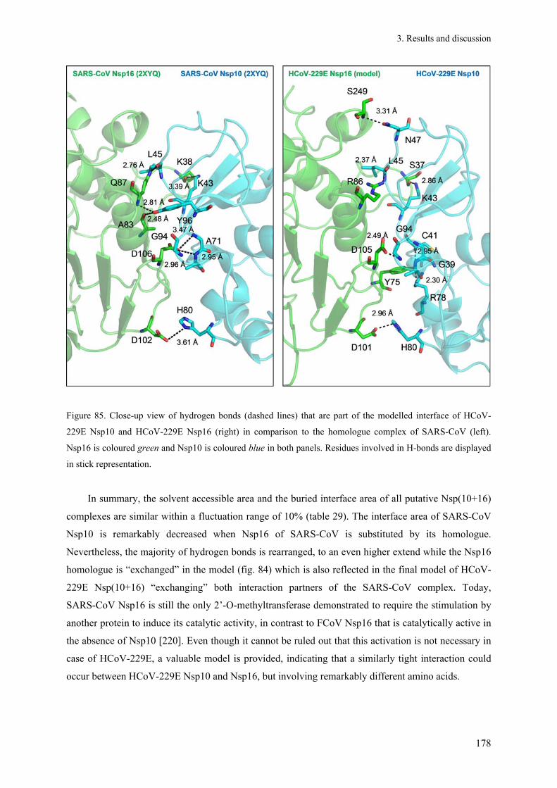
3. Results and discussion
178
Figure 85. Close-up view of hydrogen bonds (dashed lines) that are part of the modelled interface of HCoV-
229E Nsp10 and HCoV-229E Nsp16 (right) in comparison to the homologue complex of SARS-CoV (left).
Nsp16 is coloured green and Nsp10 is coloured blue in both panels. Residues involved in H-bonds are displayed
in stick representation.
In summary, the solvent accessible area and the buried interface area of all putative Nsp(10+16)
complexes are similar within a fluctuation range of 10% (table 29). The interface area of SARS-CoV
Nsp10 is remarkably decreased when Nsp16 of SARS-CoV is substituted by its homologue.
Nevertheless, the majority of hydrogen bonds is rearranged, to an even higher extend while the Nsp16
homologue is “exchanged” in the model (fig. 84) which is also reflected in the final model of HCoV-
229E Nsp(10+16) “exchanging” both interaction partners of the SARS-CoV complex. Today,
SARS-CoV Nsp16 is still the only 2’-O-methyltransferase demonstrated to require the stimulation by
another protein to induce its catalytic activity, in contrast to FCoV Nsp16 that is catalytically active in
the absence of Nsp10 [220]. Even though it cannot be ruled out that this activation is not necessary in
case of HCoV-229E, a valuable model is provided, indicating that a similarly tight interaction could
occur between HCoV-229E Nsp10 and Nsp16, but involving remarkably different amino acids.

4. Conclusion
179
4. Conclusion Addressing the question whether coronaviral pre-processed polyproteins, e.g. pp710, pp79 and
pp78, already act as functional (preformed) building blocks of the RTC, it was shown in this study that
polymerase activity, secondary structure and oligomerization characteristics detected for the precursor
states remain largely comparable to that of the mature Nsps. The observation that a mutation within
the GXXXG-motif, important for dimerization of mature Nsp9 [102; 104], is dispensable for the
oligomerization of pp79 and pp710 indicates that this dimerization site is uncovered not before Nsp9
is N-terminally processed. Considering that the dimerization of Nsp9 is essential for viral replication
[104], this observation could explain why processing of pp89 is not dispensable in vivo, as observed
by Deming et al. [107]. Nevertheless, cleavage of the respective site was determined to be comparably
slow in agreement with the relatively high KD-value of the inhibitory peptide derived from the Nsp8-
Nsp9 cleavage site and with previous studies [65; 191]. In contrast, cleavage of pp910 is dispensable
for viability of MHV in vivo [107], corresponding to the observation that Nsp9 and Nsp10 do not share
a joint binding interface that could, for instance, impair dimerization of Nsp9. Dimerization of SARS-
CoV pp910 was observed by Z. J. Miknis (dissertation, State University of New York, unpublished) in
a SAXS experiment. However, in light of the rapid processing of pp910 by the main protease and the
missing interaction of both mature Nsps, the physiological impact of the precursor pp910 is most
likely rather low or absent.
As observed for other enzymatic activities of mature Nsps so far, the detected polymerase
activity, i.e. at least the nucleotide ligand epitopes determined by STD-NMR spectroscopy, appears to
be highly conserved for polyproteins of SARS-CoV and of the distantly related Alphacoronavirus
HCoV-229E. A high level of conservation of the SAM and RNA binding site of HCoV-229E Nsp16
and Nsp10 and its complex is also indicated, compared to the reported SARS-CoV Nsp(10+16)
complex [112; 114]. Despite the known degree of sequence variability at the interface, conserved
interaction epitopes are expected for the building blocks of the RTC of different coronaviruses, as
widely suggested in this study for Nsp10 and Nsp16. Since the overall fold of mammalian and viral
methyltransferases, e.g. Nsp16 is highly conserved, it seems to be more promising in terms of
developing antiviral compounds to focus on Nsp10 and its binding interfaces, instead to interfere with
the activity of Nsp16 itself, as suggested by [181]. Along with other putative regulatory functions of
Nsp10 [117; 118] this statement highlights an important role of Nsp10 in coronavirus research.
In terms of oligomerization it was not possible to verify the formation of a specific
hexadecameric 8:8-Nsp(7+8) complex, which was reported by [98] in the context of a high-resolution
crystal structure. However, smaller hetero-oligomers exist in solution, perhaps in addition to a
hexadecamer. Therefore, the significant different oligomerization states identified for homologue

4. Conclusion
180
complexes strongly depend on the applied method for investigation and the associated (artificial)
conditions. A dodecameric state of Nsp10 composed of trimers observed within a protein crystal
structure [131], for example, is in contradiction to all results obtained by analysing isolated Nsp10 in
solution. In a wide frame of experimental conditions Nsp10 was demonstrated to be monomeric, while
a comparably large oligomer of the Nsp(10+16) complex is sterically improbable. Different high
molecular weight oligomers observed for pp710 were highly heterogeneous and not yet correlated to a
physiological function, beside the possibility to provide an increased stability towards stress in a host
cell, e.g. induced by proteases or temperature. The detected equilibrium of the monomeric and dimeric
state of pp710 appears to be physiologically more relevant. The presented low-resolution model of
pp710 that is strongly supported by mass spectrometry data indicates that the dimerization via the C-
terminal domain of Nsp8 appears to be conserved for pp710 and pp78 during maturation of Nsps as
well as for Alpha- and Betacoronaviruses. In fact, a significant flexibility of the N-terminus of the
Nsp8 domain within pp710 and pp78 is strongly suggested.
Finally, specific interactions of SARS-CoV main protease with pp710 cleavage sites lacking the
crucial glutamine in P1 position were observed. However, even though both proteins can form a
homodimer there is no significant formation of specific high-order oligomers of the protease-
polyprotein complex, probably influenced by a dependence on the protein concentration, by an
asymmetric fold of the protomers within a dimer [52] or sterical limitations. Based on reports
concerning the monomer-dimer equilibrium of SARS-CoV Mpro [48; 132] it is conceivable that the
interaction is at least partly a 1:1 interaction involving different native full-length substrates – like in a
set of mass spectrometry experiments.
The major aim of future studies will still be the elucidation of high-resolution structures of
precursor states of coronaviral Nsps. Probably involving novel crystallographic techniques, both
pp710 and partly processed fragments as well as polyprotein-Mpro-complexes mentioned above
represent important targets to confirm the low-resolution models and the structural and functional
suggestions presented in this study. A clarification of further Nsp-Nsp-interactions and intra-
polyprotein-domain-interactions will broaden the basis of structural knowledge to understand the
architecture of the coronaviral RTC complex and to develop perspectives and strategies to specifically
interfere with its formation. In addition, next to the well-established attempts to target the coronaviral
main protease Nsp5 for drug development, also Nsp10 appears to be an interesting target for
modulation of the methyltransferase activity of its interaction partner Nsp16. In this context, additional
efforts might be promising to target oligomeric states of Nsps or precursor proteins by dimerization
inhibitors and to investigate the effect on virus replication. A mechanism to inhibit the essential
dimerization of the SARS-CoV main protease by a peptide was previously described [221].

181
5. Summary Coronaviruses (CoVs) comprise a group of enveloped (-)ssRNA viruses. SARS-CoV and the
currently emerging MERS-CoV characterized by a fatality rate of around 50% are considered to be a
major world-wide health issue with high economic impact. Although most coronaviral infections are
comparably harmless for humans (e.g. for HCoV-229E), SARS-CoV and MERS-CoV clearly
demonstrated that severe respiratory diseases are likely to be spread by novel highly pathogenic
coronavirus species in the future, infecting various animals and humans. The coronaviral main
protease has been identified as a suitable target to treat infections, but such compounds did not reach
the market yet. A large hypothetical transcriptase/replicase complex (RTC) of coronaviruses is
assumed to be another promising drug target, even though its formation remains poorly understood so
far. It is established that the RTC essentially involves several mature non-structural-proteins (Nsps)
released from the primary polyprotein translation products of the viral RNA by viral proteases, while
the impact of their sequentially cleaved polyprotein-precursors is unknown.
Addressing important questions of structure, oligomeric composition and function of polyprotein
precursors that may act as putative RTC building blocks, the C-terminus of SARS-CoV polyprotein 1a
comprising Nsp7-10 (pp710) was recombinantly produced in E. coli and purified. The homologue
protein of the distantly related Alphacoronavirus HCoV-229E that causes common cold was purified
for comparison as well. In addition, specific fragments of these large pp710 molecules that were
observed along the pathway of processing by a viral main protease in vitro were purified to allow a
structural comparison with incompletely cleaved polyprotein intermediates and mature non-structural
proteins. For the first time, a complex cleavage reaction of a viral polyprotein was monitored at high
mass accuracy by mass spectrometry, revealing for example a 2:2-interaction state of mature SARS-
CoV Nsp7 and Nsp8. It was shown that RNA-dependent RNA polymerase activity of Nsp8 does not
require a hexadecameric interaction with Nsp7, which was previously reported. Nsp8 is capable of de
novo synthesis of RNA fragments (> 10 nt), even in the precursor state covalently linked to Nsp7 and
Nsp9. The binding epitope of nucleotides resulting in catalytic activity was characterized for two Nsp8
precursors by saturation transfer difference NMR spectroscopy. The observation of catalytic activity
and interaction with nucleic acids promotes the idea of functionally relevant polyproteins acting as
RTC building blocks. Furthermore, size exclusion-chromatography and small-angle X-ray scattering
(SAXS) demonstrate a concentration-dependant oligomerization of SARS-CoV and HCoV-229E
pp710, which is hypothesized to represent a regulatory mechanism during coronavirus infection. A
low-resolution structural model for the domain interactions within the monomer of pp710 as well as
for the intermolecular dimer interactions is proposed, verified by SAXS data of truncated variants and
mass spectrometry of cross-linking experiments. Based on SAXS data it is further suggested that the
interaction of Nsp7 and Nsp8 is preformed in the “polyprotein state“, without significant

182
rearrangements during proteolytic maturation, exploiting a putative flexibility of the N-terminus of
Nsp8 to interact with the C-terminus of Nsp7.
For proteolytic processing of a polyprotein, coronaviral main proteases require a glutamine in P1-
position of the recognition site. Exchanging this essential residue by asparagine inhibits the cleavage
completely, but interestingly still allows a strong interaction with the active centre of the protease.
This state of interaction, likely sharing structural aspects with the native processing pathway in vivo,
was investigated involving full-length polyprotein substrates. Peptides derived from main protease
recognition sites carrying a P1-position mutation actually displayed inhibition of the protease activity;
the lowest KD-value determined by SPR spectroscopy is in the low µM range. The interaction was
verified by native mass spectrometry. Consequently, these peptides comprise a starting point for novel
main protease inhibitors.
The high-resolution structure of HCoV-229E Nsp10 was solved by X-ray crystallography,
comprising the first Nsp10 structure of an Alphacoronavirus. As demonstrated, Nsp10 containing two
highly conserved and occupied Zn2+-binding sites is monomeric in solution. Interaction with Nsp7,
Nsp8 or Nsp9 was not observed in a wide frame of experimental conditions. In silico modelling
predicts an interaction with HCoV-229E Nsp16 and therefore the modulation of the Nsp16 2’-O-
methyltransferase activity highly similar to that reported for SARS-CoV Nsp10 and Nsp16. Sequence
variations within the interface are compensated by establishing alternative interactions.
In summary, pp710 as well as its partly processed and full processed maturation products that are
assumed to constitute building blocks of the essential coronaviral RTC have been characterized in
terms of function and structure applying state-of-the-art methods in structural biology. The direct
comparison of equivalent proteins from Alphacoronavirus HCoV-229E and Betacoronavirus SARS-
CoV overall revealed a high structural and functional conservation, despite the distant relationship
within the Coronaviridae which enlightens conserved key-events in the process of RTC-formation of
Alpha- and Betacoronaviruses. Significant differences were only detected in the tertiary structure of
pp710 and pp78, according to SAXS.

183
6. Zusammenfassung
Coronaviren (CoV) sind membranumhüllte (-)ssRNA Viren, die Menschen bzw. verschiedene
Tiere infizieren und in der Regel eher milde Erkältungskrankheiten auslösen. Das SARS-CoV und das
sich aktuell ausbreitende MERS-CoV, welches bislang eine Sterblichkeitsrate von etwa 50% aufweist,
repräsentieren jedoch Beispiele, die ein ernstes weltumspannendes Gesundheitsproblem darstellen.
Neben den vielen bekannten nicht-pathogenen Coronaviren, wie z.B. HCoV-229E, müssen in Zukunft
auch weitere Ausbrüche von schweren Atemwegserkrankungen befürchtet werden, die durch neuartige
hoch-pathogene Coronaviren, die den Wirt wechseln, ausgelöst werden. Die coronavirale Haupt-
Protease wurde bereits als effektives Ziel zur Behandlung von Infektionen identifiziert, bisher haben
derartige Medikamente allerdings noch keine Marktreife erlangt. Eine weitere perspektivische
Zielstruktur könnte der essentielle Transkriptase/Replikase-Komplex (RTC) darstellen, dessen
Existenz und Aufbau unter anderem aus viralen Nicht-Struktur-Proteinen (Nsp) postuliert wird. Die
genaue Zusammensetzung des RTC und vor allem die Bedeutung der primären Translationsprodukte
des Virus, also der Polyproteine, aus denen durch virale Proteasen zunächst teilweise prozessierte
Intermediate und final die reifen Nsps sequentiell freigesetzt werden, sind derzeit jedoch nahezu
vollständig unbekannt.
Um wichtige Fragestellungen bezüglich der Struktur, der Stöchiometrie von oligomeren
Zuständen sowie der Funktion von Polyprotein-Precursorn, die als mögliche Bausteine des RTC
fungieren können, wurde ein großer Teil des C-Terminus des SARS-CoV Polyproteins 1a, bestehend
aus den vier Domänen Nsp7-Nsp10 (pp710), rekombinant in E. coli produziert und gereinigt. Das
homologe Protein des entfernt verwandten Erkältungserregers HCoV-229E wurde für vergleichende
Untersuchungen ebenfalls hergestellt und gereinigt. Zusätzlich wurden spezifische Fragmente isoliert,
die während der proteolytischen Prozessierung von pp710 durch die virale Haupt-Protease
nachgewiesen wurden, um einen direkten Vergleich mit unvollständig prozessierten Polyprotein-
Intermediaten und reifen Nsps zu ermöglichen. Meines Wissens wurde zum ersten Mal eine solche
komplexe Prozessierungsreaktion eines viralen Polyproteins mit hoher Massengenauigkeit durch
native Massenspektrometrie verfolgt, wodurch unter Anderem ein 2:2-Interaktionszustand von SARS-
CoV Nsp7 und der RNA-abhängigen RNA-Polymerase Nsp8 in Lösung identifiziert werden konnte.
Weiterhin wurde nachgewiesen, dass die Polymerase-Aktivität nicht zwingend einen hexadecameren
Zustand von SARS-CoV Nsp7 und Nsp8 erfordert, der zuvor postuliert wurde. Bereits im Polyprotein-
Zustand vor der N- und C-terminalen Prozessierung ist die Nsp8-Domäne in der Lage, größere RNA-
Fragmente (> 10 nt) zu synthetisieren. Das Bindungsepitop von Nukleotiden an zwei Nsp8-
Vorläuferproteine wurde mittels Sättigungs-Transfer-Differenz NMR Spektroskopie charakterisiert.
Der Nachweis einer Interaktion mit verschiedenen Nukleinsäuren und der katalytischen Aktivität von
Polyproteinen bekräftigt die Hypothese, dass unvollständig prozessierte Polyproteine bereits
funktionelle Bausteine des RTC mit „vorgeformten“ Struktureigenschaften sein können. Weiterhin

184
wurde durch Größen-Ausschluss-Chromatographie (SEC) und Röntgen-Kleinwinkel-Beugung
(SAXS) eine konzentrationsabhängige Oligomerisierung von SARS-CoV und HCoV-229E pp710
nachgewiesen, was möglicherweise einen Regulationsmechanismus während der Coronavirus-
Infektion darstellen könnte. Ein strukturelles Modell der Inter-Domänen-Interaktionen im pp710-
Monomer sowie der intermolekularen Dimer-Interaktionen wurde mit niedriger Auflösung erstellt und
basierend auf SAXS- und Massenspektrometrie-Daten verifiziert. Dieses Modell sowie das SAXS-
Modell von pp78 (Polyprotein Nsp7-Nsp8) legen nahe, dass die Interaktion von „reifem“ Nsp7 und
Nsp8 im Polyprotein-Stadium bereits vorgeformt ist, ohne dass während der Prozessierung
signifikante Umlagerungen von Nsp7 erforderlich wären. Bei konserviertem Bindungsepitop kann die
im Polyprotein-Stadium erforderlich kovalente Verknüpfung von Nsp7 und Nsp8 durch die postulierte
Flexibilität des N-Terminus von Nsp8 ermöglicht werden.
Die proteolytische Prozessierung des Polyproteins durch die coronavirale Haupt-Protease
erfordert ein Glutamin in P1-Position der Erkennungsstelle. Ein Austausch dieses essentiellen Restes
gegen Asparagin inhibiert die Katalyse vollständig, erlaubt aber interessanterweise noch immer eine
starke Interaktion von Protease und Substrat. Dieser Interaktionszustand, der wahrscheinlich
strukturelle Aspekte des nativen Prozessierungsablaufs in vivo widerspiegelt, wurde mit vollständigen
Polyprotein-Substraten mittels SAXS-Technik charakterisiert. Peptide mit einer Mutation in P1-
Position, die von den nativen Protease-Schnittstellen abgeleitet wurden, inhibierten die Protease. Der
niedrigste durch SPR-Spektroskopie bestimmte KD-Wert liegt im niedrigen µM-Bereich. Die
Interaktion wurde durch Massenspektrometrie bestätigt. Folglich repräsentieren diese Peptide einen
weiteren Ansatzpunkt zur Entwicklung neuartiger Haupt-Protease-Inhibitoren.
Des Weiteren wird mit der röntgenkristallographischen Aufklärung der Struktur des HCoV-229E
Nsp10, einem vielseitigen regulatorischen coronaviralen Protein, die erste Nsp10-Struktur eines
Alphacoronavirus präsentiert. Nsp10 beinhaltet zwei hochkonservierte und besetzte Zn2+-
Bindungsstellen und weist in Lösung eindeutig einen monomeren Zustand auf. Unter verschiedensten
experimentellen Bedingungen konnte keine Interaktion mit reifem Nsp7, Nsp8 oder Nsp9
nachgewiesen werden. Durch in silico Modellierung wurde eine Interaktion von Nsp10 mit HCoV-
229E Nsp16 und somit eine Regulation der 2’-O-Methyltransferase-Aktivität von Nsp16 entsprechend
der für SARS-CoV berichteten vorhergesagt. Sequenzvariationen im Bereich der Interaktionsflächen
werden durch Etablierung alternativer Wechselwirkungen kompensiert.
Insgesamt wurde das coronavirale Polyprotein pp710 sowie teilweise und auch vollständig
prozessierte Spaltprodukte, die als unterschiedliche „Bausteine“ des essentiellen RTC postuliert
werden, bezüglich ihrer funktionellen und strukturellen Eigenschaften charakterisiert. Der direkte
Vergleich entsprechender Proteine des Alphacoronavirus HCoV-229E und des Betacoronavirus

185
SARS-CoV zeigte generell ein hohes Ausmaß an funktioneller und struktureller Konservierung, trotz
der Entfernung beider Gruppen innerhalb der Coronavirus-Familie. Signifikante Unterschiede
beschränken sich auf die Tertiärstruktur von pp710 und pp78, basierend auf SAXS-Daten.

186
7. References
[1] Fauquet, CM.; Fargette, D. International Committee on Taxonomy of Viruses and the 3,142 unassigned species. Virology journal. 2005. 2. 64.
[2] den Boon, J A; Snijder, EJ.; Chirnside, ED.; de Vries, A A; Horzinek, MC.; Spaan, WJ. Equine arteritis virus is not a togavirus but belongs to the coronaviruslike superfamily. Journal of virology. 1991. 65. 6. 2910–2920.
[3] Snijder, EJ.; Bredenbeek, PJ.; Dobbe, JC.; Thiel, V; Ziebuhr, J; Poon, Leo L M; Guan, Y; Rozanov, M; Spaan, Willy J M; Gorbalenya, AE. Unique and conserved features of genome and proteome of SARS-coronavirus, an early split-off from the coronavirus group 2 lineage. Journal of molecular biology. 2003. 331. 5. 991–1004.
[4] Tyrrell, DA.; Bynoe, ML. Cultivation Of A Novel Type Of Common-Cold Virus In Organ Cultures. British medical journal. 1965. 1. 5448. 1467–1470.
[5] Peiris, J S M; Lai, ST.; Poon, L L M; Guan, Y; Yam, L Y C; Lim, W; Nicholls, J; Yee, W K S; Yan, WW.; Cheung, MT.; Cheng, V C C; Chan, KH.; Tsang, D N C; Yung, R W H; Ng, TK.; Yuen, KY. Coronavirus as a possible cause of severe acute respiratory syndrome. Lancet. 2003. 361. 9366. 1319–1325.
[6] Donnelly, CA.; Ghani, AC.; Leung, GM.; Hedley, AJ.; Fraser, C; Riley, S; Abu-Raddad, LJ.; Ho, L; Thach, T; Chau, P; Chan, K; Lam, T; Tse, L; Tsang, T; Liu, S; Kong, James H B; Lau, Edith M C; Ferguson, NM.; Anderson, RM. Epidemiological determinants of spread of causal agent of severe acute respiratory syndrome in Hong Kong. Lancet. 2003. 361. 9371. 1761–1766.
[7] Guan, Y; Zheng, BJ.; He, YQ.; Liu, XL.; Zhuang, ZX.; Cheung, CL.; Luo, SW.; Li, PH.; Zhang, LJ.; Guan, YJ.; Butt, KM.; Wong, KL.; Chan, KW.; Lim, W; Shortridge, KF.; Yuen, KY.; Peiris, J S M; Poon, L L M. Isolation and characterization of viruses related to the SARS coronavirus from animals in southern China. Science (New York, N.Y.). 2003. 302. 5643. 276–278.
[8] Lim, PLian; Lee, THong; Rowe, EK. Middle East Respiratory Syndrome coronavirus (MERS CoV): Update 2013. Current infectious disease reports. 2013.
[9] van der Hoek, Lia; Pyrc, K; Jebbink, MF.; Vermeulen-Oost, W; Berkhout, Ron J M; Wolthers, KC.; Wertheim-van Dillen, Pauline M E; Kaandorp, J; Spaargaren, J; Berkhout, B. Identification of a new human coronavirus. Nature medicine. 2004. 10. 4. 368–373.
[10] Rottier, Peter J M; Nakamura, K; Schellen, P; Volders, H; Haijema, BJan. Acquisition of macrophage tropism during the pathogenesis of feline infectious peritonitis is determined by mutations in the feline coronavirus spike protein. Journal of virology. 2005. 79. 22. 14122–14130.
[11] Stadler, K; Masignani, V; Eickmann, M; Becker, S; Abrignani, S; Klenk, H; Rappuoli, R. SARS--beginning to understand a new virus. Nature reviews. Microbiology. 2003. 1. 3. 209–218.
[12] Larson, HE.; Reed, SE.; TYRRELL, DA. Isolation of rhinoviruses and coronaviruses from 38 colds in adults. Journal of medical virology. 1980. 5. 3. 221–229.
[13] Pene, F; Merlat, A; Vabret, A; Rozenberg, F; Buzyn, A; Dreyfus, F; Cariou, A; Freymuth, F; Lebon, P. Coronavirus 229E-related pneumonia in immunocompromised patients. Clinical infectious diseases : an official publication of the Infectious Diseases Society of America. 2003. 37. 7. 929–932.
[14] Marra, MA.; Jones, Steven J M; Astell, CR.; Holt, RA.; Brooks-Wilson, A; Butterfield, Yaron S N; Khattra, J; Asano, JK.; Barber, SA.; Chan, SY.; Cloutier, A; Coughlin, SM.; Freeman, D; Girn, N; Griffith, OL.; Leach, SR.; Mayo, M; McDonald, H; Montgomery, SB.; Pandoh, PK.;

187
Petrescu, AS.; Robertson, AGordon; Schein, JE.; Siddiqui, A; Smailus, DE.; Stott, JM.; Yang, GS.; Plummer, F; Andonov, A; Artsob, H; Bastien, N; Bernard, K; Booth, TF.; Bowness, D; Czub, M; Drebot, M; Fernando, L; Flick, R; Garbutt, M; Gray, M; Grolla, A; Jones, S; Feldmann, H; Meyers, A; Kabani, A; Li, Y; Normand, S; Stroher, U; Tipples, GA.; Tyler, S; Vogrig, R; Ward, D; Watson, B; Brunham, RC.; Krajden, M; Petric, M; Skowronski, DM.; Upton, C; Roper, RL. The Genome sequence of the SARS-associated coronavirus. Science (New York, N.Y.). 2003. 300. 5624. 1399–1404.
[15] Raman, S; Bouma, P; Williams, GD.; Brian, DA. Stem-loop III in the 5' untranslated region is a cis-acting element in bovine coronavirus defective interfering RNA replication. Journal of virology. 2003. 77. 12. 6720–6730.
[16] Goebel, SJ.; Miller, TB.; Bennett, CJ.; Bernard, KA.; Masters, PS. A hypervariable region within the 3' cis-acting element of the murine coronavirus genome is nonessential for RNA synthesis but affects pathogenesis. Journal of virology. 2007. 81. 3. 1274–1287.
[17] Spagnolo, JF.; Hogue, BG. Host protein interactions with the 3' end of bovine coronavirus RNA and the requirement of the poly(A) tail for coronavirus defective genome replication. Journal of virology. 2000. 74. 11. 5053–5065.
[18] Sawicki, SG.; Sawicki, DL.; Siddell, SG. A Contemporary View of Coronavirus Transcription. Journal of Virology. 2006. 81. 1. 20–29.
[19] Enjuanes, L; Almazán, F; Sola, I; Zuñiga, S. Biochemical aspects of coronavirus replication and virus-host interaction. Annual review of microbiology. 2006. 60. 211–230.
[20] Thiel, V; Ivanov, KA.; Putics, A; Hertzig, T; Schelle, B; Bayer, S; Weissbrich, B; Snijder, EJ.; Rabenau, H; Doerr, HWilhelm; Gorbalenya, AE.; Ziebuhr, J. Mechanisms and enzymes involved in SARS coronavirus genome expression. The Journal of general virology. 2003. 84. Pt 9. 2305–2315.
[21] Morales, L; Mateos-Gomez, PA.; Capiscol, C; del Palacio, L; Enjuanes, L; Sola, I. Transmissible gastroenteritis coronavirus genome packaging signal is located at the 5' end of the genome and promotes viral RNA incorporation into virions in a replication independent process. Journal of Virology. 2013.
[22] Escors, D; Ortego, J; Laude, H; Enjuanes, L. The membrane M protein carboxy terminus binds to transmissible gastroenteritis coronavirus core and contributes to core stability. Journal of virology. 2001. 75. 3. 1312–1324.
[23] Lu, G; Hu, Y; Wang, Q; Qi, J; Gao, F; Li, Y; Zhang, Y; Zhang, W; Yuan, Y; Bao, J; Zhang, B; Shi, Y; Yan, J; Gao, GF. Molecular basis of binding between novel human coronavirus MERS-CoV and its receptor CD26. Nature. 2013. 500. 7461. 227–231.
[24] Bosch, BJan; de Haan, Cornelis A M; Rottier, Peter J M. Coronavirus spike glycoprotein, extended at the carboxy terminus with green fluorescent protein, is assembly competent. Journal of virology. 2004. 78. 14. 7369–7378.
[25] Wang, S; Guo, F; Liu, K; Wang, H; Rao, S; Yang, P; Jiang, C. Endocytosis of the receptor-binding domain of SARS-CoV spike protein together with virus receptor ACE2. Virus research. 2008. 136. 1-2. 8–15.
[26] Pewe, L; Zhou, H; Netland, J; Tangudu, C; Olivares, H; Shi, L; Look, D; Gallagher, T; Perlman, S. A severe acute respiratory syndrome-associated coronavirus-specific protein enhances virulence of an attenuated murine coronavirus. Journal of virology. 2005. 79. 17. 11335–11342.
[27] Tangudu, C; Olivares, H; Netland, J; Perlman, S; Gallagher, T. Severe acute respiratory syndrome coronavirus protein 6 accelerates murine coronavirus infections. Journal of virology. 2007. 81. 3. 1220–1229.
[28] Law, P. T. W. The 3a protein of severe acute respiratory syndrome-associated coronavirus induces apoptosis in Vero E6 cells. Journal of General Virology. 2005. 86. 7. 1921–1930.

188
[29] Yuan, X; Wu, J; Shan, Y; Yao, Z; Dong, B; Chen, B; Zhao, Z; Wang, S; Chen, J; Cong, Y. SARS coronavirus 7a protein blocks cell cycle progression at G0/G1 phase via the cyclin D3/pRb pathway. Virology. 2006. 346. 1. 74–85.
[30] van Hemert, Martijn J; de Wilde, Adriaan H; Gorbalenya, AE.; Snijder, EJ. The in vitro RNA synthesizing activity of the isolated arterivirus replication/transcription complex is dependent on a host factor. The Journal of biological chemistry. 2008. 283. 24. 16525–16536.
[31] Su, M; Chang, C; Chu, C; Tsai, C; Chang, K. An atypical RNA pseudoknot stimulator and an upstream attenuation signal for -1 ribosomal frameshifting of SARS coronavirus. Nucleic acids research. 2005. 33. 13. 4265–4275.
[32] Garcia (Ed.) 2012 – Viral Genomes - Molecular Structure, Diversity, Gene Expression Mechanisms and Host-Virus Interactions, InTech, ISBN: 978-953-51-0098-0
[33] Knoops, K; Kikkert, M; Worm, Sjoerd H E van den; Zevenhoven-Dobbe, JC.; van der Meer, Yvonne; Koster, AJ.; Mommaas, AMieke; Snijder, EJ. SARS-coronavirus replication is supported by a reticulovesicular network of modified endoplasmic reticulum. PLoS biology. 2008. 6. 9. e226.
[34] Angelini, MM.; Akhlaghpour, M; Neuman, BW.; Buchmeier, MJ. Severe acute respiratory syndrome coronavirus nonstructural proteins 3, 4, and 6 induce double-membrane vesicles. mBio. 2013. 4. 4.
[35] Snijder, EJ.; van Tol, H; Roos, N; Pedersen, KW. Non-structural proteins 2 and 3 interact to modify host cell membranes during the formation of the arterivirus replication complex. The Journal of general virology. 2001. 82. Pt 5. 985–994.
[36] Clementz, MA.; Kanjanahaluethai, A; O'Brien, TE.; Baker, SC. Mutation in murine coronavirus replication protein nsp4 alters assembly of double membrane vesicles. Virology. 2008. 375. 1. 118–129.
[37] Cho, JKeun; Curtis-Long, MJ.; Lee, KHo; Kim, DWook; Ryu, HWon; Yuk, HJoo; Park, KHun. Geranylated flavonoids displaying SARS-CoV papain-like protease inhibition from the fruits of Paulownia tomentosa. Bioorganic & medicinal chemistry. 2013. 21. 11. 3051–3057.
[38] Tan, J; Kusov, Y; Mutschall, D; Tech, S; Nagarajan, K; Hilgenfeld, R; Schmidt, CL. The "SARS-unique domain" (SUD) of SARS coronavirus is an oligo(G)-binding protein. Biochemical and biophysical research communications. 2007. 364. 4. 877–882.
[39] Tan, J; Vonrhein, C; Smart, OS.; Bricogne, G; Bollati, M; Kusov, Y; Hansen, G; Mesters, JR.; Schmidt, CL.; Hilgenfeld, R. The SARS-unique domain (SUD) of SARS coronavirus contains two macrodomains that bind G-quadruplexes. PLoS pathogens. 2009. 5. 5. e1000428.
[40] Kiemer, L; Lund, O; Brunak, S; Blom, N. Coronavirus 3CLpro proteinase cleavage sites: possible relevance to SARS virus pathology. BMC bioinformatics. 2004. 5. 72.
[41] Denison, MR.; Zoltick, PW.; Hughes, SA.; Giangreco, B; Olson, AL.; Perlman, S; Leibowitz, JL.; Weiss, SR. Intracellular processing of the N-terminal ORF 1a proteins of the coronavirus MHV-A59 requires multiple proteolytic events. Virology. 1992. 189. 1. 274–284.
[42] Denison, MR.; Hughes, SA.; Weiss, SR. Identification and characterization of a 65-kDa protein processed from the gene 1 polyprotein of the murine coronavirus MHV-A59. Virology. 1995. 207. 1. 316–320.
[43] Lu, XT.; Sims, AC.; Denison, MR. Mouse hepatitis virus 3C-like protease cleaves a 22-kilodalton protein from the open reading frame 1a polyprotein in virus-infected cells and in vitro. Journal of virology. 1998. 72. 3. 2265–2271.
[44] Schiller, JJ.; Kanjanahaluethai, A; Baker, SC. Processing of the coronavirus MHV-JHM polymerase polyprotein: identification of precursors and proteolytic products spanning 400 kilodaltons of ORF1a. Virology. 1998. 242. 2. 288–302.

189
[45] Schiller, JJ.; Baker, SC. Maturation of the polymerase polyprotein of the coronavirus MHV strain JHM involves a cascade of proteolytic processing events. Advances in experimental medicine and biology. 1998. 440. 135–139.
[46] Heusipp, G; Grötzinger, C; Herold, J; Siddell, SG.; Ziebuhr, J. Identification and subcellular localization of a 41 kDa, polyprotein 1ab processing product in human coronavirus 229E-infected cells. The Journal of general virology. 1997. 78 (Pt 11). 2789–2794.
[47] Hagemeijer, MC.; Rottier, Peter J M; de Haan, Cornelis A M. Biogenesis and dynamics of the coronavirus replicative structures. Viruses. 2012. 4. 11. 3245–3269.
[48] Chen, S; Jonas, F; Shen, C; Hilgenfeld, R; Higenfeld, R. Liberation of SARS-CoV main protease from the viral polyprotein: N-terminal autocleavage does not depend on the mature dimerization mode. Protein & cell. 2010. 1. 1. 59–74.
[49] Barrila, J; Bacha, U; Freire, E. Long-range cooperative interactions modulate dimerization in SARS 3CLpro. Biochemistry. 2006. 45. 50. 14908–14916.
[50] Chen, S; Hu, T; Zhang, J; Chen, J; Chen, K; Ding, J; Jiang, H; Shen, X. Mutation of Gly-11 on the dimer interface results in the complete crystallographic dimer dissociation of severe acute respiratory syndrome coronavirus 3C-like protease: crystal structure with molecular dynamics simulations. The Journal of biological chemistry. 2008. 283. 1. 554–564.
[51] Chen, S; Jonas, F; Shen, C; Hilgenfeld, R; Higenfeld, R. Liberation of SARS-CoV main protease from the viral polyprotein: N-terminal autocleavage does not depend on the mature dimerization mode. Protein & cell. 2010. 1. 1. 59–74.
[52] Chen, H; Wei, P; Huang, C; Tan, L; Liu, Y; Lai, L. Only one protomer is active in the dimer of SARS 3C-like proteinase. The Journal of biological chemistry. 2006. 281. 20. 13894–13898.
[53] Yang, H; Yang, M; Ding, Y; Liu, Y; Lou, Z; Zhou, Z; Sun, L; Mo, L; Ye, S; Pang, H; Gao, GF.; Anand, K; Bartlam, M; Hilgenfeld, R; Rao, Z. The crystal structures of severe acute respiratory syndrome virus main protease and its complex with an inhibitor. Proceedings of the National Academy of Sciences of the United States of America. 2003. 100. 23. 13190–13195.
[54] Hilgenfeld, R; Anand, K; Mesters, JR.; Rao, Z; Shen, X; Jiang, H; Tan, J; Verschueren, Koen H G. Structure and dynamics of SARS coronavirus main proteinase (Mpro). Advances in experimental medicine and biology. 2006. 581. 585–591.
[55] Steuber, H; Hilgenfeld, R. Recent advances in targeting viral proteases for the discovery of novel antivirals. Current topics in medicinal chemistry. 2010. 10. 3. 323–345.
[56] Shi, J; Sivaraman, J; Song, J. Mechanism for controlling the dimer-monomer switch and coupling dimerization to catalysis of the severe acute respiratory syndrome coronavirus 3C-like protease. Journal of virology. 2008. 82. 9. 4620–4629.
[57] Zhang, S; Zhong, N; Xue, F; Kang, X; Ren, X; Chen, J; Jin, C; Lou, Z; Xia, B. Three-dimensional domain swapping as a mechanism to lock the active conformation in a super-active octamer of SARS-CoV main protease. Protein & cell. 2010. 1. 4. 371–383.
[58] Chou, C; Chang, H; Hsu, W; Lin, T; Lin, C; Chang, G. Quaternary structure of the severe acute respiratory syndrome (SARS) coronavirus main protease. Biochemistry. 2004. 43. 47. 14958–14970.
[59] Chen, S; Chen, L; Tan, J; Chen, J; Du, L; Sun, T; Shen, J; Chen, K; Jiang, H; Shen, X. Severe acute respiratory syndrome coronavirus 3C-like proteinase N terminus is indispensable for proteolytic activity but not for enzyme dimerization. Biochemical and thermodynamic investigation in conjunction with molecular dynamics simulations. The Journal of biological chemistry. 2005. 280. 1. 164–173.
[60] Graziano, V; McGrath, WJ.; Yang, L; Mangel, WF. SARS CoV main proteinase: The monomer-dimer equilibrium dissociation constant. Biochemistry. 2006. 45. 49. 14632–14641.

190
[61] Huang, C; Wei, P; Fan, K; Liu, Y; Lai, L. 3C-like proteinase from SARS coronavirus catalyzes substrate hydrolysis by a general base mechanism. Biochemistry. 2004. 43. 15. 4568–4574.
[62] Solowiej, J; Thomson, JA.; Ryan, K; Luo, C; He, M; Lou, J; Murray, BW. Steady-State and Pre-Steady-State Kinetic Evaluation of Severe Acute Respiratory Syndrome Coronavirus (SARS-CoV) 3CL pro Cysteine Protease: Development of an Ion-Pair Model for Catalysis. Biochemistry. 2008. 47. 8. 2617–2630.
[63] Anand, K; Ziebuhr, J; Wadhwani, P; Mesters, JR.; Hilgenfeld, R. Coronavirus main proteinase (3CLpro) structure: basis for design of anti-SARS drugs. Science (New York, N.Y.). 2003. 300. 5626. 1763–1767.
[64] Goetz, DH.; Choe, Y; Hansell, E; Chen, YT.; McDowell, M; Jonsson, CB.; Roush, WR.; McKerrow, J; Craik, CS. Substrate specificity profiling and identification of a new class of inhibitor for the major protease of the SARS coronavirus. Biochemistry. 2007. 46. 30. 8744–8752.
[65] Chu, LMatthew; Choy, W; Tsai, S; Rao, Z; Ngai, S. Rapid peptide-based screening on the substrate specificity of severe acute respiratory syndrome (SARS) coronavirus 3C-like protease by matrix-assisted laser desorption/ionization time-of-flight mass spectrometry. Protein science : a publication of the Protein Society. 2006. 15. 4. 699–709.
[66] Mandadapu, SRao; Weerawarna, PM.; Prior, AM.; Uy, Roxanne Adeline Z; Aravapalli, S; Alliston, KR.; Lushington, GH.; Kim, Y; Hua, DH.; Chang, K; Groutas, WC. Macrocyclic inhibitors of 3C and 3C-like proteases of picornavirus, norovirus, and coronavirus. Bioorganic & medicinal chemistry letters. 2013. 23. 13. 3709–3712.
[67] Nguyen, Thi Thanh Hanh; Woo, H; Kang, H; van Nguyen, D; Kim, Y; Kim, D; Ahn, S; Xia, Y; Kim, D. Flavonoid-mediated inhibition of SARS coronavirus 3C-like protease expressed in Pichia pastoris. Biotechnology Letters. 2012. 34. 5. 831–838.
[68] Chen, C; Lin, Coney P C; Huang, K; Chen, W; Hsieh, H; Liang, P; Hsu, JT-A. Inhibition of SARS-CoV 3C-like Protease Activity by Theaflavin-3,3'-digallate (TF3). Evidence-based complementary and alternative medicine : eCAM. 2005. 2. 2. 209–215.
[69] Liu, X; Kim, J; Li, Y; Li, J; Liu, F; Chen, X. Tannic acid stimulates glucose transport and inhibits adipocyte differentiation in 3T3-L1 cells. The Journal of nutrition. 2005. 135. 2. 165–171.
[70] Tadera, K; Minami, Y; Takamatsu, K; Matsuoka, T. Inhibition of alpha-glucosidase and alpha-amylase by flavonoids. Journal of nutritional science and vitaminology. 2006. 52. 2. 149–153.
[71] Groot, H de; Rauen, U. Tissue injury by reactive oxygen species and the protective effects of flavonoids. Fundamental & clinical pharmacology. 1998. 12. 3. 249–255.
[72] Thanigaimalai, P; Konno, S; Yamamoto, T; Koiwai, Y; Taguchi, A; Takayama, K; Yakushiji, F; Akaji, K; Chen, S; Naser-Tavakolian, A; Schön, A; Freire, E; Hayashi, Y. Development of potent dipeptide-type SARS-CoV 3CL protease inhibitors with novel P3 scaffolds: Design, synthesis, biological evaluation, and docking studies. European journal of medicinal chemistry. 2013. 68C. 372–384.
[73] Thanigaimalai, P; Konno, S; Yamamoto, T; Koiwai, Y; Taguchi, A; Takayama, K; Yakushiji, F; Akaji, K; Kiso, Y; Kawasaki, Y; Chen, S; Naser-Tavakolian, A; Schön, A; Freire, E; Hayashi, Y. Design, synthesis, and biological evaluation of novel dipeptide-type SARS-CoV 3CL protease inhibitors: structure-activity relationship study. European journal of medicinal chemistry. 2013. 65. 436–447.
[74] Ren, Z; Yan, L; Zhang, N; Guo, Y; Yang, C; Lou, Z; Rao, Z. The newly emerged SARS-Like coronavirus HCoV-EMC also has an “Achilles’ heel”: current effective inhibitor targeting a 3C-like protease. Protein & cell. 2013. 4. 4. 248–250.
191
[75] Xue, X; Yu, H; Yang, H; Xue, F; Wu, Z; Shen, W; Li, J; Zhou, Z; Ding, Y; Zhao, Q; Zhang, XC.; Liao, M; Bartlam, M; Rao, Z. Structures of two coronavirus main proteases: implications for substrate binding and antiviral drug design. Journal of virology. 2008. 82. 5. 2515–2527.
[76] Shie, J; Fang, J; Kuo, T; Kuo, C; Liang, P; Huang, H; Wu, Y; Jan, J; Cheng, YE.; Wong, C. Inhibition of the severe acute respiratory syndrome 3CL protease by peptidomimetic alpha,beta-unsaturated esters. Bioorganic & medicinal chemistry. 2005. 13. 17. 5240–5252.
[77] Lee, C; Kuo, C; Ko, T; Hsu, M; Tsui, Y; Chang, S; Yang, S; Chen, S; Chen, H; Hsu, M; Shih, S; Liang, P; Wang, AH-J. Structural basis of inhibition specificities of 3C and 3C-like proteases by zinc-coordinating and peptidomimetic compounds. The Journal of biological chemistry. 2009. 284. 12. 7646–7655.
[78] Zhu, L; George, S; Schmidt, MF.; Al-Gharabli, SI.; Rademann, J; Hilgenfeld, R. Peptide aldehyde inhibitors challenge the substrate specificity of the SARS-coronavirus main protease. Antiviral research. 2011. 92. 2. 204–212.
[79] Lee, T; Cherney, MM.; Liu, J; James, KEllis; Powers, JC.; Eltis, LD.; James, Michael N G. Crystal structures reveal an induced-fit binding of a substrate-like Aza-peptide epoxide to SARS coronavirus main peptidase. Journal of molecular biology. 2007. 366. 3. 916–932.
[80] Chuck, C; Chen, C; Ke, Z; Chi-Cheong Wan, D; Chow, H; Wong, K. Design, synthesis and crystallographic analysis of nitrile-based broad-spectrum peptidomimetic inhibitors for coronavirus 3C-like proteases. European Journal of Medicinal Chemistry. 2013. 59. 1–6.
[81] Wu, C; King, K; Kuo, C; Fang, J; Wu, Y; Ho, M; Liao, C; Shie, J; Liang, P; Wong, C. Stable benzotriazole esters as mechanism-based inactivators of the severe acute respiratory syndrome 3CL protease. Chemistry & biology. 2006. 13. 3. 261–268.
[82] Jansson, AM. Structure of Alphacoronavirus Transmissible Gastroenteritis Virus nsp1 Has Implications for Coronavirus nsp1 Function and Evolution. Journal of Virology. 2013. 87. 5. 2949–2955.
[83] Lokugamage, KG.; Narayanan, K; Huang, C; Makino, S. Severe Acute Respiratory Syndrome Coronavirus Protein nsp1 Is a Novel Eukaryotic Translation Inhibitor That Represses Multiple Steps of Translation Initiation. Journal of Virology. 2012. 86. 24. 13598–13608.
[84] Yu, K; Ming, Z; Li, Y; Chen, C; Bao, Z; Ren, Z; Liu, B; Tao, W; Rao, Z; Lou, Z. Purification, crystallization and preliminary X-ray analysis of nonstructural protein 2 (nsp2) from avian infectious bronchitis virus. Acta Crystallographica Section F Structural Biology and Crystallization Communications. 2012. 68. 6. 716–719.
[85] Wojdyla, JA.; Manolaridis, I; Snijder, EJ.; Gorbalenya, AE.; Coutard, B; Piotrowski, Y; Hilgenfeld, R; Tucker, PA. Structure of the X (ADRP) domain of nsp3 from feline coronavirus. Acta crystallographica. Section D, Biological crystallography. 2009. 65. Pt 12. 1292–1300.
[86] Yu, M; Lee, J; Lee, JMoo; Kim, Y; Chin, Y; Jee, J; Keum, Y; Jeong, Y. Identification of myricetin and scutellarein as novel chemical inhibitors of the SARS coronavirus helicase, nsP13. Bioorganic & medicinal chemistry letters. 2012. 22. 12. 4049–4054.
[87] Brunn, A von; Teepe, C; Simpson, JC.; Pepperkok, R; Friedel, CC.; Zimmer, R; Roberts, R; Baric, R; Haas, J. Analysis of intraviral protein-protein interactions of the SARS coronavirus ORFeome. PloS one. 2007. 2. 5. e459.
[88] Pan, J; Peng, X; Gao, Y; Li, Z; Lu, X; Chen, Y; Ishaq, M; Liu, D; Dediego, ML.; Enjuanes, L; Guo, D. Genome-wide analysis of protein-protein interactions and involvement of viral proteins in SARS-CoV replication. PloS one. 2008. 3. 10. e3299.
[89] Ito, N; Mossel, EC.; Narayanan, K; Popov, VL.; Huang, C; Inoue, T; Peters, CJ.; Makino, S. Severe acute respiratory syndrome coronavirus 3a protein is a viral structural protein. Journal of virology. 2005. 79. 5. 3182–3186.

192
[90] Shen, S; Lin, P; Chao, Y; Zhang, A; Yang, X; Lim, SGee; Hong, W; Tan, Y. The severe acute respiratory syndrome coronavirus 3a is a novel structural protein. Biochemical and biophysical research communications. 2005. 330. 1. 286–292.
[91] Palaninathan, S.; Bhardwaj, K.; Alcantara, J.M.O.; Guarino, L.; Yi, L.L.; Kao, C.C.; Sacchettini, J. Crystal structure of Nsp15-H234A mutant- Hexamer in asymmetric unit. Journal of Biological Chemistry. 2008. 283. 3655 – 3664.
[92] Bhardwaj, K; Liu, P; Leibowitz, JL.; Kao, CC. The Coronavirus Endoribonuclease Nsp15 Interacts with Retinoblastoma Tumor Suppressor Protein. Journal of Virology. 2012. 86. 8. 4294–4304.
[93] Ahn, D; Choi, J; Taylor, DR.; Oh, J. Biochemical characterization of a recombinant SARS coronavirus nsp12 RNA-dependent RNA polymerase capable of copying viral RNA templates. Archives of Virology. 2012. 157. 11. 2095–2104.
[94] Imbert, I; Guillemot, J; Bourhis, J; Bussetta, C; Coutard, B; Egloff, M; Ferron, F; Gorbalenya, AE.; Canard, B. A second, non-canonical RNA-dependent RNA polymerase in SARS coronavirus. The EMBO journal. 2006. 25. 20. 4933–4942.
[95] Hansen, JL.; Long, AM.; Schultz, SC. Structure of the RNA-dependent RNA polymerase of poliovirus. Structure. 1997. 5. 8. 1109–1122.
[96] Xiao, Y; Ma, Q; Restle, T; Shang, W; Svergun, DI.; Ponnusamy, R; Sczakiel, G; Hilgenfeld, R. Nonstructural proteins 7 and 8 of feline coronavirus form a 2:1 heterotrimer that exhibits primer-independent RNA polymerase activity. Journal of virology. 2012. 86. 8. 4444–4454.
[97] te Velthuis, Aartjan J W; van den Worm, Sjoerd H E; Snijder, EJ. The SARS-coronavirus nsp7+nsp8 complex is a unique multimeric RNA polymerase capable of both de novo initiation and primer extension. Nucleic acids research. 2012. 40. 4. 1737–1747.
[98] Zhai, Y; Sun, F; Li, X; Pang, H; Xu, X; Bartlam, M; Rao, Z. Insights into SARS-CoV transcription and replication from the structure of the nsp7-nsp8 hexadecamer. Nature structural & molecular biology. 2005. 12. 11. 980–986.
[99] Li, S; Zhao, Q; Zhang, Y; Zhang, Y; Bartlam, M; Li, X; Rao, Z. New nsp8 isoform suggests mechanism for tuning viral RNA synthesis. Protein & cell. 2010. 1. 2. 198–204.
[100] Johnson, MA.; Jaudzems, K; Wüthrich, K. NMR Structure of the SARS-CoV Nonstructural Protein 7 in Solution at pH 6.5. Journal of molecular biology. 2010. 402. 4. 619–628.
[101] Ponnusamy, R; Moll, R; Weimar, T; Mesters, JR.; Hilgenfeld, R. Variable oligomerization modes in coronavirus non-structural protein 9. Journal of molecular biology. 2008. 383. 5. 1081–1096.
[102] Sutton, G; Fry, E; Carter, L; Sainsbury, S; Walter, T; Nettleship, J; Berrow, N; Owens, R; Gilbert, R; Davidson, A; Siddell, S; Poon, Leo L M; Diprose, J; Alderton, D; Walsh, M; Grimes, JM.; Stuart, DI. The nsp9 replicase protein of SARS-coronavirus, structure and functional insights. Structure (London, England : 1993). 2004. 12. 2. 341–353.
[103] Ponnusamy, R; Mesters, JR.; Ziebuhr, J; Moll, R; Hilgenfeld, R. Non structural proteins 8 and 9 of human coronavirus 229E. Advances in experimental medicine and biology. 2006. 581. 49–54.
[104] Miknis, ZJ.; Donaldson, EF.; Umland, TC.; Rimmer, RA.; Baric, RS.; Schultz, LWayne. Severe acute respiratory syndrome coronavirus nsp9 dimerization is essential for efficient viral growth. Journal of virology. 2009. 83. 7. 3007–3018.
[105] Webster, G; Genschel, J; Curth, U; Urbanke, C; Kang, C; Hilgenfeld, R. A common core for binding single-stranded DNA: structural comparison of the single-stranded DNA-binding proteins (SSB) from E. coli and human mitochondria. FEBS letters. 1997. 411. 2-3. 313–316.

193
[106] Mapelli, M; Panjikar, S; Tucker, PA. The crystal structure of the herpes simplex virus 1 ssDNA-binding protein suggests the structural basis for flexible, cooperative single-stranded DNA binding. The Journal of biological chemistry. 2005. 280. 4. 2990–2997.
[107] Deming, DJ.; Graham, RL.; Denison, MR.; Baric, RS. Processing of open reading frame 1a replicase proteins nsp7 to nsp10 in murine hepatitis virus strain A59 replication. Journal of virology. 2007. 81. 19. 10280–10291.
[108] Züst, R; Miller, TB.; Goebel, SJ.; Thiel, V; Masters, PS. Genetic interactions between an essential 3' cis-acting RNA pseudoknot, replicase gene products, and the extreme 3' end of the mouse coronavirus genome. Journal of virology. 2008. 82. 3. 1214–1228.
[109] Geng, H; Liu, Y; Chan, W; Lo, AWing-Ip; Au, DMun-Yee; Waye, MMiu-Yee; Ho, Y. The putative protein 6 of the severe acute respiratory syndrome-associated coronavirus: expression and functional characterization. FEBS letters. 2005. 579. 30. 6763–6768.
[110] Lai, MM.; Stohlman, SA. Genome structure of mouse hepatitis virus: comparative analysis by oligonucleotide mapping. Advances in experimental medicine and biology. 1981. 142. 69–82.
[111] Joseph, JS.; Saikatendu, KSingh; Subramanian, V; Neuman, BW.; Brooun, A; Griffith, M; Moy, K; Yadav, MK.; Velasquez, J; Buchmeier, MJ.; Stevens, RC.; Kuhn, P. Crystal structure of nonstructural protein 10 from the severe acute respiratory syndrome coronavirus reveals a novel fold with two zinc-binding motifs. Journal of virology. 2006. 80. 16. 7894–7901.
[112] Decroly, E; Debarnot, C; Ferron, F; Bouvet, M; Coutard, B; Imbert, I; Gluais, L; Papageorgiou, N; Sharff, A; Bricogne, G; Ortiz-Lombardia, M; Lescar, J; Canard, B. Crystal structure and functional analysis of the SARS-coronavirus RNA cap 2'-O-methyltransferase nsp10/nsp16 complex. PLoS pathogens. 2011. 7. 5. e1002059.
[113] Ke, M; Chen, Y; Wu, A; Sun, Y; Su, C; Wu, H; Jin, X; Tao, J; Wang, Y; Ma, X; Pan, J; Guo, D. Short peptides derived from the interaction domain of SARS coronavirus nonstructural protein nsp10 can suppress the 2'-O-methyltransferase activity of nsp10/nsp16 complex. Virus research. 2012. 167. 2. 322–328.
[114] Debarnot, C; Imbert, I; Ferron, F; Gluais, L; Varlet, I; Papageorgiou, N; Bouvet, M; Lescar, J; Decroly, E; Canard, B. Crystallization and diffraction analysis of the SARS coronavirus nsp10-nsp16 complex. Acta crystallographica. Section F, Structural biology and crystallization communications. 2011. 67. Pt 3. 404–408.
[115] Chen, Y; Tao, J; Sun, Y; Wu, A; Su, C; Gao, G; Cai, H; Qiu, S; Wu, Y; Ahola, T; Guo, D. Structure-function analysis of severe acute respiratory syndrome coronavirus RNA cap guanine-N7-methyltransferase. Journal of virology. 2013. 87. 11. 6296–6305.
[116] Chen, P; Jiang, M; Hu, T; Liu, Q; Chen, XS.; Guo, D. Biochemical characterization of exoribonuclease encoded by SARS coronavirus. Journal of biochemistry and molecular biology. 2007. 40. 5. 649–655.
[117] Bouvet, M; Imbert, I; Subissi, L; Gluais, L; Canard, B; Decroly, E. RNA 3'-end mismatch excision by the severe acute respiratory syndrome coronavirus nonstructural protein nsp10/nsp14 exoribonuclease complex. Proceedings of the National Academy of Sciences of the United States of America. 2012. 109. 24. 9372–9377.
[118] Donaldson, EF.; Sims, AC.; Graham, RL.; Denison, MR.; Baric, RS. Murine hepatitis virus replicase protein nsp10 is a critical regulator of viral RNA synthesis. Journal of virology. 2007. 81. 12. 6356–6368.
[119] Daffis, S; Szretter, KJ.; Schriewer, J; Li, J; Youn, S; Errett, J; Lin, T; Schneller, S; Zust, R; Dong, H; Thiel, V; Sen, GC.; Fensterl, V; Klimstra, WB.; Pierson, TC.; Buller, RMark; Gale, M; Shi, P; Diamond, MS. 2'-O methylation of the viral mRNA cap evades host restriction by IFIT family members. Nature. 2010. 468. 7322. 452–456.
[120] Li, Q; Wang, L; Dong, C; Che, Y; Jiang, L; Liu, L; Zhao, H; Liao, Y; Sheng, Y; Dong, S; Ma, S. The interaction of the SARS coronavirus non-structural protein 10 with the cellular oxido-

194
reductase system causes an extensive cytopathic effect. Journal of clinical virology : the official publication of the Pan American Society for Clinical Virology. 2005. 34. 2. 133–139.
[121] Koopmann, R; Cupelli, K; Redecke, L; Nass, K; Deponte, DP.; White, TA.; Stellato, F; Rehders, D; Liang, M; Andreasson, J; Aquila, A; Bajt, S; Barthelmess, M; Barty, A; Bogan, MJ.; Bostedt, C; Boutet, S; Bozek, JD.; Caleman, C; Coppola, N; Davidsson, J; Doak, RBruce; Ekeberg, T; Epp, SW.; Erk, B; Fleckenstein, H; Foucar, L; Graafsma, H; Gumprecht, L; Hajdu, J; Hampton, CY.; Hartmann, A; Hartmann, R; Hauser, G; Hirsemann, H; Holl, P; Hunter, MS.; Kassemeyer, S; Kirian, RA.; Lomb, L; Maia, Filipe R N C; Kimmel, N; Martin, AV.; Messerschmidt, M; Reich, C; Rolles, D; Rudek, B; Rudenko, A; Schlichting, I; Schulz, J; Seibert, MMarvin; Shoeman, RL.; Sierra, RG.; Soltau, H; Stern, S; Strüder, L; Timneanu, N; Ullrich, J; Wang, X; Weidenspointner, G; Weierstall, U; Williams, GJ.; Wunderer, CB.; Fromme, P; Spence, John C H; Stehle, T; Chapman, HN.; Betzel, C; Duszenko, M. In vivo protein crystallization opens new routes in structural biology. Nature methods. 2012. 9. 3. 259–262.
[122] Redecke, L; Nass, K; Deponte, DP.; White, TA.; Rehders, D; Barty, A; Stellato, F; Liang, M; Barends, Thomas R M; Boutet, S; Williams, GJ.; Messerschmidt, M; Seibert, MMarvin; Aquila, A; Arnlund, D; Bajt, S; Barth, T; Bogan, MJ.; Caleman, C; Chao, T; Doak, RBruce; Fleckenstein, H; Frank, M; Fromme, R; Galli, L; Grotjohann, I; Hunter, MS.; Johansson, LC.; Kassemeyer, S; Katona, G; Kirian, RA.; Koopmann, R; Kupitz, C; Lomb, L; Martin, AV.; Mogk, S; Neutze, R; Shoeman, RL.; Steinbrener, J; Timneanu, N; Wang, D; Weierstall, U; Zatsepin, NA.; Spence, John C H; Fromme, P; Schlichting, I; Duszenko, M; Betzel, C; Chapman, HN. Natively inhibited Trypanosoma brucei cathepsin B structure determined by using an X-ray laser. Science (New York, N.Y.). 2013. 339. 6116. 227–230.
[123] Boutet, S; Lomb, L; Williams, GJ.; Barends, Thomas R M; Aquila, A; Doak, RBruce; Weierstall, U; Deponte, DP.; Steinbrener, J; Shoeman, RL.; Messerschmidt, M; Barty, A; White, TA.; Kassemeyer, S; Kirian, RA.; Seibert, MMarvin; Montanez, PA.; Kenney, C; Herbst, R; Hart, P; Pines, J; Haller, G; Gruner, SM.; Philipp, HT.; Tate, MW.; Hromalik, M; Koerner, LJ.; van Bakel, N; Morse, J; Ghonsalves, W; Arnlund, D; Bogan, MJ.; Caleman, C; Fromme, R; Hampton, CY.; Hunter, MS.; Johansson, LC.; Katona, G; Kupitz, C; Liang, M; Martin, AV.; Nass, K; Redecke, L; Stellato, F; Timneanu, N; Wang, D; Zatsepin, NA.; Schafer, D; Defever, J; Neutze, R; Fromme, P; Spence, John C H; Chapman, HN.; Schlichting, I. High-resolution protein structure determination by serial femtosecond crystallography. Science (New York, N.Y.). 2012. 337. 6092. 362–364.
[124] Chapman, HN.; Fromme, P; Barty, A; White, TA.; Kirian, RA.; Aquila, A; Hunter, MS.; Schulz, J; Deponte, DP.; Weierstall, U; Doak, RBruce; Maia, Filipe R N C; Martin, AV.; Schlichting, I; Lomb, L; Coppola, N; Shoeman, RL.; Epp, SW.; Hartmann, R; Rolles, D; Rudenko, A; Foucar, L; Kimmel, N; Weidenspointner, G; Holl, P; Liang, M; Barthelmess, M; Caleman, C; Boutet, S; Bogan, MJ.; Krzywinski, J; Bostedt, C; Bajt, S; Gumprecht, L; Rudek, B; Erk, B; Schmidt, C; Hömke, A; Reich, C; Pietschner, D; Strüder, L; Hauser, G; Gorke, H; Ullrich, J; Herrmann, S; Schaller, G; Schopper, F; Soltau, H; Kühnel, K; Messerschmidt, M; Bozek, JD.; Hau-Riege, SP.; Frank, M; Hampton, CY.; Sierra, RG.; Starodub, D; Williams, GJ.; Hajdu, J; Timneanu, N; Seibert, MMarvin; Andreasson, J; Rocker, A; Jönsson, O; Svenda, M; Stern, S; Nass, K; Andritschke, R; Schröter, C; Krasniqi, F; Bott, M; Schmidt, KE.; Wang, X; Grotjohann, I; Holton, JM.; Barends, Thomas R M; Neutze, R; Marchesini, S; Fromme, R; Schorb, S; Rupp, D; Adolph, M; Gorkhover, T; Andersson, I; Hirsemann, H; Potdevin, G; Graafsma, H; Nilsson, B; Spence, John C H. Femtosecond X-ray protein nanocrystallography. Nature. 2011. 470. 7332. 73–77.
[125] Freer, AS.; Guarnaccio, L; Wafford, K; Smith, J; Steilberg, J; Culver, JN.; Harris, MT. SAXS characterization of genetically engineered tobacco mosaic virus nanorods coated with palladium in the absence of external reducing agents. Journal of colloid and interface science. 2013. 392. 213–218.
[126] Arias-Palomo, E; O'Shea, VL.; Hood, IV.; Berger, JM. The bacterial DnaC helicase loader is a DnaB ring breaker. Cell. 2013. 153. 2. 438–448.

195
[127] Sagane, Y; Hayashi, S; Matsumoto, T; Miyashita, S; Inui, K; Miyata, K; Yajima, S; Suzuki, T; Hasegawa, K; Yamano, A; Nishikawa, A; Ohyama, T; Watanabe, T; Niwa, K. Sugar-induced conformational change found in the HA-33/HA-17 trimer of the botulinum toxin complex. Biochemical and biophysical research communications. 2013. 438. 3. 483–487.
[128] Siuzdak, G; Bothner, B; Yeager, M; Brugidou, C; Fauquet, CM.; Hoey, K; Chang, CM. Mass spectrometry and viral analysis. Chemistry & biology. 1996. 3. 1. 45–48.
[129] Uetrecht, C; Heck, Albert J R. Modern biomolecular mass spectrometry and its role in studying virus structure, dynamics, and assembly. Angewandte Chemie (International ed. in English). 2011. 50. 36. 8248–8262.
[130] Zhou, M; Jones, CM.; Wysocki, VH. Dissecting the Large Noncovalent Protein Complex GroEL with Surface-Induced Dissociation and Ion Mobility-Mass Spectrometry. Analytical chemistry. 2013. 85. 17. 8262–8267.
[131] Su, D; Lou, Z; Sun, F; Zhai, Y; Yang, H; Zhang, R; Joachimiak, A; Zhang, XC.; Bartlam, M; Rao, Z. Dodecamer structure of severe acute respiratory syndrome coronavirus nonstructural protein nsp10. Journal of virology. 2006. 80. 16. 7902–7908.
[132] Wu, CGuo; Cheng, SChun; Chen, SChuan; Li, JYan; Fang, YHsuan; Chen, YHung; Chou, CYuan. Mechanism for controlling the monomer-dimer conversion of SARS coronavirus main protease. Acta crystallographica. Section D, Biological crystallography. 2013. 69. Pt 5. 747–755.
[133] Bertani, G. Studies on lysogenesis. I. The mode of phage liberation by lysogenic Escherichia coli. Journal of bacteriology. 1951. 62. 3. 293–300.
[134] Chevallet, M; Luche, S; Rabilloud, T. Silver staining of proteins in polyacrylamide gels. Nature protocols. 2006. 1. 4. 1852–1858.
[135] Adkins, S; Burmeister, M. Visualization of DNA in agarose gels as migrating colored bands: applications for preparative gels and educational demonstrations. Analytical biochemistry. 1996. 240. 1. 17–23.
[136] Mullis, KB. The unusual origin of the polymerase chain reaction. Scientific American. 1990. 262. 4. 56-61, 64-5.
[137] Sanger, F; Coulson, AR. A rapid method for determining sequences in DNA by primed synthesis with DNA polymerase. Journal of molecular biology. 1975. 94. 3. 441–448.
[138] Hanahan, D. Studies on transformation of Escherichia coli with plasmids. Journal of molecular biology. 1983. 166. 4. 557–580.
[139] Inoue, H; Nojima, H; Okayama, H. High efficiency transformation of Escherichia coli with plasmids. Gene. 1990. 96. 1. 23–28.
[140] Clarke, PR.; Hill, CR. Physical and chemical aspects of ultrasonic disruption of cells. The Journal of the Acoustical Society of America. 1970. 47. 2. 649–653.
[141] Green, Sambrook 2012 – Molecular cloning, 4th edition, Cold Spring Harbor Laboratory Press, ISBN: 1936113422
[142] Gasteiger, E; Gattiker, A; Hoogland, C; Ivanyi, I; Appel, RD.; Bairoch, A. ExPASy: The proteomics server for in-depth protein knowledge and analysis. Nucleic acids research. 2003. 31. 13. 3784–3788.
[143] Abrahams, JP.; Kraal, B; Bosch, L. Zone-interference gel electrophoresis: a new method for studying weak protein-nucleic acid complexes under native equilibrium conditions. Nucleic acids research. 1988. 16. 21. 10099–10108.
[144] Renart, J; Reiser, J; Stark, GR. Transfer of proteins from gels to diazobenzyloxymethyl-paper and detection with antisera: a method for studying antibody specificity and antigen structure. Proceedings of the National Academy of Sciences of the United States of America. 1979. 76. 7. 3116–3120.

196
[145] Kelly, SM.; Jess, TJ.; Price, NC. How to study proteins by circular dichroism. Biochimica et biophysica acta. 2005. 1751. 2. 119–139.
[146] Yang, JT.; Wu, CS.; Martinez, HM. Calculation of protein conformation from circular dichroism. Methods in enzymology. 1986. 130. 208–269.
[147] Konarev, PV.; Volkov, VV.; Sokolova, AV.; Koch, MH. J.; Svergun, DI. PRIMUS. Journal of Applied Crystallography. 2003. 36. 5. 1277–1282.
[148] Guinier, A. La Diffraction des rayons X aux très pétits angles - application à l'etude de phénomènes ultramicroscopiques…. 1939
[149] Svergun, DI. Determination of the regularization parameter in indirect-transform methods using perceptual criteria. Journal of Applied Crystallography. 1992. 25. 4. 495–503.
[150] Franke, D; Svergun, DI. DAMMIF , a program for rapid ab-initio shape determination in small-angle scattering. Journal of Applied Crystallography. 2009. 42. 2. 342–346.
[151] Volkov, VV.; Svergun, DI. Uniqueness of ab initio shape determination in small-angle scattering. Journal of Applied Crystallography. 2003. 36. 3. 860–864.
[152] Svergun, D; Barberato, C; Koch, MH. J. CRYSOL – a Program to Evaluate X-ray Solution Scattering of Biological Macromolecules from Atomic Coordinates. Journal of Applied Crystallography. 1995. 28. 6. 768–773.
[153] Petoukhov, MV.; Svergun, DI. Global rigid body modeling of macromolecular complexes against small-angle scattering data. Biophysical journal. 2005. 89. 2. 1237–1250.
[154] Svergun, DI.; Petoukhov, MV.; Koch, MH. Determination of domain structure of proteins from X-ray solution scattering. Biophysical journal. 2001. 80. 6. 2946–2953.
[155] Powell, HR.; Johnson, O; Leslie, Andrew G W. Autoindexing diffraction images with iMosflm. Acta crystallographica. Section D, Biological crystallography. 2013. 69. Pt 7. 1195–1203.
[156] Kabsch, W. Automatic indexing of rotation diffraction patterns. Journal of Applied Crystallography. 1988. 21. 1. 67–72.
[157] McCoy, AJ.; Grosse-Kunstleve, RW.; Adams, PD.; Winn, MD.; Storoni, LC.; Read, RJ. Phaser crystallographic software. Journal of Applied Crystallography. 2007. 40. Pt 4. 658–674.
[158] Murshudov, GN.; Grebenko, AI.; Barynin, V; Dauter, Z; Wilson, KS.; Vainshtein, BK.; Melik-Adamyan, W; Bravo, J; Ferrán, JM.; Ferrer, JC.; Switala, J; Loewen, PC.; Fita, I. Structure of the heme d of Penicillium vitale and Escherichia coli catalases. The Journal of biological chemistry. 1996. 271. 15. 8863–8868.
[159] Emsley, P; Cowtan, K. Coot: model-building tools for molecular graphics. Acta crystallographica. Section D, Biological crystallography. 2004. 60. Pt 12 Pt 1. 2126–2132.
[160] Emsley, P; Lohkamp, B; Scott, WG.; Cowtan, K. Features and development of Coot. Acta crystallographica. Section D, Biological crystallography. 2010. 66. Pt 4. 486–501.
[161] Rinner, O; Seebacher, J; Walzthoeni, T; Mueller, LN.; Beck, M; Schmidt, A; Mueller, M; Aebersold, R. Identification of cross-linked peptides from large sequence databases. Nature methods. 2008. 5. 4. 315–318.
[162] Walzthoeni, T; Claassen, M; Leitner, A; Herzog, F; Bohn, S; Forster, F; Beck, M; Aebersold, R. False discovery rate estimation for cross-linked peptides identified by mass spectrometry. Nature methods. 2012. 9. 9. 901–903.
[163] van den Heuvel, Robert H H; van Duijn, E; Mazon, H; Synowsky, SA.; Lorenzen, K; Versluis, C; Brouns, Stan J J; Langridge, D; van der Oost, John; Hoyes, J; Heck, Albert J R. Improving the performance of a quadrupole time-of-flight instrument for macromolecular mass spectrometry. Analytical chemistry. 2006. 78. 21. 7473–7483.

197
[164] Synowsky, SA.; Heck, AJ.R. The yeast Ski complex is a hetero-tetramer. Protein Science. 2007. 17. 1. 119–125.
[165] Morgner, N; Robinson, CV. Massign: an assignment strategy for maximizing information from the mass spectra of heterogeneous protein assemblies. Analytical chemistry. 2012. 84. 6. 2939–2948.
[166] Zhang, Y. I-TASSER server for protein 3D structure prediction. BMC bioinformatics. 2008. 9. 40.
[167] Brenke, R; Kozakov, D; Chuang, G; Beglov, D; Hall, D; Landon, MR.; Mattos, C; Vajda, S. Fragment-based identification of druggable 'hot spots' of proteins using Fourier domain correlation techniques. Bioinformatics. 2009. 25. 5. 621–627.
[168] Jo, S; Vargyas, M; Vasko-Szedlar, J; Roux, B; Im, W. PBEQ-Solver for online visualization of electrostatic potential of biomolecules. Nucleic Acids Research. 2008. 36. Web Server. W270.
[169] Hernandez, M; Ghersi, D; Sanchez, R. SITEHOUND-web: a server for ligand binding site identification in protein structures. Nucleic Acids Research. 2009. 37. Web Server. W413.
[170] Matthes, N; Mesters, JR.; Coutard, B; Canard, B; Snijder, EJ.; Moll, R; Hilgenfeld, R. The non-structural protein Nsp10 of mouse hepatitis virus binds zinc ions and nucleic acids. FEBS letters. 2006. 580. 17. 4143–4149.
[171] Schagger, H; Jagow, G von. Blue native electrophoresis for isolation of membrane protein complexes in enzymatically active form. Analytical biochemistry. 1991. 199. 2. 223–231.
[172] Raghunathan, S; Kozlov, AG.; Lohman, TM.; Waksman, G. Structure of the DNA binding domain of E. coli SSB bound to ssDNA. Nature structural biology. 2000. 7. 8. 648–652.
[173] Krishna, TS.; Kong, XP.; Gary, S; Burgers, PM.; Kuriyan, J. Crystal structure of the eukaryotic DNA polymerase processivity factor PCNA. Cell. 1994. 79. 7. 1233–1243.
[174] Bayfield, OW.; Chen, C; Patterson, AR.; Luan, W; Smits, C; Gollnick, P; Antson, AA. Trp RNA-binding attenuation protein: modifying symmetry and stability of a circular oligomer. PloS one. 2012. 7. 9. e44309.
[175] Majumdar, A; Cesario, WColón; White-Grindley, E; Jiang, H; Ren, F; Khan, MRepon; Li, L; Choi, EMan-Lik; Kannan, K; Guo, F; Unruh, J; Slaughter, B; Si, K. Critical role of amyloid-like oligomers of Drosophila Orb2 in the persistence of memory. Cell. 2012. 148. 3. 515–529.
[176] Hobson, SD.; Rosenblum, ES.; Richards, OC.; Richmond, K; Kirkegaard, K; Schultz, SC. Oligomeric structures of poliovirus polymerase are important for function. The EMBO journal. 2001. 20. 5. 1153–1163.
[177] Parks, D; Bolinger, R; Mann, K. Redox state regulates binding of p53 to sequence-specific DNA, but not to non-specific or mismatched DNA. Nucleic acids research. 1997. 25. 6. 1289–1295.
[178] Bauer, CE.; Elsen, S; Bird, TH. Mechanisms for redox control of gene expression. Annual review of microbiology. 1999. 53. 495–523.
[179] Kozin, MB.; Svergun, DI. Automated matching of high- and low-resolution structural models. Journal of Applied Crystallography. 2001. 34. 1. 33–41.
[180] Rambo, RP.; Tainer, JA. Accurate assessment of mass, models and resolution by small-angle scattering. Nature. 2013. 496. 7446. 477–481.
[181] Chen, Y; Su, C; Ke, M; Jin, X; Xu, L; Zhang, Z; Wu, A; Sun, Y; Yang, Z; Tien, P; Ahola, T; Liang, Y; Liu, X; Guo, D. Biochemical and structural insights into the mechanisms of SARS coronavirus RNA ribose 2'-O-methylation by nsp16/nsp10 protein complex. PLoS pathogens. 2011. 7. 10. e1002294.

198
[182] Muller, DR.; Schindler, P; Towbin, H; Wirth, U; Voshol, H; Hoving, S; Steinmetz, MO. Isotope-tagged cross-linking reagents. A new tool in mass spectrometric protein interaction analysis. Analytical chemistry. 2001. 73. 9. 1927–1934.
[183] Rappsilber, J. The beginning of a beautiful friendship: cross-linking/mass spectrometry and modelling of proteins and multi-protein complexes. Journal of structural biology. 2011. 173. 3. 530–540.
[184] Venselaar, H; Joosten, RP.; Vroling, B; Baakman, Coos A B; Hekkelman, ML.; Krieger, E; Vriend, G. Homology modelling and spectroscopy, a never-ending love story. European biophysics journal : EBJ. 2010. 39. 4. 551–563.
[185] Fu, C; Uetrecht, C; Kang, S; Morais, MC.; Heck, A. J. R.; Walter, MR.; Prevelige, PE. A Docking Model Based on Mass Spectrometric and Biochemical Data Describes Phage Packaging Motor Incorporation. Molecular & Cellular Proteomics. 2010. 9. 8. 1764–1773.
[186] Eyal, E; Gerzon, S; Potapov, V; Edelman, M; Sobolev, V. The limit of accuracy of protein modeling: influence of crystal packing on protein structure. Journal of molecular biology. 2005. 351. 2. 431–442.
[187] Egloff, M; Ferron, F; Campanacci, V; Longhi, S; Rancurel, C; Dutartre, H; Snijder, EJ.; Gorbalenya, AE.; Cambillau, C; Canard, B. The severe acute respiratory syndrome-coronavirus replicative protein nsp9 is a single-stranded RNA-binding subunit unique in the RNA virus world. Proceedings of the National Academy of Sciences of the United States of America. 2004. 101. 11. 3792–3796.
[188] Zuker, M. Mfold web server for nucleic acid folding and hybridization prediction. Nucleic Acids Research. 2003. 31. 13. 3406–3415.
[189] Zhang, S; Zhong, N; Xue, F; Kang, X; Ren, X; Chen, J; Jin, C; Lou, Z; Xia, B. Three-dimensional domain swapping as a mechanism to lock the active conformation in a super-active octamer of SARS-CoV main protease. Protein & cell. 2010. 1. 4. 371–383.
[190] Stobart, CC.; Sexton, NR.; Munjal, H; Lu, X; Molland, KL.; Tomar, S; Mesecar, AD.; Denison, MR. Chimeric exchange of coronavirus nsp5 proteases (3CLpro) identifies common and divergent regulatory determinants of protease activity. Journal of Virology. 2013.
[191] Chuck, C; Chong, L; Chen, C; Chow, H; Wan, DChi-Cheong; Wong, K. Profiling of substrate specificity of SARS-CoV 3CL. PloS one. 2010. 5. 10. e13197.
[192] Fan, K; Ma, L; Han, X; Liang, H; Wei, P; Liu, Y; Lai, L. The substrate specificity of SARS coronavirus 3C-like proteinase. Biochemical and Biophysical Research Communications. 2005. 329. 3. 934–940.
[193] Grum-Tokars, V; Ratia, K; Begaye, A; Baker, SC.; Mesecar, AD. Evaluating the 3C-like protease activity of SARS-Coronavirus: recommendations for standardized assays for drug discovery. Virus research. 2008. 133. 1. 63–73.
[194] Deming, DJ.; Graham, RL.; Denison, MR.; Baric, RS. MHV-A59 ORF1a replicase protein nsp7-nsp10 processing in replication. Advances in experimental medicine and biology. 2006. 581. 101–104.
[195] Ziebuhr, J; Herold, J; Siddell, SG. Characterization of a human coronavirus (strain 229E) 3C-like proteinase activity. Journal of virology. 1995. 69. 7. 4331–4338.
[196] Bost, AG.; Carnahan, RH.; Lu, XT.; Denison, MR. Four proteins processed from the replicase gene polyprotein of mouse hepatitis virus colocalize in the cell periphery and adjacent to sites of virion assembly. Journal of virology. 2000. 74. 7. 3379–3387.
[197] Quillin, ML.; Matthews, BW. Accurate calculation of the density of proteins. Acta crystallographica. Section D, Biological crystallography. 2000. 56. Pt 7. 791–794.

199
[198] Chou, C; Chang, H; Hsu, W; Lin, T; Lin, C; Chang, G. Quaternary structure of the severe acute respiratory syndrome (SARS) coronavirus main protease. Biochemistry. 2004. 43. 47. 14958–14970.
[199] Fan, K; Wei, P; Feng, Q; Chen, S; Huang, C; Ma, L; Lai, B; Pei, J; Liu, Y; Chen, J; Lai, L. Biosynthesis, purification, and substrate specificity of severe acute respiratory syndrome coronavirus 3C-like proteinase. The Journal of biological chemistry. 2004. 279. 3. 1637–1642.
[200] Li, C; Qi, Y; Teng, X; Yang, Z; Wei, P; Zhang, C; Tan, L; Zhou, L; Liu, Y; Lai, L. Maturation mechanism of severe acute respiratory syndrome (SARS) coronavirus 3C-like proteinase. The Journal of biological chemistry. 2010. 285. 36. 28134–28140.
[201] Ryu, YBae; Jeong, HJae; Kim, JHoon; Kim, YMin; Park, J; Kim, D; Nguyen, Thi Thanh Hanh; Park, S; Chang, JSun; Park, KHun; Rho, M; Lee, WSong. Biflavonoids from Torreya nucifera displaying SARS-CoV 3CL(pro) inhibition. Bioorganic & medicinal chemistry. 2010. 18. 22. 7940–7947.
[202] Chen, L; Li, J; Luo, C; Liu, H; Xu, W; Chen, G; Liew, OWah; Zhu, W; Puah, CMok; Shen, X; Jiang, H. Binding interaction of quercetin-3-β-galactoside and its synthetic derivatives with SARS-CoV 3CLpro: Structure–activity relationship studies reveal salient pharmacophore features. Bioorganic & Medicinal Chemistry. 2006. 14. 24. 8295–8306.
[203] Kim, DWook; Seo, KHye; Curtis-Long, MJ.; Oh, KYeol; Oh, J; Cho, JKeun; Lee, KHo; Park, KHun. Phenolic phytochemical displaying SARS-CoV papain-like protease inhibition from the seeds of Psoralea corylifolia. Journal of enzyme inhibition and medicinal chemistry. 2013.
[204] Lin, C; Tsai, F; Tsai, C; Lai, C; Wan, L; Ho, T; Hsieh, C; Chao, PLee. Anti-SARS coronavirus 3C-like protease effects of Isatis indigotica root and plant-derived phenolic compounds. Antiviral research. 2005. 68. 1. 36–42.
[205] Senes, A; Gerstein, M; Engelman, DM. Statistical analysis of amino acid patterns in transmembrane helices: the GxxxG motif occurs frequently and in association with beta-branched residues at neighboring positions. Journal of molecular biology. 2000. 296. 3. 921–936.
[206] Mao, G; Tan, J; Cui, M; Chui, D; Xu, X. The GxxxG motif in the transmembrane domain of AbetaPP plays an essential role in the interaction of CTF beta with the gamma-secretase complex and the formation of amyloid-beta. Journal of Alzheimer's disease : JAD. 2009. 18. 1. 167–176.
[207] Arbely, E; Granot, Z; Kass, I; Orly, J; Arkin, IT. A trimerizing GxxxG motif is uniquely inserted in the severe acute respiratory syndrome (SARS) coronavirus spike protein transmembrane domain. Biochemistry. 2006. 45. 38. 11349–11356.
[208] Chen, B; Fang, S; Tam, JP.; Liu, DXiang. Formation of stable homodimer via the C-terminal alpha-helical domain of coronavirus nonstructural protein 9 is critical for its function in viral replication. Virology. 2009. 383. 2. 328–337.
[209] Vermeer, AW.; Norde, W. The thermal stability of immunoglobulin: unfolding and aggregation of a multi-domain protein. Biophysical journal. 2000. 78. 1. 394–404.
[210] Greenfield, NJ. Using circular dichroism collected as a function of temperature to determine the thermodynamics of protein unfolding and binding interactions. Nature protocols. 2006. 1. 6. 2527–2535.
[211] Perczel, A; Park, K; Fasman, GD. Deconvolution of the circular dichroism spectra of proteins: the circular dichroism spectra of the antiparallel beta-sheet in proteins. Proteins. 1992. 13. 1. 57–69.
[212] Bressanelli, S; Tomei, L; Rey, FA.; Francesco, R de. Structural analysis of the hepatitis C virus RNA polymerase in complex with ribonucleotides. Journal of virology. 2002. 76. 7. 3482–3492.
200
[213] Butcher, SJ.; Grimes, JM.; Makeyev, EV.; Bamford, DH.; Stuart, DI. A mechanism for initiating RNA-dependent RNA polymerization. Nature. 2001. 410. 6825. 235–240.
[214] Shi, J; Wei, Z; Song, J. Dissection study on the severe acute respiratory syndrome 3C-like protease reveals the critical role of the extra domain in dimerization of the enzyme: defining the extra domain as a new target for design of highly specific protease inhibitors. The Journal of biological chemistry. 2004. 279. 23. 24765–24773.
[215] Shi, J; Song, J. The catalysis of the SARS 3C-like protease is under extensive regulation by its extra domain. The FEBS journal. 2006. 273. 5. 1035–1045.
[216] Huang, Q; Yu, L; Petros, AM.; Gunasekera, A; Liu, Z; Xu, N; Hajduk, P; Mack, J; Fesik, SW.; Olejniczak, ET. Structure of the N-terminal RNA-binding domain of the SARS CoV nucleocapsid protein. Biochemistry. 2004. 43. 20. 6059–6063.
[217] Petrov, AS.; Bernier, CR.; Hsiao, C; Okafor, CDenise; Tannenbaum, E; Stern, J; Gaucher, E; Schneider, D; Hud, NV.; Harvey, SC.; Dean Williams, L. RNA–Magnesium–Protein Interactions in Large Ribosomal Subunit. The Journal of Physical Chemistry B. 2012. 116. 28. 8113–8120.
[218] Georgieva, D; Koker, M; Redecke, L; Perbandt, M; Clos, J; Bredehorst, R; Genov, N; Betzel, C. Oligomerization of the proteolytic products is an intrinsic property of prion proteins. Biochemical and biophysical research communications. 2004. 323. 4. 1278–1286.
[219] Lugari, A; Betzi, S; Decroly, E; Bonnaud, E; Hermant, A; Guillemot, J; Debarnot, C; Borg, J; Bouvet, M; Canard, B; Morelli, X; Lécine, P. Molecular mapping of the RNA Cap 2'-O-methyltransferase activation interface between severe acute respiratory syndrome coronavirus nsp10 and nsp16. The Journal of biological chemistry. 2010. 285. 43. 33230–33241.
[220] Decroly, E; Imbert, I; Coutard, B; Bouvet, M; Selisko, B; Alvarez, K; Gorbalenya, AE.; Snijder, EJ.; Canard, B. Coronavirus nonstructural protein 16 is a cap-0 binding enzyme possessing (nucleoside-2'O)-methyltransferase activity. Journal of virology. 2008. 82. 16. 8071–8084.
[221] Wei, P; Fan, K; Chen, H; Ma, L; Huang, C; Tan, L; Xi, D; Li, C; Liu, Y; Cao, A; Lai, L. The N-terminal octapeptide acts as a dimerization inhibitor of SARS coronavirus 3C-like proteinase. Biochemical and biophysical research communications. 2006. 339. 3. 865–872.
Recent review on SARS-CoV/MERS-CoV emergence and antiviral research:
[222] Hilgenfeld, R; Peiris, M. From SARS to MERS: 10 years of research on highly pathogenic human coronaviruses. Antiviral Research. 2013. 100. 1. 286–295.
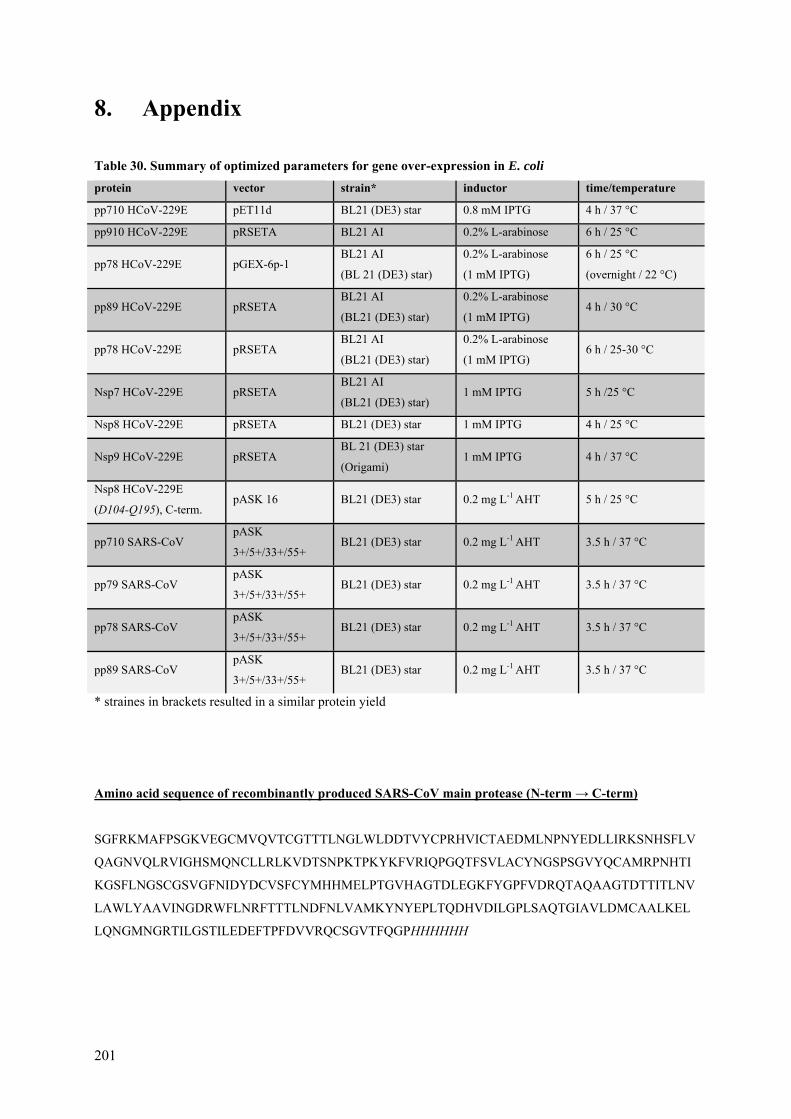
201
8. Appendix
Table 30. Summary of optimized parameters for gene over-expression in E. coli
protein vector strain* inductor time/temperature
pp710 HCoV-229E pET11d BL21 (DE3) star 0.8 mM IPTG 4 h / 37 °C
pp910 HCoV-229E pRSETA BL21 AI 0.2% L-arabinose 6 h / 25 °C
pp78 HCoV-229E pGEX-6p-1 BL21 AI
(BL 21 (DE3) star)
0.2% L-arabinose
(1 mM IPTG)
6 h / 25 °C
(overnight / 22 °C)
pp89 HCoV-229E pRSETA BL21 AI
(BL21 (DE3) star)
0.2% L-arabinose
(1 mM IPTG) 4 h / 30 °C
pp78 HCoV-229E pRSETA BL21 AI
(BL21 (DE3) star)
0.2% L-arabinose
(1 mM IPTG) 6 h / 25-30 °C
Nsp7 HCoV-229E pRSETA BL21 AI
(BL21 (DE3) star) 1 mM IPTG 5 h /25 °C
Nsp8 HCoV-229E pRSETA BL21 (DE3) star 1 mM IPTG 4 h / 25 °C
Nsp9 HCoV-229E pRSETA BL 21 (DE3) star
(Origami) 1 mM IPTG 4 h / 37 °C
Nsp8 HCoV-229E
(D104-Q195), C-term. pASK 16 BL21 (DE3) star 0.2 mg L-1 AHT 5 h / 25 °C
pp710 SARS-CoV pASK
3+/5+/33+/55+ BL21 (DE3) star 0.2 mg L-1 AHT 3.5 h / 37 °C
pp79 SARS-CoV pASK
3+/5+/33+/55+ BL21 (DE3) star 0.2 mg L-1 AHT 3.5 h / 37 °C
pp78 SARS-CoV pASK
3+/5+/33+/55+ BL21 (DE3) star 0.2 mg L-1 AHT 3.5 h / 37 °C
pp89 SARS-CoV pASK
3+/5+/33+/55+ BL21 (DE3) star 0.2 mg L-1 AHT 3.5 h / 37 °C
* straines in brackets resulted in a similar protein yield
Amino acid sequence of recombinantly produced SARS-CoV main protease (N-term → C-term)
SGFRKMAFPSGKVEGCMVQVTCGTTTLNGLWLDDTVYCPRHVICTAEDMLNPNYEDLLIRKSNHSFLV
QAGNVQLRVIGHSMQNCLLRLKVDTSNPKTPKYKFVRIQPGQTFSVLACYNGSPSGVYQCAMRPNHTI
KGSFLNGSCGSVGFNIDYDCVSFCYMHHMELPTGVHAGTDLEGKFYGPFVDRQTAQAAGTDTTITLNV
LAWLYAAVINGDRWFLNRFTTTLNDFNLVAMKYNYEPLTQDHVDILGPLSAQTGIAVLDMCAALKEL
LQNGMNGRTILGSTILEDEFTPFDVVRQCSGVTFQGPHHHHHH
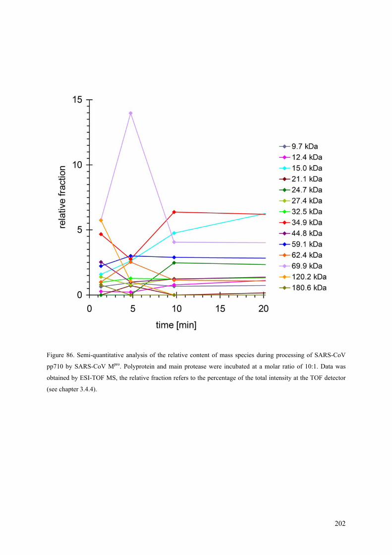
202
Figure 86. Semi-quantitative analysis of the relative content of mass species during processing of SARS-CoV
pp710 by SARS-CoV Mpro. Polyprotein and main protease were incubated at a molar ratio of 10:1. Data was
obtained by ESI-TOF MS, the relative fraction refers to the percentage of the total intensity at the TOF detector
(see chapter 3.4.4).
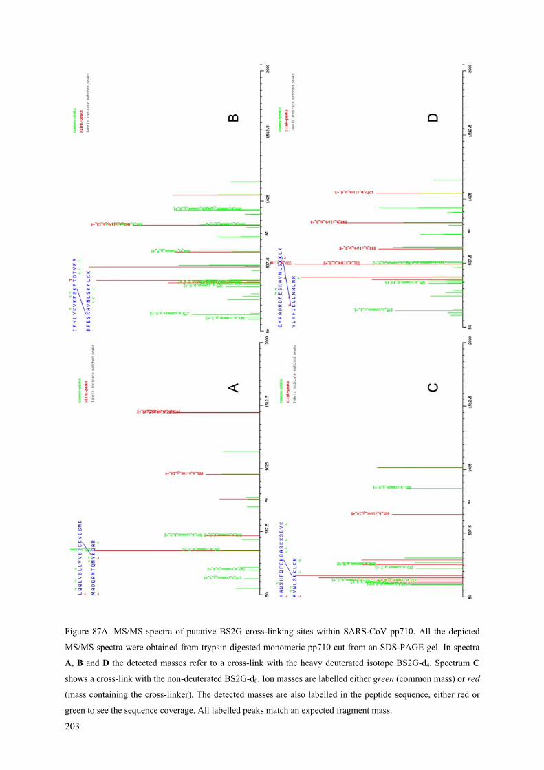
203
Figure 87A. MS/MS spectra of putative BS2G cross-linking sites within SARS-CoV pp710. All the depicted
MS/MS spectra were obtained from trypsin digested monomeric pp710 cut from an SDS-PAGE gel. In spectra
A, B and D the detected masses refer to a cross-link with the heavy deuterated isotope BS2G-d4. Spectrum C
shows a cross-link with the non-deuterated BS2G-d0. Ion masses are labelled either green (common mass) or red
(mass containing the cross-linker). The detected masses are also labelled in the peptide sequence, either red or
green to see the sequence coverage. All labelled peaks match an expected fragment mass.

204
Figure 87B. MS/MS spectra of putative BS2G cross-linking sites within SARS-CoV pp710. Spectrum E
indicates cross-linking of monomeric pp710 with BS2G-d0 cut from an SDS-PAGE gel. Spectrum F shows a
cross-link site within trimeric pp710 with BS2G-d4. Spectrum G represents a putative cross-link with BS2G-d4
in a sample of larger oligomers. Spectrum H shows a cross-linked peptide containing the light isotope BS2G-d0
in an oligomer fraction.

205
SARS-CoV 1 ASQAWQPGVAMPNLYKMQRMLLEKCDLQNYGENAVIPKGIMMNVAKYTQLCQYLNTLTLAVPYNMRVIHFGAGSDKGVAP HCoV-229E 1 -SAEWKCGYSMPGIYKTQRMCLEPCNLYNYGAGLKLPSGIMFNVVKYTQLCQYFNSTTLCVPHNMRVLHLGAGSDYGVAP Feline CoV 1 -SAEWNPGYSMPTLYKIQRMCLERCNLYNYGAQVRLPDGITTNVVKYTQLCQYLNTTTVCVPHKMRVLHLGAAGASGVAP Turkey CoV 1 ---AWTCGYNMPELYKVQNCVMEPCNIPNYGVGITLPSGIMMNVAKYTQLCQYLSKTTMCVPHNMRVMHFGAGSDKGVAP consensus 1 .. *. *..** .**.*....*.*...***.....*.**..** ********....*..**..***.* **....**** SARS-CoV 81 GTAVLRQWLPTGTLLVDSDLNDFVSDADSTLIGDCATVHTANKWDLIISDMYDPRTKHVTK----ENDSKEGFFTYLCGF HCoV-229E 80 GTAVLKRWLPHDAIVVDNDVVDYVSDADFSVTGDCATVYLEDKFDLLISDMYDGRTKAIDG----ENVSKEGFFTYINGF Feline CoV 80 GSTVLRRWLPDDAILVDNDLRDYVSDADFSVTGDCTSLYIEDKFDLLISDLYDGSTKSIDG----ENTSKDGFFTYINGF Turkey CoV 78 GSTVLKQWLPEGTLLVDNDIVDYVSDAHVSVLSDCNKYKTEHKFDLVISDMYTDNDSKRKHEGVIANNGNDDVFIYLSDF consensus 81 *. **. ***. ..**.*..*.****......**.......*.**.***.*......... .* .....*.*...* SARS-CoV 157 IKQKLALGGSIAVKITEHSWNADLYKLMGHFSWWTAFVTNVNASSSEAFLIGANYLGKP--KEQIDGYTMHANYIFWRNT HCoV-229E 156 ICEKLAIGGSIAIKVTEYSWNKKLYELVQRFSFWTMFCTSVNTSSSEAFVVGINYLGDFAQGPFIDGNIIHANYVFWRNS Feline CoV 156 IKEKLSLGGSAAIKITEFSWNKDLYELIQRFEYWTVFCTSVNTSSSEGFLIGINYLGPYCDKAIVDGNIMHANYIFWRNS Turkey CoV 158 LRNNLALGGSFAVKVTETSWHENLYDIAQDCAWWTMFCTAVNASSSEAFMIGVNYLGAS-EKVKVSGKTLHANYIFWRNC consensus 161 ....*..***.*.*.**.**...**........**.*.*.** ****.*..*.**** . .. ..*. .****.****. SARS-CoV 235 NPIQLSSYSLFDMSKFPLKLRGTAVMSLKENQINDMIYSLLEKGRLIIRENNRVVVSSDILVNN- HCoV-229E 236 TVMSLSYNSVLDLSKFNCKHKATVVVQLKDSDINDMVLSLVRSGKLLVRGNGKCLSFSNHLVS-- Feline CoV 236 TIMALSHNSVLDTPKFKCRCNNALIVNLKEKELNEMVVGLLRKGKLLIRNNGKLLNFGNHLVNVP Turkey CoV 237 NYLQTSAYSIFDVAKFDLKLKATPVVSLKTEQKDRLSFWFS------------------------ consensus 241 ....* *. *..** .........**. ...... ........... ..... .......
Figure 88. Alignment of coronaviral Nsp16 (2’-O-methyltransferase) created by Clustal Omega and visualized
by Boxshade V 3.2 (SIB).

206
Figure 89. Electrostatic surface potential calculated by Sybyl-X 1.2 comparing the interaction of two different
homologues of Nsp10 and Nsp16, particularly the resulting conserved patch of positive charge (blue/purple).

207
9. Risk and safety statements
9.1. Chemicals (GHS classification)
Table 31. Chemicals (GHS classification)
compound CAS-No. supplier GHS
hazard hazard statements
precautionary
statements
acetic acid 64-19-7 Chem-solute GHS02
GHS05 H226, H314
P280, P305+351+338,
P310
acrylamide 30% 79-06-1 Carl Roth GHS06
GHS08
H301, H312, H316,
H317, H319, H332,
H340, H350, H361f,
H372
P201, P280, P301+310,
P305+351+338,
P308+313
ATP 34369-07-8 Sigma - - -
agarose 9012-36-6 Serva - - -
ammonium
acetate 631-61-8 Sigma - - -
(NH4)2SO4 7283-20-2 Carl Roth - - -
ampicillin 69-52-3 Carl Roth GHS08 H334, H317
P280, P261,
P302+P352,
P342+P311
APS 7727-54-0 Carl Roth
GHS03
GHS07
GHS08
H272, H302, H315,
H317, H319, H334;
H335
P280, P305+351+338,
P302+352, P304+341,
P342+311
bis-Tris 6976-37-0 Sigma GHS07 H315, H319, H335 P261, P305 + P351 +
P338
bromophenol
blue 115-39-9 Applichem - - -
boric acid 10043-35-3 Carl Roth H360 P201, P202, P281,
P303+313, P405, P501
CaCl2 10043-52-4 Merck GHS07 H319 P305+351+338
citric acid 77-92-9 Sigma GHS05 H318 P305+351+338, P311
coomassie
brilliant blue
R250
6104-59-2 Serva - - -
EGCG 989-51-5
Sigma - - -

208
compound CAS-No. supplier GHS
hazard hazard statements
precautionary
statements
DMSO 67-68-5
Sigma GHS08
H315, H319
P280, P302+352, P305 +351+338, P332+313, P362
DTT 578517 Applichem GHS07 H302, H315, H319,
H335
P302+352,
P305+351+338
EDC 25952-53-8 Applichem GHS05,
GHS07 H315, H318, H335
P261, P280, P305 +
P351 + P338
EDTA 60-00-4 Sigma GHS07 H319 P305+351+338
ethanol 64-17-5 Carl Roth GHS02 H225 P210
ethidium
bromide 1239-45-8 Sigma
GHS06,
GHS08 H302, H330, H341
P260, P281, P284,
P310
glutaraldehyde 111-30-8 Applichem
GHS05,
GHS07,
GHS08,
GHS09
H302 + H332, H314,
H317, H334, H335,
H400
P261, P273, P280,
P305 + P351 + P338,
P310
glycerol 56-81-5 Sigma - - -
GSH 70-18-8 Carl Roth - - -
guanidin
hydrochlorid 50-01-1 Applichem GHS07 H302, H315, H319
P305+P351+P388,
P302+P352
HEPES 7365-45-9 Sigma - - -
hydrochloric
acid >25% 7647-01-0 Merck
GHS05,
GHS07 H314, H335
P261, P280, P310,
P305+351+338
imidazole 288-32-4 Carl Roth
GHS05,
GHS06,
GHS08
H301; H314; H361
P260, P281,
P303+P361+P353,
P301+P330+P331,
P305+P351+P338,
P308+P313
IPTG 367-93-1 Carl Roth - - -
isopropanol 67-63-0 Carl Roth GHS02,
GHS07 H225, H319, H336.
P210, P233,
P305+351+338
KCl 7447-40-7 Carl Roth - - -
K2HPO4 7758-11-4 Carl Roth - - -
KH2PO4 7778-77-0 Merck - - -

209
compound CAS-No. supplier GHS
hazard hazard statements
precautionary
statements
Li2SO4 10102-25-7 Merck GHS07 H302 -
MgCl2 7786-30-3 Carl Roth - - -
MgSO4 7487-88-9 Merck - - -
MPD 107-41-5 Carl Roth GHS07 H315, H319 -
2-mercapto-
ethanol 60-24-2
Fisher
Scientific
GHS06,
GHS09
H302, H411, H315,
H335, H311, H319
P280, P312,
P302+P350, P261,
P273, P301+P312,
P305+351+338
NaOAc 127-09-3 Applichem - - -
Na2HPO4 7558-79-4 Carl Roth - - -
NaH2PO4 10049-21-5 Applichem - - -
NaF
7681-49-4
Merck - H301, H315, H319 P301 + P310, P305
+P351 + P338
NaCl 7647-14-5 Carl Roth - - -
NaH2PO4 10049-21-5 Applichem - - -
NaOH 1310-73-2 Merck GHS05 H314 P280, P310,
P305+351+338
Na3-citrate 6132-04-3 Sigma - - -
NiSO4 10101-97-0 Applichem
GHS08,
GHS09,
GHS07
H332, H315, H334,
H317, H341, H350i,
H360D, H372
H410
P280, P273, P201,
P342+P311,
P308+P313,
P302+P352
paraffin 8002-74-2 Applichem - - -
PEG 400 25322-68-3 Sigma - - -
PEG 6000 25322-68-3 Merck - - -
PEG 8000 25322-68-3 Sigma - - -
PMSF 329-98-6 Applichem GHS06,
GHS05 H301, H314
P280,
P305+P351+P338,
P310
SDS 151-21-3 Sigma GHS02
GHS06
H228, H302, H311,
H315, H319, H335
P210, P261, P280,
P312, P305+351+338
silver nitrate 7761-88-8 Carl Roth GHS09 H272, H314, H410
P273, P280, P301+310+331, P305+351+338, P309+310

210
compound CAS-No. supplier GHS
hazard hazard statements
precautionary
statements
tannic acid 1401-55-4 Sigma - - -
TCA 76-03-9 Sigma GHS05
GHS09 H314, H410
P273, P280,
P301+330+331,
P305+351+338,
P309+P310
TEMED 110-18-9 Merck
GHS02
GHS05
GHS07
H225, H302, H314,
H332
P261, P280,
P305+351+338
Tris 1185-53-1 Fluka GHS07 H315, H319, H335 P261,
P305+351+338
yeast extract 8013-01-2 Serva - - -
ZnCl2 7646-85-7 Sigma
GHS05,
GHS07,
GHS09
H302-H314-H410
P273, P280, P305 +
P351 + P338, P310,
P501
β-OG 29836-26-8 Carl Roth - - -
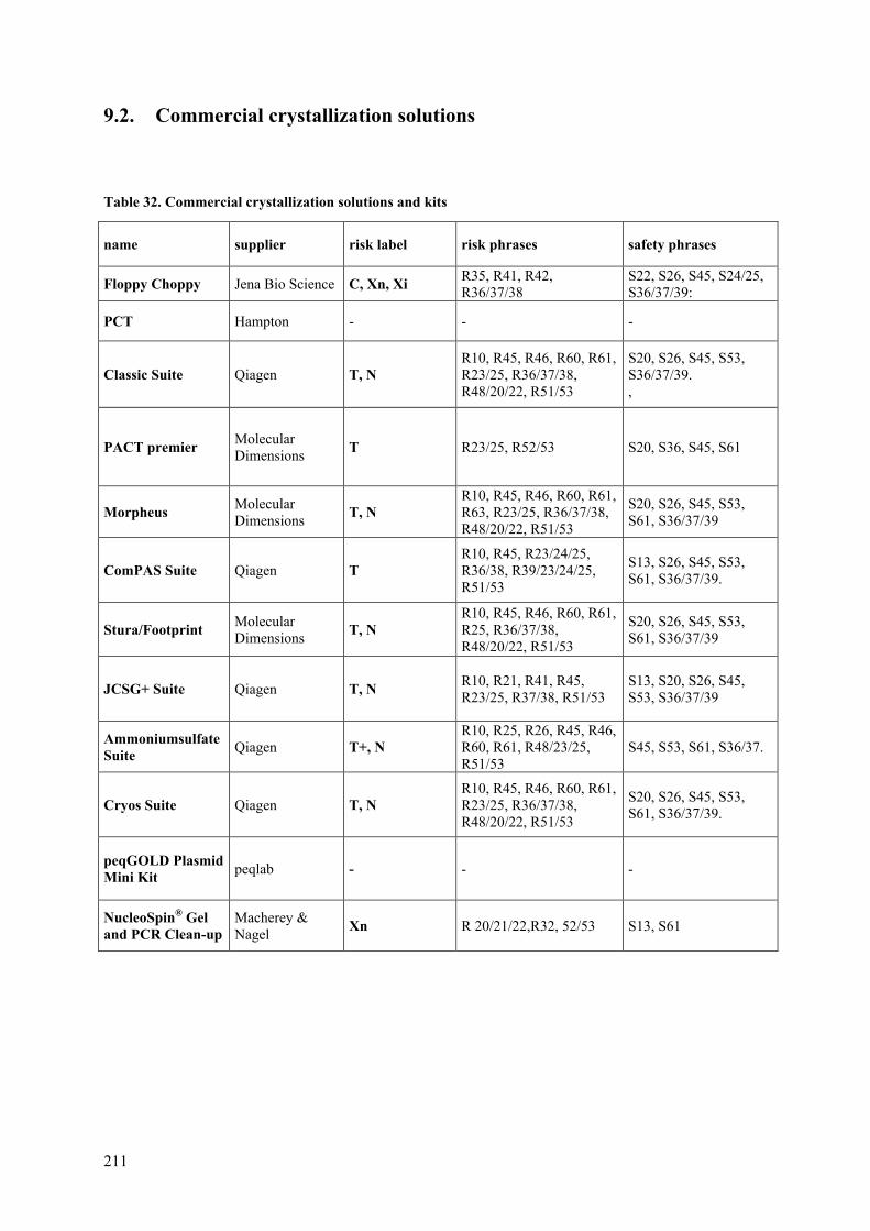
211
9.2. Commercial crystallization solutions
Table 32. Commercial crystallization solutions and kits
name supplier risk label risk phrases safety phrases
Floppy Choppy Jena Bio Science C, Xn, Xi R35, R41, R42, R36/37/38
S22, S26, S45, S24/25, S36/37/39:
PCT Hampton - - -
Classic Suite Qiagen T, N R10, R45, R46, R60, R61, R23/25, R36/37/38, R48/20/22, R51/53
S20, S26, S45, S53, S36/37/39. ,
PACT premier Molecular Dimensions
T R23/25, R52/53 S20, S36, S45, S61
Morpheus Molecular Dimensions
T, N R10, R45, R46, R60, R61, R63, R23/25, R36/37/38, R48/20/22, R51/53
S20, S26, S45, S53, S61, S36/37/39
ComPAS Suite Qiagen T R10, R45, R23/24/25, R36/38, R39/23/24/25, R51/53
S13, S26, S45, S53, S61, S36/37/39.
Stura/Footprint Molecular Dimensions
T, N R10, R45, R46, R60, R61, R25, R36/37/38, R48/20/22, R51/53
S20, S26, S45, S53, S61, S36/37/39
JCSG+ Suite Qiagen T, N R10, R21, R41, R45, R23/25, R37/38, R51/53
S13, S20, S26, S45, S53, S36/37/39
Ammoniumsulfate Suite
Qiagen T+, N R10, R25, R26, R45, R46, R60, R61, R48/23/25, R51/53
S45, S53, S61, S36/37.
Cryos Suite Qiagen T, N R10, R45, R46, R60, R61, R23/25, R36/37/38, R48/20/22, R51/53
S20, S26, S45, S53, S61, S36/37/39.
peqGOLD Plasmid Mini Kit
peqlab - - -
NucleoSpin® Gel and PCR Clean-up
Macherey & Nagel
Xn R 20/21/22,R32, 52/53 S13, S61

212
9.3. GHS and risk symbols and information about hazard-, risk-, safety-
and precaution-statements
Figure 90. Hazard symbols (according to http://www.hse.gov.uk/) for formulations and respective risk labels
Figure 91. GHS pictograms (source: http://www.unece.org/trans/danger/publi/ghs/pictograms.html).
Table 33. GHS hazard statements
GHS hazard statements
H 225 Highly flammable liquid and vapour
H 226 Flammable liquid and vapour
H 228 Flammable solid
H 272 May intensify fire; oxidizer
H 301 Toxic if swallowed
H 302 Harmful if swallowed
H 311 Toxic in contact with skin
H 312 Harmful in contact with skin
H 314 Causes severe skin burns and eye damage
H 315 Causes skin irritation
H 316 Causes mild skin irritation

213
H 317 May cause an allergic skin reaction
H 318 Causes serious eye damage
H 319 Causes serious eye irritation
H 330 Fatal if inhaled
H 331 Toxic if inhaled
H 332 Harmful if inhaled
H 334 May cause allergy or asthma symptoms or breathing difficulties if inhaled
H 335 May cause respiratory irritation
H 336 May cause drowsiness or dizziness
H 340 May cause genetic defects
H 341 Suspected of causing genetic defects
H 350 May cause cancer
H 350i May cause cancer by inhalation
H 360 May damage fertility or the unborn child
H 360D May damage the unborn child
H 361 Suspected of damaging fertility or the unborn child
H361f Suspected of damaging fertility
H 370 Causes damage to organs
H 372 Causes damage to organs through prolonged or repeated exposure
H 410 Very toxic to aquatic life with long lasting effects
H 411 Toxic to aquatic life with long lasting effects

214
Table 34. GHS precautionary statements
GHS precautionary statements
P201 Obtain special instructions before use
P210 Keep away from heat/sparks/open flames/hot surfaces – No smoking
P233 Keep container tightly closed
P260 Do not breathe dust/fume/gas/mist/vapors/spray
P261 Avoid breathing dust/fume/gas/mist/vapors/spray
P264 Wash … thoroughly after handling
P273 Avoid release to the environment
P281 Use personal protective equipment as required
P280 Wear protective gloves/protective clothing/eye protection/face protection
P284 Wear respiratory protection
P309 IF exposed or you feel unwell: ...
P310 Immediately call a POISON CENTER or doctor/physician
P311 Call a POISON CENTER or doctor/physician
P312 Call a POISON CENTER or doctor/physician if you feel unwell
P321 Specific treatment (see respective MSDS)
P362 Take off contaminated clothing and wash before reuse
P501 Dispose of contents/container to …
P301+310 IF SWALLOWED: Immediately call a POISON CENTER or doctor/physician
P301+P312 IF SWALLOWED: Call a POISON CENTER or doctor/physician if you feel unwell
P301+P330+P331 IF SWALLOWED: Rinse mouth. Do NOT induce vomiting
P302+P352 IF ON SKIN: Wash with soap and water
P303+P361+P353 IF ON SKIN (or hair): Remove/Take off immediately all contaminated clothing. Rinse skin with water/shower
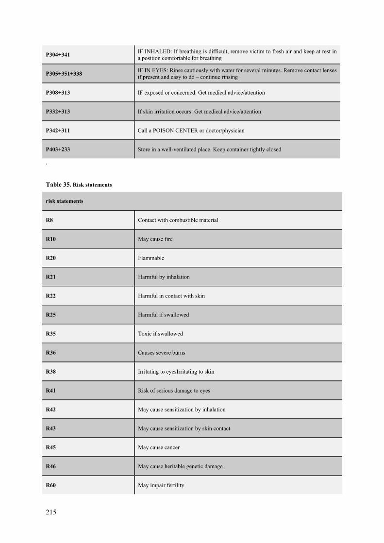
215
P304+341 IF INHALED: If breathing is difficult, remove victim to fresh air and keep at rest in a position comfortable for breathing
P305+351+338 IF IN EYES: Rinse cautiously with water for several minutes. Remove contact lenses if present and easy to do – continue rinsing
P308+313 IF exposed or concerned: Get medical advice/attention
P332+313 If skin irritation occurs: Get medical advice/attention
P342+311 Call a POISON CENTER or doctor/physician
P403+233 Store in a well-ventilated place. Keep container tightly closed
.
Table 35. Risk statements
risk statements
R8 Contact with combustible material
R10 May cause fire
R20 Flammable
R21 Harmful by inhalation
R22 Harmful in contact with skin
R25 Harmful if swallowed
R35 Toxic if swallowed
R36 Causes severe burns
R38 Irritating to eyesIrritating to skin
R41 Risk of serious damage to eyes
R42 May cause sensitization by inhalation
R43 May cause sensitization by skin contact
R45 May cause cancer
R46 May cause heritable genetic damage
R60 May impair fertility

216
R61 May cause harm to the unborn child
R39/23/24/25 Toxic: danger of very serious irreversible effects through inhalation, in contact with skin and if swallowed
R36/37/38 Irritating to eyes, respiratory system and skin
R23/24/25 Toxic by inhalation, in contact with skin and if swallowed
R20/21/22 Harmful by inhalation, in contact with skin and if swallowed
R48/20/22 Harmful: danger of serious damage to health by prolonged exposure through inhalation and if swallowed
R23/25 Toxic by inhalation and if swallowed
R36/38 Irritating to eyes and skin
R51/53 Toxic to aquatic organisms, may cause long-term adverse effects in the aquatic environment
R37/38 Irritating to respiratory system and skin
safetey statements
S20 When using do not eat or drink. Do not breathe dust
S22 In case of contact with eyes, rinse immediately with plenty of water and seek medical advice
S26 In case of accident or if you feel unwell seek medical advice immediately (show the label where possible).
S28 After contact with skin, wash immediately with plenty of ... (to be specified by the manufacturer)
S45 If swallowed, seek medical advice immediately and show this container or label
S46 Avoid exposure - obtain special instructions before use
S53 Avoid release to the environment
S61 Refer to special instructions/safety data sheet
S24/25 Avoid contact with skin and eyes
S36/37 Wear suitable protective clothing and gloves
S36/37/39 Wear suitable protective clothing, gloves and eye/face protection

217
10. Poster presentations and publications
Parts of this thesis were presented as a poster at the following conferences.
International Conference on Antivirals for Neglected and Emerging Viruses (ICAV 9),
Lübeck, 10th-13th October, 2010
S. Falke, A. Kikhney, D. Svergun, R. Hilgenfeld, L. Redecke
“First Structural insights into Polyprotein Nsp7-10 from Human Coronavirus 229E”
23rd Annual Meeting of the Society for Virology, Kiel, 6th-9th March, 2013
S. Falke, R. Schubert, C. Uetrecht, J. Schulz, A. Kikhney, D. Svergun, R. Hilgenfeld, L.
Redecke
“Structural Insights into Coronaviral Polyprotein Nsp7-10”
A talk was held by Sven Falke at the “5th International PhD Symposium” at the Helmholtz
Zentrum für Infektionsforschung (HZI), Braunschweig, 15th December, 2011
“Structural Characterization of Non-Structural Proteins and Preprocessed Polyproteins
within the Coronaviral Transcriptase/Replicase Complex”
Publications
S. Falke, M. Klinge, D. Rehders, A. Kikhney, D. Svergun, R. Hilgenfeld, L. Redecke
“First structural insights into Alphacoronavirus Nsp10 and its impact on the Interaction with Nsp16”, to be submitted
S. Falke, R. Schubert, C. Uetrecht, A. Heck, M. Kötzler, B. Meyer, A. Kikhney, M.
Kwiatkowski, H. Schlüter, D. Svergun, R. Hilgenfeld, L. Redecke
“Elucidating low-resolution structure and processing of coronaviral polyprotein Nsp7-10 in vitro”, to be submitted

218
11. Acknowledgement In the end of more than three years of this project I am so glad for meeting a large number of skilled people who
strongly supported and positively influenced my work. At first I would like to express an outstanding “thank
you” to Dr. Lars Redecke heading the junior-group SIAS for giving me the opportunity to move on in science.
His balanced mood and continuous support with reliable teamwork helped me lot. This has also included a few
consequent and strict but helpful advices at the right time and discussions beyond biochemistry. A special
gratitude is addressed to Prof. Rolf Hilgenfeld who provided the truly interesting topic and was always available
to share his huge experience, enthusiasm and knowledge in many fruitful discussions over the years.
Furthermore, I thank Prof. Christian Betzel for really valuable discussions and so many small aspects of support
and career guidance as well as the opportunity to work in his labs and use his infrastructure. I thank Prof.
Reinhard Bredehorst for being the second reviewer of the thesis and Prof. Bernd Meyer for his commitment to
be disputation referee.
From the very beginning of my project Dr. Dirk Rehders supported me with SPR spectroscopy and in silico
modelling and set a good “lab atmosphere”; therefore he deserves a huge acknowledgement. Without Dr.
Charlotte Uetrecht the native mass spectrometry experiments would not have been possible. Thank you, it was a
good cooperation – not forgetting about Prof. Albert Heck’s group in Utrecht. One important partner in terms of
speculation about coronavirus proteins is Yibei Xiao. Thank you for the cooperation on analysing polymerase
activity and for being always available by E-mail. I also thank Dr. Alexey Kikhney for a strong support in
learning how to analyze SAXS data and Prof. Dmitri Svergun and other people from EMBL for support and
coordination during the synchrotron experiments.
Ich mache die zweite Hälfte mal auf Deutsch und hoffe, dass ich niemanden vergesse. Weit oben auf der Liste
stehen auch Robin Schubert und Marco Klinge. Ein großes Lob und Dank gilt euch für viel Kreativität, großes
Engagement und Fingerspitzengefühl in der Weiterentwicklung des Projekts. Danke für viel Spaß. Ebenso gilt
mein Dank weiteren Leuten die das Projekt mit vollem Einsatz und Freude an unterschiedlichen Stellen praktisch
unterstützt haben: Timur Yorgan, Henrike Friedenberger, Bianca Schlee, Raphael Klein – und natürlich Birgitta
Fried, der immer positive gute Geist des Labors. Der Gruppe von Prof. Hartmut Schlüter dank ich für
massenspektrometrische Untersuchungen und Beratung, insbesondere Marcel Kwiatkowski war eine große
Unterstützung. Dr. Miriam Koetzler danke ich sehr für ihre Bereitschaft NMR Experimente durchzuführen und
mir die Technik etwas näher zu bringen. Darüber hinaus bedanke ich mich herzlich bei allen Doktoranden der
Graduiertenschule SDI. Natürlich bin ich noch allen Leuten zu Dank verpflichtet, die über die Jahre im Labor
waren oder immer noch dort sind bzw. den Alltag an der Uni etwas schöner, interessanter, spannender gemacht
haben. An alle Gäste und Kollegen von SIAS, den Arbeitskreisen Betzel und Hahn und an der Uni insgesamt:
Danke euch allen!
Ganz besonders danke ich meiner Familie, die mir diesen Schritt zum großen Teil erst ermöglicht hat und mich
immer zu 100% unterstützt hat. Danke an alle Freunde, ich kann mich glücklich schätzen.
H. F. V. S.

219
12. Curriculum vitae
- entfällt aus datenschutzrechtlichen Gründen -

220
Selbstständigkeitserklärung
Hiermit erkläre ich an Eides statt, dass ich die an der Universität Hamburg zur Promotion
eingereichte Dissertation mit dem Titel „Coronaviral Polyprotein Nsp7-10: Proteolytic
Processing and Dynamic Interactions within the Transcriptase/Replicase Complex“ im Institut
für Biochemie des Fachbereichs Chemie der Universität Hamburg ohne sonstige nicht
angeführte Hilfe durchgeführt und bei der Abfassung der Dissertation keine anderen als die
dort aufgeführten Hilfsmittel benutzt habe.
Ferner versichere ich, dass ich bisher an keiner in- oder ausländischen Universität ein Gesuch
um Zulassung zur Promotion eingereicht und daher weder diese noch eine andere Arbeit als
Dissertation vorgelegt habe.
Sven Falke

221

222