Real-time gene delivery vector tracking in the endo-lysosomal pathway of live cells
A Novel Mechanism of Lysosomal Acid Sphingomyelinase Maturation: REQUIREMENT FOR CARBOXYL-TERMINAL...
12
A Novel Mechanism of Lysosomal Acid Sphingomyelinase Maturation REQUIREMENT FOR CARBOXYL-TERMINAL PROTEOLYTIC PROCESSING * Received for publication, June 16, 2010, and in revised form, November 18, 2010 Published, JBC Papers in Press, November 22, 2010, DOI 10.1074/jbc.M110.155234 Russell W. Jenkins ‡ , Jolanta Idkowiak-Baldys ‡ , Fabio Simbari § , Daniel Canals ‡ , Patrick Roddy ‡ , Clarke D. Riner ¶ , Christopher J. Clarke ‡ , and Yusuf A. Hannun ‡1 From the ‡ Department of Biochemistry and Molecular Biology, Medical University of South Carolina, Charleston, South Carolina 29425, the § Department of Biomedicinal Chemistry, Institute for Advanced Chemistry of Catalonia (CSIC), Jordi Girona 18-26, Barcelona, Spain, and the ¶ Department of Biochemistry and Cellular and Molecular Biology, University of Tennessee, Knoxville, Tennessee 37996 Acid sphingomyelinase (aSMase) catalyzes the hydrolysis of sphingomyelin (SM) to form the bioactive lipid ceramide (Cer). Notably, aSMase exists in two forms: a zinc (Zn 2 )- independent lysosomal aSMase (L-SMase) and a Zn 2 -de- pendent secreted aSMase (S-SMase) that arise from alterna- tive trafficking of a single protein precursor. Despite extensive investigation into the maturation and trafficking of aSMase, the exact identity of mature L-SMase has re- mained unclear. Here, we describe a novel mechanism of aSMase maturation involving C-terminal proteolytic pro- cessing within, or in close proximity to, endolysosomes. Us- ing two different C-terminal-tagged constructs of aSMase (V5, DsRed), we demonstrate that aSMase is processed from a 75-kDa, Zn 2 -activated proenzyme to a mature 65 kDa, Zn 2 -independent L-SMase. L-SMase is recognized by a polyclonal Ab to aSMase, but not by anti-V5 or anti-DsRed antibodies, suggesting that the C-terminal tag is lost during maturation. Furthermore, indirect immunofluorescence staining demonstrated that mature L-SMase colocalized with the lysosomal marker LAMP1, whereas V5-aSMase lo- calized to the Golgi secretory pathway. Moreover, V5- aSMase possessed Zn 2 -dependent activity suggesting it may represent the common protein precursor of S-SMase and L-SMase. Importantly, the 65-kDa L-SMase, but not V5-aSMase, was sensitive to the lysosomotropic inhibitor desipramine, co-fractionated with lysosomes, and migrated at the same M r as partially purified human aSMase. Finally, three aSMase mutants containing C-terminal Niemann-Pick mutations (R600H, R600P, R608) exhibited defective pro- teolytic maturation. Taken together, these results demon- strate that mature L-SMase arises from C-terminal proteo- lytic processing of pro-aSMase and suggest that impaired C-terminal proteolysis may lead to severe defects in L-SMase function. Acid sphingomyelinase (aSMase) 2 (EC 3.1.4.12) is a soluble lysosomal hydrolase that plays a prominent role in the catabo- lism of sphingomyelin (SM) to ceramide (Cer). Interestingly, aSMase exists as two enzymatic forms, one that is targeted to the endolysosomal compartment, whereas the other is re- leased extracellularly (1). Lysosomal aSMase (L-SMase) arises from mannose 6-phosphorylation of N-glycans, which targets pro-aSMase to the endolysosomal compartment where it en- counters and tightly coordinates Zn 2 , thus becoming “Zn 2 - independent,” i.e. not requiring addition of zinc for activity (1). Acid SMase precursors that are not mannose 6-phosphor- ylated get directed to the Golgi secretory pathway and re- leased extracellularly giving rise to secretory aSMase (S- SMase) (1, 2). Cells from patients with inherited defects in the mannose 6-phosphorylation pathway (i.e. I-cell disease), se- crete large amounts of aSMase (3) and this form of aSMase is activated by Zn 2 (4). Acid SMase is first synthesized as a 75-kDa prepro-enzyme representing the full-length, N-glycosylated protein (3). The prepro-aSMase is rapidly processed to pro-aSMase (72 kDa), and within the acidic compartment matures to a 70-kDa form, and last is processed to a 52-kDa polypeptide (3). Ma- ture L-SMase is believed to represent the 70-kDa and/or the 52-kDa forms of aSMase, however, given the lack of investiga- tion into the Zn 2 requirement of these different forms of aSMase, the precise molecular identity of the mature, Zn 2 - independent L-SMase remains unclear. Hurwitz et al. (5) demonstrated that the in vivo aSMase inhibitor desipramine, induced the loss of the 70-kDa form of aSMase concomitant with the loss of L-SMase activity in treated cells. However, isoelectric focusing studies have described two forms of aSMase: a 70-kDa form and a 57-kDa form, which correlated * This work was supported, in whole or in part, by National Institutes of Health Grant P01 CA097132 from the NCI (to Y. A. H.), National Institutes of Health MSTP Training Grant GM08716 (to R. W. J.), American Heart As- sociation Pre-doctoral Fellowship AHA 081509E (to R. W. J.), a Medical University of South Carolina Hollings Cancer Center Abney Foundation scholarship (to R. W. J.), Ministerio de Educacion y Ciencia (Spain) Predoc- toral Fellowship AP2006-02190 (to F. S.), and Administrative Supple- ments Providing Summer Research Experiences for Students and Science Educators Grant HL043707-19S1 (to C. D. R.). The confocal core is sup- ported in part by National Institutes of Health NCI Medical University of South Carolina-Cancer Center Support Grant 1P30 CA138313-01. 1 To whom correspondence should be addressed: 173 Ashley Ave., P.O. Box 250509C, Charleston, SC 29425. Tel.: 843-792-4321; Fax: 843-792-4322; E-mail: [email protected]. 2 The abbreviations used are: aSMase, acid sphingomyelinase; S-SMase, se- cretory sphingomyelinase; L-SMase, lysosomal sphingomyelinase; SM, sphingomyelin; NPD, Niemann-Pick disease; PNGaseF, peptide N-gly- canase F; MEM, minimal essential medium; ANOVA, analysis of variance; IP, immunoprecipitation. THE JOURNAL OF BIOLOGICAL CHEMISTRY VOL. 286, NO. 5, pp. 3777–3788, February 4, 2011 © 2011 by The American Society for Biochemistry and Molecular Biology, Inc. Printed in the U.S.A. FEBRUARY 4, 2011 • VOLUME 286 • NUMBER 5 JOURNAL OF BIOLOGICAL CHEMISTRY 3777 at MUSC Library, on January 29, 2011 www.jbc.org Downloaded from
Transcript of A Novel Mechanism of Lysosomal Acid Sphingomyelinase Maturation: REQUIREMENT FOR CARBOXYL-TERMINAL...
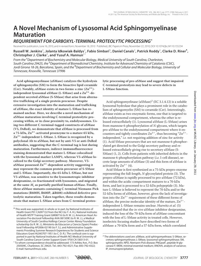
A Novel Mechanism of Lysosomal Acid SphingomyelinaseMaturationREQUIREMENT FOR CARBOXYL-TERMINAL PROTEOLYTIC PROCESSING*
Received for publication, June 16, 2010, and in revised form, November 18, 2010 Published, JBC Papers in Press, November 22, 2010, DOI 10.1074/jbc.M110.155234
Russell W. Jenkins‡, Jolanta Idkowiak-Baldys‡, Fabio Simbari§, Daniel Canals‡, Patrick Roddy‡, Clarke D. Riner¶,Christopher J. Clarke‡, and Yusuf A. Hannun‡1
From the ‡Department of Biochemistry and Molecular Biology, Medical University of South Carolina, Charleston,South Carolina 29425, the §Department of Biomedicinal Chemistry, Institute for Advanced Chemistry of Catalonia (CSIC),Jordi Girona 18-26, Barcelona, Spain, and the ¶Department of Biochemistry and Cellular and Molecular Biology, University ofTennessee, Knoxville, Tennessee 37996
Acid sphingomyelinase (aSMase) catalyzes the hydrolysisof sphingomyelin (SM) to form the bioactive lipid ceramide(Cer). Notably, aSMase exists in two forms: a zinc (Zn2�)-independent lysosomal aSMase (L-SMase) and a Zn2�-de-pendent secreted aSMase (S-SMase) that arise from alterna-tive trafficking of a single protein precursor. Despiteextensive investigation into the maturation and traffickingof aSMase, the exact identity of mature L-SMase has re-mained unclear. Here, we describe a novel mechanism ofaSMase maturation involving C-terminal proteolytic pro-cessing within, or in close proximity to, endolysosomes. Us-ing two different C-terminal-tagged constructs of aSMase(V5, DsRed), we demonstrate that aSMase is processed froma 75-kDa, Zn2�-activated proenzyme to a mature 65 kDa,Zn2�-independent L-SMase. L-SMase is recognized by apolyclonal Ab to aSMase, but not by anti-V5 or anti-DsRedantibodies, suggesting that the C-terminal tag is lost duringmaturation. Furthermore, indirect immunofluorescencestaining demonstrated that mature L-SMase colocalizedwith the lysosomal marker LAMP1, whereas V5-aSMase lo-calized to the Golgi secretory pathway. Moreover, V5-aSMase possessed Zn2�-dependent activity suggesting itmay represent the common protein precursor of S-SMaseand L-SMase. Importantly, the 65-kDa L-SMase, but notV5-aSMase, was sensitive to the lysosomotropic inhibitordesipramine, co-fractionated with lysosomes, and migratedat the same Mr as partially purified human aSMase. Finally,three aSMase mutants containing C-terminal Niemann-Pickmutations (R600H, R600P, �R608) exhibited defective pro-teolytic maturation. Taken together, these results demon-strate that mature L-SMase arises from C-terminal proteo-
lytic processing of pro-aSMase and suggest that impairedC-terminal proteolysis may lead to severe defects inL-SMase function.
Acid sphingomyelinase (aSMase)2 (EC 3.1.4.12) is a solublelysosomal hydrolase that plays a prominent role in the catabo-lism of sphingomyelin (SM) to ceramide (Cer). Interestingly,aSMase exists as two enzymatic forms, one that is targeted tothe endolysosomal compartment, whereas the other is re-leased extracellularly (1). Lysosomal aSMase (L-SMase) arisesfrom mannose 6-phosphorylation of N-glycans, which targetspro-aSMase to the endolysosomal compartment where it en-counters and tightly coordinates Zn2�, thus becoming “Zn2�-independent,” i.e. not requiring addition of zinc for activity(1). Acid SMase precursors that are not mannose 6-phosphor-ylated get directed to the Golgi secretory pathway and re-leased extracellularly giving rise to secretory aSMase (S-SMase) (1, 2). Cells from patients with inherited defects in themannose 6-phosphorylation pathway (i.e. I-cell disease), se-crete large amounts of aSMase (3) and this form of aSMase isactivated by Zn2� (4).Acid SMase is first synthesized as a 75-kDa prepro-enzyme
representing the full-length, N-glycosylated protein (3). Theprepro-aSMase is rapidly processed to pro-aSMase (72 kDa),and within the acidic compartment matures to a 70-kDaform, and last is processed to a 52-kDa polypeptide (3). Ma-ture L-SMase is believed to represent the 70-kDa and/or the52-kDa forms of aSMase, however, given the lack of investiga-tion into the Zn2� requirement of these different forms ofaSMase, the precise molecular identity of the mature, Zn2�-independent L-SMase remains unclear. Hurwitz et al. (5)demonstrated that the in vivo aSMase inhibitor desipramine,induced the loss of the 70-kDa form of aSMase concomitantwith the loss of L-SMase activity in treated cells. However,isoelectric focusing studies have described two forms ofaSMase: a 70-kDa form and a 57-kDa form, which correlated
* This work was supported, in whole or in part, by National Institutes ofHealth Grant P01 CA097132 from the NCI (to Y. A. H.), National Institutesof Health MSTP Training Grant GM08716 (to R. W. J.), American Heart As-sociation Pre-doctoral Fellowship AHA 081509E (to R. W. J.), a MedicalUniversity of South Carolina Hollings Cancer Center Abney Foundationscholarship (to R. W. J.), Ministerio de Educacion y Ciencia (Spain) Predoc-toral Fellowship AP2006-02190 (to F. S.), and Administrative Supple-ments Providing Summer Research Experiences for Students and ScienceEducators Grant HL043707-19S1 (to C. D. R.). The confocal core is sup-ported in part by National Institutes of Health NCI Medical University ofSouth Carolina-Cancer Center Support Grant 1P30 CA138313-01.
1 To whom correspondence should be addressed: 173 Ashley Ave., P.O. Box250509C, Charleston, SC 29425. Tel.: 843-792-4321; Fax: 843-792-4322;E-mail: [email protected].
2 The abbreviations used are: aSMase, acid sphingomyelinase; S-SMase, se-cretory sphingomyelinase; L-SMase, lysosomal sphingomyelinase; SM,sphingomyelin; NPD, Niemann-Pick disease; PNGaseF, peptide N-gly-canase F; MEM, minimal essential medium; ANOVA, analysis of variance;IP, immunoprecipitation.
THE JOURNAL OF BIOLOGICAL CHEMISTRY VOL. 286, NO. 5, pp. 3777–3788, February 4, 2011© 2011 by The American Society for Biochemistry and Molecular Biology, Inc. Printed in the U.S.A.
FEBRUARY 4, 2011 • VOLUME 286 • NUMBER 5 JOURNAL OF BIOLOGICAL CHEMISTRY 3777
at MU
SC
Library, on January 29, 2011w
ww
.jbc.orgD
ownloaded from
with peaks of activity (6). The former was found in fractionswith the highest level of L-SMase activity and was assigned apI of 6.8–7.2. Importantly, aSMase protein was found in al-most all of the fractions, whereas aSMase activity was concen-trated in only 25% of the fractions. Therefore, despiteextensive investigation, the true identity of mature, Zn2�-independent L-SMase remains unknown.By virtue of its distinct cellular itinerary, S-SMase exhibits
several defining characteristics that have been used to distin-guish it from L-SMase. First, S-SMase does not encounterZn2� during its trafficking and maturation, and thus remainsZn2�-dependent (2). Second, S-SMase is trafficked throughthe distal Golgi pathway where it undergoes additional pro-cessing of N-glycans to become of the “complex” type, render-ing the enzyme partially insensitive to digestion with endogly-cosidase H (2, 3, 6). Third, it appears that L-SMase undergoesadditional N-terminal proteolytic processing as purified S-SMase begins with His60 and L-SMase begins with Gly66 (2).Mature L-SMase may undergo additional N-terminal process-ing as microsequencing of purified placental L-SMase indi-cated the N terminus began with Gly83 (7).Although N-terminal modifications to the different forms
of aSMase have been documented, evidence for the C-termi-nal modification is lacking. It has previously been suggestedthat carboxyl-terminal modification of aSMase might serve asa mechanism to regulate enzyme activity. Qiu et al. (8) de-scribed a mechanism whereby oxidation, mutation, and/ordeletion of the C-terminal Cys629 resulted in activation of theenzyme. Based on these results the authors postulated thatloss of the C-terminal Cys629 might serve as a “cysteineswitch,” as has been described for matrix metalloproteinases(9), whereby loss of C-terminal Cys residues favors hydrationof Zn2� thereby promoting enzyme activation (8). Also dis-cussed was the possible relevance of this mechanism of en-zyme activation to in vivo regulation. Given that C-terminalprocessing has been described for several lysosomal hydro-lases that follow a similar path of trafficking and maturation,such as cathepsin D (10), it is conceivable that aSMase under-goes similar proteolytic processing to generate matureL-SMase.To determine whether C-terminal processing was required
for the formation of mature L-SMase in situ we utilized cellsstably overexpressing aSMase with C-terminal V5/His orDsRed fusion tags. Here, we demonstrate that C-terminalprocessing of aSMase occurs within or near the endolysoso-mal compartment, giving rise to mature Zn2�-independentL-SMase. Mature L-SMase is recognized by an antibody di-rected to aSMase, is sensitive to the lysosomotropic inhibitordesipramine, and co-fractionates with lysosomes. Further-more, we provide evidence that C-terminal-tagged aSMaserepresent pro-aSMase, is Zn2�-dependent, and localizes tothe Golgi secretory pathway, but not the endolysosomal com-partment. Last, three C-terminal Niemann-Pick mutants ex-hibit defective C-terminal processing with loss of L-SMaseactivity. These results indicate that C-terminal processing isessential for formation of mature L-SMase.
EXPERIMENTAL PROCEDURES
Materials—MCF7 and HEK 293 cells were obtained fromATCC (Manassas, VA). RPMI and MEM culture medium,fetal bovine serum, blasticidin S-HCl, and geneticin (G418)were obtained from Invitrogen. Anti-V5 mouse monoclonalantibody was from Invitrogen. DsRed rabbit polyclonal anti-body was obtained from Clontech (Mountain View, CA). Rab-bit polyclonal LAMP-1 antibody was obtained from Abcam(Cambridge, MA). Mouse monoclonal LAMP1 antibody andDsRed goat polyclonal antibody, and HRP-labeled secondaryantibodies were from Santa Cruz Biotechnology, Inc. (SantaCruz, CA). Calreticulin rabbit polyclonal antibody was fromSigma. Rabbit anti-�-mannosidase II polyclonal antibody wasfrom Chemicon International (Temecula, CA). Rabbit anti-TGN46 antibody was from Novus (Littleton, CO). Enhancedchemiluminescence kit was from ThermoScientific (Rockford,IL). Leupeptin, pepstatin, and aprotinin were obtained fromRoche Applied Science. Desipramine, peptide N-glycanase F(PNGaseF), and all other chemicals were obtained fromSigma. Partially purified placental acid sphingomyelinase wasa gift from Dr. Gary Smith (GlaxoSmithKline), and has previ-ously been described (11). Rabbit polyclonal antisera toaSMase (number 1598) (12) was a generous gift from Drs.Edward Schuchman and Richard Kolesnick.Cell Culture and Generation of Stable Cell Lines—MCF7
cells were grown in RPMI 1640, supplemented with L-gluta-mine and 10% (v/v) fetal bovine serum. HEK 293 were grownin 10% FBS/MEM. MCF7 stably expressing V5-aSMaseWT
were previously described (13). To generate HEK 293 cellsstably expressing DsRed plasmids, cells were plated in 6-welldishes (200,000 cells/well) and transfected with 1.0 �g ofDNA using Lipofectamine 2000 according to the manufactur-er’s instructions. The following day, transfected cells weretrypsinized, pelleted, and re-plated (1:5) in 10% FBS/MEMsupplemented with 0.75 mg/ml of G418 (Invitrogen). After2–3 weeks in selective medium, transfection efficiency wasevaluated by confocal microscopy for DsRed fluorescence.Plasmids and Site-directed Mutagenesis—pEF6.V5/
His.aSMaseWT and pEF6.V5/His.aSMaseS508A were previouslydescribed (11). pEF6.V5/His.LacZ is the control plasmidpEF6/V5-His-TOPO cloning kit (Invitrogen). VSVG3-GFPwas kindly provided by Dr. Chiara Luberto (Medical Univer-sity of South Carolina), and was generated and described bythe laboratory of Dr. Kai Simons (14). pDsRed-N1-Monomer-aSMaseWT was previously described (15). DsRed-aSMaseS508A
mutant was generated via site-directed mutagenesis using theQuikChange kit from Stratagene, as previously described (11).DsRed-aSMaseR600P, DsRed-aSMaseR600H, and DsRed-aSMase�R608 were generated using the following mutagenesisprimers: underlined nucleotides represent the changed codon,“/” represents site of deleted codon, R600H Fwd, 5�-GCTCTCTGCCCATGCTGACAGCC-3�, R600H Rev, 5�-GGCTGTCAGCATGGGCAGAGAGC-3�; R600P Fwd,5�-GCTCTCTGCCCCTGCTGACAGCC-3�, R600H Rev, 5�-GGCTGTCAGCAGGGGCAGAGAGC-3�; �R608 Fwd,5�-CTGCTCTGTGC/CACCTGATGCC-3�, �R608 Rev, 5�-GGCATCAGGTG/GCACAGAGCAG-3�. For all constructs,
Proteolytic Maturation of Lysosomal Acid Sphingomyelinase
3778 JOURNAL OF BIOLOGICAL CHEMISTRY VOLUME 286 • NUMBER 5 • FEBRUARY 4, 2011
at MU
SC
Library, on January 29, 2011w
ww
.jbc.orgD
ownloaded from
plasmid DNA was sequenced (GeneWiz) to confirm muta-tions and ensure that the C-terminal DsRed tag was in-framewith the aSMase cDNA.Immunoprecipitation—Immunoprecipitation of V5-tagged
proteins was performed as previously described (15). Briefly,V5-S-SMase was immunoprecipitated directly from condi-tioned medium with 1 �g/ml of anti-V5 mAb, unless other-wise indicated, and 1:20 protein A/G-agarose beads (SantaCruz) in the presence of 1:500 protease inhibitor mixture(Sigma) by rocking overnight at 4 °C. For intracellular V5-aSMase, cells were washed two times and then scraped in ice-cold PBS. Cells were pelleted by centrifugation (5 min, 800 �g, 4 °C) and then lysed in IP buffer (20 mM Tris-HCl, pH 7.4,100 mM NaCl, 1% Nonidet P-40, 1 mM EDTA, 1 mM EGTA,1:200), each protease inhibitor mixture (Sigma), and phospha-tase inhibitor mixtures 1 and 2 (Sigma). Lysates were clarifiedby centrifugation (10 min at 10,000 � g at 4 °C), normalizedto the protein concentration, and incubated with ProteinA/G-agarose for 1 h to remove nonspecific interacting pro-teins. Cleared lystates were then incubated overnight at 4 °Cwith 1 �g/ml of primary antibody (V5) and 1:10 protein A/G-agarose using a circular rotator. Bound protein was isolatedby centrifugation (1 min, 12,000 � g), washed three timeswith lysis buffer, and the immunoprecipitates were preparedby addition of 60 �l of 2� Laemmli buffer. Equal volumeswere subjected to SDS-PAGE electrophoresis and Westernblotting. For immunotitration experiments, conditions weresimilar to those described above except that the concentra-tion of V5 mAb was adjusted (0, 1, 2, and 4 �g/ml).Glycosidase Digestion—For PNGaseF digestion, 37.5 �l of
ddH2O was added to placental L-SMase (20 �l) or V5-aSMaseimmune complexes (20 �l), followed by 10 �l of 5� reactionbuffer (250 mM sodium phosphate, pH 7.5) and 2.5 �l of de-naturation solution (2% SDS � 1 mM �-ME) to each tube.Samples were boiled for 5 min and then cooled on ice. Two �l(0.01 units) of enzyme were then added to each sample alongwith Triton X-100 (0.75% final), mixed by gentle pipeting, andincubated overnight (� 18 h) at 37 °C. The following day,samples were retrieved, 50 �l of 2� sample buffer was addedto 50 �l reaction volume, and samples were boiled again for 5min. Cleavage was monitored by Western blotting followingSDS-PAGE on a 7.5% acrylamide gel.SDS-PAGE and Immunoblotting—SDS-PAGE and immu-
noblotting were performed as described previously (15). Pri-mary antibody dilutions were V5, 1:5,000; aSMase, 1:500–1,000; DsRed, 1:500; and �-actin, 1:10,000.In Vitro aSMase Activity Assay—S-SMase and L-SMase
were measured as described previously (15).Differential Centrifugation—2 � 106 DsRed-aSMaseWT
HEK were seeded in 10-cm dishes in 10% FBS/MEM and al-lowed to grow for 2 days. Cells were then shifted to serum-free medium (10.0 ml of 0.1% BSA/MEM per dish) for 18 h.Adherent cells were washed, scraped, and pelleted (5 min,1100 rpm, 4 °C) in ice-cold PBS. Following aspiration of thePBS wash, cell pellets were resuspended in 1.0 ml of BufferL/F, 10 mM Tris-HCl (pH 7.5), 300 mM sucrose, 5 mM EDTA,1:200 protease inhibitor mixture (Sigma), 1:200 phosphataseinhibitor mixture 1 (Sigma), 1:200 phosphatase inhibitor mix-
ture 2 (Sigma), and incubated on ice for 1 h. Samples werethen homogenized by passage through a 281⁄2-gauge syringe10–15 times. Samples were then centrifuged for 5 min at1,000 � g (4 °C) to obtain the 1,000 � g (nuclear, unbrokencell) pellet. The supernatant from the 1,000 � g spin was thenspun for 10 min at 10,000 � g (4 °C) to obtain the 10,000 � g(heavy membrane) pellet. The supernatant from the 10,000 �g spin was then spun for 1 h at 100,000 � g (4 °C) to obtainthe 100,000 � g (light membrane) pellet. The pellets fromeach spin were resuspended in 0.1 ml of buffer L/F for subse-quent analysis. To resuspend the 100,000 � g pellet buffer L/Fwas supplemented with Triton X-100 (1% final concentra-tion). Aliquots of supernatants and pellets were snap frozen inCO2(s)/MeOH bath and stored at �80 °C prior to aSMaseactivity assay.Silver Stain for Total Protein—Silver staining of polyacryl-
amide gels was performed using the SilverSNAP� Stain Kit IIfrom Pierce, according to the manufacturer’s instructions.Statistical Analysis—Data are represented as mean � S.E.,
unless otherwise indicated. Unpaired Student’s t test, one-wayANOVA with Dunnett’s post-test, and two-way ANOVA withBonferroni post-test statistical analyses were performed usingPrism/GraphPad software with additional post-test analysisusing the Prism/Graphpad website.
RESULTS
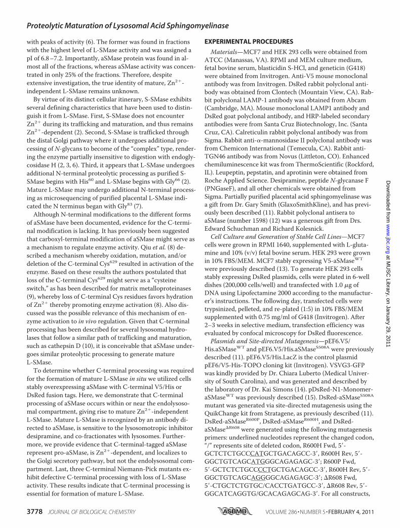
Carboxyl-terminal V5-tagged aSMase Localizes to the GolgiSecretory Pathway—It is well established that aSMase local-izes to the endolysosomal compartment (16, 17). However, inongoing studies utilizing MCF7 stably expressing a C-termi-nal-tagged V5-aSMase, we observed that V5-aSMase exhib-ited minimal colocalization with the lysosomal markerLAMP1 (Fig. 1A), but instead co-localized more strongly withVSVG3-GFP (Fig. 1B), which marks all compartments of theGolgi secretory pathway (18). Counterstaining with markersof the endoplasmic reticulum (Fig. 1C, calreticulin) and Golgi(Fig. 1, D and E, �-mannosidase II and TGN46) also demon-strated strong colocalization with markers of Golgi secretorypathway, but not the ER. To further explore this, immunofluo-rescence staining was performed using aSMase-specific anti-sera (number 1598) (12). As expected, the aSMase antibodyshowed strong colocalization with V5-aSMase (Fig. 1F). How-ever, the aSMase Ab recognized structures not detected bythe V5 Ab. As partial colocalization of aSMase with LAMP1was also evident (Fig. 1G), this indicated that the aSMase Abrecognized both mature lysosomal aSMase and V5-taggedaSMase. These results demonstrate that V5-aSMase residesprimarily in the Golgi secretory pathway, and suggest thatlysosomal aSMase had lost its C-terminal V5 tag upon entryinto the endolysosomal compartment.65-kDa aSMase Develops from 75-kDa V5-aSMase but
Lacks a V5 Tag—The loss of the ability to detect the C-termi-nal V5 tag of the lysosomally localized aSMase suggested pro-teolytic processing of aSMase within the endolysosomal com-partment. Consequently, such processing would be expectedto yield lower molecular weight forms of aSMase. To investi-gate this, cellular extracts from vector control (V5-LacZ) andV5-aSMaseWT MCF7 were assayed for L-SMase activity to
Proteolytic Maturation of Lysosomal Acid Sphingomyelinase
FEBRUARY 4, 2011 • VOLUME 286 • NUMBER 5 JOURNAL OF BIOLOGICAL CHEMISTRY 3779
at MU
SC
Library, on January 29, 2011w
ww
.jbc.orgD
ownloaded from
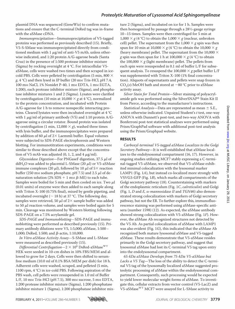
confirm L-SMase activity, and were subjected to Western blotanalysis using both anti-V5 and anti-aSMase antibodies.L-SMase activity was markedly increased (�15-fold) in V5-aSMase MCF7 compared with vector control MCF7, indicat-ing that overexpressed V5-aSMase acquired Zn2�-indepen-dent aSMase activity (Fig. 2A). Western blot analysis with V5mAb revealed a single band in V5-aSMaseWT MCF7 (75 kDa),whereas two distinct bands were evident with the aSMase Ab(75 kDa, 65 kDa) (Fig. 2B). These results indicate that a 65-kDa form of aSMase is generated from 75-kDa V5-aSMase,but is lacking the C-terminal tag. Moreover, given that Zn2�-independent L-SMase activity develops from V5-aSMase, this65-kDa aSMase likely represents lysosomal aSMase observedabove (Fig. 1D).Selective loss of 65-kDa aSMase following Desipramine
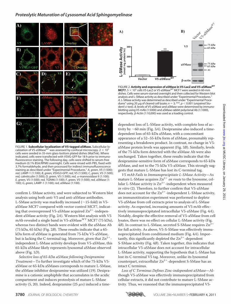
Treatment—To further investigate which of the 75-kDa V5-aSMase or 65-kDa aSMases corresponds to mature L-SMase,the aSMase inhibitor desipramine was utilized (19). Desipra-mine is a cationic amphiphile that accumulates in the acidiccompartment and induces proteolysis of mature L-SMaseactivity (5, 20). Indeed, desipramine (25 �M) induced a time-
dependent loss of L-SMase activity, with complete loss of ac-tivity by �60 min (Fig. 3A). Desipramine also induced a time-dependent loss of 65-kDa aSMase, with a concomitantappearance of a 52–55-kDa form of aSMase, presumably rep-resenting a breakdown product. In contrast, no change in V5-aSMase protein levels was apparent (Fig. 3B). Similarly, levelsof the 75-kDa form detected with the aSMase Ab were alsounchanged. Taken together, these results indicate that thedesipramine-sensitive form of aSMase corresponds to 65-kDaaSMase, and not to the 75-kDa V5-aSMase. This strongly sug-gests that mature L-SMase has lost its C-terminal tag.V5 mAb Fails to Immunoprecipitate L-SMase Activity—As
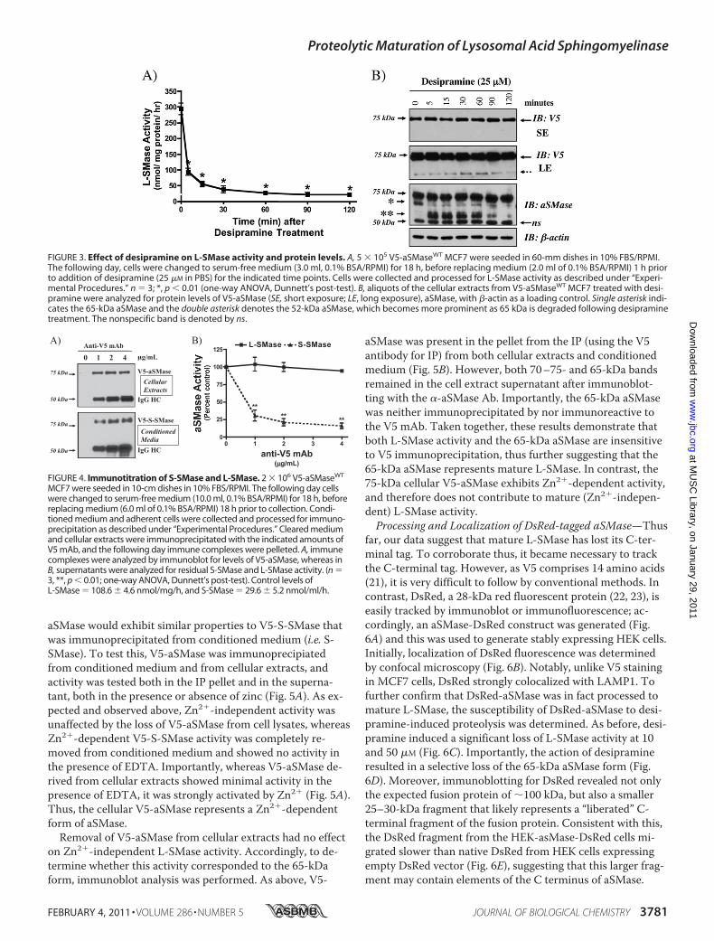
mature L-SMase acquires Zn2� en route to the lysosome, cel-lular L-SMase activity is Zn2�-independent when measuredin vitro (2). Therefore, to further confirm that V5-aSMasedoes not account for the Zn2�-independent L-SMase activity,an immunotitration experiment was performed to depleteV5-aSMase from cell extracts prior to analysis of L-SMaseactivity. As expected, increasing amounts of V5 mAb effec-tively immunoprecipitated intracellular V5-aSMase (Fig. 4A).Notably, despite the effective removal of V5-aSMase from celllysates, there was no effect on cellular L-SMase activity (Fig.4B). In contrast to L-SMase, secreted S-SMase requires Zn2�
for full activity. As above, V5-S-SMase was effectively immu-noprecipitated from conditioned medium (Fig. 4A). Impor-tantly, this significantly depleted the Zn2�-dependentS-SMase activity (Fig. 4B). Taken together, this indicates thatintracellular V5-aSMase does not account for intracellularL-SMase activity, supporting the hypothesis that L-SMase haslost its C-terminal V5 tag. Moreover, unlike its lysosomalcounterpart, extracellular Zn2�-dependent S-SMase has anintact C terminus.Loss of C Terminus Defines Zinc-independent aSMase—Al-
though V5-aSMase was effectively immunoprecipitated fromcellular extracts, it did not contribute to mature L-SMase ac-tivity. Thus, we reasoned that the immunoprecipitated V5-
FIGURE 1. Subcellular localization of V5-tagged aSMase. Subcellular lo-calization of V5-aSMaseWT was assessed by confocal microscopy. 2 � 105
cells were seeded in 35-mm glass-bottom plated dishes (MatTek). Whereindicated, cells were transfected with VSVG-GFP for 18 h prior to immuno-fluorescence staining. The following day, cells were shifted to serum-freemedium (0.1% BSA/RPMI) overnight. Cells were rinsed with PBS, fixed with3.7% formaldehyde, and then processed for indirect immunofluorescencestaining as described under “Experimental Procedures.” A, green, V5 (1:500);red, LAMP-1 (1:100); B, green, VSVG3-GFP; red, V5 (1:500); C, green, V5 (1:500);red, calreticulin (1:500); D, green, V5 (1:500); red, �-mannosidase II (1:100);E, green, V5 (1:500); red, TGN46 (1:100); F, green, V5 (1:500); red, aSMase (1:100); G, green, LAMP-1 (1:100); red, aSMase (1:100).
FIGURE 2. Activity and expression of aSMase in V5-LacZ and V5-aSMaseWT
MCF7. 5 � 105 cells V5-LacZ or V5-aSMaseWT MCF7 were seeded in 60-mmdishes. Cells were serum-starved overnight and then collected for Western blotanalysis and L-SMase activity as described under “Experimental Procedures.”A, L-SMase activity was determined as described under “Experimental Proce-dures” using 20 �g of cleared cell lysate; n � 3; ***, p 0.001 (unpaired Stu-dent’s t test). B, levels of V5-aSMase and aSMase were determined by immuno-blotting using V5 mAb (1:5000) and aSMase rabbit polyclonal Ab (1:1000),respectively. �-Actin (1:10,000) was used as a loading control.
Proteolytic Maturation of Lysosomal Acid Sphingomyelinase
3780 JOURNAL OF BIOLOGICAL CHEMISTRY VOLUME 286 • NUMBER 5 • FEBRUARY 4, 2011
at MU
SC
Library, on January 29, 2011w
ww
.jbc.orgD
ownloaded from
aSMase would exhibit similar properties to V5-S-SMase thatwas immunoprecipitated from conditioned medium (i.e. S-SMase). To test this, V5-aSMase was immunoprecipiatedfrom conditioned medium and from cellular extracts, andactivity was tested both in the IP pellet and in the superna-tant, both in the presence or absence of zinc (Fig. 5A). As ex-pected and observed above, Zn2�-independent activity wasunaffected by the loss of V5-aSMase from cell lysates, whereasZn2�-dependent V5-S-SMase activity was completely re-moved from conditioned medium and showed no activity inthe presence of EDTA. Importantly, whereas V5-aSMase de-rived from cellular extracts showed minimal activity in thepresence of EDTA, it was strongly activated by Zn2� (Fig. 5A).Thus, the cellular V5-aSMase represents a Zn2�-dependentform of aSMase.Removal of V5-aSMase from cellular extracts had no effect
on Zn2�-independent L-SMase activity. Accordingly, to de-termine whether this activity corresponded to the 65-kDaform, immunoblot analysis was performed. As above, V5-
aSMase was present in the pellet from the IP (using the V5antibody for IP) from both cellular extracts and conditionedmedium (Fig. 5B). However, both 70–75- and 65-kDa bandsremained in the cell extract supernatant after immunoblot-ting with the �-aSMase Ab. Importantly, the 65-kDa aSMasewas neither immunoprecipitated by nor immunoreactive tothe V5 mAb. Taken together, these results demonstrate thatboth L-SMase activity and the 65-kDa aSMase are insensitiveto V5 immunoprecipitation, thus further suggesting that the65-kDa aSMase represents mature L-SMase. In contrast, the75-kDa cellular V5-aSMase exhibits Zn2�-dependent activity,and therefore does not contribute to mature (Zn2�-indepen-dent) L-SMase activity.Processing and Localization of DsRed-tagged aSMase—Thus
far, our data suggest that mature L-SMase has lost its C-ter-minal tag. To corroborate thus, it became necessary to trackthe C-terminal tag. However, as V5 comprises 14 amino acids(21), it is very difficult to follow by conventional methods. Incontrast, DsRed, a 28-kDa red fluorescent protein (22, 23), iseasily tracked by immunoblot or immunofluorescence; ac-cordingly, an aSMase-DsRed construct was generated (Fig.6A) and this was used to generate stably expressing HEK cells.Initially, localization of DsRed fluorescence was determinedby confocal microscopy (Fig. 6B). Notably, unlike V5 stainingin MCF7 cells, DsRed strongly colocalized with LAMP1. Tofurther confirm that DsRed-aSMase was in fact processed tomature L-SMase, the susceptibility of DsRed-aSMase to desi-pramine-induced proteolysis was determined. As before, desi-pramine induced a significant loss of L-SMase activity at 10and 50 �M (Fig. 6C). Importantly, the action of desipramineresulted in a selective loss of the 65-kDa aSMase form (Fig.6D). Moreover, immunoblotting for DsRed revealed not onlythe expected fusion protein of �100 kDa, but also a smaller25–30-kDa fragment that likely represents a “liberated” C-terminal fragment of the fusion protein. Consistent with this,the DsRed fragment from the HEK-asMase-DsRed cells mi-grated slower than native DsRed from HEK cells expressingempty DsRed vector (Fig. 6E), suggesting that this larger frag-ment may contain elements of the C terminus of aSMase.
FIGURE 3. Effect of desipramine on L-SMase activity and protein levels. A, 5 � 105 V5-aSMaseWT MCF7 were seeded in 60-mm dishes in 10% FBS/RPMI.The following day, cells were changed to serum-free medium (3.0 ml, 0.1% BSA/RPMI) for 18 h, before replacing medium (2.0 ml of 0.1% BSA/RPMI) 1 h priorto addition of desipramine (25 �M in PBS) for the indicated time points. Cells were collected and processed for L-SMase activity as described under “Experi-mental Procedures.” n � 3; *, p 0.01 (one-way ANOVA, Dunnett’s post-test). B, aliquots of the cellular extracts from V5-aSMaseWT MCF7 treated with desi-pramine were analyzed for protein levels of V5-aSMase (SE, short exposure; LE, long exposure), aSMase, with �-actin as a loading control. Single asterisk indi-cates the 65-kDa aSMase and the double asterisk denotes the 52-kDa aSMase, which becomes more prominent as 65 kDa is degraded following desipraminetreatment. The nonspecific band is denoted by ns.
FIGURE 4. Immunotitration of S-SMase and L-SMase. 2 � 106 V5-aSMaseWT
MCF7 were seeded in 10-cm dishes in 10% FBS/RPMI. The following day cellswere changed to serum-free medium (10.0 ml, 0.1% BSA/RPMI) for 18 h, beforereplacing medium (6.0 ml of 0.1% BSA/RPMI) 18 h prior to collection. Condi-tioned medium and adherent cells were collected and processed for immuno-precipitation as described under “Experimental Procedures.” Cleared mediumand cellular extracts were immunoprecipitated with the indicated amounts ofV5 mAb, and the following day immune complexes were pelleted. A, immunecomplexes were analyzed by immunoblot for levels of V5-aSMase, whereas inB, supernatants were analyzed for residual S-SMase and L-SMase activity. (n �3, **, p 0.01; one-way ANOVA, Dunnett’s post-test). Control levels ofL-SMase � 108.6 � 4.6 nmol/mg/h, and S-SMase � 29.6 � 5.2 nmol/ml/h.
Proteolytic Maturation of Lysosomal Acid Sphingomyelinase
FEBRUARY 4, 2011 • VOLUME 286 • NUMBER 5 JOURNAL OF BIOLOGICAL CHEMISTRY 3781
at MU
SC
Library, on January 29, 2011w
ww
.jbc.orgD
ownloaded from
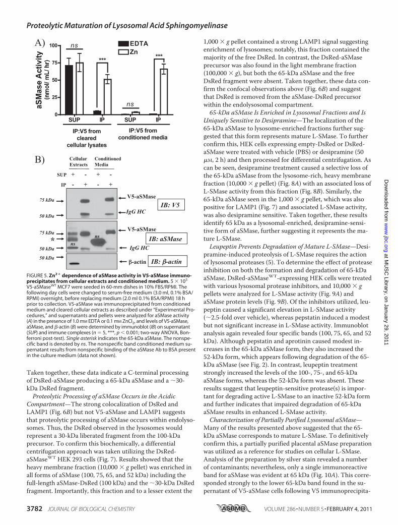
Taken together, these data indicate a C-terminal processingof DsRed-aSMase producing a 65-kDa aSMase and a �30-kDa DsRed fragment.Proteolytic Processing of aSMase Occurs in the Acidic
Compartment—The strong colocalization of DsRed andLAMP1 (Fig. 6B) but not V5-aSMase and LAMP1 suggeststhat proteolytic processing of aSMase occurs within endolyso-somes. Thus, the DsRed observed in the lysosomes wouldrepresent a 30-kDa liberated fragment from the 100-kDaprecursor. To confirm this biochemically, a differentialcentrifugation approach was taken utilizing the DsRed-aSMaseWT HEK 293 cells (Fig. 7). Results showed that theheavy membrane fraction (10,000 � g pellet) was enriched inall forms of aSMase (100, 75, 65, and 52 kDa) including thefull-length aSMase-DsRed (100 kDa) and the �30-kDa DsRedfragment. Importantly, this fraction and to a lesser extent the
1,000 � g pellet contained a strong LAMP1 signal suggestingenrichment of lysosomes; notably, this fraction contained themajority of the free DsRed. In contrast, the DsRed-aSMaseprecursor was also found in the light membrane fraction(100,000 � g), but both the 65-kDa aSMase and the freeDsRed fragment were absent. Taken together, these data con-firm the confocal observations above (Fig. 6B) and suggestthat DsRed is removed from the aSMase-DsRed precursorwithin the endolysosomal compartment.65-kDa aSMase Is Enriched in Lysosomal Fractions and Is
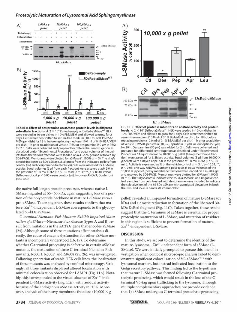
Uniquely Sensitive to Desipramine—The localization of the65-kDa aSMase to lysosome-enriched fractions further sug-gested that this form represents mature L-SMase. To furtherconfirm this, HEK cells expressing empty-DsRed or DsRed-aSMase were treated with vehicle (PBS) or desipramine (50�M, 2 h) and then processed for differential centrifugation. Ascan be seen, desipramine treatment caused a selective loss ofthe 65-kDa aSMase from the lysosome-rich, heavy membranefraction (10,000 � g pellet) (Fig. 8A) with an associated loss ofL-SMase activity from this fraction (Fig. 8B). Similarly, the65-kDa aSMase seen in the 1,000 � g pellet, which was alsopositive for LAMP1 (Fig. 7) and associated L-SMase activity,was also desipramine sensitive. Taken together, these resultsidentify 65 kDa as a lysosomal-enriched, desipramine-sensi-tive form of aSMase, further suggesting it represents the ma-ture L-SMase.Leupeptin Prevents Degradation of Mature L-SMase—Desi-
pramine-induced proteolysis of L-SMase requires the actionof lysosomal proteases (5). To determine the effect of proteaseinhibition on both the formation and degradation of 65-kDaaSMase, DsRed-aSMaseWT-expressing HEK cells were treatedwith various lysosomal protease inhibitors, and 10,000 � gpellets were analyzed for L-SMase activity (Fig. 9A) andaSMase protein levels (Fig. 9B). Of the inhibitors utilized, leu-peptin caused a significant elevation in L-SMase activity(�2.5-fold over vehicle), whereas pepstatin induced a modestbut not significant increase in L-SMase activity. Immunoblotanalysis again revealed four specific bands (100, 75, 65, and 52kDa). Although pepstatin and aprotinin caused modest in-creases in the 65-kDa aSMase form, they also increased the52-kDa form, which appears following degradation of the 65-kDa aSMase (see Fig. 2). In contrast, leupeptin treatmentstrongly increased the levels of the 100-, 75-, and 65-kDaaSMase forms, whereas the 52-kDa form was absent. Theseresults suggest that leupeptin-sensitive protease(s) is impor-tant for degrading active L-SMase to an inactive 52-kDa formand further indicates that impaired degradation of 65-kDaaSMase results in enhanced L-SMase activity.Characterization of Partially Purified Lysosomal aSMase—
Many of the results presented above suggested that the 65-kDa aSMase corresponds to mature L-SMase. To definitivelyconfirm this, a partially purified placental aSMase preparationwas utilized as a reference for studies on cellular L-SMase.Analysis of the preparation by silver stain revealed a numberof contaminants; nevertheless, only a single immunoreactiveband for aSMase was evident at 65 kDa (Fig. 10A). This corre-sponded strongly to the lower 65-kDa band found in the su-pernatant of V5-aSMase cells following V5 immunoprecipita-
FIGURE 5. Zn2� dependence of aSMase activity in V5-aSMase immuno-precipitates from cellular extracts and conditioned medium. 5 � 105
V5-aSMaseWT MCF7 were seeded in 60-mm dishes in 10% FBS/RPMI. Thefollowing day cells were changed to serum-free medium (3.0 ml, 0.1% BSA/RPMI) overnight, before replacing medium (2.0 ml 0.1% BSA/RPMI) 18 hprior to collection. V5-aSMase was immunoprecipitated from conditionedmedium and cleared cellular extracts as described under “Experimental Pro-cedures,” and supernatants and pellets were analyzed for aSMase activity(A) in the presence of 1.0 mM EDTA or 0.1 mM ZnCl2, and levels of V5-aSMase,aSMase, and �-actin (B) were determined by immunoblot (IB) on supernatant(SUP) and immune complexes (n � 5, ***, p 0.001; two-way ANOVA, Bon-ferroni post-test). Single asterisk indicates the 65-kDa aSMase. The nonspe-cific band is denoted by ns. The nonspecific band conditioned medium su-pernatant results from nonspecific binding of the aSMase Ab to BSA presentin the culture medium (data not shown).
Proteolytic Maturation of Lysosomal Acid Sphingomyelinase
3782 JOURNAL OF BIOLOGICAL CHEMISTRY VOLUME 286 • NUMBER 5 • FEBRUARY 4, 2011
at MU
SC
Library, on January 29, 2011w
ww
.jbc.orgD
ownloaded from

tion (Fig. 10B, cf. Fig. 4, A and B). Importantly, the placentalaSMase preparation also exhibited Zn2�-independent aSMaseactivity (Fig. 10C). Variations in N-glycan modifications canalso influence migration of aSMase on SDS-PAGE. Conse-
quently, V5-aSMase immune complexes and purifiedL-SMase were digested with PNGaseF to assess the nativeprotein size (Fig. 10D). Fully deglycosylated V5-aSMase mi-grated at �62–64 kDa, consistent with previous reports for
FIGURE 6. Subcellular localization and protein processing of DsRed-tagged aSMase in HEK 293. A, schematic representation of the aSMase-DsRed fu-sion protein. B, 2 � 105 DsRed-aSMaseWT HEK were seeded in 35-mm glass-bottom plated dishes (MatTek) in 10% FBS/MEM. The following day cells werechanged to serum-free medium (2.0 ml, 0.1% BSA/MEM) overnight, before being processed for indirect immunofluorescence staining. Cells were fixed, per-meabilized, blocked as described under “Experimental Procedures,” and stained for aSMase (1:100, green, �488) and LAMP1 (1:100, blue, �647). DsRed fluo-rescence was detected using the �555 channel. Arrowheads denote triple-positive structures. C, 5 � 105 DsRed-aSMaseWT HEK were seeded in 60-mmdishes in 10% FBS/MEM. The following day, cells were changed to serum-free medium (3.0 ml of 0.1% BSA/MEM) for 18 h, before replacing medium (2.0 mlof 0.1% BSA/MEM) 1 h prior to addition of desipramine (10 and 50 �M in PBS) for 2 h. Cells were collected and processed for L-SMase activity as describedunder “Experimental Procedures” (n � 3; **, p .01; one-way ANOVA, Dunnett’s post-test). D, levels of aSMase and DsRed-aSMase in cellular extracts fol-lowing desipramine treatment were determined by immunoblot (IB) with �-actin as a loading control. Single asterisk indicates 65-kDa aSMase. E, molecularweight of the liberated DsRed fragment from DsRed-aSMaseWT HEK (WT) compared with that of native, unfused DsRed (28 kDa) from HEK stably expressingpDsRed-N1-Monomer (EMPTY). Equal protein was loaded and resolved by SDS-PAGE (4 –20%). Membranes were probed with �-DsRed Ab (1:500).
FIGURE 7. Differential centrifugation of DsRed-tagged aSMase in HEK 293. 2 � 106 DsRed-empty or DsRed-aSMaseWT HEK were seeded in 10-cm dishesin 10% FBS/MEM and allowed to grow for 2 days. Cells were then shifted to serum-free medium (10.0 ml of 0.1% BSA/MEM per dish) for 18 h before collec-tion. Cells were collected and prepared for differential centrifugation as described under “Experimental Procedures,” and equal volumes were loaded on a4 –20% gel and resolved by SDS-PAGE. Membranes were blotted for aSMase, DsRed, and LAMP1, as described (n � 3). Single asterisk indicates the65-kDa aSMase. Relative enrichment of L-SMase activity in the 10,000 � g pellet (compared with whole cell extracts) is at least 4 –5-fold (data notshown). IB, immunoblot.
Proteolytic Maturation of Lysosomal Acid Sphingomyelinase
FEBRUARY 4, 2011 • VOLUME 286 • NUMBER 5 JOURNAL OF BIOLOGICAL CHEMISTRY 3783
at MU
SC
Library, on January 29, 2011w
ww
.jbc.orgD
ownloaded from
the native full-length protein precursor, whereas native L-SMase migrated at 55–60 kDa, again suggesting loss of a por-tion of the polypeptide backbone in mature L-SMase versuspro-aSMase. Taken together, these results confirm that ma-ture, Zn2�-independent L-SMase corresponds to the glycosy-lated 65-kDa aSMase.C-terminal Niemann-Pick Mutants Exhibit Impaired Matu-
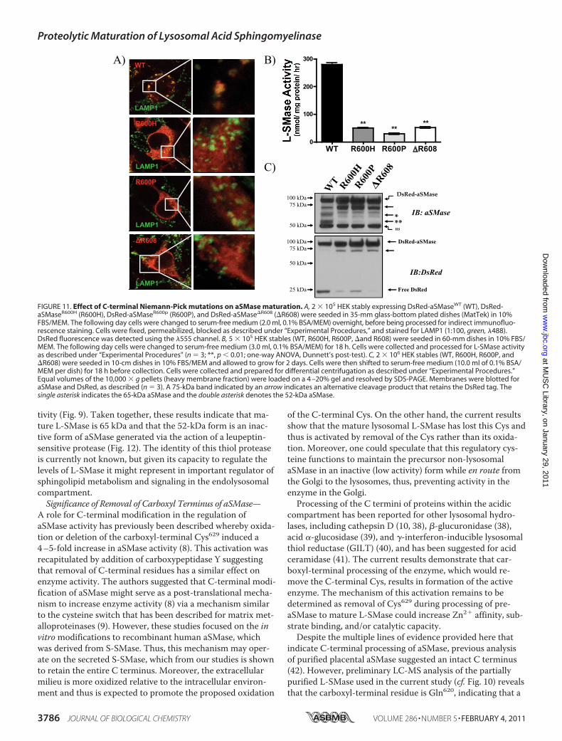
ration of aSMase—Niemann-Pick disease (types A and B) re-sult from mutations in the SMPD1 gene that encodes aSMase(24). Although some of these mutations affect catalysis di-rectly, the cause of enzyme dysfunction for other aSMase mu-tants is incompletely understood (16, 17). To determinewhether C-terminal processing is defective in certain aSMasemutants, the maturation of three C-terminal Niemann-Pickmutants, R600H, R600P, and �R608 (25, 26), was investigated.Following generation of stable HEK cells lines, the localizationof these mutants was analyzed by confocal microscopy. Strik-ingly, all three mutants displayed altered localization withminimal colocalization observed for LAMP1 (Fig. 11A). Nota-bly, this corresponded to the virtual absence of Zn2�-inde-pendent L-SMase activity (Fig. 11B), with residual activitybecause of the endogenous aSMase activity in HEK. More-over, analysis of the heavy membrane fractions (10,000 � g
pellet) revealed an impaired formation of mature L-SMase (65kDa) and a drastic reduction in formation of the liberated 30-kDa DsRed fragment (Fig. 11C). Taken together, these resultssuggest that the C terminus of aSMase is essential for properproteolytic maturation of L-SMase, and mutation of residuesin this region is sufficient to prevent formation of mature,Zn2�-independent L-SMase.
DISCUSSION
In this study, we set out to determine the identity of themature, lysosomal, Zn2�-independent form of aSMase (L-SMase). We were initially prompted to pursue this line of in-vestigation when confocal microscopic analysis failed to dem-onstrate significant colocalization of V5-aSMaseWT withlysosomal markers, but instead indicated localization to theGolgi secretory pathway. This finding led to the hypothesisthat mature L-SMase was formed following C-terminal pro-teolytic processing, which would result in the loss of the C-terminal V5-tag upon trafficking to the lysosome. Throughmultiple complementary approaches, we provide evidencethat 1) aSMase undergoes C-terminal proteolytic processing,
FIGURE 8. Effect of desipramine on aSMase protein levels in differentsubcellular fractions. A, 2 � 106 DsRed-empty or DsRed-aSMaseWT HEKwere seeded in 10-cm dishes in 10% FBS/MEM and allowed to grow for 2days. Cells were then shifted to serum-free medium (10.0 ml of 0.1% BSA/MEM per dish) for 18 h, before replacing medium (10.0 ml of 0.1% BSA/MEMper dish) 1 h prior to addition of vehicle (PBS) or desipramine (50 �M in PBS)for 2 h. Cells were collected and prepared for differential centrifugation asdescribed under “Experimental Procedures,” and equal volumes of the pel-lets from the various fractions were loaded on a 4 –20% gel and resolved bySDS-PAGE. Membranes were blotted for aSMase (1:1000) (n � 3). The singleasterisk indicates 65-kDa aSMase. B, aliquots from the indicated pellets fromcontrol (ctl) and desipramine-treated (Des) cells were assessed for L-SMaseactivity. Equal volumes (5 �l from each fraction) were assayed at pH 5.0 inthe presence of 1.0 mM EDTA (37 °C, 30 min) (n � 3; ***, p 0.001 versusDsRed-empty; #, p 0.05 versus control (ctl); two-way ANOVA, Bonferronipost-test).
FIGURE 9. Effect of protease inhibitors on aSMase activity and proteinlevels. A, 2 � 106 DsRed-aSMaseWT HEK were seeded in 10-cm dishes in10% FBS/MEM and allowed to grow for 2 days. Cells were then shifted toserum-free medium (10.0 ml of 0.1% BSA/MEM per dish) for 18 h, beforereplacing medium (10.0 ml of 0.1% BSA/MEM per dish) 1 h prior to additionof vehicle (DMSO), pepstatin (10 �M), aprotinin (5 �M), or leupeptin (50 �M)for 20 h. Desipramine (50 �M) was added for 2 h. Cells were collected andprepared for differential centrifugation as described under “ExperimentalProcedures.” Aliquots from the 10,000 � g pellet (heavy membrane frac-tion) were assessed for L-SMase activity. Equal volumes (5 �l from 10,000 �g pellet) were assayed at pH 5.0 in the presence of 1.0 mM EDTA (37 °C, 30min). Activity is expressed as % of the vehicle control (n � 3; *, p 0.05; **,p 0.01; one-way ANOVA, Dunnett’s post-test). B, equal volumes of the10,000 � g pellet (heavy membrane fraction) were loaded on a 4 –20% geland resolved by SDS-PAGE. Membranes were blotted for aSMase (1:1000)(n � 3). The single asterisk indicates the 65-kDa aSMase. As a negative con-trol, samples from cells treated with desipramine were included to indicatethe selective loss of the 65-kDa aSMase with associated elevations in boththe 100- and 75-kDa bands. IB, immunoblot.
Proteolytic Maturation of Lysosomal Acid Sphingomyelinase
3784 JOURNAL OF BIOLOGICAL CHEMISTRY VOLUME 286 • NUMBER 5 • FEBRUARY 4, 2011
at MU
SC
Library, on January 29, 2011w
ww
.jbc.orgD
ownloaded from

2) this processing event occurs in (or in close proximity to)the endolysosomal compartment, 3) this processing yields aC-terminal truncated 65-kDa protein that exhibits Zn2�-in-dependent aSMase activity, and 4) mutations in the C termi-nus prevent maturation of aSMase. Together, these resultssuggest that mature L-SMase arises from C-terminal proteo-lytic processing within the endolysosomal compartment(Fig. 12).Identity of Mature L-SMase—Acid SMase has been purified
from a variety of different sources including human urine(27–29), human brain (30), human placenta (7, 31, 32), over-expression in CHO cells (33), and Sf21 insect cells (34). Im-portantly, aSMase purified from extracellular sources (e.g.urine, conditioned medium) likely represents forms ofS-SMase, whereas aSMase purified from tissue is believed torepresent L-SMase. Human placental aSMase, which presum-ably represents L-SMase, has been purified as a 89-kDa pro-tein (31), a 75-kDa protein (7), a 70-kDa protein (35–37), anda 62-kDa protein (32).Given the variability in the reportedMr of aSMase, we uti-
lized cell lines stably expressing C-terminal-tagged aSMasefusion proteins to determine the identity of L-SMase. Usingantibodies against aSMase as well as the C-terminal fusionpeptide/protein, we were able to monitor both that status of
the C terminus (V5, DsRed) as well as the core polypeptide(aSMase). Several lines of evidence demonstrate C-terminalproteolytic processing of pro-aSMase to form matureL-SMase. First, V5-aSMase failed to colocalize with markersof the endolysosomal compartment and instead colocalizedwith markers of the Golgi secretory pathway (i.e. VSVG3-GFP, TGN46). Second, desipramine treatment failed to affectlevels of 75-kDa V5-aSMase, despite inducing a rapid loss ofL-SMase activity. This loss of L-SMase activity correlated in-stead with the disappearance of a 65-kDa form of aSMase thatwas recognized by an aSMase Ab, but not a V5 Ab, indicatingcarboxyl terminus processing of the mature active form.Third, immunoprecipitation of V5-aSMase failed to precipi-tate Zn2�-independent aSMase (L-SMase) activity, but effec-tively depleted Zn2�-dependent S-SMase activity. Fourth, thegeneration of mature L-SMase in HEK expressing DsRed-aSMase occurs with the liberation of a 25–30-kDa DsRed-immunoreactive fragment (free DsRed) that is concentratedwithin lysosome-rich fractions, providing strong support ofC-terminal processing within endolysosomal processing. Col-lectively, these findings indicate that upon trafficking to thelysosome, pro-aSMase (72 kDa) matures to L-SMase (65 kDa)via C-terminal proteolytic processing.The findings on 75-kDa V5-aSMase protein have implica-
tions on previous reports of C-terminal-tagged aSMase ana-lyzed in cell culture. Schissel et al. (2) purified FLAG-taggedaSMase from cellular homogenates and conditioned medium.C-terminal FLAG-tagged aSMase from cellular extracts mi-grated at �72 kDa and was activated nearly 5-fold by exoge-nous Zn2�, consistent with the current results. This sensitiv-ity to Zn2� was believed to result from incomplete saturationof Zn2� binding sites. Based on our results with V5-aSMaseimmunoprecipitated from cellular extracts (Figs. 3 and 4), wepropose that the 75-kDa aSMase with an intact C terminus isa pre-lysosomal form of aSMase. Consistent with this model,V5-aSMase colocalized with markers of the Golgi secretorypathway but not with lysosomes. Given that the acquisition ofZn2� by aSMase is thought to occur within the acidic com-partment, we reasoned that intracellular 75-kDa aSMase mayrepresent the common protein precursor of S-SMase andL-SMase.Proteolytic Processing of Acid Sphingomyelinase—Process-
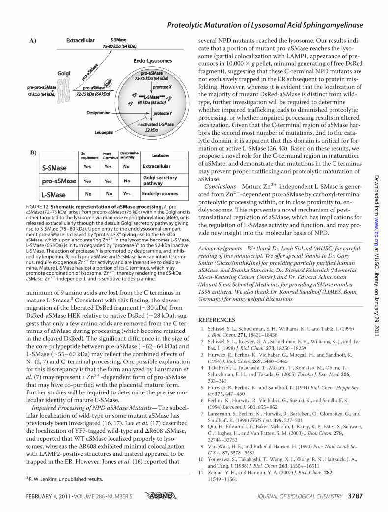
ing of aSMase has been previously described to proceed viasequential formation of prepro-aSMase (75 kDa), pro-aSMase(72 kDa), and then 70- and 52-kDa forms of aSMase (3). Gen-eration of the 70- and 52-kDa forms is absent in cells withimpaired trafficking to lysosomes (3), suggesting that either orboth the 70- and 52-kDa forms represent mature L-SMase.Desipramine promotes degradation of mature L-SMase,which proceeds through a leupeptin-sensitive protease (5).The results provided here indicate that 52-kDa aSMase isformed from the degradation of mature L-SMase (65 kDa).Desipramine induced a time-dependent loss of L-SMase ac-tivity, concomitant with the loss of 65-kDa aSMase, and theappearance of 52-kDa aSMase (Fig. 3). On the other hand,leupeptin resulted in accumulation of mature 65-kDa aSMase(as well as all precursor forms) and diminished formation of52-kDa aSMase, with an associated elevation in L-SMase ac-
FIGURE 10. Characterization of partially purified human placentalaSMase. A, equal volumes of purified L-SMase were loaded on a 4 –20% geland resolved by SDS-PAGE. One gel was processed for silver staining (see“Experimental Procedures”) and the other was processed for immunoblot-ting with �-aSMase Ab. B, an aliquot of the supernatant (SUP) from cellularextracts following IP:V5-aSMase (see Fig. 4) was loaded on a 10% gel withan aliquot of purified L-SMase and the membrane was probed with anti-aSMase antibody. The single asterisk denotes the 65-kDa aSMase. C, acidSMase activity of partially purified L-SMase was assessed with 1.0 mM EDTA(0 mM Zn2�), or 50 �M, 500 �M, or 5 mM ZnCl2 (n � 3; *, p 0.05; one-wayANOVA, Dunnett’s post-test). D, V5-aSMase immunoprecipitated from cellu-lar extracts and an aliquot of purified L-SMase were either boiled directly inLaemmli buffer (control) or digested with PNGase F (2 units/ml, 18 h, 37 °C).Equal volumes were loaded on a 7.5% gel and resolved by SDS-PAGE. Mem-branes were blotted for aSMase (1:1000) (n � 3). WCL, whole cell lysate.
Proteolytic Maturation of Lysosomal Acid Sphingomyelinase
FEBRUARY 4, 2011 • VOLUME 286 • NUMBER 5 JOURNAL OF BIOLOGICAL CHEMISTRY 3785
at MU
SC
Library, on January 29, 2011w
ww
.jbc.orgD
ownloaded from
tivity (Fig. 9). Taken together, these results indicate that ma-ture L-SMase is 65 kDa and that the 52-kDa form is an inac-tive form of aSMase generated via the action of a leupeptin-sensitive protease (Fig. 12). The identity of this thiol proteaseis currently not known, but given its capacity to regulate thelevels of L-SMase it might represent in important regulator ofsphingolipid metabolism and signaling in the endolysosomalcompartment.Significance of Removal of Carboxyl Terminus of aSMase—
A role for C-terminal modification in the regulation ofaSMase activity has previously been described whereby oxida-tion or deletion of the carboxyl-terminal Cys629 induced a4–5-fold increase in aSMase activity (8). This activation wasrecapitulated by addition of carboxypeptidase Y suggestingthat removal of C-terminal residues has a similar effect onenzyme activity. The authors suggested that C-terminal modi-fication of aSMase might serve as a post-translational mecha-nism to increase enzyme activity (8) via a mechanism similarto the cysteine switch that has been described for matrix met-alloproteinases (9). However, these studies focused on the invitromodifications to recombinant human aSMase, whichwas derived from S-SMase. Thus, this mechanism may oper-ate on the secreted S-SMase, which from our studies is shownto retain the entire C terminus. Moreover, the extracellularmilieu is more oxidized relative to the intracellular environ-ment and thus is expected to promote the proposed oxidation
of the C-terminal Cys. On the other hand, the current resultsshow that the mature lysosomal L-SMase has lost this Cys andthus is activated by removal of the Cys rather than its oxida-tion. Moreover, one could speculate that this regulatory cys-teine functions to maintain the precursor non-lysosomalaSMase in an inactive (low activity) form while en route fromthe Golgi to the lysosomes, thus, preventing activity in theenzyme in the Golgi.Processing of the C termini of proteins within the acidic
compartment has been reported for other lysosomal hydro-lases, including cathepsin D (10, 38), �-glucuronidase (38),acid �-glucosidase (39), and �-interferon-inducible lysosomalthiol reductase (GILT) (40), and has been suggested for acidceramidase (41). The current results demonstrate that car-boxyl-terminal processing of the enzyme, which would re-move the C-terminal Cys, results in formation of the activeenzyme. The mechanism of this activation remains to bedetermined as removal of Cys629 during processing of pre-aSMase to mature L-SMase could increase Zn2� affinity, sub-strate binding, and/or catalytic capacity.Despite the multiple lines of evidence provided here that
indicate C-terminal processing of aSMase, previous analysisof purified placental aSMase suggested an intact C terminus(42). However, preliminary LC-MS analysis of the partiallypurified L-SMase used in the current study (cf. Fig. 10) revealsthat the carboxyl-terminal residue is Gln620, indicating that a
FIGURE 11. Effect of C-terminal Niemann-Pick mutations on aSMase maturation. A, 2 � 105 HEK stably expressing DsRed-aSMaseWT (WT), DsRed-aSMaseR600H (R600H), DsRed-aSMaseR600p (R600P), and DsRed-aSMase�R608 (�R608) were seeded in 35-mm glass-bottom plated dishes (MatTek) in 10%FBS/MEM. The following day cells were changed to serum-free medium (2.0 ml, 0.1% BSA/MEM) overnight, before being processed for indirect immunofluo-rescence staining. Cells were fixed, permeabilized, blocked as described under “Experimental Procedures,” and stained for LAMP1 (1:100, green, �488).DsRed fluorescence was detected using the �555 channel. B, 5 � 105 HEK stables (WT, R600H, R600P, �and R608) were seeded in 60-mm dishes in 10% FBS/MEM. The following day cells were changed to serum-free medium (3.0 ml, 0.1% BSA/MEM) for 18 h. Cells were collected and processed for L-SMase activityas described under “Experimental Procedures” (n � 3; **, p 0.01; one-way ANOVA, Dunnett’s post-test). C, 2 � 106 HEK stables (WT, R600H, R600P, and�R608) were seeded in 10-cm dishes in 10% FBS/MEM and allowed to grow for 2 days. Cells were then shifted to serum-free medium (10.0 ml of 0.1% BSA/MEM per dish) for 18 h before collection. Cells were collected and prepared for differential centrifugation as described under “Experimental Procedures.”Equal volumes of the 10,000 � g pellets (heavy membrane fraction) were loaded on a 4 –20% gel and resolved by SDS-PAGE. Membranes were blotted foraSMase and DsRed, as described (n � 3). A 75-kDa band indicated by an arrow indicates an alternative cleavage product that retains the DsRed tag. Thesingle asterisk indicates the 65-kDa aSMase and the double asterisk denotes the 52-kDa aSMase.
Proteolytic Maturation of Lysosomal Acid Sphingomyelinase
3786 JOURNAL OF BIOLOGICAL CHEMISTRY VOLUME 286 • NUMBER 5 • FEBRUARY 4, 2011
at MU
SC
Library, on January 29, 2011w
ww
.jbc.orgD
ownloaded from
minimum of 9 amino acids are lost from the C terminus inmature L-Smase.3 Consistent with this finding, the slowermigration of the liberated DsRed fragment (�30 kDa) fromDsRed-aSMase HEK relative to native DsRed (�28 kDa), sug-gests that only a few amino acids are removed from the C ter-minus of aSMase during processing (which become retainedin the cleaved DsRed). The significant difference in the size ofthe core polypeptide between pre-aSMase (�62–64 kDa) andL-SMase (�55–60 kDa) may reflect the combined effects ofN- (2, 7) and C-terminal processing. One possible explanationfor this discrepancy is that the form analyzed by Lansmann etal. (7) may represent a Zn2�-dependent form of pro-aSMasethat may have co-purified with the placental mature form.Further studies will be required to determine the precise mo-lecular identity of mature L-SMase.Impaired Processing of NPD aSMase Mutants—The subcel-
lular localization of wild-type or some mutant aSMase haspreviously been investigated (16, 17). Lee et al. (17) describedthe localization of YFP-tagged wild-type and �R608 aSMase,and reported that WT aSMase localized properly to lyso-somes, whereas the �R608 exhibited minimal colocalizationwith LAMP2-positive structures and instead appeared to betrapped in the ER. However, Jones et al. (16) reported that
several NPD mutants reached the lysosome. Our results indi-cate that a portion of mutant pro-aSMase reaches the lyso-some (partial colocalization with LAMP1, appearance of pre-cursors in 10,000 � g pellet, minimal generating of free DsRedfragment), suggesting that these C-terminal NPD mutants arenot exclusively trapped in the ER subsequent to protein mis-folding. However, whereas it is evident that the localization ofthe majority of mutant DsRed-aSMase is distinct from wild-type, further investigation will be required to determinewhether impaired trafficking leads to diminished proteolyticprocessing, or whether impaired processing results in alteredlocalization. Given that the C-terminal region of aSMase har-bors the second most number of mutations, 2nd to the cata-lytic domain, it is apparent that this domain is critical for for-mation of active L-SMase (26, 43). Based on these results, wepropose a novel role for the C-terminal region in maturationof aSMase, and demonstrate that mutations in the C terminusmay prevent proper trafficking and proteolytic maturation ofaSMase.Conclusions—Mature Zn2�-independent L-SMase is gener-
ated from Zn2�-dependent pro-aSMase by carboxyl-terminalproteolytic processing within, or in close proximity to, en-dolysosomes. This represents a novel mechanism of post-translational regulation of aSMase, which has implications forthe regulation of L-SMase activity and function, and may pro-vide new insight into the molecular basis of NPD.
Acknowledgments—We thank Dr. Leah Siskind (MUSC) for carefulreading of this manuscript. We offer special thanks to Dr. GarySmith (GlaxoSmithKline) for providing partially purified humanaSMase, and Branka Stancevic, Dr. Richard Kolesnick (MemorialSloan-Kettering Cancer Center), and Dr. Edward Schuchman(Mount Sinai School of Medicine) for providing aSMase number1598 antisera. We also thank Dr. Konrad Sandhoff (LIMES, Bonn,Germany) for many helpful discussions.
REFERENCES1. Schissel, S. L., Schuchman, E. H., Williams, K. J., and Tabas, I. (1996)
J. Biol. Chem. 271, 18431–184362. Schissel, S. L., Keesler, G. A., Schuchman, E. H., Williams, K. J., and Ta-
bas, I. (1998) J. Biol. Chem. 273, 18250–182593. Hurwitz, R., Ferlinz, K., Vielhaber, G., Moczall, H., and Sandhoff, K.
(1994) J. Biol. Chem. 269, 5440–54454. Takahashi, I., Takahashi, T., Mikami, T., Komatsu, M., Ohura, T.,
Schuchman, E. H., and Takada, G. (2005) Tohoku J. Exp. Med. 206,333–340
5. Hurwitz, R., Ferlinz, K., and Sandhoff, K. (1994) Biol. Chem. Hoppe Sey-ler 375, 447–450
6. Ferlinz, K., Hurwitz, R., Vielhaber, G., Suzuki, K., and Sandhoff, K.(1994) Biochem. J. 301, 855–862
7. Lansmann, S., Ferlinz, K., Hurwitz, R., Bartelsen, O., Glombitza, G., andSandhoff, K. (1996) FEBS Lett. 399, 227–231
8. Qiu, H., Edmunds, T., Baker-Malcolm, J., Karey, K. P., Estes, S., Schwarz,C., Hughes, H., and Van Patten, S. M. (2003) J. Biol. Chem. 278,32744–32752
9. Van Wart, H. E., and Birkedal-Hansen, H. (1990) Proc. Natl. Acad. Sci.U.S.A. 87, 5578–5582
10. Yonezawa, S., Takahashi, T., Wang, X. J., Wong, R. N., Hartsuck, J. A.,and Tang, J. (1988) J. Biol. Chem. 263, 16504–16511
11. Zeidan, Y. H., and Hannun, Y. A. (2007) J. Biol. Chem. 282,11549–115613 R. W. Jenkins, unpublished results.
FIGURE 12. Schematic representation of aSMase processing. A, pro-aSMase (72–75 kDa) arises from prepro-aSMase (75 kDa) within the Golgi and iseither targeted to the lysosome via mannose 6-phosphorylation (M6P), or isreleased extracellularly through the default Golgi secretory pathway givingrise to S-SMase (75– 80 kDa). Upon entry to the endolysosomal compart-ment pro-aSMase is cleaved by “protease X” giving rise to the 65-kDaaSMase, which upon encountering Zn2� in the lysosome becomes L-SMase.L-SMase (65 kDa) is in turn degraded by “protease Y” to the 52-kDa inactiveL-SMase. The action of protease Y is promoted by desipramine, and inhib-ited by leupeptin. B, both pro-aSMase and S-SMase have an intact C termi-nus, require exogenous Zn2� for activity, and are insensitive to desipra-mine. Mature L-SMase has lost a portion of its C terminus, which maypromote coordination of lysosomal Zn2�, thereby rendering the 65-kDaaSMase, Zn2�-independent, and is sensitive to desipramine.
Proteolytic Maturation of Lysosomal Acid Sphingomyelinase
FEBRUARY 4, 2011 • VOLUME 286 • NUMBER 5 JOURNAL OF BIOLOGICAL CHEMISTRY 3787
at MU
SC
Library, on January 29, 2011w
ww
.jbc.orgD
ownloaded from
12. Rotolo, J. A., Zhang, J., Donepudi, M., Lee, H., Fuks, Z., and Kolesnick, R.(2005) J. Biol. Chem. 280, 26425–26434
13. Zeidan, Y. H., Wu, B. X., Jenkins, R. W., Obeid, L. M., and Hannun, Y. A.(2008) FASEB J. 22, 183–193
14. Toomre, D., Keller, P., White, J., Olivo, J. C., and Simons, K. (1999)J. Cell Sci. 112, 21–33
15. Jenkins, R. W., Canals, D., Idkowiak-Baldys, J., Simbari, F., Roddy, P.,Perry, D. M., Kitatani, K., Luberto, C., and Hannun, Y. A. (2010) J. Biol.Chem. 285, 35706–35718
16. Jones, I., He, X., Katouzian, F., Darroch, P. I., and Schuchman, E. H.(2008)Mol. Genet. Metab. 95, 152–162
17. Lee, C. Y., Tamura, T., Rabah, N., Lee, D. Y., Ruel, I., Hafiane, A., Iatan,I., Nyholt, D., Laporte, F., Lazure, C., Wada, I., Krimbou, L., and Genest,J. (2007) Biochemistry 46, 14969–14978
18. Presley, J. F., Cole, N. B., Schroer, T. A., Hirschberg, K., Zaal, K. J., andLippincott-Schwartz, J. (1997) Nature 389, 81–85
19. Albouz, S., Hauw, J. J., Berwald-Netter, Y., Boutry, J. M., Bourdon, R.,and Baumann, N. (1981) Biomedicine 35, 218–220
20. Kolzer, M., Werth, N., and Sandhoff, K. (2004) FEBS Lett. 559, 96–9821. Southern, J. A., Young, D. F., Heaney, F., Baumgartner, W. K., and Ran-
dall, R. E. (1991) J. Gen. Virol. 72, 1551–155722. Baird, G. S., Zacharias, D. A., and Tsien, R. Y. (2000) Proc. Natl. Acad.
Sci. U.S.A. 97, 11984–1198923. Matz, M. V., Fradkov, A. F., Labas, Y. A., Savitsky, A. P., Zaraisky, A. G.,
Markelov, M. L., and Lukyanov, S. A. (1999) Nat. Biotechnol. 17,969–973
24. Schuchman, E. H. (2007) J. Inherit. Metab. Dis. 30, 654–66325. Lee, C. Y., Lesimple, A., Denis, M., Vincent, J., Larsen, A., Mamer, O.,
Krimbou, L., Genest, J., and Marcil, M. (2006) J. Lipid Res. 47, 622–63226. Simonaro, C. M., Desnick, R. J., McGovern, M. M., Wasserstein, M. P.,
and Schuchman, E. H. (2002) Am. J. Hum. Genet. 71, 1413–141927. Quintern, L. E., and Sandhoff, K. (1991)Methods Enzymol. 197,
536–54028. Quintern, L. E., Zenk, T. S., and Sandhoff, K. (1989) Biochim. Biophys.
Acta 1003, 121–12429. Quintern, L. E., Weitz, G., Nehrkorn, H., Tager, J. M., Schram, A. W.,
and Sandhoff, K. (1987) Biochim. Biophys. Acta 922, 323–33630. Yamanaka, T., and Suzuki, K. (1982) J. Neurochem. 38, 1753–176431. Jones, C. S., Shankaran, P., and Callahan, J. W. (1981) Biochem. J. 195,
373–38232. Zou, L., Kojima, N., Kito, M., and Yagi, K. (1989) Biotechnol. Appl Bio-
chem. 11, 217–22533. He, X., Miranda, S. R., Xiong, X., Dagan, A., Gatt, S., and Schuchman,
E. H. (1999) Biochim. Biophys. Acta 1432, 251–26434. Bartelsen, O., Lansmann, S., Nettersheim, M., Lemm, T., Ferlinz, K., and
Sandhoff, K. (1998) J. Biotechnol. 63, 29–4035. Rousson, R., Parvaz, P., Bonnet, J., Rodriguez-Lafrasse, C., Louisot, P.,
and Vanier, M. T. (1993) J. Immunol. Methods 160, 199–20636. Kurth, J., and Stoffel, W. (1991) Biol. Chem. Hoppe Seyler 372, 215–22337. Sakuragawa, N. (1982) J. Biochem. 92, 637–64638. Erickson, A. H., and Blobel, G. (1983) Biochemistry 22, 5201–520539. Wisselaar, H. A., Kroos, M. A., Hermans, M. M., van Beeumen, J., and
Reuser, A. J. (1993) J. Biol. Chem. 268, 2223–223140. Arunachalam, B., Phan, U. T., Geuze, H. J., and Cresswell, P. (2000) Proc.
Natl. Acad. Sci. U.S.A. 97, 745–75041. He, X., Okino, N., Dhami, R., Dagan, A., Gatt, S., Schulze, H., Sandhoff,
K., and Schuchman, E. H. (2003) J. Biol. Chem. 278, 32978–3298642. Lansmann, S., Schuette, C. G., Bartelsen, O., Hoernschemeyer, J., Linke,
T., Weisgerber, J., and Sandhoff, K. (2003) Eur. J. Biochem. 270,1076–1088
43. Pittis, M. G., Ricci, V., Guerci, V. I., Marcais, C., Ciana, G., Dardis, A.,Gerin, F., Stroppiano, M., Vanier, M. T., Filocamo, M., and Bembi, B.(2004) Hum. Mutat. 24, 186–187
Proteolytic Maturation of Lysosomal Acid Sphingomyelinase
3788 JOURNAL OF BIOLOGICAL CHEMISTRY VOLUME 286 • NUMBER 5 • FEBRUARY 4, 2011
at MU
SC
Library, on January 29, 2011w
ww
.jbc.orgD
ownloaded from