Real-time gene delivery vector tracking in the endo-lysosomal pathway of live cells
Sensitivity to lysosome-dependent cell death is directly regulated by lysosomal cholesterol content
11
Sensitivity to Lysosome-Dependent Cell Death Is Directly Regulated by Lysosomal Cholesterol Content Hanna Appelqvist 1 *, Linnea Sandin 1 , Karin Bjo ¨ rnstro ¨m 2,3 , Paul Saftig 4 , Brett Garner 5,6 , Karin O ¨ llinger 1,7 , Katarina Ka ˚ gedal 1 1 Experimental Pathology, Department of Clinical and Experimental Medicine, Faculty of Health Sciences, Linko ¨ ping University, Linko ¨ ping, Sweden, 2 Anaesthesiology, Department of Medical and Health Sciences, Faculty of Health Sciences, Linko ¨ ping University, Linko ¨ ping, Sweden, 3 Department of Anaesthesiology UHL, County Council of O ¨ stergo ¨ tland, Linko ¨ ping, Sweden, 4 Biochemical Institute, Christian-Albrechts-University Kiel, Kiel, Germany, 5 Illawarra Health and Medical Research Institute, University of Wollongong, Wollongong, Australia, 6 School of Biological Sciences, University of Wollongong, Wollongong, Australia, 7 Department of Clinical Pathology and Clinical Genetics, County Council of O ¨ stergo ¨ tland, Linko ¨ ping, Sweden Abstract Alterations in lipid homeostasis are implicated in several neurodegenerative diseases, although the mechanisms responsible are poorly understood. We evaluated the impact of cholesterol accumulation, induced by U18666A, quinacrine or mutations in the cholesterol transporting Niemann-Pick disease type C1 (NPC1) protein, on lysosomal stability and sensitivity to lysosome-mediated cell death. We found that neurons with lysosomal cholesterol accumulation were protected from oxidative stress-induced apoptosis. In addition, human fibroblasts with cholesterol-loaded lysosomes showed higher lysosomal membrane stability than controls. Previous studies have shown that cholesterol accumulation is accompanied by the storage of lipids such as sphingomyelin, glycosphingolipids and sphingosine and an up regulation of lysosomal associated membrane protein-2 (LAMP-2), which may also influence lysosomal stability. However, in this study the use of myriocin and LAMP deficient fibroblasts excluded these factors as responsible for the rescuing effect and instead suggested that primarily lysosomal cholesterol content determineD the cellular sensitivity to toxic insults. Further strengthening this concept, depletion of cholesterol using methyl-b-cyclodextrin or 25-hydroxycholesterol decreased the stability of lysosomes and cells became more prone to undergo apoptosis. In conclusion, cholesterol content regulated lysosomal membrane permeabilization and thereby influenced cell death sensitivity. Our data suggests that lysosomal cholesterol modulation might be used as a therapeutic strategy for conditions associated with accelerated or repressed apoptosis. Citation: Appelqvist H, Sandin L, Bjo ¨ rnstro ¨ m K, Saftig P, Garner B, et al. (2012) Sensitivity to Lysosome-Dependent Cell Death Is Directly Regulated by Lysosomal Cholesterol Content. PLoS ONE 7(11): e50262. doi:10.1371/journal.pone.0050262 Editor: Kostas Vekrellis, Foundation for Biomedical Research Academy of Athens, Greece Received April 19, 2012; Accepted October 17, 2012; Published November 16, 2012 Copyright: ß 2012 Appelqvist et al. This is an open-access article distributed under the terms of the Creative Commons Attribution License, which permits unrestricted use, distribution, and reproduction in any medium, provided the original author and source are credited. Funding: This work was supported by the Swedish Research Council (2010–3463), the Deutsche Forschungsgemeinschaft, and the foundations of Olle Engqvist and A ˚ ke Wiberg. The funders had no role in study design, data collection and analysis, decision to publish, or preparation of the manuscript. Competing Interests: The authors have declared that no competing interests exist. * E-mail: [email protected] Introduction Lysosomes are acidic organelles involved in several cellular functions, including degradation of macromolecules, repair of the plasma membrane, antigen presentation, recycling of cell surface receptors and apoptosis signaling [1]. Upon a variety of cell death stimuli, lysosomal membrane permeabilization (LMP) is induced and this results in the release of lysosomal content to the cytosol. Previous studies have convincingly shown that the presence of lysosomal proteases, cathepsins, in the cytosol mediates apoptosis [2,3,4], implying that the integrity of the lysosomal membrane is of high importance for cell survival. The mechanism underlying LMP is still incompletely understood; however, a number of factors have been described to affect the stability of the lysosomal membrane, including the level of lysosome-associated membrane proteins (LAMP) and cholesterol [5]. Niemann-Pick disease type C (NPC) is a complex neurodegen- erative lysosomal storage disorder caused by mutations in the genes encoding the cholesterol transporting proteins NPC1 and NPC2. Normally, cholesterol is released from endocytosed low density lipoprotein (LDL) particles by the action of lysosomal acid lipase and is then transported, via the lysosomal NPC proteins, to the ER where it serves as a sensor for cellular cholesterol homeostasis and may be esterified [6]. Nonfunctional NPC proteins disturb cholesterol efflux from the lysosomes. Thus, NPC-mutated cells are characterized by the accumulation of unesterified cholesterol in the endo-lysosomal system [7]. Other lipids, including sphingomyelin, glycosphingolip- ids, sphingosine and bis(monoacylglycero)phosphate (BMP) accu- mulate in the lysosomes in NPC as well [8,9]. At present there is no cure for NPC, and the goal for therapeutic treatment is to diminish the lipid load. Alleviation of the NPC phenotype can be obtained by several approaches, e.g., by decreasing cholesterol levels [10], inhibiting glycosphingolipid synthesis [11] or increasing lipid degradation [12]. b-Cyclodextrin compounds has been shown to correct cholesterol transport in NPC-defective cells [13] and substantially reduce neurodegeneration and increase lifespan in Npc1 2/2 mice [14]. Several substances have the ability to decrease lysosomal cholesterol; for example, 25-hydroxycholesterol (25-HC) down-regulates cho- lesterol accumulation through homeostatic ER mechanisms by signaling cholesterol excess [15]. Lipidosis, and intracellular accumulation of phospholipids, is a side effect of certain cationic amphiphilic drugs, including quinacrine, desipramine, imipramine PLOS ONE | www.plosone.org 1 November 2012 | Volume 7 | Issue 11 | e50262
Transcript of Sensitivity to lysosome-dependent cell death is directly regulated by lysosomal cholesterol content

Sensitivity to Lysosome-Dependent Cell Death Is DirectlyRegulated by Lysosomal Cholesterol ContentHanna Appelqvist1*, Linnea Sandin1, Karin Bjornstrom2,3, Paul Saftig4, Brett Garner5,6, Karin Ollinger1,7,
Katarina Kagedal1
1 Experimental Pathology, Department of Clinical and Experimental Medicine, Faculty of Health Sciences, Linkoping University, Linkoping, Sweden, 2 Anaesthesiology,
Department of Medical and Health Sciences, Faculty of Health Sciences, Linkoping University, Linkoping, Sweden, 3 Department of Anaesthesiology UHL, County Council
of Ostergotland, Linkoping, Sweden, 4 Biochemical Institute, Christian-Albrechts-University Kiel, Kiel, Germany, 5 Illawarra Health and Medical Research Institute, University
of Wollongong, Wollongong, Australia, 6 School of Biological Sciences, University of Wollongong, Wollongong, Australia, 7 Department of Clinical Pathology and Clinical
Genetics, County Council of Ostergotland, Linkoping, Sweden
Abstract
Alterations in lipid homeostasis are implicated in several neurodegenerative diseases, although the mechanisms responsibleare poorly understood. We evaluated the impact of cholesterol accumulation, induced by U18666A, quinacrine or mutationsin the cholesterol transporting Niemann-Pick disease type C1 (NPC1) protein, on lysosomal stability and sensitivity tolysosome-mediated cell death. We found that neurons with lysosomal cholesterol accumulation were protected fromoxidative stress-induced apoptosis. In addition, human fibroblasts with cholesterol-loaded lysosomes showed higherlysosomal membrane stability than controls. Previous studies have shown that cholesterol accumulation is accompanied bythe storage of lipids such as sphingomyelin, glycosphingolipids and sphingosine and an up regulation of lysosomalassociated membrane protein-2 (LAMP-2), which may also influence lysosomal stability. However, in this study the use ofmyriocin and LAMP deficient fibroblasts excluded these factors as responsible for the rescuing effect and instead suggestedthat primarily lysosomal cholesterol content determineD the cellular sensitivity to toxic insults. Further strengthening thisconcept, depletion of cholesterol using methyl-b-cyclodextrin or 25-hydroxycholesterol decreased the stability of lysosomesand cells became more prone to undergo apoptosis. In conclusion, cholesterol content regulated lysosomal membranepermeabilization and thereby influenced cell death sensitivity. Our data suggests that lysosomal cholesterol modulationmight be used as a therapeutic strategy for conditions associated with accelerated or repressed apoptosis.
Citation: Appelqvist H, Sandin L, Bjornstrom K, Saftig P, Garner B, et al. (2012) Sensitivity to Lysosome-Dependent Cell Death Is Directly Regulated by LysosomalCholesterol Content. PLoS ONE 7(11): e50262. doi:10.1371/journal.pone.0050262
Editor: Kostas Vekrellis, Foundation for Biomedical Research Academy of Athens, Greece
Received April 19, 2012; Accepted October 17, 2012; Published November 16, 2012
Copyright: � 2012 Appelqvist et al. This is an open-access article distributed under the terms of the Creative Commons Attribution License, which permitsunrestricted use, distribution, and reproduction in any medium, provided the original author and source are credited.
Funding: This work was supported by the Swedish Research Council (2010–3463), the Deutsche Forschungsgemeinschaft, and the foundations of Olle Engqvistand Ake Wiberg. The funders had no role in study design, data collection and analysis, decision to publish, or preparation of the manuscript.
Competing Interests: The authors have declared that no competing interests exist.
* E-mail: [email protected]
Introduction
Lysosomes are acidic organelles involved in several cellular
functions, including degradation of macromolecules, repair of the
plasma membrane, antigen presentation, recycling of cell surface
receptors and apoptosis signaling [1]. Upon a variety of cell death
stimuli, lysosomal membrane permeabilization (LMP) is induced
and this results in the release of lysosomal content to the cytosol.
Previous studies have convincingly shown that the presence of
lysosomal proteases, cathepsins, in the cytosol mediates apoptosis
[2,3,4], implying that the integrity of the lysosomal membrane is of
high importance for cell survival. The mechanism underlying
LMP is still incompletely understood; however, a number of
factors have been described to affect the stability of the lysosomal
membrane, including the level of lysosome-associated membrane
proteins (LAMP) and cholesterol [5].
Niemann-Pick disease type C (NPC) is a complex neurodegen-
erative lysosomal storage disorder caused by mutations in the genes
encoding the cholesterol transporting proteins NPC1 and NPC2.
Normally, cholesterol is released from endocytosed low density
lipoprotein (LDL) particles by the action of lysosomal acid lipase and
is then transported, via the lysosomal NPC proteins, to the ER where
it serves as a sensor for cellular cholesterol homeostasis and may be
esterified [6]. Nonfunctional NPC proteins disturb cholesterol efflux
from the lysosomes. Thus, NPC-mutated cells are characterized by
the accumulation of unesterified cholesterol in the endo-lysosomal
system [7]. Other lipids, including sphingomyelin, glycosphingolip-
ids, sphingosine and bis(monoacylglycero)phosphate (BMP) accu-
mulate in the lysosomes in NPC as well [8,9]. At present there is no
cure for NPC, and the goal for therapeutic treatment is to diminish
the lipid load. Alleviation of the NPC phenotype can be obtained by
several approaches, e.g., by decreasing cholesterol levels [10],
inhibiting glycosphingolipid synthesis [11] or increasing lipid
degradation [12].
b-Cyclodextrin compounds has been shown to correct cholesterol
transport in NPC-defective cells [13] and substantially reduce
neurodegeneration and increase lifespan in Npc12/2 mice [14].
Several substances have the ability to decrease lysosomal cholesterol;
for example, 25-hydroxycholesterol (25-HC) down-regulates cho-
lesterol accumulation through homeostatic ER mechanisms by
signaling cholesterol excess [15]. Lipidosis, and intracellular
accumulation of phospholipids, is a side effect of certain cationic
amphiphilic drugs, including quinacrine, desipramine, imipramine
PLOS ONE | www.plosone.org 1 November 2012 | Volume 7 | Issue 11 | e50262

and amiodarone, used to treat e.g., depression and arrhythmias
[16,17]. The exact mechanism of action of these small lysosomo-
tropic compounds remains poorly understood, but their amphiphilic
nature allows them to accumulate in membranes and might disrupt
the activity of membrane proteins like NPC1 [18]. In addition, the
compound U18666A has been extensively used to mimic the NPC
phenotype by impairing the intracellular transport of LDL-derived
cholesterol from lysosomes [19], thus resulting in cholesterol
accumulation in this compartment.
The way in which increased lysosomal cholesterol contributes to
NPC is unknown, but it has been suggested that both lipid storage
and a concomitant inflammatory response, involving macrophages
in peripheral organs and activated glia in the central nervous
system, converge to produce the pathological lesions that
characterize the disease [10]. Recently, we reported that enhanced
lysosomal cholesterol content protects cells from LMP-dependent
apoptosis [20]. Although this finding, which has also been
confirmed by others [21], may seem counterintuitive, it is possible
Figure 1. Cholesterol modulation in human fibroblasts is associated with alterations of the lysosomal compartment. Human wtfibroblasts were treated with U18666A or quinacrine to induce cholesterol accumulation, and NPC1-mutant fibroblasts were treated with methyl-b-cyclodextrin (MbCD) or 25-hydroxy cholesterol (25-HC) to revert cholesterol storage. A) Measurement of unesterified cholesterol (n = 4) and B)representative images of filipin staining (scale bar 10 mm). C and D) Representative histogram from flow cytometric analysis of Lysotrackerfluorescence staining. M1 gate denotes the highly fluorescent population. E and F) Quantification of peak channel in the M1 population (seen in Cand D; n = 4). Data are presented as the mean 6 SD, * p#0.05.doi:10.1371/journal.pone.0050262.g001
Lysosomal Stability Is Regulated by Cholesterol
PLOS ONE | www.plosone.org 2 November 2012 | Volume 7 | Issue 11 | e50262
that cholesterol preserves the integrity of the lysosomal membrane
and thus promotes neuronal survival upon acute cellular stress.
Importantly, in both NPC1-mutant cells and U18666A treated
cells, cholesterol accumulation is associated with storage of several
other lipids [9,22], which might influence lysosomal stability. In
addition, the expression of LAMP-2 was increased by U18666A
treatment [20]. Because LAMPs have been shown to be important
for regulation of LMP [23], further studies to distinguish between
Figure 2. Manipulation of lysosomal cholesterol content modulates the cellular sensitivity to apoptosis. Cholesterol content of humanfibroblasts was modulated using U18666A, quinacrine, methyl-b-cyclodextrin (MbCD) or 25-hydroxy cholesterol (25-HC) before apoptosis wasinduced using O-methyl-serine dodecylamide hydrochloride (MSDH; 24 h). Phase contrast images of A) wt and B) NPC1-mutant fibroblasts (NPC1mut).Scale bar 20 mm. C) Viability of cultures in A and B, respectively, assessed by the MTT assay (n = 4). Viability is expressed as percentage of untreatedcultures. D) Caspase-3 like activity (n = 4–8). E) Representative curve of increase in green fluorescence during photo-oxidation of acridine orange. F)Quantification of lag time (as presented in E; n = 5–6). Data are presented as the mean 6 SD, * p#0.05.doi:10.1371/journal.pone.0050262.g002
Lysosomal Stability Is Regulated by Cholesterol
PLOS ONE | www.plosone.org 3 November 2012 | Volume 7 | Issue 11 | e50262
the LMP-modulating roles of cholesterol, sphingolipids and altered
LAMP-1 and 22 expression were undertaken. We hypothesize
that modulation of lysosomal composition affects cellular sensitiv-
ity to apoptosis and cell fate can be manipulated by the use of
agents inducing or reducing cholesterol content. We herein
provide evidence that cholesterol, and not accompanying sphin-
golipids or LAMP proteins, stabilizes lysosomes and thereby
protects from cell death.
Results
Treatment with cholesterol modifying drugs results inalterations of the lysosomal compartment
We hypothesized that modulation of lysosomal composition
affects cellular sensitivity to apoptosis and tested our theory using
human fibroblasts derived from a patient with NPC. The cells
harbor mutations in the NPC1 gene and have a negligible
expression of the NPC1 protein (results not shown). Cellular
cholesterol levels were 2-fold higher in NPC1-mutant fibroblasts
compared with wild type (wt) fibroblasts (Figure 1A). Labeling of
unesterified cholesterol using the antibiotic filipin showed a
perinuclear vesicular pattern in NPC1-mutant cells, confirming
that cholesterol accumulated in late endosomes/lysosomes
(Figure 1B). Cholesterol levels were manipulated in these two cell
models, by agents that were reported to change the lipid
composition of lysosomes. Treatment of wt fibroblasts with
U18666A or quinacrine resulted in cholesterol accumulation, as
assessed by analysis of unesterified cholesterol content and
lysosomal location was confirmed by filipin staining (Figure 1A
and B). In addition, both agents induced expansion of the
lysosomal compartment, as demonstrated by increased Lyso-
tracker fluorescence (Figure 1C and E). NPC1-mutant fibroblasts
were treated with methyl-b-cyclodextrin (MbCD) and 25-HC,
substances suggested to revert cholesterol storage. The results
verified that both agents significantly reduced cholesterol content
and decreased filipin fluorescence (Figure 1A and B). Both drugs
reduced Lysotracker fluorescence of NPC1-mutant fibroblasts
(Figure 1D and F), indicating that reversion of cholesterol load is
accompanied by normalization of the lysosomal compartment.
Cholesterol content influences lysosomal stability andaffects apoptosis sensitivity
To investigate whether lysosomal cholesterol content could be
involved in lysosomal stability and thereby affect the cellular
sensitivity to apoptosis, wt and NPC1-mutant fibroblasts were
exposed to O-methyl-serine dodecylamide hydrochloride (MSDH),
a lysosomotropic detergent previously demonstrated to induce
apoptosis via LMP [20,24]. MSDH induced a substantial loss of
viability in wt fibroblasts, while NPC1-mutant fibroblasts were less
sensitive (Figure 2A–C). Treatment of wt fibroblasts with U18666A
or quinacrine to increase lysosomal cholesterol content prior to
exposure to MSDH significantly decreased the sensitivity to
apoptosis induction (Figure 2A and C). Conversely, cholesterol
reduction in NPC1-mutant fibroblasts, induced either by MbCD or
25-HC pretreatment, increased the sensitivity to cell death
(Figure 2B and C), which further indicates that cholesterol has a
cytoprotective effect. Results obtained by the MTT viability assay
(Figure 2C) were verified by caspase-3 activity measurement
(Figure 2D) and crystal violet staining (Figure S1). This confirms
that cholesterol is an important factor in the regulation of apoptosis.
Photo-oxidation of acridine orange (AO), was applied to analyze
lysosomal membrane stability as previously described [25]. The
weak base AO accumulates in lysosomes and lysosomal rupture
can be enforced by exposure to blue light. The loss of lysosomal
integrity was measured as a distinct increase in AO-fluorescence in
the cytosol, and the lag time, from the start of laser irradiation
until rupture of lysosomes, was estimated (Figure 2E). NPC1-
mutant cells showed a longer lag time before lysosomal rupture
compared to wt cells (Figure 2F). Similarly, wt cells treated with
U18666A showed a longer lag time before lysosomal rupture
compared to untreated control wt cells (Figure 2F). This indicates
that cells with cholesterol accumulation have a more stable
lysosomal membrane. In addition, treatment of NPC1-mutant
cells with the cholesterol reducing agent MbCD resulted in a
shorter lag time before lysosomal rupture (Figure 2F), which is
consistent with decreased lysosomal membrane stability. These
results indicate that cholesterol regulates apoptosis sensitivity at the
level of LMP and is not a result of perturbation of up- or
downstream signaling.
Myriocin decreases the level of sphingomyelin in humanfibroblasts but does not affect cell death sensitivity
In addition to cholesterol, both NPC1-deficient cells and
U18666A-treated cells accumulate several other lipids, including
sphingomyelin, glycosphingolipids and sphingosine [9,22], which
have been suggested to influence the stability of lysosomes [26,27].
By employing myriocin, an inhibitor of serine palmitoyltransfer-
ase, which catalyzes the initial step in sphingolipid biosynthesis, the
levels of sphingomyelin, sphingosine and glycosphingolipids are all
reduced [28]. Sphingomyelin is the major product of the
sphingolipid biosynthetic pathway, and spectrophotometric anal-
ysis of myriocin-treated wt fibroblasts (with or without U18666A-
treatment) and NPC1-mutant fibroblasts confirmed that myriocin
was able to decrease the amount of sphingomyelin in these cells by
at least 40% (Figure 3A). Of note, in a similar experimental setting
filipin staining was demonstrated to be diminished by prolonged
myriocin treatment [22]. However, control experiments verified
that cholesterol content was not affected by myriocin treatment
(Figure 3B and C). Moreover, treatment with myriocin in our
experimental model did not change the sensitivity of cells to
MSDH-induced apoptosis (Figure 3D and E). These results were
verified by crystal violet staining (data not shown). Thus, reducing
sphingolipids in cells that maintain lysosomal cholesterol accumu-
lation does not affect LMP-induced cell death.
Lysosomal cholesterol accumulation protects corticalneurons from apoptosis
Because NPC disease is characterized by neuronal cell death
and U18666A has previously been demonstrated to be toxic to
neurons [29], we tested the effect of cholesterol accumulation in
primary cortical rat neurons. U18666A treatment induced
cholesterol redistribution into the endolysosomal system in the
neurons as evident from filipin staining (Figure 4A and B), but
there was no net increase in cellular cholesterol content
(Figure S2). The lysosomal cholesterol accumulation induced by
U18666A was non-toxic, as neither loss of viability nor activation
of caspase-3 was observed, even at concentrations up to 3 mg/ml
(Figure 4C). Importantly, U18666A-induced cholesterol accumu-
lation protected neurons from the lysosome-dependent cell death
induced by MSDH (Figure 4D and E). As oxidative stress is a
common feature of many diseases affecting the brain, the
sensitivity of neurons to H2O2-induced apoptosis was also
investigated. Cholesterol-loaded neurons were less sensitive to
oxidative stress-induced apoptosis as well (Figure 4D and E).
Lysosomal Stability Is Regulated by Cholesterol
PLOS ONE | www.plosone.org 4 November 2012 | Volume 7 | Issue 11 | e50262

Figure 3. Cholesterol, and not accumulating sphingolipids, is responsible for the apoptosis protection. Human wt fibroblasts, with orwithout U18666A treatment, and NPC1-mutant fibroblasts were treated with vehicle (dimethyl sulfoxide; DMSO) or myriocin to inhibit sphingolipidbiosynthesis. A) Sphingomyelin (n = 3), B) cholesterol content (n = 4) and C) filipin staining (scale bar 10 mm) of human fibroblasts. D) Phase contrastimages of human fibroblasts exposed to O-methyl-serine dodecylamide hydrochloride (MSDH; 24 h). Scale bar 20 mm. E) Viability of cultures in D,assessed by the MTT assay (n = 3). Viability is expressed as percentage of MSDH-untreated cultures. Data are presented as the mean 6 SD, * p#0.05,ns; non-significant.doi:10.1371/journal.pone.0050262.g003
Lysosomal Stability Is Regulated by Cholesterol
PLOS ONE | www.plosone.org 5 November 2012 | Volume 7 | Issue 11 | e50262

Cholesterol protects MEFs from apoptosis independentof LAMP expression
NPC1-mutant cells have an increased expression of LAMP-2
compared to wt fibroblasts (Figure 5A and B), and LAMP proteins
have recently been suggested to regulate the stability of the lysosomal
membrane [23]. Therefore, we decided to investigate the impor-
tance of LAMP proteins and took advantage of MEFs deficient for
either LAMP-1 (LAMP-12/2) or 22 (LAMP-22/2). Cultures were
exposed to oxidative stress induced by H2O2 addition, and a viability
analysis demonstrated that none of the MEF variants displayed any
significant difference in sensitivity to cell death compared to wt
MEFs (Figure 5C and D). In concordance, LAMP expression did not
influence lysosomal stability in MEFs, as there were no significant
differences in the lag times until lysosomal destabilization after
Figure 4. Cholesterol Accumulation in Cortical Neurons Rescues Cells from Apoptosis Induced by MSDH and Oxidative Stress.Cortical neurons were treated with U18666A. A) Filipin staining and differential interference contrast microscopy (DIC) images (scale bar 10 mm) andB) a higher magnification of filipin staining (scale bar 10 mM). C) Viability analysis and caspase-3-like activity (n = 3) after 72 h. D) Phase contrastimages (scale bar 20 mm) and E) viability analysis (MTT assay; n = 3) of cultures exposed to O-methyl-serine dodecylamide hydrochloride (MSDH) orH2O2, generated by glucose oxidase, with or without pretreatment with U18666A (48 h). Viability is expressed as percentage of untreated control.Data are presented as the mean 6 SD, * p#0.05, ns; non-significant.doi:10.1371/journal.pone.0050262.g004
Lysosomal Stability Is Regulated by Cholesterol
PLOS ONE | www.plosone.org 6 November 2012 | Volume 7 | Issue 11 | e50262

photo-oxidation of the different cell types (data not shown).
U18666A treatment rescued wt, LAMP-12/2 and LAMP-22/2
cells from oxidative stress-induced apoptosis (Figure 5C and D),
indicating that LAMP expression is not required for the protective
effect of U18666A treatment. Filipin staining verified that untreated
wt, LAMP-12/2 and LAMP-22/2 cells had relatively weak and
diffuse staining, whereas cells treated with U18666A exhibited
increased perinuclear vesicular filipin staining (Figure 5E). In
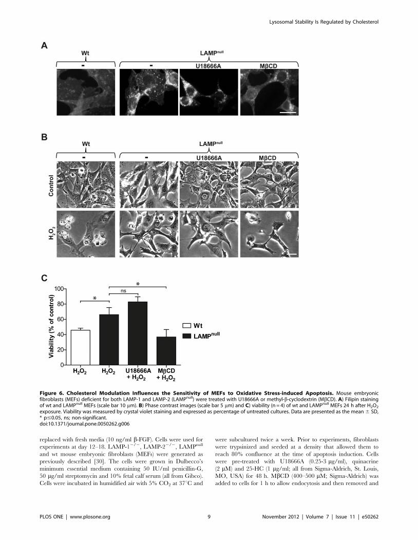
contrast to MEFs deficient for either LAMP-1 or LAMP-2, MEFs
deficient for both LAMP-1 and 22 (LAMPnull) displayed prominent,
inherent cholesterol accumulation (Figure 6A), in agreement with an
earlier study [30]. Analysis of cholesterol content demonstrated that
LAMPnull cells contained a significantly higher amount of unester-
ified cholesterol compared to wt MEFs (13.061.8 vs. 8.862.0 mg
cholesterol/mg protein; p#0.05), while cells deficient for either
LAMP-1 or LAMP-2 did not differ from wt cells. Moreover,
LAMPnull cells demonstrated a lower sensitivity than wt MEFs to
H2O2-induced cell death (Figure 6B and C). U18666A treatment
did not change the cholesterol content, as shown by filipin staining of
LAMPnull MEFs. This explains why the oxidative stress sensitivity of
LAMPnull cells was not altered by U18666A pre-treatment
(Figure 6A–C). In contrast to U18666A treatment or NPC1
mutation, cholesterol accumulation in LAMPnull MEFs is not
accompanied by the storage of other lipids [31]. Therefore, in these
cells, neither sphingolipids nor LAMP proteins could influence
lysosomal stability. Finally, we reduced the cholesterol content of
LAMPnull cells by MbCD pre-treatment. Such treatment reduced
filipin staining and sensitized cells to H2O2-induced apoptosis
(Figure 6A–C). Thus, we confirm that cholesterol accumulation
protects cells from apoptosis, and the potential protective effects of
accompanying lipids can be excluded.
Discussion
In this study we have demonstrated that cholesterol accumu-
lation stabilizes lysosomes and confers protection from acute toxic
insults induced by a lysosomotropic detergent, photo-oxidation or
oxidative stress. We provide novel mechanistic insights by showing
that neither sphingolipids, known to accumulate together with
cholesterol in lysosomes, nor LAMP proteins are involved in this
protective activity. A recent study suggested that unesterified
cholesterol modulates cellular susceptibility to ROS-induced LMP
by providing an alternative target for oxidants, thus lowering the
probability of damage to other lysosomal components [21]. Our
data regarding H2O2 exposure is consistent with this idea.
However, because our current study shows that cholesterol also
confers protection in cells exposed to the lysosomotropic
compound MSDH, although MSDH does not appear to induce
ROS production [32], an alternative explanation is that the higher
cholesterol content alters the architecture of the lysosomal
membrane, making it less sensitive to the effect of the lysosomo-
tropic detergent or oxidants. In our study, lysosomal cholesterol
levels were also shown to influence the sensitivity of lysosomes to
photo-oxidation. LAMP expression did, however, not influence
the stability of lysosomes in our experimental system, although it
was previously demonstrated that knockdown of either LAMP-1 or
LAMP-2 is sufficient to sensitize cells to photo-oxidation-induced
lysosomal destabilization [23]. LAMP-1 and 22 are estimated to
constitute approximately 50% of all lysosomal membrane proteins
[33]. Jaattela and colleagues showed that down-regulation of
LAMP proteins in human cancer cells sensitizes them to lysosomal
cell death pathways induced by various anticancer drugs,
indicating that LAMP proteins protect the lysosomal membrane
[23]. Knockdown of either LAMP-1 or LAMP-2 was sufficient to
sensitize cells to LMP in their experimental model. We found
increased expression of LAMP proteins in NPC-deficient cells in
this study and in U18666A-treated cells [20]. It is possible that the
increased expression of LAMP could contribute to the increased
lysosomal stability observed in these cells. However, the lack of
LAMP proteins did not significantly alter the sensitivity to
oxidative stress-induced apoptosis or photo-oxidation in MEFs,
whereas changes in lysosomal cholesterol had a profound effect.
As cholesterol is an important component of all cellular
membranes, including specialized lipid raft micro domains [34],
modulation of cholesterol content has the ability to induce major
changes in cell function. We suggest that cholesterol has an
important additional role in the regulation of apoptosis sensitivity
by acting at the level of permeabilization of the lysosome. In
concordance with our results, Reiners et al. conclude that
U18666A, as well as imipramine, suppresses apoptosis by
inhibiting LMP [21]. We show that alterations in cholesterol load
influences cellular sensitivity to MSDH- and oxidative stress-
induced apoptosis. MSDH is an agent that specifically targets the
lysosomal membrane and is therefore appropriate for studies of
lysosomal membrane stability. Because MSDH is an unconven-
tional apoptosis inducer, we have shown in earlier studies that
lysosomal cholesterol also protects cells from death caused by the
classical apoptosis inducers staurosporine and cisplatin [20]. If
increased cellular cholesterol content exerts its protective activity
at the lysosomes, apoptotic signaling proceeding without lysosomal
involvement should not be affected. Indeed, U18666A was shown
to only protect from cell death induced by agents that signal
apoptosis via LMP [21].
In NPC disease, all cells accumulate cholesterol in their
lysosomes, but the major clinical symptoms are due to neuronal
dysfunction. Therefore, we investigated the effect of U18666A-
induced cholesterol accumulation on apoptosis sensitivity in rat
cortical neurons. In contrast to a previously published study [29],
U18666A did not affect viability of cortical neurons in our
experimental settings. Thus, cholesterol accumulation per se is not
toxic to neurons, and cholesterol accumulation actually protected
neurons from apoptosis induced by MSDH and oxidative stress.
These results might seem contradictory, as NPC is a chronic
neurodegenerative disease (i.e., associated with neuronal death).
The reason for the neuronal vulnerability has not been elucidated,
but neurons seem to be particularly susceptible to disturbances of
lysosomal function [35,36], and cholesterol storage in lysosomes
induces additional changes in the lysosomal system. In the brains
of NPC12/2 mice, increased levels of cathepsins have been
demonstrated [37]. Furthermore, in NPC-mutant cells, fusion and
fission of late endosomes and lysosomes are reduced [38], and
vesicle trafficking is impaired [39,40]. Although cholesterol
accumulation confers protection toward acute stress, it remains
likely that the associated additional disturbances in lysosomal
function may have deleterious effects in the cell in the long run.
Noteworthy, disruption of the lysosomal system is implicated in the
development of many neurodegenerative disorders that also have a
connection to altered cholesterol homeostasis, such as Alzheimer’s,
Parkinson’s and Huntington’s diseases [35]. These disorders are
characterized by selective vulnerability of specific brain areas to
neurodegeneration and oxidative stress [41]. Interestingly, in cells
adapted to chronic oxidative stress, resistance was associated with
intracellular cholesterol accumulation. Analysis of brain tissue
reveals that stress-resistant cells in vitro showed similar features to
the less vulnerable cerebellum in mice, whereas stress-sensitive
cells resembled the highly sensitive hippocampal area [42]. These
results highlight the possibility that alterations in membrane
Lysosomal Stability Is Regulated by Cholesterol
PLOS ONE | www.plosone.org 7 November 2012 | Volume 7 | Issue 11 | e50262
cholesterol composition may be at least partly involved in the
responses allowing neurons to cope with prolonged stress.
Several factors have been suggested to be involved in the
regulation of lysosomal stability, such as the lipid composition of
the lysosomal membrane as well as lysosomal membrane proteins.
Our results demonstrate that the manipulation of lysosomal
cholesterol content can be used to modify apoptosis sensitivity.
Our data indicate that short-term lysosomal cholesterol modula-
tion might be used as a therapeutic strategy for conditions
associated with accelerated or repressed apoptosis.
Materials and Methods
Ethics statementThe animal experiments were approved by the Ethics Com-
mittee for Animal Research at Linkoping University (permit
number 101/08).
Cells and culture conditionsWt (GM05659) and NPC1-mutant (GM18436) human fibro-
blasts (passages 12–24; Coriell Institute, Camden, NJ, USA) were
cultured in Eagle’s minimum essential medium supplemented with
10% fetal calf serum, 2 mM glutamine, 50 IU/ml penicillin-G and
50 mg/ml streptomycin (all from Gibco, Paisley, UK). Cells were
incubated in humidified air with 5% CO2 at 37uC and were
subcultured once a week. Primary cultures of newborn rat neurons
were obtained essentially as described elsewhere [43]. In short,
newborn Sprauge-Dawley rats (Taconic Europe, Lilla Skensved,
Denmark) were decapitated and the cortex dissected. The cortex
tissue was sieved through a nylon mesh (80 mm) into Neurobasal A
medium, 50 IU/ml penicillin, 50 mg/ml streptomycin, 0.5 mM
glutamine and 2% B27 supplement, with addition of 5 ng/ml b-
fibroblast growth factor (b-FGF; b-fibroblast growth factor Life
Technologies, Darmstadt, Germany). Cells were plated on poly-L-
lysine coated surface at a density of 100000 cells/cm2. After 24 h,
half of the media was changed to receive a final concentration of
10 ng/ml b-FGF. Every third day, half of the medium was
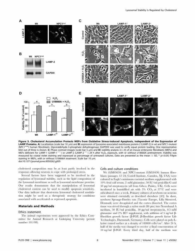
Figure 5. Cholesterol Accumulation Protects MEFs from Oxidative Stress-induced Apoptosis, Independent of the Expression ofLAMP Proteins. A) Localization (scale bar 10 mm) and B) expression of lysosome-associated membrane protein-2 (LAMP-2) in wt and NPC1-mutant(NPC1mut) human fibroblasts. Glyceraldehyde-3-phosphate dehydrogenase (GAPDH) was used to verify equal protein loading. One representativeblot out of three is shown. C) Phase contrast images (scale bar 5 mm) and D) viability analysis (n = 4) of wt mouse embryonic fibroblasts (MEFs) andMEFs deficient for LAMP-1 (LAMP-12/2) or LAMP-2 (LAMP-22/2) 24 h after H2O2 exposure, with or without U18666A pretreatment. Viability wasmeasured by crystal violet staining and expressed as percentage of untreated cultures. Data are presented as the mean 6 SD, * p#0.05) Filipinstaining in MEFs, with or without U18666A treatment. Scale bar 10 mm.doi:10.1371/journal.pone.0050262.g005
Lysosomal Stability Is Regulated by Cholesterol
PLOS ONE | www.plosone.org 8 November 2012 | Volume 7 | Issue 11 | e50262
replaced with fresh media (10 ng/ml b-FGF). Cells were used for
experiments at day 12–18. LAMP-12/2, LAMP-22/2, LAMPnull
and wt mouse embryonic fibroblasts (MEFs) were generated as
previously described [30]. The cells were grown in Dulbecco’s
minimum essential medium containing 50 IU/ml penicillin-G,
50 mg/ml streptomycin and 10% fetal calf serum (all from Gibco).
Cells were incubated in humidified air with 5% CO2 at 37uC and
were subcultured twice a week. Prior to experiments, fibroblasts
were trypsinized and seeded at a density that allowed them to
reach 80% confluence at the time of apoptosis induction. Cells
were pre-treated with U18666A (0.25-3 mg/ml), quinacrine
(2 mM) and 25-HC (1 mg/ml; all from Sigma-Aldrich, St. Louis,
MO, USA) for 48 h. MbCD (400–500 mM; Sigma-Aldrich) was
added to cells for 1 h to allow endocytosis and then removed and
Figure 6. Cholesterol Modulation Influences the Sensitivity of MEFs to Oxidative Stress-induced Apoptosis. Mouse embryonicfibroblasts (MEFs) deficient for both LAMP-1 and LAMP-2 (LAMPnull) were treated with U18666A or methyl-b-cyclodextrin (MbCD). A) Filipin stainingof wt and LAMPnull MEFs (scale bar 10 mm). B) Phase contrast images (scale bar 5 mm) and C) viability (n = 4) of wt and LAMPnull MEFs 24 h after H2O2
exposure. Viability was measured by crystal violet staining and expressed as percentage of untreated cultures. Data are presented as the mean 6 SD,* p#0.05, ns; non-significant.doi:10.1371/journal.pone.0050262.g006
Lysosomal Stability Is Regulated by Cholesterol
PLOS ONE | www.plosone.org 9 November 2012 | Volume 7 | Issue 11 | e50262

cells were chased for 24 h. This approach was shown to deplete
cholesterol from the lysosomal membrane rather than the plasma
membrane [44]. Cells were treated with myriocin (10 mM; Sigma-
Aldrich) or vehicle (dimethyl sulfoxide; DMSO) for 48 h before
analysis or apoptosis induction.
Apoptosis inductionApoptosis was induced by exposing cells to the lysosomotropic
detergent MSDH (10–15 mM; kindly provided by Gene M.
Dubowchik, Bristol-Myers Squibb, Wallingford, CT, USA),
glucose oxidase (GO; 1.6 mg/ml, Sigma-Aldrich) or H2O2 (570–
760 mM; Sigma-Aldrich). The concentrations were optimized to
induce apoptosis without necrotic contamination, as judged by
morphologic examination of cell cultures. MSDH was added in
serum-free medium for 24 h. All drugs (U18666A, quinacrine, 25-
HC, MbCD and myriocin) were omitted during the exposure.
Cells were exposed to H2O2 in serum-free medium for 2 h and
then incubated for 24 h in complete medium before analysis. GO,
an enzyme that catalyzes the oxidation of glucose and generates
H2O2, was freshly prepared prior to each experiment (1 mg/ml in
50 mM sodium acetate, pH 5.1). Neurons were exposed to GO in
complete medium for 1 h, and then the medium was exchanged to
serum free medium for 24 h.
Viability analysisAfter treatment, cell cultures were morphologically examined in
a phase contrast microscope and viability was measured using the
3-(4,5-dimethylthiazol-2-yl)-2,5-diphenyltetrazolium bromide
(MTT; Calbiochem, San Diego, CA, USA) reduction assay. Cells
were incubated with 0.25 mg/ml MTT for 2h at 37uC. The MTT
solution was then removed and the formazan product dissolved in
DMSO. The absorbance was measured at 550 nm. In addition,
the amount of surviving and thus attached cells was determined
using crystal violet staining. Cells were fixed in 4% paraformal-
dehyde for 20 min, followed by 0.04% crystal violet staining for
20 min at room temperature. The plates were washed thoroughly
by dipping in H2O and subsequently air-dried. Samples were then
solubilized in 1% Sodium dodecyl sulfate (SDS) before absorbance
was measured at 550 nm. Caspase-3-like activity was analyzed
using the substrate Ac-DEVD-AMC (Becton, Dickinson and
Company, Franklin Lakes, NJ) according to the manufacturer’s
instructions. Fluorescence was correlated to protein content.
Lipid measurementsUnesterified cholesterol content was measured in cell lysates
using the Amplex Red Cholesterol Assay Kit (Invitrogen, Paisley,
UK), as described by the manufacturer. Cholesterol amount was
correlated to protein content. Sphingomyelin content was
analyzed according to a previously described method [28].
ImmunocytochemistryCells were prepared for immuno-cytochemistry as described
elsewhere [20]. Antibodies against LAMP-2 (Southern Biotech,
Birmingham, AL, USA), followed by antibodies conjugated to
Alexa Fluor (Molecular Probes), were used. To visualize unester-
ified cholesterol, cells were stained with filipin (125 mg/ml; Sigma-
Aldrich) for 1 h at room temperature. Cover slips were washed
and mounted using Prolong gold (Invitrogen). Cells were
examined using a Nikon Eclipse E600 laser scanning confocal
microscope (Nikon, Tokyo, Japan) together with the EZC1 3.7
software (Nikon Instruments, Melville, NY, USA) or a Nikon
Eclipse TE2000U microscope (Nikon) with a Bio-Rad Radiance
2100 MP confocal system (Carl Zeiss, Jena, Germany).
Flow cytometric determination of Lysotrackerfluorescence
Cells were stained with 50 nM Lysotracker green-26 (Invitro-
gen) for 5 min at 37uC and detached by trypsinization.
Lysotracker fluorescence was analyzed in a LSR flow cytometer
(Becton Dickinson Biosciences, Franklin Lakes, NJ, USA) using a
488 nm argon laser and the resulting fluorescence was detected in
the FL1 channel using a 530628 nm filter. Data from 10000 cells
were collected and was analyzed using CellQuest software (Becton
Dickinson Biosciences).
Western blot analysisProtein separation was performed as described previously [20].
Proteins were blotted onto a nitrocellulose membrane using an
iBlot Dry Blotting System (Invitrogen). The following primary
antibodies were used: mouse anti-LAMP-2 (1:1000; Southern
Biotech, Birmingham, AL, USA) and mouse anti-glyceraldehyde-
3-phosphate dehydro-genase (GAPDH; 1:5000; Novus Biologicals,
Littleton, Co, USA).
Determination of lysosomal membrane stabilityTo analyze the integrity of lysosomes, photo-oxidation of AO
(Gurr, Poole, UK) was employed as described earlier [23]. AO is a
metachromatic dye that, when excited by blue light, emits red
fluorescence when highly concentrated inside lysosomes and green
fluorescence when diluted in the cytosol [26]. Cells seeded on
coverslips were incubated with AO (2 mg/ml) for 15 min at 37uC,
washed with phosphate buffered saline (PBS), and placed on the
stand of a Nikon Eclipse E600 laser scanning confocal microscope.
AO was excited using a 488 nm light from a 100-mW diode laser,
and loss of lysosomal proton gradient was followed by capturing
laser scanning micrographs every 330 ms in a channel defined by
bandpass filters for 495–555 nm. Green fluorescence intensity in
pre-defined areas was subsequently analyzed using Volocity
(PerkinElmer, Waltham, MA, USA) and plotted. The loss of
lysosomal integrity was determined as the lag time from the start of
blue laser irradiation until the rupture of lysosomes induced an
increase of green fluorescence in the cytosol (Figure 3E).
Statistical analysisAll experiments were repeated at least three times and the
results are presented as the means and standard deviations of
independent samples. Data were statistically evaluated using a
nonparametric Kruskal-Wallis test, followed by Mann-Whitney U
test for comparison of two groups. P values #0.05 were considered
to be significant and marked with an asterisk in figures.
Supporting Information
Figure S1 Viability of human fibroblasts after MSDH exposure
as assessed by crystal violet staining. Human wt fibroblasts were
treated with U18666A or quinacrine to induce cholesterol
accumulation, and NPC1-mutant fibroblasts were treated with
methyl-b-cyclodextrin (MbCD) or 25-hydroxy cholesterol (25-HC)
to revert cholesterol storage. Viability of cultures assessed by
crystal violet staining (n = 4). Viability is expressed as percentage of
untreated cultures. Data are presented as the mean 6 SD,
* p#0.05.
(TIF)
Figure S2 Measurement of cholesterol content in primary
neuronal cultures. Cultures of rat neurons were treated with
U18666A (0.5–3 mg/ml, 48 h) and the unesterified cholesterol
Lysosomal Stability Is Regulated by Cholesterol
PLOS ONE | www.plosone.org 10 November 2012 | Volume 7 | Issue 11 | e50262

content was measured (n = 3). Data are presented as the mean 6
SD, ns; non-significant.
(TIF)
Acknowledgments
We thank Ida Eriksson for technical assistance, Sangeeta Nath for help
with confocal microscopy and Ann-Charlotte Johansson and Alex
Schneede for scientific input.
Author Contributions
Conceived and designed the experiments: HA LS KB PS BG KO KK.
Performed the experiments: HA LS. Analyzed the data: HA LS BG KO
KK. Wrote the paper: HA LS KB PS BG KO KK.
References
1. Repnik U, Stoka V, Turk V, Turk B (2012) Lysosomes and lysosomal cathepsins
in cell death. Biochim Biophys Acta 1824: 22–33.2. Guicciardi ME, Deussing J, Miyoshi H, Bronk SF, Svingen PA, et al. (2000)
Cathepsin B contributes to TNF-alpha-mediated hepatocyte apoptosis bypromoting mitochondrial release of cytochrome c. Journal of Clinical
Investigation 106: 1127–1137.
3. Foghsgaard L, Wissing D, Mauch D, Lademann U, Bastholm L, et al. (2001)Cathepsin B acts as a dominant execution protease in tumor cell apoptosis
induced by tumor necrosis factor. Journal of Cell Biology 153: 999–1010.4. Roberg K, Kagedal K, Ollinger K (2002) Microinjection of cathepsin D induces
caspase-dependent apoptosis in fibroblasts. American Journal of Pathology 161:
89–96.5. Johansson AC, Appelqvist H, Nilsson C, Kagedal K, Roberg K, et al. (2010)
Regulation of apoptosis-associated lysosomal membrane permeabilization.Apoptosis 15: 527–540.
6. Simons K, Ikonen E (2000) How cells handle cholesterol. Science 290: 1721–1726.
7. Sokol J, Blanchette-Mackie J, Kruth HS, Dwyer NK, Amende LM, et al. (1988)
Type C Niemann-Pick disease. Lysosomal accumulation and defectiveintracellular mobilization of low density lipoprotein cholesterol. Journal of
Biological Chemistry 263: 3411–3417.8. Chevallier J, Chamoun Z, Jiang G, Prestwich G, Sakai N, et al. (2008)
Lysobisphosphatidic acid controls endosomal cholesterol levels. Journal of
Biological Chemistry 283: 27871–27880.9. Lloyd-Evans E, Platt FM (2010) Lipids on trial: the search for the offending
metabolite in Niemann-Pick type C disease. Traffic 11: 419–428.10. Liu B, Ramirez CM, Miller AM, Repa JJ, Turley SD, et al. (2010) Cyclodextrin
overcomes the transport defect in nearly every organ of NPC1 mice leading toexcretion of sequestered cholesterol as bile acid. J Lipid Res 51: 933–944.
11. Lachmann RH, te Vruchte D, Lloyd-Evans E, Reinkensmeier G, Sillence DJ, et
al. (2004) Treatment with miglustat reverses the lipid-trafficking defect inNiemann-Pick disease type C. Neurobiology of Disease 16: 654–658.
12. Devlin C, Pipalia NH, Liao X, Schuchman EH, Maxfield FR, et al. (2010)Improvement in lipid and protein trafficking in Niemann-Pick C1 cells by
correction of a secondary enzyme defect. Traffic 11: 601–615.
13. Abi-Mosleh L, Infante RE, Radhakrishnan A, Goldstein JL, Brown MS (2009)Cyclodextrin overcomes deficient lysosome-to-endoplasmic reticulum transport
of cholesterol in Niemann-Pick type C cells. Proceedings of the NationalAcademy of Sciences of the United States of America 106: 19316–19321.
14. Davidson CD, Ali NF, Micsenyi MC, Stephney G, Renault S, et al. (2009)
Chronic cyclodextrin treatment of murine Niemann-Pick C disease amelioratesneuronal cholesterol and glycosphingolipid storage and disease progression.
PLoS One 4: e6951.15. Lange Y, Ye J, Rigney M, Steck TL (1999) Regulation of endoplasmic reticulum
cholesterol by plasma membrane cholesterol. Journal of Lipid Research 40:2264–2270.
16. Klingenstein R, Lober S, Kujala P, Godsave S, Leliveld SR, et al. (2006)
Tricyclic antidepressants, quinacrine and a novel, synthetic chimera thereofclear prions by destabilizing detergent-resistant membrane compartments.
Journal of Neurochemistry 98: 748–759.17. Anderson N, Borlak J (2006) Drug-induced phospholipidosis. FEBS Lett 580:
5533–5540.
18. Kaufmann AM, Krise JP (2008) Niemann-Pick C1 functions in regulatinglysosomal amine content. Journal of Biological Chemistry 283: 24584–24593.
19. Liscum L, Faust JR (1989) The intracellular transport of low density lipoprotein-derived cholesterol is inhibited in Chinese hamster ovary cells cultured with 3-
beta-[2-(diethylamino)ethoxy]androst-5-en-17-one. Journal of Biological Chem-istry 264: 11796–11806.
20. Appelqvist H, Nilsson C, Garner B, Brown AJ, Kagedal K, et al. (2011)
Attenuation of the lysosomal death pathway by lysosomal cholesterolaccumulation. American Journal of Pathology 178: 629–639.
21. Reiners JJ Jr, Kleinman M, Kessel D, Mathieu PA, Caruso JA (2011)Nonesterified cholesterol content of lysosomes modulates susceptibility to
oxidant-induced permeabilization. Free Radic Biol Med 50: 281–294.
22. Lloyd-Evans E, Morgan AJ, He X, Smith DA, Elliot-Smith E, et al. (2008)Niemann-Pick disease type C1 is a sphingosine storage disease that causes
deregulation of lysosomal calcium. Nature Medicine 14: 1247–1255.
23. Fehrenbacher N, Bastholm L, Kirkegaard-Sorensen T, Rafn B, Bottzauw T, et
al. (2008) Sensitization to the lysosomal cell death pathway by oncogene-induced
down-regulation of lysosome-associated membrane proteins 1 and 2. Cancer
Research 68: 6623–6633.
24. Li W, Yuan X, Nordgren G, Dalen H, Dubowchik GM, et al. (2000) Induction
of cell death by the lysosomotropic detergent MSDH. FEBS Letters 470: 35–39.
25. Zdolsek JM, Olsson GM, Brunk UT (1990) Photooxidative damage to lysosomes
of cultured macrophages by acridine orange. Photochem Photobiol 51: 67–76.
26. Kagedal K, Zhao M, Svensson I, Brunk UT (2001) Sphingosine-induced
apoptosis is dependent on lysosomal proteases. Biochemical Journal 359: 335–
343.
27. Caruso JA, Mathieu PA, Reiners JJ Jr (2005) Sphingomyelins suppress the
targeted disruption of lysosomes/endosomes by the photosensitizer NPe6 during
photodynamic therapy. Biochem J 392: 325–334.
28. Glaros EN, Kim WS, Garner B (2010) Myriocin-mediated up-regulation of
hepatocyte apoA-I synthesis is associated with ERK inhibition. Clin Sci (Lond)
118: 727–736.
29. Cheung NS, Koh CH, Bay BH, Qi RZ, Choy MS, et al. (2004) Chronic
exposure to U18666A induces apoptosis in cultured murine cortical neurons.
Biochemical and Biophysical Research Communications 315: 408–417.
30. Eskelinen EL, Schmidt CK, Neu S, Willenborg M, Fuertes G, et al. (2004)
Disturbed cholesterol traffic but normal proteolytic function in LAMP-1/
LAMP-2 double-deficient fibroblasts. Mol Biol Cell 15: 3132–3145.
31. Schneede A, Schmidt CK, Holtta-Vuori M, Heeren J, Willenborg M, et al.
(2011) Role for LAMP-2 in endosomal cholesterol transport. J Cell Mol Med 15:
280–295.
32. Zhao M, Antunes F, Eaton JW, Brunk UT (2003) Lysosomal enzymes promote
mitochondrial oxidant production, cytochrome c release and apoptosis.
Eur J Biochem 270: 3778–3786.
33. Saftig P, Schroder B, Blanz J (2010) Lysosomal membrane proteins: life between
acid and neutral conditions. Biochem Soc Trans 38: 1420–1423.
34. Lingwood D, Simons K (2010) Lipid rafts as a membrane-organizing principle.
Science 327: 46–50.
35. Nixon RA, Yang DS, Lee JH (2008) Neurodegenerative lysosomal disorders: a
continuum from development to late age. Autophagy 4: 590–599.
36. Bellettato CM, Scarpa M (2010) Pathophysiology of neuropathic lysosomal
storage disorders. J Inherit Metab Dis 33: 347–362.
37. Amritraj A, Peake K, Kodam A, Salio C, Merighi A, et al. (2009) Increased
activity and altered subcellular distribution of lysosomal enzymes determine
neuronal vulnerability in Niemann-Pick type C1-deficient mice. American
Journal of Pathology 175: 2540–2556.
38. Goldman SD, Krise JP (2010) Niemann-Pick C1 functions independently of
Niemann-Pick C2 in the initial stage of retrograde transport of membrane-
impermeable lysosomal cargo. J Biol Chem 285: 4983–4994.
39. Choudhury A, Sharma DK, Marks DL, Pagano RE (2004) Elevated endosomal
cholesterol levels in Niemann-Pick cells inhibit rab4 and perturb membrane
recycling. Mol Biol Cell 15: 4500–4511.
40. Ganley IG, Pfeffer SR (2006) Cholesterol accumulation sequesters Rab9 and
disrupts late endosome function in NPC1-deficient cells. J Biol Chem 281:
17890–17899.
41. Double KL, Reyes S, Werry EL, Halliday GM (2010) Selective cell death in
neurodegeneration: why are some neurons spared in vulnerable regions? Prog
Neurobiol 92: 316–329.
42. Clement AB, Gamerdinger M, Tamboli IY, Lutjohann D, Walter J, et al. (2009)
Adaptation of neuronal cells to chronic oxidative stress is associated with altered
cholesterol and sphingolipid homeostasis and lysosomal function. J Neurochem
111: 669–682.
43. Hansson E, Ronnback L (1989) Primary cultures of astroglia and neurons from
different brain regions. In: Shahar A d, Vernadakis A, Haber B, editors. A
dissection and tissue culture manual of nervous system. 1st ed. New York: Liss.
pp. 92–104.
44. Rosenbaum AI, Zhang G, Warren JD, Maxfield FR (2010) Endocytosis of beta-
cyclodextrins is responsible for cholesterol reduction in Niemann-Pick type C
mutant cells. Proceedings of the National Academy of Sciences of the United
States of America 107: 5477–5482.
Lysosomal Stability Is Regulated by Cholesterol
PLOS ONE | www.plosone.org 11 November 2012 | Volume 7 | Issue 11 | e50262