Lipoproteins are critical TLR2 activating toxins in group B streptococcal sepsis
Upload
independentCategory
view
0download
0
MOLECULAR AND CELLULAR BIOLOGY, May 2007, p. 3337–3352 Vol. 27, No. 90270-7306/07/$08.00�0 doi:10.1128/MCB.01544-06Copyright © 2007, American Society for Microbiology. All Rights Reserved.
HOXA10 Controls Osteoblastogenesis by DirectlyActivating Bone Regulatory and Phenotypic Genes�
Mohammad Q. Hassan,1 Rahul Tare,1† Suk Hee Lee,1 Matthew Mandeville,1 Brian Weiner,1Martin Montecino,2 Andre J. van Wijnen,1 Janet L. Stein,1 Gary S. Stein,1 and Jane B. Lian1*
Department of Cell Biology and Cancer Center, University of Massachusetts Medical School, Worcester, Massachusetts 01655,1 andDepartamento de Bioquimica y Biologia Molecular, Facultad de Ciencias Biologicas, Universidad de Concepcion, Concepcion, Chile2
Received 18 August 2006/Returned for modification 4 October 2006/Accepted 9 February 2007
HOXA10 is necessary for embryonic patterning of skeletal elements, but its function in bone formationbeyond this early developmental stage is unknown. Here we show that HOXA10 contributes to osteogeniclineage determination through activation of Runx2 and directly regulates osteoblastic phenotypic genes. Inresponse to bone morphogenic protein BMP2, Hoxa10 is rapidly induced and functions to activate the Runx2transcription factor essential for bone formation. A functional element with the Hox core motif was charac-terized for the bone-related Runx2 P1 promoter. HOXA10 also activates other osteogenic genes, including thealkaline phosphatase, osteocalcin, and bone sialoprotein genes, and temporally associates with these targetgene promoters during stages of osteoblast differentiation prior to the recruitment of RUNX2. Exogenousexpression and small interfering RNA knockdown studies establish that HOXA10 mediates chromatin hyper-acetylation and trimethyl histone K4 (H3K4) methylation of these genes, correlating to active transcription.HOXA10 therefore contributes to early expression of osteogenic genes through chromatin remodeling. Impor-tantly, HOXA10 can induce osteoblast genes in Runx2 null cells, providing evidence for a direct role inmediating osteoblast differentiation independent of RUNX2. We propose that HOXA10 activates RUNX2 inmesenchymal cells, contributing to the onset of osteogenesis, and that HOXA10 subsequently supports boneformation by direct regulation of osteoblast phenotypic genes.
Patterning and development of the skeleton are complexprocesses involving signaling proteins and transcription factorsthat function as determinants for bone formation. Among theprincipal regulatory cascades for the development of the skel-eton are Hox genes that determine the position and shape of atissue element (19, 54) and the bone morphogenetic proteins(BMPs), which induce the differentiation of mesenchymal cellsto osteoblast and chondroblast lineages (18, 84). BMP2 signal-ing leads to the induction of a number of transcription factors,including RUNX2 (28) and OSTERIX (42, 56), which areessential for bone development (34, 45, 69). Several HOX andhomeodomain proteins have been identified as molecular tar-gets of BMP-mediated gene transcription during early stagesof bone formation by microarray gene expression profilingstudies (3, 33). The present study was aimed at characterizinga BMP2-inducible gene, the Homeobox a10 (HOXA10) gene,as a candidate for contributing to commitment and develop-ment of the osteoblast phenotype.
Mammalian Hox genes (homologues of the Drosophila ho-meotic genes) encode transcription factors crucial in regionaldevelopment along the anterior-posterior axis during embryo-genesis (44, 64). The mouse and human genomes contain 39Hox genes, which are grouped into four clusters, Hoxa, Hoxb,Hoxc, and Hoxd, positioned on four separate chromosomes in
13 paralogs (38, 44). Hoxa10 is a member of the Abdominal B(Abd B) class of homeobox protein-encoding genes, represent-ing the most 5� genes in the cluster consisting of paralog genesfrom Hoxa 9 to Hoxa 13 (4, 44, 50). The corresponding class ofhomeobox proteins binds preferentially to a consensus core ofTTAT or TTAC, which is distinct from the TAAT homeodo-main consensus core binding site recognized by MSX and DLXproteins (4). HOXA10 DNA binding is influenced by flankingsequences and the formation of complexes with HOXA10-interacting proteins of the MEIS and PBX classes of transcrip-tion factors, as well as other coregulatory proteins, such ashistone deacetylase 2 (13, 51, 60, 72, 74, 81).
Several of the Hox genes are essential for normal skeletaldevelopment (31, 64). The nonparalogous Hoxa10 and Hoxd11genes cooperate in the development of the forelimbs and axialskeleton and are required to globally pattern the mammalianskeleton (6, 7, 26, 68, 80). Inactivation of the paralogousHoxa10 and Hoxd10 genes results in alterations in the forma-tion of the forelimbs and hind limbs. Hoxa10�/� mice revealedan active role for the gene in modeling the femur, tibia, andfibula (12, 79). Transgenic expression of HOXA10 in pre-somitic mesoderm of the mouse resulted in vertebrae withoutribs (11). HOXA10 is expressed in the presomitic mesoderm,which develops into the axial skeleton and cooperates withother Hox genes (e.g., Hoxd11) for normal skeletal develop-ment (11, 26, 31, 64). Despite the considerable genetic evi-dence that HOXA10 has critical skeletal functions, targetgenes of HOXA10 in bone have not been identified.
In this study we have characterized HOXA10 regulation ofthe key osteogenic factor Runx2, as well as RUNX2 targetgenes, identifying Hoxa10-specific regulatory elements in pro-
* Corresponding author. Mailing address: Department of Cell Biol-ogy and Cancer Center, University of Massachusetts Medical School,55 Lake Avenue North, Worcester, MA 01655-0106. Phone: (508)856-5625. Fax: (508) 856-6800. E-mail: [email protected].
† Present address: University Orthopedics, Bone and Joint ResearchGroup, General Hospital, Southampton, United Kingdom.
� Published ahead of print on 26 February 2007.
3337
on March 13, 2016 by guest
http://mcb.asm
.org/D
ownloaded from
moters of the Runx2, osteocalcin (OC), alkaline phosphatase,and bone sialoprotein osteoblast-related genes. This discoveryof a Hox regulatory factor in activating Runx2 provides novelinsights for a mechanism that regulates a transcription factoressential for bone formation (43, 58). Although Runx2 is rap-idly induced in response to BMP2 and present in developinglimbs, somites, and mesenchymal condensations prior to chon-drogenic and osteoblast differentiation (23, 46, 76), a Smad-responsive element has not been defined. Thus, BMP2-in-duced HOXA10 represents a key regulator of Runx2transcription during embryogenesis. Our studies also show thatHOXA10 can regulate osteoblast genes independent ofRUNX2. We have thus identified an additional role forHOXA10 in postnatal bone formation and maintenance of theosteoblast phenotype. We propose that HOXA10 functions intwo capacities: as a component of a BMP2 signaling cascadeprior to RUNX2 to mediate the developmental induction ofosteogenesis and during osteoblast differentiation to regulatethe temporal expression of bone phenotypic genes to driveosteoblast maturation through mechanisms involving chroma-tin remodeling of gene promoters.
(Brian Weiner’s contribution to this paper is in fulfillment of aWorcester Polytechnic Institute undergraduate thesis project.)
MATERIALS AND METHODS
Cell cultures. C3H10T1/2 and NIH 3T3 cells were maintained in Dulbecco’sminimal essential medium (MEM) (GIBCO) supplemented with 10% fetal bo-vine serum (FBS) (Atlanta Biologicals, Georgia). C3H10T1/2 cells were inducedto osteogenesis by BMP2 (300 ng/ml). MC3T3 cells were maintained in �-MEMsupplemented with 10% FBS. Primary rat osteoblast cells were isolated fromcalvaria according to the procedures described previously (34). The rat osteo-sarcoma cell line ROS 17/2.8, representing a mature osteoblast phenotype, wasmaintained in F-12 medium supplemented with 5% FBS (70). Cells were cul-tured under osteogenic conditions with MEM (GIBCO-BRL) supplementedwith 10% FBS, 50 �g of ascorbic acid/ml, and 10 mM �-glycerol phosphate. Bonemarrow stromal cells were isolated from 8-week-old C57BL6 mice and culturedin MEM containing 10% FBS (29).
Runx2 null cells were isolated from calvarial tissues of mouse embryos (17.5days postcoitum) of the Runx2 null mouse and immortalized using mouse telom-erase (TERT). Characterization of this cell line has been described previously(1). The BMP2 used in these studies was a kind gift from John Wozney (WyethResearch, Women’s Health and Musculoskeletal Biology).
Transfection and reporter assays. The Hoxa10 expression clone containing themouse cDNA (1.2 kb) of the Hoxa10-1 variant (55 kDa) containing the transac-tivation domain was kindly provided by Richard L. Maas (Harvard MedicalSchool, Boston, MA) (4). Transient transfections were performed in six-wellplates at 50 to 70% confluence with 5 �l of FuGENE6 transfection reagent withwild-type (WT) and deleted promoter reporter DNA according to the manufac-turer’s instructions (Roche, Indianapolis, IN). A tagged Hoxa10 expression vec-tor (pcDNA3.1-Xpress-Hoxa10) was constructed was used along with a Runx2expression vector (pcDNA3.1-HA-Runx2) (85) in this study. For control of ex-pression, vector pcDNA3.1 was transfected according to the experimental con-dition. The following Runx2 WT and deletion promoter reporter plasmids (21)were used: the WT Runx2 0.6-kb fragment and the deletion series (�490, �458,�351, �288, �128) fragments. These fragments were cloned in pGL3 basicluciferase vector (Promega, Madison WI). Cells transfected with either Runx2promoter luciferase or �208 OC promoter chloramphenicol acetyltransferase(CAT) reporter constructs along with Hoxa10, Runx2, or control vector wereharvested 24 to 36 h after transfection, and all lysates were assayed for luciferaseor CAT activity according to the manufacturer’s instructions (Promega, Madi-son, WI). All results were normalized to the luciferase activity resulting fromtransfection of the promoterless pGL3 luciferase construct (Promega, Madison,WI). The OC promoter activity was assayed by using the �208-bp promoterDNA fragment from rat OC genes cloned in the pCAT basic vector (Promega,Madison, WI) (32). The percent CAT conversion was the average of values forsix similar transfection samples.
cDNA synthesis and QPCR. RNA was isolated from cultures of MC3T3,NIH 3T3, and C3H10T1/2 cells by use of TRIzol reagent (Invitrogen, Carls-bad, CA) according to the manufacturer’s protocol. cDNAs were synthesizedwith oligo(dT) primers by use of a SuperScript first-strand cDNA synthesis kit(Invitrogen) according to the manufacturer’s protocol. Gene expression wasassessed by real-time quantitative PCR (RT-QPCR) using Power SYBRgreen PCR master mix (Applied Biosystems, California). Primers used forPCRs are listed in Table 1.
Electrophoretic mobility shift analysis (EMSA). Recombinant HOXA10 pro-tein was translated using Promega TNT coupled with a rabbit reticulocyte lysatesystem (Promega). WT and mutant oligonucleotides containing the Hoxa10
TABLE 1. Primers for real-time PCR assays
Primer forindicated gene Sequence
Bone marker primers(mouse)
BSPForward ...................5�-GCA CTC CAA CTG CCC AAG A-3�Reverse ....................5�-TTT TGG AGC CCT GCT TTC TG-3�
ALPForward ...................5�-TTG TGC CAG AGA AAG AGA GAG A-3�Reverse ....................5�-GTT TCA GGG CAT TTT TCA AGG T-3�
Runx2Forward ...................5�-CGG CCC TCC CTG AAC TCT-3�Reverse ....................5�-TGC CTG CCT GGG ATC TGT A-3�
OCForward ...................5�-CTG ACA AAG CCT TCA TGT CCA A-3�Reverse ....................5�-GCG GGC GAG TCT GTT CAC TA-3�
Hoxa10Forward ...................5�-TTC GCC GGA GAA GGA CTC-3�Reverse ....................5�-TCT TTG CTG TGA GCC AGT TG-3�
Primers for ChIP(rat)
Runx2Forward ...................5�-TCA GCA TTT GTA TTC TAT CCA AAT CC-3�Reverse ....................5�-TGG CAT TCA GAA GGT TAT AGC TTT T-3�
ALPForward ...................5�-CCT GTG CAT TTC CCA ACA CGG CGG-3�Reverse ....................5�-CCA CTT CCC AGG CAG TGG AGA CAG-3�
BSPForward ...................5�-GTT TAA ATG CTT AAG TCG TTT GC-3�Reverse ....................5�-GGC TGT GGG TTC TCA CCA GAA A-3�
OCForward ...................5�-GGC AGC CTC TGA TTG TGT CC-3�Reverse ....................5�-TAT ATC CAC TGC CTG AGC GG-3�
Control 3� UTRprimers forChIP (mouse)
OCForward ...................5�-GAT CCC ATA TCA GCC AGC AC-3�Reverse ....................5�-GAC TGC CCT GGA TCA CAA GT-3�
Runx2Forward ...................5�-CGT CCA CCT GTT CCA AAG TT-3�Reverse ....................5�-GGC ATT GCC ATT TTC AGT TT-3�
BSPForward ...................5�-CCT TTT CGG TGA TTG CAG TT-3�Reverse ....................5�-AAG GTT GAG GGT GTC AGT GG-3�
ALPForward ...................5�-TTG TTC CTC TTG CCT CAG GT-3�Reverse ....................5�-TGA CAA TCA CAT GGC CTC TC-3�
Control 3� UTRprimers forChIP (rat)
OCForward ...................5�-GCA CTG CAC AGA TGT GGA AC-3�Reverse ....................5�-CAG GTT TTC CCT TTC TCA GG-3�
Runx2Forward ...................5�-TGC TTT GCA ACC AAA TCA AG-3�Reverse ....................5�-TCT GAA GGG AAG CTT TGG AA-3�
BSPForward ...................5�-TGG AAG ATG CTT GAT GAC CA-3�Reverse ....................5�-AAG GGG TCA GAG GAC AAG GT-3�
ALPForward ...................5�-CCA GTG TGA TCC CCA GAA CT-3�Reverse ....................5�-TGT CTG TAG CAA TCC CAC CA-3�
3338 HASSAN ET AL. MOL. CELL. BIOL.
on March 13, 2016 by guest
http://mcb.asm
.org/D
ownloaded from

binding site derived from the Runx2 P1 promoter site 1 (WT, 5�G CAT TCAGAA GGT TAT AGC TTT 3�; mutant, 5�G CAT TCA GAA GGC GAT AGCTTT 3� [underlining indicates the HOXA10 binding site, and boldface indicatesthe mutation]) were end labeled with [�-32P]ATP by use of T4 polynucleotidekinase (New England Biolabs, Massachusetts). The detailed procedures havebeen described previously (34). Anti-HOXA10 (N20) or a nonspecific antibody(either �-actin or normal immunoglobulin G [IgG] as indicated) (Santa Cruz)were used for the immunoshift studies. Complexes were visualized by autora-diography of a 6.5% acrylamide gel.
Immunoblotting. Each well of a six-well plate was lysed in 50 �l lysis buffer(2% sodium dodecyl sulfate [SDS], 10 mM dithiothreitol, 10% glycerol, 12%urea, 10 mM Tris-HCl [pH 7.5], 1 mM phenylmethylsulfonyl fluoride, 1� pro-tease inhibitor cocktail [Roche], 25 �M MG132 [proteosome inhibitor fromCalbiochem]) and boiled for 5 min. Equal amounts of total protein were ana-lyzed by SDS-polyacrylamide gel electrophoresis and probed with suitable anti-bodies. Immunocomplexes were detected using Western Lightning chemilumi-nescence reagent (Perkin Elmer, Boston, MA).
Antibodies. The following antibodies were purchased from Santa Cruz Bio-technology. HOXA10 N20 (SC-17158) was for chromatin immunoprecipitation(ChIP) and EMSA, and A20 (5C-17159) was for Western blotting. RUNX2antibodies were PEBP2�A (M-70 [SC-10758]), PEBP2�A (C-19 [SC-8566]), andactin (I-19 [SC-1616]). Mouse monoclonal RUNX2 antibody was a generous giftfrom Yoshi Ito and Kosei Ito (National University, Singapore, Republic ofSingapore). Anti-hyperacetylated histone H4 (Penta) and anti-trimethyl histoneH3 (Lys4), clone MC315, were purchased from Upstate Cell Signaling Solutions(Charlottesville, VA). The mouse monoclonal anti-Xpress antibody was obtainedfrom Invitrogen (Carlsbad, CA). Mouse monoclonal antibody against RNA poly-merase II (Pol II) (clone 8WG16) was obtained from Covance (Princeton, NJ)and used in ChIP studies.
ChIP assays. The procedure for ChIP in primary rat osteoblasts has alreadybeen described (34). Control primer pairs from 3� untranslated regions (UTR) ofthe genes were used to verify specific and nonspecific binding of DNA fragments(Table 1). IgG antibody was used as a control for nonspecific pull-down ofimmunocomplexes. Sequential ChIP studies were performed using the primarypull-down from one antibody, which was divided into equal aliquots for thesecond pull-down with antibodies specific for coregulatory molecules. Instead ofbeing eluted in 1% SDS and 100 mM Na2HCO3 after cross-linking and washing,immunocomplexes were eluted in 10 mM dithiothreitol. The eluate was furtherdiluted 1:40 in ChIP dilution buffer (0.01% SDS, 1.1% Triton X-100, 1.2 mMEDTA, 167 mM Tris-HCl [pH 8.1], 167 mM NaCl) and used for the secondimmunoprecipitations. Aliquots (2 to 3 �l) of DNA samples from differentpull-downs were assayed by either radioactive labeling or RT-QPCR using PowerSYBR green PCR master mix (Applied Biosystems, California) for the detectionof specific DNA fragments with primers in the proximal promoters of bone-related genes that encompass the Hoxa10 binding sites (Table 1).
Immunohistochemistry and immunofluorescence. Long bones from normalnewborn mice were fixed with 4% paraformaldehyde in 0.1 M cacodylate buffer,pH 7.4, for 48 h, dehydrated, and embedded in paraffin by standard procedures.Paraffin-embedded tissues (5-�m sections) were immunoperoxidase labeled andblocked by 0.3% H2O2 in absolute methanol for 30 min at room temperature. Toreveal antigens, sections were put in a 1 mM Tris solution, pH 9.0, supplementedwith 0.5 mM EGTA. The sections for RUNX2 immunohistochemistry wereheated in a microwave oven for 10 min after EGTA treatment, while a normalsteaming antigen retrieval method was used for HOXA10. Nonspecific immu-noglobulin binding was prevented by incubating the sections in 50 mM NH4Cl for30 min followed by blocking with phosphate-buffered saline supplemented with1% bovine serum albumin, 0.05% saponin, and 0.2% gelatin. Serial sections wereincubated overnight at 4°C with anti-rabbit RUNX2 (M-70 and C-19) or anti-goat HOXA10 (N20) antibody diluted (1:100) in phosphate-buffered saline sup-plemented with 0.1% bovine serum albumin and 0.3% Triton X-100. Equalamounts of the respective blocking peptides (2 �g/ml) for HOXA10 and RUNX2were also used. Normal IgG was used as a nonspecific control. Immunolabelingcontrols were performed by using antibodies preabsorbed with immunizing pep-tides. Labeling was visualized with the horseradish peroxidase-conjugated sec-ondary antibody (P448, 1:200; Dako).
MC3T3 cells were plated at a density of 0.6 � 105 cells/well on gelatin-coatedcoverslips in six-well plates. Cells were processed for in situ immunofluorescenceanalyses, which were carried out as described previously (39). HOXA10 wasdetected by a goat polyclonal antibody at a dilution of 1:100 (Santa Cruz Bio-technology). The secondary antibody used was Alexa 488–anti-goat antibody(Molecular Probes) at a dilution of 1:800. For overexpressed HOXA10 protein,HeLa cells at 0.5 � 105cells/well on gelatin-coated coverslips were transfectedwith 0.5 �g of Hoxa10 expression construct, and 24 h after transfection the
coverslip was processed for immunofluorescence study (39). Xpress-HOXA10was detected by a mouse monoclonal antibody against the Xpress tag at adilution of 1:3,000 (Invitrogen). The secondary antibody used was Alexa 568–anti-mouse antibody (Molecular Probes, Eugene, OR) at a dilution of 1:800.Slides were examined on a Zeiss Axioplan 2 microscope fitted with epifluores-cence (Carl Zeiss, Jena, Germany) attached to a charge-coupled-device camera.Images were saved and processed using Metamorph imaging software, version6.1 (Universal Imaging, Downingtown, PA).
RNA interference (RNAi) of Hoxa10. The mouse MC3T3-E1 osteoblastic cellsat 30 to 50% confluence were transfected using Oligofectamine (Invitrogen LifeTechnologies) with small interfering RNA (siRNA) duplexes specific for murineHoxa10 r(CCA AAU UAU CCC ACA ACA A)dTdT and r(UUG UUG UGGGAU AAU UUG G)dCdG obtained from QIAGEN Inc. (Stanford, CA). Sixdifferent sets of siRNA duplexes at different concentrations were used to eval-
FIG. 1. Hoxa10 expression in relation to osteogenic differentiation.(A) Premyogenic C2C12 cells were treated with 100 ng/ml BMP2 for24 h. Total RNA was isolated at different time points (0, 1, 2, 4, 6, 8,12, 16, 20, and 24 h). Five micrograms of total RNA was reversetranscribed with oligo(dT) primer and amplified (RT-QPCR) by gene-specific primers. (Top) Hoxa10 expression (�BMP, �BMP) was nor-malized to Gapdh expression (�BMP, �BMP), and relative transcriptlevels were plotted; (bottom) the induction of Runx2 expression withBMP2 treatment is shown for the same time course. Error bars rep-resent triplicate analyses of each sample from two independent exper-iments. (B) Temporal expression of Hoxa10 and the osteogenic mark-ers Runx2 and ALP during MC3T3 cell growth and differentiation.cDNAs from different time points were amplified using bone-specificgene primers (Table 1). Expression values from RT-QPCR were nor-malized to Gapdh values. Error bars represent triplicate sample anal-yses from one experiment. Independent experiments exhibited similartemporal expressions (data not shown).
VOL. 27, 2007 MECHANISMS FOR HOXA10 REGULATION OF OSTEOGENESIS 3339
on March 13, 2016 by guest
http://mcb.asm
.org/D
ownloaded from

uate the target specificity and knockdown efficiency. The cells were also trans-fected with control siRNA duplexes specific for green fluorescent protein by useof the same concentrations to check the transfection efficiency. The siRNAexperiment was carried out for 72 h. Total RNA and proteins from the specificsiRNA oligonucleotide-treated, untreated, Oligofectamine-treated and nonspe-cific-oligonucleotide-treated cells were analyzed by Western blotting and RT-QPCR. To study chromatin modifications after Hoxa10 siRNA treatment, cellswere harvested 72 h posttransfection and used for immunoprecipitation withtrimethyl histone H3K4 and hyperacetylated H4 antibody for ChIP analyses.
RESULTS
Hoxa10 is a BMP2-responsive gene correlating to the induc-tion of Runx2-mediated osteogenesis. Hoxa10 was identified asan early BMP2-responsive gene in an Affymetrix cDNA pro-filing study of BMP2-mediated osteogenic induction of pre-myogenic C2C12 cells (3). During the 24-h time course,Hoxa10 expression clustered into a group of genes in which theosteogenic transcription factor Runx2 was also present. Wecarried out independent experiments in this study and quanti-tated gene expression by RT-QPCR to validate the microarrayreport (Fig. 1A). Both Hoxa10 and Runx2 mRNAs are presentat very low levels in untreated cells, immediately upregulatedby BMP2 within 2 h, and then partly downregulated, but theyexhibit a second spike at 6 h. After the 8-h commitment pointto osteogenesis, when bone phenotypic markers (alkalinephosphatase and OC) are expressed (3), Hoxa10 mRNA re-mains at a steady state, while Runx2 continuously increases.
To further evaluate the expression of Hoxa10 and Runx2
during the development of the osteoblast phenotype, we ex-amined the temporal expression of Hoxa10 during the differ-entiation of osteoprogenitor MC3T3 cells. Hoxa10 is endog-enously expressed at low levels in the proliferating cells (day 7)and is induced four- to fivefold upon the maturation of thephenotype, concurrent with cellular multilayering (day 10 inthis experiment) (Fig. 1B). Peak mRNA expression levels ofHoxa10 are transient and downregulated by 50% by day 16, theonset of mineral deposition in MC3T3 cells. Expression ofRunx2 and alkaline phosphatase phenotypic genes continu-ously increases during differentiation. These observations sug-gest that Hoxa10 expression is important at the early stages ofosteoblast differentiation. We further showed that either treat-ment of pluripotent C3H10T1/2 cells with BMP2 or growth ofadherent bone marrow cells in osteogenic media results in atwofold increase in Hoxa10 concomitant with the expression ofthe osteoblast phenotype (data not shown). These studies im-plicate HOXA10 in establishing early events in the develop-ment of the osteoblast phenotype.
To appreciate the role of HOXA10 in osteogenesis in vivo,we examined its expression in bone cell populations in situ inrelation to endochondral bone formation and skeletal devel-opment. Sections of long bones and vertebrae from newbornmice were treated with specific antibodies to detect RUNX2and HOXA10 (Fig. 2). We found strong expression ofHOXA10 in the periosteum, the hypertrophic chondrocytezone of the growth plate, and osteoblasts on all bone surfaces.
FIG. 2. Coexpression of HOXA10 and RUNX2 in vivo and in vitro. (A) HOXA10 and RUNX2 are visualized in hypertrophic chondrocytesand osteoblast lineage cells in long bones and vertebrae as indicated. Immunohistochemistry was performed using anti-HOXA10 antibody andanti-RUNX2 antibody. For nonspecific controls, normal rabbit IgG was substituted for primary antibody, and blocking peptides specific for theHOXA10 and RUNX2 antibodies were used. Bone and vertebra sections are shown at �10 and �40 magnification as indicated for the boxed areasat the growth plate and the primary spongiosa. Trabeculae show robust expression of both proteins in surface osteoblasts. The first column showspeptide blocking controls (magnification, �10). (B) In situ immunofluorescence examination of endogenous HOXA10 protein in MC3T3 cells todemonstrate nuclear staining with few cytoplasmic foci using the Santa Cruz antibody. (C) Nuclear localization of transfected Xpress-taggedHOXA10 is also observed for HeLa cells detected with anti-Xpress antibody (see Materials and Methods). DAPI, 4�,6�-diamidino-2-phenylindole.
3340 HASSAN ET AL. MOL. CELL. BIOL.
on March 13, 2016 by guest
http://mcb.asm
.org/D
ownloaded from
RUNX2 was also expressed in these cell populations. Weakexpressions of HOXA10 and RUNX2 were found in the flat-tened cells that represent the prehypertrophic chondrocytephenotype. It is noteworthy that both HOXA10 and RUNX2were absent from the epiphyseal chondrocytes that representpermanent hyaline cartilage. Similar observations were madefor vertebrae tissue. We conclude from these studies thatRUNX2 and HOXA10 are expressed in osteogenic lineagecells and maturing chondrocytes at the growth plate in both theaxial and appendicular skeletons.
While most immunohistochemical staining is nuclear, somehypertrophic chondrocytes and osteoblasts have cytoplasmicstaining representing either background or specific HOXA10staining. To address a potential artifact of cytoplasmic staining,we examined the endogenous localization of HOXA10 by in situimmunofluorescence in isolated osteoblasts. Punctate nuclearstaining was observed, with a few foci detected in the cytoplasmsof MC3T3 cells (Fig. 2B). However, 100% nuclear localizationwas observed with exogenously expressed HOXA10 protein inHeLa cells by use of the Xpress tag antibody (Fig. 2C). Thesefindings indicate that the localization of the HOXA10 isoformwith transactivation activity is nuclear.
The coordinate BMP2 induction of Hoxa10 and Runx2 inresponse to BMP2-induced osteogenesis in several cell models,taken together with the profiles of expression of these genes
during the maturation of MC3T3 cells as well as their coex-pression in osteogenic lineage cells, suggests a functional rela-tionship between RUNX2 and HOXA10.
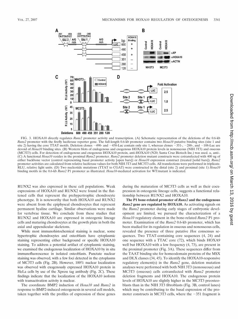
The P1 bone-related promoter of Runx2 and the endogenousRunx2 gene are regulated by HOXA10. As activating signals onthe Runx2 promoter during early stages of embryonic devel-opment are limited, we pursued the characterization of aHoxa10 regulatory element in the bone-related Runx2 P1 pro-moter. Examination of the Runx2 0.6-kb promoter, which hasbeen studied for its regulation in osseous and nonosseous cells,revealed the presence of three putative Hox consensus se-quences. Two TTAT-containing core sequences (4, 24) andone sequence with a TTAC core (72), which binds HOXA9well but HOXA10 with a low frequency (4, 73), are present inthe proximal promoter (Fig. 3A). These sequences differ fromthe TAAT binding site for homeodomain proteins of the MSXand DLX classes (34, 45). To identify the HOXA10-responsiveregulatory element(s) in the Runx2 gene, deletion mutationanalyses were performed with both NIH 3T3 (nonosseous) andMC3T3 (osseous) cells cotransfected with Runx2 promoterdeletion fragments and HOXA10. The endogenous proteinlevels of HOXA10 are slightly higher in the MC3T3 preosteo-blasts than in the NIH 3T3 fibroblasts (Fig. 3B, control lanes)which may be contributing to the basal expression of the pro-moter constructs in MC3T3 cells, where the �351 fragment is
FIG. 3. HOXA10 directly regulates Runx2 promoter activity and transcription. (A) Schematic representation of the deletions of the 0.6-kbRunx2 promoter with the firefly luciferase reporter gene. The full-length 0.6-kb promoter contains two Hoxa10 putative binding sites (site 1 andsite 2) having the core TTAT motifs. Deletion clones �490- and �458-Luc contain only site 1, whereas clones �351-, �288-, and �108-Luc aredevoid of Hoxa10 binding sites. (B) Western blots of endogenous and exogenous HOXA10 protein levels in nonosseous (NIH 3T3) and osseous(MC3T3) cells. For detection of endogenous and exogenous HOXA10 protein, anti-HOXA10 (N20; Santa Cruz Biotech Inc.) was used. �, anti-.(C) A functional Hoxa10 resides in the proximal Runx2 promoter. Runx2 promoter deletion mutant constructs were cotransfected with 400 ng ofeither backbone vector (control representing basal promoter activity [open bars]) or Hoxa10 expression construct (treated [solid bars]). Runx2promoter activities are calculated from relative luciferase values for both NIH 3T3 and MC3T3 cells. All transfections were performed in triplicate.RLU, relative light units. (D) Two nucleotide mutations (TTAT to CGAT) were constructed in the distal (site 2) and proximal (site 1) Hoxa10binding motifs in the 0.6-kb Runx2 P1 promoter as illustrated. Hoxa10-mediated activation for WT/mutant is indicated.
VOL. 27, 2007 MECHANISMS FOR HOXA10 REGULATION OF OSTEOGENESIS 3341
on March 13, 2016 by guest
http://mcb.asm
.org/D
ownloaded from

reduced 20% (Fig. 3C). In the presence of exogenousHOXA10, a 2- to 2.5-fold increase in promoter activity of the�600, �490, and �458 fragments is observed, suggesting thatthe proximal TTAT site 1 is sufficient for HOXA10 regulationof Runx2 (Fig. 3C). Furthermore, complete loss of HOXA10responsiveness occurs with the �351 Runx2-Luc fragment,which lacks the putative site 1 for HOXA10 binding. Further-more, mutation of site 1 blocked HOXA10-mediated tran-scriptional activation (Fig. 3D). The site 2 mutation had noeffect on HOXA10 induction of Runx2, i.e., the construct re-tained the same 2.5-fold activation level as the WT. The TTACcore site, located at �420/�417, cannot be functional, as thesite 1 mutation showed no HOXA10-induced change in pro-moter activity. Taken together, these results are consistent withthe location of a functional Hoxa10 site in the proximal pro-moter fragment at �353/�351, a regulatory element mediatingHOXA10 activation of the Runx2 P1 promoter.
Sequence-specific binding of HOXA10 to the proximal reg-ulatory motif was validated in EMSA by WT and mutant oli-gonucleotide binding studies (Fig. 4A and B). Confirming themutation studies, site 2 does not form a HOX protein-DNAcomplex with osteoblast nuclear extracts (Fig. 4B). In con-trast, site 1 binds in vitro-transcribed and -translated (IVTT)HOXA10 protein and HOXA10 from nuclear extracts of os-teoblasts, as indicated by antibody supershifts (Fig. 4B). Torule out the possibility of redundant HOX protein binding tosite 1 because other HOX factors are expressed in osteoblasts(20, 30), we further examined the interaction of other paralo-gous HOX proteins expressed in MC3T3 cells with Runx2 site1. Antibody specificities were established by testing IVTTHOXA10 protein bound to probe with supershift analyses us-ing antibodies specific to other Abd B class proteins. None ofthese antibodies cross-reacted with HOXA10 (Fig. 4C, leftpanel), nor did they supershift/block shift the HOXA10 com-plex from nuclear extracts bound to Runx2 site 1 (Fig. 4C, rightpanel). In conclusion, the deletion and site-directed mutagen-esis of putative Hox elements in the 0.6-kb Runx2 promoterdemonstrates that HOXA10 protein binds preferentially to theRunx2 TTAT site 1.
To address the biological significance of the HOXA10-me-diated positive regulation of the Runx2 gene at the onset ofosteoblast differentiation, cellular levels of HOXA10 were in-hibited by siRNA knockdown in two osteogenic cell models.Preosteoblastic MC3T3 cells were treated with several Hoxa10siRNA duplex oligonucleotides for 72 h, which resulted in theinhibition of HOXA10 cellular protein levels compared withthe nonsilencing duplex control. Figure 5A shows results fortwo siRNAs, one of which only modestly decreased HOXA10protein (siRNA2; decrease of 10 to 15% from densitometrymeasurements), while siRNA1 exhibited a robust dose-depen-dent inhibitory effect on HOXA10 and also caused a significantdose-related decrease in RUNX2 protein (Fig. 5B).
The potency of HOXA10 in regulating Runx2 at the onset ofBMP2-induced osteogenesis was examined with C2C12 cells(Fig. 5C). Cells were first treated with siRNA for 48 h, whichwas followed by BMP2 for 2 h. Hoxa10 siRNA resulted in a50% knockdown of Hoxa10, a decrease which prevented thetwofold BMP2 induction of Hoxa10 mRNA in the control(absence of Hoxa10 siRNA). Runx2 was reduced nearly 75% byHoxa10 siRNA in the absence of BMP2. In the presence of
BMP2 and Hoxa10 siRNA, Runx2 was not induced to a levelover that seen for the control. However, the inhibition was less(33% decrease) than in the absence of BMP2 (50% de-crease). This finding is likely the consequence of BMP2 induc-tion of other transcription factors that can upregulate Runx2.Thus, we were prompted to examine levels of Dlx3, a BMP2early response gene in this cell model (35). Dlx3 was inducedover the control in the presence of Hoxa10 siRNA and BMP2(data not shown). These studies define HOXA10 as a signifi-
FIG. 4. Specificity of the Hoxa10 site 1 for HOXA10 interactions.(A) Oligonucleotide sequences are shown for the WT and the Hoxa10site mutations and compared to that for the mouse consensus optimalHoxa10 binding site (4). Underlining indicates the core binding site;lowercase indicates mutant nucleotides. (B) DNA binding activity ofHOXA10 is demonstrated by EMSA. Ten femtomoles of labeled dou-ble-stranded oligonucleotides derived from the WT or mutant (Mut)probe as indicated was incubated with 2 �l of IVTT HOXA10 or 5 �gof MC3T3 nuclear extract (NE). Self-competition (Self), antibody su-pershift, and nonspecific antibody (NS Ab) controls are shown. Themutant double-stranded oligonucleotide for Hoxa10 does not bindnuclear proteins. The top unlabeled arrows indicate the supershiftedband; the lower unlabeled arrows show the position of the HOXA10-specific band, below which is a nonspecific complex (arrows labeledNS). �, anti-; C, control. (C) IVTT of indicated HOX proteins iscomplexed with the Hox site 1 in Runx2 to show the specificity of theHOXA10 antibody. Nuclear extracts of MC3T3 osteoblasts were usedin EMSA studies to show the specificity of the HOXA10 interaction(arrow) with the site 1 Runx2 probe. GFP, green fluorescent protein;EV, empty vector.
3342 HASSAN ET AL. MOL. CELL. BIOL.
on March 13, 2016 by guest
http://mcb.asm
.org/D
ownloaded from
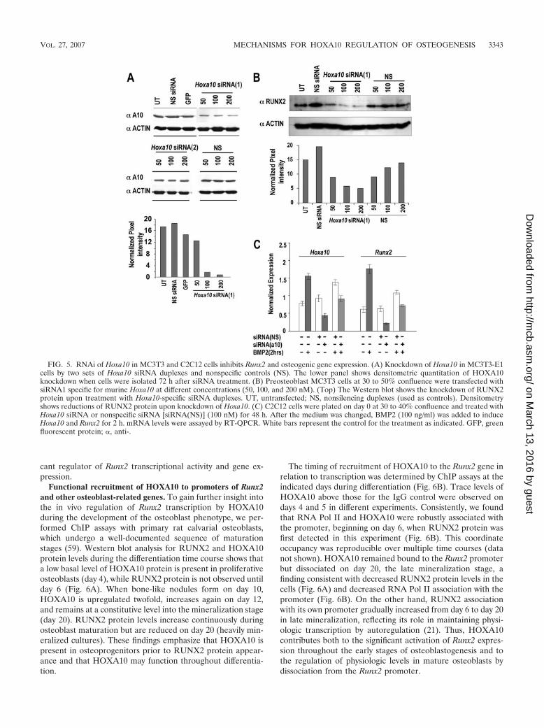
cant regulator of Runx2 transcriptional activity and gene ex-pression.
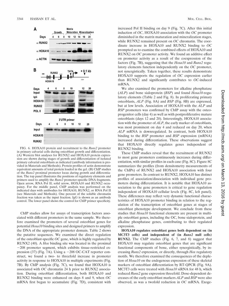
Functional recruitment of HOXA10 to promoters of Runx2and other osteoblast-related genes. To gain further insight intothe in vivo regulation of Runx2 transcription by HOXA10during the development of the osteoblast phenotype, we per-formed ChIP assays with primary rat calvarial osteoblasts,which undergo a well-documented sequence of maturationstages (59). Western blot analysis for RUNX2 and HOXA10protein levels during the differentiation time course shows thata low basal level of HOXA10 protein is present in proliferativeosteoblasts (day 4), while RUNX2 protein is not observed untilday 6 (Fig. 6A). When bone-like nodules form on day 10,HOXA10 is upregulated twofold, increases again on day 12,and remains at a constitutive level into the mineralization stage(day 20). RUNX2 protein levels increase continuously duringosteoblast maturation but are reduced on day 20 (heavily min-eralized cultures). These findings emphasize that HOXA10 ispresent in osteoprogenitors prior to RUNX2 protein appear-ance and that HOXA10 may function throughout differentia-tion.
The timing of recruitment of HOXA10 to the Runx2 gene inrelation to transcription was determined by ChIP assays at theindicated days during differentiation (Fig. 6B). Trace levels ofHOXA10 above those for the IgG control were observed ondays 4 and 5 in different experiments. Consistently, we foundthat RNA Pol II and HOXA10 were robustly associated withthe promoter, beginning on day 6, when RUNX2 protein wasfirst detected in this experiment (Fig. 6B). This coordinateoccupancy was reproducible over multiple time courses (datanot shown). HOXA10 remained bound to the Runx2 promoterbut dissociated on day 20, the late mineralization stage, afinding consistent with decreased RUNX2 protein levels in thecells (Fig. 6A) and decreased RNA Pol II association with thepromoter (Fig. 6B). On the other hand, RUNX2 associationwith its own promoter gradually increased from day 6 to day 20in late mineralization, reflecting its role in maintaining physi-ologic transcription by autoregulation (21). Thus, HOXA10contributes both to the significant activation of Runx2 expres-sion throughout the early stages of osteoblastogenesis and tothe regulation of physiologic levels in mature osteoblasts bydissociation from the Runx2 promoter.
FIG. 5. RNAi of Hoxa10 in MC3T3 and C2C12 cells inhibits Runx2 and osteogenic gene expression. (A) Knockdown of Hoxa10 in MC3T3-E1cells by two sets of Hoxa10 siRNA duplexes and nonspecific controls (NS). The lower panel shows densitometric quantitation of HOXA10knockdown when cells were isolated 72 h after siRNA treatment. (B) Preosteoblast MC3T3 cells at 30 to 50% confluence were transfected withsiRNA1 specific for murine Hoxa10 at different concentrations (50, 100, and 200 nM). (Top) The Western blot shows the knockdown of RUNX2protein upon treatment with Hoxa10-specific siRNA duplexes. UT, untransfected; NS, nonsilencing duplexes (used as controls). Densitometryshows reductions of RUNX2 protein upon knockdown of Hoxa10. (C) C2C12 cells were plated on day 0 at 30 to 40% confluence and treated withHoxa10 siRNA or nonspecific siRNA [siRNA(NS)] (100 nM) for 48 h. After the medium was changed, BMP2 (100 ng/ml) was added to induceHoxa10 and Runx2 for 2 h. mRNA levels were assayed by RT-QPCR. White bars represent the control for the treatment as indicated. GFP, greenfluorescent protein; �, anti-.
VOL. 27, 2007 MECHANISMS FOR HOXA10 REGULATION OF OSTEOGENESIS 3343
on March 13, 2016 by guest
http://mcb.asm
.org/D
ownloaded from
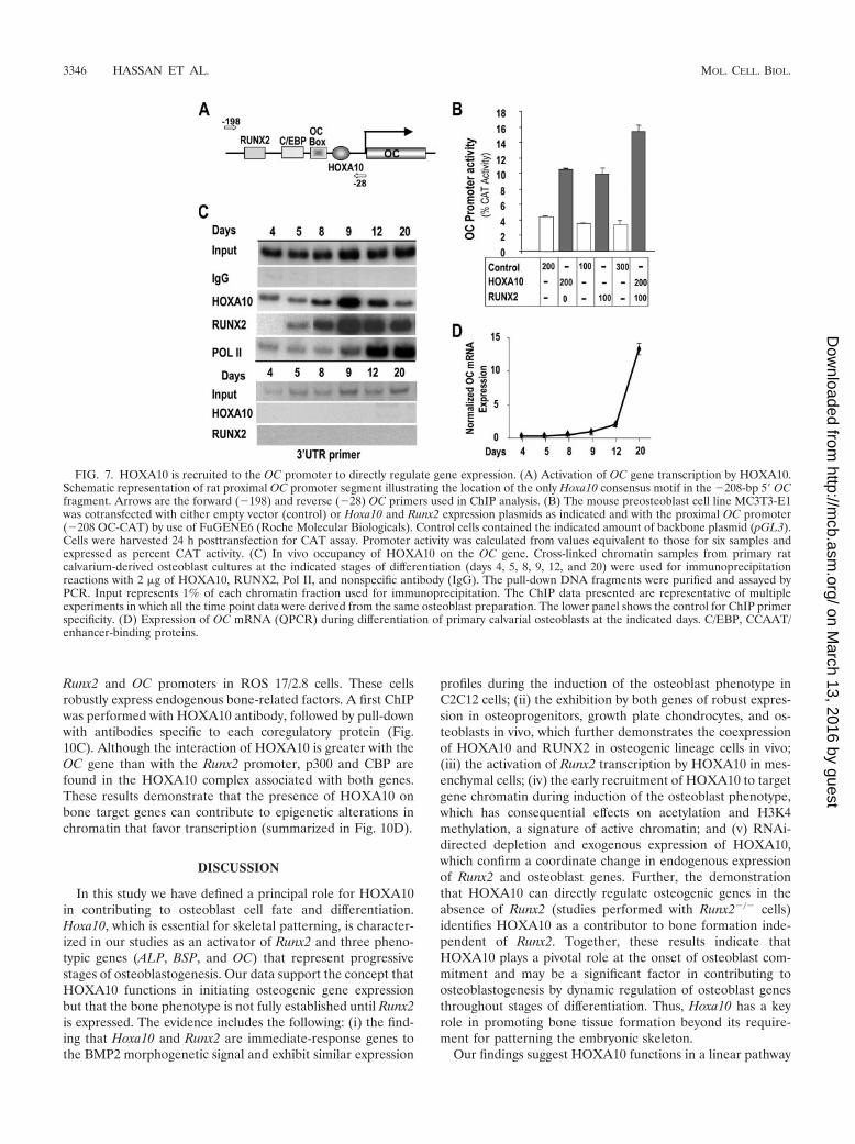
ChIP studies allow for assays of transcription factors asso-ciated with different promoters in the same sample. We there-fore examined the promoters of several osteoblast genes forpotential Hoxa10 binding sites and designed primers to amplifythe DNA of the appropriate promoter domain. Table 2 showsthe putative sequences. We examined the direct regulationof the osteoblast-specific OC gene, which is highly regulated byRUNX2 (40). A Hox binding site was located in the proximal�208 promoter segment, which exhibits tissue-restricted ex-pression (37) (Fig. 7A). Using a �208 OC-CAT reporter con-struct, we found a two- to threefold increase in promoteractivity in response to HOXA10 in multiple experiments (Fig.7B). By ChIP analysis (Fig. 7C), we found that HOXA10 isassociated with OC chromatin 24 h prior to RUNX2 associa-tion. During osteoblast differentiation, both HOXA10 andRUNX2 binding were enhanced on days 8 and 9, when OCmRNA first began to accumulate (Fig. 7D), consistent with
increased Pol II binding on day 9 (Fig. 7C). After this initialinduction of OC, HOXA10 association with the OC promoterdiminished in the matrix maturation and mineralization stages,while RUNX2 remained present on OC chromatin. The coor-dinate increase in HOXA10 and RUNX2 binding to OCprompted us to examine the combined effects of HOXA10 andRUNX2 on OC promoter activity. We found an additive effecton promoter activity as a result of the coexpression of thefactors (Fig. 7B), suggesting that the Hoxa10 and Runx2 regu-latory elements function independently on the OC promoter,not synergistically. Taken together, these results demonstrateHOXA10 supports the regulation of OC expression earlierthan RUNX2 and significantly contributes to OC-inducedmRNA.
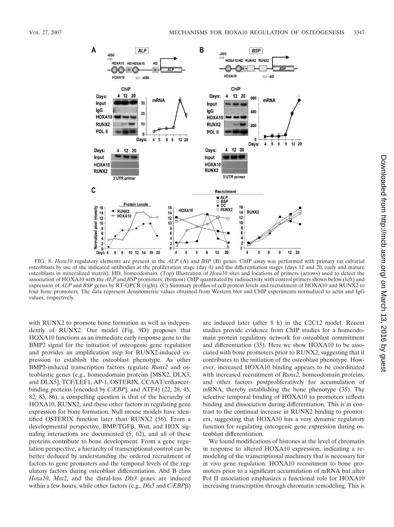
We also examined the promoters for alkaline phosphatase(ALP) and bone sialoprotein (BSP) and found Hoxa10-regu-latory elements (Table 2 and Fig. 8). In proliferating primaryosteoblasts, ALP (Fig. 8A) and BSP (Fig. 8B) are expressed,but at low levels. Association of HOXA10 with the ALP andBSP promoters was confirmed by ChIP assay with the osteo-progenitor cells (day 4) as well as with postproliferative matureosteoblasts (days 12 and 20). Interestingly, HOXA10 associa-tion with the promoter of ALP, the early marker of osteoblasts,was most prominent on day 4 and reduced on day 20, whenALP mRNA is downregulated. In contrast, both HOXA10binding to the BSP promoter and BSP expression (mRNA)increased during differentiation. These observations suggestthat HOXA10 directly regulates genes independent ofRUNX2 binding.
These ChIP studies reveal that the recruitment of RUNX2to most gene promoters continuously increases during differ-entiation, with similar profiles in each case (Fig. 8C). Figure 8Csummarizes the profiles (from densitometry measurements ofthe ChIPs) of RUNX2 and HOXA10 association with fourgene promoters. In contrast to RUNX2, HOXA10 has distinctprofiles of association/dissociation with individual gene pro-moters during differentiation. It is notable that HOXA10 as-sociation to the gene promoters is critical to gene regulationindependent of HOXA10 cellular levels (Fig. 8C, left panel).These differences may reflect very dynamic functional charac-teristics of HOXA10 promoter binding in relation to the reg-ulation of the transcription of osteoblast genes at stages ofosteoblast phenotype development. We conclude from thesestudies that Hoxa10 functional elements are present in multi-ple osteoblast genes, including the OC, bone sialoprotein, andalkaline phosphatase genes, contributing to their regulatedexpression.
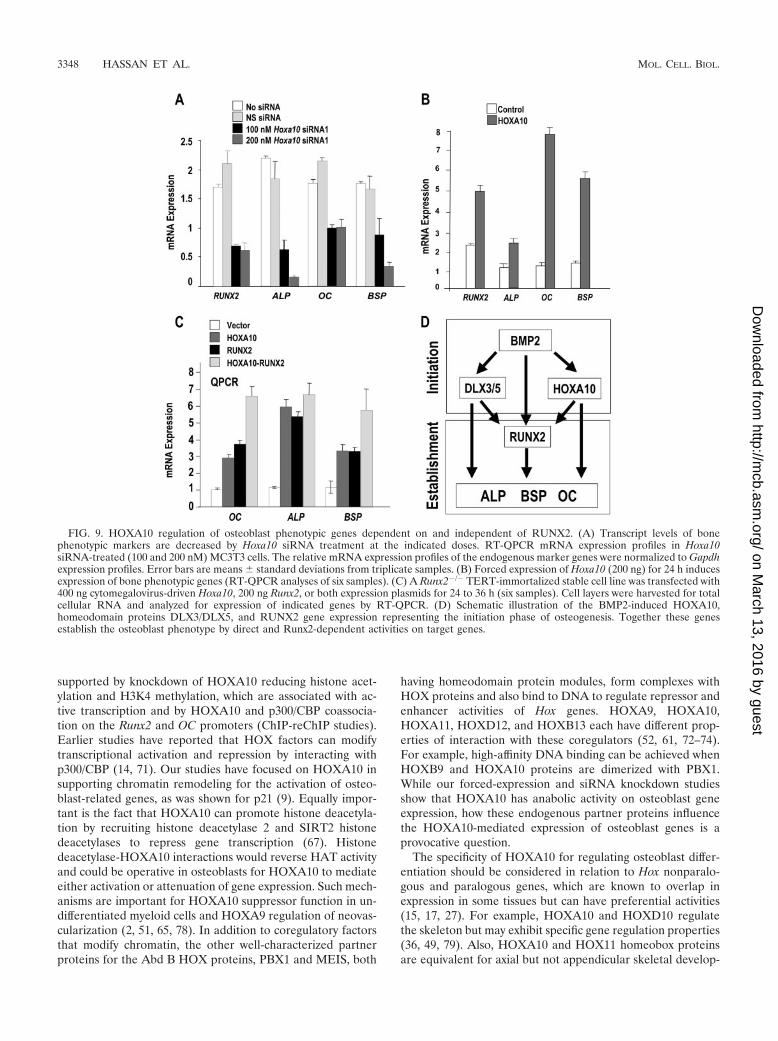
HOXA10 regulates osteoblast genes both dependent on (inMC3T3 cells) and independent of (in Runx2 null cells)RUNX2. The ChIP studies (Fig. 6, 7, and 8) suggest thatHOXA10 may regulate osteoblast genes that are significantfunctional components of bone, either synergistically, by in-creasing Runx2 expression, or directly, through Hox regulatorymotifs. We therefore examined the consequences of the deple-tion of Hoxa10 on the endogenous expression of these skeletalmarkers of osteoblast differentiation by RT-QPCR (Fig. 9A).MC3T3 cells were treated with Hoxa10 siRNA for 48 h, whichreduced Runx2 gene expression threefold. Dose-dependent de-creases of the early osteoblast markers ALP and BSP were alsoobserved, as was a twofold reduction in OC mRNA. Exoge-
FIG. 6. HOXA10 protein and recruitment to the Runx2 promoterin primary calvarial cells during osteoblast growth and differentiation.(A) Western blot analyses for RUNX2 and HOXA10 protein expres-sion are shown during stages of growth and differentiation of isolatedprimary calvarial osteoblasts as indicated (antibody information is pro-vided in Materials and Methods). Protein profiles of actin demonstrateequivalent amounts of total protein loaded in the gel. (B) ChIP studiesof the Runx2 proximal promoter locus during growth and differentia-tion. The top panel illustrates the positions of regulatory elements andprimers used to amplify the Runx2 promoter-specific DNA fragments.Open arrow, RNA Pol II; solid arrow, HOXA10 and RUNX2 occu-pancy. For the middle panel, ChIP analysis was performed on theindicated days with antibodies for HOXA10, RUNX2, or RNA Pol II(see Materials and Methods). One percent of the soluble chromatinfraction was taken as the input fraction. IgG is shown as an antibodycontrol. The lower panel shows the control for ChIP primer specificity.
3344 HASSAN ET AL. MOL. CELL. BIOL.
on March 13, 2016 by guest
http://mcb.asm
.org/D
ownloaded from
nous expression of HOXA10 in MC3T3 cells (Fig. 9B) showsthat Runx2 and ALP expression is increased twofold, while themature osteoblast-related genes OC and BSP are induced be-tween five- and eightfold.
To address the possibility of the direct regulation of osteoblastgenes, we tested the competency of HOXA10 to induce theirexpression independent of RUNX2 by using a TERT-immortal-ized preosteoblastic cell line derived from Runx2 null mice (1)(Fig. 9C). Cells were transfected with Hoxa10 and/or Runx2 ex-pression vector, and total cellular RNA was analyzed 24 h later.Expression profiling by real-time PCR revealed that HOXA10induced OC, ALP, and BSP to an extent the same as that seen fortheir induction by RUNX2 (three- to fourfold for OC or BSP andfive- to sixfold for ALP). The presence of HOXA10 and RUNX2together resulted in an additive induction for OC and BSP, geneswhose expression continues to increase during differentiation(Fig. 7 and 8), but not ALP (Fig. 8).
HOXA10 activation of osteogenic genes OC, ALP, and BSPin Runx2 null cells, taken together with the association ofHOXA10 with binding elements in these promoters, supportsa RUNX2-independent direct role for HOXA10 in promotingosteoblast differentiation. However, these promoters are alsoRunx2 responsive, and HOXA10 directly induces Runx2,thereby contributing to HOXA10 RUNX2-dependent tran-scription of osteoblast genes. The BMP2-initiated induction ofHoxa10, Runx2, and other transcriptional factors (e.g., DLX3and DLX5) indicates that these factors all contribute specificregulatory functions to support bone formation. This combi-natorial control is schematically illustrated in Fig. 9D.
HOXA10 contributes to chromatin modification of osteo-blast target genes for induced transcription. The early recruit-ment of HOXA10, prior to RUNX2, on the OC, ALP, and BSPgene promoters suggests that HOXA10 may contribute to re-modeling the chromatin of these phenotypic genes. The re-
modeling of chromatin structure is mediated in part by en-zymes that topologically alter DNA interactions with histonesor that covalently modify the core histone proteins H3 and H4(41). Acetylation of histone H4 and H3K4 methylation aremodifications that strongly correlate with transcriptionally ac-tive chromatin (10, 75, 77). To test this hypothesis, we exam-ined the effects of Hoxa10 depletion on histone acetylation andH3K4 methylation of gene promoters, which reflect transcrip-tionally active chromatin. Cells were treated with Hoxa10-spe-cific siRNA for 72 h (Fig. 10A), and these chromatin modifi-cations were assayed by ChIP analysis for the four bonephenotypic genes (Fig. 10B). We confirmed a 50% knockdownof Hoxa10 by siRNA and a 50% reduction in HOXA10 bindingto OC, ALP, and BSP promoters but a modest decrease in therecruitment of HOXA10 to the RUNX2 promoter (Fig. 10B,left panels). We observed a significant 50% reduction in theacetylation of OC, BSP, and Runx2 chromatin but a modestdecrease in ALP chromatin acetylation in cells treated withHoxa10 siRNA relative to control levels (nonsilencing siRNA)(Fig. 10B). On the other hand, we found in multiple studies(n 3) a significant decrease in the H3K4 methylation of ALPand BSP, while the methylation of OC and Runx2 chromatinwas less decreased.
Upon RNAi-mediated depletion of HOXA10, we found animpairment of histone modifications in the chromatin of theRunx2, OC, ALP, and BSP genes, suggesting that coregulatoryproteins with histone acetyltransferase (HAT) activity may bepresent in a HOXA10 complex associated with chromatin. Toaddress this mechanism of chromatin remodeling by HOXA10binding to target genes, we examined the association of co-regulatory proteins having HAT activity, p300 and CBP, withHOXA10 (Fig. 10C). We performed sequential chromatin im-munoprecipitation studies (ChIP-reChIP) to determine the invivo interactions of HOXA10 with coactivator proteins on the
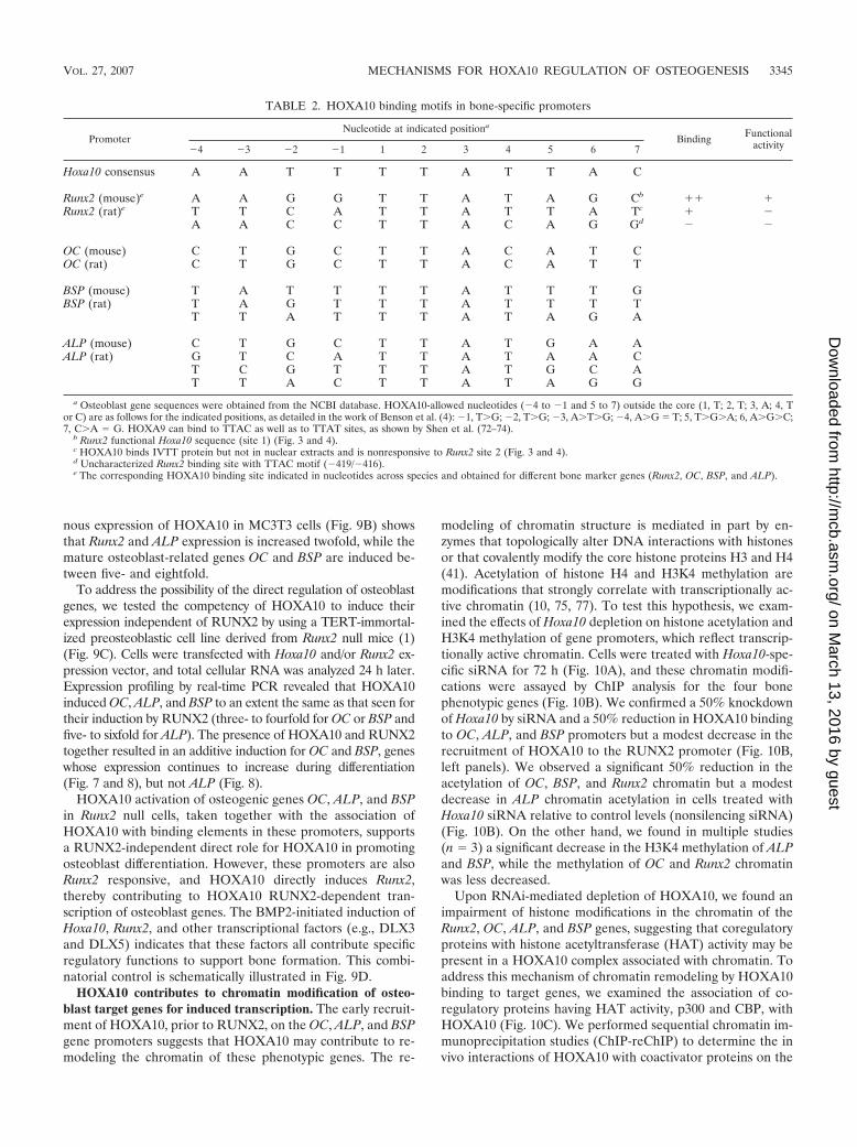
TABLE 2. HOXA10 binding motifs in bone-specific promoters
PromoterNucleotide at indicated positiona
Binding Functionalactivity�4 �3 �2 �1 1 2 3 4 5 6 7
Hoxa10 consensus A A T T T T A T T A C
Runx2 (mouse)e A A G G T T A T A G Cb �� �Runx2 (rat)e T T C A T T A T T A Tc � �
A A C C T T A C A G Gd � �
OC (mouse) C T G C T T A C A T COC (rat) C T G C T T A C A T T
BSP (mouse) T A T T T T A T T T GBSP (rat) T A G T T T A T T T T
T T A T T T A T A G A
ALP (mouse) C T G C T T A T G A AALP (rat) G T C A T T A T A A C
T C G T T T A T G C AT T A C T T A T A G G
a Osteoblast gene sequences were obtained from the NCBI database. HOXA10-allowed nucleotides (�4 to �1 and 5 to 7) outside the core (1, T; 2, T; 3, A; 4, Tor C) are as follows for the indicated positions, as detailed in the work of Benson et al. (4): �1, TG; �2, TG; �3, ATG; �4, AG T; 5, TGA; 6, AGC;7, CA G. HOXA9 can bind to TTAC as well as to TTAT sites, as shown by Shen et al. (72–74).
b Runx2 functional Hoxa10 sequence (site 1) (Fig. 3 and 4).c HOXA10 binds IVTT protein but not in nuclear extracts and is nonresponsive to Runx2 site 2 (Fig. 3 and 4).d Uncharacterized Runx2 binding site with TTAC motif (�419/�416).e The corresponding HOXA10 binding site indicated in nucleotides across species and obtained for different bone marker genes (Runx2, OC, BSP, and ALP).
VOL. 27, 2007 MECHANISMS FOR HOXA10 REGULATION OF OSTEOGENESIS 3345
on March 13, 2016 by guest
http://mcb.asm
.org/D
ownloaded from
Runx2 and OC promoters in ROS 17/2.8 cells. These cellsrobustly express endogenous bone-related factors. A first ChIPwas performed with HOXA10 antibody, followed by pull-downwith antibodies specific to each coregulatory protein (Fig.10C). Although the interaction of HOXA10 is greater with theOC gene than with the Runx2 promoter, p300 and CBP arefound in the HOXA10 complex associated with both genes.These results demonstrate that the presence of HOXA10 onbone target genes can contribute to epigenetic alterations inchromatin that favor transcription (summarized in Fig. 10D).
DISCUSSION
In this study we have defined a principal role for HOXA10in contributing to osteoblast cell fate and differentiation.Hoxa10, which is essential for skeletal patterning, is character-ized in our studies as an activator of Runx2 and three pheno-typic genes (ALP, BSP, and OC) that represent progressivestages of osteoblastogenesis. Our data support the concept thatHOXA10 functions in initiating osteogenic gene expressionbut that the bone phenotype is not fully established until Runx2is expressed. The evidence includes the following: (i) the find-ing that Hoxa10 and Runx2 are immediate-response genes tothe BMP2 morphogenetic signal and exhibit similar expression
profiles during the induction of the osteoblast phenotype inC2C12 cells; (ii) the exhibition by both genes of robust expres-sion in osteoprogenitors, growth plate chondrocytes, and os-teoblasts in vivo, which further demonstrates the coexpressionof HOXA10 and RUNX2 in osteogenic lineage cells in vivo;(iii) the activation of Runx2 transcription by HOXA10 in mes-enchymal cells; (iv) the early recruitment of HOXA10 to targetgene chromatin during induction of the osteoblast phenotype,which has consequential effects on acetylation and H3K4methylation, a signature of active chromatin; and (v) RNAi-directed depletion and exogenous expression of HOXA10,which confirm a coordinate change in endogenous expressionof Runx2 and osteoblast genes. Further, the demonstrationthat HOXA10 can directly regulate osteogenic genes in theabsence of Runx2 (studies performed with Runx2�/� cells)identifies HOXA10 as a contributor to bone formation inde-pendent of Runx2. Together, these results indicate thatHOXA10 plays a pivotal role at the onset of osteoblast com-mitment and may be a significant factor in contributing toosteoblastogenesis by dynamic regulation of osteoblast genesthroughout stages of differentiation. Thus, Hoxa10 has a keyrole in promoting bone tissue formation beyond its require-ment for patterning the embryonic skeleton.
Our findings suggest HOXA10 functions in a linear pathway
FIG. 7. HOXA10 is recruited to the OC promoter to directly regulate gene expression. (A) Activation of OC gene transcription by HOXA10.Schematic representation of rat proximal OC promoter segment illustrating the location of the only Hoxa10 consensus motif in the �208-bp 5� OCfragment. Arrows are the forward (�198) and reverse (�28) OC primers used in ChIP analysis. (B) The mouse preosteoblast cell line MC3T3-E1was cotransfected with either empty vector (control) or Hoxa10 and Runx2 expression plasmids as indicated and with the proximal OC promoter(�208 OC-CAT) by use of FuGENE6 (Roche Molecular Biologicals). Control cells contained the indicated amount of backbone plasmid (pGL3).Cells were harvested 24 h posttransfection for CAT assay. Promoter activity was calculated from values equivalent to those for six samples andexpressed as percent CAT activity. (C) In vivo occupancy of HOXA10 on the OC gene. Cross-linked chromatin samples from primary ratcalvarium-derived osteoblast cultures at the indicated stages of differentiation (days 4, 5, 8, 9, 12, and 20) were used for immunoprecipitationreactions with 2 �g of HOXA10, RUNX2, Pol II, and nonspecific antibody (IgG). The pull-down DNA fragments were purified and assayed byPCR. Input represents 1% of each chromatin fraction used for immunoprecipitation. The ChIP data presented are representative of multipleexperiments in which all the time point data were derived from the same osteoblast preparation. The lower panel shows the control for ChIP primerspecificity. (D) Expression of OC mRNA (QPCR) during differentiation of primary calvarial osteoblasts at the indicated days. C/EBP, CCAAT/enhancer-binding proteins.
3346 HASSAN ET AL. MOL. CELL. BIOL.
on March 13, 2016 by guest
http://mcb.asm
.org/D
ownloaded from
with RUNX2 to promote bone formation as well as indepen-dently of RUNX2. Our model (Fig. 9D) proposes thatHOXA10 functions as an immediate early response gene to theBMP2 signal for the initiation of osteogenic gene regulationand provides an amplification step for RUNX2-induced ex-pression to establish the osteoblast phenotype. As otherBMP2-induced transcription factors regulate Runx2 and os-teoblastic genes (e.g., homeodomain proteins [MSX2, DLX3,and DLX5], TCF/LEF1, AP-1, OSTERIX, CCAAT/enhancer-binding proteins [encoded by C/EBP], and ATF4) (22, 28, 45,82, 83, 86), a compelling question is that of the hierarchy ofHOXA10, RUNX2, and these other factors in regulating geneexpression for bone formation. Null mouse models have iden-tified OSTERIX function later than RUNX2 (56). From adevelopmental perspective, BMP/TGF�, Wnt, and HOX sig-naling interactions are documented (5, 62), and all of theseproteins contribute to bone development. From a gene regu-lation perspective, a hierarchy of transcriptional control can bebetter deduced by understanding the ordered recruitment offactors to gene promoters and the temporal levels of the reg-ulatory factors during osteoblast differentiation. Abd B classHoxa10, Msx2, and the distal-less Dlx3 genes are inducedwithin a few hours, while other factors (e.g., Dlx5 and C/EBP�)
are induced later (after 8 h) in the C2C12 model. Recentstudies provide evidence from ChIP studies for a homeodo-main protein regulatory network for osteoblast commitmentand differentiation (35). Here we show HOXA10 to be asso-ciated with bone promoters prior to RUNX2, suggesting that itcontributes to the initiation of the osteoblast phenotype. How-ever, increased HOXA10 binding appears to be coordinatedwith increased recruitment of Runx2, homeodomain proteins,and other factors postproliferatively for accumulation ofmRNA, thereby establishing the bone phenotype (35). Theselective temporal binding of HOXA10 to promoters reflectsbinding and dissociation during differentiation. This is in con-trast to the continual increase in RUNX2 binding to promot-ers, suggesting that HOXA10 has a very dynamic regulatoryfunction for regulating osteogenic gene expression during os-teoblast differentiation.
We found modifications of histones at the level of chromatinin response to altered HOXA10 expression, indicating a re-modeling of the transcriptional machinery that is necessary forin vivo gene regulation. HOXA10 recruitment to bone pro-moters prior to a significant accumulation of mRNA but afterPol II association emphasizes a functional role for HOXA10increasing transcription through chromatin remodeling. This is
FIG. 8. Hoxa10 regulatory elements are present in the ALP (A) and BSP (B) genes. ChIP assay was performed with primary rat calvarialosteoblasts by use of the indicated antibodies at the proliferation stage (day 4) and the differentiation stages (days 12 and 20, early and matureosteoblasts in mineralized matrix). HD, homeodomain. (Top) Illustration of Hoxa10 sites and locations of primers (arrows) used to detect theassociation of HOXA10 with the ALP and BSP promoters; (bottom) ChIP quantitated by radioactivity with control primers shown below (left) andexpression of ALP and BSP genes by RT-QPCR (right). (C) Summary profiles of cell protein levels and recruitment of HOXA10 and RUNX2 tofour bone promoters. The data represent densitometric values obtained from Western blot and ChIP experiments normalized to actin and IgGvalues, respectively.
VOL. 27, 2007 MECHANISMS FOR HOXA10 REGULATION OF OSTEOGENESIS 3347
on March 13, 2016 by guest
http://mcb.asm
.org/D
ownloaded from
supported by knockdown of HOXA10 reducing histone acet-ylation and H3K4 methylation, which are associated with ac-tive transcription and by HOXA10 and p300/CBP coassocia-tion on the Runx2 and OC promoters (ChIP-reChIP studies).Earlier studies have reported that HOX factors can modifytranscriptional activation and repression by interacting withp300/CBP (14, 71). Our studies have focused on HOXA10 insupporting chromatin remodeling for the activation of osteo-blast-related genes, as was shown for p21 (9). Equally impor-tant is the fact that HOXA10 can promote histone deacetyla-tion by recruiting histone deacetylase 2 and SIRT2 histonedeacetylases to repress gene transcription (67). Histonedeacetylase-HOXA10 interactions would reverse HAT activityand could be operative in osteoblasts for HOXA10 to mediateeither activation or attenuation of gene expression. Such mech-anisms are important for HOXA10 suppressor function in un-differentiated myeloid cells and HOXA9 regulation of neovas-cularization (2, 51, 65, 78). In addition to coregulatory factorsthat modify chromatin, the other well-characterized partnerproteins for the Abd B HOX proteins, PBX1 and MEIS, both
having homeodomain protein modules, form complexes withHOX proteins and also bind to DNA to regulate repressor andenhancer activities of Hox genes. HOXA9, HOXA10,HOXA11, HOXD12, and HOXB13 each have different prop-erties of interaction with these coregulators (52, 61, 72–74).For example, high-affinity DNA binding can be achieved whenHOXB9 and HOXA10 proteins are dimerized with PBX1.While our forced-expression and siRNA knockdown studiesshow that HOXA10 has anabolic activity on osteoblast geneexpression, how these endogenous partner proteins influencethe HOXA10-mediated expression of osteoblast genes is aprovocative question.
The specificity of HOXA10 for regulating osteoblast differ-entiation should be considered in relation to Hox nonparalo-gous and paralogous genes, which are known to overlap inexpression in some tissues but can have preferential activities(15, 17, 27). For example, HOXA10 and HOXD10 regulatethe skeleton but may exhibit specific gene regulation properties(36, 49, 79). Also, HOXA10 and HOX11 homeobox proteinsare equivalent for axial but not appendicular skeletal develop-
FIG. 9. HOXA10 regulation of osteoblast phenotypic genes dependent on and independent of RUNX2. (A) Transcript levels of bonephenotypic markers are decreased by Hoxa10 siRNA treatment at the indicated doses. RT-QPCR mRNA expression profiles in Hoxa10siRNA-treated (100 and 200 nM) MC3T3 cells. The relative mRNA expression profiles of the endogenous marker genes were normalized to Gapdhexpression profiles. Error bars are means � standard deviations from triplicate samples. (B) Forced expression of Hoxa10 (200 ng) for 24 h inducesexpression of bone phenotypic genes (RT-QPCR analyses of six samples). (C) A Runx2�/� TERT-immortalized stable cell line was transfected with400 ng cytomegalovirus-driven Hoxa10, 200 ng Runx2, or both expression plasmids for 24 to 36 h (six samples). Cell layers were harvested for totalcellular RNA and analyzed for expression of indicated genes by RT-QPCR. (D) Schematic illustration of the BMP2-induced HOXA10,homeodomain proteins DLX3/DLX5, and RUNX2 gene expression representing the initiation phase of osteogenesis. Together these genesestablish the osteoblast phenotype by direct and Runx2-dependent activities on target genes.
3348 HASSAN ET AL. MOL. CELL. BIOL.
on March 13, 2016 by guest
http://mcb.asm
.org/D
ownloaded from

FIG. 10. siRNA knockdown of Hoxa10 decreases histone acetylation and H3K4 methylation of osteogenic genes. (A) MC3T3 cells were treatedwith Hoxa10 siRNA and nonspecific control (NS) for 72 h. The knockdown effect is shown by Western blotting. (B) DNA samples from ChIP withHOXA10, nonspecific IgG, methylated histone K4 (H3K4), and acetylated histone H4 (ACH4) were amplified by gene-specific (Sp) and 3� controlUTR primers (Table 1) for the indicated gene promoters. The left panel for each gene shows the effect of Hoxa10 siRNA on HOXA10 recruitmentin ChIP assay of the indicated promoter. The right panel shows the status of H3K4 methylation or H4 acetylation of the chromatin modificationby Hoxa10-specific knockdown. (C) ChIP-reChIP assays were performed to identify the association of coregulatory factors p300 and CBP withHOXA10 on the Runx2 and OC promoters. A HOXA10 ChIP was performed with HOXA10 antibody, and the resulting chromatin immunopre-cipitate (second input) was subjected to reChIP with the indicated antibodies (�) (secondary pull-down). Normal IgG and green fluorescent protein(GFP) antibodies provided controls. The DNA fragments for Runx2 and OC were amplified as described in Materials and Methods. (D) Schematicillustration to show how HOXA10 belongs to an epigenetic coregulatory complex for remodeling chromatin to induce transcription of osteogenicgenes. We propose that HOXA10 may be among the earliest factors recruited to bone promoters. HOXA10 recruitment is followed by thecoordinated occupancy of other bone-related transcription factors for maximal expression of individual genes during stages of differentiation. HD,homeodomain.
VOL. 27, 2007 MECHANISMS FOR HOXA10 REGULATION OF OSTEOGENESIS 3349
on March 13, 2016 by guest
http://mcb.asm
.org/D
ownloaded from
ment (87). The TTAT regulatory element is recognized byseveral HOX proteins; however, protein-DNA interactions aswell as protein-protein interactions are dependent on the con-textual sequence of the core TTAT motif (8, 13, 57). Ouranalysis of the functional specificity of the Runx2 Hoxa10 site 1and no other core motifs in the 0.6-kb Runx2 promoter isconsistent with other studies evaluating the specificities ofHoxa10 sites (4, 72, 74). Although several HOX proteins arelikely present in osteoblast nuclear extracts (e.g., HOXA9),our findings revealed a supershifted/block shifted complex bythe HOXA10 antibody. However, it is possible that other HOXprotein interactions may occur at this and potentially otherHOX motifs within the Runx2 gene. Our data do support theconcept that the Runx2 site 1 (TTAT) has a high degree ofspecificity for HOXA10 binding. Interestingly, the OC gene ischaracterized by a TTAC core motif, implicating this motif inthe formation of complexes that support a high degree of OCexpression in mature osteoblasts. It is also noteworthy that theflanking nucleotides are distinct among the four target genesstudied here (Table 2). This finding implies that their se-quences may hold information for the dynamic association ofHOXA10 with an individual gene throughout differentiation.
HOXA10 is expressed in many different tissues in which itsfunctions have been addressed and may be related to its activitiesin bone tissue. Progesterone and 17�-estradiol, hormones thatregulate turnover of adult bone (53, 63), increase Hoxa10 expres-sion (16, 48). HOXA10 regulates hematopoietic differentiation inpart via activation of p21 (WAF1/CIP1), the cyclin-dependentkinase inhibitor (9, 25). In the spinal cord, expression of Hoxa10is confined to the postmitotic cell population (15). We find thatHOXA10 is expressed in proliferating cells and upregulated inpostproliferative mature osteoblasts in vitro. HOXA10 is also atarget of induction by vitamin D3 (66), a hormone that increasesthe expression of osteoblast-related genes. Thus, HOXA10 ap-pears to have a broad role in gene regulation for contributing tocell differentiation. Our studies have added to the growing rec-ognition that Hox genes have important functions in the adult,including now the skeleton (55).
In conclusion, our characterization of HOXA10 positive reg-ulation of genes that represent major functional componentsfor bone formation, including those encoding RUNX2 (a boneessential transcription factor), alkaline phosphatase (requiredfor matrix mineralization), bone sialoprotein (an importantcell matrix-binding phosphoprotein with hydroxyapatite nucle-ation capabilities), and OC (the calcium binding bone-specificprotein) (47), solidifies the concept that HOXA10 is an im-portant regulator of gene expression throughout the progres-sion of bone formation. These novel functions for HOXA10 inregulating target genes for osteoblast differentiation and boneformation in the postnatal skeleton have broad implications forHOXA10 functions in normal bone metabolism and bone-related disorders.
ACKNOWLEDGMENTS
Studies reported were supported in part by grants from the NationalInstitutes of Health (DE12528, AR39588, AR48818, and P30DK32520).
The contents of this work are solely our responsibility and do notnecessarily represent the official views of the National Institutes ofHealth.
We thank Richard L. Maas (Department of Medicine, Brigham andWomen’s Hospital, Harvard Medical School, Boston, MA) for re-agents, Anthony Imbalzano (University of Massachusetts MedicalSchool) for helpful discussions, Dana Fredericks for technical assis-tance, and Judy Rask for manuscript preparation.
REFERENCES
1. Bae, J.-S., S. Gutierrez, R. Narla, J. Pratap, R. Devados, J. L. Stein, J. B.Lian, G. S. Stein, and A. Javed. 2007. Reconstitution of Runx2/Cbfa1 nullcells identifies Runx2 functional domains required for osteoblast differenti-ation and responsiveness to osteogenic regulators BMP2, TGF� and1,25(OH)2D3. J. Cell. Biochem. 100:434–449.
2. Bae, N. S., M. J. Swanson, A. Vassilev, and B. H. Howard. 2004. Humanhistone deacetylase SIRT2 interacts with the homeobox transcription factorHOXA10. J. Biochem. (Tokyo) 135:695–700.
3. Balint, E., D. Lapointe, H. Drissi, C. van der Meijden, D. W. Young, A. J. vanWijnen, J. L. Stein, G. S. Stein, and J. B. Lian. 2003. Phenotype discovery bygene expression profiling: mapping of biological processes linked to BMP-2-mediated osteoblast differentiation. J. Cell. Biochem. 89:401–426.
4. Benson, G. V., T. H. Nguyen, and R. L. Maas. 1995. The expression patternof the murine Hoxa-10 gene and the sequence recognition of its homeodo-main reveal specific properties of Abdominal B-like genes. Mol. Cell. Biol.15:1591–1601.
5. Bondos, S. 2006. Variations on a theme: Hox and Wnt combinatorial regu-lation during animal development. Sci. STKE 2006:e38.
6. Boulet, A. M., and M. R. Capecchi. 2002. Duplication of the Hoxd11 genecauses alterations in the axial and appendicular skeleton of the mouse. Dev.Biol. 249:96–107.
7. Boulet, A. M., and M. R. Capecchi. 2004. Multiple roles of Hoxa11 andHoxd11 in the formation of the mammalian forelimb zeugopod. Develop-ment 131:299–309.
8. Brake, R. L., U. R. Kees, and P. M. Watt. 2002. A complex containing PBX2contributes to activation of the proto-oncogene HOX11. Biochem. Biophys.Res. Commun. 294:23–34.
9. Bromleigh, V. C., and L. P. Freedman. 2000. p21 is a transcriptional target ofHOXA10 in differentiating myelomonocytic cells. Genes Dev. 14:2581–2586.
10. Bulger, M. 2005. Hyperacetylated chromatin domains: lessons from hetero-chromatin. J. Biol. Chem. 280:21689–21692.
11. Carapuco, M., A. Novoa, N. Bobola, and M. Mallo. 2005. Hox genes specifyvertebral types in the presomitic mesoderm. Genes Dev. 19:2116–2121.
12. Carpenter, E. M., J. M. Goddard, A. P. Davis, T. P. Nguyen, and M. R.Capecchi. 1997. Targeted disruption of Hoxd-10 affects mouse hindlimbdevelopment. Development 124:4505–4514.
13. Chang, C. P., L. Brocchieri, W. F. Shen, C. Largman, and M. L. Cleary. 1996.Pbx modulation of Hox homeodomain amino-terminal arms establishes dif-ferent DNA-binding specificities across the Hox locus. Mol. Cell. Biol. 16:1734–1745.
14. Chariot, A., C. van Lint, M. Chapelier, J. Gielen, M. P. Merville, and V.Bours. 1999. CBP and histone deacetylase inhibition enhance the transacti-vation potential of the HOXB7 homeodomain-containing protein. Oncogene18:4007–4014.
15. Choe, A., H. Q. Phun, D. D. Tieu, Y. H. Hu, and E. M. Carpenter. 2006.Expression patterns of Hox10 paralogous genes during lumbar spinal corddevelopment. Gene Expr. Patterns 6:730–737.
16. Daftary, G. S., and H. S. Taylor. 2006. Endocrine regulation of HOX genes.Endocr. Rev. 27:331–355.
17. Davis, A. P., D. P. Witte, H. M. Hsieh-Li, S. S. Potter, and M. R. Capecchi.1995. Absence of radius and ulna in mice lacking hoxa-11 and hoxd-11.Nature 375:791–795.
18. de la Fuente, L., and J. A. Helms. 2005. Head, shoulders, knees, and toes.Dev. Biol. 282:294–306.
19. Depew, M. J., C. A. Simpson, M. Morasso, and J. L. Rubenstein. 2005.Reassessing the Dlx code: the genetic regulation of branchial arch skeletalpattern and development. J. Anat. 207:501–561.
20. Dobreva, G., M. Chahrour, M. Dautzenberg, L. Chirivella, B. Kanzler, I.Farinas, G. Karsenty, and R. Grosschedl. 2006. SATB2 is a multifunctionaldeterminant of craniofacial patterning and osteoblast differentiation. Cell125:971–986.
21. Drissi, H., Q. Luc, R. Shakoori, S. Chuva de Sousa Lopes, J.-Y. Choi, A.Terry, M. Hu, S. Jones, J. C. Neil, J. B. Lian, J. L. Stein, A. J. van Wijnen,and G. S. Stein. 2000. Transcriptional autoregulation of the bone relatedCBFA1/RUNX2 gene. J. Cell. Physiol. 184:341–350.
22. Drissi, H., A. Pouliot, J. L. Stein, A. J. van Wijnen, G. S. Stein, and J. B.Lian. 2002. Identification of novel protein/DNA interactions within the pro-moter of the bone-related transcription factor Runx2/Cbfa1. J. Cell. Bio-chem. 86:403–412.
23. Ducy, P., R. Zhang, V. Geoffroy, A. L. Ridall, and G. Karsenty. 1997. Osf2/Cbfa1: a transcriptional activator of osteoblast differentiation. Cell 89:747–754.
24. Ekker, S. C., D. G. Jackson, D. P. von Kessler, B. I. Sun, K. E. Young, and
3350 HASSAN ET AL. MOL. CELL. BIOL.
on March 13, 2016 by guest
http://mcb.asm
.org/D
ownloaded from

P. A. Beachy. 1994. The degree of variation in DNA sequence recognitionamong four Drosophila homeotic proteins. EMBO J. 13:3551–3560.
25. Eklund, E. A. 2006. The role of HOX genes in myeloid leukemogenesis.Curr. Opin. Hematol. 13:67–73.
26. Favier, B., F. M. Rijli, C. Fromental-Ramain, V. Fraulob, P. Chambon, andP. Dolle. 1996. Functional cooperation between the non-paralogous genesHoxa-10 and Hoxd-11 in the developing forelimb and axial skeleton. Devel-opment 122:449–460.
27. Fromental-Ramain, C., X. Warot, S. Lakkaraju, B. Favier, H. Haack, C.Birling, A. Dierich, P. Dolle, and P. Chambon. 1996. Specific and redundantfunctions of the paralogous Hoxa-9 and Hoxd-9 genes in forelimb and axialskeleton patterning. Development 122:461–472.
28. Gaur, T., C. J. Lengner, H. Hovhannisyan, R. A. Bhat, P. V. N. Bodine, B. S.Komm, A. Javed, A. J. van Wijnen, J. L. Stein, G. S. Stein, and J. B. Lian.2005. Canonical WNT signaling promotes osteogenesis by directly stimulat-ing RUNX2 gene expression. J. Biol. Chem. 280:33132–33140.
29. Gaur, T., C. J. Lengner, S. Hussain, B. Trevant, D. Ayers, J. L. Stein, P. V. N.Bodine, B. S. Komm, G. S. Stein, and J. B. Lian. 2006. Secreted frizzledprotein 1 regulates Wnt signaling for BMP induced chondrocyte differenti-ation. J. Cell. Physiol. 208:87–96. [Epub ahead of print.]
30. Gersch, R. P., F. Lombardo, S. C. McGovern, and M. Hadjiargyrou. 2005.Reactivation of Hox gene expression during bone regeneration. J. Orthop.Res. 23:882–890.
31. Goodman, F. R. 2002. Limb malformations and the human HOX genes.Am. J. Med. Genet. 112:256–265.
32. Gutierrez, S., A. Javed, D. Tennant, M. van Rees, M. Montecino, G. S. Stein,J. L. Stein, and J. B. Lian. 2002. CCAAT/enhancer-binding proteins (C/EBP) � and � activate osteocalcin gene transcription and synergize withRunx2 at the C/EBP element to regulate bone-specific expression. J. Biol.Chem. 277:1316–1323.
33. Harris, S. E., D. Guo, M. A. Harris, A. Krishnaswamy, and A. Lichtler. 2003.Transcriptional regulation of BMP-2 activated genes in osteoblasts usinggene expression microarray analysis: role of Dlx2 and Dlx5 transcriptionfactors. Front. Biosci. 8:s1249–s1265.
34. Hassan, M. Q., A. Javed, M. I. Morasso, J. Karlin, M. Montecino, A. J. vanWijnen, G. S. Stein, J. L. Stein, and J. B. Lian. 2004. Dlx3 transcriptionalregulation of osteoblast differentiation: temporal recruitment of Msx2, Dlx3,and Dlx5 homeodomain proteins to chromatin of the osteocalcin gene. Mol.Cell. Biol. 24:9248–9261.
35. Hassan, M. Q., R. S. Tare, S. Lee, M. Mandeville, M. I. Morasso, A. Javed,A. J. van Wijnen, J. L. Stein, G. S. Stein, and J. B. Lian. 2006. BMP2commitment to the osteogenic lineage involves activation of Runx2 by Dlx3and a homeodomain transcriptional network. J. Biol. Chem. 281:40515–40526.
36. Hedlund, E., S. L. Karsten, L. Kudo, D. H. Geschwind, and E. M. Carpenter.2004. Identification of a Hoxd10-regulated transcriptional network and com-binatorial interactions with Hoxa10 during spinal cord development. J. Neu-rosci. Res. 75:307–319.
37. Hoffmann, H. M., T. L. Beumer, S. Rahman, L. R. McCabe, C. Banerjee, F.Aslam, J. A. Tiro, A. J. van Wijnen, J. L. Stein, G. S. Stein, and J. B. Lian.1996. Bone tissue-specific transcription of the osteocalcin gene: role of anactivator osteoblast-specific complex and suppressor hox proteins that bindthe OC box. J. Cell. Biochem. 61:310–324.
38. Izpisua-Belmonte, J. C., H. Falkenstein, P. Dolle, A. Renucci, and D.Duboule. 1991. Murine genes related to the Drosophila AbdB homeoticgenes are sequentially expressed during development of the posterior part ofthe body. EMBO J. 10:2279–2289.
39. Javed, A., B. Guo, S. Hiebert, J.-Y. Choi, J. Green, S.-C. Zhao, M. A.Osborne, S. Stifani, J. L. Stein, J. B. Lian, A. J. van Wijnen, and G. S. Stein.2000. Groucho/TLE/R-Esp proteins associate with the nuclear matrix andrepress RUNX (CBF�/AML/PEBP2�) dependent activation of tissue-spe-cific gene transcription. J. Cell Sci. 113:2221–2231.
40. Javed, A., S. Gutierrez, M. Montecino, A. J. van Wijnen, J. L. Stein, G. S.Stein, and J. B. Lian. 1999. Multiple Cbfa/AML sites in the rat osteocalcinpromoter are required for basal and vitamin D responsive transcription andcontribute to chromatin organization. Mol. Cell. Biol. 19:7491–7500.
41. Jenuwein, T., and C. D. Allis. 2001. Translating the histone code. Science293:1074–1080.
42. Kim, Y. J., H. N. Kim, E. K. Park, B. H. Lee, H. M. Ryoo, S. Y. Kim, I. S.Kim, J. L. Stein, J. B. Lian, G. S. Stein, A. J. van Wijnen, and J. Y. Choi.2006. The bone-related Zn finger transcription factor Osterix promotes pro-liferation of mesenchymal cells. Gene 366:145–151.
43. Komori, T., H. Yagi, S. Nomura, A. Yamaguchi, K. Sasaki, K. Deguchi, Y.Shimizu, R. T. Bronson, Y.-H. Gao, M. Inada, M. Sato, R. Okamoto, Y.Kitamura, S. Yoshiki, and T. Kishimoto. 1997. Targeted disruption of Cbfa1results in a complete lack of bone formation owing to maturational arrest ofosteoblasts. Cell 89:755–764.
44. Krumlauf, R. 1994. Hox genes in vertebrate development. Cell 78:191–201.45. Lee, M. H., Y. J. Kim, W. J. Yoon, J. I. Kim, B. G. Kim, Y. S. Hwang, J. M.
Wozney, X. Z. Chi, S. C. Bae, K. Y. Choi, J. Y. Cho, J. Y. Choi, and H. M.Ryoo. 2005. Dlx5 specifically regulates Runx2-II expression by binding to
homeodomain response elements in the Runx2 distal promoter. J. Biol.Chem. 280:35579–35587.
46. Lengner, C. J., M. Q. Hassan, R. W. Serra, C. Lepper, A. J. van Wijnen, J. L.Stein, J. B. Lian, and G. S. Stein. 2005. Nkx3.2 mediated repression ofRUNX2 promotes chondrogenic differentiation. J. Biol. Chem. 280:15872–15879.
47. Lian, J. B., A. Javed, S. K. Zaidi, C. Lengner, M. Montecino, A. J. vanWijnen, J. L. Stein, and G. S. Stein. 2004. Regulatory controls for osteoblastgrowth and differentiation: role of Runx/Cbfa/AML factors. Crit. Rev. Eu-karyot. Gene Expr. 14:1–41.
48. Lim, H., L. Ma, W. G. Ma, R. L. Maas, and S. K. Dey. 1999. Hoxa-10regulates uterine stromal cell responsiveness to progesterone during implan-tation and decidualization in the mouse. Mol. Endocrinol. 13:1005–1017.
49. Lin, A. W., and E. M. Carpenter. 2003. Hoxa10 and Hoxd10 coordinatelyregulate lumbar motor neuron patterning. J. Neurobiol. 56:328–337.
50. Lowney, P., J. Corral, K. Detmer, M. M. LeBeau, L. Deaven, H. J. Lawrence,and C. Largman. 1991. A human Hox 1 homeobox gene exhibits myeloid-specific expression of alternative transcripts in human hematopoietic cells.Nucleic Acids Res. 19:3443–3449.
51. Lu, Y., I. Goldenberg, L. Bei, J. Andrejic, and E. A. Eklund. 2003. HoxA10represses gene transcription in undifferentiated myeloid cells by interactionwith histone deacetylase 2. J. Biol. Chem. 278:47792–47802.
52. Mann, R. S., and M. Affolter. 1998. Hox proteins meet more partners. Curr.Opin. Genet. Dev. 8:423–429.
53. Manolagas, S. C., S. Kousteni, and R. L. Jilka. 2002. Sex steroids and bone.Recent Prog. Horm. Res. 57:385–409.
54. Merabet, S., J. Pradel, and Y. Graba. 2005. Getting a molecular grasp onHox contextual activity. Trends Genet. 21:477–480.
55. Morgan, R. 2006. Hox genes: a continuation of embryonic patterning?Trends Genet. 22:67–69.
56. Nakashima, K., X. Zhou, G. Kunkel, Z. Zhang, J. M. Deng, R. R. Behringer,and B. de Crombrugghe. 2002. The novel zinc finger-containing transcriptionfactor osterix is required for osteoblast differentiation and bone formation.Cell 108:17–29.
57. Neuteboom, S. T., and C. Murre. 1997. Pbx raises the DNA binding speci-ficity but not the selectivity of antennapedia Hox proteins. Mol. Cell. Biol.17:4696–4706.
58. Otto, F., A. P. Thornell, T. Crompton, A. Denzel, K. C. Gilmour, I. R.Rosewell, G. W. H. Stamp, R. S. P. Beddington, S. Mundlos, B. R. Olsen,P. B. Selby, and M. J. Owen. 1997. Cbfa1, a candidate gene for cleidocranialdysplasia syndrome, is essential for osteoblast differentiation and bone de-velopment. Cell 89:765–771.
59. Owen, T. A., M. Aronow, V. Shalhoub, L. M. Barone, L. Wilming, M. S.Tassinari, M. B. Kennedy, S. Pockwinse, J. B. Lian, and G. S. Stein. 1990.Progressive development of the rat osteoblast phenotype in vitro: reciprocalrelationships in expression of genes associated with osteoblast proliferationand differentiation during formation of the bone extracellular matrix. J. Cell.Physiol. 143:420–430.
60. Pineault, N., C. Abramovich, H. Ohta, and R. K. Humphries. 2004. Differ-ential and common leukemogenic potentials of multiple NUP98-Hox fusionproteins alone or with Meis1. Mol. Cell. Biol. 24:1907–1917.
61. Pineault, N., C. D. Helgason, H. J. Lawrence, and R. K. Humphries. 2002.Differential expression of Hox, Meis1, and Pbx1 genes in primitive cellsthroughout murine hematopoietic ontogeny. Exp. Hematol. 30:49–57.
62. Pockwinse, S. M., J. B. Lawrence, R. H. Singer, J. L. Stein, J. B. Lian, andG. S. Stein. 1993. Gene expression at single cell resolution associated withdevelopment of the bone cell phenotype: ultrastructural and in situ hybrid-ization analysis. Bone 14:347–352.
63. Riggs, B. L., S. Khosla, and L. J. Melton III. 2002. Sex steroids and theconstruction and conservation of the adult skeleton. Endocr. Rev. 23:279–302.
64. Rijli, F. M., and P. Chambon. 1997. Genetic interactions of Hox genes inlimb development: learning from compound mutants. Curr. Opin. Genet.Dev. 7:481–487.
65. Rossig, L., C. Urbich, T. Bruhl, E. Dernbach, C. Heeschen, E. Chavakis, K.Sasaki, D. Aicher, F. Diehl, F. Seeger, M. Potente, A. Aicher, L. Zanetta, E.Dejana, A. M. Zeiher, and S. Dimmeler. 2005. Histone deacetylase activity isessential for the expression of HoxA9 and for endothelial commitment ofprogenitor cells. J. Exp. Med. 201:1825–1835.
66. Rots, N. Y., M. Liu, E. C. Anderson, and L. P. Freedman. 1998. A differentialscreen for ligand-regulated genes: identification of HoxA10 as a target ofvitamin D3 induction in myeloid leukemic cells. Mol. Cell. Biol. 18:1911–1918.
67. Saleh, M., I. Rambaldi, X. J. Yang, and M. S. Featherstone. 2000. Cellsignaling switches HOX-PBX complexes from repressors to activators oftranscription mediated by histone deacetylases and histone acetyltrans-ferases. Mol. Cell. Biol. 20:8623–8633.
68. Salsi, V., and V. Zappavigna. 2006. Hoxd13 and Hoxa13 directly control theexpression of the EphA7 ephrin tyrosine kinase receptor in developing limbs.J. Biol. Chem. 281:1992–1999.
69. Shao, J. S., S. L. Cheng, J. M. Pingsterhaus, N. Charlton-Kachigian, A. P.
VOL. 27, 2007 MECHANISMS FOR HOXA10 REGULATION OF OSTEOGENESIS 3351
on March 13, 2016 by guest
http://mcb.asm
.org/D
ownloaded from

Loewy, and D. A. Towler. 2005. Msx2 promotes cardiovascular calcificationby activating paracrine Wnt signals. J. Clin. Investig. 115:1210–1220.
70. Shen, J., M. A. Montecino, J. B. Lian, G. S. Stein, A. J. van Wijnen, and J. L.Stein. 2002. Histone acetylation in vivo at the osteocalcin locus is functionallylinked to vitamin D-dependent, bone tissue-specific transcription. J. Biol.Chem. 277:20284–20292.
71. Shen, W. F., K. Krishnan, H. J. Lawrence, and C. Largman. 2001. The HOXhomeodomain proteins block CBP histone acetyltransferase activity. Mol.Cell. Biol. 21:7509–7522.
72. Shen, W. F., J. C. Montgomery, S. Rozenfeld, J. J. Moskow, H. J. Lawrence,A. M. Buchberg, and C. Largman. 1997. AbdB-like Hox proteins stabilizeDNA binding by the Meis1 homeodomain proteins. Mol. Cell. Biol. 17:6448–6458.
73. Shen, W. F., S. Rozenfeld, A. Kwong, L. G. Komuves, H. J. Lawrence, and C.Largman. 1999. HOXA9 forms triple complexes with PBX2 and MEIS1 inmyeloid cells. Mol. Cell. Biol. 19:3051–3061.
74. Shen, W. F., S. Rozenfeld, H. J. Lawrence, and C. Largman. 1997. TheAbd-B-like Hox homeodomain proteins can be subdivided by the ability toform complexes with Pbx1a on a novel DNA target. J. Biol. Chem. 272:8198–8206.
75. Sims, R. J., III, and D. Reinberg. 2006. Histone H3 Lys 4 methylation: caughtin a bind? Genes Dev. 20:2779–2786.
76. Smith, N., Y. Dong, J. Pratap, J. B. Lian, P. Kingsley, A. J. van Wijnen, J. L.Stein, E. M. Schwarz, R. J. O’Keefe, G. S. Stein, and M. H. Drissi. 2005.Overlapping expression of Runx1(Cbfa2) and Runx2(Cbfa1) transcriptionfactors supports cooperative induction of skeletal development. J. Cell.Physiol. 203:133–143.
77. Sterner, D. E., and S. L. Berger. 2000. Acetylation of histones and transcrip-tion-related factors. Microbiol. Mol. Biol. Rev. 64:435–459.
78. Thorsteinsdottir, U., G. Sauvageau, M. R. Hough, W. Dragowska, P. M.
Lansdorp, H. J. Lawrence, C. Largman, and R. K. Humphries. 1997. Over-expression of HOXA10 in murine hematopoietic cells perturbs both myeloidand lymphoid differentiation and leads to acute myeloid leukemia. Mol. Cell.Biol. 17:495–505.
79. Wahba, G. M., S. L. Hostikka, and E. M. Carpenter. 2001. The paralogousHox genes Hoxa10 and Hoxd10 interact to pattern the mouse hindlimbperipheral nervous system and skeleton. Dev. Biol. 231:87–102.
80. Wellik, D. M., and M. R. Capecchi. 2003. Hox10 and Hox11 genes arerequired to globally pattern the mammalian skeleton. Science 301:363–367.
81. Williams, T. M., M. E. Williams, and J. W. Innis. 2005. Range of HOX/TALE superclass associations and protein domain requirements forHOXA13:MEIS interaction. Dev. Biol. 277:457–471.
82. Xiao, G., D. Jiang, C. Ge, Z. Zhao, Y. Lai, H. Boules, M. Phimphilai, X.Yang, G. Karsenty, and R. T. Franceschi. 2005. Cooperative interactionsbetween activating transcription factor 4 and Runx2/Cbfa1 stimulate osteo-blast-specific osteocalcin gene expression. J. Biol. Chem. 280:30689–30696.
83. Yang, X., and G. Karsenty. 2004. ATF4, the osteoblast accumulation ofwhich is determined post-translationally, can induce osteoblast-specific geneexpression in non-osteoblastic cells. J. Biol. Chem. 279:47109–47114.
84. Yoon, B. S., and K. M. Lyons. 2004. Multiple functions of BMPs in chon-drogenesis. J. Cell. Biochem. 93:93–103.
85. Zaidi, S. K., A. Javed, J.-Y. Choi, A. J. van Wijnen, J. L. Stein, J. B. Lian, andG. S. Stein. 2001. A specific targeting signal directs Runx2/Cbfa1 to sub-nuclear domains and contributes to transactivation of the osteocalcin gene.J. Cell Sci. 114:3093–3102.
86. Zambotti, A., H. Makhluf, J. Shen, and P. Ducy. 2002. Characterization of anosteoblast-specific enhancer element in the CBFA1 gene. J. Biol. Chem.277:41497–41506.
87. Zhao, Y., and S. S. Potter. 2002. Functional comparison of the Hoxa 4, Hoxa10, and Hoxa 11 homeoboxes. Dev. Biol. 244:21–36.
3352 HASSAN ET AL. MOL. CELL. BIOL.
on March 13, 2016 by guest
http://mcb.asm
.org/D
ownloaded from
Copyright © 2022 FDOKUMEN