The Formin Daam1 and Fascin Directly Collaborate to Promote Filopodia Formation
7
Current Biology 23, 1–7, July 22, 2013 ª2013 Elsevier Ltd All rights reserved http://dx.doi.org/10.1016/j.cub.2013.06.013 Report The Formin Daam1 and Fascin Directly Collaborate to Promote Filopodia Formation Richa Jaiswal, 1 Dennis Breitsprecher, 1 Agnieszka Collins, 1 Ivan R. Corre ˆa, Jr., 2 Ming-Qun Xu, 2 and Bruce L. Goode 1, * 1 Department of Biology, Brandeis University, Waltham, MA 02454, USA 2 New England Biolabs, Ipswich, MA 01938, USA Summary Filopodia are slender cellular protrusions that dynamically extend and retract to facilitate directional cell migration, pathogen sensing, and cell-cell adhesion [1–4]. Each filopo- dium contains a rigid and organized bundle of parallel actin filaments, which are elongated at filopodial tips by formins and Ena/VASP proteins [5–10]. However, relatively little is known about how the actin filaments in the filopodial shaft are spatially organized to form a bundle with appropriate dimensions and mechanical properties. Here, we report that the mammalian formin Daam1 (Disheveled-associated activator of morphogenesis 1) is a potent actin-bundling protein and localizes all along the filopodial shaft, which dif- fers from other formins that localize specifically to the tips. Silencing of Daam1 led to severe defects in filopodial number, integrity, and architecture, similar to silencing of the bundling protein fascin. This led us to investigate the potential relationship between Daam1 and fascin. Fascin and Daam1 coimmunoprecipitated from cell extracts, and silencing of fascin led to a striking loss of Daam1 localization to filopodial shafts, but not tips. Furthermore, purified fascin bound directly to Daam1, and multicolor single-molecule TIRF imaging revealed that fascin recruited Daam1 to and stabilized Daam1 on actin bundles in vitro. Our results reveal an unanticipated and direct collaboration between Daam1 and fascin in bundling actin, which is required for proper filopodial formation. Results and Discussion Endogenous Daam1 Localizes All along the Filopodial Shafts Filopodia contain unbranched actin filaments that are orga- nized into parallel arrays and crosslinked along their lengths in the filopodial shaft [11, 12]. There is little understanding of how the filaments in filopodia are organized, but the actin- bundling protein fascin plays a critical role in keeping filopodia rigid and straight [11–13]. Silencing of fascin leads to filopodia with a ‘‘wavy’’ appearance, in which the actin bundle is bent, buckled, and loosely organized [12]. Importantly, fascin deple- tion does not abolish bundling of filaments in filopodia, sug- gesting that other bundling proteins may contribute to this process [12]. Here, we report a role for the formin protein Daam1 (Disheveled-associated activator of morphogenesis 1) in bundling actin to drive filopodia formation. In a variety of cell types, formins localize to the tips of filopodial and lamellipodial protrusions and have been demonstrated to be critical for actin nucleation and elongation at these sites [5, 7, 8, 14, 15]. However, many formins have the additional biochemical activity of bundling actin filaments, even though the potential relevance of this activity in vivo has remained largely unexplored. We performed immuno- staining of endogenous Daam1 in B16F1 mouse melanoma cells and found that, unlike other reported formins, Daam1 localizes along the entire filopodial shaft and throughout the lamellipodium (Figures 1A and 1B). Line-scan analysis re- vealed a strong colocalization of Daam1 with F-actin in these structures (Figure 1C). This localization to filopodia was veri- fied with a second anti-Daam1 antibody (see Figure S1A avail- able online). Similar colocalization of Daam1 and F-actin in filopodia was observed in other cell lines, including NIH 3T3 filbroblasts, N2A neuroblastoma cells, and RAW 264.7 macro- phages (Figure S1B). Furthermore, EGFP-tagged full-length Daam1 (EGFP-FL-Daam1) and constitutively active Daam1 (EGFP-Daam1DDAD) both localized to filopodia in B16F1 cells (Figure S1C). Daam1 Silencing Causes Severe Defects in Filopodia Formation To analyze the potential role of Daam1 in filopodia formation, we silenced Daam1 in B16F1 cells using small interfering oligonucleotides (siDaam1) directed against its N terminus. Silencing was confirmed by western blot analysis (Figure S1D) and by loss of Daam1 immunostaining in siDaam1-treated cells (Figure S1E). Daam1 silencing led to pronounced defects, including filopodia with a wavy appearance (Figure 1D), remi- niscent of the defects caused by fascin silencing in B16F1 cells [12, 16]. This observation suggested that Daam1, like fascin, is required for proper structural integrity of filopodia. In addition, there was an w2.5-fold decrease in the density of filopodia in siDaam1-treated cells compared to cells treated with control scrambled oligos (Figure 1E), again similar to defects caused by fascin silencing and consistent with a destabilization of filopodia structure. These phenotypes were also rescued by expression of an RNAi-resistant EGFP-CDaam1 (encompass- ing FH1-FH2-COOH) (Figure 1E), demonstrating that the filopodial phenotypes arise from loss of Daam1 and that the C-terminal half of Daam1 (used in all of the biochemical exper- iments below) is sufficient to perform Daam1’s critical role (or roles) in filopodia formation. A similar striking loss of filopo- dia was observed upon Daam1 silencing in three additional cell lines (NIH 3T3, N2A, and RAW 264.7; Figure S1B), indicating that Daam1 is critical for filopodia formation in diverse cells types. A number of formins, including Drosophila DAAM, are capable of bundling actin filaments in vitro [16–23], but there have been few studies addressing their in vivo roles as bun- dlers. We considered whether mouse Daam1 might function in this capacity within the filopodia shaft. Therefore, we next silenced fascin to directly compare the effects to Daam1 silencing. Silencing of fascin alone caused defects similar to those previously reported [12], which included a drastic reduc- tion in filopodia density and the appearance of aberrant wavy filopodia (Figures 1F and 1H). Cosilencing of Daam1 and fascin led to even stronger defects in filopodia density, although *Correspondence: [email protected] Please cite this article in press as: Jaiswal et al., The Formin Daam1 and Fascin Directly Collaborate to Promote Filopodia Formation, Current Biology (2013), http://dx.doi.org/10.1016/j.cub.2013.06.013
-
Upload
independent -
Category
Documents
-
view
1 -
download
0
Transcript of The Formin Daam1 and Fascin Directly Collaborate to Promote Filopodia Formation
Please cite this article in press as: Jaiswal et al., The Formin Daam1 and Fascin Directly Collaborate to Promote Filopodia Formation,Current Biology (2013), http://dx.doi.org/10.1016/j.cub.2013.06.013
The Formin Daam1 and Fasc
Current Biology 23, 1–7, July 22, 2013 ª2013 Elsevier Ltd All rights reserved http://dx.doi.org/10.1016/j.cub.2013.06.013
Reportin
Directly Collaborateto Promote Filopodia Formation
Richa Jaiswal,1 Dennis Breitsprecher,1 Agnieszka Collins,1
Ivan R. Correa, Jr.,2 Ming-Qun Xu,2 and Bruce L. Goode1,*1Department of Biology, Brandeis University, Waltham,MA 02454, USA2New England Biolabs, Ipswich, MA 01938, USA
Summary
Filopodia are slender cellular protrusions that dynamicallyextend and retract to facilitate directional cell migration,
pathogen sensing, and cell-cell adhesion [1–4]. Each filopo-dium contains a rigid and organized bundle of parallel actin
filaments, which are elongated at filopodial tips by forminsand Ena/VASP proteins [5–10]. However, relatively little is
known about how the actin filaments in the filopodial shaftare spatially organized to form a bundle with appropriate
dimensions and mechanical properties. Here, we reportthat the mammalian formin Daam1 (Disheveled-associated
activator of morphogenesis 1) is a potent actin-bundlingprotein and localizes all along the filopodial shaft, which dif-
fers from other formins that localize specifically to the tips.Silencing of Daam1 led to severe defects in filopodial
number, integrity, and architecture, similar to silencing ofthe bundling protein fascin. This led us to investigate the
potential relationship between Daam1 and fascin. Fascinand Daam1 coimmunoprecipitated from cell extracts, and
silencing of fascin led to a striking loss of Daam1 localization
to filopodial shafts, but not tips. Furthermore, purified fascinbound directly to Daam1, and multicolor single-molecule
TIRF imaging revealed that fascin recruited Daam1 to andstabilized Daam1 on actin bundles in vitro. Our results reveal
an unanticipated and direct collaboration between Daam1and fascin in bundling actin, which is required for proper
filopodial formation.
Results and Discussion
Endogenous Daam1 Localizes All along the FilopodialShafts
Filopodia contain unbranched actin filaments that are orga-nized into parallel arrays and crosslinked along their lengthsin the filopodial shaft [11, 12]. There is little understanding ofhow the filaments in filopodia are organized, but the actin-bundling protein fascin plays a critical role in keeping filopodiarigid and straight [11–13]. Silencing of fascin leads to filopodiawith a ‘‘wavy’’ appearance, in which the actin bundle is bent,buckled, and loosely organized [12]. Importantly, fascin deple-tion does not abolish bundling of filaments in filopodia, sug-gesting that other bundling proteins may contribute to thisprocess [12]. Here, we report a role for the formin proteinDaam1 (Disheveled-associated activator of morphogenesis1) in bundling actin to drive filopodia formation.
In a variety of cell types, formins localize to the tips offilopodial and lamellipodial protrusions and have been
*Correspondence: [email protected]
demonstrated to be critical for actin nucleation and elongationat these sites [5, 7, 8, 14, 15]. However, many formins have theadditional biochemical activity of bundling actin filaments,even though the potential relevance of this activity in vivohas remained largely unexplored. We performed immuno-staining of endogenous Daam1 in B16F1 mouse melanomacells and found that, unlike other reported formins, Daam1localizes along the entire filopodial shaft and throughout thelamellipodium (Figures 1A and 1B). Line-scan analysis re-vealed a strong colocalization of Daam1 with F-actin in thesestructures (Figure 1C). This localization to filopodia was veri-fied with a second anti-Daam1 antibody (see Figure S1A avail-able online). Similar colocalization of Daam1 and F-actin infilopodia was observed in other cell lines, including NIH 3T3filbroblasts, N2A neuroblastoma cells, and RAW 264.7 macro-phages (Figure S1B). Furthermore, EGFP-tagged full-lengthDaam1 (EGFP-FL-Daam1) and constitutively active Daam1(EGFP-Daam1DDAD) both localized to filopodia in B16F1 cells(Figure S1C).
Daam1 Silencing Causes Severe Defects in Filopodia
FormationTo analyze the potential role of Daam1 in filopodia formation,we silenced Daam1 in B16F1 cells using small interferingoligonucleotides (siDaam1) directed against its N terminus.Silencing was confirmed by western blot analysis (Figure S1D)and by loss of Daam1 immunostaining in siDaam1-treatedcells (Figure S1E). Daam1 silencing led to pronounced defects,including filopodia with a wavy appearance (Figure 1D), remi-niscent of the defects caused by fascin silencing in B16F1 cells[12, 16]. This observation suggested that Daam1, like fascin, isrequired for proper structural integrity of filopodia. In addition,there was an w2.5-fold decrease in the density of filopodia insiDaam1-treated cells compared to cells treated with controlscrambled oligos (Figure 1E), again similar to defects causedby fascin silencing and consistent with a destabilization offilopodia structure. These phenotypes were also rescued byexpression of an RNAi-resistant EGFP-CDaam1 (encompass-ing FH1-FH2-COOH) (Figure 1E), demonstrating that thefilopodial phenotypes arise from loss of Daam1 and that theC-terminal half of Daam1 (used in all of the biochemical exper-iments below) is sufficient to perform Daam1’s critical role(or roles) in filopodia formation. A similar striking loss of filopo-diawas observed uponDaam1 silencing in three additional celllines (NIH 3T3, N2A, and RAW 264.7; Figure S1B), indicatingthat Daam1 is critical for filopodia formation in diverse cellstypes.A number of formins, including Drosophila DAAM, are
capable of bundling actin filaments in vitro [16–23], but therehave been few studies addressing their in vivo roles as bun-dlers. We considered whether mouse Daam1 might functionin this capacity within the filopodia shaft. Therefore, we nextsilenced fascin to directly compare the effects to Daam1silencing. Silencing of fascin alone caused defects similar tothose previously reported [12], which included a drastic reduc-tion in filopodia density and the appearance of aberrant wavyfilopodia (Figures 1F and 1H). Cosilencing of Daam1 and fascinled to even stronger defects in filopodia density, although

Figure 1. Daam1 Localizes to Filopodia Shafts and Is Required for Filopodia Integrity
(A) Colocalization of Daam1 and actin. B16F1 cells were fixed and stained with rhodamine phalloidin and anti-Daam1 antibody.
(B) Magnified view of boxed region in (A).
(C) Line-scan analysis of Daam1 and actin fluorescence intensities along the filopodium boxed in (B).
(D) Daam1 silencing leads to a decrease in filopodia density and altered filopodial morphology.
(E) Average filopodia density (number of filopodia per 20 mm of cell periphery) in siDaam1-treated cells, and cells rescued by expression of EGFP-CDaam1
(n = 10 cells; p < 0.001). Error bars represent SD.
(F) Silencing of fascin leads to a decrease in filopodia density and altered filopodial morphology, reminiscent of Daam1 silencing.
(G) F-actin staining in cells where fascin and Daam1 were cosilenced.
(H) Quantification of average filopodia densities. Error bars represent SD (n = 10 cells; p < 0.001).
(I) Silencing of fascin reduces Daam1 localization to filopodia shafts, but not tips.
(J) Line-scan analysis of fluorescence intensities of Daam1 and actin along the filopodium boxed in (I).
See also Figure S1.
Current Biology Vol 23 No 142
Please cite this article in press as: Jaiswal et al., The Formin Daam1 and Fascin Directly Collaborate to Promote Filopodia Formation,Current Biology (2013), http://dx.doi.org/10.1016/j.cub.2013.06.013
some remaining short F-actin-rich foci or nubs were observed(Figures 1G and 1H). These results indicate that fascin andDaam1 are each critical for maintaining the normal densityand appearance of filopodia. In addition, we observed in fas-cin-silenced cells a drastic reduction in Daam1 localizationalong filopodia shafts, although Daam1 still localized to filopo-dia tips (Figures 1I, 1J, and S1F). These observations show thatfascin is required for proper localization of Daam1 to filopodialactin bundles and suggest that Daam1 may perform separateroles in the filopodia shafts versus tips.
Daam1 Is a Potent Actin Filament BundlerTo better understand the activities of Daam1 that underlie itsin vivo role (or roles) in filopodia formation, we purified theC-terminal half of human Daam1, CDaam1 (FH1-FH2-COOH),
and analyzed its effects on actin filament dynamics and orga-nization in vitro. Similar to many other formins, in pyrene-actinassembly assays CDaam1 directly stimulated actin polymeri-zation and protected growing barbed ends of filaments frominhibition by capping protein (Figure 2A). Furthermore, actinnucleation by CDaam1 was suppressed by profilin (Figure 2A),and an I698A mutation in CDaam1 abolished actin nucleationactivity (Figure 2B) [24–26].In addition to its polymerization effects, CDaam1 bundled
actin filaments in a concentration-dependent manner inlow-speed sedimentation assays (Figures 2C and S2A). Toexamine actin filament bundling by CDaam1 in real time, weused total internal reflection fluorescence (TIRF) microscopyto visualize Oregon green (OG)-labeled actin filaments. Nano-molar concentrations of CDaam1 induced bundle formation,
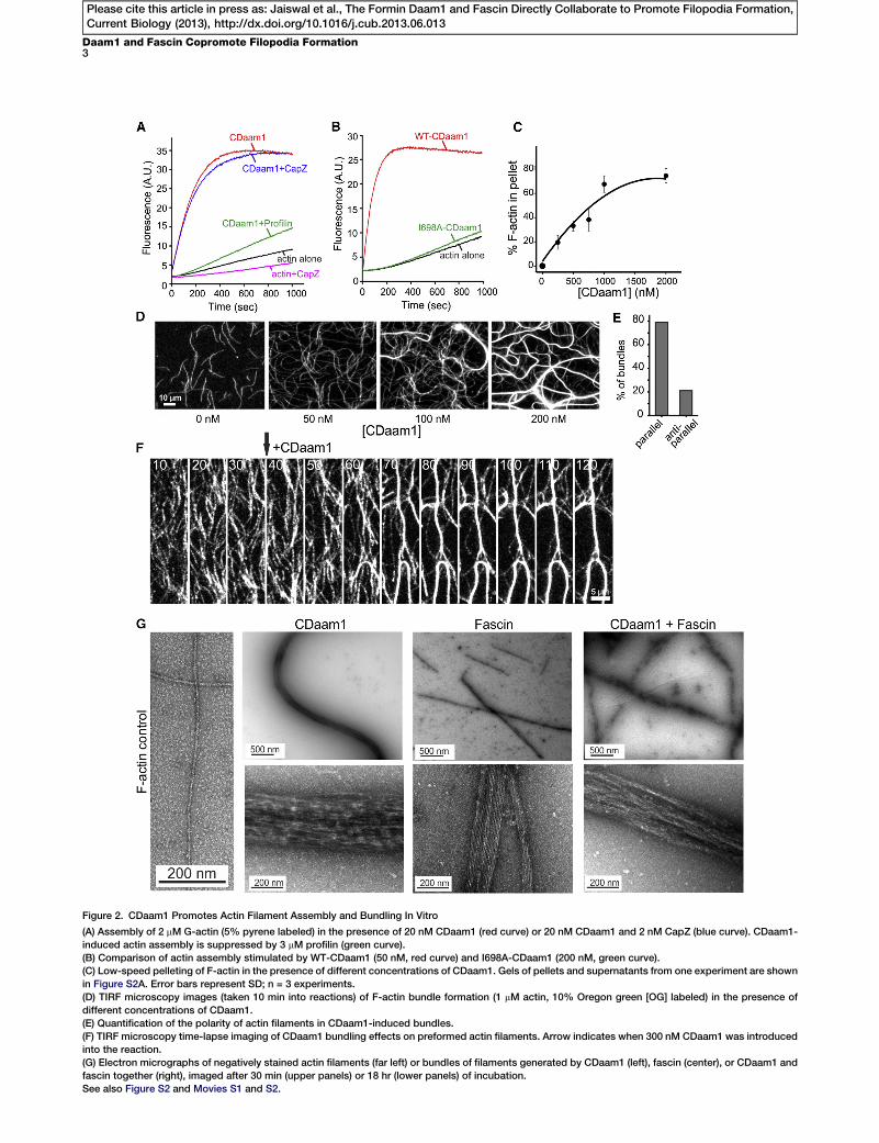
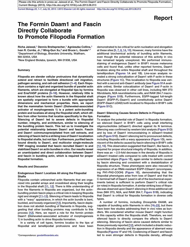
Figure 2. CDaam1 Promotes Actin Filament Assembly and Bundling In Vitro
(A) Assembly of 2 mM G-actin (5% pyrene labeled) in the presence of 20 nM CDaam1 (red curve) or 20 nM CDaam1 and 2 nM CapZ (blue curve). CDaam1-
induced actin assembly is suppressed by 3 mM profilin (green curve).
(B) Comparison of actin assembly stimulated by WT-CDaam1 (50 nM, red curve) and I698A-CDaam1 (200 nM, green curve).
(C) Low-speed pelleting of F-actin in the presence of different concentrations of CDaam1. Gels of pellets and supernatants from one experiment are shown
in Figure S2A. Error bars represent SD; n = 3 experiments.
(D) TIRF microscopy images (taken 10 min into reactions) of F-actin bundle formation (1 mM actin, 10% Oregon green [OG] labeled) in the presence of
different concentrations of CDaam1.
(E) Quantification of the polarity of actin filaments in CDaam1-induced bundles.
(F) TIRF microscopy time-lapse imaging of CDaam1 bundling effects on preformed actin filaments. Arrow indicates when 300 nM CDaam1 was introduced
into the reaction.
(G) Electron micrographs of negatively stained actin filaments (far left) or bundles of filaments generated by CDaam1 (left), fascin (center), or CDaam1 and
fascin together (right), imaged after 30 min (upper panels) or 18 hr (lower panels) of incubation.
See also Figure S2 and Movies S1 and S2.
Daam1 and Fascin Copromote Filopodia Formation3
Please cite this article in press as: Jaiswal et al., The Formin Daam1 and Fascin Directly Collaborate to Promote Filopodia Formation,Current Biology (2013), http://dx.doi.org/10.1016/j.cub.2013.06.013

Current Biology Vol 23 No 144
Please cite this article in press as: Jaiswal et al., The Formin Daam1 and Fascin Directly Collaborate to Promote Filopodia Formation,Current Biology (2013), http://dx.doi.org/10.1016/j.cub.2013.06.013
and the abundance of bundles scaled with increasing CDaam1concentrations (Figure 2D). Furthermore, we observed zipper-ing of polymerizing filaments into bundles (Figure S2B;Movie S1), reminiscent of previously reported fascin- or fim-brin-induced filament zippering [27, 28]. Analysis of bundlepolarity by identification of the fast-growing barbed ends offilaments in time-lapse imaging revealed that approximately80% of CDaam1-induced bundles consisted of filamentswith their barbed ends pointing in the same direction (Figures2E and S2C; Movie S1). This parallel arrangement is similarto the filament organization observed in filopodia [27, 29,30]. We then tested whether, in addition to being able to bun-dle filaments that are actively growing, Daam1 could bundlepreformed filaments. After polymerizing filaments in TIRFchambers, we flowed in 300 nM CDaam1 in the absence ofactin monomers, and we observed rapid bundling, whichreached completion by w50 s after flow-in (Figure 2F;Movie S2).
Our in vivo observations showing that two different bundlingproteins, fascin and Daam1, are required to organize actin intoa structure that can support normal filopodial protrusionprompted us to compare the ultrastructural effects of fascinand Daam1 on bundle formation in vitro using transmissionelectron microscopy (TEM) (Figures 2G and S2D). Fascin pro-duced very straight bundles consisting of flattened arrays ofregularly spaced filaments, enabling measurement of averagenumber of filaments per bundle (17.7 6 1.6) and averageinterfilament distance (8.2 6 0.5 nm) [16, 31] (Figure S2D). Incontrast, CDaam1 produced wavy, densely packed, roundedbundles, precluding any analysis of the number of filamentsper bundle or interfilament distances. Interestingly, bundlesformed in the combined presence of CDaam1 and fascinshowed intermediate levels of bundle straightness, rounded-ness, and width, and intermediate levels of interfilamentspacing and organization. Thus, the composite bundlesappear to acquire distinct properties from each bundler, whichmay help explain their corequirement for filopodia formationin vivo.
Fascin Directly Binds and Recruits Daam1 to ActinFilament Bundles
Our in vivo observation that fascin is required for Daam1 local-ization to filopodial shafts prompted us to test whetherCDaam1 and fascin physically associate. From B16F1 cellsexpressing GFP alone, GFP-CDaam1, or GFP-FL-Daam1from plasmids, we found that endogenous fascin coimmuno-precipitated with both GFP-Daam1 constructs, but not withGFP alone, suggesting that fascin interacts with the C-terminalhalf of Daam1 (Figure 3A). Furthermore, purified CDaam1bound to immobilized GST-fascin, but not GST (Figure 3B),demonstrating that fascin-Daam1 interactions are direct.
To better understand the functional relationship betweenfascin and Daam1, we employed two-color TIRF microscopyto directly visualize labeled Daam1 molecules interacting withactin bundles. A purified SNAP-CDaam1 fusion protein waslabeled with benzylguanine-Dy649 fluorescent dye. SNAP-649-CDaam1 nucleated actin assembly in bulk assays indistin-guishably from unlabeled CDaam1 (Figure S3A). Furthermore,it enhanced filament elongation rates in TIRF assays in aprofilin-dependent manner (Figure S3B). Single SNAP-649-CDaam1 molecules processively moved on growing barbedends of actin filaments in the presence and absence of profilin(Figures 3C and S3C; Movie S3), as observed for SNAP-mDia1[32]. At concentrations above 100 nM, SNAP-649-CDaam1
induced actin bundle formation, similar to untagged CDaam1,and decorated the bundles (Figure 3D). Thus, SNAP-649-CDaam1 was functionally equivalent to CDaam1 in all of itseffects on actin dynamics and organization.To analyze dynamics of SNAP-649-CDaam1 molecules on
actin bundles, we used low nanomolar concentrations ofSNAP-649-CDaam1, which reduced the background andenabled detection of single-molecule binding events. Becausethese concentrations of SNAP-649-CDaam1 do not bundle fil-aments, we prebundled filaments with 300 nM of unlabeledCDaam1, fascin, or fimbrin (as a control) and then flowedin lower concentrations (1–10 nM) of SNAP-649-CDaam1.SNAP-649-CDaam1 molecules rarely interacted with the sidesof single (nonbundled) filaments but readily associated withthe barbed ends (Figure 3C; R.J., unpublished data). Whenfilaments were bundled by fascin or unlabeled CDaam1,SNAP-649-CDaam1 molecules bound to the sides of bundles(Figure 3E). In contrast, when filaments were bundled by fim-brin, SNAP-649-CDaam1 showed no interactions with thesides of bundles (Figure 3E). Thus, SNAP-649-CDaam1 recruit-ment to bundles is highly specific, consistent with the directinteraction between fascin and CDaam1.As an additional control, we asked whether a different for-
min, mDia1, which lacks bundling activity and is absent fromfilopodial shafts [22, 33, 34], was recruited to the sidesof bundles. SNAP-tagged mDia1 (SNAP-649-mDia1-C, FH1-FH2-COOH) readily associated with barbed ends of filamentsand remained processively attached during elongation asexpected [32]. However, no specific interactions of SNAP-649-mDia1-C with the sides of fascin-generated bundleswere detected (Figure 3E). Thus, interactions of SNAP-649-CDaam1 with the sides of bundles are highly specific.Quantification of the number of SNAP-649-CDaam1 andSNAP-649-mDia1-C spots per unit length of bundle furtherrevealed that SNAP-649-CDaam1 interactions are more pro-nounced for fascin-induced bundles compared to CDaam1-induced bundles (Figure 3F). Taken together with our otherobservations, fascin appears to directly recruit Daam1 tobundles.Finally, we analyzed the dynamics of single SNAP-649-
CDaam1 molecules in CDaam1- and fascin-generated actinbundles to gain insights into the mechanism of Daam1 interac-tions with these structures. SNAP-649-CDaam1 moleculeswere observed to transiently bind CDaam1-bundled filaments,with themajority of dwell times being shorter than 50 s (Figures4A, 4C, and S3D; Movie S4). However, in fascin-inducedbundles, SNAP-649-CDaam1 dwell times greatly increased(Figures 4B, 4D, and S3D; Movie S4), suggesting that fascinstabilizes CDaam1 associations with the bundles. SNAP-649-CDaam1 molecules displayed two-dimensional diffusionalong CDaam1-induced bundles, as determined by mean-square displacement analysis (Figures 4A and 4E; Movie S5).In contrast, diffusion was abrogated in fascin-induced bundlesas indicated by the spatially static spots (Figures 4B and 4D;Movie S4). Thus, fascin confines SNAP-CDaam1 in the bundle,which is remarkably consistent with our in vivo observationsshowing that Daam1 localization to filopodial shafts is dimin-ished after fascin silencing.
Concluding Remarks
In summary, our results reveal a critical role for the forminDaam1 in organizing filaments in the filopodial shaft, and inmaintaining the structural integrity of filopodia. Until now, fas-cin has been the only bundler shown to both localize all along
Figure 3. Fascin Directly Binds and Recruits Daam1 to Actin Filament Bundles
(A) Coimmunoprecipitation in B16F1 cells of endogenous fascin with plasmid-expressed GFP-CDaam1 and GFP-FL-Daam1, but not GFP. Blots were
probed with antibodies to fascin (top) and GFP (bottom).
(B) Immunoblot showing direct binding between purified fascin and CDaam1. The blot was probed with anti-Daam1 antibodies and was quantified for levels
of soluble CDaam1 in pellet fractions after incubation with GST or GST-fascin beads. Error bars represent SD, n = 3.
(C) Dual-color TIRF microscopy of an elongating actin filament (green), conditions as above, processively capped by a SNAP-649-CDaam1 molecule (red).
Time is given in seconds.
(D) F-actin bundling by SNAP-649-CDaam1. Actin (1 mM; 10%OG labeled) was polymerized in the presence of 300 nM SNAP-649-CDaam1, producing bun-
dles heavily decorated with SNAP-649-CDaam1.
(E) Single-molecule binding of SNAP-649-CDaam1 to preformed F-actin bundles. Actin (1 mM; 10%OG labeled) was polymerized in the presence of 300 nM
CDaam1, fascin, or fimbrin (Sac6) to generate actin filament bundles, 1 nMSNAP-649-CDaam1or SNAP-649-mDia1-Cwas introduced, and binding of formin
molecules (red) to bundles (green) was monitored. Arrows mark formin molecules associated with filament/bundle barbed ends.
(F) Quantification of the number of SNAP-649-CDaam1 or SNAP-649-mDia1-C spots per mm length of bundle. Error bars represent SD (n = 3 experiments;
more than 20 bundles were scored for each condition).
See also Figure S3 and Movie S3.
Daam1 and Fascin Copromote Filopodia Formation5
Please cite this article in press as: Jaiswal et al., The Formin Daam1 and Fascin Directly Collaborate to Promote Filopodia Formation,Current Biology (2013), http://dx.doi.org/10.1016/j.cub.2013.06.013
the filopodia shaft and be required for filopodia integrity. Wefound that Daam1 similarly localizes along the shafts anddemonstrated that both Daam1 and fascin are required forproper filopodia formation. Furthermore, we found that fascinand Daam1 directly associate, and that fascin recruits Daam1to filopodial shafts in vivo. In vitro, fascin also is sufficient to re-cruit Daam1 to actin bundles and restrict Daam1 diffusionalong bundles. Together, these data reveal an unanticipatedand direct collaboration between these two actin bundlers,where each is critical in vivo for the proper formation offilopodia.
Finally, our results expand the in vivo roles of formins toinclude actin bundling. Until now, formin cellular functions inregulating the actin cytoskeleton have focused primarily onactin filament nucleation and elongation, and there havebeen few in vivo investigations into their potential roles as
bundlers. Even though bundling activities have beendescribed in vitro for many formins [17, 22], the in vivo rele-vance of these activities has remained in question. Our find-ings demonstrate a critical role for the formin Daam1 inbundling filaments and collaborating with fascin to give the fi-lopodia shaft structural integrity. Formins also decorate anumber of other actin arrays in vivo that contain bundled fila-ments (e.g., stress fibers, cytokinetic rings, stereocilia, invado-podia, bristles, and sarcomeres) [35–40], where their bundlingactivities may be relevant.
Supplemental Information
Supplemental Information includes three figures, Supplemental Experi-
mental Procedures, and fivemovies and can be found with this article online
at http://dx.doi.org/10.1016/j.cub.2013.06.013.
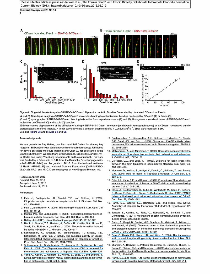
Figure 4. Single-Molecule Analysis of SNAP-649-CDaam1 Dynamics on Actin Bundles Generated by Unlabeled CDaam1 or Fascin
(A and B) Time-lapse imaging of SNAP-649-CDaam1 molecules binding to actin filament bundles produced by CDaam1 (A) or fascin (B).
(C and D) Kymographs of SNAP-649-CDaam1 binding to bundles from experiments as in (A) and (B). Histograms show dwell times of SNAP-649-CDaam1
molecules on CDaam1 (C) and fascin (D) bundles.
(E) Mean-square displacement of the diffusion of a single SNAP-649-CDaam1 molecule (as shown in kymograph above) on a CDaam1-generated bundle
plotted against the time interval. A linear curve fit yields a diffusion coefficient of D = 0.06825 mm2 s21. Error bars represent SEM.
See also Figure S3 and Movies S4 and S5.
Current Biology Vol 23 No 146
Please cite this article in press as: Jaiswal et al., The Formin Daam1 and Fascin Directly Collaborate to Promote Filopodia Formation,Current Biology (2013), http://dx.doi.org/10.1016/j.cub.2013.06.013
Acknowledgments
We are grateful to Ray Habas, Jan Faix, and Jeff Gelles for sharing key
reagents; EdDougherty for assistancewith confocalmicroscopy; JeffGelles
for advice on single-molecule imaging; and Chen Xu for assistance in the
Brandeis EM facility. We also thank Brian Graziano, BrookeMcCartney, Avi-
tal Rodal, and Casey Ydenberg for comments on the manuscript. This work
was funded by a fellowship to D.B. from the Deutsche Forschungsgemein-
schaft (BR 4116-1/1) and by grants to B.L.G. from the National Institutes
of Health (GM083137) and National Science Foundation (DMR-MRSEC-
0820429). I.R.C. and M.-Q.X. are employees of New England Biolabs, Inc.
Received: April 9, 2013
Revised: May 25, 2013
Accepted: June 5, 2013
Published: July 11, 2013
References
1. Faix, J., Breitsprecher, D., Stradal, T.E., and Rottner, K. (2009).
Filopodia: complex models for simple rods. Int. J. Biochem. Cell Biol.
41, 1656–1664.
2. Faix, J., and Rottner, K. (2006). The making of filopodia. Curr. Opin. Cell
Biol. 18, 18–25.
3. Mattila, P.K., and Lappalainen, P. (2008). Filopodia: molecular architec-
ture and cellular functions. Nat. Rev. Mol. Cell Biol. 9, 446–454.
4. Ridley, A.J. (2011). Life at the leading edge. Cell 145, 1012–1022.
5. Block, J., Stradal, T.E., Hanisch, J., Geffers, R., Kostler, S.A., Urban, E.,
Small, J.V., Rottner, K., and Faix, J. (2008). Filopodia formation induced
by active mDia2/Drf3. J. Microsc. 231, 506–517.
6. Schirenbeck, A., Arasada, R., Bretschneider, T., Stradal, T.E.,
Schleicher, M., and Faix, J. (2006). The bundling activity of vasodi-
lator-stimulated phosphoprotein is required for filopodium formation.
Proc. Natl. Acad. Sci. USA 103, 7694–7699.
7. Schirenbeck, A., Bretschneider, T., Arasada, R., Schleicher, M., and
Faix, J. (2005). The Diaphanous-related formin dDia2 is required for
the formation and maintenance of filopodia. Nat. Cell Biol. 7, 619–625.
8. Yang, C., Czech, L., Gerboth, S., Kojima, S., Scita, G., and Svitkina, T.
(2007). Novel roles of formin mDia2 in lamellipodia and filopodia forma-
tion in motile cells. PLoS Biol. 5, e317.
9. Breitsprecher, D., Kiesewetter, A.K., Linkner, J., Urbanke, C., Resch,
G.P., Small, J.V., and Faix, J. (2008). Clustering of VASP actively drives
processive, WH2 domain-mediated actin filament elongation. EMBO J.
27, 2943–2954.
10. Mallavarapu, A., and Mitchison, T. (1999). Regulated actin cytoskeleton
assembly at filopodium tips controls their extension and retraction.
J. Cell Biol. 146, 1097–1106.
11. DeRosier, D.J., and Edds, K.T. (1980). Evidence for fascin cross-links
between the actin filaments in coelomocyte filopodia. Exp. Cell Res.
126, 490–494.
12. Vignjevic, D., Kojima, S., Aratyn, Y., Danciu, O., Svitkina, T., and Borisy,
G.G. (2006). Role of fascin in filopodial protrusion. J. Cell Biol. 174,
863–875.
13. Otto, J.J., Kane, R.E., andBryan, J. (1979). Formation of filopodia in coe-
lomocytes: localization of fascin, a 58,000 dalton actin cross-linking
protein. Cell 17, 285–293.
14. Block, J., Breitsprecher, D., Kuhn, S., Winterhoff, M., Kage, F., Geffers,
R., Duwe, P., Rohn, J.L., Baum, B., Brakebusch, C., et al. (2012). FMNL2
drives actin-based protrusion and migration downstream of Cdc42.
Curr. Biol. 22, 1005–1012.
15. Harris, E.S., Gauvin, T.J., Heimsath, E.G., and Higgs, H.N. (2010).
Assembly of filopodia by the formin FRL2 (FMNL3). Cytoskeleton 67,
755–772.
16. Jansen, S., Collins, A., Yang, C., Rebowski, G., Svitkina, T., and
Dominguez, R. (2011). Mechanism of actin filament bundling by fascin.
J. Biol. Chem. 286, 30087–30096.
17. Barko, S., Bugyi, B., Carlier, M.F., Gombos, R., Matusek, T., Mihaly, J.,
and Nyitrai, M. (2010). Characterization of the biochemical properties
and biological function of the formin homology domains of Drosophila
DAAM. J. Biol. Chem. 285, 13154–13169.
18. Esue, O., Harris, E.S., Higgs, H.N., and Wirtz, D. (2008). The filamentous
actin cross-linking/bundling activity of mammalian formins. J. Mol. Biol.
384, 324–334.
19. Michelot, A., Derivery, E., Paterski-Boujemaa, R., Guerin, C., Huang, S.,
Parcy, F., Staiger, C.J., and Blanchoin, L. (2006). A novel mechanism for
the formation of actin-filament bundles by a nonprocessive formin. Curr.
Biol. 16, 1924–1930.
20. Harris, E.S., and Higgs, H.N. (2006). Biochemical analysis of mammalian
formin effects on actin dynamics. Methods Enzymol. 406, 190–214.
Daam1 and Fascin Copromote Filopodia Formation7
Please cite this article in press as: Jaiswal et al., The Formin Daam1 and Fascin Directly Collaborate to Promote Filopodia Formation,Current Biology (2013), http://dx.doi.org/10.1016/j.cub.2013.06.013
21. Machaidze, G., Sokoll, A., Shimada, A., Lustig, A., Mazur, A.,
Wittinghofer, A., Aebi, U., and Mannherz, H.G. (2010). Actin filament
bundling and different nucleating effects of mouse Diaphanous-related
formin FH2 domains on actin/ADF and actin/cofilin complexes. J. Mol.
Biol. 403, 529–545.
22. Harris, E.S., Rouiller, I., Hanein, D., and Higgs, H.N. (2006). Mechanistic
differences in actin bundling activity of two mammalian formins, FRL1
and mDia2. J. Biol. Chem. 281, 14383–14392.
23. Moseley, J.B., and Goode, B.L. (2005). Differential activities and regula-
tion of Saccharomyces cerevisiae formin proteins Bni1 and Bnr1 by
Bud6. J. Biol. Chem. 280, 28023–28033.
24. Neidt, E.M., Skau, C.T., and Kovar, D.R. (2008). The cytokinesis formins
from the nematode worm and fission yeast differentially mediate actin
filament assembly. J. Biol. Chem. 283, 23872–23883.
25. Okada, K., Bartolini, F., Deaconescu, A.M., Moseley, J.B., Dogic, Z.,
Grigorieff, N., Gundersen, G.G., and Goode, B.L. (2010). Adenomatous
polyposis coli protein nucleates actin assembly and synergizes with
the formin mDia1. J. Cell Biol. 189, 1087–1096.
26. Paul, A.S., and Pollard, T.D. (2008). The role of the FH1 domain and
profilin in formin-mediated actin-filament elongation and nucleation.
Curr. Biol. 18, 9–19.
27. Breitsprecher, D., Koestler, S.A., Chizhov, I., Nemethova, M.,Mueller, J.,
Goode, B.L., Small, J.V., Rottner, K., and Faix, J. (2011). Cofilin cooper-
ates with fascin to disassemble filopodial actin filaments. J. Cell Sci.
124, 3305–3318.
28. Skau, C.T., and Kovar, D.R. (2010). Fimbrin and tropomyosin competi-
tion regulates endocytosis and cytokinesis kinetics in fission yeast.
Curr. Biol. 20, 1415–1422.
29. Lewis, A.K., and Bridgman, P.C. (1992). Nerve growth cone lamellipodia
contain two populations of actin filaments that differ in organization and
polarity. J. Cell Biol. 119, 1219–1243.
30. Small, J.V. (1988). The actin cytoskeleton. Electron Microsc. Rev. 1,
155–174.
31. Ishikawa, R., Sakamoto, T., Ando, T., Higashi-Fujime, S., and Kohama,
K. (2003). Polarized actin bundles formed by human fascin-1: their
sliding and disassembly on myosin II and myosin V in vitro.
J. Neurochem. 87, 676–685.
32. Breitsprecher, D., Jaiswal, R., Bombardier, J.P., Gould, C.J., Gelles, J.,
and Goode, B.L. (2012). Rocket launcher mechanism of collaborative
actin assembly defined by single-molecule imaging. Science 336,
1164–1168.
33. Suraneni, P., Rubinstein, B., Unruh, J.R., Durnin, M., Hanein, D., and Li,
R. (2012). The Arp2/3 complex is required for lamellipodia extension and
directional fibroblast cell migration. J. Cell Biol. 197, 239–251.
34. Sarmiento, C., Wang, W., Dovas, A., Yamaguchi, H., Sidani, M., El-Sibai,
M., Desmarais, V., Holman, H.A., Kitchen, S., Backer, J.M., et al. (2008).
WASP family members and formin proteins coordinate regulation of cell
protrusions in carcinoma cells. J. Cell Biol. 180, 1245–1260.
35. Hotulainen, P., and Lappalainen, P. (2006). Stress fibers are generated
by two distinct actin assembly mechanisms in motile cells. J. Cell
Biol. 173, 383–394.
36. Li, D., Hallett, M.A., Zhu, W., Rubart, M., Liu, Y., Yang, Z., Chen, H.,
Haneline, L.S., Chan, R.J., Schwartz, R.J., et al. (2011). Dishevelled-
associated activator of morphogenesis 1 (Daam1) is required for heart
morphogenesis. Development 138, 303–315.
37. Sato, A., Khadka, D.K., Liu, W., Bharti, R., Runnels, L.W., Dawid, I.B.,
and Habas, R. (2006). Profilin is an effector for Daam1 in non-canonical
Wnt signaling and is required for vertebrate gastrulation. Development
133, 4219–4231.
38. Severson, A.F., Baillie, D.L., and Bowerman, B. (2002). A Formin
Homology protein and a profilin are required for cytokinesis and Arp2/
3-independent assembly of cortical microfilaments in C. elegans.
Curr. Biol. 12, 2066–2075.
39. Takeya, R., Taniguchi, K., Narumiya, S., and Sumimoto, H. (2008). The
mammalian formin FHOD1 is activated through phosphorylation by
ROCK and mediates thrombin-induced stress fibre formation in endo-
thelial cells. EMBO J. 27, 618–628.
40. Watanabe, S., Ando, Y., Yasuda, S., Hosoya, H., Watanabe, N., Ishizaki,
T., and Narumiya, S. (2008). mDia2 induces the actin scaffold for the
contractile ring and stabilizes its position during cytokinesis in NIH
3T3 cells. Mol. Biol. Cell 19, 2328–2338.