Demonstration of Reversible Priming of Human Neutrophils Using Platelet-Activating Factor
9
1996 88: 4330-4337 E Kitchen, AG Rossi, AM Condliffe, C Haslett and ER Chilvers platelet- activating factor Demonstration of reversible priming of human neutrophils using http://bloodjournal.hematologylibrary.org/content/88/11/4330.full.html Updated information and services can be found at: Articles on similar topics can be found in the following Blood collections http://bloodjournal.hematologylibrary.org/site/misc/rights.xhtml#repub_requests Information about reproducing this article in parts or in its entirety may be found online at: http://bloodjournal.hematologylibrary.org/site/misc/rights.xhtml#reprints Information about ordering reprints may be found online at: http://bloodjournal.hematologylibrary.org/site/subscriptions/index.xhtml Information about subscriptions and ASH membership may be found online at: Copyright 2011 by The American Society of Hematology; all rights reserved. Society of Hematology, 2021 L St, NW, Suite 900, Washington DC 20036. Blood (print ISSN 0006-4971, online ISSN 1528-0020), is published weekly by the American For personal use only. on March 28, 2014. by guest bloodjournal.hematologylibrary.org From For personal use only. on March 28, 2014. by guest bloodjournal.hematologylibrary.org From
-
Upload
independent -
Category
Documents
-
view
3 -
download
0
Transcript of Demonstration of Reversible Priming of Human Neutrophils Using Platelet-Activating Factor

1996 88: 4330-4337
E Kitchen, AG Rossi, AM Condliffe, C Haslett and ER Chilvers platelet- activating factorDemonstration of reversible priming of human neutrophils using
http://bloodjournal.hematologylibrary.org/content/88/11/4330.full.htmlUpdated information and services can be found at:
Articles on similar topics can be found in the following Blood collections
http://bloodjournal.hematologylibrary.org/site/misc/rights.xhtml#repub_requestsInformation about reproducing this article in parts or in its entirety may be found online at:
http://bloodjournal.hematologylibrary.org/site/misc/rights.xhtml#reprintsInformation about ordering reprints may be found online at:
http://bloodjournal.hematologylibrary.org/site/subscriptions/index.xhtmlInformation about subscriptions and ASH membership may be found online at:
Copyright 2011 by The American Society of Hematology; all rights reserved.Society of Hematology, 2021 L St, NW, Suite 900, Washington DC 20036.Blood (print ISSN 0006-4971, online ISSN 1528-0020), is published weekly by the American
For personal use only.on March 28, 2014. by guest bloodjournal.hematologylibrary.orgFrom For personal use only.on March 28, 2014. by guest bloodjournal.hematologylibrary.orgFrom
Demonstration of Reversible Priming of Human Neutrophils Using Platelet-Activating Factor
By Elizabeth Kitchen, Adriano G. Rossi, Alison M. Condliffe, Christopher Haslett, and Edwin R. Chilvers
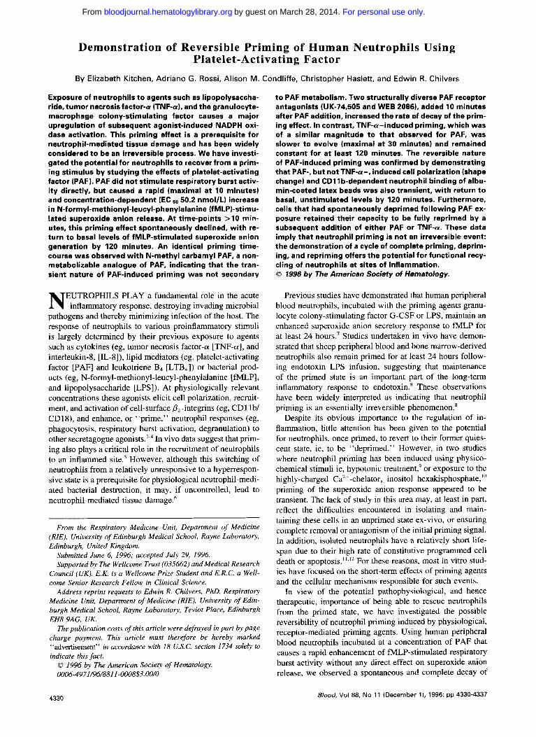
Exposure of neutrophils t o agents such as lipopolysaccha- ride, tumor necrosis factor-a (TNF-a), and the granulocyte- macrophage colony-stimulating factor causes a major upregulation of subsequent agonist-induced NADPH oxi- dase activation. This priming effect is a prerequisite for neutrophil-mediated tissue damage and has been widely considered t o be an irreversible process. We have investi- gated the potential for neutrophils t o recover from a prim- ing stimulus by studying the effects of platelet-activating factor (PAF). PAF did not stimulate respiratory burst activ- i t y directly, but caused a rapid (maximal at 10 minutes) and concentration-dependent (EC 50.2 nmol/L) increase in N-formyl-methionyl-leucyl-phenylalanine (fMLP)-stimu- lated superoxide anion release. At time-points >10 min- utes, this priming effect spontaneously declined, wi th re- turn t o basal levels of fMLP-stimulated superoxide anion generation by 120 minutes. An identical priming time- course was observed with N-methyl carbamyl PAF, a non- metabolizable analogue of PAF, indicating that the tran- sient nature of PAF-induced priming was not secondary
EUTROPHILS PLAY a fundamental role in the acute N inflammatory response, destroying invading microbial pathogens and thereby minimizing infection of the host. The response of neutrophils to various proinflammatory stimuli is largely determined by their previous exposure to agents such as cytokines (eg, tumor necrosis factor-a [TNF-a], and interleukin-8, [IL-8]), lipid mediators (eg, platelet-activating factor [PAF] and leukotriene B4 [LTB,]) or bacterial prod- ucts (eg, N-formyl-methionyl-leucyl-phenylalanine [fMLP], and lipopolysaccharide [LPS]). At physiologically relevant concentrations these agonists elicit cell polarization, recruit- ment, and activation of cell-surface p,-integrins (eg, CD1 lb/ CD18), and enhance, or “prime,” neutrophil responses (eg, phagocytosis, respiratory burst activation, degranulation) to other secretagogue agonist^.'.^ In vivo data suggest that prim- ing also plays a critical role in the recruitment of neutrophils to an inflammed site.5 However, although this switching of neutrophils from a relatively unresponsive to a hyperrespon- sive state is a prerequisite for physiological neutrophil-medi- ated bacterial destruction, it may, if uncontrolled, lead to neutrophil-mediated tissue damage.6
From the Respiratory Medicine Unit, Department of Medicine (RIE), University of Edinburgh Medical School, Rayne Laboratory, Edinburgh, United Kingdom.
Submitted June 6, 1996; accepted July 29, 1996. Supported by The Wellcome Trust (035662) and Medical Research
Council (UK). E.K. is a Wellcome Prize Student and E.R.C. a Well- come Senior Research Fellow in Clinical Science.
Address reprint requests to Edwin R. Chilvers, PhD, Respiratoly Medicine Unit, Department of Medicine (RIE), University of Edin- burgh Medical School, Rayne Laboratory, Teviot Place, Edinburgh EH8 9AG, UK.
The publication costs of this article were defrayed in part by page charge payment. This article must therefore be hereby marked “advertisement” in accordance with 18 U.S.C. section 1734 solely to indicate this fact. 0 1996 by The American Society of Hematology. 0006-497~/96/8811-0008$3.00/0
t o PAF metabolism. Two structurally diverse PAF receptor antagonists (UK-74,505 and WEB 2086). added 10 minutes after PAF addition, increased the rate of decay of the prim- ing effect. In contrast, TNF-a-induced priming, which was of a similar magnitude t o that observed for PAF, was slower to evolve (maximal at 30 minutes) and remained constant for at least 120 minutes. The reversible nature of PAF-induced priming was confirmed by demonstrating that PAF-, but not TNF-a-, induced cell polarization (shape change) and CDI 1 b-dependent neutrophil binding of albu- min-coated latex beads was also transient, wi th return t o basal, unstimulated levels by 120 minutes. Furthermore, cells that had spontaneously deprimed following PAF ex- posure retained their capacity t o be fully reprimed by a subsequent addition of either PAF or TNF-a. These data imply that neutrophil priming is not an irreversible event: the demonstration of a cycle of complete priming, deprim- ing, and repriming offers the potential for functional recy- cling of neutrophils at sites of inflammation. 0 7996 by The American Society of Hematology.
Previous studies have demonstrated that human peripheral blood neutrophils, incubated with the priming agents granu- locyte colony-stimulating factor G-CSF or LPS, maintain an enhanced superoxide anion secretory response to fMLP for at least 24 hours.’ Studies undertaken in vivo have demon- strated that sheep peripheral blood and bone marrow-derived neutrophils also remain primed for at least 24 hours follow- ing endotoxin LPS infusion, suggesting that maintenance of the primed state is an important part of the long-term inflammatory response to endotoxin.R These observations have been widely interpreted as indicating that neutrophil priming is an essentially irreversible phenomenon.’
Despite its obvious importance to the regulation of in- flammation, little attention has been given to the potential for neutrophils, once primed, to revert to their former quies- cent state, ie, to be “deprimed.” However, in two studies where neutrophil priming has been induced using physico- chemical stimuli ie, hypotonic treatment,’ or exposure to the highly-charged Ca2’-chelator, inositol hexakisphosphate,”’ priming of the superoxide anion response appeared to be transient. The lack of study in this area may, at least in part, reflect the difficulties encountered in isolating and main- taining these cells in an unprimed state ex-vivo, or ensuring complete removal or antagonism of the initial priming signal. In addition, isolated neutrophils have a relatively short life- span due to their high rate of constitutive programmed cell death or apoptosis.”,12 For these reasons, most in vitro stud- ies have focused on the short-term effects of priming agents and the cellular mechanisms responsible for such events.
In view of the potential pathophysiological, and hence therapeutic, importance of being able to rescue neutrophils from the primed state, we have investigated the possible reversibility of neutrophil priming induced by physiological, receptor-mediated priming agents. Using human peripheral blood neutrophils incubated at a concentration of PAF that causes a rapid enhancement of fMLP-stimulated respiratory burst activity without any direct effect on superoxide anion release, we observed a spontaneous and complete decay of
4330 Blood, Vol88, No 11 (December I ) , 1996: pp 4330-4337
For personal use only.on March 28, 2014. by guest bloodjournal.hematologylibrary.orgFrom

REVERSIBLE PRIMING OF HUMAN NEUTROPHILS USING PAF 433 1
the PAF-primed superoxide anion response and PAF- induced CD1 lb/CD18 activation and to a lesser extent, PAF-induced shape change. The rate of neutrophil recovery following PAF addition could be enhanced by the use of selective PAF receptor antagonists (WEB 2086 and UK- 74,505). Furthermore, the deprimed cells retained their full capacity to be reprimed by an alternative priming agent (TNF-a) or a further addition of PAF. The ability of neutro- phils to participate in a complete priming/depriming/reprim- ing cycle allows far greater flexibility in the control of neu- trophil behavior at an inflammatory site than was hitherto realized.
MATERIALS AND METHODS
Neutrophil preparation. Peripheral venous blood was taken from healthy adult volunteers, anticoagulated with 4 mL 3.8% so- dium citrate/40 mL blood, and centrifuged (300g) for 20 minutes. Neutrophils were isolated exactly as previously detailed' using dex- tran sedimentation and discontinuous plasma-Percoll gradients. This isolation technique yields neutrophils that display very low levels of basal shape change (<8% assessed flow-cytometrically) or direct fMLP-induced superoxide anion generation. The purified neutrophils were washed sequentially in platelet-poor plasma, phosphate-buf- fered saline (PBS) without, and PBS with, CaC1, and MgCIZ. Cell punty and viability (trypan blue exclusion) were routinely >95% (<0.5% monocyte contamination) and >99.5%, respectively.
Neutrophils (IO6 in 90 pL PBS containing CaCl2 and MgCI2) were equilibrated in a gently shaking water-bath for 5 minutes at 37°C. Priming agents were added in a IO-pL volume to achieve the required drug concentrations (PAF 1 nmollL to I O pmol/L, TNF-a 200 U/mL) and incubations continued for the peri- ods stated. Preliminary experiments demonstrated this concentration of TNF-a to be optimal in causing maximal enhancement of fMLP- induced superoxide anion generation with minimal direct respiratory burst activation (data not shown). To determine the ICso values of the PAF receptor antagonists used, neutrophils were incubated with WEB 2086 or UK-74,505 (both at 10 nmolL to 10 pmol/L) for 30 minutes before the addition of priming agents. For investigations examining the reversibility of PAF-induced priming, neutrophils were treated with PAF (1 pmoUL) for I O minutes before addition of 1 p m o m WEB 2086 or UK-74,505. fMLP (100 nmol/L) or buffer (PBS) was added to samples (final volume 1 mL) 10 minutes before the addition of an equal volume of 2.5% glutaraldehyde. Samples were analyzed for shape change by flow cytometry (Coulter EPICS Profile 11; Coulter Electronics, Luton, UK) using a slight modifica- tion of a previously published method.I3 Percentage shape change was calculated from the mean forward light scatter of each sample by gating on the non-shape changed neutrophil population. The values obtained using this method correlate closely with those derived by direct visual assessment of shape change, with the exception that the flow cytometric method of assessment slightly overestimates the extent of basal shape ~ h a n g e . ' ~
Superoxide anion release assay. Neutrophils were isolated, equilibrated at 37"C, and incubated with PAF or PBS exactly as detailed above, except that cytochrome C (800 pL, 1 mg/mL) was added immediately before the addition of MLP. One of each set of quadruplicate determinations included superoxide dismutase (375 U) to allow confirmation of the specificity of cytochrome C reduction. Reactions were stopped by placing the cells on ice, followed by centrifugation ( I 2,50Og, 2 minutes, 4°C). The superoxide dismutase- inhibitable reduction of cytochrome C was determined in each super- natant by measuring the peak absorbance between 535 and 565 nm using a Pye-Unicam scanning spectrophotometer, and expressed as nanomoles superoxide anions generated per 10' neutrophils. In ex-
Shape change assay.
periments designed to assess the ability of PAF-recovered neutro- phils to be reprimed with either TNF-a or a further addtion of PAF, cells were treated with PAF (1 pmol/L) or PBS for 120 minutes, before a final incubation with PAF (1 pmol/L, 10 minutes) or TNF- a (200 U/mL, 30 minutes) and assessment of MLP-stimulated su- peroxide anion release.
To examine the effects of hypotonic challenge on neutrophil func- tion, cells (lo6 in 250 fiL PBS) were equilibrated at 37°C as outlined above and incubated for 19 minutes in PBS containing 80 pmol/L cytochrome C, with 150 mmol/L NaCl (isotonic incubations) or 50 mmol/L NaCl (hypotonic incubations). Neutrophils were then treated for 1 minute with 20 pL of either 5 m o m NaCl (to reverse hypoto- nicity to isotonicity) or PBS (to retain hypotonicity or isotonicity). The effects of the hypotonic challenge itself on basal and fMLP- stimulated superoxide anion generation, together with the ability of these cells to deprime following restoration of isotonicity and there- after be reprimed with PAF, was then assessed as detailed above.
Neurophil adhesion to albumin-coated latex heads (A CLB). Fluorescent latex beads (2.5% packed voUvol stock solution as pur- chased) were washed (three times) in PBS, resuspended at 2.5% (vol/vol) in PBS containing IO mg/mL human serum albumin, and incubated for 10 minutes at 25°C. The resultant ACLB were again washed (three times) in PBS and finally resuspended at 0.75% (vol/ vol). Neutrophils (175 pL aliquots at 107/mL, in PBS with CaC1, and MgCI2) were incubated in a shaking water-bath at 37°C for 5 minutes, and then treated with PAF (1 pmoUL), TNF-a (200 UlmL) or PBS (all added in a 15-pL volume) for 0 to 120 minutes. ACLB (25 pL of 0.75% voUvol solution) were added to each tube 15 minutes before the termination of each reaction, except for time points <15 minutes where the beads were added before the agonist. Neutrophils were then fixed by the addition of 0.5 mL of 0.5% glutaraldehyde. After 30 minutes at room temperature, nonadherent ACLB were removed by washing with PBS (three times) and bead- binding to the neutrophil assessed using an EPICS Profile I1 (Coulter Electronics), as previously detailed.I4
All values are expressed as means 5 standard error of mean (SEM) of (n) number of separate experiments. Values, where applicable, were compared by ANOVA or the Student's f- test for paired data, with P < .05 considered to be significant. Significant differences between groups were determined by the New- man-Keuls procedure.
fMLP, PAF, superoxide dismutase (SOD), cyto- chrome C, PBS (with or without CaCIz and MgClz), dextran-500, Percoll, human serum albumin and glutaraldehyde (25%) were pur- chased from Sigma (Poole, UK). TNF-a was obtained from Gen- zyme (Cambridge, MA). One micron fluorescent microspheres were purchased as a 2.5% solids-latex (2.5% vol/vol) stock solution from Polysciences Inc through the UK supplier Park Scientific (Notting- ham, UK). WEB 2086 and UK-74,505 were gifts from Boehringer Ingelheim Ltd (Berks, UK) and Dr J. Parry (Pfizer, Sandwich, UK), respectively. 1 - 0- alkyl-2-N-methylcarbamyl-glycerophosph~holine (N-methyl carbamyl PAF) was obtained from Calbiochem (Notting- ham, UK).
Statistics.
Materials.
RESULTS
PAF is an established and important inflammatory media- tor in vivo.I5 It was selected for this study because its priming effect in neutrophils is rapid, receptor-mediated," associated with minimal direct activation of superoxide anion re- lease,17318 and inhibitable by specific, high-affinity PAF re- ceptor antagonists. Under our own experimental conditions, PAF (1 nmoVL to 10 pmol/L) did not affect spontaneous superoxide anion release (Fig lA), but caused a rapid (maxi- mal at 10 minutes), concentration-dependent increase in fMLP-stimulated superoxide anion release (ECS0 50.2 nmol/
For personal use only.on March 28, 2014. by guest bloodjournal.hematologylibrary.orgFrom

20 -
15 -
10-
5 -
KITCHEN ET AL
L, Fig 1 A). and a similar concentration-dependent increase in shape change (EC5,, I I O nmol/L, Fig 1B). In the shape change experiments. I O pmol/L PAF appeared to have less effect than I pmol/L PAF, but this correlated with the light- microscopic observation of large, round. "glassy"-looking cells, suggestive of cell swelling. In view of these findings. a PAF concentration of 1 pmol/L was chosen for all further priming studies.
PAF-inclrrcerl priming of sirpero.\-icle nriiori gerierntiori it1
rieirtropkils is reser.sikle. Figure 2 illustrates how the length of the initial PAF incubation period affects the subse- quent enhancement of superoxide anion release in response to fMLP. The ability of PAF to prime the fMLP-response was maximal after a IO-minute PAF preincubation. with the priming effect decaying thereafter, to approach unprimed levels by 2 hours (Fig 2). Notably, the pattern of reversal of the PAF priming effect was consistently biphasic. with an initial rapid loss in priming occurring within I5 to 30 minutes -' -' -7 -6 -5 of PAF addition, followed by a second. slower phase of decay. This progressive decline in the magnitude of the primed fMLP-superoxide anion response was not due to cell necrosis, as viability (assessed by trypan blue exclusion) was routinely >95% for all time points studied.
0
1% [PAF (W1
6o 1 Antagonists T
40 -
30 - 20 -
10 - 0-
T
i 0 -9 -8 -7 -6 -5
Fig 1. Effect of PAF on basal and fMLP-stimulated superoxide anion generation and shape change in human neutrophils. (A) Concentration- response data for superoxide anion generation. Isolated human neutro- phils ( lo6 in 90 pL PBSl were equilibrated for 5 minutes at 37°C and incubated with 10 p L of PAF (10 nmollL to 10 pmollL final concentra- tion) for 10 minutes. Cells were then stimulated with 100 nmol/L fMLP (hatched ban) or buffer (closed bars) for 10 minutes in the presence of cytochrome C (1 mg/mL), in a final volume of 1 mL. Reactions were terminated by placing samples on ice and superoxide anion release was assessed by scanning spectrophotometry. Values represent mean of triplicate determinations from a single experiment, representative of six. (6) Concentration-response data for shape change. Neutrophils were incubated as outlined above for superoxide anion generation, except that buffer replaced the cytochrome C and reactions were termi- nated by the addition of 1 mL 2.596 glutaraldehyde. Samples were ana- lyzed by flow cytometry and percent shape change calculated from the mean forward light scatter values, by gating on the non-shape changed population. Values represent mean 2 SEM for six independent experi- ments each performed in duplicate.
Time (min) Fig 2. Time-course for PAF-priming of fMLP-stimulated superox-
ide anion release in human neutrophils. Isolated human neutrophils (10' in 80 pL PBS) were equilibrated for 5 minutes at 37°C and incu- bated with 10 pL PAF (1 pmollL1 (closed symbols) or buffer (open symbols) for 10 minutes. A 10-pL aliquot of WEB 2086 (1 pmollL, triangles), UK-74,505 (1 pmollL, diamonds). or buffer (circles) was added 10 minutes after PAF. Cells were incubated for a further 0 to 120 minutes before a final 10-minute stimulation with 100 nmollL fMLP (circles, triangles, diamonds) or buffer (squares) in the presence of cytochrome C (1 mglmL). Reactions were terminated at the appro- priate times by placing the cells on ice and superoxide anion release assessed by scanning spectrophotometry. Data points represent mean values for triplicate determinations from three separate experi- ments. SEM values were all <10 YO of mean and are omitted for reasons of clarity.
For personal use only.on March 28, 2014. by guest bloodjournal.hematologylibrary.orgFrom
REVERSIBLE PRIMING OF HUMAN NEUTROPHILS USING PAF
In an attempt to elucidate some of the possible factors responsible for this time-dependent reversal of PAF-medi- ated neutrophil priming, additional experiments were under- taken to assess the role of PAF metabolism and PAF receptor desensitization. Firstly, PAF was substituted by a nonmetab- olizable analogue, N-methyl carbamyl PAF. This resulted in a near identical time course (data not shown) to that illus- trated in Fig 2, indicating that PAF degradation was not responsible for the loss in the priming effect. Secondly, we examined the ability of two specific, but structurally differ- ent, PAF receptor antagonists, WEB 2086 and UK-74,505, to influence the rate of depriming. Preliminary studies estab- lished optimal conditions for the use of these antagonists: a 30-minute preincubation with UK-74,505 caused a concen- tration-dependent (IC5,, 68 nmoVL) and complete (at 1 pmoY L) inhibition of the PAF-primed superoxide anion response, whereas inhibition by WEB 2086 was biphasic and incom- plete (55% ? 4% inhibition with 10 pmoVL WEB 2086, data not shown). Neither of these compounds, at the concen- trations used, affected neutrophil viability nor superoxide anion release in control or fMLP-treated cells (data not shown). Thus, to investigate the influence of PAF receptor blockade on the rate of decay of PAF-induced priming, both antagonists were used at a concentration of 1 pmol/L. When WEB 2086 (1 pmoVL) or UK-74,505 (1 pmol/L) was added 10 minutes after PAF, a small but significantly faster (P < .05) rate of decay of the PAF-primed superoxide anion re- sponse was observed (Fig 21, with UK-74,505 having the greater effect. This data suggests that although PAF receptor desensitization or uncoupling may play a role in the decay of the priming effect, this process is not complete following a 10-minute incubation with 1 pmoVL PAF.
In marked contrast to the priming time course observed with PAF, TNF-a-induced priming, although slower to evolve (maximal at 30 minutes), remained constant for at least 2 hours (nanomoles superoxide anion released: at 30 minutes: fMLP (100 nmoVL) 4.6 ? 1.6, TNF-a (200 U/mL) + fMLP 20.1 t 3.2; at 2 hours: fMLP 5.0 ? 0.7, TNF-a + fMLP 19.2 2 4.9, n = 3) .
Transient effects of PAF on neutrophil shape change and ACLB binding. To validate our observations of neutrophil recovery following PAF-mediated priming of the respiratory burst, we undertook further time course studies to examine the effects of PAF on neutrophil shape change and CD1 lb activa- tion. Shape change was chosen because previous studies have demonstrated a tight correlation between priming of the super- oxide anion response and the extent or proportion of neutmphils that have undergone cell polarization.* In addition, the relatively weak priming effect of L-8 on superoxide anion generation has been reported to partially reverse with time without any concomitant recovery of CDllb expression."
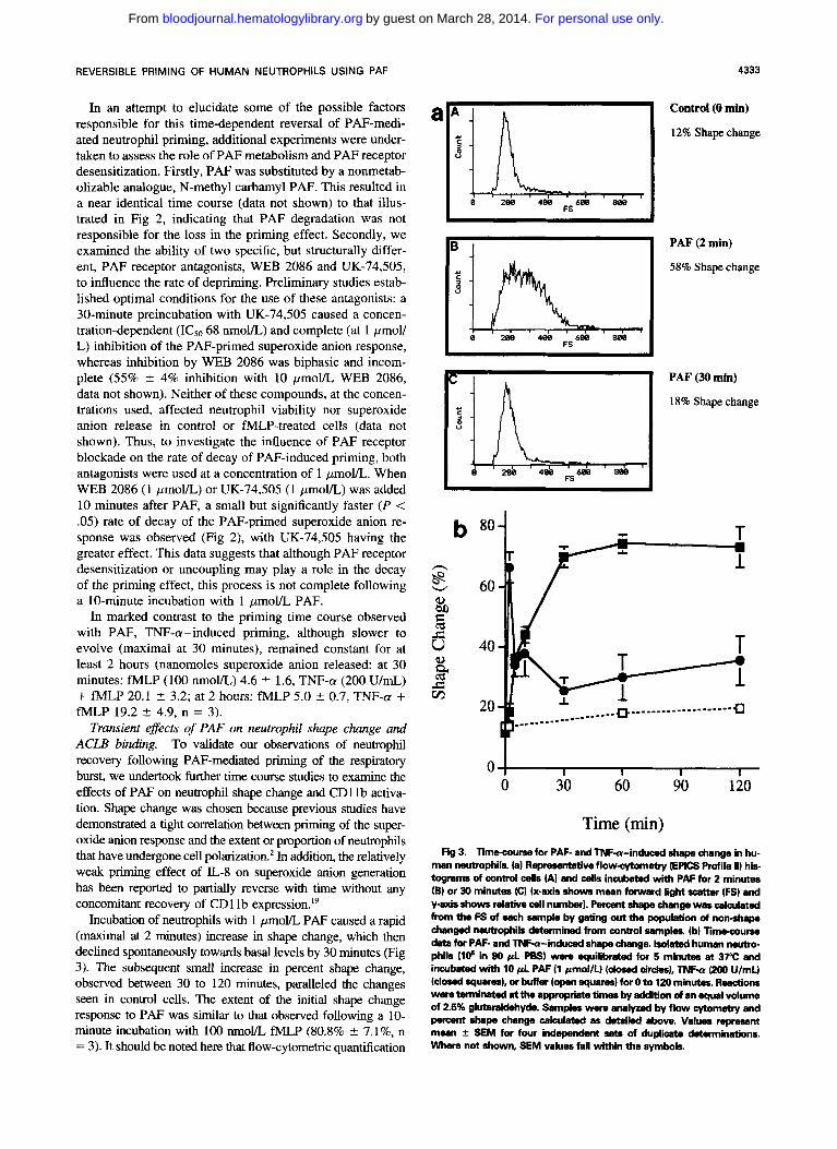
Incubation of neutrophils with 1 pmoYL PAF caused a rapid (maximal at 2 minutes) increase in shape change, which then declined spontaneously towards basal levels by 30 minutes (Fig 3). The subsequent small increase in percent shape change, observed between 30 to 120 minutes, paralleled the changes seen in control cells. The extent of the initial shape change response to PAF was similar to that observed following a 10- minute incubation with 1 0 0 nmoVL fMLP (80.8% +. ".l%, n = 3) . It should be noted here that flow-cytometric quantification
a;L; 4333
Control (0 min)
12% Shape change
8 288 488 Fs688 888
i 1 k 1 , I
58% Shapechange
PAF (2 min)
a m a a 6 m a w FS e$k, ~ '~
PAF (30 min)
18% Shape change
e 288 488 Fs6a0
n
g W
f @ 5 JZ m
b 80
60
40
20 - 7 """
f i I
A L
g """"1 """"
I)"""""""""" CI
0 1 0 30 60 90 120
Time (min) Fig 3. Timecourse for PAF- and TNFa-induced shape change in hu-
man neutrophils. la) Ruprusentative flowcytometry (EPICS M i l e ll) his tograms of control cells (A) and cells incubated with PAF for 2 minutes (B) or 30 minutes (C) (x-axis shows mean forward light scattcw (FS) and y-axis shows relative call number). Percent shape change was c a l a ~ l a t e d from the FS of each sample by gating out the population of non-shape changed neutrophils determined from control samples. (b) limecourse data for PAF- and TNFa-induced shape change. Isolated human nemo- phils (l@ in 90 p l PBS) wem equilibmted for 5 minutes at 37°C and incubated with 10 p l PAF (1 pmoVU (dosed cirdes), TNFa (200 U/mLl (closed squares), or buffer (open squares) for 0 to 120 minutes. Reactions wem tenninated at the appropriate timer by addiion d an equal volume of 2.5% glutarakkhyde. Samples were analyzed by flow cytometry and percent shape change calculated as &tailed above. Values r q m a e n t mean f SEM for four ind" sets of duplicate daaminations. Where not shown, SEM values fall within the symbols.
For personal use only.on March 28, 2014. by guest bloodjournal.hematologylibrary.orgFrom

4334 KITCHEN ET AL
a
; j h, , , , , , 1 1 , , , r i I 10
LFL 188
L
; 1 L,,;, , , ,
1 10 100 LFL
Control (0 min)
12% ACLB binding
PAF (10 min)
48% ACLB binding
b n
g W
a W2
U 6 2 3 4 v1 g
2 Y El 1
60 l T
C PAF (120 min)
j L
2 1 % ACLB binding
LFL I
n l " I
0 30 60 90 120 I I I I
Time (min)
Fig 4. Time-course for PAF- and TNF-a-induced binding of ACLB. (a) Representative flow-cytometry histograms of control cells (A) and cells incubated with PAF for 10 minutes (B) or 120 minutes (C) (x-axis shows logarithmic scale green fluorescence (LFL) and y-axis shows relative cell number). The lowest fluorescent (far left) peak represents neutrophils with no attached ACLB then, with increasing binding, single, double, and triple bead peaks (correlated by fluorescence microscopy) can be distinguished. The percent neutrophils with attached ACLB was calculated by gating out the far left peak determined from time-matched control samples. (b) Time-course data for PAF- and TNF-a-induced binding of ACLB. Isolated human neutrophils (1.75 x lo6 in 175 pL PES) were equilibrated for 5 minutes at 37°C and incubated with 15 pL PAF (1 pmol/L) (closed circles), TNF-a (200 UlmL) (closed squares), or buffer (open squares) for 0 to 120 minutes. ACLB (25 pLI were added 15 minutes before termination of the reaction with 0.5 mL 0.5% glutaraldehyde, except for time points <l5 minutes where the beads were added before the agonist. Samples were analyzed by flow cytometry and the percent neutrophils with attached ACLB was calculated, as detailed above. Values represent mean f SEM for four independent sets of determinations, each performed in duplicate. Where not shown, SEM values fall within the symbols.
of shape change, although a convenient and highly reproducible assay, gives consistently higher levels of basal shape change (approximately 8%") than those obtained by direct visual as- sessment of cell morphology. Our data confirm, however, that PM-induced shape change, like priming of the superoxide anion response, is a transient event. Neutrophil shape change to TNF-a (200 U/mL) was of a similar magnitude (70.1% 5 1.8%, n = 4) to that induced by PAF, but was slower to evolve (plateau at 30 minutes) and remained constant for the 2-hour incubation period (Fig 3).
As a third index of priming, we followed the P,-integrin- dependent binding of ACLB over a 2-hour incubation with either PAF or TNF-a. Again, PAF (l pmol/L) induced a time-dependent increase in ACLB binding, maximal after 10 minutes, which declined to reach control levels by 2 hours (Fig 4). TNF-a (200 U/mL) also augmented ACLB binding but, unlike PAF, the extent of bead binding reached a plateau at 30 minutes and remained constant for the ensuing 90- minute incubation period (Fig 4).
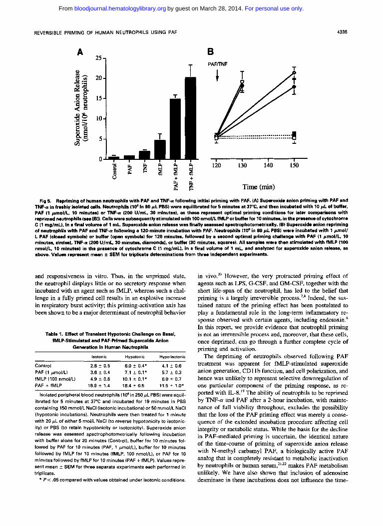
Repriming of neutrophils with PAF or TNF-a. We next investigated whether neutrophils that had primed and then spontaneously deprimed during a 120-minute incubation with PAF were capable of being reprimed. Figure 5 demon- strates that PAF-recovered neutrophils retain their full capac-
ity to be reprimed when challenged again with either PAF (1 pmoVL, 10 minutes) or TNF-a (200 U/mL, 30 minutes), generating similar amounts of superoxide anions upon fMLP stimulation as freshly-primed cells. This finding was re- peated when neutrophils were primed and deprimed using a modification of a previously described hypotonic challenge protocol.' Under isotonic conditions, fh4LP (100 nmol/L) and PAF ( 1 pmol/L) alone elicited little superoxide anion release, with PAF enhancing the fMLP-stimulated superox- ide anion response by 3.8-fold (Table 1). A 20-minute hypo- tonic challenge resulted in a modest (twofold) priming of the fMLP response, which recovered towards control levels when isotonicity was restored for l minute. Subsequent treat- ment with PAF reprimed the fMLP response, albeit to a slightly lower level than that observed in cells maintained under isotonic conditions. Cell viability was routinely >95% for all conditions studied. Thus, like cells that had deprimed following PAF exposure, osmotically primed and deprimed neutrophils also retained their capacity to be primed for a second time by a physiological agonist such as PAF.
DISCUSSION
The neutrophil can exist in a number of different func- tional states and this has a significant bearing on its behavior
For personal use only.on March 28, 2014. by guest bloodjournal.hematologylibrary.orgFrom
REVERSIBLE PRIMING OF HUMAN NEUTROPHILS USING PAF 4335
A
2 - 2
g b 'a 2
e,
A m 2%
< a
35 F4>
kg 3 -
251 T 20 - I
15 - 10 -
- U i i k $ $ $
$ i
B PAFlTNF
-r I
............................ .""""........"""....." I .L
I I 1 I 120 130 140 150
Time (min)
Fig 5. Repriming of human neutrophils with PAF and TNF-a following initial priming with PAF. (A) Superoxide anion priming with PAF and TNF-U in freshly isolated cells. Neutrophils (10' in 90 pL P e s ) were equilibrated for 5 minutes at 37%, and then incubated with l 0 p L of buffer, PAF (1 pmol/L, 10 minutes) or TNF-a (200 UlmL, 30 minutes), as these represent optimal priming conditions for later comparisons with reprimed neutrophils (see [B]). Cells were subsequently stimulated with 100 nmollLfMLP or buffer for 10 minutes, in the presence of cytochrome C (1 mglmL), in a final volume of 1 mL. Superoxide anion release was finally assessed spectrophotometrically. (B) Superoxide anion repriming of neutrophils wkh PAF and TNF-a following a 120-minute incubation with PAF. Neutrophils llO* in 80 pL PBS) were incubated with l pmoll L PAF (closed symbols) or buffer (open symbols) for 120 minutes, followed by a second optimal priming challenge with PAF (1 pmollL, 10 minutes, circles), TNF-a (200 UlmL, 30 minutes, diamonds), or buffer (30 minutes, squares). All samples were then stimulated with fMLP (100 nmol/L, 10 minutes) in the presence of cytochrome C (1 mglmL), in a final volume of 1 mL, and analyzed for superoxide anion release, as above. Values represent mean f SEM for triplicate determinations from three independent experiments.
and responsiveness in vitro. Thus, in the unprimed state, the neutrophil displays little or no secretory response when incubated with an agent such as fMLP, whereas such a chal- lenge in a fully primed cell results in an explosive increase in respiratory burst activity; this priming-activation axis has been shown to be a major determinant of neutrophil behavior
Table 1. Effect of Transient Hypotonic Challenge on Basal, fMLP-Stimulated and PAF-Primed Superoxide Anion
Generation in Human Neutrophils
Isotonic Hypotonic Hypo-Isotonic
Control 2.8 ? 0.5 6.0 t- 0.4' 4.1 ? 0.6 PAF (1 pmol/L) 3.6 t- 0.4 7.1 2 0.1* 5.7 lr 0.3 fMLP (100 nmoi/L) 4.9 2 0.6 10.1 2 0.1* 6.0 ? 0.7 PAF + fMLP 18.9 ? 1.4 18.4 2 0.5 11.5 t 1.0*
Isolated peripheral blood neutrophils (IO6 in 250pL PBS) were equil- ibrated for 5 minutes at 37°C and incubated for 19 minutes in PBS containing 150 mmol/L NaCl (isotonic incubations) or 50 mmol/L NaCl (hypotonic incubations). Neutrophils were then treated for 1 minute with 20 .uL of either 5 mol/L NaCl (to reverse hypotonicity to isotonic- ity) or PBS (to retain hypotonicity or isotonicity). Superoxide anion release was assessed spectrophotometrically following incubation with buffer alone for 20 minutes (Control), buffer for 10 minutes fol- lowed by PAF for 10 minutes (PAF, 1 pmol/L), buffer for 10 minutes followed by fMLP for 10 minutes (fMLP. 100 nmol/L), or PAF for 10 minutes followed by fMLP for 10 minutes (PAF + fMLP). Values repre- sent mean t- SEM for three separate experiments each performed in triplicate.
* P < .05 compared with values obtained under isotonic conditions.
in vivo?' However, the very protracted priming effect of agents such as LPS, G-CSF, and GM-CSF, together with the short life-span of the neutrophil, has led to the belief that priming is a largely irreversible proce~s.'~' Indeed, the sus- tained nature of the priming effect has been postulated to play a fundamental role in the long-term inflammatory re- sponse observed with certain agents, including endotoxin.' In this report, we provide evidence that neutrophil priming is not an irreversible process and, moreover, that these cells, once deprimed, can go through a further complete cycle of priming and activation.
The depriming of neutrophils observed following PAF treatment was apparent for fMLP-stimulated superoxide anion generation, CD1 l b function, and cell polarization, and hence was unlikely to represent selective downregulation of one particular component of the priming response, as re- ported with IL-8.I9 The ability of neutrophils to be reprimed by TNF-a and PAF after a 2-hour incubation, with mainte- nance of full viability throughout, excludes the possibility that the loss of the PAF priming effect was merely a conse- quence of the extended incubation procedure affecting cell integrity or metabolic status. While the basis for the decline in PAF-mediated priming is uncertain, the identical nature of the time-course of priming of superoxide anion release with N-methyl carbamyl PAF, a biologically active PAF analog that is completely resistant to metabolic inactivation by neutrophils or human seIum,2'~zz makes PAF metabolism unlikely. We have also shown that inclusion of adenosine deaminase in these incubations does not influence the time-
For personal use only.on March 28, 2014. by guest bloodjournal.hematologylibrary.orgFrom

4336
course of PAF-primed superoxide anion responses, which excludes a secondary effect mediated via adenosine release and autocrine activation of cyclic adenosine monophosphate (AMP)-dependent protein kinase pathways (Kitchen, Rossi and Chilvers, unpublished observations, February 1995). Al- though homologous receptor desensitization may underlie the transient nature of the PAF signal, the increased rate of decay of fMLP-induced superoxide anion release following UK-74,505 or WEB 2086 addition and the return of a fully competent PAF priming response after 2 hours suggests that PAF receptor uncoupling/desensitization is both incomplete and transient. These data are consistent with previous studies demonstrating rapid activation-induced uncoupling and in- ternalization of PAF receptors followed by subsequent recep- tor re-expression.” The recovery of PAF receptor number and function is likely to reflect extensive membrane attach- ment and metabolism of PAF (approximately l pmol/107 neut rophi l~ /min) .~~~*~
From our own comparisons of PAF and TNF-(U and other published observations, it would appear that neutrophil prim- ing in vitro falls into three categories: ( l) fully reversible (eg, that induced by PAF, osmotic swelling, or inositol hexa- kisphosphate); (2) partially reversible (eg, with IL-l’ or IL- 8); or (3) largely irreversible (eg, with GM-CSF”, G-CSF, or LPS). Further studies would be required to categorize TNF-(U because its effects did not show any signs of recovery over the 2-hour incubation period used in this study, but have been reported to decay over 24 hours.7 It is also intriguing to note that only agents in group (3) are able to modulate the rate of neutrophil apoptosis, which again testifies to the long duration of action of this class of agents.” While it is clear that neither the efficacy nor extent of the initial priming signal dictates the reversibility of the primed state (because PAF, TNF-a [Fig 51, LPS, and GM-CSF [data not shown] induce equivalent levels of priming), it is possible that the duration of the priming signal andor its rate of onset are key determinants. However, in the absence of any clear mechanistic basis for neutrophil priming, it is also possible that the above agents use discrete signalling pathways to induce their priming effects.
This current observation of reversible priming may allow the pro-inflammatory, and potentially tissue-damaging, ef- fects of neutrophil priming/activation to be counteracted by a process other than apoptosis or the pharmacological inhibi- tion of neutrophil activation. It is unlikely that neutrophils within an inflammatory focus would be exposed in isolation to PAF or other “transient” priming agents. However, as endothelial cell-associated PAF has been shown to play a central role in neutrophil priming and migration through IL- 10-treated human umbilical vein endothelial cell mono- layers in vitro,27 and endogenously formed PAF is involved in leukocyte extravasation induced by IL-1 in vivo,2x any delay in cell exit through an activated endothelial surface may permit cell recovery and the return of unprimed neutro- phils to the circulation. Thus, the recognition that neutrophils have the potential to deprime allows an additional point of control in the early stages of the acute inflammatory re- sponse, whereby cells may return to their former quiescent state and potentially re-enter the circulating neutrophil pool. These deprimed neutrophils, once fully recovered, could
KITCHEN ET AL
again attain their maximal priming potential and mount sub- sequent responses, as dictated by ensuing inflammatory chal- lenges. In contrast, priming agents with a longer duration of action would maintain neutrophils in the primed state for a much longer period of time and may, therefore, play a dis- tinct role in vivo, in the wake of‘ a more widespread or prolonged inflammatory insult.
REFERENCES I . Guthrie LA, McPhail LC, Henson PM, Johnston RB Jr: Prim-
ing of neutrophils for enhanced release of oxygen metabolites by bacterial lipopolysaccharide. Evidence for increased activity of the superoxide-producing enzyme. J Exp Med 160: 1656, 1984
2. Haslett C, Guthrie LA, Kopaniak MM, Johnston RJ, Henson PM: Modulation of multiple neutrophil functions by preparative methods or trace concentrations of bacterial lipopolysaccharide. Am J Pathol ll9:101, 1985
3. Vadas MA, Nicola N, Lopez AF, Metcalf D, Johnson G, Per- eira A: Mononuclear cell-mediated enhancement of granulocyte function in man. J Immunol 133:202, 1984
4. Cross AS, Lowell GH, Palmblad J, Sadoff JC, Young L, Berger M: Mechanism of priming of human neutrophils by a soluble lym- phoblastoid cell factor. J Immunol 135:2074, 1985
5. Worthen GS, Haslett C, Rees AJ, Gumbay RS, Henson JE, Henson PM: Neutrophil-mediated pulmonary vascular injury: syner- gistic effect of trace amounts of lipopolysaccharide and neutrophil stimuli on vascular permeability and neutrophil sequestration in the lung. Am Rev Respir Dis 136:19, 1987
6. Smedly LA, Tonnesen RA, Sandhaus RA, Haslett C, Guthrie LA, Johnston RB, Henson PM, Worthen CS: Neutrophil-mediated injury to endothelial cells: enhancement by endotoxin and essential role of neutrophil elastase. J Clin Invest 77: 1233, 1986
7. Ichinose Y, Hara N. Ohta M, Aso H, Chikama H, Kawasaki M, Kubota I, Shimizu T, Yagawa K: Recombinant granulocyte colony- stimulating factor and lipopolysaccharide maintain the phenotype of and superoxide generation by neutrophils. Infect Immun 58:1647, 1990
8. Carey LA, Albertine KH, Gee MH: In vivo endotoxin exposure increases neutrophil counts without maintenance of primed superox- ide anion release in short-term cultures of sheep bone marrow. J Leukoc Biol 56:145, 1994
9. Edashige K, Watanabe Y, Sato EF, Takehara Y, Utsumi K: Reversible priming and protein-tyrosyl phosphorylation in human peripheral neutrophils under hypotonic conditions. Arch Biochem Biophys 302:343, 1993
10. Kitchen E, Condliffe AM, Rossi AG, Haslett C, Chilvers ER: Characterization of inositol hexakisphosphate (InsP,)-mediated priming in human neutrophils: Lack of extracellular [3H]-InsP6 re- ceptors. Br J Pharmacol 117:979, 1996
1 1 . Savill JS, Henson PM, Haslett C: Phagocytosis of aged neu- trophils by macrophages is mediated by a novel “charge-sensitive” recognition mechanism. J Clin Invest 84 15 18, 1989
12. Savill JS, Dransfield I, Hogg N, Haslett C: Vitronectin recep- tor-mediated phagocytosis of cells undergoing apoptosis. Nature 343: I70, 1990
13. Cole AT, Garlick NM, Galvin AM, Hawkey CJ, Robins RA: A flow cytometric method to measure shape change of human neu- trophils. Clin Sci 89:549, 1995
14. Stocks SC, Kerr MA, Haslett C, Dransfield I: CD66-depen- dent neutrophil activation: a possible mechanism for vascular selec- tin-mediated regulation of neutrophil adhesion. J Leukoc Biol58:40. 1995
15. Archer CB, Page CP. Morley J, MacDonald DM: Accumula- tion of inflammatory cells in response to intracutaneous platelet acti- vatlng factor (PAF-acether) in man. Br J Dermatol 112:285, 1985
For personal use only.on March 28, 2014. by guest bloodjournal.hematologylibrary.orgFrom

REVERSIBLE PRIMING OF HUMAN NEUTROPHILS USING PAF 4337
16. Dent G, Ukena D, Chanez P, Sybrecht G, Barnes P: Charac- terization of PAF receptors on human neutrophils using the specific antagonist, WEB 2086: correlation between receptor binding and function. FEBS Lett 244:365, 1989
17. Ingraham LM, Coates TD, Allen JM, Higgins CP, Baehner RL, Boxer LA: Metabolic, membrane, and functional responses of human polymorphonuclear leukocytes to platelet-activating factor. Blood 59:1259, 1982
18. Gay JC, Beckman JK, Zaboy KA, Lukens JN: Modulation of neutrophil oxidative responses to soluble stimuli by platelet-activat- ing factor. Blood 67:931, 1986
19. Roberts PJ, Pizzey AR. Khwaja A, Carver JE, Mire-Sluis AR, Linch DC: The effects of interleukin-8 on neutrophil f Met Leu Phe receptors, CD1 l b expression and metabolic activity, in comparison and combination with other cytokines. Br J Haematol 84:586, 1993
20. Warren JS, Mandel DM, Johnson KJ, Ward PA: Evidence for the role of platelet-activating factor in immune complex vasculitis in the rat. J Clin Invest 83:669, 1989
21. O’Flaherty JT, Redman JF, Schmitt JD, Ellis JM, Surles JR, Marx MH, Piantadosi C, Wykle RL: I-0-alkyl-2-N-methylcarbamyl- glycerophosphocholine: A biologically potent, non-metabolizable analog of platelet activating factor. Biochem Biophys Res Commun 147:18, 1987
22. Tessner TG, O’Flaherty JT, Wykle RL: Stimulation of plate-
let-activating factor synthesis by a nonmetabolizable bioactive ana- log of platelet-activating factor and influence of arachidonic acid metabolites. J Biol Chem 264:4794, 1989
23. O’Flaherty JT, Jacobson DP, Redman J F Regulation of plate- let-activating factor receptors and the desensitization response in polymorphonuclear neutrophils. Biochem J 288:241, 1992
24. OFlaherty JT, Surles JR, Redman J, Jacobson D, Piantadosi C, Wykle RL: Binding and metabolism of platelet-activating factor by human neutrophils. J Clin Invest 78:381, 1986
25. O’Flaherty JT, Chabot MC, Redman J, Jacobson D, Wykle RL: Receptor-independent metabolism of platelet-activating factor in myelogenous cells. FEBS Lett 250:341, 1989
26. Lee A, Whyte MKB, Haslett C: Prolongation of in vitro life- span and functional longevity of neutrophils by inflammatory media- tors acting through inhibition of apoptosis. J Leukoc Biol 54:283, 1993
27. Hill ME, Bird ID, Daniels RH, Elmore MA, Finnen MJ: Endothelial cell-associated platelet-activating factor primes neutro- phils for enhanced superoxide production and arachidonic acid re- lease during adhesion to but not transmigration across IL-I p -treated endothelial monolayers. J Immunol 153:3673, 1994
28. Nourshargh S , Larkin SW, Das A, Williams TJ: Interleukin- 1-induced leukocyte extravasation across rat mesenteric microves- sels is mediated by platelet-activating factor. Blood 85:2553, 1995
For personal use only.on March 28, 2014. by guest bloodjournal.hematologylibrary.orgFrom