Synthesis of thermosensitive microgels with a tunable magnetic core
Upload
independentCategory
view
0download
0
ORIGINAL ARTICLE
Inclusion complexes of c-cyclodextrin and carboxyl-modifiedc-cyclodextrin with C60: synthesis, characterizationand controlled release application via microgels
Fifere Adrian Æ Tania Budtova Æ Elena Tarabukina ÆMariana Pinteala Æ Spulber Mariana Æ Cristian Peptu ÆValeria Harabagiu Æ Bogdan C. Simionescu
Received: 10 September 2008 / Accepted: 7 January 2009 / Published online: 23 January 2009
� Springer Science+Business Media B.V. 2009
Abstract Carboxyl modified c-cyclodextrin (CDSA) with
a substitution degree of about 9.5 was prepared by the
esterification of c-cyclodextrin (CD) with succinic anhy-
dride in pyridine at 90 �C. The chemical composition and
the structure of CDSA were characterized by FT-IR,
MALDI-TOF, X-ray diffraction pattern, potentiometric
titration and TGA. Modified and native c-cyclodextrin
associate with fullerene (C60) in DMF-toluene mixture
resulting 1:1 CDSA:C60 and CD:C60 inclusion complexes.
Aqueous solutions of native cyclodextrin, carboxyl-modi-
fied cyclodextrin and their inclusion complexes with C60
were used as microgel solvent (or swelling agent) for
controlled release application. The release of solutions was
induced by shear stress and demonstrated using rheo-
optical set-up.
Keywords Carboxyester modified c-cyclodextrin �Fullerene C60 � Inclusion complexes �Shear induced release from microgel
Introduction
Making fullerenes ‘‘soluble’’ in water is of great interest
for their specific biological applications like enzyme
inhibition, antiviral activity, DNA cleavage, photody-
namic therapy, electron transfer, etc. [1]. As already
known, the large spherical fullerene C60 can be reason-
ably well dissolved in non-polar solvents, such as toluene
and dichloromethane, is moderately soluble in nonpolar,
aprotic and polar, nonaqueous solvents, but is totally
insoluble in water. Until recently, the solubilisation of C60
in aqueous solution was possible only by chemical deri-
vatisation of fullerene, which leads to a partial loss of its
aromatic character [2]. One example of fullerene deriva-
tive was described in 1993 by Schinazi et al. [3], which
tested carboxyl-modified fullerene for antiviral activity in
cells acutely and chronically infected with HIV-1 and in
cell free systems, suggesting that carboxylic water-soluble
fullerene derivatives have potential selective activity
against HIV-1.
The most recent and successful strategy to improve the
water-solubility of C60 is to surround it with water-soluble
host molecules such as calixarenes [4–6], cyclodextrins [7–
9] or to incorporate C60 in polymeric structures [10]. The
main advantage of using non-covalent linkages is the fact
that the electronic structure of fullerene remains only
slightly changed as compared to the structure of derivatized
fullerene. In particular, this difference was shown for pairs
of charge transfer complexes of poly(N-vinylpyrrolidone)
(PVP) with fullerene C60 (PVP/C60) or with modified C60
(PVP/modified C60) [11]. That is why unique properties of
fullerene caused by its structure can be revealed full-bodily
if it is complexed with polymers.
Cyclodextrins (CDs) are cyclic oligosaccharides having
a hydrophilic external surface which confers aqueous
F. Adrian � M. Pinteala � S. Mariana (&) � C. Peptu �V. Harabagiu � B. C. Simionescu
Petru Poni Institute of Macromolecular Chemistry,
700487 Iasi, Romania
e-mail: [email protected]
T. Budtova
Mines ParisTech, Centre de Mise en Forme des Materiaux
(CEMEF), 06904 Sophia-Antipolis, France
E. Tarabukina
Institute of Macromolecular Compounds, Russian Academy
of Sciences, 199004 St. Petersburg, Russia
123
J Incl Phenom Macrocycl Chem (2009) 64:83–94
DOI 10.1007/s10847-009-9539-4

solubility, and a relatively hydrophobic inner cavity able to
incorporate molecules of appropriate size. b- and c-CD
have been reported to form inclusion complexes with C60.
Because the dimension of CD cavity (0.57 nm for b-CD;
0.95 nm for c-CD) is smaller than the size of C60
(1.00 nm), a full accommodation of C60 inside CD is not
possible. Thus, theoretically two CD molecules are needed
to bound one C60, i.e., a 2:1 b- or c-CD:C60 complex is
formed [12, 13]. Even a full accommodation of C60, 1:1
inclusion complexes between cyclodextrins and C60, as
described in this paper, have already described in the lit-
erature [14–17]. To use the unique properties of CDs as
host macrocycles for inclusion complexation, a wide
variety of their chemically modified derivatives have been
prepared [18, 19].
The goal of our investigation is to prepare and charac-
terize fullerene/c-cyclodextrin complexes, with native and
chemically modified c-CDs and to demonstrate one option
of their controlled delivery. The cyclodextrin was esterified
with succinic anhydride leading to the formation of a
macrocycle functionalized with carboxyl groups (CDSA).
CD/C60 and CDSA/C60 complexes were dissolved in water
and used as a solvent for a synthetic superabsorbent mi-
crogel, the latter being a model carrier. Solvent release
from the gel was induced by shear stress and demonstrated
with rheo-optical tool using the same principle as described
in literature [20].
Materials and methods
Materials
c-Cyclodextrin (CD) (Aldrich) was dried at low pressure
and 80 �C for 48 h. Pyridine was dried by distillation on
potassium hydroxide. Fullerene-C60 (C60) was purchased
from MER Corporation and used without further purifica-
tion. All other reagents and solvents, succinic anhydride
(SA), toluene and dimethylformamide (DMF) were from
Aldrich and used as received.
Aqua Keep 10 SHNF hydrogel (AK) was kindly pro-
vided by Kobo Products Inc. It is composed of 75–25%
poly(sodium acrylate-co-acrylic acid) chemically cross-
linked with N,N0-methylenebisacrylamide. The initial state
of the gel was a powder of dry spherical particles with
diameter of 5–60 lm.
A silicone oil, polydimethylsiloxane (PDMS) Rhodorsil
47 V 200000 (Rhodia, France) with viscosity of 230 Pa s,
was selected as suspending matrix for swollen microgels.
Silicone oil was chosen because it is transparent, chemi-
cally inert with respect to swollen gels and the solvents
used and sufficiently viscous, enabling to exert the stresses
necessary to deform the gel particles.
Methods
Complex characterization
FT-IR characterisation was performed on a Bruker Vertex
70, instrument using KBr disc method. NMR spectra were
obtained on a DRX 400 Avance Bruker 400 MHz spec-
trometer. UV–vis absorptions were measured on an Analytik
Specord 200 Jena spectrophotometer. MALDI-TOF mass
spectrum of CDSA was recorded on a Reflex IV (Bruker
Saxonia) instrument. The samples were prepared by mixing
the compound solution (10 mg/mL) in a 1:1 v/v acetoni-
trile:water mixture, with a saturated solution of a-cyano-4-
hydroxycinnamic acid matrix in the same solvent mixture in
a volumetric ratio of 1/100. Then, 1 lL of the resulting
mixture was placed onto the sample plate and the solvent
was evaporated at room temperature. Spectra were recorded
in positive linear mode. All experiments were carried out
collecting 200 laser shots with laser power slightly above
threshold to obtain a clear signal. The calibration was
performed using a Bruker peptide mixture for the 1,000–
3,000 Da m/z span. TGA analyses were performed in
oxygen with a temperature scanning rate of 10 K/min on a
Q-1500 D MOM Budapest system. The wide angle X-ray
diffraction (WAXD) patterns were recorded on a Bruker
AXS D8 advance X-ray diffractometer with scanning scope
of 0�–40�, scanning speed of 4�/min, using Cu Ka radiation.
Titration of carboxylic groups in CDSA
and CDSA/C60 complex
The potentiometric titration was done with a TitrolineAl-
pha Plus apparatus. The aqueous solution of 0.003 g/L of
CDSA or CDSA/C60 complex was titrated with 10-2 M
aqueous sodium hydroxide. The equivalence points were
calculated as an average of three measurements and the
molar content of carboxylic units was calculated with the
relation (1):
NCOOH ¼MCD
Va
Vb� CCDSA
CNaOHMNaOH �MSA
ð1Þ
where NCOOH is the average molar number of carboxylic
groups in CDSA; MNaOH, MCD and MSA are the molar
masses of sodium hydroxide, unmodified cyclodextrin and
succinic anhydride, respectively; Va and Vb are the volume
of CDSA and of sodium hydroxide solution at equivalence
point, respectively; CCDSA and CNaOH are the concentrations
of CDSA and NaOH solutions in g/L. An average molar
content of 9.6 carboxylic groups was obtained for CDSA.
From the average number of carboxylic groups on each
CDSA macrocycle, the molar ratio between components in
CDSA/C60 complex of about 1:1 was calculated with the
relation (2).
84 J Incl Phenom Macrocycl Chem (2009) 64:83–94
123
CDSA
C60
ðmolarÞ ¼ MC60
NCOOHVa
Vb� CCDSA=C60
CNaOHMNaOH �MSA
ð2Þ
where MC60 is the molecular weight of C60 and CCDSA/C60
is the concentration of CDSA/C60 complex in g/L.
Determination of microgel swelling degree
In order to determine gel swelling degree at equilibrium,
aqueous solutions of CD, CDSA, CD/C60 or CDSA/C60 of
different concentrations (from 0 to 1 g/dL for CD and CDSA
and from 0 to 0.34 g/dL for CD/C60 and CDSA/C60 com-
plexes) were added drop by drop to precisely weighted dry
gel particles until the suspension becomes fluid due to the
presence of the excess of solvent. At each step of solvent
addition, the suspension was let to relax after absorbing the
solvent. The weight of solvent added just before the excess
of solvent appeared was noted and the degree of swelling at
equilibrium Q was calculated as the ratio between the weight
of absorbed solvent and the weight of the dry particles.
Rheo-optics
For the rheo-optical experiments, microgel was swollen up
to 70% of the equilibrium value by simply adding a certain
amount of solution to the dry particles, which leads to
complete solution absorption by the gel. With this proce-
dure the solution concentration inside the gel is the same as
in the prepared one.
Rheo-optical set-up was used to observe the shear-
induced release of the aqueous solutions of CD, CDSA,
CD/C60 and CDSA/C60 from a swollen microgel, as
described in [21] for model systems. A simple shear flow
was generated by a counter rotating plate–plate system [20,
22, 23]. Two plates are transparent and rotate in opposite
directions. An optical microscope placed above the upper
rotating plate allows observations in the plane formed by
the flow and vorticity directions. All the experiments were
recorded by a CCD camera coupled to the image acquisi-
tion complex. The gap between the plates was 0.7 mm. By
adjusting the relative velocities of the plates, a selected
particle immersed in the PDMS can be immobilized in the
laboratory framework and its behaviour can be monitored.
The shear rate was varied between 0.5 and 25 s-1. All the
experiments were performed at room temperature. A
detailed description of sample preparation and experi-
mental procedure is given in literature [23].
The initial swollen gel is spherical. Under shear, the gel
deforms, solvent is released and can be detached [20, 23].
After stopping the shear, gel recovers its initial spherical
shape. In order to determine gel volume loss, gel diameter
before and after shearing was measured and the relative
volume loss DV was calculated as DV = (V0 - V(t))/V0,
where V(t) and V0 are gel volume at a moment t and t = 0,
respectively. The experiments were performed in the fol-
lowing way: a selected particle was subjected to a constant
shear rate during a certain time (from 10 s to 2 min), then
shear was stopped and the diameter of the relaxed gel was
measured. In subsequent runs, the particle was submitted
to the increasing shear rates by increasing the rotation
velocities of the plates. The gel diameter was measured by
analysing the recorded tapes with the help of image anal-
ysis software Visilog from Noesis 2000. The experimental
errors of rheo-optical measurements (shear rate, dimen-
sions of objects) were smaller than 10%.
Results and discussion
Synthesis
Synthesis of CDSA
CDSA was prepared by the esterification of CD with SA in
pyridine at 90 �C, according to a procedure proposed by
Gonzalez and Rossi [21] for the chemical modification of
b-CD with SA. The procedure was as follows. Into a three
necked round bottomed vessel, equipped with magnetic
stirrer, reflux condenser and argon inlet, a solution of
5.34 g (53.4 mmol) SA in 72 mL anhydrous pyridine was
added under stirring over a solution of 6.00 g (4.6 mmol)
CD in 100 mL anhydrous pyridine at 60 �C. The mixed
solutions were heated at 90–100 �C under stirring and the
obtained opalescent solution was diluted with 40 mL
anhydrous pyridine. The reaction was continued at this
temperature for 9 h under stirring in argon atmosphere, and
then the solvent was evaporated in vacuum. The solid was
washed several times with diethyl ether to remove all traces
of pyridine and suspended in acetone under stirring over-
night to remove the unreacted anhydride. The purified
CDSA was obtained as a white solid in about 90% yield.
Synthesis of the CD/C60 complex (Scheme 1)
Into a three necked round bottomed flask provided with
reflux condenser, magnetic stirrer, argon inlet and dropping
funnel, a solution of 201 mg (0.155 mmol) CD in 30 mL
water was prepared and heated at 100 �C. A solution of
60 mg (0.083 mmol) C60 in 30 mL toluene was added drop
wise through the dropping funnel and the heterogeneous
mixture was maintained under strong stirring and inert
atmosphere at 100–107 �C for 3 days. After cooling up the
mixture, three liquid phases (water uncoloured phase,
emulsion violet phase and toluene dark violet phase) and a
violet precipitate were obtained. The precipitate, the
J Incl Phenom Macrocycl Chem (2009) 64:83–94 85
123

aqueous and emulsion phases were mixed, diluted with
20 mL water and centrifuged together to completely sep-
arate the complex. The obtained complex was dried,
washed firstly with toluene until no coloration of the sol-
vent was observed and secondly with cold water to separate
the uncomplexed C60 and CD, respectively. After drying,
64.4 mg of violet solid complex were obtained.
Synthesis of the CDSA/C60 complex (Scheme 1)
Into a three necked round bottomed flask, provided with
reflux condenser, magnetic stirrer, argon inlet and dropping
funnel, a solution of 250 mg (0.114 mmol) CDSA in
23 mL DMF was introduced. A solution of 40 mg
(0.057 mmol) C60 in 23 mL toluene was added drop wise
through the dropping funnel. The mixture was stirred at
room temperature for 10 min and then at 90 �C for 48 h.
The solvents were then evaporated under vacuum and the
obtained solid was washed with large amounts of toluene
until no C60 was observed in toluene phase. The brown
solid product was washed with diethyl ether to remove
solvent traces and three times with small amounts of ethyl
alcohol to remove the unreacted CDSA. About 88.8 mg of
brown solid complex were obtained after purification.
Characterization of CDSA and of cyclodextrin/C60
complexes
The synthesis of modified cyclodextrin and of CD/C60 and
CDAS/C60 complexes is described in Methods Section and
Scheme 1 Synthesis of CDSA,
CD/C60 and CDSA/C60
complexes
86 J Incl Phenom Macrocycl Chem (2009) 64:83–94
123

schematically presented in Scheme 1. All new compounds
were extensively characterized using FT-IR, NMR, UV-
vis, TGA, potentiometric titration and mass spectrometry.
FT-IR absorption spectra
The FT-IR absorption spectra of the initial c-cyclodextrin,
CDAS and their complexes with C60 are presented in
Fig. 1, from a to d, respectively. The modification of CD is
clearly demonstrated by the presence of the carbonyl ester
high band at 1733 in CDSA spectrum (Fig. 1b). Moreover,
some CD specific vibrations were diminished (708 cm-1)
and others were shifted with 1 to 7 cm-1 through its
asymmetrical esterification. As previously showed by Bo-
ulas et al. [24], the complexation of CD with C60
determines minimal shifts (less than 6 cm-1) of the ring
vibrations (583, 708, 758 and 943 cm-1), of the coupled
C–O–C stretching/O–H bending vibrations (1158 cm-1),
and of the coupled C–O/C–C stretching/O–H bending
vibrations (1027 and 1080 cm-1). Larger shift, about
9 cm-1 was registered for the coupled C–H/O–H bending
vibrations at 1416 cm-1, which may indicate the interac-
tions between CD inner cavity and C60 (Fig. 1a, c).
Contrary to CD/C60, the CDSA/C60 complex showed
higher shifts of 6–8 cm-1 for some of the cyclodextrin ring
vibrations (586 and 946 cm-1) (Fig. 1b, d).
The FT-IR spectra of C60 complexes with both CD and
CDSA (Fig. 1c, d) show that cyclodextrin absorptions are
practically covering fullerene bands. Only the stronger
band at 526 cm-1, visible in FT-IR spectra of both com-
plexes, can be clearly attributed to C60 [24].
1H-NMR and 13C-NMR characterization
A deeper structural analysis was performed by 1H- (Figs. 2,
3, 4) and 13C-NMR (Figs. 5, 6) in D2O and in DMSO-d6.
Figures 2 and 3 compare the 1H-NMR resonance peaks of
unmodified and modified CDs in D2O and DMSO-d6,
respectively. As previously shown by Schneider et al. [25],
the chemical shifts of different protons belonging to CD
ring are not identical in these two solvents due to sub-
stantial differences in the populations around the C5–C6
bond and in the rate of exchange of OH protons (much
faster for D2O as compared to DMSO-d6). The position of
resonance peaks is also dependent on the acquisition tem-
perature (24.2 �C in our experiments).
As one can see from Figs. 2 and 3, the anomeric protons
appearing as unique doublet signals in non-modified CD at
5.10 ppm in D2O (Fig. 2a) and 4.89 in DMSO-d6 (Fig. 3a)
are splitted into two signals in modified CD, denoting an
asymmetric substitution of CD ring. In D2O (Fig. 2b), the
anomeric signals are larger and centred at 5.10 and
5.30 ppm. In DMSO-d6 (Fig. 3b), a first peak remain
unchanged at 4.89 ppm and the second one is larger
between 5.10 and 5.12 ppm. Moreover, following the
esterification of the sixth position, the peaks corresponding
to the H6 protons in the spectra of CDSA are enlarged and
downfield shifted as compared to CD. The peaks of Ha and
Hb methylene protons linked to ester and carboxylic
groups, respectively, in the spectrum of CDSA performed
in DMSO-d6 are masked by the solvent (Fig. 3b), but they
are identified as quite large signals of two superposed
triplets, at 2.70–2.89 ppm in the spectrum performed in
D2O (Fig. 2b) [26].
The 1H-NMR spectra and peak values of CD/C60 and
of CDSA/C60 complexes performed in DMSO-d6 areFig. 1 FT-IR spectra of CD (a), CDSA (b), CD/C60 complex (c),
CDSA/C60 complex (d)
J Incl Phenom Macrocycl Chem (2009) 64:83–94 87
123

presented in Fig. 4. One can observe that only small dif-
ferences in the chemical shifts of CD/C60 or CDSA/C60 as
compared to the non complexed corresponding macrocycle
molecules (Fig. 3) are evidenced. These differences are
mainly due to a conformational change to a more conical
structure in the host molecule induced by complexation, as
suggested for CD/C60 system in [27].
Figures 5 and 6 contain 13C-NMR data of modified and
unmodified CDs and of their C60 complexes in DMSO-d6.
Fig. 2 1H-NMR spectra of CD (a) and CDSA (b) in D2O
Fig. 3 1H-NMR spectra of CD (a) and CDSA (b) in DMSO-d6
Fig. 4 1H-NMR spectra of CD/C60 complex (a) and CDSA/C60
complex (b) in DMSO-d6
Fig. 5 13C-NMR of CD (a) and CD/C60 complex (b) in DMSO-d6
88 J Incl Phenom Macrocycl Chem (2009) 64:83–94
123

The 13C-NMR chemical shifts are much better indicators
for the structural variation. The observed 13C peak posi-
tions of CD (Fig. 5a) and CDSA (Fig. 6a) vary from 60.03
to 63.00 ppm (d = 3 ppm) for C6 and from 72.22 to 69.21
(d = 3.01 ppm) for C5, showing very few correlation with
other constituent induced shifts, as Anderson et al. [27]
previously pointed out. The 13C shifts of C4 reflect syn and
anty orientation of the C6–OR group with respect to the
C5–C4 bond, taking into consideration the classical chair
conformation of glucopyranose units that does not depend
on the C6 substituent [28]. In CDSA spectrum, the reso-
nance peaks corresponding to methylene, ester and
carboxyl carbon atoms belonging to SA derived substituent
are also visible at 28.58, 172.01 and 173.48 ppm, respec-
tively. As for the CD/C60 complex, its 13C-NMR
characteristic peaks (Fig. 5b) are slightly upfield shifted
(d =-0.06 to 0.08 ppm) as compared to the initial CD,
while for CDSA/C60 complex (Fig. 6b) higher downfield
and upfield shifts for C1 and C2 (d = ?0.12 7 ?
0.31 ppm) and for C3 and C6 (d = -0.14 7 –0.25 ppm),
respectively, were observed when compared to the non-
complexed CDSA. The differences in the chemical shifts of
cyclodextrins and of their complexes are due to confor-
mational changes of the macrocyclic molecules upon
complexation. In addition, 13C-NMR spectra of CD/C60
and CDSA/C60 complexes show signals at 142.00 and
142.77 ppm, respectively, which were slight upfield shifted
from the 143.00 ppm peak of pristine C60. These minor
differences mean that CD cavities only slightly disturbed
the local magnetic field of C60 [25].
Mass spectrometry characterization
The molecular structure of CDSA was also proved by
MALDI-TOF analysis (Fig. 7). The resulted peaks are
sodium adducts ranging from 1719 to 2519 Da with a mass
increment of 100 Da. The m/z values of each peak can be
calculated by the formula: X = 1296 (c-CD) ? n100
(SA) ? 23 (Na), where n is the molar number of SA units.
The determined degree of functionalization is ranging
from 6 to 13. The most abundant product was the CD
bearing 10 SA units. The peaks with lower intensity, which
are doubling the main series, are K adducts, provided that
K ions can be usually found as an impurity in laboratory
glassware. Sodium adducts of the carboxylic Na salts
formed during the MALDI ionization process can also be
observed. The result confirms the average values of 9.5 and
Fig. 6 13C-NMR spectra of CDSA (a) and CDSA/C60 complex (b) in
DMSO-d6
Fig. 7 MALDI-TOF spectra of
CDSA
J Incl Phenom Macrocycl Chem (2009) 64:83–94 89
123

9.6 of the substitution degree of CDSA obtained from the
potentiometric titration, respectively, and the structure
proposed in Scheme 1.
The mixture of differently substituted homologues was
used without separation for the preparation of CDSA/C60
complexes.
UV–vis absorption spectra
The UV–vis absorption spectra of CD/C60 and CDSA/C60
complexes in water solution are presented in Fig. 8. The
strong narrow absorptions at 214, 260 and 332 nm for CD/
C60 and at 214, 258, and 332 nm for CDSA/C60, the spike
at 410 and 408 nm for CD/C60 and CDAS/C60, and the
small broad band centred at about 500 nm confirmed the
formation of CD/C60 [28] and CDSA/C60 complexes. The
spectra match those reported by Anderson et al. [29] for
CD/C60. It is obvious that the spectra are very close to C60
spectrum in n-hexane [30], which shows characteristic
signals at 211, 256, and 328 nm, a small shoulder at
404 nm and the broad band centred at ca. 550 nm. The
resemblance between the spectra of CD/C60 or CDAS/C60
complexes in water solutions and C60 in n-hexane clearly
shows that there is no chemical modification of C60. The
blue shift of the broad band is due to the more polar
environment characteristic for water solvent [30]. This
broad band is assigned to the HOMO-LUMO transition for
C60 and consequently the energetic gap is higher for the
complex as compared to C60 alone in n-hexane. Even the
resemblance of the spectra of C60 in n-hexane and the
complexes of CD and CDSA in water is obvious; we
cannot assume the same absorptivity of C60 in n-hexane
and complexes due to solvatochromic changes and
dielectric constant of the solvent [31, 32].
Complexation ratio determined by thermogravimetric
analysis
The composition of complexes was roughly calculated for
solid complexes from their TGA curves as compared to
those corresponding to the non-complexed cyclodextrins
and C60 precursors after water removal, considering the
second step of weight losses (Fig. 9). TGA curves for CD,
CDSA and C60 are quite different. The decomposition
temperatures of CD and CDSA are 325 and 300 �C,
respectively, while that for C60 is 400 �C. Reading the
weight losses corresponding to the known decomposition
temperatures of the initial materials, the proportions
between the components in the complexes can be deter-
mined. For example, for solid CD/C60 complex the CD and
C60 contents were found to be 66% and 34%, respectively,
giving a 1:1 ratio between CD and C60 (theoretical values
for 1:1 complex are 62.42% CD and 34. 68% C60). Similar
composition for CD/C60 complex was previously reported
by Pierre Boulas [24]. In the same manner, the contents of
CDSA and C60 of about 77% and 23% were determined
from the TGA curves and a CDSA/C60 ratio of about 1:1
was calculated for the solid CDSA/C60 complex (theoret-
ical contents of CDSA and C60 in a 1:1 complex are
75.34% and 24.66%, respectively). The composition of the
Fig. 8 Electronic absorption
spectra of water solutions of (a),
0.02 g/dL CD/C60 complex; (b),
0.03 g/dL CDSA/C60 complex
Fig. 9 TGA curves of pure C60, CD and CDSA and their complexes
with C60
90 J Incl Phenom Macrocycl Chem (2009) 64:83–94
123

complexes determined by TGA is in agreement with NMR
and potentiometric titration data.
X-ray diffraction pattern
Figure 10 presents a comparison of WAXD data of CD,
C60, CDSA, CD/C60 and CDSA/C60 inclusion complexes at
room temperature, at 2h varying from 1 to 40�. CD and C60
samples have highly crystalline structure as indicated by
the tiny reflexes of different intensities. Major peaks cor-
responding to CD are at 2h = 12.0, 16.2, 18.6, 21.6, 24.8�and the ones of C60 are at 2h = 10.93, 17.74, 20.85. They
are similar to those reported in literature [33]. The dif-
fraction peaks of CD and CD/C60 at low angles (2h around
7.5�) indicate the formation of the channel type structures
[34]. The CD/C60 complex shows different diffraction
pattern when compared to its CD and C60 precursors. On
the contrary, CDSA diffraction pattern is a typical one for
amorphous products, denoting the complete disturbing of
the crystalline structure of CD through its asymmetrical
esterification. The same amorphous structure was evi-
denced for its complex with C60.
Microgel swelling in aqueous solutions of cyclodextrins
and of their complexes with C60
It is known that the swelling of polyacrylate hydrogels is
sensitive to pH of the solvent. As the aqueous solutions of
above described C60 complexes with CD and CDSA are
intended to be incorporated into such a hydrogel, the
dependence of the pH of all studied solutions on their
concentrations was first measured. The solutions of the
cyclodextrins and those of their complexes were analyzed
in concentration range 0–1.0 and 0–0.034 g/dL, respec-
tively. The upper limit of the concentration interval for
complex solutions was imposed by their solubility limit in
water. As expected, CD and CD/C60 solutions exhibit no
concentration dependence of pH value, while CDSA and
CDSA/C60 solutions present a decrease of pH from 6.6 to
3.0 and 4.0, respectively, due to the increase of carboxylic
group concentration.
The equilibrium swelling degree (Q) of gel microparti-
cles was studied as a function of solution concentration
(Fig. 11). No buffer was used to modify the pH value. The
degree of gel swelling at equilibrium in pure water is
310 g/g. The presence of dissolved CD practically does not
influence the gel swelling within experimental errors in the
studied concentration range (line 1 in Fig. 11a). In CDSA
solutions, Q strongly decreases with the increase of
Fig. 10 X-Ray diffraction pattern of CD (a), CDSA (b), CD/C60
complex (c) and CDSA/C60 complex (d)
Fig. 11 Variation of the gel
swelling degree at equilibrium
as a function of (a),
cyclodextrins and (b),
complexes concentrations.
Lines are given to guide the eye.
Dotted line in (b) corresponds to
the beginning of curve 2 from
(a)
J Incl Phenom Macrocycl Chem (2009) 64:83–94 91
123

cyclodextrin concentration due to the increase of solution
acidity (Fig. 11a, line 2). A decrease of gel swelling was
recorded for CD/C60 due to the increase of solution
hydrophobicity (line 1 in Fig. 11b) and, by consequence,
the decrease of solvent quality, because of the presence of
fullerene. Thus, as far as data for CD/C60 practically
coincide with the ones for CDSA shown by dotted line for
comparison (they are the same as in Fig. 11a but taken only
at low CDSA concentrations), it seems that the decrease of
the solvent quality induced by the presence of C60 is rather
strong. The complexation of CDSA with fullerene
decreases further the gel swelling (Fig. 11b, line 2) because
both effects, the hydrophobisation of the solvent and the
decrease of pH are acting when CDSA/C60 solution is used
as gel swelling agent. However, because of the lower sol-
ubility of cyclodextrin (modified or not)/C60 complexes it
is difficult to give quantitative conclusions about the
influence of fullerenes on gel swelling degree.
Shear-induced release of cyclodextrins, modified
cyclodextrins and their complexes
with C60 from hydrogels
The release induced by shear of aqueous solutions of CD,
CDSA and of their complexes with fullerene C60 from
microgel particles was studied using rheo-optical counter-
rotating set-up [16]. Microgel was swollen to Q* = 0.7Q
in the aqueous solutions of the systems mentioned above.
This swelling degree was chosen to ensure complete
absorbance of solution by the gel (no free solvent left). The
concentration of solutions was the same in all cases,
0.03 g/dL, which is just below the solubility limit for the
complexes of C60 with CD or CDSA.
A relative volume loss for a microgel particle, DV, was
monitored as a function of time and shear rate. The diameter
of a swollen microgel particle at rest was 0.11–0.16 mm. In
our previous study it was shown that the relative volume
loss does not depend on the initial size of a particle swollen
to the same degree in the same solution [16]. The depen-
dencies of DV vs. strain (strain = (shear rate) 9 time (1/
s) 9 s) for each system were obtained for several particle
sizes. The average dependences DV = f(strain) for each
solution were then calculated.
The values are presented in Fig. 12. First, up to 70% of
solution contained in the gel can be released under shear,
whatever is the solution type, with or without C60, and
this does not seem to be the limit because the curves are
not saturating. Second, the rate of gel volume loss in
CDSA solutions and its complex with C60 is higher than
that in non-modified cyclodextrin and its complex with
C60. CD and CD/C60 are thermodynamically better sol-
vents for gels as compared with modified cyclodextrin
and its complex, because of CDSA acidity. Thus CDSA
and CDSA/C60 are easier to ‘‘extract’’ from the gel. No
significant influence of C60 on the release from microgel
was noticed. This can be probably explained by very
dilute solution concentration: a slight decrease in solvent
thermodynamic quality due to the presence of hydro-
phobic C60 is masked by rheo-optical experimental errors
that are around 10%.
The microgels swollen in solutions of fullerene inclu-
sion complexes with CD and CDSA can be used as
appropriate carriers for fullerene controlled delivery. The
release of fullerene-containing inclusion complexes can be
varied in a wide range of gel volume loss: from a small
volume loss to almost complete deswelling by means of
strain variations, i.e. time or/and shear rate change.
Determination of the C60 CDSA complex composition
by titration of carboxylic groups in CDSA and CDSA/
C60complex
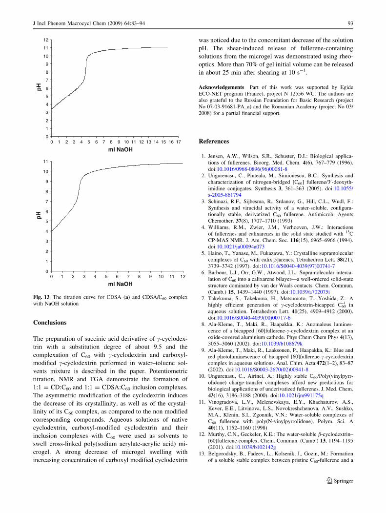
As observable in Fig. 13 a and b the titration curves of
carboxylic groups in CDSA and CDSA/C60 complex are
similar to those characteristic for a week monoprotic acid,
and the equivalence point can be estimated from the
inflection point in the titration curve.
The equivalence point determined from Fig. 13 a rep-
resents the degree of substitution/mol CD. Applying the
Eq. 1 we obtained an average molar content equal to 9.6
carboxylic groups/mol CD.
The equivalence point determined from Fig. 13 b and
that determined from Fig. 13 a allows the determination of
the number of CD with carboxylic groups/mol of C60, the
molar ratio CDAS/C60 is 1:1.
Fig. 12 Relative volume loss of a microgel swollen in aqueous
solutions of CDSA (line 1, open points); CDSA/C60 (line 1, solid
points); CD (line 2, open points); CD/C60 (line 2, solid points) as a
function of strain. Lines are given to guide the eye
92 J Incl Phenom Macrocycl Chem (2009) 64:83–94
123
Conclusions
The preparation of succinic acid derivative of c-cyclodex-
trin with a substitution degree of about 9.5 and the
complexation of C60 with c-cyclodextrin and carboxyl-
modified c-cyclodextrin performed in water–toluene sol-
vents mixture is described in the paper. Potentiometric
titration, NMR and TGA demonstrate the formation of
1:1 = CD:C60 and 1:1 = CDSA:C60 inclusion complexes.
The asymmetric modification of the cyclodextrin induces
the decrease of its crystallinity, as well as of the crystal-
linity of its C60 complex, as compared to the non modified
corresponding compounds. Aqueous solutions of native
cyclodextrin, carboxyl-modified cyclodextrin and their
inclusion complexes with C60 were used as solvents to
swell cross-linked poly(sodium acrylate-acrylic acid) mi-
crogel. A strong decrease of microgel swelling with
increasing concentration of carboxyl modified cyclodextrin
was noticed due to the concomitant decrease of the solution
pH. The shear-induced release of fullerene-containing
solutions from the microgel was demonstrated using rheo-
optics. More than 70% of gel initial volume can be released
in about 25 min after shearing at 10 s-1.
Acknowledgements Part of this work was supported by Egide
ECO-NET program (France), project N 12556 WC. The authors are
also grateful to the Russian Foundation for Basic Research (project
No 07-03-91681-PA_a) and the Romanian Academy (project No 03/
2008) for a partial financial support.
References
1. Jensen, A.W., Wilson, S.R., Schuster, D.I.: Biological applica-
tions of fullerenes. Bioorg. Med. Chem. 4(6), 767–779 (1996).
doi:10.1016/0968-0896(96)00081-8
2. Ungurenasu, C., Pinteala, M., Simionescu, B.C.: Synthesis and
characterization of nitrogen-bridged [C60] fullerene/30-deoxyth-
imidine conjugates. Synthesis 3, 361–363 (2005). doi:10.1055/
s-2005-861794
3. Schinazi, R.F., Sijbesma, R., Srdanov, G., Hill, C.L., Wudl, F.:
Synthesis and virucidal activity of a water-soluble, configura-
tionally stable, derivatized C60 fullerene. Antimicrob. Agents
Chemother. 37(8), 1707–1710 (1993)
4. Williams, R.M., Zwier, J.M., Verhoeven, J.W.: Interactions
of fullerenes and calixarenes in the solid state studied with 13C
CP-MAS NMR. J. Am. Chem. Soc. 116(15), 6965–6966 (1994).
doi:10.1021/ja00094a073
5. Haino, T., Yanase, M., Fukazawa, Y.: Crystalline supramolecular
complexes of C60 with calix[5]arenes. Tetrahedron Lett. 38(21),
3739–3742 (1997). doi:10.1016/S0040-4039(97)00741-7
6. Barbour, L.J., Orr, G.W., Atwood, J.L.: Supramolecular interca-
lation of C60 into a calixarene bilayer—a well-ordered solid-state
structure dominated by van der Waals contacts. Chem. Commun.
(Camb.) 15, 1439–1440 (1997). doi:10.1039/a702075i
7. Takekuma, S., Takekuma, H., Matsumoto, T., Yoshida, Z.: A
highly efficient generation of c-cyclodextrin-bicapped C60–1 in
aqueous solution. Tetrahedron Lett. 41(25), 4909–4912 (2000).
doi:10.1016/S0040-4039(00)00717-6
8. Ala-Kleme, T., Maki, R., Haapakka, K.: Anomalous lumines-
cence of a bicapped [60]fullerene-c-cyclodextrin complex at an
oxide-covered aluminium cathode. Phys Chem Chem Phys 4(13),
3055–3060 (2002). doi:10.1039/b108679k
9. Ala-Kleme, T., Maki, R., Laaksonen, P., Haapakka, K.: Blue and
red photoluminescence of bicapped [60]fullerene-c-cyclodextrin
complex in aqueous solutions. Anal. Chim. Acta 472(1–2), 83–87
(2002). doi:10.1016/S0003-2670(02)00941-8
10. Ungurenasu, C., Airinei, A.: Highly stable C60/Poly(vinylpyrr-
olidone) charge-transfer complexes afford new predictions for
biological applications of underivatized fullerenes. J. Med. Chem.
43(16), 3186–3188 (2000). doi:10.1021/jm991175q
11. Vinogradova, L.V., Melenevskaya, E.Y., Khachaturov, A.S.,
Kever, E.E., Litvinova, L.S., Novokreshchenova, A.V., Sushko,
M.A., Klenin, S.I., Zgonnik, V.N.: Water-soluble complexes of
C60 fullerene with poly(N-vinylpyrrolidone). Polym. Sci. A
40(11), 1152–1160 (1998)
12. Murthy, C.N., Geckeler, K.E.: The water-soluble b-cyclodextrin–
[60]fullerene complex. Chem. Commun. (Camb.) 13, 1194–1195
(2001). doi:10.1039/b102142g
13. Belgorodsky, B., Fadeev, L., Kolsenik, J., Gozin, M.: Formation
of a soluble stable complex between pristine C60-fullerene and a
0
1
2
3
4
5
6
7
8
9
10
11
12
0 1 2 3 4 5 6 7 8 9 10 11 12 13 14 15 16 17
ml NaOH
pH
0
1
2
3
4
5
6
7
8
9
10
11
ml NaOH
pH
0 1 2 3 4 5 6 7 8 9 10 11 12
Fig. 13 The titration curve for CDSA (a) and CDSA/C60 complex
with NaOH solution
J Incl Phenom Macrocycl Chem (2009) 64:83–94 93
123
native blood protein. ChemBioChem 7(11), 1783–1789 (2006).
doi:10.1002/cbic.200600237
14. Eddaoudi, H., Deratani, A., Tingri, S., Sinan, F., Seta, P.: Ful-
lerene membrane transport in PVA films. Polym. Int. 52(8),
1390–1395 (2003). doi:10.1002/pi.1240
15. Bodor, N., Huang, M.J., Watts, J.D.: Theoretical am1 studies of
inclusion complexes of a- and b-cyclodextrins with methylated
benzoic acids and phenol, and c-cyclodextrin with buckminster-
fullerene. J. Incl. Phenom. Macrocycl. Chem. 25(1–3), 97–102
(2005). doi:10.1007/BF01041545
16. Liu, Y., Wang, H., Liang, P., Zhang, Y.H.: Water-soluble
supramolecular fullerene assembly mediated by metallobridged
beta-cyclodextrins. Angew. Chem. Int. Ed. 43(20), 2690–2694
(2004). doi:10.1002/anie.200352973
17. Ying, Y., Zhiqiang, S., Yongshan, M., Mei, X., Jiechao, G.:
Highly water-soluble [60]Fullerene-ethylenediamino- ? p
p-cyclo-
dextrin inclusion complex: the synthesis and self-assembly with
poly (Acrylic Acid). Supramol. Chem. 20(3), 295–299 (2008).
doi:10.1080/10610270701242372
18. Boger, J., Corcoran, R.J., Lehn, J.M.: Cyclodextrin chemistry
selective modification of all primary hydroxyl groups of a- and
b-cyclodextrins. Helv. Chim. Acta 61, 2190–2218 (1978). doi:
10.1002/hlca.19780610622
19. Takeo, K., Mitouh, H., Uemura, K.: Selective modification of
cyclomalto-oligosccharides via tert-butyldimethylsilylation.
Carbohydr. Res. 187(2), 203–221 (1989). doi:10.1016/0008-
6215(89)80004-7
20. Zeo, U., Tarabukina, E., Budtova, T.: Kinetics of shear-induced
gel deswelling/solvent release. J. Control Release 108(1), 73–83
(2005). doi:10.1016/j.jconrel.2005.07.012
21. Gonzalez, C.J., Rossi, R.H.: Synthesis and complexation prop-
erties of an amphiphilic cyclodextrin. Arkivoc XII, 87–99 (2001)
22. Zanina, A., Budtova, T.: Hydrogel under shear: a rheo-optical
study of the particle deformation and solvent release. Macro-
molecules 35(5), 1973–1975 (2002). doi:10.1021/ma011425o
23. Vervoort, S., Budtova, T.: Shear-induced solvent release from a
swollen microgel in the vorticity direction. Colloid Surf.
A Physicochem. Eng. Asp. 262(1–3), 132–138 (2005). doi:
10.1016/j.colsurfa.2005.04.013
24. Boulas, P., Kutner, W., Jones, M.T., Kadish, K.M.: Bucky basket
ball stabilization of electrogenerated C60•– radical monoanion in
water by means of cyclodextrin inclusion chemistry. J. Phys.
Chem. 98(4), 1282–1287 (1994). doi:10.1021/j100055a039
25. Schneider, H.J., Hacket, F., Rudiger, V.: NMR studies of cyclo-
dextrins and cyclodextrin complexes. Chem. Rev. 98(5), 1755–
1785 (1998). doi:10.1021/cr970019t
26. Cucinotta, V., Giuffrida, A., Maccarrone, G., Messina, M.,
Puglisi, A., Torrisi, A., Vecchio, G.: The 6-derivative of
b-cyclodextrin with succinic acid: a new chiral selector for CD-
EKC. J. Pharm. Biomed. Anal. 37(5), 1009–1014 (2005). doi:
10.1016/j.jpba.2004.08.023
27. Anderson, T., Westman, G., Wennerstrom, O., Sundahl, M.:
NMR and UV-VIS investigation of water-soluble fullerene–60-c-
cyclodextrin complex. J. Chem. Soc., Perkin Trans. 2(5), 1097–
1101 (1994) doi:10.1039/p29940001097
28. Priyadarsini, K.I., Mohan, H., Tyagi, A.K., Mittal, J.P.: Inclusion
complex of c-cyclodextrin-C60 formation, characterization, and
photophysical properties in aqueous solutions. J. Phys. Chem.
98(17), 4756–4759 (1994). doi:10.1021/j100068a044
29. Anderson, T., Nilsson, K., Sundahl, M., Westman, G., Wenner-
strom, O.: C60 embedded in c-cyclodextrin: a water-soluble
fullerene. J. Chem. Soc. Chem. Commun. 8, 604–606 (1992)
30. Ajie, H., Alvarez, M.M., Anz, S.J., Beck, R.D., Diederich, F.,
Fostiropoulos, K., Huffman, Wolfgang, D.R., Kratschmer, W.,
Rubin, Y., Schriver, K.E., Sensharma, D., Whetten, R.L.: Char-
acterization of the soluble all-carbon molecules C60 and C70.
J. Phys. Chem. 94(24), 8630–8633 (1990) doi:10.1021/j100387a004
31. Sun, Y.P., Ma, B., Bunker, C.E., Liu, B.: All-carbon polymers
(Polyfullerenes) from photochemical reactions of fullerene clus-
ters in room-temperature solvent mixtures. J. Am. Chem. Soc.
117(51), 12705–12711 (2008). doi:10.1021/ja00156a007
32. Mrzel, A., Mertelj, A., Omerzu, A., Cyopic, M., Mihailovic, D.:
Investigation of encapsulation and solvatochromism of fullerenes
in binary solvent mixtures. J. Phys. Chem. B 103(51), 11256–
11260 (2008)
33. Tseng, W.Y., Chen, Y.H., Khairullin, I.I., Cheng, S., Hwang,
L.P.: NMR study of solid C60(c-cyclodextrin)2. Solid State Nucl.
Mag. 8(4), 219–229 (1997). doi:10.1016/S0926-2040(97)00013-1
34. Marangoci, N., Fifere, A., Farcas, A., Perichaud, A., Harabagiu,
V., Pinteala, M., Simionescu, B.C.: Synthesis and characteriza-
tion of polyrotaxanes based on cyclodextrins and viologen
modified polydimethylsiloxanes. High Perform. Polym. Online-
First. Published on 11 December 2007 as doi:10.1177/
0954008307082152
94 J Incl Phenom Macrocycl Chem (2009) 64:83–94
123
Copyright © 2022 FDOKUMEN