Detailed investigation of a γ-cyclodextrin inclusion complex with l-thyroxine for improved...
11
1 23 Journal of Inclusion Phenomena and Macrocyclic Chemistry and Macrocyclic Chemistry ISSN 0923-0750 Volume 74 Combined 1-4 J Incl Phenom Macrocycl Chem (2012) 74:397-405 DOI 10.1007/s10847-012-0133-9 Detailed investigation of a γ-cyclodextrin inclusion complex with l-thyroxine for improved pharmaceutical formulations Jaya Lakkakula, Rui Werner Maçedo Krause, Derek Tantoh Ndinteh, S. P. Vijaylakshmi & Ashok M. Raichur
-
Upload
independent -
Category
Documents
-
view
1 -
download
0
Transcript of Detailed investigation of a γ-cyclodextrin inclusion complex with l-thyroxine for improved...
1 23
Journal of Inclusion Phenomena andMacrocyclic Chemistryand Macrocyclic Chemistry ISSN 0923-0750Volume 74Combined 1-4 J Incl Phenom Macrocycl Chem (2012)74:397-405DOI 10.1007/s10847-012-0133-9
Detailed investigation of a γ-cyclodextrininclusion complex with l-thyroxine forimproved pharmaceutical formulations
Jaya Lakkakula, Rui Werner MaçedoKrause, Derek Tantoh Ndinteh,S. P. Vijaylakshmi & Ashok M. Raichur
1 23
Your article is protected by copyright and
all rights are held exclusively by Springer
Science+Business Media B.V.. This e-offprint
is for personal use only and shall not be self-
archived in electronic repositories. If you
wish to self-archive your work, please use the
accepted author’s version for posting to your
own website or your institution’s repository.
You may further deposit the accepted author’s
version on a funder’s repository at a funder’s
request, provided it is not made publicly
available until 12 months after publication.

ORIGINAL ARTICLE
Detailed investigation of a c-cyclodextrin inclusion complexwith L-thyroxine for improved pharmaceutical formulations
Jaya Lakkakula • Rui Werner Macedo Krause •
Derek Tantoh Ndinteh • S. P. Vijaylakshmi •
Ashok M. Raichur
Received: 6 September 2011 / Accepted: 23 February 2012 / Published online: 15 March 2012
� Springer Science+Business Media B.V. 2012
Abstract Thyroxine is a naturally occurring human hor-
mone produced by the thyroid gland. Clinical applications
of thyroxine to treat several chronic disorders are limited
by poor water solubility and instability under physio-
logical conditions. An inclusion complex of levo-thyroxine
(L-thyroxine), the active form of the hormone with gamma
cyclodextrin (c-CD) has been obtained and studied with the
aim of improving oral delivery rather than the injection
formulation of the sodium salt. In addition to greater
patient acceptability, inclusion complexes often improve
aqueous solubility and bioavailability, stability, and reduce
toxicity of drugs, thus providing enhanced pharmaceutical
formulations. Physicochemical characterization of the
inclusion complex was carried out using Fourier transform
infrared spectroscopy, X-ray diffractometry, differential
scanning calorimetry, scanning electron microscopy and
proton nuclear magnetic resonance spectroscopy. Inter-
molecular dipolar interactions for the inclusion complex
were also studied using 2 dimensional ROESY experi-
ments. Formation of the inclusion complex between the
protons H3 and H5 of cyclodextrin with aromatic protons
of thyroxine was confirmed by their dipolar interaction.
Molecular modelling was used to understand the basis for
the complex formation and predict the formation of other
complexes. Interestingly, we found that L-thyroxine forms
an inclusion complex only with the larger c-CD and not
with other available alpha and beta forms.
Keywords c-Cyclodextrin � Levo-thyroxine �Hypothyroidism � Inclusion complex
Introduction
Levo-thyroxine (L-thyroxine) is a synthetic and natural
hormone (Fig. 1) used for treating hypothyroidism and
other thyroid conditions, and is commonly administered as
a daily oral tablet or in the ionized form as an injection
formulation. In hypothyroidism, the thyroid gland does not
produce enough thyroid hormone or the body metabolises
the hormone too rapidly. This hormone is essential for
many vital metabolic functions in the body, especially for
energy. Some of the symptoms of hypothyroidism there-
fore include mood swings, lethargy, increased weight and
intolerance to cold [1]. L-Thyroxine referred to as T4
(tetraiodothyronine), is secreted by the thyroid gland and
transported around the body. The action of the deiodinase
enzymes on T4 in peripheral tissues leads to the loss of an
iodine atom, forming T3 (triiodothyronine), which acts on
the thyroid receptor in cell nuclei [2]. Patients suffering
from hypothyroidism are treated with both T3 and T4.
Absorption of the drugs occurs mostly in the intestine, with
some 62–82 % of L-thyroxine being absorbed within the
first three hours of passing into the lower intestine (i.e.
jejunum and ileum) [3]. Patients are under daily medication
as the drug is not stored in the body, and is often rapidly
degraded in the stomach. To overcome these problems and
for better patient compliance efforts have been put to
design drug delivery systems for controlled and sustained
release of these and other hormones.
J. Lakkakula � R. W. M. Krause (&) � D. T. Ndinteh
Department of Applied Chemistry, Center for Nanomaterials
Science, University of Johannesburg, Doornfontein,
Johannesburg 2028, South Africa
e-mail: [email protected]
S. P. Vijaylakshmi � A. M. Raichur
Department of Materials Engineering, Indian Institute of
Science, Bangalore 560 012, India
123
J Incl Phenom Macrocycl Chem (2012) 74:397–405
DOI 10.1007/s10847-012-0133-9
Author's personal copy

There is a need for pharmaceutical formulations that
will increase the duration of the availability of the hor-
mones in the body. In this way there would be a reduction
in the frequency of administration of the drugs and hence
an increased patient compliance, but also this process
avoids the potentially dangerous effects of too much hor-
mone in the body.
A possible way to approach the above stated goal may
be oral drug formulation that will enable delivery of the
drug in a controlled manner. Carriers have always played
an important role in designing proper drug formulation for
prolonged release and action of drug. For controlled release
of dosage forms for various drugs, cyclodextrin has played
a pivotal role as a carrier material [4]. Cyclodextrins can
also be used as complexing molecules as they have a
unique property of forming inclusion complexes in solid
and liquid states [5].
Cyclodextrins (CDs) are cyclic, torus-shaped non-toxic
oligomers of amylose with external hydrophilic surface and
internal hydrophobic interiors. The most commonly avail-
able CDs are a-, b-, and c-cyclodextrins that have 6, 7 and
8 glucose units, respectively (Fig. 2). The characteristics of
cyclodextrin allow the formation of inclusion complexes
with many organic molecules where polarity of guest
molecule plays a key role in the complex formation [6, 7].
The driving forces are both the stabilisation of a hydro-
phobic guest, and the entropically favourable expulsion of
water molecules from the lipophilic CD cavity. However,
steric interactions start to operate as the size of side
included guest increases, thereby also reducing the number
of molecules that can be complexed [6]. Entry of lipophilic
guest molecule into the hydrophobic cavity of cyclodextrin
leads to formation of inclusion complex with the dis-
placement of water molecules seated inside the torus [8].
Improved aqueous solubility and membrane permeation
have been studied by formulating thyroxine with cyclo-
dextrin [9]. Yet no complete characterisation of the inclu-
sion complex between thyroxine and gamma cyclodextrin
has been reported.
Generally, guest/host systems of various compounds
with cyclodextrins have been prepared and characterised.
The formation of inclusion complexes of phenols with
b-CD have been used for waste water treatment and studied
using mass spectrometry, FTIR, surface tension and ultra-
violet visible spectroscopy [10], infra-red spectroscopy,
XRD and DSC was used to characterize the inclusion of
aromatic benzene moieties such as those of miconazole
nitrate into the cavity of b-CD [11].
The aqueous inclusion complex between substituted
b-CDs and ebastine was studied using 13CNMR, 2D NMR,
and 1H NMR [12]. Emission spectra, stoichiometry, change
in entropy and enthalpy of inclusion complex was studied
using fluorescence and molecular mechanics [13], while
OH
O
II
II
H2N
O OH
Fig. 1 Plane and spatial representations of L-thyroxine molecular
structure
O
OHHO
OH
O
OOH
HOOHO
OOH
OH
OH
O
OOH
OH
OH
O
O
OH
OH
HO
O OH
OHHO
O
OOH
HO
HO
O
O
OH
OH
HO
O
O
Fig. 2 Plane and spatial
representations of molecular
structure c-CD
398 J Incl Phenom Macrocycl Chem (2012) 74:397–405
123
Author's personal copy

crystal structure, packing and intermolecular hydrogen
bonding of inclusion complex was calculated using X-ray
crystallography [14]. Spectroscopy remains amongst the
most important tools for investigating various cyclodextrin
phenomena [15] and in this work we investigate the com-
plexation of L-thyroxine and c-CD and characterise the
complex using NMR, 2D NMR, XRD, FTIR, DSC, SEM
and molecular modelling.
Materials and methods
Materials
Cyclodextrin was obtained from Wacker Chemie and
L-thyroxine purchased from Sigma-Aldrich (Germany) and
both were used as received. All reagents were of analytical
grade. Deionized water was used throughout the experiment.
Methods
Preparation of inclusion complexes
The inclusion complex of c-cyclodextrin with L-thyroxine
was affected using a method similar to that adopted for
making pharmaceutical compositions containing the same,
with minor changes [9]. The inclusion complexes were
formed by dissolving 20 ml of a 10 % c-CD in water and
adding 500 mg of L-thyroxine under intensive stirring
(600 rpm) at room temperature for 12 h. The resulting
opalescent solution was then filtered using a 0.22 lm cel-
lulose acetate filter and clear filtrate was freeze-dried
yielding a white solid.
Solid state characterization of inclusion complex
PXRD
The powder X-ray diffraction patterns were recorded using
X-Pert PRO, PANalytical diffractometer system, operated
at a voltage of 40 kV and a current of 30 mA. The pure
c-CD, L-thyroxine and inclusion complex were analyzed in
the 2h angle range of 3–600.
1HNMR
1HNMR was recorded with BRUKER Avance 400 MHz or
500 MHz spectrometer in deuterated DMSO and refer-
enced to residual solvent.
2D-ROESY experiments were carried out in phase
sensitive mode, set up applying a continuous wave (CW)
with spin lock for mixing. Spectra were obtained on the
inclusion complex using spin lock of 180x 180-x pulses
using purge pulses before d1 with a 90� high power pulse.
DSC
DSC was carried out in a temperature range of 25–350 �C
under an argon flow maintained at 80 ml/min and the
scanning rate was 5 �C/min. The sample was weighed and
placed in an aluminium pan whereas empty aluminium pan
was used as a reference.
FTIR
Infrared spectra of the inclusion complex, host (c-CD) and
the guest molecule (L-thyroxine) was studied using a
BRUKER ALPHA-P spectrometer. The spectra were
recorded from 3,600 to 600 cm-1 in attenuated total
reflectance (ATR) mode.
SEM
Vacuum dried formulations were prepared on silicon
wafers and coated with gold. The samples were examined
using SIRION High resolution FEI scanning electron
microscope (SEM) at an accelerating voltage of 20 kV.
Molecular modelling
Molecular mechanics and dynamics were performed using
Allinger’s MM2 force field [16, 17] on Cambridge soft
Chem3D softwareTM
on a Pentium CPU.
Gamma cyclodextrin was geometry-optimised using a
combination of a Truncated Newton and Polak Ribiere
optimiser, to a Root-Mean-Square (RMS) gradient of 0.01.
Optimisation of the geometry of the L-thyroxine and the
complex was performed similarly. All parameters in the
forcefield are from the ‘‘MM2 (1991) Parameter Set’’, as
provided by N. L. Allinger, University of Georgia, apart
from the C(sp2)–O–C(sp2) bond angle in thyroxine, which
was modelled on the C(sp2)–O–(Csp3) bond angle.
Modelling of the c-CD/L-thyroxine complexes were
completed by manually docking the optimised L-thyroxine
structure inside each of the optimised c-CD structures as a
starting geometry and then performing a full-geometry
optimization inside a static solvent (water) cage. Manual
docking was performed for both possible arrangements of
the drug (i.e. amino-acid towards the CD primary or sec-
ondary face). The dielectric constant for electrostatic
interactions was set at 1.
The optimised structures were subjected to a series of sim-
ulated annealing heating and cooling cycles between 46 and
370 K, in steps of 2 ps and a heating rate of 1.00 kcal/atom/ps
and allowing for energy minimization after each 10 steps.
J Incl Phenom Macrocycl Chem (2012) 74:397–405 399
123
Author's personal copy
Results and discussions
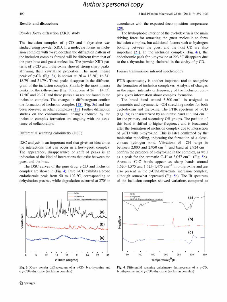
Powder X-ray diffraction (XRD) study
The inclusion complex of c-CD and L-thyroxine was
studied using powder XRD. If a molecule forms an inclu-
sion complex with c-cyclodextrin the diffraction pattern of
the inclusion complex formed will be different from that of
the pure host and guest molecules. The powder XRD pat-
terns of c-CD and L-thyroxine showed strong sharp peaks,
affirming their crystalline properties. The most intense
peak of c-CD (Fig. 3a) is shown at 2h = 12.28�, 16.34�,
18.79� and 21.79�. These peaks disappear in the diffracto-
gram of the inclusion complex. Similarly the most intense
peaks for the L-thyroxine (Fig. 3b) appear at 2h = 14.53�,
17.56� and 23.21� and these peaks also are not found in the
inclusion complex. The changes in diffractogram confirm
the formation of inclusion complex [18] (Fig. 3c) and has
been observed in other complexes [19]. Further diffraction
studies on the conformational changes induced by the
inclusion complex formation are ongoing with the assis-
tance of collaborators.
Differential scanning calorimetry (DSC)
DSC analysis is an important tool that gives an idea about
the interactions that can occur in a host–guest complex.
The appearance, disappearance or shift of peaks is an
indication of the kind of interactions that exist between the
guest and the host.
The DSC curves of the pure drug, c-CD and inclusion
complex are shown in (Fig. 4). Pure c-CD exhibits a broad
endothermic peak from 50 to 102 �C, corresponding to
dehydration process, while degradation occurred at 270� in
accordance with the expected decomposition temperature
[20].
The hydrophobic interior of the cyclodextrin is the main
driving force for attracting the guest molecule to form
inclusion complex, but additional factors such as hydrogen
bonding between the guest and the host CD are also
important [21]. In the inclusion complex (Fig. 4c), the
endothermic peak for L-thyroxine at 223 �C disappears due
to the L-thyroxine being sheltered in the cavity of c-CD.
Fourier transmission infrared spectroscopy
FTIR spectroscopy is another important tool to recognize
the formation of inclusion complexes. Analysis of changes
in the signal intensity or frequency of the inclusion com-
plex gives information about complex formation.
The broad band around 3,300 cm-1 is assigned to
symmetric and asymmetric –OH stretching modes for both
cyclodextrin and thyroxine. The FTIR spectrum of c-CD
(Fig. 5a) is characterized by an intense band at 3,284 cm-1
for the primary and secondary OH groups. The position of
this band is shifted to higher frequency and is broadened
after the formation of inclusion complex due to interaction
of c-CD with L-thyroxine. This is later confirmed by the
molecular modelling, indicating the formation of a close-
contact hydrogen bond. Vibrations of –CH range in
between 2,800 and 2,950 cm-1, and band at 2,924 cm-1
confirm the presence of L-thyroxine in the complex, as well
as a peak for the aromatic C–H at 3,057 cm-1 (Fig. 5b).
Aromatic C–C bands appear as sharp bands around
1,620–1,575 and 1,525–1,475 cm-1 in L-thyroxine and are
also present in the c-CD/L-thyroxine inclusion complex,
although somewhat depressed (Fig. 5c). The IR spectrum
of the inclusion complex showed variations compared to
6 9 12 15 18 21 24 27 30
2 Theta (degree)
G-CD
L-Thyroxine
G-CD+L-Thyroxine(inclusion complex)
cou
nts
/sec
(a)
(b)
(c)
Fig. 3 X-ray powder diffractogram of a c-CD, b L-thyroxine and
c c-CD/L-thyroxine (inclusion complex)
50 100 150 200 250 300 350
Temperature(0 )C
G-CD L-Thyroxine G-CD+L-Thyroxine(inclusion complex)
Hea
tflo
w
(a)
(b)
(c)
Fig. 4 Differential scanning calorimetry thermograms of a c-CD,
b L-thyroxine and c c-CD/L-thyroxine (inclusion complex)
400 J Incl Phenom Macrocycl Chem (2012) 74:397–405
123
Author's personal copy
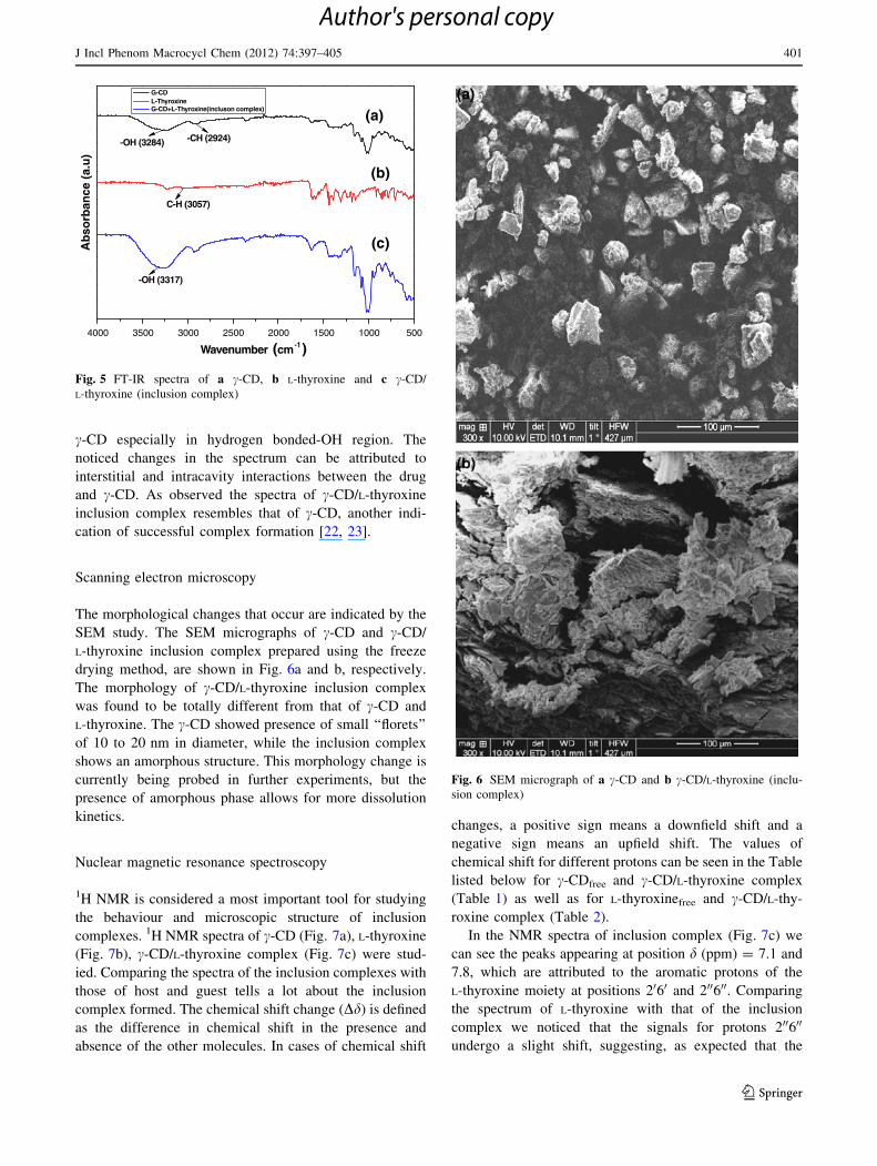
c-CD especially in hydrogen bonded-OH region. The
noticed changes in the spectrum can be attributed to
interstitial and intracavity interactions between the drug
and c-CD. As observed the spectra of c-CD/L-thyroxine
inclusion complex resembles that of c-CD, another indi-
cation of successful complex formation [22, 23].
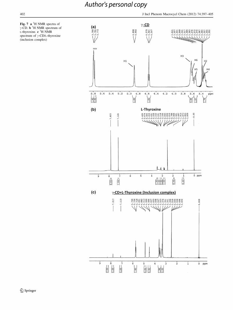
Scanning electron microscopy
The morphological changes that occur are indicated by the
SEM study. The SEM micrographs of c-CD and c-CD/
L-thyroxine inclusion complex prepared using the freeze
drying method, are shown in Fig. 6a and b, respectively.
The morphology of c-CD/L-thyroxine inclusion complex
was found to be totally different from that of c-CD and
L-thyroxine. The c-CD showed presence of small ‘‘florets’’
of 10 to 20 nm in diameter, while the inclusion complex
shows an amorphous structure. This morphology change is
currently being probed in further experiments, but the
presence of amorphous phase allows for more dissolution
kinetics.
Nuclear magnetic resonance spectroscopy
1H NMR is considered a most important tool for studying
the behaviour and microscopic structure of inclusion
complexes. 1H NMR spectra of c-CD (Fig. 7a), L-thyroxine
(Fig. 7b), c-CD/L-thyroxine complex (Fig. 7c) were stud-
ied. Comparing the spectra of the inclusion complexes with
those of host and guest tells a lot about the inclusion
complex formed. The chemical shift change (Dd) is defined
as the difference in chemical shift in the presence and
absence of the other molecules. In cases of chemical shift
changes, a positive sign means a downfield shift and a
negative sign means an upfield shift. The values of
chemical shift for different protons can be seen in the Table
listed below for c-CDfree and c-CD/L-thyroxine complex
(Table 1) as well as for L-thyroxinefree and c-CD/L-thy-
roxine complex (Table 2).
In the NMR spectra of inclusion complex (Fig. 7c) we
can see the peaks appearing at position d (ppm) = 7.1 and
7.8, which are attributed to the aromatic protons of the
L-thyroxine moiety at positions 2060 and 200600. Comparing
the spectrum of L-thyroxine with that of the inclusion
complex we noticed that the signals for protons 200600
undergo a slight shift, suggesting, as expected that the
4000 3500 3000 2500 2000 1500 1000 500
Ab
sorb
ance
(a.
u)
Wavenumber (cm-1)
G-CD L-Thyroxine G-CD+L-Thyroxine(incluson complex)
-OH (3284) -CH (2924)
C-H (3057)
-OH (3317)
(a)
(c)
(b)
Fig. 5 FT-IR spectra of a c-CD, b L-thyroxine and c c-CD/
L-thyroxine (inclusion complex)
Fig. 6 SEM micrograph of a c-CD and b c-CD/L-thyroxine (inclu-
sion complex)
J Incl Phenom Macrocycl Chem (2012) 74:397–405 401
123
Author's personal copy
γ
γ
(a)
(b)
(c)
Fig. 7 a 1H NMR spectra of
c-CD. b 1H NMR spectrum of
L-thyroxine. c 1H NMR
spectrum of c-CD/L-thyroxine
(inclusion complex)
402 J Incl Phenom Macrocycl Chem (2012) 74:397–405
123
Author's personal copy
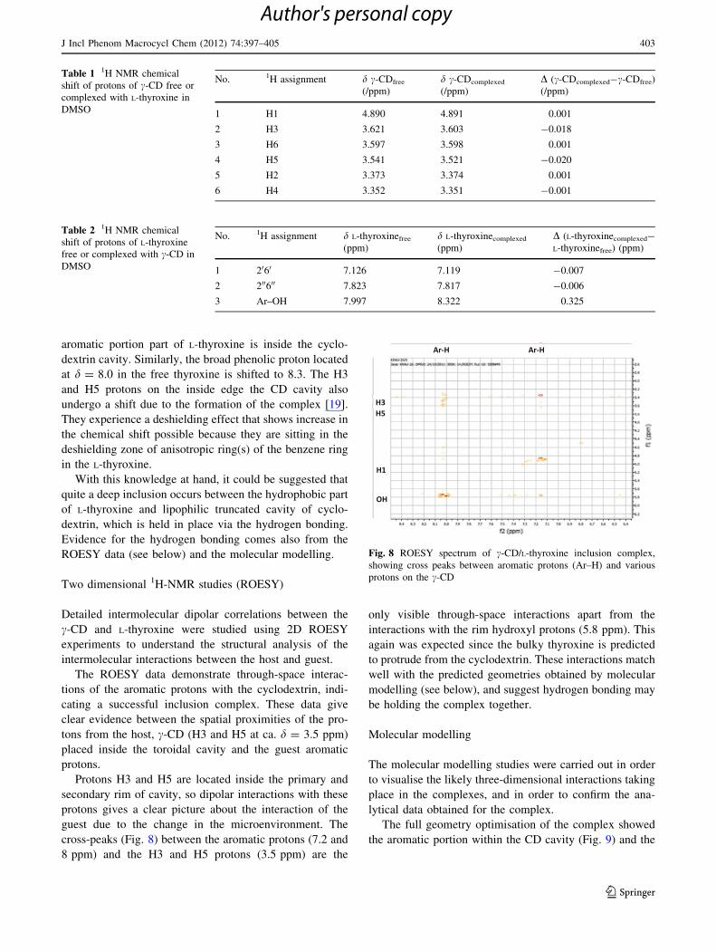
aromatic portion part of L-thyroxine is inside the cyclo-
dextrin cavity. Similarly, the broad phenolic proton located
at d = 8.0 in the free thyroxine is shifted to 8.3. The H3
and H5 protons on the inside edge the CD cavity also
undergo a shift due to the formation of the complex [19].
They experience a deshielding effect that shows increase in
the chemical shift possible because they are sitting in the
deshielding zone of anisotropic ring(s) of the benzene ring
in the L-thyroxine.
With this knowledge at hand, it could be suggested that
quite a deep inclusion occurs between the hydrophobic part
of L-thyroxine and lipophilic truncated cavity of cyclo-
dextrin, which is held in place via the hydrogen bonding.
Evidence for the hydrogen bonding comes also from the
ROESY data (see below) and the molecular modelling.
Two dimensional 1H-NMR studies (ROESY)
Detailed intermolecular dipolar correlations between the
c-CD and L-thyroxine were studied using 2D ROESY
experiments to understand the structural analysis of the
intermolecular interactions between the host and guest.
The ROESY data demonstrate through-space interac-
tions of the aromatic protons with the cyclodextrin, indi-
cating a successful inclusion complex. These data give
clear evidence between the spatial proximities of the pro-
tons from the host, c-CD (H3 and H5 at ca. d = 3.5 ppm)
placed inside the toroidal cavity and the guest aromatic
protons.
Protons H3 and H5 are located inside the primary and
secondary rim of cavity, so dipolar interactions with these
protons gives a clear picture about the interaction of the
guest due to the change in the microenvironment. The
cross-peaks (Fig. 8) between the aromatic protons (7.2 and
8 ppm) and the H3 and H5 protons (3.5 ppm) are the
only visible through-space interactions apart from the
interactions with the rim hydroxyl protons (5.8 ppm). This
again was expected since the bulky thyroxine is predicted
to protrude from the cyclodextrin. These interactions match
well with the predicted geometries obtained by molecular
modelling (see below), and suggest hydrogen bonding may
be holding the complex together.
Molecular modelling
The molecular modelling studies were carried out in order
to visualise the likely three-dimensional interactions taking
place in the complexes, and in order to confirm the ana-
lytical data obtained for the complex.
The full geometry optimisation of the complex showed
the aromatic portion within the CD cavity (Fig. 9) and the
Table 1 1H NMR chemical
shift of protons of c-CD free or
complexed with L-thyroxine in
DMSO
No. 1H assignment d c-CDfree
(/ppm)
d c-CDcomplexed
(/ppm)
D (c-CDcomplexed-c-CDfree)
(/ppm)
1 H1 4.890 4.891 0.001
2 H3 3.621 3.603 -0.018
3 H6 3.597 3.598 0.001
4 H5 3.541 3.521 -0.020
5 H2 3.373 3.374 0.001
6 H4 3.352 3.351 -0.001
Table 2 1H NMR chemical
shift of protons of L-thyroxine
free or complexed with c-CD in
DMSO
No. 1H assignment d L-thyroxinefree
(ppm)
d L-thyroxinecomplexed
(ppm)
D (L-thyroxinecomplexed-
L-thyroxinefree) (ppm)
1 2060 7.126 7.119 -0.007
2 200600 7.823 7.817 -0.006
3 Ar–OH 7.997 8.322 0.325
Fig. 8 ROESY spectrum of c-CD/L-thyroxine inclusion complex,
showing cross peaks between aromatic protons (Ar–H) and various
protons on the c-CD
J Incl Phenom Macrocycl Chem (2012) 74:397–405 403
123
Author's personal copy
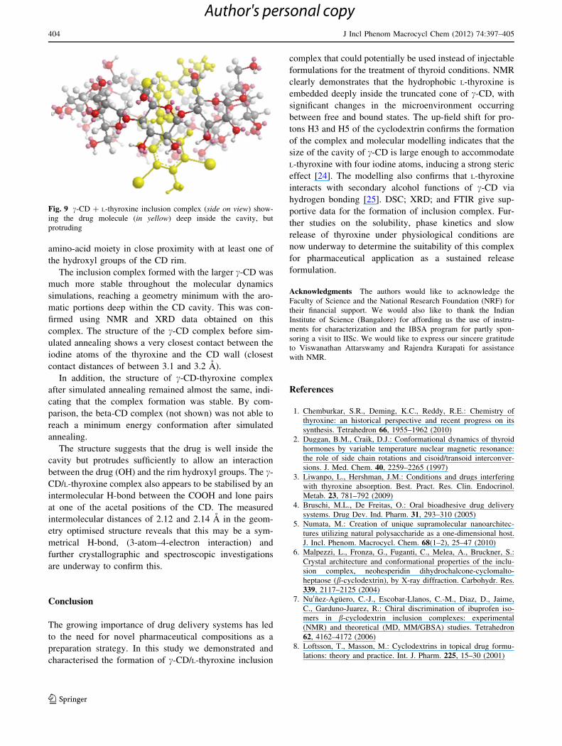
amino-acid moiety in close proximity with at least one of
the hydroxyl groups of the CD rim.
The inclusion complex formed with the larger c-CD was
much more stable throughout the molecular dynamics
simulations, reaching a geometry minimum with the aro-
matic portions deep within the CD cavity. This was con-
firmed using NMR and XRD data obtained on this
complex. The structure of the c-CD complex before sim-
ulated annealing shows a very closest contact between the
iodine atoms of the thyroxine and the CD wall (closest
contact distances of between 3.1 and 3.2 A).
In addition, the structure of c-CD-thyroxine complex
after simulated annealing remained almost the same, indi-
cating that the complex formation was stable. By com-
parison, the beta-CD complex (not shown) was not able to
reach a minimum energy conformation after simulated
annealing.
The structure suggests that the drug is well inside the
cavity but protrudes sufficiently to allow an interaction
between the drug (OH) and the rim hydroxyl groups. The c-
CD/L-thyroxine complex also appears to be stabilised by an
intermolecular H-bond between the COOH and lone pairs
at one of the acetal positions of the CD. The measured
intermolecular distances of 2.12 and 2.14 A in the geom-
etry optimised structure reveals that this may be a sym-
metrical H-bond, (3-atom–4-electron interaction) and
further crystallographic and spectroscopic investigations
are underway to confirm this.
Conclusion
The growing importance of drug delivery systems has led
to the need for novel pharmaceutical compositions as a
preparation strategy. In this study we demonstrated and
characterised the formation of c-CD/L-thyroxine inclusion
complex that could potentially be used instead of injectable
formulations for the treatment of thyroid conditions. NMR
clearly demonstrates that the hydrophobic L-thyroxine is
embedded deeply inside the truncated cone of c-CD, with
significant changes in the microenvironment occurring
between free and bound states. The up-field shift for pro-
tons H3 and H5 of the cyclodextrin confirms the formation
of the complex and molecular modelling indicates that the
size of the cavity of c-CD is large enough to accommodate
L-thyroxine with four iodine atoms, inducing a strong steric
effect [24]. The modelling also confirms that L-thyroxine
interacts with secondary alcohol functions of c-CD via
hydrogen bonding [25]. DSC; XRD; and FTIR give sup-
portive data for the formation of inclusion complex. Fur-
ther studies on the solubility, phase kinetics and slow
release of thyroxine under physiological conditions are
now underway to determine the suitability of this complex
for pharmaceutical application as a sustained release
formulation.
Acknowledgments The authors would like to acknowledge the
Faculty of Science and the National Research Foundation (NRF) for
their financial support. We would also like to thank the Indian
Institute of Science (Bangalore) for affording us the use of instru-
ments for characterization and the IBSA program for partly spon-
soring a visit to IISc. We would like to express our sincere gratitude
to Viswanathan Attarswamy and Rajendra Kurapati for assistance
with NMR.
References
1. Chemburkar, S.R., Deming, K.C., Reddy, R.E.: Chemistry of
thyroxine: an historical perspective and recent progress on its
synthesis. Tetrahedron 66, 1955–1962 (2010)
2. Duggan, B.M., Craik, D.J.: Conformational dynamics of thyroid
hormones by variable temperature nuclear magnetic resonance:
the role of side chain rotations and cisoid/transoid interconver-
sions. J. Med. Chem. 40, 2259–2265 (1997)
3. Liwanpo, L., Hershman, J.M.: Conditions and drugs interfering
with thyroxine absorption. Best. Pract. Res. Clin. Endocrinol.
Metab. 23, 781–792 (2009)
4. Bruschi, M.L., De Freitas, O.: Oral bioadhesive drug delivery
systems. Drug Dev. Ind. Pharm. 31, 293–310 (2005)
5. Numata, M.: Creation of unique supramolecular nanoarchitec-
tures utilizing natural polysaccharide as a one-dimensional host.
J. Incl. Phenom. Macrocycl. Chem. 68(1–2), 25–47 (2010)
6. Malpezzi, L., Fronza, G., Fuganti, C., Melea, A., Bruckner, S.:
Crystal architecture and conformational properties of the inclu-
sion complex, neohesperidin dihydrochalcone-cyclomalto-
heptaose (b-cyclodextrin), by X-ray diffraction. Carbohydr. Res.
339, 2117–2125 (2004)
7. Nu0nez-Aguero, C.-J., Escobar-Llanos, C.-M., Diaz, D., Jaime,
C., Garduno-Juarez, R.: Chiral discrimination of ibuprofen iso-
mers in b-cyclodextrin inclusion complexes: experimental
(NMR) and theoretical (MD, MM/GBSA) studies. Tetrahedron
62, 4162–4172 (2006)
8. Loftsson, T., Masson, M.: Cyclodextrins in topical drug formu-
lations: theory and practice. Int. J. Pharm. 225, 15–30 (2001)
Fig. 9 c-CD ? L-thyroxine inclusion complex (side on view) show-
ing the drug molecule (in yellow) deep inside the cavity, but
protruding
404 J Incl Phenom Macrocycl Chem (2012) 74:397–405
123
Author's personal copy
9. Fischer, W., Bracher, D.: Thyroxine/cyclodextrin complexes and
pharmaceutical compositions containing the same. United States
Patent (1999)
10. Bonenfant, D., Niquette, P., Mimeault, M., Furtos-Matei, A.,
Hausler, R.: UV–Vis and FTIR spectroscopic analyses of inclu-
sion complexes of nonylphenol and nonylphenol ethoxylate with
b-cyclodextrin. Water Res. 43, 3575–3581 (2009)
11. Wang, J.-H., Cai, Z.: Investigation of inclusion complex of
miconazole nitrate with b-cyclodextrin. Carbohydr. Polym. 72,
255–260 (2008)
12. Maddens, T., Velaz, I., Machın, R., Isasi, J.R., Martın, C.:
Complexation of ebastine with b-cyclodextrin derivatives. J. Incl.
Phenom. Macrocycl. Chem. 70(3–4), 415–419 (2011)
13. Serna, L., Marino, A.D., Mendicuti, F.: Inclusion complexes of a
bichromophoric diester containing anthracene and naphthalene
groups with a- and b-cyclodextrins: thermodynamics and molec-
ular mechanics. Spectrochim. Acta Part A 61, 945–1954 (2005)
14. Wang, E.-J., Liana, Z.-X., Cai, J.: The crystal structure of the 1:1
inclusion complex of b-cyclodextrin with benzamide. Carbohydr.
Res. 342, 767–771 (2007)
15. Zhao, W., Chao, J., Du, R., Huang, S.: Spectroscopic studies on
the inclusion behavior between caffenic acid and c-cyclodextrin,
Online FirstTM
(2011). doi:10.1007/s10847-011-9930-9
16. Allinger, N.L.: Conformational analysis. 130. MM2. A hydro-
carbon force field utilizing V1 and V2 torsional terms. J. Am.
Chem. Soc. 99, 8127–8134 (1977)
17. Sprague, J.T., Tai, J.C., Yuh, Y., Allinger, N.L.: The MMP2
calculational method. J. Comput. Chem. 8, 581–603 (1987)
18. Xua, D., Wanga, X., Ding, L.: Spectroscopic studies on the inter-
action of c-cyclodextrin–daunorubicin inclusion complex with
herring sperm DNA. Carbohydr. Polym. 83(3), 1257–1262 (2010)
19. Anselmi, C., Centini, M., Ricci, M., Buonocore, A., Granata, P.,
Tsuno, T., Facino, R.M.: Analytical characterization of a ferulic
acid/c-cyclodextrin inclusion complex. J. Pharm. Biomed. Anal.
40, 875–881 (2006)
20. Kawasaki, J., Satou, D., Takagaki, T., Nemoto, T., Kawaguchi,
A.: Structural features of inclusion complexes of c-cyclodextrin
with various polymers. Polymer 48, 1127–1138 (2007)
21. Wulff, M., Alden, M.: Solid state studies of drug–cyclodextrin
inclusion complexes in PEG 6000 prepared by a new method.
Eur. J. Pharm. Sci. 8, 269–281 (1999)
22. Stancanelli, R., Crupi, V., Luca, L.D., Ficarra, P., Ficarra, R.,
Gitto, R., Guardo, M., Iraci, N., Majolino, D., Tommasini, S.,
Venuti, V.: Improvement of water solubility of non-competitive
AMPA receptor antagonists by complexation with b-cyclodex-
trin. Bioorg. Med. Chem. 16, 8706–8712 (2008)
23. Lu, J., Shin, I.D., Nojima, S., Tonelli, A.E.: Formation and
characterization of the inclusion compounds between poly(e-
caprolactone)-poly(ethylene oxide)-poly(e-caprolactone) triblock
copolymer and a- and c-cyclodextrin. Polymer 41, 5871–5883
(2000)
24. Peani, C., Creminon, C., Grassi, J., Pradelles, P., Perly, B., Dje-
daini-Pilard, F.: NMR investigations of the inclusion of thyroxine
and derivatives in natural cyclodextrins. J. Incl. Phenom. Mac-
rocycl. Chem. 33, 307–319 (1999)
25. Shahgaldian, P., Hegner, M., Pieles, U.: A cyclodextrin self-
assembled monolayer (SAM) based surface plasmon resonance
(SPR) sensor for enantioselective analysis of thyroxine. J. Incl.
Phenom. Macrocycl. Chem. 53, 35–39 (2005)
J Incl Phenom Macrocycl Chem (2012) 74:397–405 405
123
Author's personal copy