Effect of methyl-beta-cyclodextrin on the viability and ...
7
Lee et al. J of Biol Res-Thessaloniki (2016) 23:5 DOI 10.1186/s40709-016-0043-x RESEARCH Effect of methyl-beta-cyclodextrin on the viability and acrosome damage of sex-sorted sperm in frozen-thawed bovine semen Seunghyung Lee 1† , Yong‑Seung Lee 1† , Sang‑Hee Lee 2 , Boo‑Keun Yang 1 and Choon‑Keun Park 1* Abstract Background: The regulation of methyl‑beta‑cyclodextrin (MBCD) on cryodamage on X‑ and Y‑sperm during cryo‑ preservation of semen was investigated. The semen was collected from ten healthy bulls of proven fertility by an arti‑ ficial vagina. The bovine sperm treated with MBCD fresh solution (0, 1, 5, 10, and 20 mM). The sperms were evaluated for viability and acrosome damage using flow cytometry. Moreover, X‑ and Y‑sperm in frozen‑thawed bovine semen were sorted by flow cytometry after Hoechst 33342‑dyed, and the viability and acrosome damage of sperms were analyzed. Results: Sperm viability in frozen‑thawed semen was decreased by MBCD (p < 0.05), also the acrosome damage of sperm was significantly increased (p < 0.05). Moreover, we sorted X‑ and Y‑sperm from frozen‑thawed bovine semen for observing the viability and acrosome damage on the separated X‑ and Y‑sperm after MBCD treatment. Viability of X‑sperm was significantly lower than that of Y‑sperm (p < 0.05). Also, acrosome damage of X‑sperm was significantly higher than Y‑sperm (p < 0.05). Conclusions: Methyl‑beta‑cyclodextrin enhances damage of sperm in frozen‑thawed bovine semen, and X‑sperm is more sensitive than Y‑sperm in cell damage. These results demonstrate that MBCD can inhibit viability of spermatozoa in frozen‑thawed bovine semen (for X‑sperm, especially). Keywords: Methyl‑beta‑cyclodextrin, Sex sorting, Viability, Acrosome damage, Spermatozoa © 2016 Lee et al. This article is distributed under the terms of the Creative Commons Attribution 4.0 International License (http:// creativecommons.org/licenses/by/4.0/), which permits unrestricted use, distribution, and reproduction in any medium, provided you give appropriate credit to the original author(s) and the source, provide a link to the Creative Commons license, and indicate if changes were made. The Creative Commons Public Domain Dedication waiver (http://creativecommons.org/publicdomain/ zero/1.0/) applies to the data made available in this article, unless otherwise stated. Background e technology of controlling the sex of mammalian off- spring is of great importance in the livestock industry [1]. ere has been great interest in sex pre-selection in bovine, and gained clear economic benefits and man- agement advantages. Sex is determined by the pres- ence or absence of the Y chromosome in mammals. In the process of spermatogenesis, X- and Y-sperm can be produced, and the sex of the embryo is determined by the chromosome of the sperm. e X-bearing sperm (X-sperm) in bovine is larger and longer than the Y-bear- ing sperm (Y-sperm), having 3.8 % more DNA [2, 3]. e X-sperm swims more slowly and has a longer life span than Y-sperm [4]. Sorting for X- and Y-sperm popu- lations based on differences in DNA content by flow cytometry was recommended for improving the selection of X- and Y-sperm [5] and it is widely used in livestock animals. Flow cytometry is able to analyze the biological characteristics of several thousand cells in real time using fluorescent dye or fluorescent antibodies and can actively separate and isolate cells having specified properties. e use of flow cytometry also provides a new opportunity to discover differences in biological characteristics between separated populations of X- and Y-sperm [6, 7]. Open Access Journal of Biological Research-Thessaloniki *Correspondence: [email protected] † Seunghyung Lee and Yong‑Seung Lee authors contributed equally to this study 1 College of Animal Life Sciences, Kangwon National University, Chuncheon 24341, Republic of Korea Full list of author information is available at the end of the article
-
Upload
khangminh22 -
Category
Documents
-
view
5 -
download
0
Transcript of Effect of methyl-beta-cyclodextrin on the viability and ...

Lee et al. J of Biol Res-Thessaloniki (2016) 23:5 DOI 10.1186/s40709-016-0043-x
RESEARCH
Effect of methyl-beta-cyclodextrin on the viability and acrosome damage of sex-sorted sperm in frozen-thawed bovine semenSeunghyung Lee1†, Yong‑Seung Lee1†, Sang‑Hee Lee2, Boo‑Keun Yang1 and Choon‑Keun Park1*
Abstract
Background: The regulation of methyl‑beta‑cyclodextrin (MBCD) on cryodamage on X‑ and Y‑sperm during cryo‑preservation of semen was investigated. The semen was collected from ten healthy bulls of proven fertility by an arti‑ficial vagina. The bovine sperm treated with MBCD fresh solution (0, 1, 5, 10, and 20 mM). The sperms were evaluated for viability and acrosome damage using flow cytometry. Moreover, X‑ and Y‑sperm in frozen‑thawed bovine semen were sorted by flow cytometry after Hoechst 33342‑dyed, and the viability and acrosome damage of sperms were analyzed.
Results: Sperm viability in frozen‑thawed semen was decreased by MBCD (p < 0.05), also the acrosome damage of sperm was significantly increased (p < 0.05). Moreover, we sorted X‑ and Y‑sperm from frozen‑thawed bovine semen for observing the viability and acrosome damage on the separated X‑ and Y‑sperm after MBCD treatment. Viability of X‑sperm was significantly lower than that of Y‑sperm (p < 0.05). Also, acrosome damage of X‑sperm was significantly higher than Y‑sperm (p < 0.05).
Conclusions: Methyl‑beta‑cyclodextrin enhances damage of sperm in frozen‑thawed bovine semen, and X‑sperm is more sensitive than Y‑sperm in cell damage. These results demonstrate that MBCD can inhibit viability of spermatozoa in frozen‑thawed bovine semen (for X‑sperm, especially).
Keywords: Methyl‑beta‑cyclodextrin, Sex sorting, Viability, Acrosome damage, Spermatozoa
© 2016 Lee et al. This article is distributed under the terms of the Creative Commons Attribution 4.0 International License (http://creativecommons.org/licenses/by/4.0/), which permits unrestricted use, distribution, and reproduction in any medium, provided you give appropriate credit to the original author(s) and the source, provide a link to the Creative Commons license, and indicate if changes were made. The Creative Commons Public Domain Dedication waiver (http://creativecommons.org/publicdomain/zero/1.0/) applies to the data made available in this article, unless otherwise stated.
BackgroundThe technology of controlling the sex of mammalian off-spring is of great importance in the livestock industry [1]. There has been great interest in sex pre-selection in bovine, and gained clear economic benefits and man-agement advantages. Sex is determined by the pres-ence or absence of the Y chromosome in mammals. In the process of spermatogenesis, X- and Y-sperm can be produced, and the sex of the embryo is determined by
the chromosome of the sperm. The X-bearing sperm (X-sperm) in bovine is larger and longer than the Y-bear-ing sperm (Y-sperm), having 3.8 % more DNA [2, 3]. The X-sperm swims more slowly and has a longer life span than Y-sperm [4]. Sorting for X- and Y-sperm popu-lations based on differences in DNA content by flow cytometry was recommended for improving the selection of X- and Y-sperm [5] and it is widely used in livestock animals. Flow cytometry is able to analyze the biological characteristics of several thousand cells in real time using fluorescent dye or fluorescent antibodies and can actively separate and isolate cells having specified properties. The use of flow cytometry also provides a new opportunity to discover differences in biological characteristics between separated populations of X- and Y-sperm [6, 7].
Open Access
Journal of Biological Research-Thessaloniki
*Correspondence: [email protected] †Seunghyung Lee and Yong‑Seung Lee authors contributed equally to this study1 College of Animal Life Sciences, Kangwon National University, Chuncheon 24341, Republic of KoreaFull list of author information is available at the end of the article

Page 2 of 7Lee et al. J of Biol Res-Thessaloniki (2016) 23:5
The main function of sperm is to produce embryos through fertilizing the oocyte, and capacitation indicates the fertility of sperm. Therefore, the difference in acro-some reaction between X- and Y-sperm during capacita-tion is important. Mammalian sperm undergo a series of biochemical transformations in the female reproductive tract that are collectively known as capacitation [8]. The signaling pathways involved in sperm capacitation have been characterized and current evidence indicates that cholesterol efflux, pH, Ca2+, actin polymerization, cAMP, and tyrosine phosphorylation are important regulatory components [9–14]. Cholesterol, a major structural con-stituent of the plasma membrane, plays an important role as a regulator of plasma membrane function [15]. Methyl-beta-cyclodextrin (MBCD), which is a cyclic heptasaccharide consisting of beta gluco-pyranose units effectively extracts sperm sterols, resulting in the disrup-tion of detergent resistant sperm membranes [16, 17].
Therefore, this study investigated the effect of MBCD on acrosome damage and viability of X- and Y-sperm in bovine frozen-thawed semen for improving the selection of X- and Y-sperm.
ResultsSperm was damaged by MBCD in frozen‑thawed bovine semenSperm viability in frozen-thawed bovine semen was decreased by MBCD (Fig. 1, p < 0.05). In the first set of experiments, the effect of MBCD on viability was evalu-ated using different MBCD concentrations (0, 1, 5, 10, and 20 mM). Viability of the treated-MBCD samples were significantly decreased in frozen-thawed sperm (52.2 ± 1.8 %, 37.5 ± 2.1 %, 19.4 ± 1.1 %, 16 ± 0.5 %, and 7.7 ± 0.9, respectively). We also observed sperm acrosome damage after MBCD treatment in all tested concentrations (0, 1, 5, 10, and 20 mM, Fig. 2). In result, acrosome damage was significantly increased by MBCD (6.3 ± 1.1 %, 9.2 ± 0.5 %, 19.4 ± 0.9 %, 28.4 ± 2.8 %, and 45.4 ± 1.0, respectively, p < 0.05).
Effects of MBCD on the viability and acrosome damage of sperm in sex‑sorted X‑ and Y‑spermIn this experiment, frozen-thawed sperms dyed with Hoechst 33342 were separated to X- and Y-sperm pop-ulations based on differences in DNA contents (Fig. 3). As shown on the overlaid histogram, FITC–PNA-dyed X-sperm had a higher fluorescent intensity. Then, the via-bility and acrosome damage using sorted X- and Y-sperm were evaluated. Y-sperm viability was significantly higher than that of X-sperm on non-treated MBCD (Fig. 4, p < 0.05). After frozen-thawed sperms treated with 1 and 5 mM MBCD (73.1 ± 1.7 % and 66.3 ± 0.2, respectively), Y-sperm viability was also higher than X-sperm, but
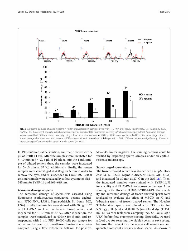
not 10 and 20 mM MBCD (35.4 ± 3.1 % and 14.3 ± 1.0, respectively). Acrosome damage of X-sperms was sig-nificantly higher (p < 0.05) than Y-sperms in all MBCD concentrations (9.5 ± 1.3 %, 15.2 ± 1.1 %, 29.5 ± 1.9 %, 39.7 ± 3.3 %, and 52.4 ± 0.8 %, respectively—Fig. 5).
DiscussionWe analyzed the difference in membrane damage to the bovine X- and Y-sperm. The method developed by Johnson et al. [18] for sexing sperm on the basis of their DNA content was applied. Our investigation was to test the sexing procedure to Johnson et al. method in cryo-preserved sperm. Flow cytometry can sort sperm at >90 % accuracy [5]. In this study, X- and Y-sperm were separated according to Hoechst 33342 intensity by flow cytometry, but the sorting procedure likely causes dam-age to sperm by laser and sorting stress [2]. Therefore, the multi-fluorescence-stained (Hoechst 33342, FITC–PNA or SYBR-14/PI) sperms were separated into popula-tions of X- and Y-based on differential DNA content. The biological characteristics of sperm were analyzed in each population by multi-analytic ability of flow cytometry.
Methyl-beta-cyclodextrin is very efficient in stimulating the efflux of membrane cholesterol from the sperm cells in vitro [16]. The efflux of cholesterol induces changes in membrane architecture and fluidity, and it thereby facilitates membrane-dependent processes like the acrosome reaction [19]. In this study, increased acrosome damage to sperm after MBCD treatment was observed by FITC-PNA staining (Figs. 3, 4). Lectin FITC-PNA binds exclusively to the outer acrosome membrane of damaged sperm [20]. Also, viability of sperms dyed with SYBR-14 and PI was shown to decrease with MBCD concentrations increased on the flow cyto-metric dot plot (Figs. 5 and 6). The viability of sperm was decreased, since MBCD damaged sperm membranes.
These results could suggest that acrosome reactions in X-sperm could occur more quickly than in Y-sperm. The X-sperm may have a lower cholesterol/phospholipid ratio in sperm membrane than Y-sperm. It is generally known that MBCD extracts cholesterol from the sperm plasma membrane [21]. The cholesterol decrease in this membrane also plays a role in the induction of the acro-some reaction. The cholesterol/phospholipid ratio of the sperm membrane is a major determinant in membrane fluidity and stability during cryopreservation [22]. Cho-lesterol reduces the transition temperature of membrane, and maintains them in a fluid state at reduced temper-atures, thereby, reducing the membrane damage that occurs at low temperatures [23]. Therefore, X-sperm may be more sensitive to low temperatures than the Y-sperm. In this study, viability of Y-sperm (72.3 ± 1.9 %) in con-trol group without MBCD was significantly higher than that of X-sperm (51.5 ± 3.0 %). These differences were

Page 3 of 7Lee et al. J of Biol Res-Thessaloniki (2016) 23:5
apparently caused by the cholesterol ratio in membrane and sensitivity to cold shock in X- and Y-sperm.
ConclusionBovine sperms suffered acrosome membrane damage and viability by MBCD. X-sperm had greater cell mem-brane damage than Y-sperm. These results may be due to differences in the cholesterol/phospholipid ratio between X- and Y-sperm membranes. Also this study was a new attempt to analyze the physiological difference between the X- and Y-sperm using flow cytometry.
MethodsPreparation of semen and MBCDThe semen was collected from ten healthy bulls of proven fertility (Hoengseong Livestock Cooperative Farm, Hoengseong, Korea) using an artificial vagina
technique method (3 times per bull). All procedures that involved the use of animals were approved by the Kangwon National University Institutional Animal Care and Use Committee (KIACUC-09-0139). The ejaculated semen was transported to the laboratory at 25 °C within 1 h. The collected semen was diluted 1:1 (v/v) with the Triladyl extender (Minitüb, Tiefenbach, Germany). After maintenance at room temperature for 10 min, the semen was diluted to 5 × 10⁷ spermatozoa in 1 mL of Trila-dyl extender, and incubated with MBCD fresh solution (0, 1, 5, 10, and 20 mM) for 15 min at 25 °C. The incu-bated semen samples were centrifuged (400×g, 5 min) to remove MBCD from sperm before freezing.
Freezing and thawingSemen treated with MBCD was re-suspended with Triladyl extender containing 20 % egg yolk, and the
Fig. 1 Effect of MBCD on viability of frozen‑thawed bovine sperm. After staining with SYBR‑14 and PI, the viability of sperm was analyzed by flow cytometry method (fluorescent intensity of SYBR‑14 and PI, top). Data were appeared as means ± SEM, n = 30 (p < 0.05, bottom)

Page 4 of 7Lee et al. J of Biol Res-Thessaloniki (2016) 23:5
Fig. 2 Effect of MBCD on acrosome damage of frozen‑thawed bovine sperm. Acrosome damage in sperm was analyzed by flow cytometry, and the staining patterns were verified by inspecting sperm samples under an epifluorescence microscope (top). Data were appeared as means ± SEM, n = 30 (p < 0.05, bottom)
Fig. 3 Dot plot and histogram of X‑ and Y‑chromosomes after Hoechst 33342 staining for sex sorting in frozen‑thawed bovine sperm. Frozen‑thawed sperm was automatically sorted into X‑ and Y‑sperm populations by flow cytometry. The sex‑sorted sperm based on differences of DNA contents. A dot plot is displaying Hoechst 33342 fluorescent intensity versus side‑angle scatter (SSC, a) and forward angle scatter (FSC, b). Histogram is displaying Hoechst 33342 fluorescent intensity of sorted X‑ and Y‑sperm (c)

Page 5 of 7Lee et al. J of Biol Res-Thessaloniki (2016) 23:5
semen cooled to 5 °C for 6 h. After cooling the semen was packaged into 0.5 mL straws cooled to −120 °C for 10 min before being plunged into liquid nitrogen for storage using static nitrogen vapor. For this study, the frozen sperms were thawed in a water bath at 37 °C for 45 s. The first prepared sperm samples were fluorescent-stained, and evaluated for viability and acrosome damage using flow cytometry (FACS Aria II, BD Biosciences, San Jose, CA, USA) and the staining patterns were verified by inspecting sperm samples under an epifluorescence microscope (Olympus, BX5, Tokyo, Japan). We used a dual blue/green filter set. The second prepared sperm samples were dyed with Hoechst 33342 and separated by
the differential DNA content, the X and Y chromosomes, and then the viability and acrosome damage of sperm were analyzed.
Sperm viabilityThe LIVE/DEADⓇ Sperm Viability Kit (L-7011, Invit-rogen, Eugene, OR, USA) was used in the fluorescence-based assay method to analyze the viability of sperm. The sperms stained with SYBR-14 (live cell) and pro-pidium iodide (PI, dead cell) and determined by flow cytometry. Briefly, the frozen-thawed semen samples washed in 1 mL phosphate buffered saline solution (PBS, 70,013, Invitrogen) and diluted semen samples in
Fig. 4 Hoechest 33342‑dyed sperms were separated to X and Y populations based on differences of DNA content by flow cytometry. Green spots fluorescent intensity in X chromosome sperm. Blue spots fluorescent intensity in Y chromosome sperm (top). The frozen‑thawed X and Y sperm dyed with SYBR‑14 and PI, and viability was analyzed by fluorescence intensity in flow cytometric dot plot (bottom). a–i Different letters are significantly different in viability of X (a–e) and Y (f–i) sperm (p < 0.05). *Different letters are significantly difference in viability between X and Y sperm (p < 0.05)
Page 6 of 7Lee et al. J of Biol Res-Thessaloniki (2016) 23:5
HEPES-buffered saline solution, and then treated with 5 μL of SYBR-14 dye. After the samples were incubated for 5–10 min at 37 °C, 5 μL of PI added into the 1 mL sam-ple of diluted semen; then, the samples were incubated for 5–10 min at 37 °C, additionally. Finally, the semen samples were centrifuged at 400×g for 5 min in order to remove the dyes, and re-suspended in 1 mL PBS. 10,000 cells per sample were analyzed by a flow cytometer, 515–545 nm for SYBR-14 and 665–685 nm.
Acrosome damage of spermThe acrosome damage of sperm was assessed using fluorescein isothiocyanate-conjugated peanut aggluti-nin (FITC-PNA, L7381, Sigma-Aldrich, St. Louis, MO, USA). Briefly, the samples were stained with 50 ng mL−1 of FITC-PNA in 1 mL of frozen-thawed semen and incubated for 5–10 min at 37 °C. After incubation, the samples were centrifuged at 400×g for 5 min and re-suspended with 1 mL PBS. 10,000 cells per sample for acrosome damage of frozen-thawed bovine sperm were analyzed using a flow cytometer, 488 nm for positive,
515–545 nm for negative. The staining patterns could be verified by inspecting sperm samples under an epifluo-rescence microscope.
Sex‑sorting of spermatozoaThe frozen-thawed semen was stained with 40 μM Hoe-chst 33342 (B2261, Sigma-Aldrich, St. Louis, MO, USA) and incubated for 30 min at 37 °C in the dark [24]. Then, the incubated samples were stained with SYBR-14/PI for viability and FITC-PNA for acrosome damage. After staining with Hoechst 33342, SYBR-14/PI, the viabil-ity and acrosome damage of frozen-thawed sperm were analyzed to evaluate the effect of MBCD on X- and Y-bearing sperm of frozen-thawed semen. The Hoechst 33342-stained sperm was diluted with BTS containing 5 % egg yolk (v/v) and 0.002 % (w/v) food dye (FD&C no. 40, Warner Jenkinson Company Inc., St. Louis, MO, USA) before flow cytometry sorting. Especially, we used a food dye for selecting the viable sperm in sex sorting, because the reagent can penetrate cell membrane and quench fluorescent intensity of dead sperm. As shown in
Fig. 5 Acrosome damage of X and Y sperm in frozen‑thawed semen. Samples dyed with FITC‑PNA after MBCD treatment (0, 1, 5, 10, and 20 mM). Red line FITC fluorescent intensity in X chromosome sperm. Black line FITC fluorescent intensity in Y chromosome sperm (top). Acrosome damage was observed by FITC fluorescence intensity using a flow cytometer (bottom). a–i Different letters are significantly different in percentages of acro‑some damage after treatment with various MBCD concentrations in X (a–e) and Y (f–i) sperm (p < 0.05). *Different letters are significantly difference in percentages of acrosome damage in X and Y sperm (p < 0.05)

Page 7 of 7Lee et al. J of Biol Res-Thessaloniki (2016) 23:5
• We accept pre-submission inquiries
• Our selector tool helps you to find the most relevant journal
• We provide round the clock customer support
• Convenient online submission
• Thorough peer review
• Inclusion in PubMed and all major indexing services
• Maximum visibility for your research
Submit your manuscript atwww.biomedcentral.com/submit
Submit your next manuscript to BioMed Central and we will help you at every step:
Fig. 3, the population of X- and Y-sperm was automati-cally sorted by Hoechst 33258, side-angle scatter (SSC), and forward angle scatter (FSC) using a FACS Aria II [25], and analyzed by flow cytometry for the viability and acrosome damage.
Statistical analysisThe results were analyzed using SAS program (software version 9.1, SAS Institute Inc., Cary, NC, USA). Treat-ment groups were compared for differences using Dun-can’s modified multiple range tests. Data were presented as mean ± SEM and a 5 % probability was considered significant.
Authors’ contributionsSL coordination of the study, interpretation of the data, drafted the manu‑script, wrote the manuscript. Y‑SL interpretation of the data, pre‑experiment, wrote part of the manuscript. S‑HL prepared the extracts, statistical analysis. B‑KY interpretation of the data, writing assistance. C‑KP conceived the study, coordination of the study, interpretation of the data, helped to draft the manuscript. All authors read and approved the final manuscript.
Author details1 College of Animal Life Sciences, Kangwon National University, Chuncheon 24341, Republic of Korea. 2 Institute of Animal Resources, Kang‑won National University, Chuncheon 24341, Republic of Korea.
AcknowledgementsThis research was supported by Agricultural Biotechnology Development Pro‑gram (IPET31060‑05‑1‑CG000), Food and Rural Affairs, Ministry of Agriculture, Republic of Korea.
Competing interestsThe authors declare that they have no competing interests.
Received: 27 October 2015 Accepted: 30 March 2016
References 1. Garner DL, Seidel GE. History of commercializing sexed semen for cattle.
Theriogenology. 2008;69:886–95. 2. Garner DL. Flow cytometric sexing of mammalian sperm. Theriogenol‑
ogy. 2006;65:943–57. 3. Johnson LA, Flook JP, Look MV. Flow cytometry of X and Y chromosome‑
bearing sperm for DNA using an improved preparation method and staining with Hoechst 33342. Gamete Res. 1987;17:203–12.
4. Flaherty S, Matthews CD. Application of modern molecular techniques to evaluate sperm sex selection methods. Mol Hum Reprod. 1996;2:937–42.
5. Seidel GE Jr. Sperm sexing technology‑the transition to commercial appli‑cation. An introduction to the symposium “Update on sexing mammalian sperm”. Theriogenology. 2009;71:1–3.
6. Alkmin DV, Parrilla I, Tarantini T, Parlapan L, Del Olmo D, Vazquez JM, et al. Intra‑ and interboar variability in flow cytometric sperm sex soring. Theriogenology. 2014;82:501–8.
7. Muratori M, Tamburrino L, Marchiani S, Cambi M, Olivito B, Azzari C, et al. Investigation on the origin of sperm DNA fragmentation: role of apopto‑sis, immaturity and oxidative stress. Mol Med. 2015;21:109–22.
8. Chang MC. Fertilizing capacity of spermatozoa deposited into fallopian tubes. Nature. 1951;168:697–8.
9. Davis BK. Timing of fertilization in mammals: sperm cholesterol/phospho‑lipid ratio as a determinant of the capacitation interval. Proc Natl Acad Sci USA. 1981;78:7560–4.
10. Parrish JJ, Susko‑Parrish JL, Graham JK. In vitro capacitation of bovine spermatozoa: role of intracellular calcium. Theriogenology. 1999;51:461–72.
11. Breitbart H. Role and regulation of intracellular calcium in acrosomal exocytosis. J Reprod Immunol. 2002;53:151–9.
12. Etkovitz N, Rubinstein S, Daniel L, Breitbart H. Role of PI3‑kinase and PI4‑kinase in actin polymerization during bovine sperm capacitation. Biol Reprod. 2008;77:263–73.
13. Hung PH, Miller MG, Meyers SA, VandeVoort CA. Sperm mitochondrial integrity is not required for hyperactivated motility, zona binding, or acro‑some reaction in rhesus macaque. Biol Reprod. 2008;79:367–75.
14. de Lamirande E, O’Flaherty C. Sperm activation: role of reactive oxygen species and kinases. Biochim Biophys Acta. 2008;1784:106–15.
15. Yeagle PL. Cholesterol and the cell membrane. Biochim Biophys Acta. 1985;822:267–87.
16. Chiu PC, Chung MK, Tsang HY, Koistinen R, Koistinen H, Seppala M, et al. Glycodelin‑S in human seminal plasma reduces cholesterol efflux and inhibits capacitation of spermatozoa. J Biol Chem. 2005;280:25580–9.
17. Pitha J, Irie T, Sklar PB, Nye JS. Drug solubilizers to aid pharmacologists: amorphous cyclodextrin derivatives. Life Sci. 1988;43:493–502.
18. Johnson LA, Flook JP, Hawk HW. Sex preselection in rabbits: live births from X and Y sperm separated by DNA and cell sorting. Biol Reprod. 1989;41:199–203.
19. Zarintash RJ, Cross NL. Unesterified cholesterol content of human sperm regulates the response of the acrosome to the agonist, progesterone. Biol Reprod. 1996;55:19–24.
20. Flesch FM, Voorhout WF, Colenbrander B, van Golde LM, Gadella BM. Use of lectins to characterize plasma membrane preparations from boar spermatozoa: a novel technique for monitoring membrane purity and quantity. Biol Reprod. 1998;59:1530–9.
21. Besenicar MP, Bavdek A, Kladnik A, Macek P, Anderluh G. Kinetics of cho‑lesterol extraction from lipid membranes by methyl‑β‑cyclodextrin—a surface plasmon resonance approach. Biochim Biophys Acta. 2008;1778:175–84.
22. Rajoriya JS, Prasad JK, Ramteke SS, Perumal P, Ghosh SK, Singh M, et al. Enriching membrane cholesterol improves stability and cryosurvival of buffalo spermatozoa. Anim Reprod Sci. 2016;164:72–81.
23. Amann RP, Pickett BW. Principles of cryopreservation and a review of cryopreservation of stallion spermatozoa. J Equine Vet Sci. 1987;7:145–73.
24. Gao QH, Wei HJ, Luo J, Han CM, Schoenian S, Du HZ, et al. Flow cytomet‑ric sexing of X‑ and Y‑chromosome‑bearing sperm in Sika deer (Cervus nippon). Small Ruminant Res. 2009;81:100–4.
25. Yoo H‑J, Lee K‑J, Lee Y‑S, Yoon P‑S, Park J‑J, Kim H‑C, et al. Analysis of sexed sperm by flow cytometry in Hanwoo (Korean native cattle). Reprod Dev Biol. 2012;36:1–6.