Polysaccharide Based Formulations for Mucosal Drug Delivery
66
1 Polysaccharide Based Formulations for Mucosal Drug Delivery: A Review Joshua Boateng, Obinna Okeke, Sajjad Khan Department of Pharmaceutical, Chemical and Environmental Sciences, Faculty of Engineering and Science, University of Greenwich at Medway, Central Avenue, Chatham Maritime, ME4 4TB, Kent, UK. # Correspondence: Dr Joshua Boateng Email: [email protected]; [email protected] Telephone: +44 (0) 208 3318980 Fax: +44 (0) 208 3319805 brought to you by CORE View metadata, citation and similar papers at core.ac.uk provided by Greenwich Academic Literature Archive
-
Upload
khangminh22 -
Category
Documents
-
view
1 -
download
0
Transcript of Polysaccharide Based Formulations for Mucosal Drug Delivery

1
Polysaccharide Based Formulations for Mucosal Drug Delivery: A Review
Joshua Boateng, Obinna Okeke, Sajjad Khan
Department of Pharmaceutical, Chemical and Environmental Sciences, Faculty of
Engineering and Science, University of Greenwich at Medway, Central Avenue, Chatham
Maritime, ME4 4TB, Kent, UK.
# Correspondence: Dr Joshua Boateng
Email: [email protected]; [email protected]
Telephone: +44 (0) 208 3318980
Fax: +44 (0) 208 3319805
brought to you by COREView metadata, citation and similar papers at core.ac.uk
provided by Greenwich Academic Literature Archive

2
Abstract
There has been increased interest in novel drug delivery systems to be administered via
mucosal routes as an alternative to the currently used traditional routes such as parenteral
(injections) and oral routes of administration. This is due to the several advantages they offer
including avoiding first pass metabolism in the liver for oral administration and local activity
which avoids the need for high systemic doses. To achieve the foregoing objectives,
bioadhesive vehicles are required that ensure prolonged residence time to achieve systemic
bioavailability via substantial drug absorption or significant drug concentration for local action.
The drug delivery system is also required to be non-deleterious to the site of application and
be well tolerated by vulnerable groups such as paediatric and geriatric patients. These essential
characteristics are mainly satisfied by naturally occurring polymers, including polysaccharide
based polymers which have the advantage of biocompatibility, biodegradability and therefore
safety. This review discusses various bioadhesive polymers of polysaccharide origin
formulated into a variety of dosage forms for drug delivery via the body’s mucosal (moist)
surfaces including ocular, oral (buccal and sublingual), nasal, gastrointestinal and vaginal
mucosa, as well as moist wound sites. The anatomy and / or physiology of each site, coupled
with the unique challenges each poses, the strategies employed for ensuring therapeutic
efficacy, as well as the current state of the art will also be covered.
Key words: Buccal, gastrointestinal, mucosal delivery, nasal, polysaccharides, ocular, vaginal,
wounds.

3
1. Introduction
1.1 Overview
Drug delivery is most commonly achieved via oral administration of dosage forms such as
tablets, capsules and liquids, representing about 70% of all pharmaceutical drug formulations.
However, this is fraught with several problems including first pass metabolism in the liver,
degradation in the gastrointestinal tract for acid labile drugs such as proteins and peptides and
risk of poor uptake for children, the severely infirmed (e.g. comatose patients) and geriatric
patients. The rejection rate of such oral dosage forms is higher than for other routes, due to
factors such as unpleasant taste [1], difficulty in swallowing and the risk of choking. Though
the alternative traditional parenteral route using injections is effective and avoids the above
limitations, it presents several challenges as well, including pain, irritation at site of injection
and the need for highly trained personnel for safe and effective administration. All these
result in poor patient compliance with consequent poor clinical outcomes, which can be
severe in certain diseases. There has been increased efforts in recent decades to develop novel
alternative systems for drug delivery based on factors such as therapeutic concerns,
biopharmaceutics and physico-chemical properties of the drug, such as poor solubility and
instability via tradition routes.
These factors are important and are mainly aimed to improve safety, efficacy and
patient compliance and ultimately help to increase product life cycle [1].
A major goal of novel drug delivery systems is appropriate targeting to direct the drug in
question to its intended site of action, minimize drug degradation and loss, increase
bioavailability, increase the fraction of drug accumulated at the site of action while preventing
or limiting harmful and unwanted side effects.
One of the major areas of current interest, which addresses a significant number of the
challenges highlighted above, is the development of bioadhesive (mucoadhesive) delivery
systems for drug administration via one or more of the body’s mucosal surfaces (routes). The
characteristic features of transmucosal routes, such as large surface area and network of blood
vessels, make such routes interesting sites for both systemic and local delivery of drugs. In
addition, they provide the ability to bypass the hepatic first pass metabolism and degradation
of drug in the stomach by delivering the drugs directly to the bloodstream thereby increasing
bioavailability [2]. The transmucosal surfaces that have been under investigation for potential
drug administration for systemic therapeutic action include the oral (buccal and sublingual) [3],

4
vaginal [4], nasal [5], ocular [6] and wound surfaces [7]. Apart from overcoming the limitations
of oral (gastrointestinal) and parenteral administration, that is avoiding first pass metabolism
and pain respectively, they are particularly advantageous in cases where only small doses are
required at the local mucosal site, thus avoiding the need for unnecessarily high systemic doses,
for example in local infections, where antibiotics are required [8, 9].
In order to achieve effective mucosal administration, the delivery matrix (system)
needs to satisfy certain criteria, particularly being biocompatible, bioadhesive
(mucoadhesive), biodegradable and easily processed into various dosage forms. Most of the
above mentioned novel drug delivery systems can be prepared using such synthetic or
biomaterial based polymers. However, the naturally occurring biomaterials have been used
extensively due to their well-known biocompatibility and biodegradable nature, in addition to
most of them being bioadhesive. A common group of such naturally occurring biomaterials is
polysaccharide based polymers, ranging from common materials such as starch, to more
complex examples such as chitosan and sodium alginate, obtained from various natural
sources or in semi-synthetic form [10].
In this article, we review the current state of the art of mucosal delivery systems
designed using polysaccharide based polymers with bio (muco) adhesive characteristics for
an application via the various mucosal routes [buccal/sublingual, gastrointestinal (emphasis
on colonic delivery), nasal, ocular and, vaginal as well as wounds surfaces due to the moist
environment in a wound environment]). The molecular basis of bioadhesion and its
importance are briefly discussed. Different formulation approaches, the unique challenges of
each route (including barriers by their structural architecture and physiology), examples of
systems available both in the literature and in some cases commercially, will be reviewed.
Finally, the prospects of having such systems in routine clinical patient use, in the medium to
long term future, are briefly discussed.
1.2 Bioadhesion (mucoadhesion)
The terms bioadhesion and mucoadhesion are sometimes used interchangeably, though
they actually mean slightly different things. Bioadhesion defines adhesion between two
materials where at least one material is of biological origin and is generally used when
interaction occurs between adhesive polymers and an epithelial surface directly, such as a
wound surface. Mucoadhesion on the other hand, involves adhesion with the mucus layer
covering a biological tissue or membrane. The adhesion force/bond is dependent on
parameters such as hydrophilicity (progress bioadhesion), stage of hydration and rate of

5
polymer erosion after being in contact with the hydrating surface. Apart from the function of
increasing the retention time of the drug on the mucosal surface to enhance the
bioavailability, some polymers can be used as enzyme inhibitors and penetration enhancers. It
has been reported that the presence of polymers absorb water from the epithelial cells to
widen the tight junction [11] and in the process allow easy penetration of drug molecules
across the membrane into the systemic circulation.
In general, mucoadhesion and bioadhesion are described as bonding between polymers
and mucosal tissues or any biological surface as shown in figure 1.
<Figure 1 here>
Mucoadhesion occurs because of various adhesive bonds at the interface between the
mucosal membrane and the mucoadhesive agent [12, 13]. These bonds include (a) ionic
bonds: where two oppositely charged ions attract each other via electrostatic interactions and
form a strong bond; (b) covalent bonds: which are very strong bonds in which electrons are
shared in space, between the bonded atoms in order to fill the orbitals; (c) hydrogen bonds: a
hydrogen atom, when covalently bonded to an electronegative atom such as an oxygen,
fluorine or nitrogen, carries a slightly positive charge and, hence, is attracted to
electronegative atoms. The mucosal membrane and mucoadhesive material share the
hydrogen atom, though this bond is usually weaker than ionic or covalent bonds; (d) van der
Waals forces: these are some of the weakest forms of interaction that arise from dipole-dipole
attractions in polar molecules, and dispersion forces with non-polar substances: (e)
hydrophobic forces: give rise to a hydrophobic effect and occur when non-polar groups are
present in an aqueous solution [12, 13].
1.2.1 Theories of bioadhesion
The mechanism of polymer attachment to a mucosal surface is not yet fully
understood. However, certain theories of bioadhesion have been proposed suggesting that it
might occur via physical entanglement and/or chemical interactions, such as electrostatic,
hydrophobic, hydrogen bonding, and van der Waal’s interactions [14]. A variety of factors
affect the mucoadhesive properties of polymers, such as molecular weight, flexibility,
hydrogen bonding capacity, cross-linking density, charge, concentration, hydration of a
polymer and the environmental factors [15]. The processes involved in the formation of

6
bioadhesive bonds have been described in three steps – (a) wetting and swelling of polymer
to permit intimate contact with biological tissue; (b) interpenetration (entanglement) of
bioadhesive polymer chains with mucin chains and (c) formation of weak chemical bonds
between the entangled chains [16]. The various theories proposed to explain the mechanisms
of bio (muco) adhesion include electronic, adsorption, wetting, fracture and diffusion and the
reader is referred to more extensive texts and reviews on the physico-chemical and
biomechanical principles that underpin these proposed theories [13, 17, 18].
1.3 Mucoadhesive polymers
Mucoadhesive polymers include a large and diverse group of molecules covering
biodegradable grafted co-polymers and thiolated polymers and are used in bioadhesive
formulations either alone or in combination with others. These formulations are often water-
soluble and when in dry form, they attract water from the biological surface and this water
transfer results in a strong interaction [16]. The ideal mucoadhesive polymer should possess
certain characteristics regarded as essential for effective function as a bioadhesive drug
delivery system [19]. These include being non-toxic and non-irritant, possessing good
spreading, swelling, solubility and biodegradable properties. In addition, they should possess
adhesive properties both in the dry and liquid/gel state, be biocompatible and possess good
viscoelastic, peel, tensile and shear strength properties as well as demonstrate local enzyme
inhibition and penetration enhancement properties.
1.3.1 Classification
Bioadhesive polymers are classified as below depending upon various characteristics
such as; (i) source: (natural and synthetic polymers), (ii) aqueous solubility (water soluble
water and insoluble, (iii) first and second generation (cationic, anionic and non-ionic
polymers), and (iv) potential bioadhesive forces (electrostatic interactions, hydrogen bonds and
covalent bonds) [20]. Currently, bioadhesive (mucoadhesive) polymers are classified as ‘first and
second generation’.
1.3.1.1 First generation polymers
The older generation of mucoadhesive polymers is referred to as ‘off-the-shelf’
polymers [21]. They lack specificity and targeting capability and adhere to mucus non-
specifically, and suffer short retention times due to the high turnover rate of mucus. Examples
7
include anionic polymers such as sodium carboxymethylcellulose, alginate and carrageenan
[23, 24, 25, 26], cationic polymers such as chitosan and its derivatives [27, 28].
1.3.1.2 Second generation polymers
The new generation of mucoadhesive polymers can adhere directly to the cell surface,
rather than to mucus and they interact with the cell by means of specific receptor or covalent
bonding instead of non-specific mechanism [22]. These include lectin-mediated bioadhesive
polymers which are naturally occurring proteins that play a fundamental role in biological
recognition phenomena involving cells and highly heterogeneous proteins [29]. This potential
has been observed for materials such as polyacrylic acids in the dry state, wheat germ agglutinin
and concanavalin A [30]. Such systems could offer duality of function in that lectin based
platforms could not only allow targeted specific attachment, but also additionally offer a
method of controlled drug delivery of macromolecular pharmaceuticals via active cell-
mediated drug uptake [31]. The adhesive properties of bacterial cells, as a more complicated
adhesion system, have recently been investigated. The ability of bacteria to adhere to a specific
target is derived from particular cell-surface components or appendages, known as fimbriae
that facilitate adhesion to other cells or inanimate surfaces [32].
1.3.1.3 Enzyme inhibiting polymers
It has been shown that some mucoadhesive polymers can act as enzyme inhibitors and
important in delivering therapeutic compounds that are specifically prone to extensive
enzymatic degradation, such as proteins and peptide drugs [33]. Investigations have
demonstrated that polymers such as poly (acrylic acid), operate through a competitive
mechanism with proteolytic enzymes. Circular dichroism studies suggest that Ca2+ depletion,
mediated by the presence of some mucoadhesive polymers, causes the secondary structure of
trypsin to change, and initiates a further auto degradation of the enzyme [34].
1.3.1.4 Thiolated polymers
Thiolated polymers are capable of forming disulphide bonds with cysteine-rich subdomains
of mucus glycoproteins covering mucosal membranes. These are the special class of
multifunctional polymers also called thiomers [35]. Thiomers are capable of forming intra-
and inter chain disulphide bonds within the polymeric network leading to strongly improved
cohesive properties and stability of drug delivery systems such as matrix tablets. These
hydrophilic macromolecules exhibit free thiol groups on the polymeric backbone. These

8
functional groups have enabled various features of well-established polymeric excipients such
as poly (acrylic acid) and chitosan to be significantly improved [36]. Due to the formation of
strong covalent bonds with mucus glycoproteins, thiomers show the strongest mucoadhesive
properties of all polymeric excipients via thiol-disulphide exchange reaction and an oxidation
process [37].
1.3.2 Mucoadhesion measurement techniques
There are different approaches used to evaluate the mucoadhesive performance of
polymers and polymeric dosage forms. These include texture analyser, [38-41], rheometric
measurements [42] and attenuated total reflection-Fourier transform infrared spectroscopy
[43, 44] methods. The texture analyser (TA) technique measures the maximum force required
to separate the polymer or dosage form from the surface of a mucosal substrate after a
specified contact time and applied force. This method evaluates stickiness, work of adhesion
(WOA) and cohesiveness of dosage forms. Stickiness is the maximum force required to
separate the probe attached to films and wafers from the given mucosal substrate such as
mucin equilibrated gelatine substrate (i.e. maximum detachment force) whereas total amount
of work or energy involved in the probe withdrawal from the substrate is calculated from the
area under the forces versus distance curve and cohesiveness is the intermolecular attraction
between the substrate and formulations and determined by the travel distance in mm on the
force versus distance profile [38] (see figure 2). The rheometric method involves studying the
extent of interpenetration of mucin (or moisture) with polymeric gels by measuring
differences in the rheological parameters of polymeric gel and their mixture with mucin [42].
The attenuated total reflection-Fourier transform infrared spectroscopy (ATR-FTIR)
approach involves the study of chain interpenetration or diffusion occurring between
polymers or dosage forms (e.g. films and wafers) and mucosal fluid such as mucin solution
[43] [44] or simulated wound fluid [40].
<Figure 2 here>
1.4 Polysaccharides
Polysaccharides are carbohydrates made up repeating monosaccharide or disaccharide units
joined together by glycosidic bonds such as starch and glycogen. Most polysaccharides are

9
naturally occurring which make them attractive choices in traditional applications as food and
pharmaceutical additives or in the form of excipients as binders, sweeteners, bulking agents,
film coatings and suspending agents. Further, polysaccharides are abundantly present in
nature, have wide availability, are inexpensive and are available in a variety of structures with
varied properties [45]. They can easily be modified chemically and biochemically and are
highly stable, safe, nontoxic, hydrophilic and gel forming and in addition biodegradable.
These include naturally occurring polysaccharides obtained from plant (e.g. guar gum,
inulin), animal (e.g. chitosan and chondroitin sulphate), algae (e.g. alginates, xanthan) or
microorganism (e.g. dextran) origin as well as starch and certain cellulosic polymers mainly
from plant sources. The most common polysaccharides used as mucoadhesive polymer can
be further divided into positively charged and negatively charged polysaccharide based on
their charge property. The most commonly used positively charged polysaccharide is chitosan
while the most commonly used negatively charged polysaccharides are alginate, pectin and
hyaluronic acid [46].
However, polysaccharides as well as other naturally occurring polymers also possess
further unique functional characteristics such as swelling and hydration which control drug
release but also impact on the mechanism of mucoadhesion. These two functional
characteristics (swelling and mucoadhesion), coupled with their biocompatible properties
have made them become commonly used polymers for various dosage forms used as drug
delivery vehicles via the various mucosal surfaces outlined above. The rest of this review will
cover each mucosal surface individually and the various polysaccharide and their
corresponding dosage forms employed either alone or in combination to deliver drugs across
these surfaces, either for systemic absorption or for local action within or around the mucosal
environment.
2. Ocular drug delivery
The human eye is a very sensitive organ to exogenous materials such as debris,
microorganisms and drugs [47] and therefore, formulations designed for ocular drug delivery
should be simple, non-invasive (to prevent irritation, inflammation or infection, to maintain
the visual clarity of the eye), as well as be able to penetrate the physiological eye barriers and
reach the site of action. The eyeball has an approximate spherical shape and is situated in the
orbit comprising three concentric: layers outer fibrous (sclera and cornea), the middle
vascular (choroid, ciliary body and iris) and the inner nervous (retina)

10
layers [48]. The eye could also be divided into chambers i.e. the anterior chamber, the
posterior chamber and the vitreous cavity [49].
The lachrymal film, is a dynamic fluid that is constantly renewed, therefore limiting
the retention time of a drug on the eye surface [50] and protects the eye by acting as a defense
against pathogens and a barrier against any drug penetration [51]. Another critical barrier in
ocular therapeutics is the conjunctiva which is an epithelium about 100 times more permeable
than the cornea for large hydrophilic compounds. Its role is to protect the eye and functions
as a passive physical barrier [51].
2.1 Ocular conditions
Eye conditions can be classified as peri-ocular and intraocular. Peri-ocular conditions
occur around the eye and can cause irritation to different parts of the eye, e.g. blepharitis,
trachoma, conjunctivitis, and dry eye [52]. Intraocular conditions represent the infection of the
inner parts of the eye and can affect the retina, the iris, the aqueous and the vitreous humour,
with the most common being glaucoma. Glaucoma can be treated by application of topical
drugs that constrict the pupil and tense the edge of the iris, which, in turn, make the surface
more permeable to aqueous humour [49].
2.2 Polysaccharide-based ocular delivery systems
2.2.1 Topical ophthalmic preparations
The design of ocular drug delivery formulations is very challenging and requires an
understanding of what can be tolerated by the eyes, of the physiology of the eyes and also of
what the factors affecting ocular drug administration and absorption are (physiological and
formulation factors (figure 3). It is very difficult in ocular therapeutics to achieve and to
maintain an effective drug concentration at the site of action for a prolonged period of time to
achieve the desired therapeutic response.
<Figure 3 here>
The available topical ophthalmic preparations include solutions, suspensions, ointments, gels
and films to treat conditions such as inflammation, infection, allergy, glaucoma, dry eye as
well for instilling local anaesthetics and diagnostic agents [53]. Liquid drops cannot be
considered optimal in the treatment of ocular diseases because of their low bioavailability
with only 5% of the instilled dose able to penetrate the cell membranes into the eye.
Therefore frequent instillation of the dosage form in question is required which may lead to
11
systemic side effects and patient non-compliance [53]. As a result, current novel systems for
drug delivery to the eye involve the use of bioadhesive polymers, including polysaccharides,
that are not only safe but ensure prolonged residence time and controlled release of the drug to
allow improved bioavailability and improved patient compliance due to reduced need for
regular application. The commonly used polysaccharide polymers reported in the literature
have been extensively evaluated by Ludwig [54] in his review paper. These include: chitosan
[55-63], hyaluronic acid [64-73], polygalacturonic acid, xyloglucan, xanthan gum, pullulan,
guar gum, scleroglucan [74-82] carrageenan [79, 83-84], gellan gum [85] and pectin [86, 87].
The factors that affect the formulation development of ophthalmic preparations include
osmolality, pH, surface tension and viscosity and most of these are extensively discussed in
other texts [53]. However, the viscosity is addressed here a bit more extensively, as it is directly
affected by the type of polysaccharide polymer used and also affects other functional properties
such as swelling and mucoadhesion. In many ophthalmic solutions viscosity-enhancing
polymers are added to prolong drug retention time in the pre-corneal tear film and therefore to
enhance drug absorption. The viscosity-enhancing polymers reduce drainage rate and increase
the thickness of the pre-corneal tear film due to their ability to drag water and stabilize the
aqueous layer.
Water-soluble hydrophilic polymers hydroxypropylmethylcellulose helps to increase
viscosity from 400cps up to about 15,000cps as well as increasing lacrimal film stability which
helps to increase residence time of drug in the cul-de-sac and thus helps increasing the
absorption and eventual bioavailability [88, 89].
2.2.2 Ocular inserts (films)
Films are made of polymers that can be natural, synthetic or semi-synthetic and the
drugs contained can be in the form of either dispersion or solution [90]. Acyclovir,
phenylpherine, diclofenac sodium and natamicin are examples of drugs that can be contained
within the ocular inserts. The ocular inserts can be either solid or semi-solid and
biodegradable or non-biodegradable. The biodegradable films don’t need to be removed,
whereas the non-biodegradable films need to be removed after a given period of time [91].
Solvent cast composite ocular inserts combining polyvinylalcohol and sodium
carboxymethylcellulose have been reported for ocular delivery of ciprofloxacillin for topical
infections using a rabbit model [92]. In this study, esterification of the polymers was

12
confirmed by Fourier transform infra-red spectroscopy while surface smoothness of 7.3nm
was obtained. Comparison of corneal penetration of the ocular insert with an eye drop
solution using a model dye (fluorescein) showed higher penetration for the ocular insert
which were also proved to be non-toxic from the in vivo study using albino rabbits.
Flurbiprofen loaded lipid based nanocarriers coated with chitosan oligosaccharides have
been investigated for potential ocular drug delivery by Qiuhua and co-workers [93]. In their
study, gamma scintigraphy was used to investigate the residence time of the coated
nanoparticles which is proportional to bioadhesivity and showed that the clearance of the
coated carriers was significantly reduced compared to the corresponding uncoated control
carrier particles. Further, the coated formulations showed a 2.4 fold increase in corneal
penetration. Both these results in the functional performance of coated carriers shows their
potential as a possible ophthalmic drug delivery system and particularly, the importance of
polysaccharides on effective bioadhesion and permeation after application.
In a related study, Li and co-workers coated liposomes with low molecular weight
chitosan and investigated them for potential ocular drug delivery application using diclofenac
as a model drug [94]. Their results showed that coating with chitosan changed the surface
charge, increased particle size but with no change in drug encapsulation efficiency.
Furthermore, the chitosan coated liposomes showed prolonged drug release, improved
physical and chemical stability, prolonged retention (bioadhesion) and enhanced drug
penetration across the cornea compared to the non-coated formulation and pure drug solution
(drops). Continuous application of the coated liposomes over a seven day period, showed no
irritation or toxicity. Xu and co-workers developed injectable in situ hydrogels from
crosslinking glycol chitosan and oxidised alginate for ocular delivery of the drug avastin
which is used to treat age related macular degeneration and proliferative diabetic retinopathy
[95]. They controlled the hydrogel degradation rate by varying the concentration of oxidised
alginate whilst avastin encapsulated within the hydrogel showed a biphasic release with an
initial burst release phase in four hours, followed by sustained release over three days.
Ilva and co-workers developed and compared various ion-activated in situ forming gels that
form cross links with commonly available cations in tear fluid, with a resultant increase in
corneal contact time, in vitro. In their study, gellan gum, xanthan gum, carrageenan and
alginate, together with HPMC and chitosan, were characterised for gelling behaviour,
rheological and textural characteristics, gel microstructure, contact angle and release of a
model hydrophilic drug [96]. Their results showed that the systems exhibited physically
entangled polymer networks that were able to disentangle upon shear stress, which

13
prolonged the release of the model hydrophilic drug, compared to a solution based dosage form.
In addition, HPMC and chitosan gels showed no structural changes upon addition of cations,
whilst gellan gum and carrageenan gels showed significant increase in viscosity,
pseudoplasticity and hardness in the presence Ca2+ and K+ respectively. Solvent cast
xyloglucan films have been employed for the delivery of ciprofloxacin with a percentage
loading of 95.45% of expected dose and total cumulative release of 98.5% of the initial drug
content following sustained type in vitro release, which was determined to follow anomalous
transport release mechanism [97]. Miyazaki and co-authors reported on thermos-reversible in
situ forming gels from enzyme hydrolysed xyloglucan for sustained ocular delivery of
pilocarpine hydrochloride and showed a square root of time release kinetics over six hours [98].
Some of the commercially available mucoadhesive ocular products include Ocusert®
(pilocarpine), BODI® (antibiotics), NyoGel ® (timolol) and Pilogel® (pilocarpine
hydrochloride).
3. Intraoral mucosa delivery
There are various reasons for the formulation of drugs into appropriate oral mucosa
dosage forms; one of which relates to accurate measurement of the dose. For example, in
children, the dose required varies with age and weight and also differs significantly from the
adult dose.
2.3.1 Sublingual and buccal mucosa drug delivery
The sublingual route of drug administration is widely studied and known to be relatively
permeable compared to other oral mucosal surfaces. The sublingual route can provide rapid
absorption and easy accessibility to the drug for systemic delivery, especially for quick-
dissolving dosage forms [99, 100]. Currently available sublingual products have been
developed for several purpose such as mental illness, in the cases where patient compliance is
important for treating chronic conditions such as depression and schizophrenia [101].
The buccal mucosa refers to the membrane lining the inside of the cheek and has
excellent accessibility, an expanse of smooth muscle and relatively immobile mucosa (figure
4), hence suitable for the administration of retentive dosage forms. It has relatively low enzyme
activity compared to the gastro-intestinal tract, painless administration and easy dosage form
withdrawal [102].
<Figure 4 here>

14
Buccal formulations have been developed to allow prolonged localised therapy and
enhanced systemic delivery. The buccal mucosa, however, while avoiding first-pass effects,
remains a significant barrier to drug absorption, especially for biopharmaceutical products
[103, 104]. An important application of buccal drug delivery is in the areas of paediatric (and
geriatric) drug administration due to the risk or fear of chocking [105, 106]. The size of the
delivery system varies with the type of formulation, for example, a buccal tablet may be
approximately 5–8mm in diameter, whereas a flexible buccal patch may be as large as 10–
15cm2 in area. The thickness of the delivery device is usually restricted to below 1 mm [14,
107]. Different types of buccal formulations such as; tablets, patches and films, semisolids and
powders are used depending upon the desirable pharmacological action [108].
Buccal drug delivery systems present various advantages and limitations including
bypassing of the gastrointestinal tract and hepatic portal system, thus increasing the
bioavailability of orally administered drugs. The buccal mucosa provides improved patient
compliance by avoiding pain associated with injections and extent of perfusion is aided by the
rich supply of blood (2.0 ml/sec /cm2). Further, a relatively rapid onset of action can be
achieved compared to the gastrointestinal route and the formulation can be removed if therapy
is required to be discontinued. In addition, buccal formulations can be used in cases of
unconsciousness and less cooperative patients as these have difficulties in swallowing oral
dosage form. Nausea and vomiting are avoided because medications do not interfere with the
oesophagus and its functions whilst drugs which show poor bioavailability via the oral
gastrointestinal route can be administered conveniently. For example, drugs such as
pantoprazole sodium, which are unstable in the acidic environment of the stomach or are
destroyed by the enzymatic or alkaline environment of the intestine [109].
As far as limitations are concerned, drugs which irritate the oral mucosa, have a bitter taste,
cause allergic reactions or discoloration of the teeth cannot be formulated for buccal delivery.
If the formulation contains antimicrobial agents, it affects the natural microbes in the buccal
cavity and patients can also not eat/drink/speak normally whilst the swallowing of saliva can
also potentially lead to the loss of dissolved or suspended drug. In addition, only those drugs
which are absorbed by passive diffusion can be administered by this route and drugs which
are unstable at buccal pH cannot be administered by this route. Finally, the buccal mucosa
membrane has low permeability, when compared specifically to the sublingual membrane
[110].

15
Because the buccal route is usually used in relatively more extended drug delivery
compared to the sublingual route, bioadhesive formulations are more favoured. Bioadhesive
polymers that have been used in buccal drug delivery to maintain formulations are hydrophilic
macro molecules containing numerous hydrogen bonds [111].
Bioadhesive polymers require some important structural characteristics which include
strong hydrogen bonding groups, strong anionic or cationic charges, high molecular weight,
chain flexibility and surface energy properties [112, 113]. Some polysaccharide polymers
achieve bioadhesion through a covalent attachment between a cysteine residues present in
mucin and the polymer of choice [114, 115]. Regardless of the dosage form, the drug must be
released from the delivery system and subsequently taken up by the oral mucosa. The drug
release from the dosage is often retarded because of poor solubility and the introduction of
cyclodextrin has been widely used to solubilise and increase the absorption of poorly water-
soluble drugs delivered via the buccal mucosa [116].
3.2 Polysaccharide-based buccal delivery systems
The challenges encountered in the formulation development of mucoadhesive drug
delivery systems have been discussed by Mizrahi and Domb [117] and Salamat-Miller [22].
The most frequently used mucoadhesive polysaccharide based polymers used in buccal mucosa
drug delivery include sodium alginate, chitosan and its derivatives, pectin and carrageenan
[118]. Various polysaccharide based formulations have been employed for delivery across the
buccal and sublingual membranes for both small and macromolecules. These include gels
films, tablets, wafers (xerogels), nano particles usually incorporated into gels, films. These are
summarised in table below showing the type of polysaccharide, dosage form and the drug used
for the study whilst selected references are reviewed in more detail.
Zeng and colleagues [119] developed buccal hydrogels that were sensitive to
temperature for the delivery of salbutamol by combining poloxamer, xanthan gum and
sodium chloride and characterising various functional characteristics such as gelation
temperature, micellization temperature, gelation time, gel strength, in vitro release (using
membrane-less and membrane based method). The above properties varied depending on the
three main components mentioned above and showed potential clinical application for buccal
delivery of salbutamol to achieve rapid systemic activity. Martin and co-workers synthesized

16
palmitoyl glycol chitosan hydrogels with different hydrophobicities by physical crosslinking
and loaded them with a model hydrophobic drug (denbufylline) for buccal drug delivery
[120]. Sodium glycodeoxycholate which is a soluble detergent was used as permeation
enhancer. The resulting crosslinked hydrogels were characterised using H nuclear magnetic
resonance spectroscopy, hydration, erosion, mucoadhesion, scanning electron microscopy
and buccal absorption across rabbit buccal mucosa membrane (using carbopol, denbufylline
and sodium glycodeoxycholate containing tablets as controls). Their results showed that
denbufylline reduced the porosity, erosion and hydration of the gels while the permeation
enhancer increased the hydration and erosion rates. Though the gels were all mucoadhesive
this was comparable to the control tablets. The buccal absorption studies showed that the
drug was detectable in the systemic circulation 30 minutes after administration for the most
hydrophobic hydrogel and this was sustained over a 5 hour period. However, drug released
from the control tablets was only detected after 60 minutes and the release was not sustained
as was the case for the crosslinked hydrogels.
Nystatin loaded microspheres from alginate and chitosan coated were prepared by
internal gelation method as antifungal delivery systems for the treatment of oral candidiasis,
and characterised by size and size distribution, shape, encapsulation efficiency, Zeta
potential, swelling, mucoadhesion, in vitro drug release and in vivo studies [121]. The
microspheres were spherical in shape and ranged in size from 85 – 135 μm with negative
potential (showing stability) and optimised encapsulation efficiency as well as swelling and
mucoadhesive behaviour. The alginate and chitosan formulations both showed a
concentration dependent release of the nystatin loaded within the microspheres and showed
strong fungicidal activity against Candida albicans but with no tissue damage. Furthermore,
the in vivo studies showed that the drug was not detectable in the systemic circulation,
suggesting it did not cross the oral mucosa membrane but acted locally, implying safety and
therefore reduced unwanted side effects.
Kassem and co-workers developed buccal adhesive tablets for sustained delivery of
buspirone hydrochloride with the aim of improving systemic bioavailability [122]. The tablet
formulation development involved the use of a 5 x 3 factorial design, setting polymer type
(carbopol, hydroxypropylmethylcellulose, sodium alginate, sodium carboxymethylcellulose
and guar gum) at five levels whilst polymer to drug ratios were set at three different levels
(combinations) and various dependent variables (mucoadhesion force, ex vivo mucoadhesion
time, percent drug release after 8 hours and time to release 50% of drug) employed. The
tablets were characterised for content uniformity, weight variation, thickness, diameter,

17
hardness, friability, swelling index, surface pH, mucoadhesion strength / time and in vitro
drug release. It was observed that the cup and core formulations adhered to the buccal
mucosa for 8 hours, showed the highest percent drug release over the same time period with a
zero order release profile. Further, pharmacokinetic experiments of the cup and core formula
in human volunteers showed a 5.6 fold increase in drug bioavailability in comparison to oral
commercial tablets with excellent in vitro and in vivo correlations.
Bilayered mucoadhesive tablets loaded with curcumin for unidirectional buccal
delivery have been prepared using a natural buccoadhesive polymer from cashew nut tree
gum with ethyl cellulose as an impermeable backing layer [123]. The tablets with
mucoadhesive strength of 13.99 g were stable and released drug over 60 days at both high
and low humidities and temperatures. Drug release was found to be via non-Fickian or
anomalous diffusion kinetics and suggested as a potential buccal adhesive tablet for
enhancing bioavailability of curcumin by avoiding first pass metabolism. Ameye and co,
produced spray dried starch/carbopol mixtures in different proportions, for evaluation as
potential bioadhesive tablets for buccal administration of miconazole [124]. They observed
that all the spray-dried composite formulations showed a comparable or better bioadhesive
capacity compared to a reference formulation with the spray drying procedure generally
improving the bioadhesive performance. Further, the effect of modifying additive (carbopol)
concentration on the in vivo adhesion time of placebo tablets and in vitro miconazole nitrate
release were tested and showed that formulations containing the ratio starch / carbopol of
70/30 showed the longest in vivo adhesion time compared to very low and very high carbopol
concentrations. Lower and higher carbopol concentrations had a shorter in vivo adhesion
time. It was observed that the composite formulations containing between 15 and 30% of
carbopol sustained the in vitro miconazole nitrate release over 20 hours whilst very high and
and very low carbopol concentrations showed a faster in vitro miconazole release.
Furthermore, in vivo studies in dogs using a different drug (testosterone) showed that the
optimised spray dried mixture could be loaded with 60% of drug but still maintained the in
vivo bioadhesion and pharmacokinetic profiles.
In an innovative application study, gum from the plant Hakea gibbosa (Hakea) was
used to formulate and characterise sustained-release and mucoadhesive buccal tablets using
rabbit buccal mucosa model with chlorpheniramine as model drug [125]. The plasm
concentration of the drug was plotted against time following application of the tablets the
buccal mucosa of rabbits. Further, mucoadhesive strength was determined by the force of
detachment as a function of time. Their results showed that the force of detachment for the

18
mucoadhesive buccal tablets increased with increasing concentration of the Hakea gum
between 5 and 90 minutes. On the contrary, it was also noted that the presence of additives
such as sodium bicarbonate or tartaric acid or increasing the concentration of the drug did not
impact on the mucoadhesive strength, suggesting that the mucoadhesive function was largely
attributable to the gum content. They concluded that “the novel, natural gum, Hakea gibbosa,
may not only be used to sustain the release of chlorpheniramine from a unidirectional-release
buccal tablet, but also demonstrate that the tablets are sufficiently mucoadhesive for clinical
application”. Further, the mucoadhesion could be controlled by varying the content of the
Hakea within the tablets and represents a viable approach for buccal drug administration as
an alternative to the commonly used oral route.
Kianfar co-authors [25] have reported on novel solvent cast films comprising kappa
carrageenan as film forming polymer and pluronic acid for buccal delivery of a model
insoluble drug, ibuprofen. The films were physically characterized using texture analysis, hot
stage microscopy, differential scanning calorimetry, thermogravimetric analysis, scanning
electron microscopy, x-ray powder diffraction, and in vitro drug dissolution. Optimized films
were obtained from gels containing 2.5% w/w of kappa carrageenan, 4% w/w poloxamer
with polyethylene glycol as plasticiser, whilst only a maximum of 0.8% w/w ibuprofen could
be incorporated into the gels to obtain films with optimum characteristics. Texture analysis
confirmed that optimum film flexibility was achieved from gels containing 5.5% w/w and
6.5% w/w of PEG 600 for blank films and ibuprofen loaded films respectively.
Thermogravimetric analysis showed residual water content of approximately 5% whilst
differential scanning calorimetry showed glass transition for ibuprofen at −53.87°C, a unified
melt peak for PEG 600/poloxamer mixture at 32.74°C and the existence of ibuprofen in
amorphous form, which was confirmed by X-ray powder diffraction. In vitro drug dissolution
studies showed that amorphous ibuprofen was released from the films at a faster rate than the
pure crystalline drug, suggesting a successful formulation of a carrageenan and poloxamer
based drug delivery system with potential for buccal delivery of an insoluble drug.
In a related follow up study, the functional performance of the optimised carrageenan
/ poloxamer films, loaded with two different drugs (hydrophilic and hydrophobic) having
different solubilities were compared [126]. In this study, the authors aimed to formulate and
characterize stable carrageenan based buccal films with desirable drug (paracetamol and
indomethacin) loading capacity and characterized by texture analysis, thermogravimetric
analysis, differential scanning calorimetry, scanning electron microscopy, X-ray powder
diffraction, and in vitro drug release studies. In this case, optimized films were obtained from

19
aqueous gels comprising 2.5% w/w carrageenan, 4% w/w poloxamer and with maximum
drug loading of 1.6% w/w and 0.8 % w/w respectively for paracetamol and indomethacin.
Interestingly, the residual water content was approximately 5% similar to that observed for
the ibuprofen loaded films previously described suggesting that this is largely dependent on
the polymer rather than the drug content. In addition, differential scanning calorimetry
showed glass transition peaks for both drugs suggesting the presence of amorphous forms of
both drugs which was confirmed by X-ray powder diffraction, again, as was the case for
ibuprofen. Finally, drug dissolution studies showed cumulative percent release of
paracetamol up to 45% whilst indomethacin showed 57% interestingly, possibly due to the
amorphous conversion.
With the aid of 9 (3 x 3) factorial design, tamarind seed xyloglucan bi-layer films
were developed as novel mucoadhesive delivery system for buccal delivery of rizatriptan
benzoate [127]. The drug loaded layer comprised xyloglucan and carbopol whilst the backing
layer contained ethylcellulose. The independent variables employed were concentrations of
the polysaccharide and added carbopol whilst three dependent variables of tensile strength,
bioadhesion force and drug release were considered. Using differential scanning calorimetry,
they showed that there were no interactions between rizatriptan and the two polymers. Drug
diffusion and permeation were carried out using a Franz diffusion cell apparatus and
bioadhesion of porcine buccal mucosa measured with the help of a texture analyser. The drug
loaded film showed a cumulative diffusion of 93.45% through the porcine buccal mucosa,
suggesting that xyloglucan polysaccharide has potential as mucoadhesive polymeric film for
buccal delivery of the drug rizatriptan.
In an interesting set of experiments, Giovino and co-workers designed a novel
mucoadhesive chitosan film incorporating insulin loaded nanoparticles for the buccal delivery
of the peptide drug as an alternative to the traditional parenteral route [128]. The nanoparticles
were prepared by double emulsion solvent evaporation method using polyethylene glycol-b-
polylactide co-polymer in the presence of polyvinylalcohol and the optimised formulation
loaded with the insulin at various concentrations (2, 5, 10 % relative to co-polymer weight).
The initial results showed successful encapsulation of the insulin with high encapsulation
efficiency (70% for particles loaded with 2% insulin), mono disperse (polydispersity index of
0.2) and spherical appropriate nanoparticles with average diameter > 300nm that were stable
(negative zeta potential) and also released the encapsulated drug during in vitro dissolution
studies in biphasic sustained fashion. Chitosan films incorporating 3 mg of insulin loaded
nanoparticles were obtained by dissolving the polymer in dilute acetic acid to obtain gels into

20
which the drug loaded nanoparticles were dispersed and then subsequently dried to obtain the
composite mucoadhesive films intended for buccal insulin administration.
In the follow up study, the selected optimised chitosan films embedded with insulin
loaded nanoparticles were further characterised for functional characteristics including
swelling, mucoadhesion (peak adhesive force, total work of adhesion and cohesiveness) using
texture analyser, film erosion and nanoparticle release using dynamic laser scattering, insulin
conformational stability using circular dichroism and Fourier transform infra-red spectroscopy
and permeation through EpiOralTM buccal tissue [129]. Their results showed that formulations
containing 3mg of nanoparticles per film, produced optimised films with excellent
mucoadhesion and swelling properties. Dynamic laser scattering measurements showed that
the erosion of the chitosan backbone controlled the release of nanoparticles from the films,
preceding insulin release from the films after 6 hours. Relative to the pure insulin, the chitosan
films yielded a 1.8-fold enhancement of ex vivo insulin permeation via EpiOralTM buccal tissue
construct with flux and apparent permeation coefficient of 0.1 g/cm2/hour and 4×10⁻² cm2/hour
respectively for insulin released from chitosan films loaded with 3% of drug loaded
nanoparticles. Circular dichroism and Fourier transform infra-red spectroscopy showed that the
conformational structure of the insulin released from nanoparticles embedded within the
chitosan films was maintained during formulation as well as during drug release.
In a recent study, Khan and co-authors reported on novel solvent cast films prepared
various hydrophilic polymers including the polysaccharides sodium alginate and carrageenan
as well as metolose, hydroxypropylmethylcellulose and methylcellulose equivalent for the
paediatric buccal delivery of the proton pump inhibitor omeprazole used in treating peptic

21
ulcers [130]. Aqueous and ethanolic gels of both polymers were prepared and dried in an oven
to obtain the films and the tensile properties determined to select optimum films for further
analysis and drug loading. Preliminary observations showed only sodium alginate and metolose
films satisfied expected ideal criteria and further tested. However, initial observations revealed
the poor stability of omeprazole under aqueous environments and required the addition of L-
arginine to stabilise the gels. The stabilised films were characterised to optimise plasticiser
content and casting solvent, prior to drug loading using tensile testing with the help of a texture
analyser. Further characterisation studies were performed using differential scanning
calorimetry, thermogravimetric analysis X-ray diffraction, scanning electron microscopy. The
differential scanning calorimetry and X-ray diffraction data suggested molecular dispersion of
drug within the polymeric matrix whilst plasticised films prepared from ethanolic gels
containing omeprazole: L-arginine 1: 2 were the most ideal in terms of transparency, ease of
peeling and flexibility.
Composite dispersions combining the polysaccharide sodium alginate and the
inorganic gum magnesium aluminium silicate have been used to prepare films incorporating
nicotine for buccal delivery as a nicotine replacement therapy system [131]. The
physicochemical properties, in vitro mucoadhesivity, drug content, drug release and
permeation of nicotine released from the composite films were investigated. Nicotine which
is basic was protonated under acid and neutral pH conditions thus interacting with the
negatively charged magnesium aluminium silicate via an electrostatic interactions which
resulted in the formation of nicotine magnesium aluminium silicate flocculates which acted
as micro-reservoirs within the films and a pH of 5 was found to ensure minimal loss of
nicotine during drying. The release of nicotine from the films and permeation across the
model mucosal membrane was explained by a matrix diffusion controlled mechanism. In
addition, the drug loaded composite films were bioadhesive and suggested as a potential
means of buccal delivery of nicotine.
Shelider and co-workers have described a novel double layered adhesive patch for
buccal delivery of zolmitriptan [132]. Three different polymers were employed; xanthan as
mucoadhesive polymer, hydroxypropylmethylcellulose as film former and polyvinyl alcohol
to improve tensile strength of the film patch. The effect of xanthan and polyvinyl alcohol
concentrations on dependent variables such as in vitro drug release, ex vivo mucoadhesive
strength and swelling index were investigated using a 32 factorial design. The in vitro drug
release studies of optimized formulation showed rapid initial drug release of 43.15% within
15 minutes, followed by sustained drug release over a 5 hour period. Further, permeability of

22
drug was enhanced by 3.29 times with the addition of 4% dimethyl sulfoxide resulting in a
total of 29.10% of drug crossing the membrane after 5 hours with no buccal mucosa tissue
damage from histopathological studies.
Ayensu and co-authors have reported on the effect of membrane dialysis on the
characteristics of chitosan based lyophilised wafers loaded with bovine serum albumin as
model protein drug for buccal drug delivery and characterised by X-ray diffraction, attenuated
total reflectance Fourier transform infra-red spectroscopy, circular dichroism, scanning
electron microscopy, hydration capacity, in vitro mucoadhesivity and drug dissolution [137].
Their results showed that the dialysed wafers demonstrated enhanced mucoadhesion and drug
release properties while newly formed sodium acetate in the undialysed wafers caused
increased crystallinity with poor mucoadhesion and drug release properties. In a related study,
both chitosan and thiolated chitosan based wafers loaded with bovine serum albumin were
prepared by freeze-drying of aqueous gels and the effect of an annealing step during the
freezing stage on functional characteristics determined with the help of analytical techniques
including circular dichroism, infrared spectroscopy, X-ray diffraction and scanning electron
microscopy as well as swelling and mucoadhesion [138]. Swelling capacities of 1110 ± 23.3%
and 480 ± 18.2% were obtained for the chitosan and thiolated chitosan formulations
respectively with thiolation showing a significant improvement in mucoadhesive performance
of the wafers (xerogels). In vitro drug dissolution studies showed BSA release of 91.5 ± 3.7%
and 94.4 ± 7.3% from the chitosan and thiolated-chitosan xerogels respectively which are very
high and demonstrate the potential of lyophilised chitosan based wafers with optimised
mucoadhesion characteristics for buccal mucosa delivery of protein based drugs.
Boateng and Araego [139] have developed a composite freeze-dried wafer for protein
drug delivery via the buccal mucosa using two naturally occurring polysaccharides i.e. chitosan
and sodium alginate and model protein drug in the form of bovine serum albumin.
Functional characterisation studies (swelling, mucoadhesion and in vitro drug dissolution)
were performed together with physical characterisation (morphology and crystallinity) were
performed using scanning electron microscopy and X-ray diffraction respectively. Following
2 hours of dissolution testing, the results showed that the release of BSA was dependent on
both the sodium alginate and protein content. Further, the presence of chitosan acted as a
suitable modifier to the mucoadhesion properties of sodium alginate and show the potential of
developing a sustained delivery system for macromolecules by combining chitosan and sodium
alginate for buccal mucosa drug delivery of macromolecules.

23
Table 1. Summary of published polysaccharide based systems used for buccal drug delivery
Polysaccharide(s) Drug Formulation Year/Reference
Xanthan gum Salbutamol Gel 2014 [119]
Glycol chitosan Denbufylline Gel 2003 [120]
Alginate / chitosan Nystatin Gel 2015 [121]
Guar gum, sodium alginate Buspirone Tablet 2014 [122]
Anacardium occidentale
gum
Curcumin Tablet 2012 [123]
Starch/carbopol Miconazole nitrate Tablets 2005 [124]
Hakea Chlorpheniramine
maleate
Tablets 1999 [125]
Carrageenan Ibuprofen Film 2011 [25]
Carrageenan Paracetamol,
indomethacin
Film 2012 [126]
Xyloglucan Rizatriptan benzoate Film 2013 [127]
Chitosan Insulin Film 2012 [128]
Chitosan Insulin Film 2013 [129]
Carrageenan, sodium
alginate
Omeprazole Film 2015 [130]
Alginate-magnessium
aluminium silicate
Nicotine Film 2009 [131]
Xanthan gum Zolmitriptan Film 2014 [132]
Okra polymer Zolmitriptan Film 2014 [133]
Catechol-chitosan Lidocaine Patch 2015 [134]
Chitosan BSA Wafer 2012a [137]
Chitosan, thiolated chitosan BSA Xerogels 2012b [138]
Thiolated chitosan BSA Wafer 2012 [39]
Laminated thiolated
chitosan
BSA Wafer 2014 [135]
Chitosan BSA Wafer 2012 [136]
Thiolated chitosan Insulin Xerogels 2014 [141]
Carrageenan Ibuprofen, paracetamol Wafer 2014 [26]
Chitosan, sodium alginate BSA Wafer 2015 [139]

24
In an in vitro and ex vivo study, the mucoadhesive and drug release characteristics of
buccal discs containing fluconazole, prepared by compressing gum cordia and lactose was
studied [140]. Their results showed that bioadhesion was significantly dependent upon the
concentration of gum cordia present within the buccal discs while the release of fluconazole
from the buccal discs was significantly dependent on the pressure applied during
compression. Kianfar and co-workers developed freeze-dried mucosal wafers using
carrageenan and pluronic acid for potential buccal delivery of model soluble (paracetamol)
and insoluble (ibuprofen) drugs [26]. Their results showed acceptable water content between
0.9 and 1.5% (thermogravimetric analysis) and amorphous conversion of original crystalline
drugs into amorphous forms after the formulation process (differential scanning calorimetry
and X-ray diffraction) which remained stable after 6 months. They also showed that the
formulations exhibited ideal mechanical and mucoadhesion properties expected of a buccal
mucosa delivery system and released both drugs in a sustained fashion over a two hour
period.
Boateng and co-workers developed freeze-dried mucoadhesive xerogels from
thiolated chitosan gels loaded with loaded with insulin for buccal mucosa delivery [141] in
the presence of enzyme inhibitor (glutathione) and permeation enhancer (aprotinin) to
enhance drug permeation. To ensure uni-directional release, the xerogels were coated on one
side with an impermeable ethylcellulose film layer. The formulations were characterised for
degree of deacetylation (nuclear magnetic resonance spectroscopy), amount of immobilised
thiol groups (Ellman’s reaction), molecular weight (gel permeation chromatography),
stability (attenuated total reflectance Fourier transform infra-red spectroscopy and circular
dichroism), in vitro and ex vivo permeation by means of EpiOralTM and sheep buccal
membrane. Their results showed that the insulin loaded xerogels showed a 1.7 fold increase
in permeation through the EpiOralTM buccal tissue in the presence of aprotinin when
compared to the pure drug whilst the permeation decreased for formulations containing the
enzyme inhibitor glutathione. The aprotinin also enhanced the permeation of insulin across
sheep buccal membrane which was well correlated with the results from permeation through
the EpiOralTM tissue.
Commercialized buccal delivery systems available in the market include ZuplenzTM
(ondansetron), BenadrylTM (diphenhydramine) and Gas-X (simethicone), Triaminic thin strips
(phenylephrine, Pedia-lax Thin Strips (senna), Theraflu (diphenhydramine). In addition,
insulin buccal spray or hydrocortisone buccal tablets are available on the market.

25
4. Nasal mucosa drug delivery
Drugs can be delivered directly to the circulatory system through the highly vascular mucosa
surface of the nasal cavity thereby bypassing the hepatic first-pass effect and other
degradation conditions in the intestines [142]. The major advantage of the nasal route over
conventional parenteral route in terms of systemic delivery is based on patient compliance
and its link to the brain via the putative pathway in the case of rapid crisis treatment. This
provides more rapid and specific effect compared to the parenteral route [143]. However,
nasal formulations are difficult to quantify and might result in overdose of drug [144] and can
also be affected by mucocilliary clearance [145]. Over the years, nasal formulations such as
sumatriptan, zolmitriptan and dihydroergotamine mesylate have been approved and
commercially available for the treatment of migraine. Commercially available peptide drugs
via nasal mucosal route include desmopressin, salmon calcitonin and nafarelin. Other
available commercial products that exploit the advantages of the nasal mucosa as a systemic
delivery route have also been developed especially in the treatment of pain, vaccination and
erectile dysfunction [143].
4.1 Nasal physiology and anatomy
The vestibular, olfactory and the respiratory regions are the three different functional
regions of the nasal cavity (figure 5). The nasal vestibule is found at the entrance of the nose
and comprises features such as the nasal hairs and keratinised epithelial cells. The nasal
vestibular region is less permeable as a result of the presence of keratinised cells. The
olfactory region is located in the roof of the nasal cavity and contains specialized nerve cells
which are sensitive to smell and is directly linked to the brain.
<Figure 5 here>
The region with the most drug absorption is the respiratory region containing the
major part of the nasal cavity. The factors that contribute to its high drug absorption include
high vascularity, large surface area, and high amount of nasal secretion [143, 146, 147]. Drug
transport through the nasal mucosal membrane as with other membranes, can be achieved via
the transcellular (i.e. transport across the cell) and paracellular (i.e. transport between cells)
routes. Drugs transported via the transcellular route are usually lipophilic drugs while
hydrophilic drugs are believed to be transported via the paracellular pathway [148].

26
4.2 Polysaccharide-based nasal delivery systems
Nasal formulations include gels, liquids, powdered particulates and pressurised
metered dose inhalers [149] with powder and pressurised metered dose inhalers being the
most common. Commercially available formulations for nasal delivery have been achieved
using pectin polysaccharide for the delivery of fentanyl [150]. Nasal formulations can be
enhanced for optimum absorption of drugs especially polar drugs with the use of both the
bioadhesive effect of polysaccharides-based mucoadhesive polymers as well as absorption
enhancers such as cyclodextrin (an oligosaccharide), surfactants, bile salts, fatty acids and
phospholipids [148]. Cho and co-workers demonstrated use of hydroxypropyl-β-cyclodextrin
combined with chitosan and poloxamer for enhanced absorption in the nasal cavity. Their
studies showed an improved bioavailability of fexofenadine hydrochloride in animal model
(i.e. rabbit) owing to the fact that chitosan and hydroxypropyl-β-cyclodextrin are permeation
enhancers [151].
The nasal systemic route has gained interest in the delivery of vaccines given that it is
the first portal of entry for inhaled pathogenic microorganisms, its richness in lymphoid tissue
and its ability to initiate both mucosal and systemic immune response [143]. Lui and co-
authors used of an ammonium salt chitosan polysaccharide in the preparation of ovalbumin/
N-trimethylaminoethylmethacrylate chitosan conjugates for nasal administration and
demonstrated an induced systemic and mucosal response in mice with nasal administration of
antigen conjugated trimethylaminoethylmethacrylate chitosan [152]. In a related study, a
nasal Shigellosis vaccine was developed for inducing mucosal immune response [153] using
chitosan nanofibers as the carrier. The antigen-containing chitosan nanofibrous membranes
were obtained by electrospinning acidified chitosan solutions (using acetic acid) and directly
administered to guinea pigs into their nasal cavity. Their results showed higher antibody
responses in the guinea pigs immunised intra-nasally with evidence of protection against
infection challenge with wild-type S flexneri 2a in a kerato-conjunctivitis Sereny test.
27
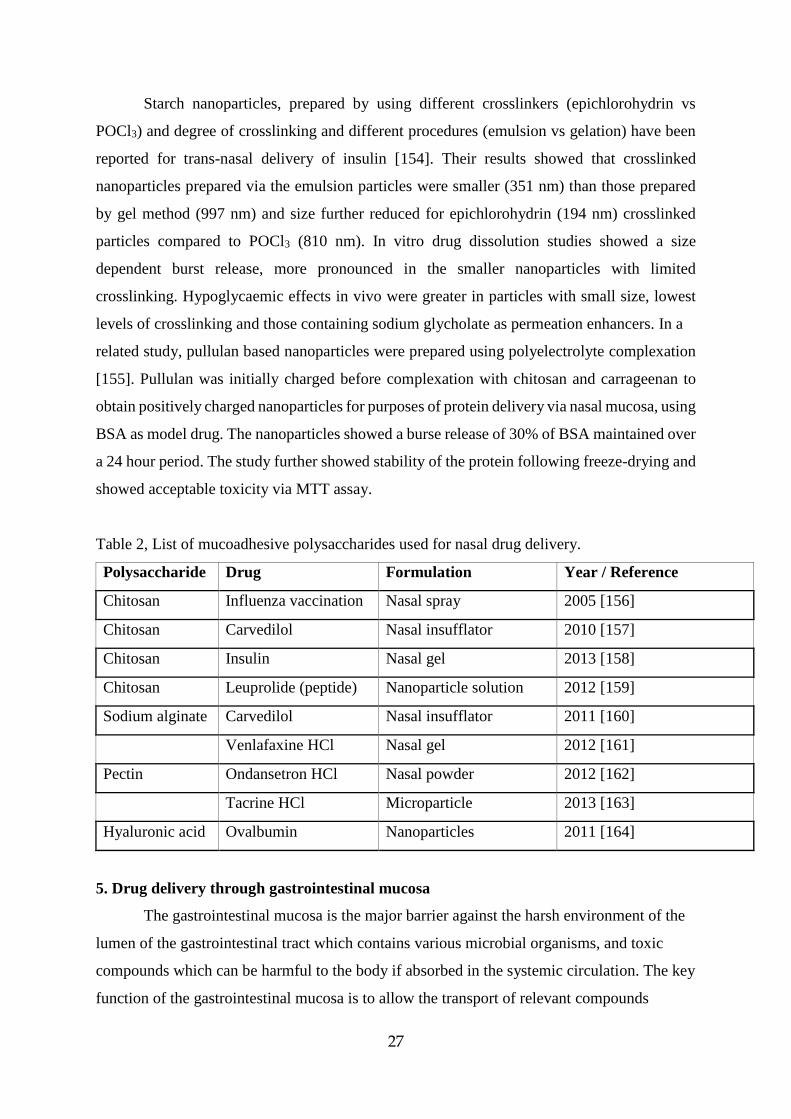
Starch nanoparticles, prepared by using different crosslinkers (epichlorohydrin vs
POCl3) and degree of crosslinking and different procedures (emulsion vs gelation) have been
reported for trans-nasal delivery of insulin [154]. Their results showed that crosslinked
nanoparticles prepared via the emulsion particles were smaller (351 nm) than those prepared
by gel method (997 nm) and size further reduced for epichlorohydrin (194 nm) crosslinked
particles compared to POCl3 (810 nm). In vitro drug dissolution studies showed a size
dependent burst release, more pronounced in the smaller nanoparticles with limited
crosslinking. Hypoglycaemic effects in vivo were greater in particles with small size, lowest
levels of crosslinking and those containing sodium glycholate as permeation enhancers. In a
related study, pullulan based nanoparticles were prepared using polyelectrolyte complexation
[155]. Pullulan was initially charged before complexation with chitosan and carrageenan to
obtain positively charged nanoparticles for purposes of protein delivery via nasal mucosa, using
BSA as model drug. The nanoparticles showed a burse release of 30% of BSA maintained over
a 24 hour period. The study further showed stability of the protein following freeze-drying and
showed acceptable toxicity via MTT assay.
Table 2, List of mucoadhesive polysaccharides used for nasal drug delivery.
Polysaccharide Drug Formulation Year / Reference
Chitosan Influenza vaccination Nasal spray 2005 [156]
Chitosan Carvedilol Nasal insufflator 2010 [157]
Chitosan Insulin Nasal gel 2013 [158]
Chitosan Leuprolide (peptide) Nanoparticle solution 2012 [159]
Sodium alginate Carvedilol Nasal insufflator 2011 [160]
Venlafaxine HCl Nasal gel 2012 [161]
Pectin Ondansetron HCl Nasal powder 2012 [162]
Tacrine HCl Microparticle 2013 [163]
Hyaluronic acid Ovalbumin Nanoparticles 2011 [164]
5. Drug delivery through gastrointestinal mucosa
The gastrointestinal mucosa is the major barrier against the harsh environment of the
lumen of the gastrointestinal tract which contains various microbial organisms, and toxic
compounds which can be harmful to the body if absorbed in the systemic circulation. The key
function of the gastrointestinal mucosa is to allow the transport of relevant compounds

28
including nutrients, drugs and water across the epithelial membrane whilst keeping out
harmful materials including microorganisms.
5.1 Gastrointestinal anatomy
The gastrointestinal barrier comprises mainly two parts: (a) the intrinsic barrier (made up of
epithelial cells which line the walls of the digestive tract, held together by very tight
junctions) and (b) the extrinsic barrier (comprising secretions and other factors not physically
part of the epithelium but contribute to the maintenance of their integrity towards its barrier
function). These secretions include mucus, bicarbonates, hormones and cytokines,
prostaglandins, growth factors, trefoil proteins, antibiotic peptides and antibodies and
immunoglobulins. However, for purposes of this review, the mucus which forms part of the
extrinsic gastrointestinal barrier will be the focus of attention and the reader is referred to
more specific anatomical, physiological and biochemical sources of peer reviewed
information for the other components outlined above [165].
<Figure 6 here>
5.2 Polysaccharide-based gastrointestinal mucosa delivery systems
As already noted, the environment within the gastrointestinal tract can be harsh to
labile drugs including proteins and peptides and therefore these drugs have traditionally not
been administered via the oral route, but rather via the parenteral injections. However, there
has been recent attempts at delivering such drugs across the gastrointestinal mucosa barrier
by use of various bioadhesive polymers including polysaccharides which either have intrinsic
permeation enhancing properties or used to formulate delivery systems incorporating natural
or synthetic permeation enhancers [166]. As is the case for the other mucosal routes, chitosan
is the most common polysaccharide owing to its biocompatibility, its bioadhesivity and,
permeation enhancing characteristics.
Guggi and co-workers prepared a delivery system for delivering calcitonin based on
various chitosan derivatives in a composite system [167]. They synthesized chitosan–4-
thiobutylamidine (as mucoadhesive fixer) conjugated to chitosan–pepstatin A (pepsin
inhibitor conjugated to mucoadhesive chitosan), incorporated into mini-tablets and used for
delivering the protein drug via the stomach mucosa. Protein permeation was further enhanced
by use of glutathione as part of the formulation. Their results showed that the chitosan–pepsin
inhibitor conjugate provided appropriate protection of the calcitonin.

29
However, the most common delivery system employed are encapsulated colloidal
systems, usually in the form of nanoparticles, given their easy manipulation (e.g. pegylation)
for targeting purposes and the extra protection afforded by encapsulating the target drug of
interest [168]. Pullulan polysaccharide were combined with the enteric polymer Eudragit to
prepare microparticles with gastric acid resistance as well as controlled drug release for oral
delivery of risedronate [169]. The microparticles were prepared by spray drying and
characterised for yield, size, encapsulating efficiency, morphology, moisture levels and in
vitro dissolution characteristics. Their results showed suitable physical properties and most
interestingly a 100 % encapsulation efficiency, resistant to simulated gastric fluid whilst
showing prolonged release in intestinal fluid. Further, when the particles were compressed
together with or without polyvinylpyrrolidone into tablets, they still maintained gastro
resistance as well as prolonged release in intestinal medium and therefore provide great
potential as an alternative oral delivery system.
Shina and Kumria [170] have reviewed several natural polysaccharides used either
alone or in combination with other organic or inorganic components for colonic drug delivery
and summarised in table 3.
Modified psyllium polysaccharide hydrogels have been proposed as potential drug
delivery vehicles for methotrexate for the treatment of gastrointestinal tract cancer [209].
Swelling and drug release characterisation studies on the hydrogel formulations showed
Fickian diffusion at different pH values suggesting the system can release the drug in
different parts of the GIT in appropriate doses over a reasonable time frame in a controlled
manner. In an in vitro study, composite calcium alginate and carboxymethylcellulose beads
with pH responsive swelling and mucoadhesion behaviour as well as biodegradability
induced by micro-organisms present in the colon, have been proposed for colon targeted
delivery of 5-fluoro-uracil. Beads prepared by ionic gelation were physically characterized
using scanning electron microscopy, X-ray diffraction, energy dispersive X-ray analysis
(EDAX), differential scanning calorimetry and texture analysis which showed higher
swelling and mucoadhesion within a simulated colonic environment. The composite beads
also degraded slowly in simulated colonic fluid which was accelerated in the presence of
microflora commonly present in the colon region. Further, in vitro drug release showed
greater than 90% total drug release when colonic enzymes were present and
carboxymethylcellulose modulated the drug release when analysed by fluorescence recovery
after photo-bleaching. Testing of the drug loaded beads against colon adenocarcinoma cells
suggested a potential application of these beads for colon specific drug delivery.

30
Table 3 Polysaccharides investigated for colon-specific drug delivery with their dosage forms
and summary of the results obtained. Adapted from Shina and Kumria (2003) [170].
Polysaccharide Drug Formulation Year / Reference
Chitosan 5-(6)-Carboxy fluorescein) Enteric-coated capsules 1997 [171]
Chitosan Insulin Enteric-coated chitosan
capsules
1997 [171]
Chitosan R68070 Enteric-coated chitosan
capsules
1999a [172]
Chitosan Sodium diclofenac Enteric-coated
chitosan microspheres
1998 [173]
Chitosan Acetaminophen Cores coated with
chitosan and phytin
1998[174]
Chitosan succinate /
phthallate
Sodium diclofenac Matrices 1999 [175]
Pectin (calcium salt) Indomethacin Matrices 1993 [176]
Pectin Indomethacin Compression coated/
matrix tablets
1995 [177]
Pectin Insulin Compression coated/
matrix tablets
1995 [177]
Pectin Radioactive tracer Enteric-coated matrix
tablets
1997 [178]
Methoxylated
pectinate
Radioactive tracer Compression coat 1994 [179]
Amidated pectin Paracetamol Matrix tablets 1997 [180]
Amidated pectin Indomethacin
Sulphamethoxazole
Chitosan-coated amidated
pectin beads
1997 [181]
Amidated pectin/
calcium pectinate
Ropivacaine Matrix tablet 2000 [182]
Pectin
Paracetamol Ethyl cellulose film
coating
1996 [183]
Pectin Theophylline Mixed film with coating 2000a, 2000b
[184, 185]
Pectin / chitosan Technetium-99 Mixed film of pectin,
chitosan and HPMC
1999a, 1999b
[186, 187]
Pectin and chitosan Indomethacin/ paracetamol Compression coat 1998 [188]
Guar gum Dexamethasone/
budesonide
Matrix tablet 1997 [189]
Guar gum Dexamethasone Matrix tablet (radio
labelled)
1997 [190]
Guar gum Indomethacin Matrix tablet 1998 [191]
Guar gum Technetium-99m-DTPA Matrix tablet 1998 [192]

31
Guar gum 5-ASA Compression
coat
1999 [193]
Cross-linked guar – – 1995a [194]
Cross-linked guar Hydrocortisone Hydrogels 2000 [195]
Crosslinked dextran – Hydrogels 1995a [196]
pH-sensitive
dextran
Bovine serum albumin
(BSA)
Hydrogels 1999 [197]
Cross-linked
dextran
Hydrocortisone Capsules 1998 [198]
Dextran fatty acid
esters
Theophylline Films 1997 [199]
Inulin – Films 1996 [200]
Inulin – Hydrogels 1998 [201]
Cross-linked
chondroitin
Indomethacin Matrix tablet 1992a [202]
Amylose 5-ASA Coated pellets 1996a [203]
Amylose/ethyl
cellulose (1:4)
Glucose Coated cores 1996 [204]
Amylose/ethylcellul
ose coating
5-ASA Coated pellets 2000a [205]
Starch Radioactive tracer Enteric-coated
capsules
2000 [206]
Calcium alginate 5-ASA Double coated swellable
beads
1992 [207]
Locust bean gum Theophylline Film 1995 [208]
In a related study, Feng and co-workers used chitosan based nanogels obtained by
electrostatic interaction between chitosan and carboxy-methyl chitosan for delivering drugs
against colorectal cancer and showed significant effect on cell viability owing to the improved
mucoadhesion which allowed a higher accumulation of drug (doxorubicin) concentration
within the cancer site [210].
pH responsive hydrogel beads obtained by grafting polyacrylamide onto kappa-
carrageenan and sodium alginate (SA), for targeting ketoprofen to the intestine have been
reported by Kulkarni and co [211]. The grafted polyacrylamide-carrageenan was synthesised
by free-radical polymerization with alkaline hydrolysis whilst the drug loaded hydrogels were
obtained by ionic gelation and covalent crosslinking and showed amorphous conversion of

32
the drug within the beads as analysed using differential scanning calorimetry and X-ray
diffraction. The beads which were spherical in shape (scanning electron microscopy), exhibited
pH sensitive swelling in a pulsatile fashion and showed that the drug release increased when
pH changed from acid to alkaline conditions. Further in vivo studies in albino rats showed that
the beads retarded the release of ketoprofen in the stomach (acidic pH) and therefore resulting
in reduced ulceration, haemorrhage and gastric mucosa erosion associated with the drug. This
finding is interesting and shows great potential for targeting such drugs to the small intestines
for systemic absorption.
Similarly, Prezotti and co-workers [212] obtained circular beads containing gellan gum
and pectin by ionotropic gelation, but using Al3+ as crosslinker, with high yield and entrapment
efficiencies and sizes ranging between 728.95 and 924.56 μm but increased with higher
polymer and crosslinker concentrations. Thermal analysis and Fourier transform infra-red
spectroscopy showed the absence of drug–polymers interactions but high mucoadhesion both
in vitro and ex vivo. Further, the beads showed high erosion under acidic pH whilst swelling
was enhanced at pH of 7.4, suggesting pH depended drug (ketoprofen) release characteristics
which was determined to be via a super case 2 type sustained release over a 6 hour period at
pH 7.4.
Different commercial products incorporating 5 amino salicylic acid are marketed
under different brand names (Pentasa, Asacol, Salofalk, Lialda, and Mezavant). Lipfen®, is a
flurbiprofen liquid suppository containing HP-beta-CD. Chronotropic™ is a “two-pulse”
colonic device for oral delivery of insulin and consists of an inner swellable/erodible
hydroxylpropylmethylcellulose (HPMC) layer surrounding a tablet core as well as an another
HPMC external coating on the exterior. Other products include Desmopressin containing
antidiuretic hormone and Sandimmune® Soft Gelatin Capsules containing cyclosporin
6. Vaginal mucosa drug delivery
The vaginal route of drug administration is an excellent route for systemic drug delivery
considering its large mucosal surface area and high blood vessel network, a common feature
in other mucosa sites. Challenges of vaginal mucosa drug delivery include vaginal irritation,
sexual activity and menstrual cycle, urination, hygiene of the individual and cultural
background [213].

33
6.1 Vaginal physiology and anatomy
The vagina is made up of muscles with an elastic lining that provide lubrication and
sensation, and links the uterus to the external genitalia called the vulva. Its diameter range
from 2.1 to 5.0cm whiles its length range from 8.4 to 11.3cm. The walls of the vagina consist
of three layers: the stratified squamous epithelial layer, the lamina propria, the muscular layer
and the tunica adventitia made up areolar connective tissues [214, 215] (figure 7). Although
the mucosal surface of the vagina does not have glands, it is usually composed of fluid which
are secreted by other components such as endometrial and tubal, cervical mucus, exfoliating
epithelial cells, Bartholin’s and Skene’s glands and transudation of leukocytes and vaginal
cells from blood vessels [216].
<Figure 7 here>
The normal microbial flora of the vagina such as Lactobacillus acidophilus helps in
regulating and maintaining the pH of the vagina by producing lactic acid with pH 3.8 and 4.2.
However, the pH of the vagina is usually altered as a result of menstruation and sexual
intercourse due to the alkalinity of semen and vaginal transudates. Absorption of drugs
administered via transmucosal routes are usually by transcellular (i.e. transport across the
cell) and paracellular (i.e. transport between cells). Dissolution of drugs in the vaginal lumen
and penetration of the membrane are important processes in the absorption of drugs from a
designed vaginal drug delivery system. Physiological factors such as change in hormonal
levels vaginal secretion volume and composition, pH and sexual activities could affect the
release and absorption of drug [213, 216].
6.2 Polysaccharide-based vaginal mucoadhesive systems
The different dosage forms of the vaginal route include solutions, suppositories, gels,
foams, vaginal rings, tampons, sponges, creams, ointments, doushes, pessaries, soft gelatin
capsules and tablets [215]. Recently, polymeric films have been considered as a novel dosage
form in vaginal mucosal delivery. Research focus of films has been aimed at solving genital
problems such as contraceptives and microbicides [217]. Mucosal delivery of drugs in the
vagina can either be systemic or locally. Local mucosal delivery involves the delivery of drug
to the tissues of the vaginal cavity as is the case in the treatment of vaginitis. In other to
achieve optimum characteristics such as bioadhesion, retention, distribution and release,
formulation of vaginal drug delivery system will require the use of a mucoadhesive
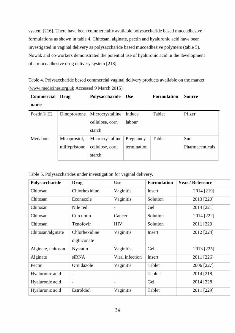
34
system [216]. There have been commercially available polysaccharide based mucoadhesive
formulations as shown in table 4. Chitosan, alginate, pectin and hyaluronic acid have been
investigated in vaginal delivery as polysaccharide based mucoadhesive polymers (table 5).
Nowak and co-workers demonstrated the potential use of hyaluronic acid in the development
of a mucoadhesive drug delivery system [218].
Table 4. Polysaccharide based commercial vaginal delivery products available on the market
(www.medicines.org.uk Accessed 9 March 2015)
Commercial
name
Drug Polysaccharide Use Formulation Source
Postin® E2 Dinoprostone Microcrystalline
cellulose, corn
starch
Induce
labour
Tablet Pfizer
Medabon Misoprostol,
milfepristone
Microcrystalline
cellulose, corn
starch
Pregnancy
termination
Tablet Sun
Pharmaceuticals
Table 5. Polysaccharides under investigation for vaginal delivery.
Polysaccharide Drug Use Formulation Year / Reference
Chitosan Chlorhexidine Vaginitis Insert 2014 [219]
Chitosan Econazole Vaginitis Solution 2013 [220]
Chitosan Nile red - Gel 2014 [221]
Chitosan Curcumin Cancer Solution 2014 [222]
Chitosan Tenofovir HIV Solution 2011 [223]
Chitosan/alginate Chlorhexidine
digluconate
Vaginitis Insert 2012 [224]
Alginate, chitosan Nystatin Vaginitis Gel 2013 [225]
Alginate siRNA Viral infection Insert 2011 [226]
Pectin Ornidazole Vaginitis Tablet 2006 [227]
Hyaluronic acid - - Tablets 2014 [218]
Hyaluronic acid - - Gel 2014 [228]
Hyaluronic acid Estroldiol Vaginitis Tablet 2011 [229]

35
6.3 Prevention of human immune deficiency (HIV) virus infection
One of the most productive fields of research in terms of vaginal delivery is in the use of
topical microbicides in the prevention of HIV transmission. The microbicides prevent early
transmission of the virus at the mucosal level and contiguous side when administered to the
vaginal mucosa. The most common anti-HIV microbicides approved molecules are tenofovir
and dapivirine. Meng and co-workers designed a polysaccharide based formulation loaded
with tenofovir using chitosan based nanoparticles. Chitosan was used as an adhesive
component to improve retention time of the formulation. The result of their research
demonstrate chitosan nanoparticle as a potential microbicide delivery system with a safe
bioadhesive and control release properties [223]. In another approach, Belletti and co-
workers investigated tenofovir loaded nanoparticle of chitosan and/or poly-(d,l-lactide-co-
glycolide) (PLGA). They also investigated the strategies at which the nanoparticles were
prepared for more efficient drug loading. In their findings it was concluded that
PLGA/chitosan nanoparticles could be an effective and attractive tenofovir antiretroviral
carrier [230].
6.4 Vaginal infections
Vaginal infections are known to cause vaginitis (inflammation of the vagina) which is highly
prevalent in women within reproductive age. The most common cause of vaginitis is vaginal
candidiasis caused by fungi such as Candida albicans. Other causes of vaginitis are known to
include aerobic vaginitis caused by the disruption of Lactobacillus spp followed by signs of
inflammation and the manifestation of a predominantly aerobic microflora such as
Escherichia coli and Streptococcus agalactiae [231, 232]. The management of vaginitis is
usually by the administration of antifungal azoles and antibacterial small molecules such as
imidazoles (for candidiasis) and quinolone (for aerobic vaginitis). These molecules are
known to hinder the activities of abnormal and invasive microorganisms however, they
exhibit poor local pharmacokinetics with commercially available products associated to
safety issues [233].
Abruzzo and co-workers demonstrated the use of polysaccharide complexes of
chitosan and alginate in the development of vaginal insert loaded with chlorhexidine
digluconate for local vaginal delivery. The use of chitosan and alginate polysaccharides in the
reported formulation was to obtain a good mucoadhesion between the insert and the mucosal
surface of the vaginal cavity [224]. Other polysaccharides such as pectin have been studied as
a mucoadhesive polymer in the treatment of vaginitis. Baloglu and colleagues demonstrated

36
the use of various polymers including pectin as mucoadhesive polysaccharide in the
development of vaginal tablets loaded with ornidazole. The formulation containing pectin
demonstrate enhanced mucoadhesive properties, controlled release and non-toxic
characteristics [227].
6.5 Delivery of nucleic acids
Another field of study in vaginal mucosal delivery in terms of viral infections responsible for
diseases such as HIV, genital herpes and cervical cancer is the use of nucleic acids such as
small interfering RNA (siRNA) or plasmid DNA. siRNA can be used to target both viral and
host gene or combined together. The vaginal route offers self-administration as well as low
toxicity in terms of siRNA delivery against sexually transmitted diseases. However, siRNAs
are susceptible to vaginal degradation, less likely to internalise into the cell and when they
do, their ability to escape the endosome is low. There is therefore need for delivery systems
that can transport siRNA via transcellular and paracellular pathways [234]. To achieve this,
there are two categories of vectors used in the transport of therapeutic siRNA; viral and non-
viral vectors. The viral vectors involve the use of viruses such as retrovirus, lentivirus and
adenovirus, while non-viral vectors include liposome-based cationic transfection reagent
[235], macromolecular conjugation [236], and polymeric nanoparticles [234].
Mucoadhesive polymeric formulation can be used to improve the delivery of designed
vectors by decreasing the effect of the physiological factors that affect absorption of vector as
well as increasing the retention time of the vector on the mucosal surface. Alginate
polysaccharide has been under investigation in the development of an alginate scaffold
system for the delivery of siRNA through the vaginal mucosal. Wu and co, developed a
PEGylated lipoplex-entrapped alginate scaffolds (PLAS) using alginate polysaccharides.
Their studies demonstrated a significant improvement in the delivery siRNA via intravaginal
route by entrapping PEGylated lipoplexes in a negatively charged alginate scaffold system
compared to conventional lipoplexes [237].
7. Drug delivery in the context of wound therapy
A wound can be defined as an injury or disruption to anatomical structure and
function resulting from simple or sever break in the skin, extending into regions such as the
subcutaneous tissue. The damage can extend to other tissues such as muscles, tendons,
nerves, vessels and even the bone [7, 238, 239]. It has been suggested that wound healing

37
involves a series of overlapping molecular events requiring extensive communication
between cells and various physiological processes (figure 8). These comprise haemostasis
and inflammatory phase, proliferative phase, remodelling and scar maturation phase [7, 240].
A wide range of wound dressings are available for a variety of wound types including
foam dressings, alginates, films, hydrocolloids and hydrogels. These have absorbent
properties and their main aim is to form gels upon contact with the wound exudate. Most of
these dressings provide and maintain a moist wound environment and are recognised for
optimal healing. However, many factors may delay the phase of healing process, because not
all dressings have the contents required for an ideal property of a dressing for optimal healing
of the wound. As a result, many wound dressings have been extensively studied as drug
delivery systems which take active part to improve wound healing [241, 242]. These are
usually prepared using various natural and synthetic polymers including hydrocolloids,
hydrogels and polysaccharides such as xanthan gum, alginates, chitosan and its derivatives,
carrageenan, hyaluronic acid and collagen. The latter group (polysaccharides) have attracted
significant interest due to their hydrophilicity, bioadhesion, biocompatibility and
biodegradability.
<Figure 8 here>
7.2 Polysaccharide-based wound delivery systems
Bioactive composite films comprising the polysaccharide carrageenan combined with
polyethylene oxide have been reported for use in chronic wounds. The films were loaded
with streptomycin to target bacterial infections and diclofenac to target the inflammatory
phase of wound healing [241, 244]. In a parallel study, freeze-dried wafers comprising
carrageenan and polyethylene oxide or sodium alginate and polyethylene oxide loaded with
the above two drugs were also prepared and characterised [242]. In both type of formulations
(films and wafers), the results showed sustained release of both drugs over a 72 hour period
whilst antibacterial studies showed synergistic antibacterial action of the two drugs against
three main infection causative bacteria which were Staphyloccocus aureus, Pseudomonas
aeruginosa and Escherichia coli when compared to the streptomycin on its own. The results
were interesting as they suggest the possibility of targeting more than one phase of wound
healing by treating infection and preventing reinfection over a three day period whilst at the
same time reducing swelling and pain associated with inflammation. In addition, the
synergistic action between the two drugs allowed the use of a lower dose of the streptomycin

38
which is advantageous in terms of lowering the exposure of newly formed cells to high doses
of both drugs, and therefore reduce the chances of toxicity. Furthermore, the release of drug
over three days due to high swelling and moisture holding capacity which controlled high
exudate levels will reduce the need for too frequent dressing changes which is a major cause
of patient non-compliance that can result in serious complications, especially for chronic
wounds.
Lyophilized wafers prepared from karaya gum were formulated and loaded with four
different antimicrobial agents (neomycin, povidone iodine, chlorhexidine and silver
sulphadiazine) as potential therapeutic dressings for exuding wounds against methicillin-
resistant Staphylococcus aureus [245]. They showed that the wafers swelled in simulated
wound fluid and released the compounds to different extents, with povidone iodine and
chlorhexidine being effective in the absence of proteins whilst neomycin was enhanced by
the presence protein in the form of bovine serum albumin with silver sulfadiazine showing
the lowest antibacterial potency. In a related study, the same group tested the effect of
chlorhexidine digluconate loaded polysaccharide based wafers against Pseudomonas
aeruginosa [246]. They produced different formulations containing the drug and functionally
characterized for simulated wound fluid absorption and holding capacity, rheological
characteristics of hydrated gels and in vitro efficacy against the bacteria in question and the
results of the different formulations compared. Their results showed that the drug reduced the
absorption of the simulated wound fluid by a factor between 11 – 50% and reduced the
rheological consistency of gels containing no sodium alginate by 10-65%. The drug
dissolution results showed that wafers containing karaya gum exhibited sustained release of
the drug within 24 hours with the highest amount released significantly higher the minimum
inhibitory concentration against the bacteria. The fitting of the release data to Korsmeyer
Peppas equation showed an anomalous diffusion mechanism for most of the wafers except
those containing xanthan.
In a further follow up study, Labovitiadi and co-workers investigated the effect of
gamma irradiation as an alternative sterilization technique to autoclaving, on lyophilized
wafers prepared from gels of guar gum, karaya gum, sodium alginate and xanthan gum,
loaded with chlorhexidine digluconate [247]. They determined the rheological (flow
properties) behaviour of irradiated wafers compared to non - irradiated controls as well as in
vitro activity against Pseudomonas aeruginosa using disc diffusion test and in vitro drug
dissolution studies by means of a diffusion cell set up. Their results showed a significant
reduction in consistency, yield stress or efficacy for the irradiated wafers compared to the
39
control, with the exception of xanthan gum. This is significant as the performance of an
intended drug delivery dressings is expected to remain consistent throughout formulation,
sterilization, handling and application to ensure its therapeutic wound healing effect. In the
case of a wound healing, any dressing or medicated dressing (delivery system) is expected to
be sterile since it makes direct contact with blood capillaries and in some cases with veins
and arteries with risk of direct entry of toxins or bacterial infection into the systemic
circulation. Since moist heat autoclaving is not possible with most polymeric dressings,
gamma radiation is one of the major sources of rendering wound management products
sterile. However, gamma radiation, because of the high energy it transmits, also has the
ability to degrade such polymers by interfering with the polymer chain integrity. It is
therefore vital to ensure that the appropriate dose is selected that removes all microbial
contamination whilst maintaining dressing structural integrity and therefore functional
performance in terms of exudate absorption capacity, swelling, viscosity and eventual control
of drug release.
Fibrous alginate hydrogel dressings loaded with tetracycline as an anti-bacterial drug
have been prepared [248]. The dressings were prepared with three dimensional plotting by
varying different processing conditions (parameters) including air pressure, nozzle diameter,
layer increment, calcium concentration, alginate concentration and speed of the nozzle along
x and y coordinates and compared with standard film controls. The dressings were then
characterized for fibre size, porosity, tensile characteristics, degradation, swelling capacity,
drug release, water vapour transmission rate and bacterial inhibition based on the different
variables noted above. The results showed the fibres had larger fibre size, lower porosity,
higher elastic modulus and tensile strength, reduced degradation and lower swelling capacity
which were all significant when compared to the control samples. Further, the drug release
and antibacterial activity were not significantly different from those of the control films,
whilst the water vapour transmission rates were only slightly different than the commercial
control dressings. They concluded that the fibrous samples absorbed fluid faster, were more
flexible which allowed better conformation around the wound contours whilst also providing
better sustained release of the model antibiotic drug. Composite freeze-dried wafers
comprising sodium alginate and gelatin have been reported for the delivery of the
antimicrobial drug silver sulphadiazine [249]. The combination of both alginate and gelatin
were observed to improve functional characteristics of the dressing such as swelling,
mucoadhesion and drug release, compared to those containing only one polymer [249]

40
Singh and Pal [250] reported on the synthesis and characterisation of Sterculia
polysaccharide crosslinked polyvinylalcohol based hydrogel film dressings for prolonged
delivery of an antibacterial drug. The films were characterised for tensile, mucoadhesion,
permeability, blood compatibility, surface morphology, in vitro drug release and
antimicrobial activity. The films absorbed between 4.80 and 6.32 gram fluid per gram of /g
of gel with swelling occurring via a Case II diffusion mechanism whilst the drug release
followed non-Fickian and Case II diffusion mechanisms depending on the type of poly vinyl
alcohol. The polymeric films were permeable to oxygen and water vapour but not permeable
to micro-organisms Further, Sterculia–PVA hydrogel wound dressing showed better blood
compatibility as compared to Sterculia-PVA-AAm.
Carboxybutylchitosan and agarose based films and foam type wound dressings loaded
with quercetin and thymol as anti-inflammatory and anaesthetic respectively have been
prepared using supercritical solvent impregnation by means of supercritical carbon dioxide
[251]. The formulations were characterised for sustained release and fluid handling
characteristics. The results showed that the drug loading amounts increased at higher
pressures and temperatures and showed that the relative amounts quercetin and thymol loaded
can be “tuned” by changing the operational pressure-temperature conditions. They also
showed that quercetin was released in a more sustained fashion owing to its higher molecular
volume, lower water solubility and favoured interactions with the carboxybutylchitosan.
Furthermore, the dressings exhibited ideal fluid and water vapour absorption capacities
coupled with adequate water vapour transmission rates expected for an ideal [7] wound
dressing.
Ribeiro and co-authors have developed hydrogel dressings combining dextran
incorporating chitosan microparticles encapsulating growth factors for wound healing of skin
burns using both in vitro and in vivo characterisation [252]. In their study, the dressings were
characterised for morphology using scanning electron microscopy, cytotoxicity profile and
degradation by-products as well as application onto burn skin wounds using an animal model.
The growth factor loaded dressings showed appropriately rapid wound healing within the
expected time period whilst histological analysis showed the absence of reactive or
granulomatous inflammatory reaction in skin lesions. Further, the dressing components and
degradation by-products were biocompatible and contributed to restoration of skin
architecture.
In a recent article, Mohandas and co-workers reported on development of composite
chitosan and hyaluronic acid based sponges loaded with fibrin nanoparticles incorporating

41
vascular endothelial growth factor to target multiple phases of wound healing for accelerated
therapeutic action in diabetic wounds [253]. They characterised the dressings for porosity,
swelling, biodegradation, mechanical properties and haemostatic potential of the sponges
whilst release of the growth factor from the sponges were evaluated using ELISA. Further,
cell viability and attachment of the composite sponges were evaluated using human dermal
fibroblast cells and human umbilical vein endothelial cells and observed capillary like tube
formation for the growth factor loaded dressings when compared with blank control sponges.
Apart from the above systems, other drugs delivered to wound sites using
polysaccharide polymers include antioxidants [254], debriding agents [255] and peptides
[256, 257]. However, a detailed and extensive evaluation is not provided here as this falls
outside the scope of the current review and the reader is encouraged to consult more specific
literature on these.
The main polysaccharide based wound dressings available commercially fall under
three main categories; hydrocolloid (mainly carboxymethylcellulose, pectin or gelatin)
dressings, alginate (sodium or calcium alginate) dressings and biomaterials
(collagen/hyaluronic acid) based tissue engineered matrices and are summarised in the table
below. To avoid any conflicts of interest and inadvertent product endorsements, the company
names are not provided and the reader is invited to find these out if of interest. Each of these
dressings can be used alone in passive wound healing or loaded with various active
ingredients such as antimicrobial or debriding agents where they act as drug delivery systems.
For extensive read about these products, the reader should please refer to the review by
Boateng et al [7]
Table 6 Polysaccharide based commercial dressings available on the market
Hydrocolloid dressing Alginate dressing Biomaterial dressing
GranuflexTM Algisite® M Alloskin
AquacelTM Algosteril® Biobrane®
ComfeelTM Kaltocarb® Apligraf®
TegasorbTM Seasorb® Dermagraft®
Sorbalgon®
Kaltostat ®
Sorbsan®

42
8 Chemically modified polysaccharide polymers
An important strategy for improving mucoadhesive performance to enhance
formulation retention time at the target absorption site and ultimate improvement is
bioavailability is the use of derivatives of naturally occurring bioadhesive polymers. These
include thiolated chitosan [35, 36, 37], carboxybutylchitosan [251], thiolated pectin, [258,
259] and caboxymethyl xanthan [260, 261, 262]. The reader is referred to the references
provided for more in depth evaluation of the unique characteristics of these chemically
modified natural polysaccharides and their use as drug delivery systems for drug delivery
across the various mucosal routes as discussed above.
9. Summary conclusion
This review has covered various polysaccharides used for mucosal drug delivery as an
alternative to the traditionally used oral and parenteral routes. It is apparent that
polysaccharides possess most of the ideal characteristics expected for effective drug
administration via mucosal membranes, including biocompatibility (safety), bioadhesion,
hydration and swelling, ease of formulation, including water solubility and biodegradability.
In addition, some polysaccharides such as chitosan have smart properties which allow
modulation of drug release based on external stimuli, such as pH and temperature. In
addition, chitosan possesses, various reactive functionalities which allow easy manipulation
to produce versatile derivatives with enhanced ideal properties such as bioadhesion,
controlled drug release as well as permeation enhancement. Furthermore, these
polysaccharides can be formulated into a wide range of dosage forms ranging from simple
solutions, swollen gels, tablets, films and patches, particulate systems and wafers (xerogels).
It is also interesting to note that these various polysaccharides and the different dosage forms
have been used to deliver a wide range of active pharmaceutical agents including small
molecules, large macromolecules, biological agents for a variety of disease conditions that
locally affect the various mucosal routes, as well as for absorption into the systemic
circulation to exert the required therapeutic effect.
Other approaches such as bladder tumors and intravesical drug delivery are also an
important routes of administration for polysaccharide based carriers for active molecules.
However, these fall outside the scope of the current review and the reader is referred to the
relevant texts which discuss these in much more detail.
With the above in mind, it is expected that more sophisticated and novel formulations
for drug administration across these mucosal routes will be reported in the near to medium

43
term, which should ultimately result in new commercial products on the market. One such
area is the delivery of recombinant and vaccine products, with the latter providing a viable
option for achieving mucosal immunization. It is hoped that such systems will continue to
advance and hopefully move from bench to bedside for patient use in the long term.
Conflict of interest
The authors report no conflicts of interest
Acknowledgments
The authors are grateful to the publishers and owners for granting permission for
reproduction of the various images reproduced used in this review.
REFERENCES
[1] Panda BP, Dey NS, Rao MEB. Development of innovative orally fast disintegrating film dosage forms:
A review. Int J Pharm Sci Nanotech. 2012; 5(2): 1666–74.
[2] Patel V, Desai T, Chavda B, Katira R. Extemporaneous dosage form for Oral liquids. Pharmacophore.
2011; 2(2): 86-103.
[3] Satter M, Sayed OM, Lane ME. Oral transmucosal drug delivery – Current status and future prospects.
Int J Pharm. 2014; 471: 498-06.
[4] Hussain A, Ahsan F. The vagina as a route for systemic drug delivery. J Contr Rel. 2005; 103(2): 301-
13.
[5] Hussain A, Majumder QH, Ahsan F. Inhaled insulin is better absorbed when administered as a dry
powder compared to solution in the presence or absence of alkylglycosides. Pharm Res. 2006; 23(1):
138-47.
[6] Nargarwal RC, Kant S, Singh PN, Maiti P, Pandit JK. Polymeric nanoparticulate system: A potential
approach for ocular drug delivery. J Contr Rel. 2009; 136)1): 2-13.
[7] Boateng JS, Matthews KH, Stevens HNE, Eccleston GM. Wound healing dressings and drug delivery
systems: A review. J Pharm Sci. 2008; 97(8): 2892-923.
[8] Suzuki Y, Tanihara M, Nishimura Y, Suzuki K, Kakimaru Y, Shimizu Y. A new drug delivery system
with controlled release of antibiotic only in the presence of infection. J Biomed Mater Res. 1998; 42(1):
112-6.
[9] Aubyn L, Boateng J. Formulation, Design and Functional Characterization of a Novel Vaginal Mucosa
Drug Delivery System. Proceedings of American Association of Pharmaceutical Scientists (AAPS 2013),
R6221, San Antonio, Texas, USA.
[10] Jana S, Ghandi A, Sen KK, Basu SK. Natural polymers and their application in drug delivery and
biomedical field. J PharmSciTech. 2011; 1(1): 16-27.
[11] Patel V, Liu F, Brown M. Modeling the oral cavity: In vitro and in vivo evaluations of buccal drug
delivery systems. J Contr Rel. 2012; 161(1): 746-56.

44
[12] Gupta SK, Singhvi IJ, Shirsati M, Karwani G, Agarwal A, Agarwak A. Buccal Adhesive Drug Delivery
System: A Review. Asian J Biochem Pharm Res. 2011; 2(1): 105-14.
[13] Smart JD. Theories of Mucoadhesion in Khutoryanskiy VV (Ed). Mucoadhesive Materials and Drug
Delivery Systems. 2014; Chapter 7 pp John Wiley and Sons Ltd, West Sussex, UK.
[14] Bobade NN, Atram SC, Wankhade VP, Pande SD, Tapar KK. A Review on Buccal Drug Delivery
System. Int J Pharm Pharm Sci Res. 2013; 3(1): 35-40.
[15] Roy S, Prabhakar B. Bioadhesive Polymeric Platforms for Transmucosal Drug Delivery Systems – a
Review. Trop J Pharm Res. 2010; 9(1): 91-104.
[16] Sudhakar Y, Kuotsu K, Bandyopadhyay AK. Buccal bioadhesive drug delivery – a promising option for
orally less efficient drugs. J Contr Rel. 2006; 114(1): 15-40.
[17] Tangri P, Khurana S, Madhav S. Muchoadhesive drug delivery: Mechanism and methods of evaluation.
Int J Pharma Bio Sci. 2011; 2(1): 458-67.
[18] Junginger H, Hoogstraate J, Verhoef C. 1999. Recent advances in buccal drug delivery and absorption -
in vitro and in vivo studies. J Contr Rel. 1999; 62(1-2): 149-59.
[19] Punitha S, Girish Y. 2010. Polymers in mucoadhesive buccal drug delivery system – A review. Int J Res
Pharm Sci. 2010; 1(2): 170-86.
[20] Alexander A, Swarna A, Tripathi D. Polymers and Permeation Enhancers: Specialized Components of
Mucoadhesives. Stamford J Pharm Sci. 2011; 4(1): 91-95.
[21] Kalani M, Yunus R. 2011. Application of supercritical antisolvent method in drug encapsulation: a
review. Int J Nanomed. 2011; 6(1): 1429-42.
[22] Salamaat-Miller N, Chittchang M, Johnson T. The use of mucoadhesive polymers in buccal drug
delivery. Adv Drug Del Rev. 2005; 57: 1666-91.
[23] Abruzzo A, Bigucci F, Cerchiara T, Cruciani F, Vitali B, Luppi B. Mucoadhesive chitosan/gelatin films
for buccal delivery of propranolol hydrochloride. Carbo Polym, 2012; 87(1): 581-88.
[24] Rossi S, Bonferoni MC, Ferrari F, Bertoni M, Caramella C. Characterization of mucin interaction with
three viscosity grades of sodium carboxymethylcellulose. Comparison between rheological and tensile
testing. Eur J Pharm Sci. 1996; 4(3): 189–96.
[25] Kianfar F, Chowdhry BZ, Antonijevic M, Boateng JS. Formulation development of a carrageenan based
delivery system for buccal drug delivery using ibuprofen as a model drug. J Biomater Nano Biotech.
2011; 2(5A), 582-95.
[26] Kianfar F, Antonijevic M, Chowdhry BZ, Boateng JS. Lyophilized wafers comprising κ-carrageenan &
pluronic acid for buccal drug delivery using model soluble and insoluble drugs. Coll Surf B: Biointerf.
2013; 103: 99– 106.
[27] Le Brun P, Fox P, De Vries M, Bodde H. In vitro penetration of some β-adrenoreceptor blocking drugs
through porcine buccal mucosa. Int J Pharm. 1989; 49(2): 141-45.
[28] Andrews G, Laverty T, Jones D. Mucoadhesive polymeric platforms for controlled drug delivery. Eur J
Pharm Biopharm. 2009; 71(3): 505-18.
[29] Arya A, Chandra A, Sharma V, Pathak K. Fast Dissolving Oral Films: An Innovative Drug Delivery
System and Dosage Form. Int J ChemTech Res. 2010; 2(1): 576-83.
45
[30] Dojo M, Azuma T, Saito T, Ohtani M, Muramatsu A, Kuriyama M. 2001. Effects of CYP2C19 gene
polymorphism on cure rates for Helicobacter pylori infection by triple therapy with proton pump inhibitor
(omeprazole or rabeprazole), amoxycillin and clarithromycin in Japan. Digestive Liver Dis. 2001; 33(8):
671-75.
[31] Lehr C. 2000. lectin-mediated drug delivery: the second generation of bioadhesives. J Contr Rel. 2000;
65: 19-29.
[32] Nibha K, Pancholi S. An Overview on: Sublingual Route for Systemic Drug Delivery. Int J Res Pharm
Biomed Sci. 2012; 3(2): 913-23.
[33] Jeng-Lin H. Role of proton pump inhibitors in the management of peptic ulcer bleeding. World J
Gastrointest Pharmacol Ther. 2010; 1(2): 51-53.
[34] Khairnar GA, Sayyad FJ. Development of buccal drug delivery system based on mucoadhesive polymers.
Int J PharmTech Res. 2010; 2(2): 719-35.
[35] Schirm, E. Tobi H, de Vries TW, Choonara I, De Jong-van den Berg LT. Lack of appropriate
formulations of medicines for children in the community. Acta Paediatrica. 2003; 92(3): 1486-89.
[36] Bemkop A. Thiomers: A new generation of mucoadhesive polymers. Adv Drug Del Rev. 2005; 57(11):
1569-82.
[37] Punitha S, Girish Y. Polymers in mucoadhesive buccal drug delivery system – A review. Int J Res Pharm
Sci. 2010; 1(2): 170-186.
[38] Thirawong N, Nunthanid J, Puttipipatkhachorn S, Sriamornsak P. (2007) 'Mucoadhesive properties of
various pectins on gastrointestinal mucosa: An in vitro evaluation using texture analyzer', Eur J Pharm
Biopharm. 2007; 67(1): 132-40.
[39] Ayensu I, Boateng JS. Development and evaluation of lyophilized thiolated-chitosan wafers for buccal
delivery of protein. J Sci Tech. 2012; 32 (2): 46-55.
[40] Boateng JS, Pawar HV, Tetteh J. 2013. Polyox and carrageenan based composite film dressing containing
anti-microbial and anti-inflammatory drugs for effective wound healing. Int J Pharm. 2013; 441(1-
2):181-91.
[41] Smart JD. The basics and underlying mechanisms of mucoadhesion. Adv Drug Del Rev. 2005;
57(11):1556-68.
[42] Tamburic S, Craig DQM. A comparison of different in vitro methods for measuring mucoadhesive
performance. Eur J Pharm Biopharm. 1997; 44(2): 159-67.
[43] Saiano F, Pitarresi G, Cavallaro G, Licciardi M, Giammona G. (2002) Evaluation of mucoadhesive
properties of alpha,beta-poly(N-hydroxyethyl)-DL-aspartamide and alpha,beta-poly(aspartylhydrazide)
using ATR-FTIR spectroscopy, Polymer, 2002; 43(23): 6281-86.
[44] Jabbari E, Wisniewski N, Peppas NA. Evidence of mucoadhesion by chain interpenetration at a
poly(acrylic acid)/mucin interface using ATR-FTIR spectroscopy. J Contr Rel. 1993; 26(2): 99-108.
[45] Hovgaard L, Brøndsted H. Current applications of polysaccharides in colon targeting. Crit Rev Ther
Drug Carrier Syst. 1996; 13(3-4): 185-23.
[46] Liu Z, Jiao Y, Wang Y, Zhou C, Zhang Z. Polysaccharides-based nanoparticles as drug delivery
systems. Adv Drug Del Rev. 2008; 60(15): 1650–62.
[47] Lai S, Wang Y, Hanes J. Mucus-penetrating nano particles for drug and gene delivery to mucosal tissues.

46
Adv Drug Del Rev. 2009; 61(2): 158–71.
[48] Cristescu D, Salavastru C, Voiculescu B, Niculescu C. Carmaciu R. Biologie. 1st ed. Bucuresti: Corint.
2008; pp.44-48.
[49] Martini F, Nath, J, Batrholomew E. Fundamentals of anatomy & physiology. 9th ed. San Francisco, CA:
Pearson Benjamin Cummings. 2012; pp.556-74.
[50] De la Fuente M, Raviña M, Paolicelli P, Sanchez A, Seijo B, Alonso M. Chitosan-based nanostructures:
a delivery platform for ocular therapeutics. Adv Drug Del Rev. 2010; 62(1): 100 - 17.
[51] Ye T, Yuan K, Zhang W, Song S, Chen F, Yang X, Wang S, Bi J, Pan W. Prodrugs incorporated into
nanotechnology-based drug delivery systems for possible improvement in bioavailability of ocular drugs
delivery. Asian J Pharm Sci. 2013; 8(4): 207-17.
[52] Pflugfelder S, Karpecki P, Perez V. Treatment of Blepharitis: Recent Clinical Trials. The Ocular Surf.
2014; 12(4): 273-84.
[53] Aulton M. Pharmaceutics, The science of dosage form design. 4th ed. Edinburgh: Churchill Livingstone.
2013.
[54] Ludwig A. The use of mucoadhesive polymers in ocular drug delivery. Adv Drug Del Rev. 2005; 57:
1595– 1639.
[55] Felt O, Furrer P, Mayer JM, Plazonnet B, Buri P, Gurny R. Topical use of chitosan in ophthalmology:
tolerance, assessment and evaluation of precorneal retention. Int J Pharm. 1999; 180: 185–93.
[56] Rossi S, Ferrari F, Bonferoni MC, Caramella C. Characterization of chitosan hydrochloride–mucin
rheological interaction: influence of polymer concentration and polymer: mucin weight ratio. Eur J
Pharm Sci. 2001; 12: 479–85.
[57] Alonso MJ, Sanchez A, The potential of chitosan in ocular drug delivery. J Pharm Pharmacol. 2003; 55:
1451– 63.
[58] Artursson P, Lindmark T, Davis SS, Illum L. Effect of chitosan on the permeability of monolayers of
intestinal epithelial cells (Caco-2). Pharm Res. 1994; 11: 1358–61.
[59] Dodane V, Khan MA, Mervin JR. Effect of chitosan on epithelial permeability and structure, Int J Pharm.
1999; 182: 21– 32.
[60] Di Colo G, Zambito Y, Burgalassi S, Saettone MF. Effects of chitosan and of its N-trimethyl and N-
carboxymethyl derivatives on the ocular pharmacokinetics of ofloxacin in rabbits. Proceedings 30th Int
Symp Contr Rel Bioact Mater. (Glasgow), 2003.
[61] Genta I, Conti B, Prugini P, Pavanetto F, Spadaro A, Puglisi P. Bioadhesive microspheres for ophthalmic
administration of acyclovir. J Pharm Pharmacol. 1997; 49: 737–42.
[62] Ferrari F, Rossi S, Bonferoni M, Caramella C, Karlsen J. Characterisation of rheological and
mucoadhesive properties of three grades of chitosan hydrochloride. Farmaco1. 997; 52: 493– 97.
[63] Li J, Xu Z. Physical characterisation of a chitosan-based hydrogel delivery system. J Pharm Sci. 2002;
91: 1669–77.
[64] Donabedian DH, Martin L, Eng DJ. Compositions comprising cationic polysaccharides and cationic
drugs. EP 0 888 770 A1 (1999).
[65] Hartmann V, Keipert S. Physico-chemical, in vitro and in vivo characterisation of polymers for ocular
use. Pharmazie. 2000; 55: 440–43.

47
[66] Gurny R, Ibrahim H, Aebi A, Buri P, Wilson CG, Washington N, Edman P, Camber O. Design and
evaluation of controlled release systems for the eye. J Contr Rel. 1987; 6: 367–73.
[67] Camber O, Edman P. Sodium hyaluronate as an ophthalmic vehicle: some factors governing its effect on
the ocular absorption of pilocarpine. Curr Eye Res. 1989; 6: 563–67.
[68] Saettone MF, Giannaccini B, Chetoni P, Torracca MT, Monti D. Evaluation of high- and low-molecular-
weight fractions of sodium hyaluronate and an ionic complex as adjuvants for topical ophthalmic vehicles
containing pilocarpine. Int J Pharm. 1991; 72: 131–39.
[69] Durrani AM, Farr SJ, Kellaway IW. Influence of molecular weight and formulation pH on the precorneal
clearance rate of hyaluronic acid in the rabbit eye. Int J Pham. 1995; 118: 243–50.
[70] Bucolo C, Spadaro A, Mangiafico S. Pharmacological evaluation of a new timolol/pilocarpine
formulation. Ophthalm Res. 1998; 30: 101–8.
[71] Herrero-Vanrell R, Fernandez-Carballido A, Frutos G, Cadorniga R. Enhancement of the mydriatic
response to tropicamide by bioadhesive polymers. J Ocul Pharmacol Ther. 2000; 16: 419–28.
[72] Camber O, Lundgren P. Diffusion of some low molecular weight compounds in sodium hyaluronate.
Acta Pharm Suec. 1985; 22: 315–20.
[73] Kyyro¨nen K, Hume L, Benedetti L, Urtti A, Topp E, Stella V. Methylprednisolone esters of hyaluronic
acid in ophthalmic drug delivery: in vitro and in vivo release. Int J. Pharm. 1992; 80: 161–69.
[74] Greaves JL, Wilson CG. Treatment of diseases of the eye with mucoadhesive delivery systems. Adv.
Drug Deliv Rev. 1993; 11: 349–83.
[75] Sintzel MB, Bernatchez SF, Tabatabay C, Gurny R. Biomaterials in ophthalmic drug delivery. Eur J
Pharm Biopharm. 1996; 42: 358–74.
[76] Madsen F, Eberth K, Smart JD. A rheological examination of the mucoadhesive/mucus interaction: the
effect of mucoadhesive type and concentration. J Control Rel. 1998; 50: 167–78.
[77] Meseguer G, Gurny R, Buri P, Rozier A, Plazonnet B. Gamma scintigraphic study of precorneal drainage
and assessment of miotic response in rabbits of various ophthalmic formulations containing pilocarpine.
Int J Pharm. 1993; 95: 229–34.
[78] Burgalassi S, Chetoni P, Saettone MF. Hydrogels for ocular delivery of pilocarpine: preliminary
evaluation in rabbits of the influence of viscosity and of drug solubility. Eur J Pharm. 1996; 42: 385–92.
[79] Saettone MF, Monti D, Torracca MT, Chetoni P. Mucoadhesive ophthalmic vehicles: evaluation of
polymeric lowviscosity formulations. J Ocul Pharmacol 1994; 10: 83–92.
[80] Burgalassi S, Panichi P, Chetoni P, Saettone MF, Boldrini E. Development of a simple dry eye model in
the albino rabbit and evaluation of some tear substitutes. Ophthalm. Res. 1999; 31: 229–35.
[81] Albasini M, Ludwig A. Evaluation of polysaccharides intended for ophthalmic use in ocular dosage
forms. Farmaco. 1995; 50: 633–42.
[82] D’Amico M, Di Filippo C, Lampa E, Boldrini E, Rossi F, Ruggiero A, Filippelli A. Effects of timolol
and timolol with tamarind seed polysaccharide on intraocular pressure in rabbits. Pharm Pharmacol
Commun. 1999; 5: 361–64.
[83] Verschueren E, Van Santvliet L, Ludwig A. Evaluation of various carrageenans as ophthalmic
viscolysers. S.T.P. Pharma Sci. 1996; 6: 203–10.

48
[84] Saettone MF, Monti D, Giannaccini B, Salminen L, Huupponen R. Macromolecular ionic complexes of
cyclopentolate for topical ocular administration. Preparation and preliminary evaluation in albino rabbits.
S.T.P. Pharma Sci. 1992; 2: 68–75.
[85] Burgalassi S, Chetoni P, Panichi L, Boldrini E, Saettone MF. Xyloglucan as a novel vehicle for timolol:
pharmacokinetics and pressure lowering activity in rabbits. J Ocul Pharmacol Ther. 2000; 16: 497–509.
[86] Giannavola C, Bucolo C, Maltese A, Paolino D, Vandelli MA, Puglisi G, Lee VHL, Fresta M. Influence
of preparations on acyclovir-loaded poly-d,l-lactic acid nanospheres and effect of PEG coating on ocular
drug bioavailability. Pharm Res 2003; 20: 584–90.
[87] Giunchedi, Conte U, Chetoni P, Saettone MF. Pectin microspheres as ophthalmic carriers for piroxicam:
evaluation in vitro and in vivo in albino rabbits. Eur J Pharm Sci. 1999; 9: 1–7.
[88] Liu Z, Li J, Nie S, Liu H, Ding P, Pan W. Study of an alginate/HPMC-based in situ gelling ophthalmic
delivery system for gatifloxacin. Int J Pharm. 2006; 315(5): P12-7.
[89] Pygall SR, Kujawinski S, Timmins P, Melia CD. The suitability of tris(hydroxylmethyl) aminomethane
(THAM) as a buffering system for hydroxypropyl methylcellulose (HPMC) hydrophilic matrices
containing a weak acid drug. Int J Pharm. 2010; 387(5): P93-102.
[90] Gupta S, Songara R and Lokwani P. Ocular inserts: An overview. Int J Pharm Res Dev. 2011; 3(6): 141-
48.
[91] Kumari A, Sharma P, Garg V, Garg G. Ocular inserts - Advancement in therapy of eye diseases. J Adv
Pharm Tech Res. 2010; 1(3): 291.
[92] Jain D, Carvalho E, Banerjee R. Biodegradable hybrid polymeric membranes for ocular drug delivery.
Acta Biomater. 2010; 6(4): 1370-79.
[93] Luo Q, Zhao J, Zhang X, Pan W. Nanostructured lipid carrier (NLC) coated with Chitosan
Oligosaccharides and its potential use in ocular drug delivery system. Int J Pharm. 2011; 403(2): 185–
91.
[94] Li N, Zhuang C, Wang M, Sun X, Nie S, Pan W. Liposome coated with low molecular weight chitosan
and its potential use in ocular drug delivery. Int J Pharm. 2009; 379(1): 131–38.
[95] Xu X, Weng Y, Xu L, Chen H. Sustained release of avastin® from polysaccharides cross-linked hydrogels
for ocular drug delivery. Int J Biol Macromol. 2013; 60: 272– 76.
[96] Rupenthal ID, Green CR, Alany RG. Comparison of ion-activated in situ gelling systems for ocular drug
delivery. Part 1: physicochemical characterisation and in vitro release. Int J Pharm. 2011; 411(1): 69-77.
[97] Mahajan HS, Deshmukh SR. Development and evaluation of gel-forming ocular films based on
xyloglucan. Carbo Polym. 2015; 122: 243–47.
[98] Miyazaki S, Suzuki S, Kawasaki N, Endo K, Takahashi A, Attwood D. In situ gelling xyloglucan
formulations for sustained release ocular delivery of pilocarpine hydrochloride. Int J Pharm. 2001;
229(2): 29–36.
[99] Kurosaki Y, Takatori T, Nishimura H, Nakayama T, Kimura T. Regional variation in oral mucosal drug
absorption permeability and degree of keratinization in hamster oral cavity. Pharm Res. 1991; 8: 1297‐
1301.
[100] Narang N, Sharma J. Sublingual mucosa as a route for systemic drug delivery. Int J Pharm Pharm Sci.
2011; 3(2): 18-22.

49
[101] Ghosh TK, Chatterjee DJ, Pfister WR. Quick dissolving oral dosage forms: Scientific and regulatory
considerations from a clinical pharmacology and biopharmaceutical perspective. In: Ghosh TK and
Pfister WR (Eds). Drug Delivery to the Oral Cavity Molecules to Market. NY, USA: CRC Press. 2005:
337‐356.
[102] Verma N, Chattopadhyay. Polymeric platform for mucoadhesive buccal drug delivery system: A Review.
Int J Curr Pharm Res. 2011; 3(3): 3-8.
[103] Aggrawal J, Gurpeet S, Saini S, Rana A. Fast dissolving films: A novel Approach to oral drug delivery.
International Research Journal of Pharmacy. 2011; 2(12): 69-74.
[104] Shojaie AH. Buccal mucosa as a route for systemic drug delivery: A review. J Pharm Pharm Sci 1998;
1(1): 15‐30.
[105] Chul J, Tordoff J, Kennedy J. Relth D. Trends in accessibility to medicines for children in New Zealand:
1998–2002. Brit J Clin Pharmacol. 2004; 57(3): 322-27.
[106] Liu F, Ranmal S, Batchelor HK, Orlu-Gul M, Ernest TB, Thomas IW, Flanagan T, Tuleu C. Patient-
Centred Pharmaceutical Design to Improve Acceptability of Medicines: Similarities and Differences in
Paediatric and Geriatric Populations. Drugs. 2014; 74(16): 1871–89.
[107] Boateng JS, Stevens HN, Eccleston GM, Auffret AD, Humphrey MJ, Matthews KH. Development and
mechanical characterization of solvent-cast polymeric films as potential drug delivery systems to
mucosal surfaces. Drug Dev Ind Pharm. 2009; 35(8): 986-96.
[108] Senel S, Ikinci G, Kas S, Yousefi‐Rad A, Hincal A. Chitosan films and hydrogels of chlorhexidine
gluconate for oral mucosal delivery. Int J Pharm. 2000; 193: 197–203.
[109] Reddy PC, Chaitanya KSC, Rao YM., A review on bioadhesive buccal drug delivery systems: current
status of formulation and evaluation methods. Daru J Pharm Sci. 2011; 19(6): 385-403.
[110] Neelagiri R, Reddy M, Rao N. Buccal patch as drug delivery system: An overview. Int J Curr Pharm
Res. 2013; 5(2): 40-7.
[111] Caon T, Jin L, Simões CMO, Norton RS, Nicolazzo JA. Enhancing the Buccal Mucosal Delivery of
Peptide and Protein Therapeutics. Pharm Res. 2014; 32(1): 1485-91.
[112] Peppas NA, Buri PA. Surface interfacial and molecular aspects of polymer bioadhesion on soft tissues.
J Contr Rel. 1985; 2: 257-76.
[113] Boddupalli BM, Mohammed ZNK, Nath RA, Banji D. Mucoadhesive drug delivery system: An
overview. J Adv Pharm Technol Res. 2010; 1(4):381-7.
[114] Wagh MP, Joshi OU, Patel JS, Jain VR. Thiomers: A new generation of Mucoadhesive Polymers. Res J
Pharm Tech. 2009; 2(2): 250-5.
[115] Prabaharan M, Gong S. Novel thiolated carboxymethyl chitosan-g-β-cyclodextrin as mucoadhesive
hydrophobic drug delivery carriers. Carb Polym. 2008; 73: 117–25.
[116] Jug M, Becirevic-Lacan M, Bengez S. Novel cyclodextrin-based film formulation intended for buccal
delivery of atenolol. Drug Dev Ind Pharm. 2009; 35: 796-807.
[117] Mizrahi B, Domb AJ. Mucoadhesive polymers for delivery of drugs to the oral cavity. Recent Pat Drug
Del Formul. 2008; 2(2): 108-19.
[118] Chopra S, Mahdi S, Kaur J, Iqbal Z, Talegaonkar S, Ahmad FJ. Advances and potential applications of
chitosan derivatives as mucoadhesive biomaterials in modern drug delivery. J Pharm Pharmacol. 2006;

50
58(8): 1021-32.
[119] Zeng N, Dumortier G, Maury M, Mignet N, Boudy V. Influence of additives on a thermosensitive
hydrogel for buccal delivery of salbutamol: Relation between micellization, gelation, mechanic and
release properties. Int J Pharm. 2014; 467(2): 70-83.
[120] Martin L, Wilson CG, Koosha F, Uchegbu IF. Sustained buccal delivery of the hydrophobic drug
denbufylline using physically cross-linked palmitoyl glycol chitosan hydrogels. Eur J Pharm Biopharm.
2003; 55(1): 35–45.
[121] Martín MJ, Calpena AC, Fernández F, Mallandrich M, Gálvez P, Clares B. Development of alginate
microspheres as nystatin carriers for oral mucosa drug delivery. Carb Polym. 2015; 117: 140–49.
[122] Kassem MAA, ElMeshad AN, Fares AR. Enhanced bioavailability of buspirone hydrochloride via cup
and core buccal tablets: Formulation and in vitro / in vivo evaluation. Int J Pharm. 2014; 463(1): 68–80.
[123] Jawahar KN, Wake P, Jain K, Sood S. Development of buccal tablets for curcumin using
ANACARDIUM OCCIDENTALE gum. Carb Polym. 2012; 88(4): 1177–83.
[124] Ameye D, Mus D, Foreman P, Remon JP. Spray-dried Amioca starch/Carbopol 974P mixtures as buccal
bioadhesive carriers. Int J Pharm. 2005; 301(1-2): 170-80.
[125] Alur HH, Pather SI, Mitra AK, Johnston TP. Transmucosal sustained-delivery of chlorpheniramine
maleate in rabbits using a novel, natural mucoadhesive gum as an excipient in buccal tablets. Int J Pharm.
1999; 188(1): 1-10.
[126] Kianfar F, Chowdhry BZ, Antonijevic MD, Boateng JS. Novel films for drug delivery via the buccal
mucosa using model soluble and insoluble drugs. Drug Dev Ind Pharm. 2012; 38(10): 1207-20.
[127] Avachat AM, Gujar KN, Wagh KV. Development and evaluation of tamarind seed xyloglucan-based
mucoadhesive buccal films of rizatriptan benzoate. Carbo Polym. 2013; 91(2): 537–42.
[128] Giovino C, Ayensu I, Tetteh J, Boateng JS. Development and characterisation of chitosan films
impregnated with insulin loaded PEG-b-PLA nanoparticles (NPs): a potential approach for buccal
delivery of macromolecules. Int J Pharm. 2012; 428(1-2): 143-51.
[129] Giovino C, Ayensu I, Tetteh J, Boateng J. An integrated buccal delivery system combining chitosan films
impregnated with peptide loaded PEG-b-PLA nanoparticles. Coll Surf B: Biointerf. 2013; 112: 9-15.
[130] Khan S, Boateng J, Trivedi V, Mitchell J. Formulation, Characterisation and Stabilisation of Buccal Films
for Paediatric Drug Delivery of Omeprazole. AAPS PharmTech. 2015; DOI: 10.1208/s12249-014-0268-
7.
[131] Pongjanyakul T, Suksri H. Alginate-magnesium aluminum silicate films for buccal delivery of nicotine.
Coll Surf B: Biointerf. 2009; 74(1): 103–13.
[132] Shiledar RR, Tagalpallewar AA, Kokare CR. Formulation and in vitro evaluation of xanthan gum-based
bilayered mucoadhesive buccal patches of zolmitriptan. Carb Polym. 2014; 101: 1234-42.
[133] Kaur G, Singh D, Brar V. Bioadhesive okra polymer based buccal patches as platform for controlled
drug delivery. Int J Biol Macromol. 2014; 70: 408–19.
[134] Xu J, Strandman S, Zhu JXX, Barralet J, Cerruti M. Genipin-crosslinked catechol-chitosan
mucoadhesive hydrogels for buccal drug delivery. Biomater. 2015; 37: 395–404.
[135] Boateng J, Ayensu I. Preparation and characterisation of laminated thiolated chitosan-based freeze-dried
wafers for potential buccal delivery of macromolecules. Drug Dev Ind Pharm. 2014; 40(5): 611-18.

51
[136] Ayensu I, Mitchell JC, Boateng JS. Development and physico-mechanical characterisation of lyophilised
chitosan wafers as potential protein drug delivery systems via the buccal mucosa. Coll Surf B. Biointerf.
2012; 91: 258–265.
[137] Ayensu I, Mitchell JC, Boateng JS. Effect of membrane dialysis on characteristics of lyophilised chitosan
wafers for potential buccal delivery of proteins. Int J Biol Macromol. 2012; 50: 905-09.
[138] Ayensu I, Mitchell JC, Boateng JS. In-vitro characterisation of thiolated chitosan based lyophilized
formulations for buccal mucosa delivery of proteins. Carbo Polym. 2012; 89: 935-41.
[139] Boateng J, Areago D. Composite sodium alginate and chitosan based wafers for buccal delivery of
macromolecules. Austin J Anal Pharm Chem. 2014; 1(5): 1022.
[140] Ahuja M, Kumar S, Kumar A. Evaluation of mucoadhesive potential of gum cordia, an anionic
polysaccharide. Int J Biol Macromol. 2013; 55: 109–12.
[141] Boateng J, Mitchell J, Pawar H, Ayensu I. Functional characterisation and permeation studies of
lyophilized thiolated chitosan xerogels for buccal delivery of insulin. Pept Prot Lett. 2014; 21(11): 1163-
75.
[142] Chang SF, Chien YW. Intranasal drug administration for systemic medication. Pharm Int. 1984; 5(12):
287-8.
[143] Illum L. Nasal drug delivery—possibilities, problems and solutions. J Contr Rel. 2003; 87(1–3): 187-98.
[144] Patel RS, McGarry GW. Most patients overdose on topical nasal corticosteroid drops: An accurate
delivery device is required. J Laryngol Otol. 2001; 115(8): 633-35.
[145] Soane RJ, Frier M, Perkins AC, Jones NS, Davis SS, Illum L. Evaluation of the clearance characteristics
of bioadhesive systems in humans. Int J Pharm. 1999; 178(1): 55-65.
[146] Arora P, Sharma S, Garg S. Permeability issues in nasal drug delivery, Drug Discov Today. 2002a; 7(18):
967-75.
[147] Türker S, Onur E, Ózer Y. Nasal route and drug delivery systems. Pharm World Sci. 2004; 26(3): 137-
42.
[148] Arora P, Sharma S, Garg S. Permeability issues in nasal drug delivery. Drug Discov Today. 2002b; 7(18):
967-75.
[149] Kublik H, Vidgren MT. Nasal delivery systems and their effect on deposition and absorption. Adv Drug
Del Rev. 1998; 29(1–2): 157-77.
[150] Watts P, Smith A, Perelman M. Nasal delivery of fentanyl. Drug Del Trans Res. 2013; 3(1): 75-83.
[151] Cho HJ, Balakrishnan P, Park EK, Song KW, Hong SS, Jang TY, Kim KS, Chung SK, Shim CK, Kim
DD. Poloxamer/cyclodextrin/chitosan-based thermoreversible gel for intranasal delivery of fexofenadine
hydrochloride. J Pharm Sci. 2011; 100(2): 381-91.
[152] Liu Z, Jiao Y, Wang Y, Zhou C, Zhang Z. Polysaccharides-based nanoparticles as drug delivery systems.
Adv Drug Del Rev. 2008; 60(15): 1650-62.
[153] Jahantigh D, Saadati M, Ramandi MF, Mousavi M, Zand AM. Novel Intranasal Vaccine Delivery System
by Chitosan Nanofibrous Membrane Containing N-Terminal Region of Ipad Antigen as a Nasal
Shigellosis Vaccine, Studies in Guinea Pigs. J Drug Del Sci Tech. 2014; 24(1): 33–9.
[154] Jain AK, Khar RK, Ahmed FJ, Diwan PV. Effective insulin delivery using starch nanoparticles as a
potential trans-nasal mucoadhesive carrier. Eur J Pharm Biopharm. 2008; 69(2): 426–35.
52
[155] Dionísio M, Cordeiro C, Remuñán-López C, Seijo B, Rosa da Costa AM, Grenha A. Pullulan-based
nanoparticles as carriers for transmucosal protein delivery. Eur J Pharm Sci. 2013; 50(1): 102–13.
[156] Read RC, Naylor SC, Potter CW, Bond J, Jabbal-Gill I, Fisher A, Illum L, Jennings R. Effective nasal
influenza vaccine delivery using chitosan. Vaccine. 2005; 23(35): 4367-74.
[157] Patil S, Babbar A, Mathur R, Mishra A, Sawant K. Mucoadhesive chitosan microspheres of carvedilol
for nasal administration. J Drug Target. 2010; 18(4): 321-31.
[158] Nazar H, Caliceti P, Carpenter B, El-Mallah AI, Fatouros DG, Roldo M, van der Merwe SM, Tsibouklis
J. A once-a-day dosage form for the delivery of insulin through the nasal route: in vitro assessment and
in vivo evaluation. Biomater Sci. 2013; 1(3): 306-14.
[159] Shahnaz G, Vetter A, Barthelmes J, Rahmat D, Laffleur F, Iqbal J, Perera G, Schlocker W, Dünnhaput S,
Augustijns P. Thiolated chitosan nanoparticles for the nasal administration of leuprolide: bioavailability
and pharmacokinetic characterization. Int J Pharm. 2012; 428(1): 164-170.
[160] Patil SB, Kaul A, Babbar A, Mathur R, Mishra A, Sawant KK. In vivo evaluation of alginate microspheres
of carvedilol for nasal delivery. J Biomed Mater Res Part B: Appl Biomater. 2012; 100B (1): 249-55.
[161] Basu S, Maity S. Preparation and characterisation of mucoadhesive nasal gel of venlafaxine
hydrochloride for treatment of anxiety disorders. Indian J Pharm Sci. 2012; 74(5): 428.
[162] Mahajan HS, Tatiya BV, Nerkar PP. Ondansetron loaded pectin based microspheres for nasal
administration: In vitro and in vivo studies. Powder Tech. 2012; 221: 168-76.
[163] Saladini B, Bigucci F, Cerchiara T, Gallucci MC, Luppi B. Microparticles based on chitosan/pectin
polyelectrolyte complexes for nasal delivery of tacrine hydrochloride. Drug Del Trans Res. 2013; 3(1):
33-41.
[164] Verheul RJ, Slütter B, Bal SM, Bouwstra JA, Jiskoot W, Hennink WE. Covalently stabilized trimethyl
chitosan-hyaluronic acid nanoparticles for nasal and intradermal vaccination. J Contr Rel. 2011; 156(1):
46-52.
[165] http://www.vivo.colostate.edu/hbooks/pathphys/digestion/stomach/gibarrier.html- Accessed 1 March
2015.
[166] Singh D, Sharma PK, Sara UVS. Development, optimization and evaluation of solid dosage form of
Thiocolchicoside by using absorption enhancers. Der Pharmacia Lettre. 2013; 5(3): 405-14.
[167] Guggi D, Krauland AH, Schnurch AB. Systemic peptide delivery via the stomach: in vivo evaluation of
an oral dosage form for salmon calcitonin. J Contr Rel. 2003; 92: 125–35.
[168] Pawar VK, Meher JG, Singh Y, Chaurasia M, Reddy BS, Chourasia MK. Targeting of gastrointestinal
tract for amended delivery of protein/peptide therapeutics: Strategies and industrial perspectives. J Contr
Rel. 2014; 168–83.
[169] de Arce Velasquez A, Ferreira LM, Stangarlin MFL, de Bona da Silva C, Rolim CMB, Cruz L. Novel
Pullulan–Eudragit® S100 blend microparticles for oral delivery of risedronate: Formulation, in vitro
evaluation and tableting of blend microparticles. Mater Sci Eng: C. 2014; 38: 212–17.
[170] Sinha VR, Kumria R. Microbially triggered drug delivery to the colon. Eur J Pharm Sci. 2003; 18(1): 3–
18.

53
[171] Tozaki H, Komoike J, Tada C, Maruyama T, Terabe A, Suzuki T, Wakerly Z,. An investigation into citrus
pectins for colonic drug specific drug delivery: Improvement of insulin absorption from the rat Wakerly,
Z., Fell, J.T., Attwood, D., Parkins, D., 1996. Pectin / ethylcellulcolon. J Pharm Sci. 1995; 86: 1016–21.
[172] Tozaki H, Fujita T, Odoriba T, Terabe A, Suzuki T, Tanaka C, Okabe S, Shoo Muranishi S, Yamamoto A.
Colon specific delivery of R68070, a new thromboxane synthase inhibitor, using chitosan capsules:
Therapeutic effects against 2,4,6-trinitrobenzene sulfonic acid-induced ulcerative colitis in rats. Life Sci.
1999a; 64: 1155–62.
[173] Lorenzo-Lamosa ML, Remunan-Lopez C, Vila-Jato JL, Alonson MJ. Design of microencapsulated
chitosan microspheres for colonic drug delivery. J Contr Rel. 1998; 52: 109–18.
[174] Tominaga S, Takaizawa T, Yamada M. Colon drug delivery system. J Pol Appl. 1998; 10: 642.
[175] Aiedeh K, Taha MO. Synthesis of chitosan succinate and chitosan phthalate and their evaluation as
suggested matrices in orally administered, colon specific drug delivery system. Arch. Pharm (Weinheim).
1999; 332: 103–7.
[176] Rubinstein A, Radai R, Ezra M, Pathak S, Rokem JS. In vitro evaluation of calcium pectinate: A potential
colon-specific drug delivery carrier. Pharm Res. 1993; 10: 258–63.
[177] Rubinstein A, Radai R. In vitro and in vivo analysis of colon specificity of calcium pectinate
formulations. Eur J Pharm Biopharm. 1995; 41: 291–95.
[178] Adkin AD, Kenyon CJ, Lerner EI, Landau I, Strauss E, Caron D, Penhasi A, Rubinstein A, Wilding IR.
The use of scintigraphy to provide ‘proof of concept’ for novel polysaccharide preparation designed for
colonic drug delivery. Pharm Res 1997; 14: 103–7.
[179] Ashford M, Fell JT, Attwood D, Sharma HL, Woodhead PJ. Studies on pectin formulation for colonic
drug delivery. J Control Rel. 1994; 30: 225–32.
[180] Wakerly Z, Fell J, Attwood D, Parkins D. Studies on amidated pectins as potential carriers in colonic
drug delivery. J Pharm Pharmacol. 1997; 49: 622–25.
[181] Munjeri O, Collett JH, Fell JT. Hydrogel beads based on amidated pectins for colon-specific drug
delivery: the role of chitosan in modifying drug release. J Control Rel. 1997; 46: 273–78.
[182] Ahrabi SF, Madsen G, Dyrstad K, Sande SA, Graffner C. Development of pectin matrix tablets for
colonic delivery of model drug ropivacaine. Eur J Pharm Sci 2000; 10: 43–52.
[183] Wakerly Z, Fell JT, Attwood D, Parkins D. Pectin / ethylcelluose film coating formulations for colonic
drug delivery. Pharm Res. 1996; 13: 1210–12.
[184] Semde R, Amighi K, Devleeschouwe MJ, Moes AJ. Effect of pectinolytic enzymes on the theophylline
release from pellets coated with water insoluble polymers containing pectin HM or calcium pectinate.
Int J Pharm. 2000a; 197: 169–79.
[185] Semde R, Amighi K, Devleeschouwe MJ, Moes AJ. Studies of pectin HM/Eudragit RL/Eudragit NE film
coating formulations intended for colonic drug delivery. Int J Pharm. 2000b; 197: 181–92.
[186] MacLeod GS, Fell JT, Collett JH, Sharma HL, Smith AM. Selective drug delivery to the colon using
pectin:chitosan:hydroxypropyl methylcellulose film coated tablets. Int J Pharm. 1999a; 187: 251–57.
[187] MacLeod GS, Fell JT, Collett JH. An in vitro investigation into the potential for biomodal drug release
from pectin / chitosan HPMC-coated tablets. Int J Pharm. 1999b; 188: 11–8.

54
[188] Fernandez-Hervas MJ, Fell JT. Pectin / chitosan mixtures as coatings for colon-specific drug delivery:
an in vitro evaluation. Int J Pharm. 1998; 169: 115–19.
[189] Wong D, Larrabeo S, Clifford K, Tremblay J, Friend DR. USP dissolution apparatus III (reciprocating
cylinder) for screening of guar-based colonic delivery formulation. J Contr. Rel. 1997; 47: 173–79.
[190] Kenyon CJ, Nardi RV, Wong D, Hooper G, Wilding IR, Friend DR. Colonic delivery of dexamethasone:
a pharmacoscintig- raphic evaluation. Aliment Pharmacol Ther. 1997; 11: 205–13.
[191] Rama Prasad YV, Krishnaiah YS, Satyanarayana S. In vitro evaluation of guar gum as a carrier for colon-
specific drug delivery. J Contr. Rel. 1998; 51: 281–87.
[192] Krishnaiah YSR, Satyanarayana S, Rama Prasad YV, Narasimha Rao S. Gamma scintigraphic studies on
guar gum matrix tablets for colonic drug delivery in healthy human volunteers. J Contr Rel. 1998; 55:
245–52.
[193] Krishnaiah YSR, Satyanaryana S, Rama Prasad YV. Studies of guar gum compression-coated 5-
aminosalicylic acid tablets for colon- specific drug delivery. Drug Dev Ind Pharm. 1999; 25: 651–57.
[194] Rubinstein A, Gliko-Kabir I. Synthesis and swelling dependent enzymatic degradation of borax modified
guar gum for colonic delivery purpose. STP Pharm Sci. 1995a; 5: 41–6.
[195] Gliko-Kabir I, Yagen B, Baluom M, Rubinstein A. Phosphated cross-linked guar for colon-specific drug
delivery. II. In vitro and in vivo evaluation in the rat. J Contr Rel 2000; 63: 129–34.
[196] Brondsted H, Hovgaard L, Simonsen L. Dextran hydrogels for colon specific drug delivery. III. In vitro
and in vivo degradation. STP Pharm. 1995a; 5: 60–4.
[197] Chiu HC, Hsiue GH, Lee YP, Huang LW. Synthesis and characterization of pH-sensitive dextran
hydrogels as a potential colon-specific drug delivery system. J Biomater Sci Polym. 1999; 10: 591–608.
[198] Brondsted H, Andersen C, Hovgaard L. Cross linked dextran-a new capsule material for colon targeting
of drug. J Contr Rel. 1998; 53: 7–13.
[199] Hirsch S, Binder V, Kolter K, Kesselhut JF, Bauer KH. Lauroyldextran as a coating material for site-
specific drug delivery to pectinthe colon: in vitro dissolution of coated tablets. Proc Int Symp Contr Rel
Bioact Mater 1997; 24: 379–80.
[200] Vervoort L, Kinget R. In vitro degradation by colonic bacteria of inulin HP incorporated in Eudragit
films. Int J Pharm. 1996; 129: 185–90.
[201] Vervoort L, Rambant P, Van den Mooter G, Augustijns P, Kinget R. Inulin hydrogels. II. In vitro
degradation study. Int J Pharm. 1998; 172: 137–45.
[202] Rubinstein A, Nakar D, Sintov A. Chondroitin sulphate: A potential biodegradable carrier for colon-
specific drug delivery. Int J Pharm. 1992a; 84: 141–50.
[203] Milojevic S, Newton JM, Cummings JH, Gibson GR, Botham RL, Ring SG, Stockham M, Allwood MC.
Amylose as a coating for drug delivery to the colon: Preparation and in vitro evaluation using 5-
aminosalicylic acid pellets. J Contr Rel 1996a; 38: 75–84.
[204] Cummings JH, Milojevic S, Harding M, Coward WA, Gibson GR, Botham RL, Ring SG, Wraight EP,
Stockham MA, Alwood MC, Newton JM. In vivo studies of amylose- and ethylcel- lulose-coated [13C]
glucose microspheres as a model for drug delivery to the colon. J Contr Rel. 1996; 40: 123–31.
[205] Siew LF, Basit AW, Newton JM. The properties of amylose-ethylcellulose films cast from organic based
solvents as potential coatings for colonic drug delivery. Eur J Pharm Sci. 2000a; 11: 133–39.

55
[206] Vilivalam VD, Illum L, Iqbal K. Starch capsules: an alternative system for oral drug delivery. Pharm Sci
Technol. Today 2000; 3: 64–9.
[207] Shun YL, Ayres JW. Calcium alginate beads as core carriers of 5-aminosalicylic acid. Pharm Res. 1992;
9: 1128-31.
[208] Bauer KH, Kesselhut JF. Novel pharmaceutical excipients for colon targeting. STP Pharm Sci. 1995; 5:
54–9.
[209] Singh B, Bala R. Polysaccharide based hydrogels as controlled drug delivery system for GIT cancer. Int
J Biol Macromol. 2014; 65: 524–33.
[210] Feng C, Li J, Kong M, Liu Y, Cheng XJ, Li Y, Park HJ, Chen XG. Surface charge effect on
mucoadhesion of chitosan based nanogels for local anti-colorectal cancer drug delivery. Coll Surf B:
Biointerf. 2015; (In Press).
[211] Kulkarni RV, Boppana R, Mohan GK, Mutalik S, Kalyane NV. pH-responsive interpenetrating network
hydrogel beads of poly(acrylamide)-G-carrageenan and sodium alginate for intestinal targeted drug
delivery: Synthesis, in vitro and in vivo evaluation. J Coll Interf Sci. 2012; 367(1): 509–17.
[212] Prezotti FG, Cury BSF, Evangelista RC. Mucoadhesive beads of gellan gum/pectin intended to
controlled delivery of drugs. Carbo Polym. 2014; 113: 286–95.
[213] Hussain A, Ahsan F. The vagina as a route for systemic drug delivery. J Contr Rel. 2005; 103(2): 301-13.
[214] das Neves J, Bahia MF. Gels as vaginal drug delivery systems. Int J Pharm. 2006; 318(1–2): 1-14.
[215] de Araújo Pereira RR, Bruschi ML. Vaginal mucoadhesive drug delivery systems. Drug Dev Ind Pharm.
2012; 38(6): 643-52.
[216] Vermani K, Garg S. The scope and potential of vaginal drug delivery. Pharm Sci Tech Today. 2000; 3(10):
359-64.
[217] Machado RM, Palmeira-De-Oliveira A, Martinez-De-Oliveira J, Palmeira-De-Oliveira R. Vaginal films
for drug delivery. J Pharm Sci. 2013; 102(7): 2069-81.
[218] Nowak J, Laffleur F, Bernkop-Schnürch A. Preactivated hyaluronic acid: A potential mucoadhesive
polymer for vaginal drug delivery. Int J Pharm. 2014; 478(1): 383-89.
[219] Bigucci F, Abruzzo A, Vitali B, Saladini B, Cerchiara, T, Gallucci MC Luppi B. Vaginal inserts based on
chitosan and carboxymethylcellulose complexes for local delivery of chlorhexidine: Preparation,
characterization and antimicrobial activity, Int J Pharm. 2015; 478(2): 456-63.
[220] Parodi B, Russo E, Caviglioli G, Baldassari S, Gaglianone N, Schito AM, Cafaggi S. A chitosan
lactate/poloxamer 407-based matrix containing Eudragit RS microparticles for vaginal delivery of
econazole: Design and in vitro evaluation. Drug Dev Ind Pharm. 2013; 39(12): 1911-20.
[221] Frank LA, Sandri G, D’Autilia F, Contri RV, Bonferoni MC, Caramella C, Frank AG, Pohlmann AR
Guterres SS. Chitosan gel containing polymeric nanocapsules: a new formulation for vaginal drug
delivery. Int J Nanomed. 2014; 9: 3151.
[222] Berginc K, Suljaković S, Škalko-Basnet N, Kristl A. Mucoadhesive liposomes as new formulation for
vaginal delivery of curcumin. Eur J Pharm Biopharm. 2014; 87(1): 40-6.
[223] Meng J, Sturgis TF, Youan BBC. Engineering tenofovir loaded chitosan nanoparticles to maximize
microbicide mucoadhesion. Eur J Pharm Sci. 2011; 44(1–2): 57-67.

56
[224] Abruzzo A, Bigucci F, Cerchiara T, Saladini B, Gallucci MC, Cruciani F, Vitali B, Luppi B.
Chitosan/alginate complexes for vaginal delivery of chlorhexidine digluconate. Carbo Polym. 2013;
91(2): 651-58.
[225] Martín-Villena MJ, Fernández-Campos F, Calpena-Campmany AC, Bozal-de Febrer N, Ruiz-Martínez
MA, Clares-Naveros B. Novel microparticulate systems for the vaginal delivery of nystatin:
Development and characterization. Carbo Polym. 2013; 94(1): 1-11.
[226] Wu SY, Chang HI, Burgess M, McMillan NAJ. Vaginal delivery of siRNA using a novel PEGylated
lipoplex-entrapped alginate scaffold system. J Contr Rel. 2011; 155(3): 418-26.
[227] Baloğlu E, Özyazıcı M, Hızarcıoğlu SY, Şenyiğit T, Özyurt D, Pekçetin C. Bioadhesive controlled release
systems of ornidazole for vaginal delivery. Pharm Dev Tech. 2006; 11(4): 477-84.
[228] Liu SB, Liu SL, Gan XL, Zhou Q, Hu LN. The effects of hyaluronic acid vaginal gel on the vaginal
epithelium of ovariectomized rats. Gynecol Endocrinol. 2014; (0): 1-6.
[229] Ekin M, Yaşar L, Savan K, Temur M, Uhri M, Gencer I, Kıvanç E. The comparison of hyaluronic acid
vaginal tablets with estradiol vaginal tablets in the treatment of atrophic vaginitis: a randomized
controlled trial. Archives Gynecol Obstet. 2011; 283(3): 539-43.
[230] Belletti D, Tosi G, Forni F, Gamberini MC, Baraldi C, Vandelli MA Ruozi B. Chemico-physical
investigation of tenofovir loaded polymeric nanoparticles. Int J Pharm. 2012; 436(1-2): 753-63.
[231] Sobel JD. Genital candidiasis, Medicine. 2005; 33(10): 62-5.
[232] Donders GGG, Bellen G, Rezeberga D. Aerobic vaginitis in pregnancy. BJOG: An Int J Obstet Gynaecol.
2011; 118(10): 1163-70.
[233] Das Neves J, Pinto E, Teixeira B, Dias G, Rocha P, Cunha T, Santos B, Amaral MH, Bahia MF. Local
treatment of vulvovaginal candidosis: General and practical considerations. Drugs. 2008; 68(13): 1787-
802.
[234] Yang S, Chen Y, Ahmadie R, Ho EA. Advancements in the field of intravaginal siRNA delivery. J Contr
Rel. 2013; 167(1): 29-39.
[235] Palliser D, Chowdhury D, Wang QY, Lee SJ, Bronson RT, Knipe DM, Lieberman J. An siRNA-based
microbicide protects mice from lethal herpes simplex virus 2 infection. Nature. 2006; 439(7072): 89-94.
[236] Wu Y, Navarro F, Lal A, Basar E, Pandey RK, Manoharan M, Feng Y, Lee SJ, Lieberman J, Palliser D.
Durable protection from Herpes Simplex Virus-2 transmission following intravaginal application of
siRNAs targeting both a viral and host gene. Cell Host Microb. 2009; 5(1): 84-94.
[237] Wu SY, Chang HI, Burgess M, McMillan NAJ. Vaginal delivery of siRNA using a novel PEGylated
lipoplex-entrapped alginate scaffold system. J Contr Rel 2011; 155(3): 418-26.
[238] Velnar T, Bailey T, Smrkolj V. The wound healing process: an overview of the cellular and molecular
mechanics. The J Int Med Res. 2009; 37: 1528-42.
[239] Thu HE, Zulfakar MH, Ng SF. Alginate based bilayer hydrocolloid films as potential slow-release
modern wound dressing. Int J Pharm. 2012; 434(1): 375-83.
[240] Guo S, Dipietro LA. Factors affecting wound healing. J Dent Res. 2010; 89(3): 219-29.
[241] Pawar H, Tetteh J, Boateng J. Preparation and characterization of novel wound healing film dressings
loaded with streptomycin and diclofenac. Coll Suf B: Biointerf. 2013; 102: 102–10.

57
[242] Pawar HV, Boateng JS, Ayensu I, Tetteh J. Multifunctional medicated lyophilised wafer dressing for
effective chronic wound healing. J Pharm Sci. 2014; 103(6): 1720-33.
[243] Beanes RS, Dang C, Soo C, Ting K. Skin repair and scar formation: the central role of TGF-[beta]. Exp
Rev Mol Med. 2003; 5: 1-22.
[244] Boateng JS, Pawar HV, Tetteh J. Polyox and carrageenan based composite film dressing containing anti-
microbial and anti-inflammatory drugs for effective wound healing. Int J Pharm. 2013; 441(1): 181-91.
[245] Labovitiadi O, Lamb AJ, Matthews KH. In vitro efficacy of antimicrobial wafers against methicillin-
resistant Staphylococcus aureus. Ther Deliv. 2012; 3(4): 443-55.
[246] Labovitiadi O, Lamb AJ, Matthews KH. Lyophilised wafers as vehicles for the topical release of
chlorhexidine digluconate—Release kinetics and efficacy against Pseudomonas aeruginosa. Int J Pharm.
2012; 439(1): 157-64.
[247] Labovitiadi O, O'Driscoll NH, Lamb AJ, Matthews KH. Rheological properties of gamma-irradiated
antimicrobial wafers and in vitro efficacy against Pseudomonas aeruginosa. Int J Pharm. 2013; 453(2):
462-72.
[248] Peng CW, Lin HY, Wang HW, Wu WW. The influence of operating parameters on the drug release and
anti-bacterial performances of alginate wound dressings prepared by three-dimensional plotting. Mater
Sci Eng: C. 2012; 32(8): 2491-500.
[249] Boateng JS, Burgos-Amador R, Okeke O, Pawar HV. Composite alginate and gelatin based bio-
polymeric wafers containing silver sulfadiazine for wound healing. Int J Biol Macromol. 2015; 79: 63-
71.
[250] Baljit S, Lok P. Sterculia crosslinked PVA and PVA-poly(AAm) hydrogel wound dressings for slow
drug delivery: Mechanical, mucoadhesive, biocompatible and permeability properties. J Mech Behav
Biomed Mater. 2012; 9: 9–21.
[251] Dias AMA, Braga MEM, Seabra IJ, Ferreira P, Gil MH, de Sousa HC. Development of natural-based
wound dressings impregnated with bioactive compounds and using supercritical carbon dioxide. Int J
Pharm. 2011; 408(1–2): 9–19.
[252] Ribeiro MP, Morgado PI, Miguel SP, Coutinho P, Correia IJ. Dextran-based hydrogel containing
chitosan microparticles loaded with growth factors (epidermal growth factor and vascular endothelial
growth factor) to be used in wound healing. Mater Sci Eng: C. 2013; 33(5): 2958–66.
[253] Mohandas A, Anisha BS, Chennazhi KP, Jayakumar R. Chitosan–hyaluronic acid/VEGF loaded fibrin
nanoparticles composite sponges for enhancing angiogenesis in wounds. Coll Surf B: Biointerf. 2015;
127: 105-13.
[254] Eroğlu İ, Gökçe EH, Tsapis N, Tanrıverdi ST, Gökçe G, Fattal E, Özer Ö. Evaluation of characteristics
and in vitro antioxidant properties of RSV loaded hyaluronic acid–DPPC microparticles as a wound
healing system. Coll Surf B: Biointerf. 2015; 126: 50-7.
[255] Singh D, Singh R. Papain incorporated chitin dressings for wound debridement sterilized by gamma
radiation. Radiation Phys Chem. 2012; 81(11): 1781-85.
[256] Moura LI, Dias AM, Leal EC, Carvalho L, de Sousa HC, Carvalho E. Chitosan-based dressings loaded
with neurotensin—An efficient strategy to improve early diabetic wound healing. Acta Biomater. 2014;
10(2): 843-57.
http://www.ncbi.nlm.nih.gov/pubmed/?term=Matthews%20KH%5BAuthor%5D&cauthor=true&cauthor_uid=22834076

58
[257] Moura LI, Dias AM, Suesca E, Casadiegos S, Leal EC, Fontanilla MR, Carvalho L, de Sousa HC,
Carvalho E. Neurotensin-loaded collagen dressings reduce inflammation and improve wound healing in
diabetic mice. Biochimi et Biophys Acta (BBA)-Mol Basis Dis. 2014; 1842(1): 32-43.
[258] Hintzen F, Haupstein S, Perera G, Bernkop-Schnürch A. Synthesis and IN VITRO characterization of
entirely S-protected thiolated pectin for drug delivery. Eur J Pharm Biopharm. 2013; 85(3):1266-73.
[259] Sharma R, Ahuja M. Thiolated pectin: Synthesis, characterization and evaluation as a mucoadhesive
polymer. Carb Polym. 2011; 85(3): 658-63.
[260] Maity S, Sa B. Ca-carboxymethyl xanthan gum mini matrices: Swelling, erosion and their impact on
drug release mechanism. Int J Biol Macromol. 2014; 68: 78-85.
[261] Maiti S, Mukherjee S. Controlled drug delivery attributes of co-polymer micelles and xathan-O-
carboxymethyl hydrogel particles. Int J Biol Macromol. 2014; 70: 37-43.
[262] Ahuja M, Kumar A, Singh K. Synthesis, characterization and in vitro release behaviour of carboxymethyl
xanthan. Int J Biol Macromol. 2012; 51(5): 1086-90.
FIGURES
Figure 1 Adhesion process between polymer and mucous membrane (Reproduced with
permission from [12].
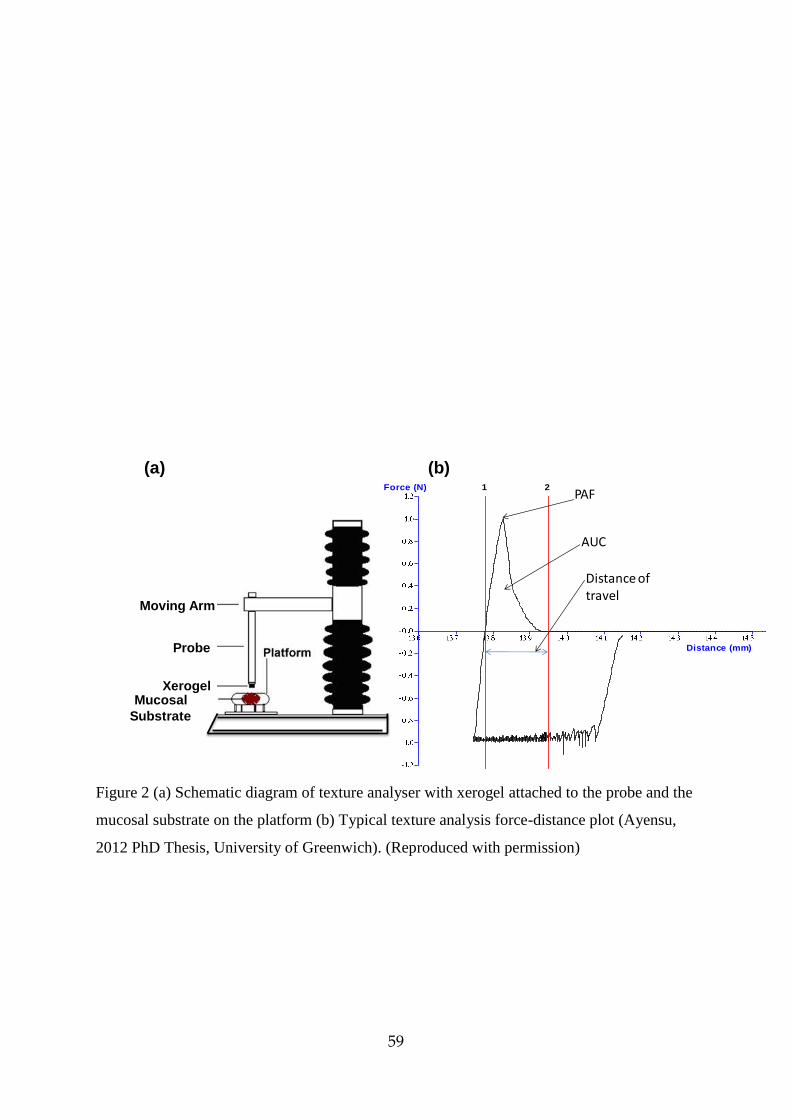
59
Figure 2 (a) Schematic diagram of texture analyser with xerogel attached to the probe and the
mucosal substrate on the platform (b) Typical texture analysis force-distance plot (Ayensu,
2012 PhD Thesis, University of Greenwich). (Reproduced with permission)
Moving Arm
Probe
XerogelMucosal
Substrate
Force (N)
Distance (mm)
1 2PAF
Distance of travel
AUC
(a) (b)

60
Figure 3 Cartoon showing cross section of the eye and illustrating the challenges of ocular
delivery including the various routes via which drugs can be effectively delivered across the
ocular barriers. (Available at: http://www.cchem.berkeley.edu/cjrgrp/group/peng.html -
Accessed 16 March 2015) Reproduced with permission.

61

62
Figure 4 A drawn schematic cross section of the buccal mucosa showing the various layers
requiring penetration to reach the systemic circulation.
Oral epithelial membrane
Basement membrane
Lamina propia
Submucosa containing blood vessels
and nerves
Muscle and / or bone

63
Figure 5 The main regions of the nasal – pharyngeal cortex for potential drug delivery
(Available at:
http://www.urmc.rochester.edu/encyclopedia/content.aspx?ContentTypeID=90&ContentID=
P02027) (Accessed 16 March 2015). Reproduced with permission.

64
Figure 6 Cross section of the lumen of the small intestines, showing the various components
within the lumen and on the mucosa lining. Reproduced with permission from [165].

65
Figure 7 Structure of the walls of the vaginal wall including the mucosal epithelium.
Available at http://teachmeanatomy.info/wp-content/uploads/Histological-Layers-of-the-
Vagina-TeachMeAnatomy.png (Accessed on 19 March 2015). Reproduced with permission

66
Figure 8 Schematic representation of different stages involved in the wound healing process
(a) wound or injury (b) haemostasis (c) inflammatory phase (d) proliferation (e) remodelling
and scar maturation. Reproduced with permission [243].


