Polysaccharide-Based Nanoparticles for Colon-Targeted Drug ...
22
Review Polysaccharide-Based Nanoparticles for Colon-Targeted Drug Delivery Systems Yubia De Anda-Flores 1, * , Elizabeth Carvajal-Millan 1, * , Alma Campa-Mada 1 , Jaime Lizardi-Mendoza 1 , Agustin Rascon-Chu 2 , Judith Tanori-Cordova 3 and Ana Luisa Martínez-López 4 Citation: De Anda-Flores, Y.; Carvajal-Millan, E.; Campa-Mada, A.; Lizardi-Mendoza, J.; Rascon-Chu, A.; Tanori-Cordova, J.; Martínez-López, A.L. Polysaccharide-Based Nanoparticles for Colon-Targeted Drug Delivery Systems. Polysaccharides 2021, 2, 626–647. https://doi.org/10.3390/ polysaccharides2030038 Academic Editors: Alessandra Braga Ribeiro, Ricardo J. B. Pinto and Karin Stana Kleinschek Received: 30 May 2021 Accepted: 30 July 2021 Published: 7 August 2021 Publisher’s Note: MDPI stays neutral with regard to jurisdictional claims in published maps and institutional affil- iations. Copyright: © 2021 by the authors. Licensee MDPI, Basel, Switzerland. This article is an open access article distributed under the terms and conditions of the Creative Commons Attribution (CC BY) license (https:// creativecommons.org/licenses/by/ 4.0/). 1 Biopolymers-CTAOA, Research Center for Food and Development (CIAD, A.C.), Carretera Gustavo Enrique Astiazarán Rosas No. 46, Hermosillo 83304, Mexico; [email protected] (A.C.-M.); [email protected] (J.L.-M.) 2 Biotechnology-CTAOV, Research Center for Food and Development (CIAD, A.C.) Carretera Gustavo Enrique Astiazarán Rosas No. 46, Hermosillo 83304, Mexico; [email protected] 3 Department of Polymers and Materials Research, University of Sonora, Hermosillo 83000, Mexico; [email protected] 4 NANO-VAC Research Group, Department of Chemistry and Pharmaceutical Technology, University of Navarra, 31008 Pamplona, Spain; [email protected] * Correspondence: [email protected] (Y.D.A.-F.); [email protected] (E.C.-M.) Abstract: Polysaccharide biomaterials have gained significant importance in the manufacture of nanoparticles used in colon-targeted drug delivery systems. These systems are a form of non- invasive oral therapy used in the treatment of various diseases. To achieve successful colonic delivery, the chemical, enzymatic and mucoadhesive barriers within the gastrointestinal (GI) tract must be analyzed. This will allow for the nanomaterials to cross these barriers and reach the colon. This review provides information on the development of nanoparticles made from various polysaccharides, which can overcome multiple barriers along the GI tract and affect encapsulation efficiency, drug protection, and release mechanisms upon arrival in the colon. Also, there is information disclosed about the size of the nanoparticles that are usually involved in the mechanisms of diffusion through the barriers in the GI tract, which may influence early drug degradation and release in the digestive tract. Keywords: nanoparticles; polysaccharide; drug delivery; colon 1. Introduction Polysaccharides are biopolymers constituted by simple sugar monomers. They are usually isolated from plant material, marine plants, and exogenous metabolites of some bacteria. The polysaccharides chain comprises monosaccharide units linked by glycosidic bonds with hydroxyl, carboxyl, and amino groups. Carbohydrates are stable, non-toxic, hydrophilic, and biodegradable biomolecules in the human body [1,2]. Polysaccharides can be used as matrices for encapsulation, immobilization, and controlled release for many active compounds. This matrix are also used in the food, pharmaceutical, biomedical, and chemical industries. Polysaccharides are the most abundant biomolecules, followed by proteins, lipids, and nucleic acids. Polysaccharides studied as a matrix for encapsulation include chitosan, pectin, alginate, starch, and dextran [3,4]. Encapsulation occurs when liquid and solid particles are trapped in a matrix or coated by a polymer material. The preparation of a proper encapsulating agent is an essential factor in enhancing encapsulation efficiency. Encapsulating agents must have the ability to preserve bioactive compounds under different processes and storage conditions [5]. A controlled release system will increase drug retention time and enhance paracellular and transcellular absorption. The release mechanisms occur by diffusion, biodegradation, and osmosis [6,7]. The application of nanotechnology to encapsulate nutrients and drugs has aroused significant interest and demand due to its great benefits. In addition, nanotech- nology has been associated with safety, environmental, ethical, and regulatory problems Polysaccharides 2021, 2, 626–647. https://doi.org/10.3390/polysaccharides2030038 https://www.mdpi.com/journal/polysaccharides
-
Upload
khangminh22 -
Category
Documents
-
view
1 -
download
0
Transcript of Polysaccharide-Based Nanoparticles for Colon-Targeted Drug ...

Review
Polysaccharide-Based Nanoparticles for Colon-Targeted DrugDelivery Systems
Yubia De Anda-Flores 1,* , Elizabeth Carvajal-Millan 1,* , Alma Campa-Mada 1, Jaime Lizardi-Mendoza 1 ,Agustin Rascon-Chu 2 , Judith Tanori-Cordova 3 and Ana Luisa Martínez-López 4
�����������������
Citation: De Anda-Flores, Y.;
Carvajal-Millan, E.; Campa-Mada, A.;
Lizardi-Mendoza, J.; Rascon-Chu, A.;
Tanori-Cordova, J.; Martínez-López,
A.L. Polysaccharide-Based
Nanoparticles for Colon-Targeted
Drug Delivery Systems.
Polysaccharides 2021, 2, 626–647.
https://doi.org/10.3390/
polysaccharides2030038
Academic Editors: Alessandra
Braga Ribeiro, Ricardo J. B. Pinto and
Karin Stana Kleinschek
Received: 30 May 2021
Accepted: 30 July 2021
Published: 7 August 2021
Publisher’s Note: MDPI stays neutral
with regard to jurisdictional claims in
published maps and institutional affil-
iations.
Copyright: © 2021 by the authors.
Licensee MDPI, Basel, Switzerland.
This article is an open access article
distributed under the terms and
conditions of the Creative Commons
Attribution (CC BY) license (https://
creativecommons.org/licenses/by/
4.0/).
1 Biopolymers-CTAOA, Research Center for Food and Development (CIAD, A.C.), Carretera Gustavo EnriqueAstiazarán Rosas No. 46, Hermosillo 83304, Mexico; [email protected] (A.C.-M.); [email protected] (J.L.-M.)
2 Biotechnology-CTAOV, Research Center for Food and Development (CIAD, A.C.) Carretera Gustavo EnriqueAstiazarán Rosas No. 46, Hermosillo 83304, Mexico; [email protected]
3 Department of Polymers and Materials Research, University of Sonora, Hermosillo 83000, Mexico;[email protected]
4 NANO-VAC Research Group, Department of Chemistry and Pharmaceutical Technology,University of Navarra, 31008 Pamplona, Spain; [email protected]
* Correspondence: [email protected] (Y.D.A.-F.); [email protected] (E.C.-M.)
Abstract: Polysaccharide biomaterials have gained significant importance in the manufacture ofnanoparticles used in colon-targeted drug delivery systems. These systems are a form of non-invasive oral therapy used in the treatment of various diseases. To achieve successful colonic delivery,the chemical, enzymatic and mucoadhesive barriers within the gastrointestinal (GI) tract must beanalyzed. This will allow for the nanomaterials to cross these barriers and reach the colon. This reviewprovides information on the development of nanoparticles made from various polysaccharides, whichcan overcome multiple barriers along the GI tract and affect encapsulation efficiency, drug protection,and release mechanisms upon arrival in the colon. Also, there is information disclosed about the sizeof the nanoparticles that are usually involved in the mechanisms of diffusion through the barriers inthe GI tract, which may influence early drug degradation and release in the digestive tract.
Keywords: nanoparticles; polysaccharide; drug delivery; colon
1. Introduction
Polysaccharides are biopolymers constituted by simple sugar monomers. They areusually isolated from plant material, marine plants, and exogenous metabolites of somebacteria. The polysaccharides chain comprises monosaccharide units linked by glycosidicbonds with hydroxyl, carboxyl, and amino groups. Carbohydrates are stable, non-toxic,hydrophilic, and biodegradable biomolecules in the human body [1,2]. Polysaccharidescan be used as matrices for encapsulation, immobilization, and controlled release for manyactive compounds. This matrix are also used in the food, pharmaceutical, biomedical, andchemical industries. Polysaccharides are the most abundant biomolecules, followed byproteins, lipids, and nucleic acids. Polysaccharides studied as a matrix for encapsulationinclude chitosan, pectin, alginate, starch, and dextran [3,4].
Encapsulation occurs when liquid and solid particles are trapped in a matrix or coatedby a polymer material. The preparation of a proper encapsulating agent is an essentialfactor in enhancing encapsulation efficiency. Encapsulating agents must have the abilityto preserve bioactive compounds under different processes and storage conditions [5]. Acontrolled release system will increase drug retention time and enhance paracellular andtranscellular absorption. The release mechanisms occur by diffusion, biodegradation, andosmosis [6,7]. The application of nanotechnology to encapsulate nutrients and drugs hasaroused significant interest and demand due to its great benefits. In addition, nanotech-nology has been associated with safety, environmental, ethical, and regulatory problems
Polysaccharides 2021, 2, 626–647. https://doi.org/10.3390/polysaccharides2030038 https://www.mdpi.com/journal/polysaccharides

Polysaccharides 2021, 2 627
related to human health and environmental impacts. Information related to the security ofnanoparticles is limited. In general, nanoparticles have unique characteristics, such as theirsmall size. This property allows them to cross different biological barriers (e.g., intestinaland mucosal epithelial cells) [8,9].
Natural biopolymers are considered biodegradable and safe for human consumptionbecause they are not toxic and do not affect cell viability, making them ideal for drugdelivery systems [10,11]. Other kinds of metallic, solid, and polymeric nanoparticles canbe toxic for humans. Researchers have used strategies of coating toxic nanoparticles withbiopolymers to reduce their toxicity [12,13]. At the moment, no legislation regulates theuse of nanoparticles in the food and pharmaceutical industries. Therefore, most countrieshave regulations for risk assessment when using nanotechnology. Relevant regulatoryagencies include the U.S. Food and Drug Administration (FDA), the European Union, theAustralian Government Department of Health, and Health Canada [14].
It is necessary to develop and appropriately evaluate nanomaterials to determine theirrisk to human health. When manufacturing these nanoparticles, the elements that must beconsidered are particle and distribution size, shape, state of aggregation/disaggregation,solubility, surface charge, and surface morphology [15,16]. The size and morphologyof nanoparticles enhance their functionality, and are dependent on the manufacturingtechnique for each nanomaterial. These properties allow a high loading capacity, highencapsulation efficiency, stability, sustained release profile, bioavailability of bioactivecompounds, and biocompatibility [17,18].
The nanoparticle technology based on polysaccharides plays a vital role in controllingdrugs, bioactive agents, and genes for oral administrations. These release systems canoccur by diffusion (barrier/matrix), degradation (chemical or physical matrix), or changesin the environment (e.g., pH, ionic strength, and pressure) [19]. Most drugs or bioactiveagents administered orally are absorbed in the upper GI tract, but their delivery in thecolon is necessary for additional results in specific therapies. Some areas of interest arecolonic delivery of peptides and proteins, probiotic bacteria, and microbiota replacementtherapies [20,21]. The main release mechanisms in the colon are degradation by colonicmicrobiota, time and pH-controlled release. Colon release formulations are generallydesigned to prevent degradation in the stomach and upper GI tract. The colon’s mainfunction is the absorption of water, ions, and the storage of feces. The large intestineis colonized by many bacteria—approximately 1012 per gram of intestinal content. Themicroorganisms in the colon are beneficial for human health because they are responsiblefor fermenting indigestible dietary fiber [22].
This review focuses on polysaccharide-based nanoparticles used for oral adminis-tration in colon-directed therapies, such as drug administration, peptides, and proteins.This contribution aims to present important information from the literature that showsvarious polysaccharide-based nanomaterials. Therefore, these nanomaterials can reach thecolon through physiological barriers due to their morphological characteristics, such assize, shape, and surface charge.
2. Polysaccharides
Various polymers with complex structures and specific functions have developednaturally. These include amino acids, nucleobases, and mono- and disaccharides. Accord-ing to the nature of their heteroatom present in the main chain natural polymers can beclassified into four types, as shown in Table 1 [23,24]. Polysaccharides are biopolymersisolated from plant, animal, microbial, and algae sources. These are made up of more thanten monosaccharide units linked by O-glycosidic bonds. Due to their great abundancein hydroxyl groups polysaccharides can be modified by carboxylation, esterification, andamination, thereby improving their functional properties [25]. According to their nature,polysaccharides have excellent biocompatibility, biodegradability, non-toxicity, and cellspecificity. Therefore, they are considered ideal for various biomedical applications, suchas drug, gene delivery, and wound dressing [26,27].

Polysaccharides 2021, 2 628
Table 1. Natural polymers and their types.
Natural Polymers Types Examples
Hydrocarbon polymers Natural rubber
Carbon-oxygen Carbohydrates Cellulose; starch; chitin; chitosan;pullulan
Carbon-oxygen-nitrogen/Sulphur Proteins Soya protein; gelatin; casein
Carbon-oxygen-nitrogen-phosphorus Nucleic acids DNA, RNA
2.1. Chitosan
Chitosan is a unique linear poly-cationic polysaccharide derived from chitin bydeacetylation. It is found primarily in the exoskeleton of crustaceans, such as shrimpand crabs. Its chemical structure consists of a chain of β-(1-4)-linked glucosamine andN-acetyl D-glucosamine units. It has functional groups such as polyhydroxy and amino,and these groups can lead to hydrogen bonds. Chitosan is the second most abundantpolysaccharide after cellulose. It is used for pharmaceutical applications due to its reactivefunctional groups, biocompatibility, biodegradability, gel-forming ability, non-toxicity, highcharge density, and low pH solubility. This polysaccharide can interact electrostaticallywith mucus or negatively charged mucosal surfaces, giving it an excellent mucoadhesiveproperty for developing systems for oral administration. The presence of its positivelycharged amino group (–NH3+) can interact with negatively charged proteoglycans on thecell surface, enabling better intestinal absorption of the drug by opening the tight junctionsbetween the epithelial cells [28].
2.2. Hyaluronic Acid
Hyaluronic acid is a non-sulfated, negatively charged glycosaminoglycan composedof D-glucuronic acid and N-acetyl-D-glucosamine linked by β-(1-3) and β-(1-4) bonds. Itis a macromolecule produced and secreted by cells as a linear polymer that is not boundto a polypeptide. It is a biocompatible biopolymer of natural origin present in the skin,connective tissues, synovial fluid of the joints, neural tissues, vitreous humor, and has theability to regulate lubrication. This polysaccharide can be chemically modified due to itsgroups, such as the carboxylic acid of glucuronic acid, primary and secondary hydroxylgroups, and N-acetyl groups, which can alter its properties such as hydrophobicity, bio-logical activity, viscoelasticity, water retention, biocompatibility, cell proliferation, woundregeneration, and specific signal transduction and cell interactions through cell surfacereceptors [29].
2.3. Pectin
Pectin is a linear heteropolysaccharide that constitutes the cell wall of plants andconsists of a linear unbranched chain of α-(1-4) linked D-galacturonic acid units (homo-galacturonan) with uronic acids. This polysaccharide contains hydroxyl and carboxylgroups, residues esterified with methyl ether distributed in its linear chain, and a certainquantity of neutral sugars present in the side chains. It is principally composed of galac-turonic acid, methyl ester, and sugar units such as arabinose, galactose, and rhamnose.Pectin can form hydrogels in the presence of its ionized carboxyl groups (–COO−) thatinteract with its positively charged anions. For this reason, it is an anionic, biodegradable,biocompatible, and non-toxic polysaccharide used for its mucoadhesive properties in theoral delivery of drugs to the colon. Pectin can remain intact in the upper gastrointestinaltract and be degraded in the colon by pectinases [30].
2.4. Guar Gum
Guar gum is a non-ionic polysaccharide. It is extracted from the seeds of Cyamopsistetragonolobus and consists of a linear chain of (1-4)-β-D-mannopyranosil units with α-D-

Polysaccharides 2021, 2 629
galactopyranosyl units attached by (1–6) ramifications. This polysaccharide presents anextraordinarily viscous property due to its intermolecular chain entanglement of galactoseside chains. It is a biocompatible and biodegradable polysaccharide with the ability to formgels in aqueous solutions, and is used to formulate hydrophilic matrices for drug deliverydue to its enzymatic degradation in the colon [31].
2.5. Dextran
Dextran is a complex branched glucan consisting of α-D-(1-6) glycosidic links andbranched at α-(1-3). It is obtained naturally from the lactic acid bacterium. It is a hy-drophilic, biodegradable, and biocompatible polysaccharide with abundant hydroxylgroups. This polysaccharide is biocompatible, highly hydrophilic, and shows low proteinadsorption. Like other polysaccharides, dextran has many hydroxyl groups, allowing itto be easily conjugated with drugs and proteins to prevent drug absorption in the smallintestine [32].
2.6. Alginate
Alginate is an anionic polysaccharide consisting of β-(1-4)-D-mannuronic acid andα-(1-4) L-guluronic acid residues. The α-(1-4) L-guluronic bonds can be cross-linked withdivalent ions by sodium ion exchange, forming a gel matrix and the retention of encapsu-lated charges. Due to their anionic nature, they can interact with cationic compounds. It isextracted mainly from brown marine algae (Laminaria hyperborean) and soil bacteria (Azobac-ter vinelandii). This polysaccharide has physicochemical properties such as biodegradability,biocompatibility, low immunogenicity, good mucoadhesion, and non-toxicity. Due toits composition, sequence of arrangement, and molecular weight, the alginate also hasfunctional groups such as polyhydroxy and carboxyl distributed throughout its chain. It ishighly reactive and with the possibility of chemical modification (oxidation, amidation,esterification, and sulphation) [33].
2.7. Arabinoxylans
Arabinoxylans (AX) are non-starch polysaccharides found mainly in the cell wall,outer layer, and endosperm of cereals. The AX are composed of a linear chain of β-(1-4)xylose units branched to arabinose units in positions C(O)-3 and C(O)-2. Arabinose can beesterified with monomeric or dimeric ferulic acid (AF). AX are desirable polysaccharidesfor application in the food, pharmaceutical, and biomedical industries. Similarly, theyhave become an attractive alternative due to their biodegradability, biocompatibility, non-toxicity, hydrophilic, and gelling properties. Due to their functional properties, AX can actas prebiotics, antioxidants, emulsifiers, and immunomodulators. The gels made from thispolysaccharide have been studied for their potential as a drug delivery system directedto the colon because they are biocompatible and have the ability to retain water. Thesecovalent gels can cross the conditions of the upper gastrointestinal tract and be fermentedin the colon by the colonic microbiota to release the drug [34,35].
3. Polysaccharide Based-Nanoparticles
Drugs can be loaded into a polysaccharide matrix or be bound to the external surface,improving their aqueous solubility and stability (e.g., proteins). They are used to enhance slowenzymatic degradation, targeting mucosal recognition, receptor binding, adhesion, and trans-port systems [36,37]. Polysaccharides have presented advantages and benefits when they areused as biomaterials for the manufacture of nanoparticles. Compared to nanoparticles synthe-sized from other materials, such as metals or synthetic polymers, polysaccharide nanoparticlesdo not present toxicity problems due to their biocompatibility and biodegradability, thusproviding safety, better storage, and physiological stability [38,39].
Nanotechnology-based drug delivery systems aim to target different routes of admin-istration. When designing polysaccharide-based nanoparticles for use in biomedicine andother industries, it is important to consider the route of administration, retention time, and

Polysaccharides 2021, 2 630
therapy [40]. These factors may be dependent on the polysaccharide used, its size, andits structure. One possible disadvantage when using polysaccharides for manufacturingnanoparticles is their high molecular weights. In addition, in many cases, the low solu-bility of some polysaccharides limits their potential in chemical modification [27]. Dueto the polysaccharide properties, the size of polysaccharide-based nanoparticles has beenconsidered between 10 and 1000 nm [41,42].
These nanoparticles can carry drugs, proteins, peptides, or DNA material to target aspecific organ or cell. Depending on the technique, the drug or other active compound canadhere, dissolve, encapsulate, or entrap into the nanoparticle matrix [43]. The significantinterest in this technology is due to the fact that the nanoparticles can target specific organsor Diana cells, and reduce side effects. Nanoparticles have a larger surface area, andtheir smaller size allows them to enter smaller capillaries and cells. Nanoparticle sizeand surface charge are highly important because many secondary properties, such assurface area, toxicity, degradation, targeting, and absorption mechanism, are related [18].Polysaccharide-based nanoparticles have shown significant potential in biological andpharmaceutical applications. Natural polysaccharides used to fabricate nanoparticlesinclude chitosan, alginate, pectin, guar gum, dextran, hyaluronic acid, and arabinoxylan(Table 2).
Previously studied polysaccharides used to fabricate nanoparticles include the manu-facture of acrylate chitosan-based nanoparticles cross-linked with tripolyphosphate anions(TPPs). The authors obtained chitosan nanoparticles with an average diameter of 356 nmand a Z potential of 13.7 mV [44]. In another study, nanoparticles were synthesized by β-lactoglobulin-pectin to encapsulate an anticancer drug (bipyridine ethyl dithiocarbamate Pt(II) nitrate), and they obtained nanoparticles with sizes of 200–250 nm [45]. The fabricatednanoparticles based on bovine serum albumin (BSA)-dextran conjugate to encapsulate cur-cumin showed nanoparticles with a size of 115 nm [46]. Similarly, arabinoxylan cross-linkedwith laccase was developed and showed a hydrodynamic diameter of 328 nm [47].
Table 2. Natural polysaccharides, properties, and their degradation mechanism.
Polysaccharide ChainCharacteristics Properties Crosslinker Degradation
Mechanism Comments Reference
Chitosanβ-(1-4)-linked
N-acetylD-glucosamine
-Positive Zpotential-pKa 6.5-Protonated atacidic and neutralpH-Mucoadhesive-Cationic
CalciumTPP Enzymatic
Degradation byenzymes
(β-glucosidase)[48]
Alginate
β-(1-4)-D-mannuronic acid
and α-(1-4)L-guluronic acid
residues
-Water-soluble-Anionic-Hydrophobicity
Divalentcations
Ca2+, Cu2+,Zn2+ or Mn2+
pH-responsive
Degradation byenzymes (glu-curonidases
etc.)
[33,49]
Pectin(1-4)-linked
α-D-galacturonicacid residues
-Water-soluble-High methoxy-Low methoxy
Ca2+ ionsLacasse
EnzymaticDegradation by
Bacteroidesspecies
[50,51]
Guar Gum
(1-4)- β-D-mannopyranoseunits with α-D-
galactopyranosylunits attached by
(1-6) linkages
-Water-soluble Epichlorohydrin pH-responsiveand enzymatic
Degradation bybacteria
(Bacteroides,ruminococci,
bifidobacteria)
[19,31]
Dextranα-(1-6) glycosidic
links and branchedat α-(1-3) position
-Mucoadhesive-Hydrophilic Diamine Enzymatic
Degradation byesterases and
endodex-tranases
[52]
Polysaccharides 2021, 2 631
Table 2. Cont.
Polysaccharide ChainCharacteristics Properties Crosslinker Degradation
Mechanism Comments Reference
HyaluronicAcid
D-glucuronic acidand N-acetyl-D-
glucosamine linkedby β-(1-3) andβ-(1-4) bond
-Anionic-Water-absorption-Water soluble
pHtemperature
ionic
pH-responsiveand enzymatic
Degradation byhyaluronidases [53]
Arabinoxylan
Xylose β-1-4linkages andα-L-arabinose
substitutions (α-1-3and α-1-2)
-Highly ferulated-Water-soluble-Neutral
LaccasePeroxidase Enzymatic
Degradation bybacteria (Bifi-dobacterium
andBacteroides)
[34,54]
(TPP) sodium tripolyphosphate.
3.1. Physicochemical Characteristics of Polysaccharide-Based Nanoparticles3.1.1. Size and Shape
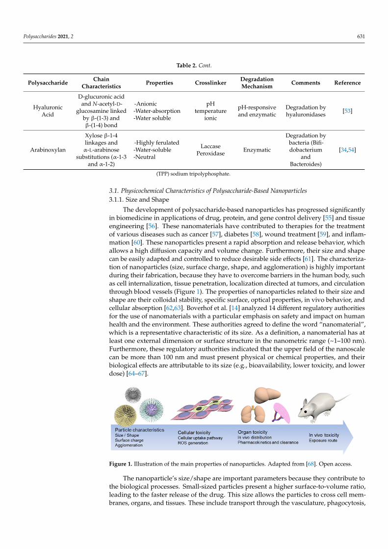
The development of polysaccharide-based nanoparticles has progressed significantlyin biomedicine in applications of drug, protein, and gene control delivery [55] and tissueengineering [56]. These nanomaterials have contributed to therapies for the treatmentof various diseases such as cancer [57], diabetes [58], wound treatment [59], and inflam-mation [60]. These nanoparticles present a rapid absorption and release behavior, whichallows a high diffusion capacity and volume change. Furthermore, their size and shapecan be easily adapted and controlled to reduce desirable side effects [61]. The characteriza-tion of nanoparticles (size, surface charge, shape, and agglomeration) is highly importantduring their fabrication, because they have to overcome barriers in the human body, suchas cell internalization, tissue penetration, localization directed at tumors, and circulationthrough blood vessels (Figure 1). The properties of nanoparticles related to their size andshape are their colloidal stability, specific surface, optical properties, in vivo behavior, andcellular absorption [62,63]. Boverhof et al. [14] analyzed 14 different regulatory authoritiesfor the use of nanomaterials with a particular emphasis on safety and impact on humanhealth and the environment. These authorities agreed to define the word “nanomaterial”,which is a representative characteristic of its size. As a definition, a nanomaterial has atleast one external dimension or surface structure in the nanometric range (~1–100 nm).Furthermore, these regulatory authorities indicated that the upper field of the nanoscalecan be more than 100 nm and must present physical or chemical properties, and theirbiological effects are attributable to its size (e.g., bioavailability, lower toxicity, and lowerdose) [64–67].
Polysaccharides 2021, 4, x 7 of 23
shape are their colloidal stability, specific surface, optical properties, in vivo behavior, and cellular absorption. [62,63]. Boverhof et al. [14] analyzed 14 different regulatory authori-ties for the use of nanomaterials with a particular emphasis on safety and impact on hu-man health and the environment. These authorities agreed to define the word “nano-material,” which is a representative characteristic of its size. As a definition, a nano-material has at least one external dimension or surface structure in the nanometric range (~1–100 nm). Furthermore, these regulatory authorities indicated that the upper field of the nanoscale can be more than 100 nm and must present physical or chemical properties, and their biological effects are attributable to its size (e.g., bioavailability, lower toxicity, and lower dose) [64–67].
Figure 1. Illustration of the main properties of nanoparticles. Adapted from [68]. Open access.
The nanoparticle’s size/shape are important parameters because they contribute to the biological processes. Small-sized particles present a higher surface-to-volume ratio, leading to the faster release of the drug. This size allows the particles to cross cell mem-branes, organs, and tissues. These include transport through the vasculature, phagocyto-sis, endocytosis, and intracellular transport. However, the size should not be too small because it has been established that the size threshold in renal excretion is usually 6 to 8 nm. In addition, sizes smaller than 6 nm can be coated with serum proteins circulating through the bloodstream, which favors an increase in the hydrodynamic diameter, block-ing renal excretion [69]. Conversely, nanoparticles larger than 100 nm are more likely to accumulate at the injection site or be trapped by macrophages in the spleen, lung, and liver [70].
3.1.2. Superficial Charge and Aggregation The surface charge measures the Z potential and represents a simple measurement
of the particles electrical characteristics. This property determines the stability of colloidal dispersions and is affected by the ionic strength and pH of the dispersion. The Z potential indicates the degree of repulsion between adjacent particles, charged in a dispersion [19]. Aggregation occurs when several particles are joined together strongly, resulting in the sum of surface areas of the individual particles. The size and shape of the aggregation have a significant influence on the properties of the nanomaterial. These aggregates are
Figure 1. Illustration of the main properties of nanoparticles. Adapted from [68]. Open access.
The nanoparticle’s size/shape are important parameters because they contribute tothe biological processes. Small-sized particles present a higher surface-to-volume ratio,leading to the faster release of the drug. This size allows the particles to cross cell mem-branes, organs, and tissues. These include transport through the vasculature, phagocytosis,

Polysaccharides 2021, 2 632
endocytosis, and intracellular transport. However, the size should not be too small becauseit has been established that the size threshold in renal excretion is usually 6 to 8 nm. Inaddition, sizes smaller than 6 nm can be coated with serum proteins circulating throughthe bloodstream, which favors an increase in the hydrodynamic diameter, blocking renalexcretion [69]. Conversely, nanoparticles larger than 100 nm are more likely to accumulateat the injection site or be trapped by macrophages in the spleen, lung, and liver [70].
3.1.2. Superficial Charge and Aggregation
The surface charge measures the Z potential and represents a simple measurement ofthe particles electrical characteristics. This property determines the stability of colloidaldispersions and is affected by the ionic strength and pH of the dispersion. The Z potentialindicates the degree of repulsion between adjacent particles, charged in a dispersion [19].Aggregation occurs when several particles are joined together strongly, resulting in thesum of surface areas of the individual particles. The size and shape of the aggregationhave a significant influence on the properties of the nanomaterial. These aggregates areindivisible structures and cannot be separated by external forces [14]. The surface chargedetermines the dispersion, aggregation, or flocculation of the particles. It can influencestability, and a neutral charge leads to instability of the nanoparticles, causing precipitation.A high Z potential confers stability to the particles, thus preventing their aggregation.When this value is low, the particles tend to attract each other and form flocs. Negativelycharged nanoparticles have low phagocytic absorption, increasing their time in the blood-stream. Additionally, positively charged particles cause increased phagocytosis due tothe interaction of the anionic cell membrane with the nanoparticles [71]. As the particlepasses through the systemic circulation, a positive surface charge facilitates interaction withplasma proteins, resulting in aggregation. Therefore, the charge density in the nanoparticleswhen interacting with body fluids can be altered [18,69,72]. The use of nanotechnology indrug delivery enables better targeting of delivery to cells and tissues [55].
3.2. Cellular Uptake Mechanisms
The cellular uptake mechanisms to deliver biological substances and therapeuticagents require internalization to specific compartments or organelles to affect a cellularlevel. First, nutrient uptake, communication between cells, their microenvironment, andcell adhesion occur through the plasma membrane by different mechanisms. Due to of theirconcentration gradients, hydrophobic or nonpolar compounds can easily diffuse throughthe plasma membrane. Then, ions and amino acids are transported across the plasmamembrane by active transport mechanisms, such as integral membrane protein pumpsor ion channels. Finally, nanoscale hydrophilic biomacromolecules are conventionallytransported into the cell by endocytosis [73].
Endocytosis is an active transport process in which a cell membrane wraps itselfaround an object and encloses it in vesicles or vacuoles pinched off from its cytoplasmicmembrane. Endocytosis is divided into two main mechanisms: phagocytosis and pinocyto-sis (Figure 2). Phagocytosis is the primary uptake for larger particles by the cell membraneforming an internal phagosome, and occurs principally in certain types of phagocytes(macrophages and neutrophils). Phagocytes can take up relatively large particles (1 mi-cron). This mechanism occurs when a particle, cells, and pathogens are recognized byphagocytes circulating through the blood and tissues. They attach to the phagocytes’ sur-face receptors, allowing further internalization. Once recognized, the plasma membrane ofphagocytes encloses the particles within a cup-shaped protrusion and fuses to produce aphagosome that is pinched off inward [74].
Pinocytosis highlights the uptake of smaller particles. Pinocytosis is subdivided intoclathrin-dependent and clathrin-independent pathways, divided into caveolae-mediatedendocytosis, micropinocytosis, and other endocytic mechanisms. The clathrin-mediatedendocytosis mechanism is the main endocytosis pathway, through which cells absorbnecessary materials. This is known as receptor-mediated uptake, which involves the

Polysaccharides 2021, 2 633
capture of particular macromolecules (ligands) or ligand-coated particles after attaching toreceptors on the cell membrane’s surface. This binding (receptor-ligand) initiates a processin the plasma membrane. It results in the formation of “coated pits”, where the receptorconcentration is greater than that in the remaining area of the cell membrane. Non-specificadsorptive uptake is a receptor-independent mechanism, and the uptake occurs throughhydrophobic or electrostatic interactions, eventually allowing the internalization process.The macropinocytosis mechanism is a process that allows the uptake of larger particles thatcannot easily cross other endocytosis pathways. In this mechanism, the plasma membranegenerates ruffles of diverse shapes that absorb extracellular fluids via closing the membraneruffles and forming large vesicles (macropinosomes) [75].
Polysaccharides 2021, 4, x 9 of 23
Figure 2. Different mechanisms of endocytosis. Adapted from [76]. Open access.
3.3. Polysaccharide Based-Nanoparticles and their Impact on Cellular Uptake The biological response of the nanoparticles is measured in animal cell culture stud-
ies before starting in vivo administration [77]. In a biological system, nanoparticles can easily pass through the membranes and most biological barriers, and easily penetrate the body, cells, organs, and tissues, presenting problems such as potential toxicity [8]. The physicochemical characteristics of the nanoparticles contribute to the cellular uptake mechanisms. The smaller sizes of the nanoparticles allow them to internalize capillaries and thus easily target the cell, providing greater therapeutic efficiency and less toxicity. Materials internalized by endocytosis are delivered to lysosomes and are subject to deg-radation after the cleavage of the budding vesicles. Delivery of nanoparticles into lyso-somes via clathrin-mediated endocytosis has been a widely used route for drug delivery [78]. The intracellular disposition of chitosan nanoparticles in macrophages has been pre-viously studied. Chitosan nanoparticles with a size of 250 nm were prepared and collo-cated in a murine macrophage cell line (RAW 264.7) as a model macrophage. The results showed that the nanoparticles follow two mechanisms of macrophage (clathrin-mediated endocytosis pathway and phagocytosis) internalization, indicating cellular digestion of the nanoparticles [79]. In another study, alginate-based nanoparticles were fabricated. The in vitro study was performed in Caco-2 cells. The results showed that nanoparticles with a size of 50/120 nm internalized via clathrin-mediated endocytosis, and the mechanisms for nanoparticles with sizes of 420 and 730 nm were caveolae-mediated endocytosis and macropinocytosis, respectively [80]. In addition, in a study using chitosan-based nanopar-ticles (CS-NPs), the CS-NPs showed an average diameter of 281 nm and a negative surface charge (−0.3 mV). The cell experiments indicated that CS-NPs presented higher cell inter-nalization efficiency in Raw 264.7 macrophages. These results show excellent potential for the use of these CS-NPs as a macrophage-targeting drug delivery system in further studies [81].
Nanoparticle behavior and motion in blood flow, membrane adhesion strength, and cell uptake pathways and effectiveness are influenced by particle shape. The surface charge of nanoparticles can also influence the uptake mechanism. Positively charged na-noparticles can be successfully absorbed by cells through electrostatic interactions with
Figure 2. Different mechanisms of endocytosis. Adapted from [76]. Open access.
3.3. Polysaccharide Based-Nanoparticles and their Impact on Cellular Uptake
The biological response of the nanoparticles is measured in animal cell culture stud-ies before starting in vivo administration [77]. In a biological system, nanoparticles caneasily pass through the membranes and most biological barriers, and easily penetrate thebody, cells, organs, and tissues, presenting problems such as potential toxicity [8]. Thephysicochemical characteristics of the nanoparticles contribute to the cellular uptake mech-anisms. The smaller sizes of the nanoparticles allow them to internalize capillaries and thuseasily target the cell, providing greater therapeutic efficiency and less toxicity. Materialsinternalized by endocytosis are delivered to lysosomes and are subject to degradationafter the cleavage of the budding vesicles. Delivery of nanoparticles into lysosomes viaclathrin-mediated endocytosis has been a widely used route for drug delivery [78]. Theintracellular disposition of chitosan nanoparticles in macrophages has been previouslystudied. Chitosan nanoparticles with a size of 250 nm were prepared and collocated in amurine macrophage cell line (RAW 264.7) as a model macrophage. The results showedthat the nanoparticles follow two mechanisms of macrophage (clathrin-mediated endo-cytosis pathway and phagocytosis) internalization, indicating cellular digestion of thenanoparticles [79]. In another study, alginate-based nanoparticles were fabricated. Thein vitro study was performed in Caco-2 cells. The results showed that nanoparticles witha size of 50/120 nm internalized via clathrin-mediated endocytosis, and the mechanismsfor nanoparticles with sizes of 420 and 730 nm were caveolae-mediated endocytosis andmacropinocytosis, respectively [80]. In addition, in a study using chitosan-based nanopar-

Polysaccharides 2021, 2 634
ticles (CS-NPs), the CS-NPs showed an average diameter of 281 nm and a negative surfacecharge (−0.3 mV). The cell experiments indicated that CS-NPs presented higher cell in-ternalization efficiency in Raw 264.7 macrophages. These results show excellent potentialfor the use of these CS-NPs as a macrophage-targeting drug delivery system in furtherstudies [81].
Nanoparticle behavior and motion in blood flow, membrane adhesion strength, andcell uptake pathways and effectiveness are influenced by particle shape. The surface chargeof nanoparticles can also influence the uptake mechanism. Positively charged nanoparticlescan be successfully absorbed by cells through electrostatic interactions with the negativelycharged cell plasma membrane (clathrin-mediated endocytosis). In contrast, negativelycharged nanoparticles are more likely to use caveolae-mediated routes [82].
The cytotoxicity of chitosan nanoparticles in Caco-2 cells was evaluated previously [83].The nanoparticles had sizes of 25 and 333 nm and a Z potential of 5.3 mV. The resultsshowed that in vitro cytotoxicity was mainly influenced by the particle size and not by itssurface charge. The Caco-2 cells were more sensitive to cytotoxicity with 25 nm nanoparti-cles because these were able to internalize the cells. Similarly, silica nanoparticles (SiNPs)coated with chitosan (CH) and alginate (A) for the encapsulation of insulin have beenpreviously reported [13]. The SiNP-CHs were synthesized with sizes of ~600 nm and witha Z potential of −13 mV, and the SiNP-A presented sizes of ~390 nm and a Z potentialof +19 mV. Cytotoxicity was evaluated by the viability of HepG2 and Caco-2 cells. Atconcentrations of 100 µg/mL, nanoparticles coated with chitosan and alginate did notshow toxicity. In the same manner, insulin-loaded chitosan-pectin nanoparticles weredeveloped [84]. Particles with a size of 240–420 nm were obtained, and their cell viabilitywas evaluated using Caco-2 cells. The particles loaded with insulin showed a moderatedecrease in cell viability, which could be attributed to the degree of acetylation of chitosanand the presence of insulin; similarly, this did not represent a toxicity problem. In addition,chitosan-g-poly(methyl methacrylate) nanoparticles thiolate by conjugating N-acetyl cys-teine were synthesized [85]. The authors obtained nanoparticle sizes ranging from 100 to330 nm and a positive surface charge due to the presence of free amide groups protonatedto chitosan. Cell viability results using Caco-2 and HT29-MTX cells showed that, after4 h, nanoparticles had viability > 90%, making it acceptable for a non-cytotoxic material.The permeability studies showed that the chitosan-based nanoparticles penetrated the cellmonolayer in which mucus is present.
4. Colon
The colon, or large intestine, extends from the ileum to the rectum, with a lengthof approximately 1.5 m and an average diameter of 6 cm. It is composed primarily ofthe cecum, ascending, transverse, descending, and sigmoid colons, the rectum, and anus.A complex layer of mucosa covers the colon. The mucus secreted by the large intestinemucosa is responsible for protecting the intestinal wall from excoriation and bacterialactivity. It provides the medium for agglutination of feces [86,87]. The colon’s mainfunction is to absorb the remainder of the water and salts secreted from the small intestine.It is responsible for bacterial fermentation, synthesis of small amounts of vitamins, storage,and excretion of feces. The microbiota that resides in the colon ferments the dietaryfiber from the carbohydrates in the diet. The products of its digestion are gases (e.g.,hydrogen, carbon dioxide, nitrogen, and sometimes methane) and short-chain fatty acids(e.g., acetic, propionic, butyric, and lactic acid) [88,89]. The pH from the cecum to therectum varies from 5.5 to 7; the change in pH is influenced by the diet and people’shealth status [22]. The colon is inhabited by abundant microbiota (400 different species) ofaerobic and anaerobic microorganisms. The microbiota has a significant impact on the hostphysiology, energy homeostasis, immune system, digestion, and the synthesis of vitamins.The colonic microbiota is mostly anaerobic, and the main species belong to the generaBacteroides, Bifidobacterium, Eubacterium, and Lactobacillus. The colon harbors a populationof 1011 bacteria per gram of intestinal content. These microorganisms produce a large

Polysaccharides 2021, 2 635
panel of enzymes responsible for breaking dietary fibers that are not digested in the smallintestine [88,90,91].
The colon does not have villi as observed in the small intestine; instead, it has acrescentic mucosal fold called the plica semilunaris which increases the internal surfacearea of the colon by about 1300 cm2. The colon is composed of four prominent layers:(1) Serous (squamous epithelium) is found in regions of the colon with a peritoneal surface.(2) Muscular regions are groups of autonomous muscles that control movement andmixing within the colon. (3) Submucosa comprises connective tissue containing bloodand lymphatic vessels, fat cells, and nerve ganglia. The ganglia communicate with theinnermost layer of the colon: the mucosa. (4) The mucosa is subdivided into three layers: themuscularis mucosa, lamina propria, and epithelium (Figure 3) [86]. The epithelium secretesthe mucosa from the stomach to the colon. This secretion forms an adherent gel (mucus) onthe surface of the mucous membranes. Its function is to act as a protective barrier betweenthe underlying epithelium and the lumen due to harmful agents, destructive hydrolases,and microorganisms. It also acts as a lubricant to facilitate the passage of digestive material,protecting the underlying epithelium from mechanical stress and providing an essentialenvironment for the enteric microbiota. The colon mucus is a very sticky translucent gelwith a thickness of 830 µm [92].
Polysaccharides 2021, 4, x 11 of 23
within the colon. (3) Submucosa comprises connective tissue containing blood and lym-phatic vessels, fat cells, and nerve ganglia. The ganglia communicate with the innermost layer of the colon: the mucosa. (4) The mucosa is subdivided into three layers: the muscu-laris mucosa, lamina propria, and epithelium (Figure 3) [86]. The epithelium secretes the mucosa from the stomach to the colon. This secretion forms an adherent gel (mucus) on the surface of the mucous membranes. Its function is to act as a protective barrier between the underlying epithelium and the lumen due to harmful agents, destructive hydrolases, and microorganisms. It also acts as a lubricant to facilitate the passage of digestive mate-rial, protecting the underlying epithelium from mechanical stress and providing an essen-tial environment for the enteric microbiota. The colon mucus is a very sticky translucent gel with a thickness of 830 µm [92].
Figure 3. Histological layers of the colon. Adapted from [93]. Rights holder: © (2021) Terese Wins-low LLC, U.S. Govt. has certain rights.
5. Colon-Targeted Drug Delivery Colon-targeted drug delivery is widely studied for the local treatment of various dis-
eases, such as ulcerative colitis, colonic pathologies [60] colon cancer [94], and in the sys-temic administration of drugs, including proteins and peptides [95]. The administration of drugs and peptides for colon-targeted delivery is a challenge because they have to pass through the upper GI tract and not be absorbed. Thus, it is imperative to protect the drug during its journey through the GI tract [96]. The colon has been studied as a suitable site for the absorption of drugs, peptides, and proteins because it has a neutral pH and less diversity of digestive enzymes. In addition, the mucosa of the colon facilitates the absorp-tion of several drugs and the colon’s proteolytic activity is less than that in the small in-testine. The colon also has a long residence time of 5 days [90].
Polysaccharide-based nanoparticles protect drugs, peptides, and proteins from hy-drolysis and enzymatic degradation in the duodenum and jejunum, thus enabling proper release of the drug in the ileum or colon, improving its systemic bioavailability [97]. The administration of a drug to the colon can be oral or rectal [98]. Oral administration is the most convenient and preferred route for people because it is painless, non-invasive, and easy to handle [99]. The best candidates for colon-targeted delivery are drugs that show poor stomach absorption to the intestine (peptides and proteins) [100]. The development of formulations and new technologies, such as nanoparticles, for the delivery of drugs, peptides, and proteins depends on the physicochemical nature of the drug, the bio-
Figure 3. Histological layers of the colon. Adapted from [93]. Rights holder: © (2021) Terese WinslowLLC, U.S. Govt. has certain rights.
5. Colon-Targeted Drug Delivery
Colon-targeted drug delivery is widely studied for the local treatment of variousdiseases, such as ulcerative colitis, colonic pathologies [60] colon cancer [94], and in thesystemic administration of drugs, including proteins and peptides [95]. The administrationof drugs and peptides for colon-targeted delivery is a challenge because they have to passthrough the upper GI tract and not be absorbed. Thus, it is imperative to protect the drugduring its journey through the GI tract [96]. The colon has been studied as a suitablesite for the absorption of drugs, peptides, and proteins because it has a neutral pH andless diversity of digestive enzymes. In addition, the mucosa of the colon facilitates theabsorption of several drugs and the colon’s proteolytic activity is less than that in the smallintestine. The colon also has a long residence time of 5 days [90].
Polysaccharide-based nanoparticles protect drugs, peptides, and proteins from hy-drolysis and enzymatic degradation in the duodenum and jejunum, thus enabling properrelease of the drug in the ileum or colon, improving its systemic bioavailability [97]. Theadministration of a drug to the colon can be oral or rectal [98]. Oral administration is the

Polysaccharides 2021, 2 636
most convenient and preferred route for people because it is painless, non-invasive, andeasy to handle [94]. The best candidates for colon-targeted delivery are drugs that showpoor stomach absorption to the intestine (peptides and proteins) [99]. The developmentof formulations and new technologies, such as nanoparticles, for the delivery of drugs,peptides, and proteins depends on the physicochemical nature of the drug, the biomate-rial, and the therapy. Carriers such as biopolymer nanoparticles can be used as matrices,hydrogels, or coating agents. This type of carrier must protect the drug, peptides, andproteins from the challenging conditions of the stomach and intestine, increase intestinalabsorption in the bloodstream, allow specific cells in the human body to be reached, andensure controlled release [100,101].
6. Factors That Influence Colon-Targeted Drug Delivery
The colon as a drug delivery site offers excellent advantages due to its pH, residencetime, and low enzyme activity (Figure 4). The administration of drugs to the colon dependson its physiological factors to ensure optimal efficiency after oral administration. Thenanoparticles must be manufactured considering their residence time in the GI tract, theenvironment (pH, microorganism, food, etc.), and the intestinal fluid volume [102]. For oraladministration of drugs, peptides, and proteins, it is necessary to cross multiple barrierspresent in the GI tract, which are designed to break down nutrients and prevent the entryof pathogens. The barriers that influence the arrival and absorption of drugs, peptides, andproteins in the colon are classified into chemical, enzymatic, and mucus barriers [15]. Eachis described below, and nanoparticle studies are shown in Table 3.
Polysaccharides 2021, 4, x 12 of 23
material, and the therapy. Carriers such as biopolymer nanoparticles can be used as ma-trices, hydrogels, or coating agents. This type of carrier must protect the drug, peptides, and proteins from the challenging conditions of the stomach and intestine, increase intes-tinal absorption in the bloodstream, allow specific cells in the human body to be reached, and ensure controlled release [101,102].
6. Factors That Influence Colon-Targeted Drug Delivery The colon as a drug delivery site offers excellent advantages due to its pH, residence
time, and low enzyme activity (Figure 4). The administration of drugs to the colon de-pends on its physiological factors to ensure optimal efficiency after oral administration. The nanoparticles must be manufactured considering their residence time in the GI tract, the environment (pH, microorganism, food, etc.), and the intestinal fluid volume [103]. For oral administration of drugs, peptides, and proteins, it is necessary to cross multiple barriers present in the GI tract, which are designed to break down nutrients and prevent the entry of pathogens. The barriers that influence the arrival and absorption of drugs, peptides, and proteins in the colon are classified into chemical, enzymatic, and mucus barriers [15]. Each is described below, and nanoparticle studies are shown in Table 3.
Figure 4. Conditions in the GI tract. Reprinted by permission from John Wiley and Sons [104] Cop-yright (2021).
Figure 4. Conditions in the GI tract. Reprinted by permission from John Wiley and Sons [103]Copyright (2021).

Polysaccharides 2021, 2 637
Table 3. Polysaccharide-based nanoparticle colon-specific drug delivery.
Polysaccharide Drug Barrier Size (nm) Mechanism Study Fabrication Technique Reference
HA–GA/HA–His Doxorubicin Chemical 147.5–607.6 pH responsive In vitro Ultrasonic dispersion [104]Chitosan/Pectin Insulin Chemical 240–420 pH responsive In vitro Electrostatic self-assembly [84]
Chitosan-Heparin Oligonucleotides Chemical 145 pH responsive In vitro Spontaneous polyelectrolytecomplexation [105]
Fucoidan/Chitosan Quercetin Chemical 300–400 pH responsive In vitro Polyelectrolyte self-assembly [106]Q-AmCs Curcumin Chemical 162 pH responsive In vitro Ionic gelation [107]
Alginate-Chitosan BSA Enzymatic 320–340 Enzymatic In vitro Nano-emulsion [108]Chitosan Resveratrol Enzymatic 115 Enzymatic In vitro Synthesized block-copolymer [109]
Modified Pectin Enzymatic 64.11 Enzymatic In vitro Sonication [110]
COS-PLGA BSA Mucoadhesive 170.7 Muchoadhesiveness Mucoadhesive strengthDouble emulsion solvent
evaporation byhomogenization
[111]
ACS Mucoadhesive 356 Muchoadhesiveness Mucoadhesive strength Ionic gelation [44]Pectinate-Chitosan Curcumin Mucoadhesive 218.1 Muchoadhesiveness Mucoadhesive strength Ionic gelation [112]Thiolated Chitosan Sitagliptin Mucoadhesive 160.3 Muchoadhesiveness Mucoadhesive strength Ionic gelation [113]
Chitosan Insulin Enzymatic 220 Enzymatic In vivo Ionic gelation [114]Chitosan TDF Enzymatic 156 Enzymatic In vivo Ionic gelation [115]Chitosan Insulin Chemical 45 and 115 pH-responsive In vivo Ionic gelation [116]
Chitosan+Mucin Insulin Mucoadhesive 504.1 Muchoadhesiveness In vivo Self-gelation [117]Chitosan/Alginate Naringenin Chemical 150–300 pH responsive In vivo Ionic gelation [118]
Chitosan IFNα Chemical 36 Epithelium In vivo Ionic gelation [119]PEGylated Chitosan Rosuvastatin Chemical <200 Epithelium In vivo Mediated reaction [120]
Chitosan Vancomycin Chemical 220 pH-responsive In vivo Ionic gelation [121]Succinyl Chitosan/Alginate Quercetin Chemical 90 pH-responsive In vivo Ionic crosslinking [122]
Chitosan/Alginate Lovastatin Chemical 50–100 pH-responsive In vivo Ionic gelation [123]Chitosan-AntBiop Enzymatic 162.7 Enzymatic In vivo Ionotropic gelation [124]
Thiolated Hyaluronic Acid Insulin Mucoadhesive 75 Muchoadhesiveness In vivo Ring-opening reaction [125]Hyaluronic Acid DOX Chemical 238.1 to 156.7 pH-responsive In vivo Two-step reaction [126]
Chitosan-modified Curcumin Chemical 281 Epithelium In vivo Ionic gelation [81]
Q-AmCs: Quaternized aminated chitosan, HA–GA/HA–His: Hyaluronic acid-l-histidine conjugate, Chitosan-AntBiop-NPs: Chitosan-Antihypertensive Biopeptides Nanoparticles, BSA: bovine serum albumin,Cur/ALG-GANPs: Curcumin loaded gum Arabic-sodium alginate nanoparticles, COS-PLG ANPs: Chitosan oligosaccharide and poly (D, L-lactide-co-glycolide) (PLGA) NPs, ACS: Acrylated chitosan, TDF:Tenofovir Disoproxil Fumarate, IFNα: Interferon-alpha, PEG: polyethylene glycol.

Polysaccharides 2021, 2 638
6.1. Chemical Barrier
In colon-targeted oral drug delivery systems such as drugs, peptides, and proteinsare exposed to aggressive environments when crossing through the stomach and smallintestine until they reach the colon. The first barrier they must cross is chemical. The pHalong the GI tract destroys the administered molecules, reducing their effectiveness. Aspreviously mentioned, the colon has a neutral pH (5.5–7), unlike the stomach and smallintestine. The GI tract is divided into the upper and lower tract. The upper tract includesthe oral cavity, pharynx, esophagus, stomach (pH 1 and 3.5), and the small intestine’sinitial section (duodenum, pH 6). The lower GI tract includes the remainder of the smallintestine (jejunum, pH 6 and ileum, pH 7), and the segments of the large intestine (cecum,colon, and rectum) [22,127]. Polysaccharide-based nanoparticles can be degraded in thestomach by electrostatic interactions, degrading the polymer network and destroying thenanoparticle. Therefore, for these conditions, the approach is to design pH-dependentnanoparticle systems using resistant matrices and enteric coatings to prevent drug releasein the upper GI tract [49].
The fabrication of chitosan and guar gum-based nanoparticles to encapsulate curcuminwas previously reported [128]. Highly charged, monodisperse, hydrophilic colloidalnanoparticles were formed with an average diameter range of 250–290 nm. Under gastricstimulation conditions using a simulated gastric fluid (SGF) (2 g/L sodium chloride;3.2 g/L pepsin; pH 1.2), the nanoparticles released 16% of the load after 3 h and remainedstable, preserving their structural integrity. The release of nanoparticles in this systemwas controlled by the diffusion and swelling of the particles, which was presented in anacidic environment due to the possible protonation of the chitosan amide group. In thesimulated intestinal fluid (SIF) (6 g/L potassium dihydrogen phosphate; 3.2 g/L pancreatin;5 g/L bile salts; pH 7.0), the release of curcumin of the nanoparticles was 43% after 2 hof incubation. This could be attributable to the decrease in the solubility of chitosan atan intestinal pH close to its pKa (6.5) and the swelling of guar gum at pH ~7, resultingin greater porosity in the nanoparticles which resulted in the release of curcumin. Inanother study, polylactic-co-glycolic acid (PLGA)-modified nanoparticles using chitosanwere manufactured [129]. The size of the nanoparticles increased from 132.8 nm to 172.7 nmdue to the use of chitosan. The increase in size helped to increase the encapsulation efficiencyfrom 65.8 to 87.1%. In vitro studies showed that modified nanoparticles responded at pH5.5 by accelerating drug release. In addition, the development of modified nanogels basedon soy protein and dextran to encapsulate riboflavin has been studied [130]. Morphologicalcharacterization analyses showed spherical core-layer structures, with sizes within the rangeof 32 to 40 nm. The size allowed an encapsulation efficiency of 65.9%. In vitro studiessimulating GI tract conditions showed that these nanogels are stable in SGF and presentedgreater release in SIF.
6.2. Enzymatic Barrier
Enzymatic degradation is a challenge for polysaccharide-based nanoparticles. Somepolysaccharides are usually susceptible to various digestive enzymes found in the GItract. The breakdown of lipids and proteins occurs in the stomach by gastric lipases andpepsins, and continuous degradation in the small intestine by pancreatic enzymes (lipase,trypsin, and elastase) [127]. The polysaccharides that are not degraded by gastric andintestinal enzymes are degraded and fermented in the colon by the colonic microbiota. Thisdegradation reduces polysaccharides to oligosaccharides, and their fermentation resultsin the production of metabolites and short-chain fatty acids [131]. Polysaccharides areoften used to deliver drugs by film coating and matrix formation. Matrices based onnon-starch polysaccharides are not decomposed in the stomach because anaerobic bacteriaferment them in the colon [54]. These microbiota-activated systems have shown promisein colon-targeted drug delivery due to anaerobic microbiota and their specific enzymaticactivity. Polysaccharides fermentable by these microorganisms and used to manufacturedrug delivery systems are pectin, guar gum, inulin, chitosan, and arabinoxylan. These

Polysaccharides 2021, 2 639
polysaccharides can resist being passed through the upper GI tract and be metabolized inthe colon to release the drug, peptides, or proteins [97].
In a previous study, citrus pectin-based nanoparticles were coated using EudragitS100 [132]. These nanoparticles were designed to release 5-fluorouracil (5-FU) in the colonto attack colorectal cancer and presented a spherical shape, an average size of 218.12 nm,and a Z potential of 27.5 mV. In vitro matrix degradation studies showed that nanoparticleswere degraded by pectinase, and the drug release started after 4 h in SIF (pH 6.8). In vivostudies to evaluate site specificity showed that a smaller quantity of the drug was lost inthe upper GI tract. An increase in the concentration of 5-FU was observed in the colon dueto degradation by its microbiota.
6.3. Mucus Barrier
The mucus present in the small and large intestine is a sticky, elastic, and viscous layer.This layer is produced by goblet cells and is responsible for protecting epithelial cells bycapturing hydrophobic molecules; it also protects them from physical damage caused byeating food. Also, the mucus layer is water and mucins, which contains salts, bacteria,carbohydrates, enzymes, and immunoglobulins. These components, which are coated withproteoglycans, give the layer a negative charge [133]. Although the pore sizes of mucus gelsare around 100 to 200 nm, it has been confirmed the mucus layer can entrap molecules withhigh molecular weight and low permeability (e.g., insulin) and positively charged nanopar-ticles with a hydrophobic nature. Hence, this is one of the obstacles that nanoparticles mustcross when drugs are administered orally. Many nanomaterials tend to be immobilizedby the mucus layer and cannot reach the intestinal epithelial cells of the colon [133,134].Polysaccharides are mucoadhesive biomaterials due to their hydrophilic groups (hydroxyl,carboxyl, amide, and sulfate), which bind to mucus by hydrogen bonding and hydrophobicor electrostatic interactions [44,98]. The negatively charged mucosa attracts positivelycharged nanoparticles, such as cationic polymers (chitosan and derivatives). Nanoparticlescoated with cationic groups or thiol groups have a longer residence time in the GI tract.The mucoadhesive and mucopenetration properties of nanoparticles are important for oraladministration because mucoadherent particles are removed from mucosal tissue [15].
Mucoadhesion is defined as the adhesion between materials (mucosa-material). For-mulations designed for mucoadhesive dosing allow prolonged retention at the site, main-taining the controlled release rate of the drug, therefore increasing its bioavailability. Mu-coadhesion can be affected by various factors, such as hydrophobicity, molecular weight,cross-linking, swelling, spatial conformation, pH, and polymer concentration. The transittime of these systems depends on the physiological renewal time of the mucus layer. Theintestinal mucin turnover time is between 50 and 270 min, so the particles adhere to themucus for between 4 and 5 h [92]. In the mucus-penetrating mechanism, nanoparticlesmust avoid adhering to the mucosa. In the design of mucus-penetrating nanoparticles,the particle easily penetrates the luminal mucus layer to break through the underlyingadherent mucus layer. Once the nanoparticles penetrate, they are closer to the cells, and ahigher dose of the drug is exposed to later bind to the underlying epithelium, improvingdrug delivery [134].
The manufacture of a chitosan-based nanoparticle system (NPCS) to encapsulateinsulin has been previously reported [135]. The nanoparticles had a size of 253.2 nm and aZ potential of 28.2 mV. In this study, the effect of nanoparticles to adhere to and infiltratethe mucus layer was evaluated. The results showed that the NPCS could penetrate throughthe epithelial mucus layer and approach the epithelial cells. In another study, insulin-loaded polyurethane-alginate (PU-AG)-based nanoparticles were evaluated as a model fororal administration [136]. The nanoparticles reached sizes of 73 nm and a Z potential of−27.3 mV. Ex vivo mucoadhesion studies report that these nanoparticles presented highmucoadhesiveness and were strongly attached to the intestinal lumen. This resulted fromthe mucoadhesive property of alginate due to its carboxyl groups and the negative chargeof the nanoparticle.
Polysaccharides 2021, 2 640
6.4. Absorption Colon-Specific Drug Delivery
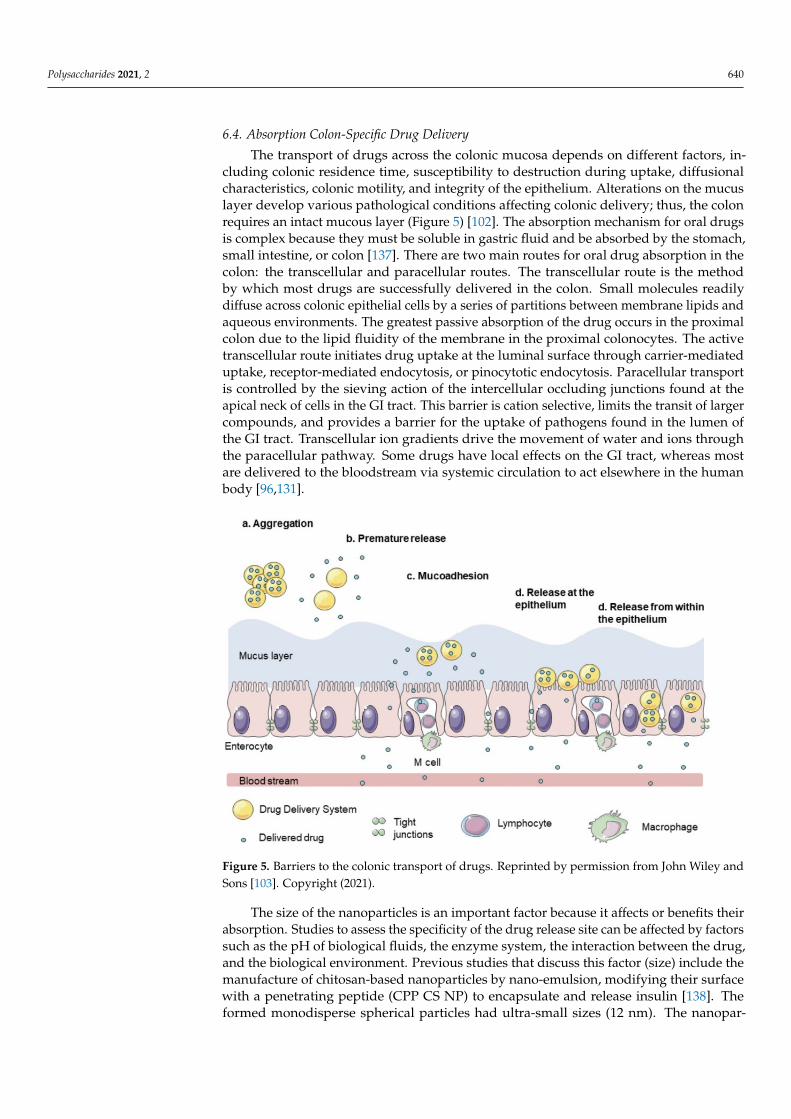
The transport of drugs across the colonic mucosa depends on different factors, in-cluding colonic residence time, susceptibility to destruction during uptake, diffusionalcharacteristics, colonic motility, and integrity of the epithelium. Alterations on the mucuslayer develop various pathological conditions affecting colonic delivery; thus, the colonrequires an intact mucous layer (Figure 5) [102]. The absorption mechanism for oral drugsis complex because they must be soluble in gastric fluid and be absorbed by the stomach,small intestine, or colon [137]. There are two main routes for oral drug absorption in thecolon: the transcellular and paracellular routes. The transcellular route is the methodby which most drugs are successfully delivered in the colon. Small molecules readilydiffuse across colonic epithelial cells by a series of partitions between membrane lipids andaqueous environments. The greatest passive absorption of the drug occurs in the proximalcolon due to the lipid fluidity of the membrane in the proximal colonocytes. The activetranscellular route initiates drug uptake at the luminal surface through carrier-mediateduptake, receptor-mediated endocytosis, or pinocytotic endocytosis. Paracellular transportis controlled by the sieving action of the intercellular occluding junctions found at theapical neck of cells in the GI tract. This barrier is cation selective, limits the transit of largercompounds, and provides a barrier for the uptake of pathogens found in the lumen ofthe GI tract. Transcellular ion gradients drive the movement of water and ions throughthe paracellular pathway. Some drugs have local effects on the GI tract, whereas mostare delivered to the bloodstream via systemic circulation to act elsewhere in the humanbody [96,131].
Polysaccharides 2021, 4, x 16 of 23
Z potential of 28.2 mV. In this study, the effect of nanoparticles to adhere to and infiltrate the mucus layer was evaluated. The results showed that the NPCS could penetrate through the epithelial mucus layer and approach the epithelial cells. In another study, insulin-loaded polyurethane-alginate (PU-AG)-based nanoparticles were evaluated as a model for oral administration [137]. The nanoparticles reached sizes of 73 nm and a Z potential of −27.3 mV. Ex vivo mucoadhesion studies report that these nanoparticles pre-sented high mucoadhesiveness and were strongly attached to the intestinal lumen. This resulted from the mucoadhesive property of alginate due to its carboxyl groups and the negative charge of the nanoparticle.
6.4. Absorption Colon-Specific Drug Delivery The transport of drugs across the colonic mucosa depends on different factors, in-
cluding colonic residence time, susceptibility to destruction during uptake, diffusional characteristics, colonic motility, and integrity of the epithelium. Alterations on the mucus layer develop various pathological conditions affecting colonic delivery; thus, the colon requires an intact mucous layer (Figure 5) [103]. The absorption mechanism for oral drugs is complex because they must be soluble in gastric fluid and be absorbed by the stomach, small intestine, or colon [138]. There are two main routes for oral drug absorption in the colon: the transcellular and paracellular routes. The transcellular route is the method by which most drugs are successfully delivered in the colon. Small molecules readily diffuse across colonic epithelial cells by a series of partitions between membrane lipids and aque-ous environments. The greatest passive absorption of the drug occurs in the proximal co-lon due to the lipid fluidity of the membrane in the proximal colonocytes. The active trans-cellular route initiates drug uptake at the luminal surface through carrier-mediated up-take, receptor-mediated endocytosis, or pinocytotic endocytosis. Paracellular transport is controlled by the sieving action of the intercellular occluding junctions found at the apical neck of cells in the GI tract. This barrier is cation selective, limits the transit of larger com-pounds, and provides a barrier for the uptake of pathogens found in the lumen of the GI tract. Transcellular ion gradients drive the movement of water and ions through the para-cellular pathway. Some drugs have local effects on the GI tract, whereas most are deliv-ered to the bloodstream via systemic circulation to act elsewhere in the human body [96,132].
Figure 5. Barriers to the colonic transport of drugs. Reprinted by permission from John Wiley andSons [103]. Copyright (2021).
The size of the nanoparticles is an important factor because it affects or benefits theirabsorption. Studies to assess the specificity of the drug release site can be affected by factorssuch as the pH of biological fluids, the enzyme system, the interaction between the drug,and the biological environment. Previous studies that discuss this factor (size) include themanufacture of chitosan-based nanoparticles by nano-emulsion, modifying their surfacewith a penetrating peptide (CPP CS NP) to encapsulate and release insulin [138]. Theformed monodisperse spherical particles had ultra-small sizes (12 nm). The nanopar-

Polysaccharides 2021, 2 641
ticles were able to penetrate Caco-2 cells in the in vitro assay through paracellular andintracellular pathways.
Similarly, the manufacture of calcium phosphate nanoparticles coated with vitaminB12-chitosan and alginate (ViTB12-Chi-CPNPs) to encapsulate and release insulin was pre-viously reported [139]. The nanoparticles were synthesized with particle sizes of <250 nmand Z potential of +32.56 mV. Studies in the monolayer of Caco-2 cells showed greateruptake of ViTB12-Chi-CPNPs. A reduction in transepithelial electrical resistance mea-surements (TEER) was observed, resulting in the paracellular transport of insulin dueto the opening of tight epithelial junctions. In another study, the author’s manufacturedalginate/dextran-based nanoparticles were loaded with insulin and coated with chitosanand albumin to improve the permeability of insulin through the intestinal epithelium [140].The nanoparticle sizes obtained were 300 nm. To study the permeability of the nanoparti-cles, they used Caco-2/HT29-MTX-/Raji B cells. This model mimics the intestinal cellularmonolayer due to its content of erythrocytes, goblet cells, and M cells in the Peyer’s patches.The results showed that these nanoparticles increased interactions with cell monolayers.
7. Conclusions and Perspectives
The use of polysaccharide-based nanoparticles for oral drug delivery to the colon hasbecome an essential strategy for treating several diseases. The use of polysaccharides tofabricate these nanomaterials improves their biocompatibility and biodegradability prop-erties. Targeted drug delivery offers attractive properties when materials are developedat a nanometric scale because their size is related to the particle’s stability, surface area,toxicity, degradation, and absorption mechanisms. Similarly, its size depends mainly onthe encapsulated drugs, the type of polysaccharide, and the manufacturing technique. Inthis review, various studies are discussed, focusing on the physicochemical properties ofnanoparticles and their potential to cross the barriers present in the GI, especially in thecolon region.
Polysaccharide-based nanoparticles have significant advantages over other nanoparti-cles (metallic, ceramic, synthetic polymers, etc.). Currently, polysaccharide-based nanopar-ticles are generally investigated in terms of their physicochemical properties, such as size,surface charge, and encapsulation efficiency, via in vitro and in vivo tests. However, oncenanoparticles are administered as drug delivery systems or as part of medical interven-tions, they tend to present specific interactions with elements of the human body, suchas organs, tissues, cells, or hormonal systems. Therefore, it is crucial to investigate thepossible side effects of these materials to a greater extent; however, this may significantlyimpact their use, application, and safety. Also, due to the extensive use and manufacture ofpolysaccharide-based nanoparticles in the nanomedicine and pharmaceutical areas, morescrutiny is needed of regulatory safety as they continue developing this field. Currently, itis known that polysaccharide-based nanoparticles are considered to be non-toxic, biocom-patible, and biodegradable; nonetheless, more in-depth research should be undertaken onthe long-term toxicity risks they may pose. Nanoparticle safety is critical because this tech-nology is increasingly playing an essential role in medicine and pharmaceuticals. Althoughregulatory organizations exist in each country, and each has established its criteria forusing nanoparticles, it is necessary to develop international legislation for the applications,safety, and restrictions of nanoparticles. This regulation would open the door to moreclinical trials and regulatory standards to set future precedents for the use and benefits ofpolysaccharide-based nanoparticles.
Author Contributions: Y.D.A.-F. writing—original draft preparation, review and editing; E.C.-M.supervision, project administration, resources, validation, writing—review and editing; A.C.-M.,A.R.-C., J.L.-M., J.T.-C. and A.L.M.-L. supervision, review and editing. All authors have read andagreed to the published version of the manuscript.

Polysaccharides 2021, 2 642
Funding: The authors thank the National Council of Science and Technology (CONACyT) andResearch Center for Food and Development (CIAD, A.C.) in Mexico for the financial support duringthe Ph.D. studies of Yubia De Anda-Flores.
Institutional Review Board Statement: Not applicable.
Informed Consent Statement: Not applicable.
Data Availability Statement: Not applicable.
Conflicts of Interest: The authors declare no conflict of interest.
References1. Liu, J.; Willför, S.; Xu, C. A review of bioactive plant polysaccharides: Biological activities, functionalization, and biomedical
applications. Bioact. Carbohydr. Diet. Fibre 2015, 5, 31–61. [CrossRef]2. Pawar, V.; Bavya, M.C.; Rohan, K.V.; Srivastava, R. Advances in Polysaccharide-Based Antimicrobial Delivery Vehicles. In Racing
for the Surface; Springer Science and Business Media LLC: Berlin/Heidelberg, Germany, 2020; pp. 267–295.3. Krishnamurthy, R. Giving Rise to Life: Transition from Prebiotic Chemistry to Protobiology. Acc. Chem. Res. 2017, 50, 455–459.
[CrossRef] [PubMed]4. Datta, L.P.; Manchineella, S.; Govindaraju, T. Biomolecules-derived biomaterials. Biomaterials 2020, 230, 119633. [CrossRef]
[PubMed]5. Shishir, M.R.I.; Xie, L.; Sun, C.; Zheng, X.; Chen, W. Advances in micro and nano-encapsulation of bioactive compounds using
biopolymer and lipid-based transporters. Trends Food Sci. Technol. 2018, 78, 34–60. [CrossRef]6. Reinholz, J.; Landfester, K.; Mailänder, V. The challenges of oral drug delivery via nanocarriers. Drug Deliv. 2018, 25, 1694–1705.
[CrossRef]7. Estevinho, B.N.; Rocha, F. Application of Biopolymers in Microencapsulation Processes. In Biopolymers for Food Design; Elsevier
BV: Amsterdam, The Netherlands, 2018; pp. 191–222.8. Gupta, R.; Xie, H. Nanoparticles in Daily Life: Applications, Toxicity and Regulations. J. Environ. Pathol. Toxicol. Oncol. 2018, 37,
209–230. [CrossRef] [PubMed]9. Patra, J.K.; Das, G.; Fraceto, L.F.; Campos, E.V.R.; del Pilar Rodriguez-Torres, M.; Acosta-Torres, L.S.; Diaz-Torres, L.A.; Grillo, R.;
Swamy, M.K.; Sharma, S.; et al. Nano based drug delivery systems: Recent developments and future prospects. J. Nanobiotechnol-ogy 2018, 16, 71. [CrossRef] [PubMed]
10. Hao, J.; Guo, B.; Yu, S.; Zhang, W.; Zhang, D.; Wang, J.; Wang, Y. Encapsulation of the flavonoid quercetin with chitosan-coatednano-liposomes. LWT 2017, 85, 37–44. [CrossRef]
11. Prabhu, S.; Chenreddy, S.; Thio, A.; Khamas, W.; Wang, J.; Thakkar, A. Preclinical systemic toxicity evaluation of chitosan-solid–lipid nanoparticle-encapsulated aspirin and curcumin in combination with free sulforaphane in BALB/c mice. Int. J. Nanomed.2016, 11, 3265–3276. [CrossRef] [PubMed]
12. Saha, S.K.; Roy, P.; Mondal, M.K.; Roy, D.; Gayen, P.; Chowdhury, P.; Babu, S.P. Development of chitosan based gold nanomaterialas an efficient antifilarial agent: A mechanistic approach. Carbohydr. Polym. 2017, 157, 1666–1676. [CrossRef] [PubMed]
13. Andreani, T.; Fangueiro, J.; Severino, P.; De Souza, A.L.R.; Martins-Gomes, C.; Fernandes, P.M.V.; Calpena, A.C.; Gremião, M.P.;Souto, E.B.; Silva, A.M. The Influence of Polysaccharide Coating on the Physicochemical Parameters and Cytotoxicity of SilicaNanoparticles for Hydrophilic Biomolecules Delivery. Nanomaterials 2019, 9, 1081. [CrossRef]
14. Boverhof, D.R.; Bramante, C.M.; Butala, J.H.; Clancy, S.F.; Lafranconi, M.; West, J.; Gordon, S.C. Comparative assessment ofnanomaterial definitions and safety evaluation considerations. Regul. Toxicol. Pharmacol. 2015, 73, 137–150. [CrossRef] [PubMed]
15. Cao, S.-J.; Xu, S.; Wang, H.-M.; Ling, Y.; Dong, J.; Xia, R.-D.; Sun, X.-H. Nanoparticles: Oral Delivery for Protein and PeptideDrugs. AAPS PharmSciTech 2019, 20, 1–11. [CrossRef] [PubMed]
16. Yu, W.; Liu, R.; Zhou, Y.; Gao, H. Size-Tunable Strategies for a Tumor Targeted Drug Delivery System. ACS Cent. Sci. 2020, 6,100–116. [CrossRef] [PubMed]
17. Tapia-Hernández, J.A.; Torres-Chávez, P.I.; Ramírez-Wong, B.; Rascon-Chu, A.; Plascencia-Jatomea, M.; Barreras-Urbina, C.G.;Vazquez, N.A.R.; Rodríguez-Félix, F. Micro- and Nanoparticles by Electrospray: Advances and Applications in Foods. J. Agric.Food Chem. 2015, 63, 4699–4707. [CrossRef]
18. Carvalho, P.; Felício, M.R.; Santos, N.; Gonçalves, S.; Domingues, M. Application of Light Scattering Techniques to NanoparticleCharacterization and Development. Front. Chem. 2018, 6, 237. [CrossRef]
19. Tiwari, A.; Verma, A.; Panda, P.K.; Saraf, S.; Jain, A.; Jain, S.K. Stimuli-responsive polysaccharides for colon-targeted drugdelivery. In Stimuli Responsive Polymeric Nanocarriers for Drug Delivery Applications; Elsevier BV: Amsterdam, The Netherlands,2019; Volume 1, pp. 547–566.
20. Naeem, M.; Awan, U.A.; Subhan, F.; Cao, J.; Hlaing, S.P.; Lee, J.; Im, E.; Jung, Y.; Yoo, J.-W. Advances in colon-targeted nano-drugdelivery systems: Challenges and solutions. Arch. Pharmacal Res. 2020, 43, 153–169. [CrossRef] [PubMed]
21. dos Santos, A.M.; Meneguin, A.B.; Akhter, D.T.; Fletcher, N.; Houston, Z.H.; Bell, C.; Thurecht, K.J.; Gremião, M.P.D. Understand-ing the role of colon-specific microparticles based on retrograded starch/pectin in the delivery of chitosan nanoparticles alongthe gastrointestinal tract. Eur. J. Pharm. Biopharm. 2021, 158, 371–378. [CrossRef]

Polysaccharides 2021, 2 643
22. Wahlgren, M.; Axenstrand, M.; Håkansson, Å.; Marefati, A.; Pedersen, B.L. In Vitro Methods to Study Colon Release: State of theArt and An Outlook on New Strategies for Better In-Vitro Biorelevant Release Media. Pharmaceutics 2019, 11, 95. [CrossRef]
23. Di Mascio, P.; Martinez, G.R.; Miyamoto, S.; Ronsein, G.E.; Medeiros, M.H.G.; Cadet, J. Singlet Molecular Oxygen Reactions withNucleic Acids, Lipids, and Proteins. Chem. Rev. 2019, 119, 2043–2086. [CrossRef] [PubMed]
24. Borchmann, D.E.; Carberry, T.P.; Weck, M. “Bio”-Macromolecules: Polymer-Protein Conjugates as Emerging Scaffolds forTherapeutics. Macromol. Rapid Commun. 2013, 35, 27–43. [CrossRef]
25. Ding, W.; Wu, Y. Sustainable dialdehyde polysaccharides as versatile building blocks for fabricating functional materials: Anoverview. Carbohydr. Polym. 2020, 248, 116801. [CrossRef]
26. Hu, H.; Xu, F.-J. Rational design and latest advances of polysaccharide-based hydrogels for wound healing. Biomater. Sci. 2020, 8,2084–2101. [CrossRef] [PubMed]
27. Barclay, T.G.; Day, C.M.; Petrovsky, N.; Garg, S. Review of polysaccharide particle-based functional drug delivery. Carbohydr.Polym. 2019, 221, 94–112. [CrossRef]
28. Ahmed, T.; Aljaeid, B. Preparation, characterization, and potential application of chitosan, chitosan derivatives, and chitosanmetal nanoparticles in pharmaceutical drug delivery. Drug Des. Dev. Ther. 2016, 10, 483–507. [CrossRef]
29. Huang, G.; Huang, H. Application of hyaluronic acid as carriers in drug delivery. Drug Deliv. 2018, 25, 766–772. [CrossRef]30. Khotimchenko, M. Pectin polymers for colon-targeted antitumor drug delivery. Int. J. Biol. Macromol. 2020, 158, 1110–1124.
[CrossRef]31. BeMiller, J.N. Guar, Locust Bean, Tara, and Cassia Gums. In Carbohydrate Chemistry for Food Scientists; Elsevier BV: Amsterdam,
The Netherlands, 2019; pp. 241–252.32. Chen, F.; Huang, G.; Huang, H. Preparation and application of dextran and its derivatives as carriers. Int. J. Biol. Macromol. 2020,
145, 827–834. [CrossRef] [PubMed]33. Severino, P.; Da Silva, C.F.; Andrade, L.N.; Oliveira, D.D.L.; Campos, J.; Souto, E.B. Alginate Nanoparticles for Drug Delivery and
Targeting. Curr. Pharm. Des. 2019, 25, 1312–1334. [CrossRef] [PubMed]34. Mendez-Encinas, M.A.; Carvajal-Millan, E.; Rascon-Chu, A.; Astiazaran-Garcia, H.F.; Rivera, D.E.V. Ferulated Arabinoxylans and
Their Gels: Functional Properties and Potential Application as Antioxidant and Anticancer Agent. Oxidative Med. Cell. Longev.2018, 2018, 1–22. [CrossRef] [PubMed]
35. Martínez-López, A.L.; Carvajal-Millan, E.; Sotelo-Cruz, N.; Micard, V.; Rascón-Chu, A.; López-Franco, Y.; Lizardi-Mendoza, J.;Canett-Romero, R. Enzymatically cross-linked arabinoxylan microspheres as oral insulin delivery system. Int. J. Biol. Macromol.2019, 126, 952–959. [CrossRef] [PubMed]
36. Song, H.; Fan, Y.; Hu, Y.; Cheng, G.; Xu, F. Polysaccharide–Peptide Conjugates: A Versatile Material Platform for BiomedicalApplications. Adv. Funct. Mater. 2021, 31, 2005978. [CrossRef]
37. Kumar, K.; Dhawan, N.; Sharma, H.; Vaidya, S.; Vaidya, B. Bioadhesive polymers: Novel tool for drug delivery. Artif. CellsNanomed. Biotechnol. 2013, 42, 274–283. [CrossRef] [PubMed]
38. Seidi, F.; Jenjob, R.; Phakkeeree, T.; Crespy, D. Saccharides, oligosaccharides, and polysaccharides nanoparticles for biomedicalapplications. J. Control. Release 2018, 284, 188–212. [CrossRef]
39. Jacob, J.; Haponiuk, J.; Thomas, S.; Gopi, S. Biopolymer based nanomaterials in drug delivery systems: A review. Mater. TodayChem. 2018, 9, 43–55. [CrossRef]
40. Sur, S.; Rathore, A.; Dave, V.; Reddy, K.R.; Chouhan, R.; Sadhu, V. Recent developments in functionalized polymer nanoparticlesfor efficient drug delivery system. Nano Struct. Nano Objects 2019, 20, 100397. [CrossRef]
41. Pereira, M.A.; Rebouças, J.D.S.; Ferraz-Carvalho, R.; de Redín, I.L.; Guerra, P.V.; Gamazo, C.; Brodskyn, C.I.; Irache, J.M.;Santos-Magalhaes, N. Poly(anhydride) nanoparticles containing cashew nut proteins can induce a strong Th1 and Treg immuneresponse after oral administration. Eur. J. Pharm. Biopharm. 2018, 127, 51–60. [CrossRef]
42. Bhatia, S. Nanoparticles Types, Classification, Characterization, Fabrication Methods and Drug Delivery Applications. In NaturalPolymer Drug Delivery Systems; Springer Science and Business Media LLC: Berlin/Heidelberg, Germany, 2016; pp. 33–93.
43. Ramos, A.P.; Cruz, M.; Tovani, C.B.; Ciancaglini, P. Biomedical applications of nanotechnology. Biophys. Rev. 2017, 9, 79–89.[CrossRef]
44. Eliyahu, S.; Aharon, A.; Bianco-Peled, H. Acrylated Chitosan Nanoparticles with Enhanced Mucoadhesion. Polymers 2018, 10,106. [CrossRef]
45. Izadi, Z.; Divsalar, A.; Saboury, A.A.; Sawyer, L. β-lactoglobulin-pectin Nanoparticle-based Oral Drug Delivery System forPotential Treatment of Colon Cancer. Chem. Biol. Drug Des. 2016, 88, 209–216. [CrossRef]
46. Fan, Y.; Yi, J.; Zhang, Y.; Yokoyama, W. Fabrication of curcumin-loaded bovine serum albumin (BSA)-dextran nanoparticles andthe cellular antioxidant activity. Food Chem. 2018, 239, 1210–1218. [CrossRef] [PubMed]
47. De Anda-Flores, Y.; Carvajal-Millan, E.; Lizardi-Mendoza, J.; Rascon-Chu, A.; Martínez-López, A.L.; Marquez-Escalante, J.;Brown-Bojorquez, F.; Tanori-Cordova, J. Covalently Cross-Linked Nanoparticles Based on Ferulated Arabinoxylans Recoveredfrom a Distiller’s Dried Grains Byproduct. Processes 2020, 8, 691. [CrossRef]
48. Cardoso, M.J.; Costa, R.R.; Mano, J.F. Marine Origin Polysaccharides in Drug Delivery Systems. Mar. Drugs 2016, 14, 34.[CrossRef]
49. Bianchera, A.; Bettini, R. Polysaccharide nanoparticles for oral controlled drug delivery: The role of drug–polymer and interpoly-mer interactions. Expert Opin. Drug Deliv. 2020, 17, 1345–1359. [CrossRef] [PubMed]

Polysaccharides 2021, 2 644
50. Lara-Espinoza, C.; Carvajal-Millán, E.; Balandrán-Quintana, R.; López-Franco, Y.; Rascón-Chu, A. Pectin and Pectin-BasedComposite Materials: Beyond Food Texture. Molecules 2018, 23, 942. [CrossRef] [PubMed]
51. Rascón-Chu, A.; Díaz-Baca, J.A.; Carvajal-Millan, E.; Pérez-López, E.; Hotchkiss, A.T.; González-Ríos, H.; Balandrán-Quintana,R.; Campa-Mada, A.C. Electrosprayed Core–Shell Composite Microbeads Based on Pectin-Arabinoxylans for Insulin Carrying:Aggregation and Size Dispersion Control. Polymers 2018, 10, 108. [CrossRef]
52. Dheer, D.; Arora, D.; Jaglan, S.; Rawal, R.; Shankar, R. Polysaccharides based nanomaterials for targeted anti-cancer drug delivery.J. Drug Target. 2016, 25, 1–16. [CrossRef]
53. Aduba, J.D.C.; Yang, H. Polysaccharide Fabrication Platforms and Biocompatibility Assessment as Candidate Wound DressingMaterials. Bioengineering 2017, 4, 1. [CrossRef]
54. Martínez-López, A.L.; Carvajal-Millan, E.; Micard, V.; Rascón-Chu, A.; Brown-Bojorquez, F.; Sotelo-Cruz, N.; López-Franco, Y.L.;Lizardi-Mendoza, J. In vitro degradation of covalently cross-linked arabinoxylan hydrogels by bifidobacteria. Carbohydr. Polym.2016, 144, 76–82. [CrossRef] [PubMed]
55. Zahin, N.; Anwar, R.; Tewari, D.; Kabir, M.T.; Sajid, A.; Mathew, B.; Uddin, M.S.; Aleya, L.; Abdel-Daim, M.M. Nanoparticles andits biomedical applications in health and diseases: Special focus on drug delivery. Environ. Sci. Pollut. Res. 2019, 27, 19151–19168.[CrossRef]
56. Wang, X.; Zhang, G.; Qi, F.; Cheng, Y.; Lu, X.; Wang, L.; Zhao, J.; Zhao, B. Enhanced bone regeneration using an insulin-loadednano-hydroxyapatite/collagen/PLGA composite scaffold. Int. J. Nanomed. 2017, 13, 117–127. [CrossRef]
57. Dadwal, A.; Baldi, A.; Kumar Narang, R. Nanoparticles as carriers for drug delivery in cancer. Artif. Cells Nanomed. Biotechnol.2018, 46, 295–305. [CrossRef] [PubMed]
58. Souto, E.B.; Souto, S.B.; Campos, J.R.; Severino, P.; Pashirova, T.N.; Zakharova, L.Y.; Silva, A.M.; Durazzo, A.; Lucarini, M.; Izzo,A.A.; et al. Nanoparticle Delivery Systems in the Treatment of Diabetes Complications. Molecules 2019, 24, 4209. [CrossRef][PubMed]
59. Tavakoli, S.; Klar, A.S. Advanced Hydrogels as Wound Dressings. Biomolecules 2020, 10, 1169. [CrossRef]60. Zhang, M.; Merlin, D. Nanoparticle-Based Oral Drug Delivery Systems Targeting the Colon for Treatment of Ulcerative Colitis.
Inflamm. Bowel Dis. 2018, 24, 1401–1415. [CrossRef] [PubMed]61. Malhaire, H.; Gimel, J.-C.; Roger, E.; Benoît, J.-P.; Lagarce, F. How to design the surface of peptide-loaded nanoparticles for
efficient oral bioavailability? Adv. Drug Deliv. Rev. 2016, 106, 320–336. [CrossRef]62. Beloqui, A.; Rieux, A.D.; Préat, V. Mechanisms of transport of polymeric and lipidic nanoparticles across the intestinal barrier.
Adv. Drug Deliv. Rev. 2016, 106, 242–255. [CrossRef]63. Tosi, G.; Duskey, J.; Kreuter, J. Nanoparticles as carriers for drug delivery of macromolecules across the blood-brain barrier. Expert
Opin. Drug Deliv. 2019, 17, 23–32. [CrossRef]64. Health Canada. 2011 Policy Statement on Health Canada’s Working Definition for Nanomaterial—Canada.ca. Available online:
https://www.canada.ca/en/health-canada/services/science-research/reports-publications/nanomaterial/policy-statement-health-canada-working-definition.html (accessed on 1 May 2021).
65. ISO 8968-1:2014. Milk and Milk Products—Determination of Nitrogen Content—Part 1: Kjeldahl Principle and Crude ProteinCalculation. Available online: https://www.iso.org/obp/ui/#iso:std:iso:8968:-1:ed-2:v1:en (accessed on 1 May 2021).
66. SCENIHR Communication on the Second Regulatory Review on Nanomaterials. Available online: https://eur-lex.europa.eu/legal-content/EN/TXT/PDF/?uri=CELEX:52012DC0572&from=EN (accessed on 1 May 2021).
67. SCENIHR Opinion on the Guidance on the Determination of Potential Health Effects of Nanomaterials Used in Medical Devices.Available online: http://ec.europa.eu/health/scientific_committees/emerging/docs/scenihr_o_045.pdf (accessed on 1 May 2021).
68. Shin, S.W.; Song, I.H.; Um, S.H. Role of Physicochemical Properties in Nanoparticle Toxicity. Nanomaterials 2015, 5, 1351–1365.[CrossRef]
69. de Crozals, G.; Bonnet, R.; Farre, C.; Chaix, C. Nanoparticles with multiple properties for biomedical applications: A strategicguide. Nano Today 2016, 11, 435–463. [CrossRef]
70. Fernández, E.F.; Santos-Carballal, B.; De Santi, C.; Ramsey, J.M.; MacLoughlin, R.; Cryan, S.-A.; Greene, C.M. Biopolymer-BasedNanoparticles for Cystic Fibrosis Lung Gene Therapy Studies. Materials 2018, 11, 122. [CrossRef] [PubMed]
71. Jafari, S.M.; Esfanjani, A.F. Instrumental analysis and characterization of nanocapsules. In Nanoencapsulation Technologies for theFood and Nutraceutical Industries; Elsevier BV: Amsterdam, The Netherlands, 2017; pp. 524–544.
72. Griffin, B.; Guo, J.; Presas, E.; Donovan, M.; Alonso, M.J.; O’Driscoll, C.M. Pharmacokinetic, pharmacodynamic and biodistributionfollowing oral administration of nanocarriers containing peptide and protein drugs. Adv. Drug Deliv. Rev. 2016, 106, 367–380.[CrossRef]
73. Zhao, J.; Stenzel, M.H. Entry of nanoparticles into cells: The importance of nanoparticle properties. Polym. Chem. 2018, 9, 259–272.[CrossRef]
74. Patel, S.; Kim, J.; Herrera, M.; Mukherjee, A.; Kabanov, A.V.; Sahay, G. Brief update on endocytosis of nanomedicines. Adv. DrugDeliv. Rev. 2019, 144, 90–111. [CrossRef]
75. Kaksonen, M.; Roux, A. Mechanisms of clathrin-mediated endocytosis. Nat. Rev. Mol. Cell Biol. 2018, 19, 313–326. [CrossRef][PubMed]
76. Manzanares, D.; Ceña, V. Endocytosis: The Nanoparticle and Submicron Nanocompounds Gateway into the Cell. Pharmaceutics2020, 12, 371. [CrossRef] [PubMed]

Polysaccharides 2021, 2 645
77. Pinelli, F.; Ortolà; Óscar, F.; Makvandi, P.; Perale, G.; Rossi, F. In vivo drug delivery applications of nanogels: A review.Nanomedicine 2020, 15, 2707–2727. [CrossRef]
78. Zhang, S.; Gao, H.; Bao, G. Physical Principles of Nanoparticle Cellular Endocytosis. ACS Nano 2015, 9, 8655–8671. [CrossRef]79. Jiang, L.Q.; Wang, T.Y.; Webster, T.J.; Duan, H.-J.; Qiu, J.Y.; Zhao, Z.M.; Yin, X.-X.; Zheng, C.L. Intracellular disposition of chitosan
nanoparticles in macrophages: Intracellular uptake, exocytosis, and intercellular transport. Int. J. Nanomed. 2017, 12, 6383–6398.[CrossRef] [PubMed]
80. Li, Q.; Liu, C.-G.; Yu, Y. Separation of monodisperse alginate nanoparticles and effect of particle size on transport of vitamin E.Carbohydr. Polym. 2015, 124, 274–279. [CrossRef]
81. Zhang, X.; Ma, Y.; Ma, L.; Zu, M.; Song, H.; Xiao, B. Oral administration of chondroitin sulfate-functionalized nanoparticles forcolonic macrophage-targeted drug delivery. Carbohydr. Polym. 2019, 223, 115126. [CrossRef]
82. Forest, V.; Pourchez, J. Preferential binding of positive nanoparticles on cell membranes is due to electrostatic interactions: Atoo simplistic explanation that does not take into account the nanoparticle protein corona. Mater. Sci. Eng. C 2017, 70, 889–896.[CrossRef] [PubMed]
83. Loh, J.W.; Saunders, M.; Lim, L.-Y. Cytotoxicity of monodispersed chitosan nanoparticles against the Caco-2 cells. Toxicol. Appl.Pharmacol. 2012, 262, 273–282. [CrossRef] [PubMed]
84. Maciel, V.B.V.; Yoshida, C.M.P.; Pereira, S.M.S.S.; Goycoolea, F.M.; Franco, T.T. Electrostatic Self-Assembled Chitosan-Pectin Nano-and Microparticles for Insulin Delivery. Molecules 2017, 22, 1707. [CrossRef]
85. Noi, I.; Schlachet, I.; Kumarasamy, M.; Sosnik, A. Permeability of Novel Chitosan-g-Poly(Methyl Methacrylate) AmphiphilicNanoparticles in a Model of Small Intestine In Vitro. Polymers 2018, 10, 478. [CrossRef] [PubMed]
86. Bharucha, A.E.; Camilleri, M. Physiology of the Colon and Its Measurement. In Shackelford’s Surgery of the Alimentary Tract, 2Volume Set; Elsevier BV: Amsterdam, The Netherlands, 2019; pp. 1676–1688.
87. Arévalo-Pérez, R.; Maderuelo, C.; Lanao, J.M. Recent advances in colon drug delivery systems. J. Control Release 2020, 327,703–724. [CrossRef]
88. Rivière, A.; Selak, M.; Lantin, D.; Leroy, F.; De Vuyst, L. Bifidobacteria and Butyrate-Producing Colon Bacteria: Importance andStrategies for Their Stimulation in the Human Gut. Front. Microbiol. 2016, 7, 979. [CrossRef]
89. Kotla, N.G.; Rana, S.; Sivaraman, G.; Sunnapu, O.; Vemula, P.K.; Pandit, A.; Rochev, Y. Bioresponsive drug delivery systems inintestinal inflammation: State-of-the-art and future perspectives. Adv. Drug Deliv. Rev. 2019, 146, 248–266. [CrossRef]
90. Amidon, S.; Brown, J.E.; Dave, V.S. Colon-Targeted Oral Drug Delivery Systems: Design Trends and Approaches. AAPSPharmSciTech 2015, 16, 731–741. [CrossRef]
91. Mendez-Encinas, M.A.; Valencia-Rivera, D.E.; Carvajal-Millan, E.; Astiazaran-Garcia, H.; Micard, V.; Rascón-Chu, A. Fermentationof Ferulated Arabinoxylan Recovered from the Maize Bioethanol Industry. Processes 2021, 9, 165. [CrossRef]
92. Netsomboon, K.; Bernkop-Schnürch, A. Mucoadhesive vs. mucopenetrating particulate drug delivery. Eur. J. Pharm. Biopharm.2016, 98, 76–89. [CrossRef]
93. Winslow, T. Colon or Rectal Cancer Stage 0. Available online: https://visualsonline.cancer.gov/details.cfm?imageid=9147(accessed on 7 May 2021).
94. Song, Q.; Jia, J.; Niu, X.; Zheng, C.; Zhao, H.; Sun, L.; Zhang, H.; Wang, L.; Zhang, Z.; Zhang, Y. An oral drug delivery systemwith programmed drug release and imaging properties for orthotopic colon cancer therapy. Nanoscale 2019, 11, 15958–15970.[CrossRef] [PubMed]
95. Zhang, L.; Sang, Y.; Feng, J.; Li, Z.; Zhao, A. Polysaccharide-based micro/nanocarriers for oral colon-targeted drug delivery. J.Drug Target. 2016, 24, 579–589. [CrossRef]
96. Lundquist, P.; Artursson, P. Oral absorption of peptides and nanoparticles across the human intestine: Opportunities, limitationsand studies in human tissues. Adv. Drug Deliv. Rev. 2016, 106, 256–276. [CrossRef] [PubMed]
97. Lee, S.H.; Bajracharya, R.; Min, J.Y.; Han, J.-W.; Park, B.J.; Han, H.-K. Strategic Approaches for Colon Targeted Drug Delivery: AnOverview of Recent Advancements. Pharmaceutics 2020, 12, 68. [CrossRef]
98. Xu, J.; Tam, M.; Samaei, S.; Lerouge, S.; Barralet, J.; Stevenson, M.M.; Cerruti, M. Mucoadhesive chitosan hydrogels as rectal drugdelivery vessels to treat ulcerative colitis. Acta Biomater. 2017, 48, 247–257. [CrossRef]
99. Muheem, A.; Shakeel, F.; Jahangir, M.A.; Anwar, M.; Mallick, N.; Jain, G.K.; Warsi, M.H.; Ahmad, F.J. A review on the strategiesfor oral delivery of proteins and peptides and their clinical perspectives. Saudi Pharm. J. 2016, 24, 413–428. [CrossRef]
100. Rajput, R.; Narkhede, J.; Naik, J.B. Nanogels as nanocarriers for drug delivery: A review. ADMET DMPK 2020, 8, 1–15. [CrossRef]101. Makvandi, P.; Ghomi, M.; Ashrafizadeh, M.; Tafazoli, A.; Agarwal, T.; Delfi, M.; Akhtari, J.; Zare, E.N.; Padil, V.V.; Zarrabi, A.;
et al. A review on advances in graphene-derivative/polysaccharide bionanocomposites: Therapeutics, pharmacogenomics andtoxicity. Carbohydr. Polym. 2020, 250, 116952. [CrossRef] [PubMed]
102. Hua, S.; Marks, E.; Schneider, J.J.; Keely, S. Advances in oral nano-delivery systems for colon targeted drug delivery ininflammatory bowel disease: Selective targeting to diseased versus healthy tissue. Nanomed. Nanotechnol. Biol. Med. 2015, 11,1117–1132. [CrossRef] [PubMed]
103. Durán-Lobato, M.; Niu, Z.; Alonso, M.J. Oral Delivery of Biologics for Precision Medicine. Adv. Mater. 2020, 32, e1901935.[CrossRef]
104. Wu, J.-L.; Tian, G.-X.; Yu, W.-J.; Jia, G.-T.; Sun, T.-Y.; Gao, Z.-Q. pH-Responsive Hyaluronic Acid-Based Mixed Micelles for theHepatoma-Targeting Delivery of Doxorubicin. Int. J. Mol. Sci. 2016, 17, 364. [CrossRef] [PubMed]

Polysaccharides 2021, 2 646
105. Pilipenko, I.; Korzhikov-Vlakh, V.; Sharoyko, V.; Zhang, N.; Schäfer-Korting, M.; Rühl, E.; Zoschke, C.; Tennikova, T. pH-SensitiveChitosan–Heparin Nanoparticles for Effective Delivery of Genetic Drugs into Epithelial Cells. Pharmaceutics 2019, 11, 317.[CrossRef] [PubMed]
106. Barbosa, A.I.; Lima, S.; Reis, S. Application of pH-Responsive Fucoidan/Chitosan Nanoparticles to Improve Oral QuercetinDelivery. Molecules 2019, 24, 346. [CrossRef] [PubMed]
107. Omer, A.; Ziora, Z.; Tamer, T.; Khalifa, R.; Hassan, M.; Mohy-Eldin, M.; Blaskovich, M. Formulation of Quaternized AminatedChitosan Nanoparticles for Efficient Encapsulation and Slow Release of Curcumin. Molecules 2021, 26, 449. [CrossRef]
108. Elgegren, M.; Kim, S.; Cordova, D.; Silva, C.; Noro, J.; Cavaco-Paulo, A.; Nakamatsu, J. Ultrasound-Assisted Encapsulation ofSacha Inchi (Plukenetia volubilis Linneo.) Oil in Alginate-Chitosan Nanoparticles. Polymers 2019, 11, 1245. [CrossRef]
109. Iglesias, N.; Galbis, E.; Díaz-Blanco, M.J.; Lucas, R.; Benito, E.; De-Paz, M.-V. Nanostructured Chitosan-Based Biomaterials forSustained and Colon-Specific Resveratrol Release. Int. J. Mol. Sci. 2019, 20, 398. [CrossRef] [PubMed]
110. Chandrarathna, H.; Liyanage, T.; Edirisinghe, S.; Dananjaya, S.; Thulshan, E.; Nikapitiya, C.; Oh, C.; Kang, D.-H.; De Zoysa, M.Marine Microalgae, Spirulina maxima-Derived Modified Pectin and Modified Pectin Nanoparticles Modulate the Gut Microbiotaand Trigger Immune Responses in Mice. Mar. Drugs 2020, 18, 175. [CrossRef]
111. Dyawanapelly, S.; Koli, U.; Dharamdasani, V.; Jain, R.; Dandekar, P. Improved mucoadhesion and cell uptake of chitosan andchitosan oligosaccharide surface-modified polymer nanoparticles for mucosal delivery of proteins. Drug Deliv. Transl. Res. 2016,6, 365–379. [CrossRef]
112. Sabra, R.; Roberts, C.J.; Billa, N. Courier properties of modified citrus pectinate-chitosan nanoparticles in colon delivery ofcurcumin. Colloid Interface Sci. Commun. 2019, 32, 100192. [CrossRef]
113. Prabahar, K.; Udhumansha, U.; Qushawy, M. Optimization of Thiolated Chitosan Nanoparticles for the Enhancement of in VivoHypoglycemic Efficacy of Sitagliptin in Streptozotocin-Induced Diabetic Rats. Pharmaceutics 2020, 12, 300. [CrossRef] [PubMed]
114. Sudhakar, S.; Chandran, S.V.; Selvamurugan, N.; Nazeer, R.A. Biodistribution and pharmacokinetics of thiolated chitosannanoparticles for oral delivery of insulin in vivo. Int. J. Biol. Macromol. 2020, 150, 281–288. [CrossRef]
115. Shailender, J.; Ravi, P.R.; Sirukuri, M.R.; Dalvi, A.; Priya, O.K. Chitosan nanoparticles for the oral delivery of tenofovir disoproxilfumarate: Formulation optimization, characterization and ex vivo and in vivo evaluation for uptake mechanism in rats. DrugDev. Ind. Pharm. 2018, 44, 1109–1119. [CrossRef]
116. He, Z.; Santos, J.L.; Tian, H.; Huang, H.; Hu, Y.; Liu, L.; Leong, K.W.; Chen, Y.; Mao, H.-Q. Scalable fabrication of size-controlledchitosan nanoparticles for oral delivery of insulin. Biomaterials 2017, 130, 28–41. [CrossRef]
117. Mumuni, M.A.; Kenechukwu, F.; Ofokansi, K.C.; Attama, A.A.; Díaz, D.D. Insulin-loaded mucoadhesive nanoparticles based onmucin-chitosan complexes for oral delivery and diabetes treatment. Carbohydr. Polym. 2020, 229, 115506. [CrossRef]
118. Maity, S.; Mukhopadhyay, P.; Kundu, P.P.; Chakraborti, A.S. Alginate coated chitosan core-shell nanoparticles for efficient oraldelivery of naringenin in diabetic animals—An in vitro and in vivo approach. Carbohydr. Polym. 2017, 170, 124–132. [CrossRef]
119. Cánepa, C.; Imperiale, J.C.; Berini, C.A.; Lewicki, M.; Sosnik, A.; Biglione, M.M. Development of a Drug Delivery System Basedon Chitosan Nanoparticles for Oral Administration of Interferon-α. Biomacromolecules 2017, 18, 3302–3309. [CrossRef] [PubMed]
120. Hirpara, M.R.; Manikkath, J.; Sivakumar, K.; Managuli, R.S.; Gourishetti, K.; Krishnadas, N.; Shenoy, R.; Jayaprakash, B.; Rao,C.M.; Mutalik, S. Long circulating PEGylated-chitosan nanoparticles of rosuvastatin calcium: Development and in vitro andin vivo evaluations. Int. J. Biol. Macromol. 2018, 107, 2190–2200. [CrossRef] [PubMed]
121. Kalhapure, R.S.; Jadhav, M.; Rambharose, S.; Mocktar, C.; Singh, S.; Renukuntla, J.; Govender, T. pH-responsive chitosannanoparticles from a novel twin-chain anionic amphiphile for controlled and targeted delivery of vancomycin. Colloids Surfaces BBiointerfaces 2017, 158, 650–657. [CrossRef]
122. Mukhopadhyay, P.; Sarkar, K.; Chakraborty, M.; Bhattacharya, S.; Mishra, R.; Kundu, P. Oral insulin delivery by self-assembledchitosan nanoparticles: In vitro and in vivo studies in diabetic animal model. Mater. Sci. Eng. C 2013, 33, 376–382. [CrossRef]
123. Thai, H.; Nguyen, C.T.; Thach, L.T.; Tran, M.T.; Mai, H.D.; Nguyen, T.T.T.; Le, G.D.; Van Can, M.; Tran, L.D.; Bach, G.L.; et al.Characterization of chitosan/alginate/lovastatin nanoparticles and investigation of their toxic effects in vitro and in vivo. Sci.Rep. 2020, 10, 909–915. [CrossRef]
124. Auwal, S.M.; Zarei, M.; Tan, C.P.; Basri, M.; Saari, N. Improved In Vivo Efficacy of Anti-Hypertensive Biopeptides Encapsulatedin Chitosan Nanoparticles Fabricated by Ionotropic Gelation on Spontaneously Hypertensive Rats. Nanomaterials 2017, 7, 421.[CrossRef] [PubMed]
125. Tian, H.; He, Z.; Sun, C.; Yang, C.; Zhao, P.; Liu, L.; Leong, K.W.; Mao, H.-Q.; Liu, Z.; Chen, Y. Uniform Core-Shell Nanoparticleswith Thiolated Hyaluronic Acid Coating to Enhance Oral Delivery of Insulin. Adv. Health Mater. 2018, 7, e1800285. [CrossRef][PubMed]
126. Tian, G.; Sun, X.; Bai, J.; Dong, J.; Zhang, B.; Gao, Z.; Wu, J. Doxorubicin-loaded dual-functional hyaluronic acid nanoparticles:Preparation, characterization and antitumor efficacy in vitro and in vivo. Mol. Med. Rep. 2018, 19, 133–142. [CrossRef]
127. Homayun, B.; Lin, X.; Choi, H.-J. Challenges and Recent Progress in Oral Drug Delivery Systems for Biopharmaceuticals.Pharmaceutics 2019, 11, 129. [CrossRef]
128. Tan, C.; Xie, J.; Zhang, X.; Cai, J.; Xia, S. Polysaccharide-based nanoparticles by chitosan and gum arabic polyelectrolytecomplexation as carriers for curcumin. Food Hydrocoll. 2016, 57, 236–245. [CrossRef]
129. Lu, B.; Lv, X.; Le, Y. Chitosan-Modified PLGA Nanoparticles for Control-Released Drug Delivery. Polymers 2019, 11, 304.[CrossRef] [PubMed]

Polysaccharides 2021, 2 647
130. Jin, B.; Zhou, X.; Li, X.; Lin, W.; Chen, G.; Qiu, R. Self-Assembled Modified Soy Protein/Dextran Nanogel Induced by Ultrasonica-tion as a Delivery Vehicle for Riboflavin. Molecules 2016, 21, 282. [CrossRef]
131. Zhang, T.; Yang, Y.; Liang, Y.; Jiao, X.; Zhao, C. Beneficial Effect of Intestinal Fermentation of Natural Polysaccharides. Nutrients2018, 10, 1055. [CrossRef] [PubMed]
132. Subudhi, M.B.; Jain, A.; Jain, A.; Hurkat, P.; Shilpi, S.; Gulbake, A.; Jain, S.K. Eudragit S100 Coated Citrus Pectin Nanoparticles forColon Targeting of 5-Fluorouracil. Materials 2015, 8, 832–849. [CrossRef]
133. McGuckin, M.; Thornton, D.; Whitsett, J.A. Mucins and Mucus. In Mucosal Immunology; Elsevier BV: Amsterdam, The Netherlands, 2015.134. García-Díaz, M.; Birch, D.; Wan, F.; Nielsen, H.M. The role of mucus as an invisible cloak to transepithelial drug delivery by
nanoparticles. Adv. Drug Deliv. Rev. 2018, 124, 107–124. [CrossRef]135. Sonaje, K.; Lin, K.-J.; Tseng, M.T.; Wey, S.-P.; Su, F.-Y.; Chuang, E.-Y.; Hsu, C.-W.; Chen, C.-T.; Sung, H.-W. Effects of chitosan-
nanoparticle-mediated tight junction opening on the oral absorption of endotoxins. Biomaterials 2011, 32, 8712–8721. [CrossRef][PubMed]
136. Bhattacharyya, A.; Mukherjee, D.; Mishra, R.; Kundu, P.P. Development of pH sensitive polyurethane–alginate nanoparticles forsafe and efficient oral insulin delivery in animal models. RSC Adv. 2016, 6, 41835–41846. [CrossRef]
137. McClements, D.J. Encapsulation, protection, and delivery of bioactive proteins and peptides using nanoparticle and microparticlesystems: A review. Adv. Colloid Interface Sci. 2018, 253, 1–22. [CrossRef] [PubMed]
138. Barbari, G.R.; Dorkoosh, F.A.; Amini, M.; Sharifzadeh, M.; Atyabi, F.; Balalaie, S.; Tehrani, N.R.; Tehrani, M.R. A novelnanoemulsion-based method to produce ultrasmall, water-dispersible nanoparticles from chitosan, surface modified withcell-penetrating peptide for oral delivery of proteins and peptides. Int. J. Nanomed. 2017, 12, 3471–3483. [CrossRef] [PubMed]
139. Verma, A.; Sharma, S.; Gupta, P.K.; Singh, A.; Teja, B.V.; Dwivedi, P.; Gupta, G.K.; Trivedi, R.; Mishra, P.R. Vitamin B12functionalized layer by layer calcium phosphate nanoparticles: A mucoadhesive and pH responsive carrier for improved oraldelivery of insulin. Acta Biomater. 2016, 31, 288–300. [CrossRef]
140. Lopes, M.; Shrestha, N.; Correia, A.; Shahbazi, M.-A.; Sarmento, B.; Hirvonen, J.T.; Veiga, F.; Seiça, R.; Ribeiro, A.; Santos, H.A.Dual chitosan/albumin-coated alginate/dextran sulfate nanoparticles for enhanced oral delivery of insulin. J. Control. Release2016, 232, 29–41. [CrossRef] [PubMed]