Stress-Induced Sphingolipid Signaling: Role of Type2 Neutral Sphingomyelinase in Murine Cell...
11
Stress-Induced Sphingolipid Signaling: Role of Type-2 Neutral Sphingomyelinase in Murine Cell Apoptosis and Proliferation Raphael Devillard 1,3 , Sylvain Galvani 1,2 , Jean-Claude Thiers 1,2 , Jean-Louis Guenet 4 , Yusuf Hannun 5 , Jacek Bielawski 5 , Anne Ne ` gre-Salvayre 1,2 , Robert Salvayre 1,2 , Nathalie Auge ´ 1,2 * 1 INSERM U858, CHU Rangueil, Toulouse, France, 2 Universite ´ Paul Sabatier de Toulouse, Faculte ´ de Me ´decine, Toulouse, France, 3 Universite ´ de Bordeaux 2, U.F.R d’Odontologie, Bordeaux, France, 4 Institut Pasteur, Unite ´ de ge ´ne ´tique des Mammife ` res, Paris, France, 5 Department of Biochemistry and Molecular Biology, Medical University of South Carolina, Charleston, South Carolina, United States of America Abstract Background: Sphingomyelin hydrolysis in response to stress-inducing agents, and subsequent ceramide generation, are implicated in various cellular responses, including apoptosis, inflammation and proliferation, depending on the nature of the different acidic or neutral sphingomyelinases. This study was carried out to investigate whether the neutral Mg 2+ - dependent neutral sphingomyelinase-2 (nSMase2) plays a role in the cellular signaling evoked by TNFalpha and oxidized LDLs, two stress-inducing agents, which are mitogenic at low concentrations and proapoptotic at higher concentrations. Methodology and Principal Findings: For this purpose, we used nSMase2-deficient cells from homozygous fro/fro (fragilitas ossium) mice and nSMase2-deficient cells reconstituted with a V5-tagged nSMase2. We report that the genetic defect of nSMase2 (in fibroblasts from fro/fro mice) does not alter the TNFalpha and oxidized LDLs-mediated apoptotic response. Likewise, the hepatic toxicity of TNFalpha is similar in wild type and fro mice, thus is independent of nSMase2 activation. In contrast, the mitogenic response elicited by low concentrations of TNFalpha and oxidized LDLs (but not fetal calf serum) requires nSMase2 activation. Conclusion and Significance: nSMase2 activation is not involved in apoptosis mediated by TNFalpha and oxidized LDLs in murine fibroblasts, and in the hepatotoxicity of TNFalpha in mice, but is required for the mitogenic response to stress- inducing agents. Citation: Devillard R, Galvani S, Thiers J-C, Guenet J-L, Hannun Y, et al. (2010) Stress-Induced Sphingolipid Signaling: Role of Type-2 Neutral Sphingomyelinase in Murine Cell Apoptosis and Proliferation. PLoS ONE 5(3): e9826. doi:10.1371/journal.pone.0009826 Editor: Rafael Linden, Universidade Federal do Rio de Janeiro (UFRJ), Brazil Received November 19, 2009; Accepted March 1, 2010; Published March 23, 2010 Copyright: ß 2010 Devillard et al. This is an open-access article distributed under the terms of the Creative Commons Attribution License, which permits unrestricted use, distribution, and reproduction in any medium, provided the original author and source are credited. Funding: INSERM, Universite Toulouse-3, Institut Francais pour la Recherche Odontologique (IFRO), Fondation pour la Recherche Medicale, Fondation Coeur et Arteres, and NIH-GM 43825. The funders had no role in study design, data collection and analysis, decision to publish, or preparation of the manuscript. Competing Interests: The authors have declared that no competing interests exist. * E-mail: [email protected] Introduction Sphingomyelin (SM) is a ubiquitous component of eukaryotic membranes, distributed mainly in the plasma membrane, which contains more than 70–80% of total cellular SM. Sphingomyelin- ases (SMases) hydrolyze the phosphodiester bond of sphingomy- elin to generate phosphorylcholine and ceramide. Ceramides and other metabolic derivatives (e.g. sphingosine and sphingosine-1- phosphate) are lipid ‘‘second messengers’’ molecules involved in the regulation of stress-induced cellular responses, including cell differentiation, proliferation, adhesion and cell death [1,2]. Apoptosis is a key event in tissue development and in various pathophysiological processes. The role of ceramide in apoptosis [3] and the balance between ceramide and sphingosine-1- phosphate have been largely investigated in various cell types [4]. However, several waves of ceramide production are observed during apoptosis and the mechanisms and roles of the specific ceramide rise are still not well defined, as ceramide generation may result from de novo synthesis or/and SM degradation by acid or neutral SMases (aSMase or nSMase) (nSMase being a generic term for an indefinite neutral SMase) [2]. Neutral SMases (nSMases) are activated by a variety of stress- inducing agents, including cytokines, oxidative stress (H2O2, oxidized lipoproteins), UV radiation, chemotherapeutic drugs, b- amyloid-peptides and lipopolysaccharide [5]. The mechanism of nSMase regulation is only partly understood, although several activators and regulators have been identified. Cytokine receptors (e.g. TNF receptor, IL-1 receptor and Fas) and associated proteins (e.g. FAN, RACK-1 and caveolin-1) have been shown to trigger nSMase activation [5,6]. Reactive oxygen and nitrogen species, GSH depletion, hydrogen peroxide and oxidative stress activate nSMase, while antioxidants, such as reduced glutathione GSH and coenzyme Q are inhibitory [5]. Various cellular mediators regulate nSMase activity including anionic phospholipids, protein kinases phospholipase A2, caveolin, Bcl-2 and Bcl-xL and proteases [5,7,8]. Considerable research on nSMase activation, regulation and physiological functions have been carried out, but only little information concerning the specific role of each nSMase is PLoS ONE | www.plosone.org 1 March 2010 | Volume 5 | Issue 3 | e9826
-
Upload
univ-bordeaux -
Category
Documents
-
view
0 -
download
0
Transcript of Stress-Induced Sphingolipid Signaling: Role of Type2 Neutral Sphingomyelinase in Murine Cell...

Stress-Induced Sphingolipid Signaling: Role of Type-2Neutral Sphingomyelinase in Murine Cell Apoptosis andProliferationRaphael Devillard1,3, Sylvain Galvani1,2, Jean-Claude Thiers1,2, Jean-Louis Guenet4, Yusuf Hannun5,
Jacek Bielawski5, Anne Negre-Salvayre1,2, Robert Salvayre1,2, Nathalie Auge1,2*
1 INSERM U858, CHU Rangueil, Toulouse, France, 2 Universite Paul Sabatier de Toulouse, Faculte de Medecine, Toulouse, France, 3 Universite de Bordeaux 2, U.F.R
d’Odontologie, Bordeaux, France, 4 Institut Pasteur, Unite de genetique des Mammiferes, Paris, France, 5 Department of Biochemistry and Molecular Biology, Medical
University of South Carolina, Charleston, South Carolina, United States of America
Abstract
Background: Sphingomyelin hydrolysis in response to stress-inducing agents, and subsequent ceramide generation, areimplicated in various cellular responses, including apoptosis, inflammation and proliferation, depending on the nature ofthe different acidic or neutral sphingomyelinases. This study was carried out to investigate whether the neutral Mg2+-dependent neutral sphingomyelinase-2 (nSMase2) plays a role in the cellular signaling evoked by TNFalpha and oxidizedLDLs, two stress-inducing agents, which are mitogenic at low concentrations and proapoptotic at higher concentrations.
Methodology and Principal Findings: For this purpose, we used nSMase2-deficient cells from homozygous fro/fro (fragilitasossium) mice and nSMase2-deficient cells reconstituted with a V5-tagged nSMase2. We report that the genetic defect ofnSMase2 (in fibroblasts from fro/fro mice) does not alter the TNFalpha and oxidized LDLs-mediated apoptotic response.Likewise, the hepatic toxicity of TNFalpha is similar in wild type and fro mice, thus is independent of nSMase2 activation. Incontrast, the mitogenic response elicited by low concentrations of TNFalpha and oxidized LDLs (but not fetal calf serum)requires nSMase2 activation.
Conclusion and Significance: nSMase2 activation is not involved in apoptosis mediated by TNFalpha and oxidized LDLs inmurine fibroblasts, and in the hepatotoxicity of TNFalpha in mice, but is required for the mitogenic response to stress-inducing agents.
Citation: Devillard R, Galvani S, Thiers J-C, Guenet J-L, Hannun Y, et al. (2010) Stress-Induced Sphingolipid Signaling: Role of Type-2 Neutral Sphingomyelinase inMurine Cell Apoptosis and Proliferation. PLoS ONE 5(3): e9826. doi:10.1371/journal.pone.0009826
Editor: Rafael Linden, Universidade Federal do Rio de Janeiro (UFRJ), Brazil
Received November 19, 2009; Accepted March 1, 2010; Published March 23, 2010
Copyright: � 2010 Devillard et al. This is an open-access article distributed under the terms of the Creative Commons Attribution License, which permitsunrestricted use, distribution, and reproduction in any medium, provided the original author and source are credited.
Funding: INSERM, Universite Toulouse-3, Institut Francais pour la Recherche Odontologique (IFRO), Fondation pour la Recherche Medicale, Fondation Coeur etArteres, and NIH-GM 43825. The funders had no role in study design, data collection and analysis, decision to publish, or preparation of the manuscript.
Competing Interests: The authors have declared that no competing interests exist.
* E-mail: [email protected]
Introduction
Sphingomyelin (SM) is a ubiquitous component of eukaryotic
membranes, distributed mainly in the plasma membrane, which
contains more than 70–80% of total cellular SM. Sphingomyelin-
ases (SMases) hydrolyze the phosphodiester bond of sphingomy-
elin to generate phosphorylcholine and ceramide. Ceramides and
other metabolic derivatives (e.g. sphingosine and sphingosine-1-
phosphate) are lipid ‘‘second messengers’’ molecules involved in
the regulation of stress-induced cellular responses, including cell
differentiation, proliferation, adhesion and cell death [1,2].
Apoptosis is a key event in tissue development and in various
pathophysiological processes. The role of ceramide in apoptosis
[3] and the balance between ceramide and sphingosine-1-
phosphate have been largely investigated in various cell types
[4]. However, several waves of ceramide production are observed
during apoptosis and the mechanisms and roles of the specific
ceramide rise are still not well defined, as ceramide generation
may result from de novo synthesis or/and SM degradation by acid
or neutral SMases (aSMase or nSMase) (nSMase being a generic
term for an indefinite neutral SMase) [2].
Neutral SMases (nSMases) are activated by a variety of stress-
inducing agents, including cytokines, oxidative stress (H2O2,
oxidized lipoproteins), UV radiation, chemotherapeutic drugs, b-
amyloid-peptides and lipopolysaccharide [5]. The mechanism of
nSMase regulation is only partly understood, although several
activators and regulators have been identified. Cytokine receptors
(e.g. TNF receptor, IL-1 receptor and Fas) and associated proteins
(e.g. FAN, RACK-1 and caveolin-1) have been shown to trigger
nSMase activation [5,6]. Reactive oxygen and nitrogen species,
GSH depletion, hydrogen peroxide and oxidative stress activate
nSMase, while antioxidants, such as reduced glutathione GSH and
coenzyme Q are inhibitory [5]. Various cellular mediators regulate
nSMase activity including anionic phospholipids, protein kinases
phospholipase A2, caveolin, Bcl-2 and Bcl-xL and proteases
[5,7,8]. Considerable research on nSMase activation, regulation
and physiological functions have been carried out, but only little
information concerning the specific role of each nSMase is
PLoS ONE | www.plosone.org 1 March 2010 | Volume 5 | Issue 3 | e9826

available, because of the relatively recent cloning of mammalian
nSMases. Several biological responses are associated to nSMase
activation, including inflammation, proliferation, differentiation,
cell growth arrest and apoptosis [5,6,9,10]. NSMase1, the first
cloned mammalian Mg2+-dependent nSMase, is localized in ER
and Golgi. However, nSMase1-knockout mice have no apparent
phenotype, nor lipid storage [5,11,12], though this enzyme could
activate a heat-induced and ceramide-mediated apoptotic signal-
ing pathway in zebrafish embryonic cells [13]. NSMase3 is
encoded by SMPD4. To date, its physiological role remains
unknown [14].
The Mg2+-dependent nSMase2, encoded by SMPD3, is
localized in the Golgi and the inner leaflet of the plasma
membrane and is implicated in signaling triggered by cytokines,
oxidative stress, amyloid b-peptide and endothelial nitric oxide
synthase regulation [15,16,17,18]. In MCF-7 cells, nSMase2 is
upregulated during cell growth and is required for confluence-
induced cell cycle arrest [9]. In smooth muscle cells and
fibroblasts, nSMase2 is required for mitogenic signaling and
DNA synthesis induced by TNFa and oxidative stress [7,8].
NSMase2 has been implicated in apoptosis induced by TNFaassociated with cycloheximide in MCF-7, by staurosporine or C2-
ceramide in neurotumor cell lines, and by H2O2 in human aortic
endothelial cells and in airways epithelial cells [5]. The deletion in
smpd3 gene is associated with an osteogenesis imperfecta
phenotype in fragilitas ossium (fro) mice [19], while mice knock-out
for nSMase2 exhibit neonatal growth retardation associated with
chondrodysplasia [20]. Neither fro/fro nor nSMase2-KO mice
exhibit any SM storage in brain and organs, and no defects of
apoptosis are observed in fro/fro [19,20].
Stress-inducing agents such as TNFa and oxidized LDLs
(oxLDLs) trigger a huge variety of cellular responses, among them
proliferation, inflammatory signaling and apoptosis [21,22,23].
OxLDLs exhibit a clear biphasic effect, since low concentration of
oxLDLs is mitogenic, whereas higher concentration is toxic[24].
Both TNFa and oxLDLs trigger nSMase activation, SM
hydrolysis, and ceramide generation, potentially involved in
apoptotic signaling. Since the precise identity and roles of the
nSMase are not clearly defined, this study was designed to evaluate
the role of nSMase2 in apoptosis and growth responses induced by
these two stress-inducing and cytotoxic agents in fibroblasts. The
use of nSMase2-deficient cells from fro/fro mice, strongly suggests
that this nSMase is not necessary for TNFa and oxLDLs induced
apoptosis in these cells but is required in the mitogenic signaling
triggered by both agents.
Methods
Chemicals[3H]Thymidine (79 Ci/mmol), [methyl-3H]choline (70 Ci/
mmol) and [choline-methyl-14C] sphingomyelin (52 mCi/mmol)
were from Perkin Elmer (Wellesley, US). RPMI 1640 containing
glutamax, DMEM, fetal calf serum (FCS) was from Invitrogen
(France). Human recombinant TNFa was from Abcys (Annecy,
France). Ac-Asp-Glu-Val-Asp-aminomethylcoumarin (Ac-DEVD-
AMC) was from Bachem (Voisins-Le-Bretonneux, France). Anti
Caspase-3 was from cell signaling (Ozyme, Saint-Quentin-en-
Yvelines). Other antibodies and reagents were obtained from
Sigma (Lisle-d’Abeau, France).
Animals and TreatmentsThe genetic background of fro/fro and wt mice was 129/SV.
Homozygous fro/fro mice, harboring a truncating mutation in
nSMase2 and fragilitas ossium (fro) phenotype [19], were genotyped
by PCR, as previously described [19], using the following primers:
59-GCCCGCAGCCATGTATAGTA-39, 59-CTCAATGGAGG-
GCACACAG-39 and 59-CAGGTTTAGGGACCCTGACG-39.
Wt and fro/fro animals (10 wt and 10 fro/fro, 5 males and 5
females in the TNFa-treated group) were housed under specific
pathogen-free conditions, maintained on a 12:12 h light-dark cycle
with lights on at 07.00, and free access to food and water (IFR-
150, Toulouse, France). At the age of 7–10 weeks, wt and fro/fro
mice (weight ,25–30 g) were injected intraperitoneally with a
single dose of D-galactosamine (800 mg/kg; Sigma) followed by
intravenous injection of murine recombinant TNFa (AbcysH)
(40 mg/kg of body weight) in a total volume of 0.1 ml PBS
containing 1% bovine serum albumin. Mice were sacrificed under
anesthesia at designated time points for histology and biochemical.
The right lateral lobe of the liver was kept for histology, and the
remainder of the tissue was immediately frozen in liquid nitrogen
for biochemical analysis.
The experimental protocol (Nu 06/858/10/06) was approved
by the institutional ethical committee for animal experiments.
LDL preparation and oxidationHuman LDLs (d 1.019–1.063) were isolated from pooled fresh
sera by sequential ultracentrifugation, dialyzed, sterilized by
filtration, and oxidized by UV-C irradiation. Mildly oxLDLs were
obtained by UV oxidation as previously described [24].
Cell CulturePrimary cultures of fibroblasts were obtained by skin biopsies
from newborn control and fro/fro mice. Briefly, skin samples were
minced and put in Petri dishes, dermis facing down. After 15 min.
of dry contact with the dishes, DMEM culture medium containing
20% FCS/penicillin/streptomycin/amphotericin A was added,
and the skin preparation was cultured at 37uC/5%CO2. After 1 to
3 weeks, cells growing around the tissue pieces were expanded,
and rapidly underwent spontaneous immortalization, as frequently
reported for primary rodent cells [25]. NSMase2 activity of
immortalized fibroblasts was similar to that of the respective
(‘primary’ non immortalized) early passages of cultured fibroblasts
and was stable over the successive passages (in particular, the
severe deficiency of nSMase2 activity remained unchanged in fro/
fro cells).
Control (wt) or fro/fro fibroblasts were routinely grown in
DMEM supplemented with 10% heat-inactivated fetal calf serum
(FCS). Before the addition of stress-inducing agents, cells were
starved overnight in serum-free RPMI-1640 (because the toxicity
of oxLDLs is higher in this medium).
Cell transfectionPlasmid containing murine V5-nSMase2 (V5-tagged smpd3) [9]
cDNA was transfected into fro/fro fibroblasts as previously
described [8]. Briefly, V5-tagged nSMase2 cDNA cloned into
the eukaryotic expression vector pEF6/V5-His was transfected
into fro/fro fibroblasts by using Lipofectamin reagent (Invitrogen),
as reported [8]. Stable transfectants were selected by adding
10 mg/ml blasticidin (Invitrogen) to the culture medium. 6
independent colonies were picked up and cultured in separate
wells, and the nSMase activity was quantified. Three clones
expressing nSMase activity at least 3 times higher than wt
fibroblasts were selected: in the paper the experiments were
performed with ‘fro-V5smpd3 clone 3’. Mock-transfected fro/fro
cells were prepared with the empty pEF6/V5-His vector, and did
not exhibit any increased nor stimulable nSMase activity, and
were used as control. In order to maintain the selection pressure,
NSMase-2 and Apoptosis
PLoS ONE | www.plosone.org 2 March 2010 | Volume 5 | Issue 3 | e9826

the transduced cells were treated again by blasticidin, every 6
passages.
Determination of nSMase activity and cellularsphingomyelin hydrolysis
The activity of nSMase was determined in cell extracts (100 mg
protein) in the presence of radiolabeled [choline-methyl-14C]-
sphingomyelin and unlabeled sphingomyelin, as previously
reported [26]. Briefly, cells were harvested and homogenized by
sonication in 0.1% Triton X-100, 10 mM MgCl2, 5 mM
dithiothreitol, 0.1 mM Na3VO4, 10 mM glycerophosphate,
750 mM ATP, 1 mM PMSF, 2 mM EDTA, 10 mM leupeptin,
and 10 mM pepstatin. Then, 100 ml of substrate containing
[choline-methyl-14C]sphingomyelin (120,000 dpm/assay) and
20 nmoles of unlabelled sphingomyelin/assay in 0.1% Triton X-
100, 20 mM HEPES buffer, pH 7.4, containing 1 mM MgCl2
was added to 100 ml of cell homogenate. After 2 h of incubation at
37uC, the liberated [methyl-14C]choline was partitioned under the
previously used conditions [26] and determined by liquid
scintillation counting.
Cellular sphingomyelin was radiolabeled by preincubating cells
with [methyl-3H]choline for 48 hours. After treatment with stress
agents, the level of radiolabeled sphingomyelin was quantified, at
the indicated time, under the previously used conditions [26].
Sphingolipid content analysisSphingolipids were extracted with chloroform/methanol from
control and TNFa or oxLDLs-stimulated wt and fro/fro fibroblasts,
and from wt and fro/fro mice livers injected with TNFa. Aliquots of
lipid extracts were used for determining total phospholipid and
sphingomyelin contents by measuring inorganic phosphorus
before and after mild alkaline methanolysis[26]. The ceramide
and S1P contents were determined by HPLC, as reported [27].
DNA synthesis, cell viability and apoptosisDNA synthesis was evaluated by [3H]thymidine incorporation
under previously described conditions [8].
The overall toxicity was evaluated by the MTT assay[24].
Apoptotic/necrotic cells were counted by fluorescence micros-
copy after staining by fluorescent DNA intercalating agents
SYTO-13 and propidium iodide (PI) [28]. Briefly, cells grown in
6-multiwell plates were incubated with the permeant DNA
intercalating green fluorescent probe SYTO-13 (0.6 mM) and the
non permeant DNA intercalating red fluorescent probe PI
(15 mM), using an inverted fluorescence microscope (Fluovert
FU, Leitz). Intact, apoptotic and necrotic cells were characterized
on the basis of their morphological features: Normal nuclei exhibit
a loose green colored chromatin, nuclei of primary necrotic cells
exhibit a loose red colored chromatin, nuclei of apoptotic cells
exhibited fragmentation associated with condensed yellow/green-
colored chromatin, while post-apoptotic necrotic cells exhibited
the same morphological features, but were red-colored. 200 cells/
well were counted, each experiment being performed at least in
triplicate. Alternatively, flow cytometry experiments after annexin-
V-FITC labeling were performed to evaluate phosphatidylserine
externalization, an early event of apoptosis. Briefly, fibroblasts
were gently trypsinized for 30 s and immediately added to 10%
FCS-containing DMEM, collected and pooled with non-attached
cells. The cells were harvested, washed, and stained with Annexin
V-fluorescein isothiocyanate and propidium iodide for 10 min at
4uC in the dark (Apoptosis Detection Kit; R&D Systems) (Annexin
V-FLUOS Kit; Roche, Mannheim,Germany) according to the
manufacturer’s instructions. After being stained, cells were
immediately analyzed on a FACScan (BD Biosciences) cytofluo-
rometer. At least 20 000 cells were analyzed per sample. All
experiments were repeated at least three times. Data analysis was
performed with Cell Quest software (Becton Dickinson).
Fluorometric assay for caspase (DEVDase) activityDEVDase (caspase) activity was determined using the fluoro-
genic substrate Ac-DEVD-AMC (N-acetyl-Asp-Glu-Val-Asp-7-
amino-4-methylcoumarin). Cells were lyzed in ice-cold lysis buffer
(10 mM HEPES (pH 7.4), 42 mM KCl, 5 mM MgCl2, 0.5%
CHAPS, 1 mM dithiothreitol, 1 mM phenylmethylsulfonyl fluo-
ride, and 2 mg/ml leupeptin). The assay mixture containing
100 mL of the cell lysate and 100 mL Ac-DEVD-AMC substrate
(final concentration 20 mmol/L) was incubated for 30 min at
25uC, and the released fluorescent product AMC was determined
by fluorometry (excitation and emission wavelengths, 355 and
440 nm, respectively).
ImmunocytochemistryCells grown on uncoated glass coverslips were fixed in 3%
paraformaldehyde for 15 min and permeabilized with 0.1%
Triton X-100, then incubated with the indicated antibodies and
finally examined by fluorescence microscopy, as previously
reported [26].
Liver histologyParaffin-embedded liver specimens from control and TNFa-
treated wt and fro/fro mice were processed and stained with
hematoxylin/eosin using standard laboratory methods, which
allowed to score the extent of hepatocellular necrosis.
Statistical AnalysisData are given as mean 6 SEM. Estimates of statistical
significance were performed by Anova (Tukey test - SigmaStat
software), values of P,0.05 being considered significant.
Results
Apoptotic or mitogenic effects of TNFa and oxLDLs onmurine fibroblasts
TNFa and oxLDLs are known to trigger the activation of the
SM/Cer pathway and to induce both mitogenic and apoptotic
responses in a dose-dependent manner and through different
mechanisms [1,3,8,29].
Preliminary experiments have shown that low concentrations of
TNFa are mitogenic for fibroblasts [8] while higher concentrations
are cytotoxic (Figure 1). The toxic effect of TNFa is generally
evaluated in the presence of cycloheximide. In our system,
cycloheximide, under the conditions usually utilized in the
literature, sensitized murine fibroblasts to the toxic effect of
TNFa, but was also toxic per se (data not shown). This led us to use
TNFa alone (in the absence of cycloheximide), in order to avoid
confusing data in the study of the toxicity (the mechanisms of
action of TNFa and cycloheximide being different, at least in
part). It may be noted that, in our experimental system (mouse
fibroblasts) human and mouse recombinant TNFa display similar
effects (data not shown), but in order to reduce the complexity of
pro-apoptotic signaling by TNFa [21,22,23], we used human
recombinant TNFa (hrTNFa), which is known to activate nSMase
through only TNF-R1, since murine TNF-R2 is insensitive to
hrTNFa [30]. Moreover, it may be noted that the expression of
TNF receptor 1 (TNFR1) was similar in wt and fro/fro fibroblasts
(data not shown).
NSMase-2 and Apoptosis
PLoS ONE | www.plosone.org 3 March 2010 | Volume 5 | Issue 3 | e9826
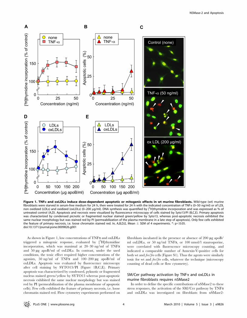
As shown in Figure 1, low concentrations of TNFa and oxLDLs
triggered a mitogenic response, evaluated by [3H]thymidine
incorporation, which was maximal at 20–30 ng/ml of TNFaand 50 mg apoB/ml of oxLDLs. In contrast, under the used
conditions, the toxic effect required higher concentrations of the
agonists, 50 ng/ml of TNFa and 100–200 mg apoB/ml of
oxLDLs. Apoptosis was evaluated by fluorescence microscopy
after cell staining by SYTO13/PI (Figure 1B,C,E). Primary
apoptosis was characterized by condensed, pyknotic or fragmented
nucleus stained green/yellow by SYTO13 whereas post-apoptotic
necrosis exhibited the same nuclear morphology but was stained
red by PI (permeabilization of the plasma membrane of apoptotic
cells). Few cells exhibited the feature of primary necrosis, i.e. loose
chromatin stained red. Flow cytometry experiments performed on
fibroblasts incubated in the presence or absence of 200 mg apoB/
ml oxLDLs, or 50 ng/ml TNFa, or 100 nmol/l staurosporine,
were correlated with fluorescence microscopy counting, and
indicated a comparable number of Annexin-V-positive cells for
both wt and fro/fro cells (Figure S1). Thus the agents were similarly
toxic for wt and fro/fro cells, whatever the technique (microscopy
counting of dead cells or flow cytometry).
SM/Cer pathway activation by TNFa and oxLDLs inmurine fibroblasts requires nSMase2
In order to define the specific contributions of nSMase2 to these
stress responses, the activation of the SM/Cer pathway by TNFaand oxLDLs was investigated on fibroblasts from nSMase2-
Figure 1. TNFa and oxLDLs induce dose-dependent apoptotic or mitogenic effects in wt murine fibroblasts. Wild-type (wt) murinefibroblasts were starved in serum-free medium for 24 h, then were treated for 24 h with the indicated concentration of TNFa (0–50 ng/ml) or of LDLnon oxidized (LDLs) and oxidized (oxLDLs) (0–200 mg/ml). DNA synthesis was quantified by [3H]thymidine incorporation and was expressed as % ofuntreated control (A,D). Apoptosis and necrosis were visualized by fluorescence microscopy of cells stained by Syto13/PI (B,C,E). Primary apoptosiswas characterized by condensed picnotic or fragmented nuclear stained green/yellow by Syto13, whereas post-apoptotic necrosis exhibited thesame nuclear morphology but was stained red by PI (permeabilization of the plasma membrane in a late step of apoptosis). Only few cells exhibitedthe feature of primary necrosis, i.e. loose chromatin stained red. In, A,B,D,E, Mean 6 SEM of 4 experiments. *: p,0.05.doi:10.1371/journal.pone.0009826.g001
NSMase-2 and Apoptosis
PLoS ONE | www.plosone.org 4 March 2010 | Volume 5 | Issue 3 | e9826

deficient fro/fro mice [19]. As expected, TNFa and oxLDLs
triggered both SM hydrolysis (Figure 2A,B) and nSMase activation
in wt fibroblasts (Figure 2D). In contrast, in fro/fro fibroblasts, SM
hydrolysis (Figure 2A,B), basal nSMase activity (Figure 2C), and
nSMase activation triggered by the two agonists (Figure 2D) were
deficient. Likewise, the ratio S1P/ceramide measured in wt and
fro/fo fibroblasts was increased two to three times in wt fibroblasts
stimulated either by oxLDLs or TNFa, while no variation was
observed in fro/fro cells (data not shown). Altogether, these data
strongly suggest that nSMase2, which is mutated and deficient in
fro/fro mice [19], is activated and required for the activation of the
SM/Cer pathway elicited by TNFa and oxLDLs in fibroblasts
(basal nSMase activity detected in fro/fro cells probably resulting
from the expression of other nSMases [2] that are apparently not
activated by TNFa and oxLDLs under the experimental
conditions used here). In these cells, the early phase (30–
120 min) of SM hydrolysis and ceramide formation appears to
be mainly attributable to nSMase2 activation.
TNFa and oxLDLs-induced apoptosis is independent ofnSMase2 activity
We then investigated whether nSMase2 is required for apoptosis
induced by TNFa, oxLDLs, or staurosporine, by comparing the
apoptotic effect of these agents in wt and fro/fro fibroblasts (Figure 3).
The time-course of oxLDLs- and TNFa-induced cell death (50%
mortality observed after 24h with oxLDLs, and 48 h with TNFa) was
assessed by the MTT assay to evaluate the overall toxicity (Figure 3A–
D). Dying cells exhibited the morphological features of apoptotic cells,
revealed by fluorescence microscopy after SYTO13/PI staining
(Figure 3E). Under the used conditions (apoptotic stress triggered by
toxic concentrations of TNFa, oxLDLs and staurosporine), fro/fro
cells underwent apoptosis similar to wt cells (and were even found
more sensitive) (Figure 3E). Consistent with the morphological data,
the apoptotic agents induced DEVDase activation, (time-course was
evaluated by the hydrolysis of the fluorogenic substrate Ac-DEVD-
AMC) (Figure 3G), and caspase-3 cleavage, (evaluated by western
blot) (Figure 3F). These effects occurred at a similar extent in wt and
Figure 2. TNFa and oxLDLs induce nSMase activation in wt but not in fro/fro fibroblasts. A,B - Sphingomyelin hydrolysis was monitoredafter metabolic labeling using [3H]choline chloride (0.5 mCi/ml) of cells, as described in the Method section. Then, cells were stimulated with oxLDLs(200 mg/ml) (A) or TNFa (50 ng/ml) (B), and the level of cellular sphingomyelin was determined at the indicated time. Results are expressed as % ofcontrol (time 0 h of stimulation). C,D -Basal nSMase activity in untreated cells (C) and in cells treated with TNFa (50 ng/ml) or oxLDLs (200 mg/ml) forthe indicated time (D). Mean 6 SEM of 3 to 5 separated experiments. * p,0.05.doi:10.1371/journal.pone.0009826.g002
NSMase-2 and Apoptosis
PLoS ONE | www.plosone.org 5 March 2010 | Volume 5 | Issue 3 | e9826

fro/fro fibroblasts (Figure 3). All these data, suggest that nSMase2 is
not required for apoptosis induced by TNFa, oxLDLs and
staurosporine in murine fibroblasts.
In fro/fro fibroblasts expressing a V5-tagged nSMase2 (fro-V5 clone
3 fibroblasts) (Figure 4A), the basal level of nSMase activity was
dramatically increased (around 3 times the level of wt) (Figure 4C).
NSMase activation and sphingomyelin hydrolysis by TNFa and
oxLDLs were also rescued in V5-fro (Fig. 4D,E). Note that mock-
transfected fro/fro cells did not exhibit any change in basal nor
stimulable nSMase activity (Fig. 4C). In spite of the broad variation in
nSMase activity, the activation of caspase 3 and the apoptotic effect of
TNFa, oxLDLs and staurosporine were similar in fro/fro and V5-fro
fibroblasts (Figure 4E–H). Taken together, all these data allow
concluding that murine fibroblasts do not require nSMase2 for cell
death induction in response to TNFa, oxLDLs, or staurosporine.
The nSMase2 defect in fro/fro mice does not preventTNFa toxicity
Because TNFa may trigger liver injury and lethal toxicity
through a mechanism involving ceramide generation [22], we
Figure 3. TNFa, oxLDLs and staurosporine induce cell apoptosis in wt and fro/fro fibroblasts. Fibroblasts were incubated with TNFa(50 ng/ml, 48 h), oxLDLs (200 mg/ml, 24 h) or staurosporine (100 nM, 6 h). Cell viability was evaluated by the MTT assay (A–C) and by countingapoptotic cells after syto13/PI labeling (D,E), as in Fig. 1. Caspase 3 activation was determined by western blot showing pro-caspase (32 kDa) andcleaved active caspase (17 kDa) (F). Time-course of DEVDase activity were measured using the fluorogenic substrate Ac-DEVD-AMC in cells treated byoxLDLs, TNFa and staurosporin, respectively (G–I). The results are mean 6 SEM of 3 to 5 separated experiments. * p,0.05 for apoptotic cell countingand DEVDase activity measurement (comparison between cells treated with or without agonist).doi:10.1371/journal.pone.0009826.g003
NSMase-2 and Apoptosis
PLoS ONE | www.plosone.org 6 March 2010 | Volume 5 | Issue 3 | e9826

Figure 4. Effects of TNFa and oxLDLs on fro/fro fibroblasts transfected with active V5-nSMase2 vector. The fro/fro fibroblasts were stablytransfected using pEF-6 plasmid containing V5-nSMase2 cDNA. After clone selection, the expression of V5-nSMase2 was evaluated by western blotusing an anti-V5 antibody (A), immunocytochemistry (B) and by enzymatic determination of nSMase activity, under basal conditions (C) and afteractivation by TNFa (50 ng/ml) or oxLDLs (200 mg/ml) (D). Time course of sphingomyelin hydrolysis induced by TNFa (50 ng/ml) or oxLDLs (200 mg/ml) were determined in V5-transfected cells under the conditions indicated in the legend to Figure 2 (E). Caspase activity was evaluated by westernblot of caspase-3 (32 KDa) (F), and by fluorometric determination of DEVDase activity (G), under the conditions of Fig. 3. Apoptosis triggered by TNFa
NSMase-2 and Apoptosis
PLoS ONE | www.plosone.org 7 March 2010 | Volume 5 | Issue 3 | e9826

compared the susceptibility of fro/fro and wt mice to the toxic
effects of TNFa (40 mg/kg) after pretreatment with the transcrip-
tion inhibitor D-galactosamine (D-GalN). In this model, D-
galactosamine was used to avoid the expression of anti-apoptotic
genes and thus to sensitize hepatic cells to TNFa, but D-
galactosamine exhibited no lethal effect per se (i.e. in the absence of
TNFa). Both fro/fro and wt mice were similarly sensitive to TNFa-
induced lethality (Figure 5A). The hepatic ceramide content,
expressed as the ratio to liver sphingomyelin, was 1.5 to 2 fold
increased in TNFa-treated wt and fro/fro mice, thereby indicating
that sphingomyelin hydrolysis mediated by TNFa treatment,
involves another sphingomyelinase, probably an acidic SMase as
reported [24], but not nSMase2. Histological study showed denser
and more eosinophilic cytoplasm and pyknotic nuclei, associated
with diffuse hemorrhagic areas, and the results were similar for
both wt and fro/fro TNFa-treated mice, (Figure 5B). It must be
noted that lower TNFa doses (10 mg/kg) were not toxic for both
wt and fro/fro mice over 48 h (data not shown).
The activation of nSMase2 is required for TNFa andoxLDLs-induced cell proliferation
Since the above data suggest that nSMase2 is not involved in the
apoptotic effect of oxLDLs and TNFa, and since nSMase activity
(50 ng/ml, 48 h incubation) or oxLDLs (200 mg/ml, 24 h incubation) was evaluated by fluorescence microscopy counting of cells labeled by syto13/PI,under conditions of Fig. 1 and 3 (H). In Fig. 4C–E, and G,H, the data are expressed as mean 6 SEM of 3 to 5 separated experiments * p,0.05; ns, notsignificant.doi:10.1371/journal.pone.0009826.g004
Figure 5. Time course of TNFa-induced toxicity in wt and fro/fro mice. Mice (10 wt or 10 fro/fro, 5 females and 5 males) were intraperitoneallyinjected with D-galactosamine (20 mg) and then injected intravenously with PBS or TNFa (40 mg/kg of body weight). A -Time course of survival of wt(red symbols) and fro/fro (green) mice treated or not by D-galactosamine and TNFa. B - Histological analysis of hematoxylin-eosin stained liversections from wt or fro/fro mice injected with D-galactosamine/PBS (sacrificed 48 hours after the injection) or D-galactosamine/TNFa (immediatelytaken off after mice death). Representative microscopy pictures of liver sections from wt and fro/fro mice (magnification, 6400).doi:10.1371/journal.pone.0009826.g005
NSMase-2 and Apoptosis
PLoS ONE | www.plosone.org 8 March 2010 | Volume 5 | Issue 3 | e9826

may play a role in the mitogenic effect of oxLDLs [26] and TNFa[8], we investigated whether the genetic defect of nSMase2 (fro/fro)
and the nSMase2 rescue (V5-fro) modulate the mitogenic effect
mediated by these agonists. As shown in Figure 6, low non-toxic
concentrations of oxLDLs and TNFa triggered nSMase activa-
tion, (and sphingomyelin hydrolysis, data not shown), mitogenic
signaling, DNA synthesis, and cell proliferation in wt fibroblasts. In
contrast, the mitogenic effect of oxLDLs and TNFa was abolished
Figure 6. Proliferation mediated by TNFa or oxLDLs requires the activation of the nSMase2. A,B - TNFa (5 ng/ml) and oxLDLs (50 mg/ml)induced nSMase activation (A) and ERK1/2 phosphorylation (B) in wt and in V5-fro) but not in fro/fro fibroblasts. It may be noted that FCS (10%)induce ERK1/2 phosphorylation in the 3 cell types, independently of nSMase2 deficiency (B). C,D - Proliferation was evaluated by DNA synthesis([3H]thymidine incorporation after 24 h incubation with the agonists) and cell count (performed after 48 h incubation with the agonists), in wt, fro/froand V5-fro fibroblasts, after treatment by TNFa, oxLDLs and FCS, as in Fig. 6A,B. In A,C,D, mean 6 SEM of 3 to 5 separated experiments. * p,0.05(comparison between cells treated with or without TNFa, as indicated).doi:10.1371/journal.pone.0009826.g006
NSMase-2 and Apoptosis
PLoS ONE | www.plosone.org 9 March 2010 | Volume 5 | Issue 3 | e9826

in fro/fro fibroblasts, as assessed by the lack of ERK1/2 activation
and DNA synthesis (Figure 6B,C). In contrast, in V5-fro fibroblasts
expressing V5-nSMase2, the activation of nSMase was rescued
(Figure 6A), as well as the activation of ERK1/2, increased DNA
synthesis, and cell proliferation (Figure 6B–D).
These data show that nSMase activation by oxLDLs and TNFais dependent on the activity of nSMase2 (Figure 6A) and is linked
to the mitogenic signaling triggered by the two agonists
(Figure 6C,D). This strongly suggests that nSMase2 is required
for the mitogenic response to the stress agonists oxLDLs and
TNFa.
Interestingly, the FCS-induced mitogenic effect was similar in
wt and fro/fro fibroblasts, thereby suggesting that nSMase2 activity
is not required for the mitogenic effect of growth factors contained
in the FCS (Figure 6B,C).
Discussion
TNFa and oxLDLs trigger the activation of nSMase2 in murine
wt fibroblasts, but the apoptotic effect of these agonists is
apparently independent of nSMase2 and SM hydrolysis, since
the genetic defect of nSMase2 (in cells from fro/fro mice) does not
alter the apoptotic response. Moreover, the hepatic toxicity
triggered in vivo by TNFa is similar in wt and fro/fro mice, thus
suggesting that nSMase2 is not required for the TNFa-induced
hepatic apoptosis. In contrast, our data show that nSMase2 is
absolutely required for the mitogenic effect induced by low
concentration of TNFa and oxLDLs.
A variety of data have been reported on signaling and biological
responses associated with the stimulation of nSMases, but only few
studies have addressed the specific nSMase that is implicated [5,6].
In the last decade, three mammalian nSMases have been cloned
[11,14,31], but their specific roles are only partly known. Precisely,
the apoptotic role of nSMase2 needs to be re-evaluated [5],
because of the unexpected phenotype of nSMase2-deficient mice,
that exhibit neonatal growth retardation and bone disease, but no
obvious defect of apoptosis [19,20]. Thus, our study was designed
to evaluate whether nSMase2 plays a role in apoptosis induced by
TNFa and by oxLDLs.
For this study, we chose TNFa and oxLDLs, two agents known
to activate nSMase, via different signaling mechanisms. TNFa is a
prototypical trigger of cellular nSMase activation (and ceramide
generation) associated with pleiotropic responses, including
apoptosis, cell proliferation and inflammation [5]. As indicated
above, TNFa was used without any protein synthesis inhibitor, in
order to exclude interference due to the cytotoxicity of
cycloheximide (or other protein inhibitor). The apoptotic effect
of TNFa was apparently independent of nSMase2 and SM
hydrolysis in murine fibroblasts, since similar apoptosis was
observed in nSMase2-deficient cells, and since rescuing the
activity of the nSMase2 did not alter the apoptosis induced by
TNFa and oxLDLs. Apoptosis by TNFa is mediated by FADD
and caspase 8 which in turn activates caspase-3 and/or Bid and
the mitochondrial apoptotic pathway [32]. An alternative
signaling pathway involving the generation of ceramide, either
through the de novo synthetic pathway, or by sphingomyelin
hydrolysis by SMases, has been hotly debated [10,33]. The role of
the acid SMase in ceramide generation and apoptosis triggered by
TNFa is largely documented [34,35], but nSMases have also been
implicated in the regulation of apoptosis by TNFa [36]. Our data
allow to conclude that nSMase2 is not required for TNFa-induced
apoptosis and that its activation by TNFa is not necessary for
triggering apoptosis in fibroblasts. However, it cannot be excluded
that nSMase2 may play a pro-apoptotic role in other cell types, as
suggested by its mutation in a murine osteosarcoma cell line and in
human leukemias [37]. This discrepancy may result from factors
regulating the level of ceramide, for instance, subcellular location,
transport and metabolic conversion, from the balance of
ceramide/sphingosine-1-phosphate, and from cell type-dependent
expression of pro-/anti-apoptotic effectors (e.g. Bcl2 family
members) [2,4].
Apoptosis triggered by oxLDLs was also found to be
independent of nSMase2 in fro/fro fibroblasts. This is consistent
with the lack of involvement of early ceramide formation and the
central role of calcium [38] in oxLDLs-induced apoptosis
[39,40,41].
In contrast, nSMase2 was required for the mitogenic response
triggered either by TNFa or oxLDLs in murine fibroblasts.
Previous reports had shown that the mitogenic response mediated
by both stress-inducing agents, depends on furin and metallopro-
teinases MT1-MMP and MMP-2 which activate nSMase2,
sphingomyelin hydrolysis, as well as the generation of sphingo-
sine-1-phosphate by sphingosine kinase [8,26]. The lack of TNFa-
or oxLDLs-induced mitogenic effect in fro/fro fibroblasts strongly
suggests that nSMase2 activation and ceramide production are
crucial for this stress-induced response. Note that the acid SMase,
which is not deficient in fro/fro fibroblasts (data not shown), is
unable to compensate for the defect in nSMase2-deficient cells.
This is in agreement with the idea that the topology of ceramide
generated by sphingomyelinases may play a crucial functional role.
Ceramide generated by the acid SMase in the outer leaflet of the
plasma membrane forms ceramide-enriched membrane platforms
that promote death-domain receptor clustering, thereby amplify-
ing apoptotic signalling [42]. In contrast, nSMase2 is located on
the inner leaflet, where it generates ceramide, hydrolyzed by
ceramidases into sphingosine, which may be phosphorylated in
turn by sphingosine kinase-1 [2,4]. This is consistent with the
hypothesis that the sphingolipid pathway located in the inner
leaflet may generate sphingosine-1-phosphate potentially involved
in cell activation, leading to cell proliferation or inflammatory
response [4].
Finally, in vivo experiments showed that nSMase2 plays
apparently no major role in hepatic apoptosis triggered by TNFa(associated to galactosamine), since wt and fro/fro mice exhibited
similar hepatotoxicity and death rate. This is in agreement with in
vitro studies, and with previous reports indicating a major role for
acid SMase and the FADD-caspase pathway in TNFa-induced
hepatocellular apoptosis [22,43,44].
Supporting Information
Figure S1 Flow cytometry. A Determination of cell death by
flow cytometry of annexin V-FITC/PI staining of wild-type (wt)
and fro/fro murine fibroblasts were seeded in 6-well plates for
48 hours and then exposed to TNFa (50 ng/ml) or oxLDLs
(200 mg/ml) for 24 hours or staurosporine (100 nM) for 6 hours.
Then, cells were harvested and stained as indicated in Material
and Methods section, and immediately used for cytometry
determination. Representative dot-plot graphs of 3 independent
experiments. The percentages of dying cells is calculated from all
annexin-V-positive cells, because microscopy examination shows
that annexin-V/PI double positive cells exhibited the morphology
of post-apoptotic necrosis. B Percentage of annexin V-positive wt
and fro/fro murine fibroblasts treated by oxLDLs, TNFa or
staurosporine. Data are expressed as mean 6 SEM from 3
separate experiments.
Found at: doi:10.1371/journal.pone.0009826.s001 (1.21 MB
TIF)
NSMase-2 and Apoptosis
PLoS ONE | www.plosone.org 10 March 2010 | Volume 5 | Issue 3 | e9826

Acknowledgments
The authors wish to thank C. Bernis, MH. Grazide, E. Mucher, L.
Rossard, Y. Bareira for the excellent technical assistance and Dr V. Gallet
(SNCF Laboratory, Toulouse) for providing human serum.
Author Contributions
Conceived and designed the experiments: RD ANS RS NA. Performed the
experiments: RD SG JCT JB NA. Analyzed the data: RD SG YAH JB
ANS RS NA. Contributed reagents/materials/analysis tools: JLG YAH.
Wrote the paper: RS NA.
References
1. Hannun YA (1996) Functions of ceramide in coordinating cellular responses to
stress. Science 274: 1855–1859.
2. Hannun YA, Obeid LM (2008) Principles of bioactive lipid signalling: lessonsfrom sphingolipids. Nat Rev Mol Cell Biol 9: 139–150.
3. Kolesnick RN, Kronke M (1998) Regulation of ceramide production andapoptosis. Annu Rev Physiol 60: 643–665.
4. Spiegel S, Milstien S (2003) Sphingosine-1-phosphate: an enigmatic signalling
lipid. Nat Rev Mol Cell Biol 4: 397–407.5. Clarke CJ, Hannun YA (2006) Neutral sphingomyelinases and nSMase2:
bridging the gaps. Biochim Biophys Acta 1758: 1893–1901.6. Auge N, Negre-Salvayre A, Salvayre R, Levade T (2000) Sphingomyelin
metabolites in vascular cell signaling and atherogenesis. Prog Lipid Res 39:207–229.
7. Coatrieux C, Sanson M, Negre-Salvayre A, Parini A, Hannun Y, et al. (2007)
MAO-A-induced mitogenic signaling is mediated by reactive oxygen species,MMP-2, and the sphingolipid pathway. Free Radic Biol Med 43: 80–89.
8. Tellier E, Negre-Salvayre A, Bocquet B, Itohara S, Hannun YA, et al. (2007)Role for furin in tumor necrosis factor alpha-induced activation of the matrix
metalloproteinase/sphingolipid mitogenic pathway. Mol Cell Biol 27:
2997–3007.9. Marchesini N, Osta W, Bielawski J, Luberto C, Obeid LM, et al. (2004) Role for
mammalian neutral sphingomyelinase 2 in confluence-induced growth arrest ofMCF7 cells. J Biol Chem 279: 25101–25111.
10. Kolesnick R, Hannun YA (1999) Ceramide and apoptosis. Trends Biochem Sci24: 224–225; author reply 227.
11. Tomiuk S, Hofmann K, Nix M, Zumbansen M, Stoffel W (1998) Cloned
mammalian neutral sphingomyelinase: functions in sphingolipid signaling? ProcNatl Acad Sci U S A 95: 3638–3643.
12. Zumbansen M, Stoffel W (2002) Neutral sphingomyelinase 1 deficiency in themouse causes no lipid storage disease. Mol Cell Biol 22: 3633–3638.
13. Yabu T, Imamura S, Yamashita M, Okazaki T (2008) Identification of Mg2+ -
dependent neutral sphingomyelinase 1 as a mediator of heat stress-inducedceramide generation and apoptosis. J Biol Chem 283: 29971–29982.
14. Krut O, Wiegmann K, Kashkar H, Yazdanpanah B, Kronke M (2006) Noveltumor necrosis factor-responsive mammalian neutral sphingomyelinase-3 is a C-
tail-anchored protein. J Biol Chem 281: 13784–13793.
15. Tomiuk S, Zumbansen M, Stoffel W (2000) Characterization and subcellularlocalization of murine and human magnesium-dependent neutral sphingomy-
elinase. J Biol Chem 275: 5710–5717.16. De Palma C, Meacci E, Perrotta C, Bruni P, Clementi E (2006) Endothelial
nitric oxide synthase activation by tumor necrosis factor alpha through neutralsphingomyelinase 2, sphingosine kinase 1, and sphingosine 1 phosphate
receptors: a novel pathway relevant to the pathophysiology of endothelium.
Arterioscler Thromb Vasc Biol 26: 99–105.17. Tani M, Hannun YA (2007) Neutral sphingomyelinase 2 is palmitoylated on
multiple cysteine residues. Role of palmitoylation in subcellular localization.J Biol Chem 282: 10047–10056.
18. Zeng C, Lee JT, Chen H, Chen S, Hsu CY, et al. (2005) Amyloid-beta peptide
enhances tumor necrosis factor-alpha-induced iNOS through neutral sphingo-myelinase/ceramide pathway in oligodendrocytes. J Neurochem 94: 703–712.
19. Aubin I, Adams CP, Opsahl S, Septier D, Bishop CE, et al. (2005) A deletion inthe gene encoding sphingomyelin phosphodiesterase 3 (Smpd3) results in
osteogenesis and dentinogenesis imperfecta in the mouse. Nat Genet 37:803–805.
20. Stoffel W, Jenke B, Block B, Zumbansen M, Koebke J (2005) Neutral
sphingomyelinase 2 (smpd3) in the control of postnatal growth and development.Proc Natl Acad Sci U S A 102: 4554–4559.
21. Wajant H (2003) Death receptors. Essays Biochem 39: 53–71.22. Ding WX, Yin XM (2004) Dissection of the multiple mechanisms of TNF-alpha-
induced apoptosis in liver injury. J Cell Mol Med 8: 445–454.
23. Chen G, Goeddel DV (2002) TNF-R1 signaling: a beautiful pathway. Science296: 1634–1635.
24. Auge N, Pieraggi MT, Thiers JC, Negre-Salvayre A, Salvayre R (1995)Proliferative and cytotoxic effects of mildly oxidized low-density lipoproteins on
vascular smooth-muscle cells. Biochem J 309 (Pt 3): 1015–1020.
25. Kittrell FS, Oborn CJ, Medina D (1992) Development of mammary
preneoplasias in vivo from mouse mammary epithelial cell lines in vitro. Cancer
Res 52: 1924–1932.
26. Auge N, Maupas-Schwalm F, Elbaz M, Thiers JC, Waysbort A, et al. (2004)
Role for matrix metalloproteinase-2 in oxidized low-density lipoprotein-induced
activation of the sphingomyelin/ceramide pathway and smooth muscle cell
proliferation. Circulation 110: 571–578.
27. Bielawski J, Szulc ZM, Hannun YA, Bielawska A (2006) Simultaneous
quantitative analysis of bioactive sphingolipids by high-performance liquid
chromatography-tandem mass spectrometry. Methods 39: 82–91.
28. Vieira O, Escargueil-Blanc I, Jurgens G, Borner C, Almeida L, et al. (2000)
Oxidized LDLs alter the activity of the ubiquitin-proteasome pathway: potential
role in oxidized LDL-induced apoptosis. FASEB J 14: 532–542.
29. Auge N, Garcia V, Maupas-Schwalm F, Levade T, Salvayre R, et al. (2002)
Oxidized LDL-induced smooth muscle cell proliferation involves the EGF
receptor/PI-3 kinase/Akt and the sphingolipid signaling pathways. Arterioscler
Thromb Vasc Biol 22: 1990–1995.
30. Brouckaert P, Libert C, Everaerdt B, Takahashi N, Cauwels A, et al. (1993)
Tumor necrosis factor, its receptors and the connection with interleukin 1 and
interleukin 6. Immunobiology 187: 317–329.
31. Hofmann K, Tomiuk S, Wolff G, Stoffel W (2000) Cloning and characterization
of the mammalian brain-specific, Mg2+-dependent neutral sphingomyelinase.
Proc Natl Acad Sci U S A 97: 5895–5900.
32. Wilson NS, Dixit V, Ashkenazi A (2009) Death receptor signal transducers:
nodes of coordination in immune signaling networks. Nat Immunol 10:
348–355.
33. Hofmann K, Dixit VM (1998) Ceramide in apoptosis–does it really matter?
Trends Biochem Sci 23: 374–377.
34. Carpinteiro A, Dumitru C, Schenck M, Gulbins E (2008) Ceramide-induced cell
death in malignant cells. Cancer Lett 264: 1–10.
35. Smith EL, Schuchman EH (2008) The unexpected role of acid sphingomyelinase
in cell death and the pathophysiology of common diseases. FASEB J 22:
3419–3431.
36. Clarke CJ, Truong TG, Hannun YA (2007) Role for neutral sphingomyelinase-2
in tumor necrosis factor alpha-stimulated expression of vascular cell adhesion
molecule-1 (VCAM) and intercellular adhesion molecule-1 (ICAM) in lung
epithelial cells: p38 MAPK is an upstream regulator of nSMase2. J Biol Chem
282: 1384–1396.
37. Kim WJ, Okimoto RA, Purton LE, Goodwin M, Haserlat SM, et al. (2008)
Mutations in the neutral sphingomyelinase gene SMPD3 implicate the ceramide
pathway in human leukemias. Blood 111: 4716–4722.
38. Escargueil-Blanc I, Andrieu-Abadie N, Caspar-Bauguil S, Brossmer R,
Levade T, et al. (1998) Apoptosis and activation of the sphingomyelin-ceramide
pathway induced by oxidized low density lipoproteins are not causally related in
ECV-304 endothelial cells. J Biol Chem 273: 27389–27395.
39. Escargueil-Blanc I, Salvayre R, Negre-Salvayre A (1994) Necrosis and apoptosis
induced by oxidized low density lipoproteins occur through two calcium-
dependent pathways in lymphoblastoid cells. FASEB J 8: 1075–1080.
40. Porn-Ares MI, Saido TC, Andersson T, Ares MP (2003) Oxidized low-density
lipoprotein induces calpain-dependent cell death and ubiquitination of caspase 3
in HMEC-1 endothelial cells. Biochem J 374: 403–411.
41. Vindis C, Elbaz M, Escargueil-Blanc I, Auge N, Heniquez A, et al. (2005) Two
distinct calcium-dependent mitochondrial pathways are involved in oxidized
LDL-induced apoptosis. Arterioscler Thromb Vasc Biol 25: 639–645.
42. Grassme H, Riethmuller J, Gulbins E (2007) Biological aspects of ceramide-
enriched membrane domains. Prog Lipid Res 46: 161–170.
43. Garcia-Ruiz C, Colell A, Mari M, Morales A, Calvo M, et al. (2003) Defective
TNF-alpha-mediated hepatocellular apoptosis and liver damage in acidic
sphingomyelinase knockout mice. J Clin Invest 111: 197–208.
44. Osawa M, Dace A, Tong KI, Valiveti A, Ikura M, et al. (2005) Mg2+ and Ca2+differentially regulate DNA binding and dimerization of DREAM. J Biol Chem
280: 18008–18014.
NSMase-2 and Apoptosis
PLoS ONE | www.plosone.org 11 March 2010 | Volume 5 | Issue 3 | e9826