Pseudomonas aeruginosa displays an epidemic population structure
Upload
independentCategory
view
1download
0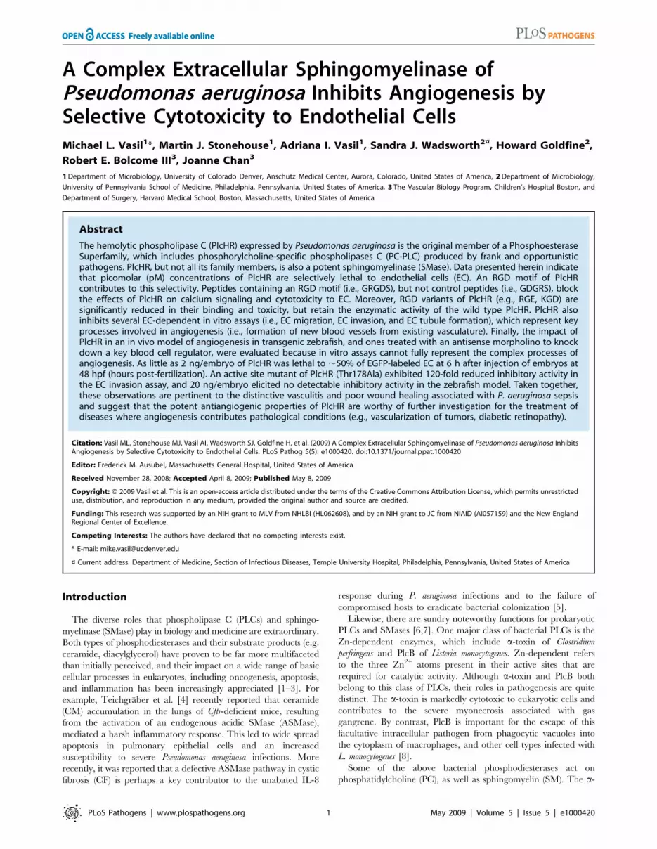
A Complex Extracellular Sphingomyelinase ofPseudomonas aeruginosa Inhibits Angiogenesis bySelective Cytotoxicity to Endothelial CellsMichael L. Vasil1*, Martin J. Stonehouse1, Adriana I. Vasil1, Sandra J. Wadsworth2¤, Howard Goldfine2,
Robert E. Bolcome III3, Joanne Chan3
1 Department of Microbiology, University of Colorado Denver, Anschutz Medical Center, Aurora, Colorado, United States of America, 2 Department of Microbiology,
University of Pennsylvania School of Medicine, Philadelphia, Pennsylvania, United States of America, 3 The Vascular Biology Program, Children’s Hospital Boston, and
Department of Surgery, Harvard Medical School, Boston, Massachusetts, United States of America
Abstract
The hemolytic phospholipase C (PlcHR) expressed by Pseudomonas aeruginosa is the original member of a PhosphoesteraseSuperfamily, which includes phosphorylcholine-specific phospholipases C (PC-PLC) produced by frank and opportunisticpathogens. PlcHR, but not all its family members, is also a potent sphingomyelinase (SMase). Data presented herein indicatethat picomolar (pM) concentrations of PlcHR are selectively lethal to endothelial cells (EC). An RGD motif of PlcHRcontributes to this selectivity. Peptides containing an RGD motif (i.e., GRGDS), but not control peptides (i.e., GDGRS), blockthe effects of PlcHR on calcium signaling and cytotoxicity to EC. Moreover, RGD variants of PlcHR (e.g., RGE, KGD) aresignificantly reduced in their binding and toxicity, but retain the enzymatic activity of the wild type PlcHR. PlcHR alsoinhibits several EC-dependent in vitro assays (i.e., EC migration, EC invasion, and EC tubule formation), which represent keyprocesses involved in angiogenesis (i.e., formation of new blood vessels from existing vasculature). Finally, the impact ofPlcHR in an in vivo model of angiogenesis in transgenic zebrafish, and ones treated with an antisense morpholino to knockdown a key blood cell regulator, were evaluated because in vitro assays cannot fully represent the complex processes ofangiogenesis. As little as 2 ng/embryo of PlcHR was lethal to ,50% of EGFP-labeled EC at 6 h after injection of embryos at48 hpf (hours post-fertilization). An active site mutant of PlcHR (Thr178Ala) exhibited 120-fold reduced inhibitory activity inthe EC invasion assay, and 20 ng/embryo elicited no detectable inhibitory activity in the zebrafish model. Taken together,these observations are pertinent to the distinctive vasculitis and poor wound healing associated with P. aeruginosa sepsisand suggest that the potent antiangiogenic properties of PlcHR are worthy of further investigation for the treatment ofdiseases where angiogenesis contributes pathological conditions (e.g., vascularization of tumors, diabetic retinopathy).
Citation: Vasil ML, Stonehouse MJ, Vasil AI, Wadsworth SJ, Goldfine H, et al. (2009) A Complex Extracellular Sphingomyelinase of Pseudomonas aeruginosa InhibitsAngiogenesis by Selective Cytotoxicity to Endothelial Cells. PLoS Pathog 5(5): e1000420. doi:10.1371/journal.ppat.1000420
Editor: Frederick M. Ausubel, Massachusetts General Hospital, United States of America
Received November 28, 2008; Accepted April 8, 2009; Published May 8, 2009
Copyright: � 2009 Vasil et al. This is an open-access article distributed under the terms of the Creative Commons Attribution License, which permits unrestricteduse, distribution, and reproduction in any medium, provided the original author and source are credited.
Funding: This research was supported by an NIH grant to MLV from NHLBI (HL062608), and by an NIH grant to JC from NIAID (AI057159) and the New EnglandRegional Center of Excellence.
Competing Interests: The authors have declared that no competing interests exist.
* E-mail: [email protected]
¤ Current address: Department of Medicine, Section of Infectious Diseases, Temple University Hospital, Philadelphia, Pennsylvania, United States of America
Introduction
The diverse roles that phospholipase C (PLCs) and sphingo-
myelinase (SMase) play in biology and medicine are extraordinary.
Both types of phosphodiesterases and their substrate products (e.g.
ceramide, diacylglycerol) have proven to be far more multifaceted
than initially perceived, and their impact on a wide range of basic
cellular processes in eukaryotes, including oncogenesis, apoptosis,
and inflammation has been increasingly appreciated [1–3]. For
example, Teichgraber et al. [4] recently reported that ceramide
(CM) accumulation in the lungs of Cftr-deficient mice, resulting
from the activation of an endogenous acidic SMase (ASMase),
mediated a harsh inflammatory response. This led to wide spread
apoptosis in pulmonary epithelial cells and an increased
susceptibility to severe Pseudomonas aeruginosa infections. More
recently, it was reported that a defective ASMase pathway in cystic
fibrosis (CF) is perhaps a key contributor to the unabated IL-8
response during P. aeruginosa infections and to the failure of
compromised hosts to eradicate bacterial colonization [5].
Likewise, there are sundry noteworthy functions for prokaryotic
PLCs and SMases [6,7]. One major class of bacterial PLCs is the
Zn-dependent enzymes, which include a-toxin of Clostridium
perfringens and PlcB of Listeria monocytogenes. Zn-dependent refers
to the three Zn2+ atoms present in their active sites that are
required for catalytic activity. Although a-toxin and PlcB both
belong to this class of PLCs, their roles in pathogenesis are quite
distinct. The a-toxin is markedly cytotoxic to eukaryotic cells and
contributes to the severe myonecrosis associated with gas
gangrene. By contrast, PlcB is important for the escape of this
facultative intracellular pathogen from phagocytic vacuoles into
the cytoplasm of macrophages, and other cell types infected with
L. monocytogenes [8].
Some of the above bacterial phosphodiesterases act on
phosphatidylcholine (PC), as well as sphingomyelin (SM). The a-
PLoS Pathogens | www.plospathogens.org 1 May 2009 | Volume 5 | Issue 5 | e1000420
toxin of C. perfringens and PlcB or L. monocytogenes hydrolyse either
PC or SM to generate DAG or CM and phosphorylcholine, while
a Bacillus cereus PC-PLC from the same zinc-dependent class of
enzymes, has no activity whatsoever on SM [9]. The SMase
activity of a-toxin however, is more critical to its role in
pathogenesis than its PC-PLC activity [10–12].
A second major class of prokaryotic phosphorylcholine
preferring PLCs, some of which are also SMases, is part of a
large Phosphoesterase/PLC Superfamily [13]. These include
bacterial and fungal PLCs, in addition to prokaryotic and
eukaryotic phosphatases. The PLC members of this family are
not Zn-dependent enzymes; rather divalent metals including Zn
and nickel readily inhibit their activity [13]. For this and other
reasons, Zn-dependent and Zn-independent bacterial PLCs each
use distinct catalytic mechanisms to hydrolyze the phosphodiester
bond between the phosphoryl head group of a choline containing
phospholipid or sphingolipid, and their DAG or CM moieties
[14].
The hemolytic phospholipase C (PlcH) of Pseudomonas aeruginosa,
the focus of the present report, is the founding member of the large
Zn-independent PLC family. Frank bacterial pathogens (i.e.
Mycobacterium tuberculosis, Bordetella pertussis & Francisella tularensis),
opportunistic bacteria (P. aeruginosa, Acinetobacter baumannii), fungal
pathogens (Aspergillus fumigatus), as well as the Class B Select Agents
Burkholderia pseudomallei & mallei, express homologous proteins
belonging to the phosphodiesterase (i.e. PLC) part of the family
[13]. Some pathogens (e.g. Mycobacterium tuberculosis, B. pseudomallei)
carry three, or as many as four genes, encoding enzymes belonging
to this class of PLCs [15–18]. All P. aeruginosa strains thus far
sequenced (7) have two genes encoding members of this family,
PlcH and PlcN [13]. In one way or another, many of these PLCs
have now been associated with the virulence of the particular
organism that produces them, but their contributions at the
cellular level remain obscure.
PlcH, which is both a PC-PLC and a SMase, is a significant
virulence determinant of P. aeruginosa in an array of infection
models. Ostroff et al. showed that a PlcH deletion mutant had a
10,000-fold increase in its LD50 in a mouse thermal injury model,
compared to that of its wild type parent [19]. Rahme et al. [20]
confirmed the attenuated phenotype of a PlcH mutant in the same
type of mouse infection model, but their mutant was constructed
using a different parental P. aeruginosa strain (i.e. PA-14). They also
reported that this PlcH mutant was attenuated in a plant (i.e.
Arabidopsis thaliana) infection model. PlcH can be a noteworthy
virulence factor in other non-mammalian hosts, as well. Hogan
and Kolter reported that a PlcH mutant was attenuated in a P.
aeruginosa infection of the mycelial phase, but not the yeast phase,
of Candida albicans [21]. This particular mutant was one of the
more attenuated ones in an assortment of other types of P.
aeruginosa mutants they tested in this model.
Early on, PlcH was predominantly characterized for its
hemolytic activity and enzymatic properties [13,22]. More recent
studies suggest that the SMase activity of PlcHR is much more
critical than its PC-PLC activity, with regard to its ability to cause
a phenomena known as ‘‘hot-cold hemolysis’’ as well as hemolysis
at 37uC [13,22,23]. The present study however, began as an effort
to assess its potential toxicity to a variety of nucleated mammalian
(i.e. non-erythrocytic) cell lines, with the objective of discerning
additional clues about its possible role at the cellular level in P.
aeruginosa infections. In the present study, we report that PlcHR is a
potent bacterial toxin, with selectivity for endothelial cells (EC)
that is, at least in part, mediated through its Arg-Gly-Asp (RGD)
motif. Based on its selective cytotoxicity for EC, PlcHR is a potent
antiangiogenic agent in assorted assays (e.g. EC invasion assays,
tube formation) that evaluate different stages of angiogenesis in
vitro, and PlcHR inhibits angiogenesis in an in vivo model in
zebrafish. These and other data, implicate PlcH in the
pathogenesis of ‘‘vasculitis’’, and the poor, angiogenesis-depen-
dent, wound healing, typically observed in sepsis associated with
this opportunistic pathogen [24,25]. From a different perspective,
it is possible that potent antiangiogenic bacterial toxins, such as
Anthrax toxin and PlcHR, might also provide novel ways to
mitigate the pathological consequences of abnormal angiogenesis
(e.g. tumor vascularization, diabetic retinopathies).
Results
PlcHR is selectively cytotoxic to endothelial cellsPlcHR, a complex heterodimer, consisting of PlcH and PlcR,
was expressed in P. aeruginosa and purified as previously described
[13,22,23]. PlcHR is hemolytic to human and sheep erythrocytes,
and had been previously reported to be moderately cytotoxic to
neutrophils and macrophage [22,23,26,27]. Yet, no one had
conducted a systematic evaluation of its cytotoxic potential for
other types of nucleated cells. Accordingly, the cytotoxic effects of
PlcHR toward an assortment of other types of eukaryotic cells was
undertaken using the following cell lines: a 1u human lung cell line
from a CF patient [28]; HeLa (epithelial); L929 mouse fibroblasts;
J774 macrophage; Chinese hamster ovary cells (CHO); and
Human Umbilical Vascular Endothelial Cells (HUVEC). As
shown in Figure 1 and Figure S1, remarkably low concentrations
(,10 ng/ml) of PlcHR were distinctly cytotoxic to HUVEC and
CHO cells. By contrast, much higher concentrations of PlcHR (i.e.
.4 mg/ml) were only slightly cytotoxic to any of the other cell
types examined including, 1u CF lung epithelial cells and J774
macrophage, that were evaluated at 6 hrs after the application of
varying amounts of PlcHR. An additional epithelial cell line, A549
lung cells was also tested, but they were even more resistant to the
cytotoxic effects of PlcHR than the 1u CF lung epithelial cells were
(data not shown).
Author Summary
Pseudomonas aeruginosa is a major bacterial opportunisticpathogen responsible for acute (e.g., sepsis) and chronicinfections (e.g., pulmonary). While it expresses assortedextracellular toxins that in one way or another contributeto pathogenesis, the precise cellular and molecularmechanisms by which these factors act is largely unknown.During septicemia, P. aeruginosa frequently causes vascu-litis and thrombosis. Endothelial cells line the entirevascular system of mammals and have sundry keyfunctions in infectious diseases, including thrombosisand inflammation. Endothelial cells are essential to theformation of new blood vessels (angiogenesis) needed forproper wound healing. This report describes in vitro and invivo experiments demonstrating that an extracellulartoxin, PlcHR, at very low (picomolar) concentrations, ishighly lethal to endothelial cells and inhibits angiogenesisin vivo. Data herein also suggest that PlcHR is selectivelytoxic to endothelial cells through its ability to bind a cellreceptor(s). These observations may be particularly rele-vant to the mechanisms by which P. aeruginosa causesvascular lesions and inhibits the healing of wounds duringseptic infections. Finally, the potent antiangiogenic attri-bute of PlcHR could be useful in the treatment ofnoninfectious diseases where angiogenesis contributestheir pathogenesis, including the vascularization of tumorsand the eye (e.g., diabetic retinopathy).
Pseudomonas aeruginosa Toxin Inhibits Angiogenesis
PLoS Pathogens | www.plospathogens.org 2 May 2009 | Volume 5 | Issue 5 | e1000420
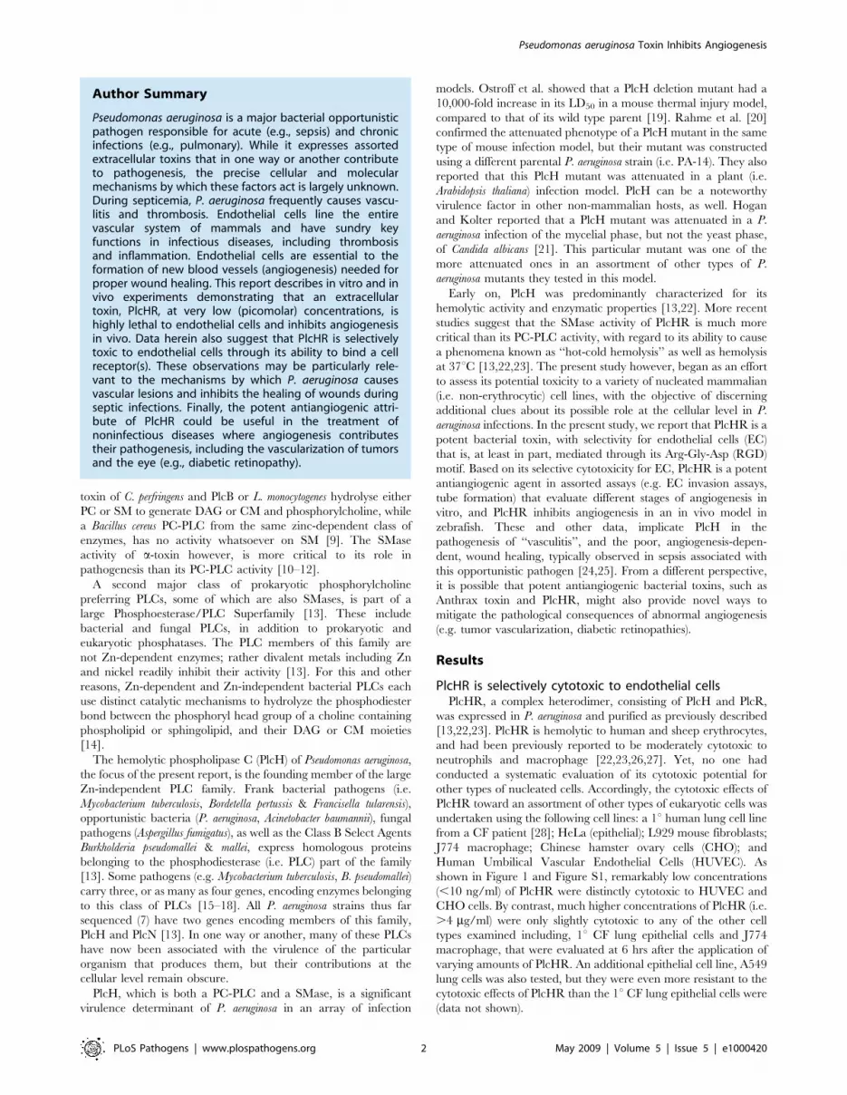
As shown in Figure 2, PlcHR does not seem to cause lysis of EC
even after 1 hour (Figure 2B), as would be expected if it had been
simply hydrolyzing their membranes through its PLC/SMase
activity. Moreover, because the outer leaflets of the membranes of
all of these cells contain equimolar concentrations of PC and SM
[29], these data suggest that the enzymatic activities of PlcHR, per
se, are not sufficient to account for its EC and CHO cell selectivity,
compared to the other types of cells that were tested (Figure 1 and
Figure S1).
PlcHR induces a rise in intracellular calcium in ECAn association between calcium and the hemolytic activity of
PlcHR has been previously noted [13], and associations between
the calcium responses of eukaryotic cells to other bacterial PLCs
have been reported [7]. Based on those observations, the effect of
PlcHR on intracellular calcium levels in EC was investigated. Only
5 ng/ml of PlcHR induced a significant increase in intracellular
calcium levels in EC, which peaked at 7 minutes (Table 1). The
rise in calcium levels could originate from the influx of
extracellular calcium or via the release of calcium from stores in
the endoplasmic reticulum (ER). The two possible pathways by
which intracellular calcium increases can be separated by
pretreatment of EC with the calcium channel blocker
SK&F96365 for 15 min (inhibits entry of extracellular calcium via
calcium channels) or by pretreatment with thapsigargin for 45 min
(depletes intracellular calcium stores in the endoplasmic reticulum
[30,31]). The 45 minute pretreatment of EC with thapsigargin
(1 mM), before application of 5 ng/ml of PlcHR (Table 1),
significantly blocked the subsequent rise in intracellular calcium
levels, that is normally observed in EC treated only with PlcHR
(5 ng/ml) (Table 1). Intracellular calcium levels at 7 minutes in EC
treated with thapsigargin and PlcHR was 5566 nM, while
intracellular calcium levels at 7 minutes in EC treated only with
PlcHR was 568658 nM. In contrast, there were no significant
differences in the intracellular calcium levels between cells that were
pre-treated with 25 mM of SK&F96365, and then subsequently
treated with PlcHR (435643 nM), compared to those treated only
with PlcHR (429648 nM) (Table 1). When PlcH alone was
compared to PlcHR a more rapid (.2 fold) release of intracellular
calcium in EC was observed, but their maximums were ultimately
virtually the same (data not shown). Also, .100 ng/ml of PlcHR
failed to induce any increase in intracellular levels of calcium in J774
macrophage (data not shown), which are resistant to the cytotoxicity
of PlcHR (Figure 1).
Figure 1. Cytotoxicity of PlcHR to assorted eukaryotic cells. Cytotoxicity (measured by LDH release) for assorted eukaryotic cell cultures usingthe CytoTox 96 Assay Kit as described in Materials and Methods. Concentrations of PlcHR (ng/ml) used are noted, and LDH release was measuredafter 6 h of incubation.doi:10.1371/journal.ppat.1000420.g001
Pseudomonas aeruginosa Toxin Inhibits Angiogenesis
PLoS Pathogens | www.plospathogens.org 3 May 2009 | Volume 5 | Issue 5 | e1000420
These data indicate that the types of cells that are susceptible or
resistant to calcium signaling by PlcHR reflect the type of cells that
are susceptible or resistant to its cytotoxicity. Because PlcHR
induces the release of calcium from the ER in EC, rather than
through calcium channels, it is particularly relevant to note that
calcium release from the ER, will in turn, activate downstream
effectors of apoptosis (e.g. calcineurin, calpains, endonucleases)
[32]. Finally, with regard to the PC-PLC and SMase activity of
PlcHR, it is important to note that increased levels of ceramide (a
SMase product), but not diacylglycerol (a PC-PLC product), can
induce the release of calcium from ER stores [33].
Induction of caspase-3 activity by PlcHRThe aspartate-specific cysteinyl proteases or caspases are a set of
mediators implicated in apoptosis. The activation of caspase-3 in
mammalian cells is a hallmark of apoptosis [34]. While the
generation of DAG from PC would be expected to induce EC
proliferation, hydrolysis of membrane SM by PlcHR, would cause
a relatively increase in CM, leading to programmed cell death (i.e.
apoptosis) [33]. We therefore chose to examine whether PlcHR
cytotoxicity could induce increased levels of caspase-3 activity,
which is one of the effector caspases that ultimately carry out
apoptosis. As shown in Figures 3, S2A and S2B, treatment of EC
with pM concentrations of PlcHR resulted in activation of caspase-
3. In experiments shown in Figure 3, both caspase-3 activation and
LDH release were measured at 16 hours post treatment with
increasing concentrations of PlcHR. Figure 3 shows that caspase-3
activity increases as the concentration of PlcHR increases until it
peaks at 6.25 ng/ml PlcHR. The level of caspase-3 activity
induced with 6.25 ng/ml PlcHR is similar to cells treated with the
apoptosis control compound camptothecin (Figure 3). Beyond
6.25 ng/ml PlcHR, caspase-3 activity begins to decrease, but the
release of LDH continues to increase until ultimately at 100 ng/ml
PlcHR there is very little caspase-3 activity and LDH release has
reached its maximum. The addition of the pan-caspase inhibitor
Z-VAD-FMK completely inhibited PlcHR activation of caspase-3
and reduced the level of LDH release (Figure S2B), indicating that
a significant portion of the cells releasing LDH are actually dying
by apoptosis
Finally, a time course for activation of caspase-3 was performed
to determine when caspase-3 was activated by treatment with
PlcHR. As shown is Figure S2A treatment of EC with as little as
2.5 ng/ml of PlcHR activated caspase-3 between 3 and 6 hours.
Caspase-3 activation was never detected in resistant cells (e.g.
Hela) when they were treated with .100 ng/ml of PlcHR (data
not shown).
Selective toxicity of PlcHR is associated with its RGDmotif
PlcH has an RGD motif (Figure S3), which is present in only
one other PLC (i.e. one of the three PlcH homologs expressed by
Burkholderia thailandensis) of the now .50 other members of this
class of enzymes. RGD motifs are associated with cell-cell and cell-
matrix interactions and they play a major role in host cell
recognition by assorted viruses, as well as bacterial virulence
factors (e.g. invasin, toxins) through their interactions with cell
surface integrin receptors [35]. Yet, the context of an RGD motif
in a protein is likewise critical to its ability to bind integrins [36],
and variants of this motif (i.e. RGE, KGE) may still be functional
for binding integrin receptors [37,38].
Several approaches were taken to assess whether the RGD motif
of PlcH is associated with its selectivity for EC or CHO cells.
PlcHR and free PlcH caused a significant increase in the level of
Figure 2. Treatment with pM (PlcHR MW = 95.6 kDA) concentrations of PlcHR induces morphological changes, but not direct lysis ofEC. (A) Untreated HUVEC. (B) HUVEC treated with 7.5 ng/ml PlcHR2 for 1 h. (C) HUVEC treated with 7.5 ng/ml PlcHR2 for 3 h. Pictures are at 2006magnification.doi:10.1371/journal.ppat.1000420.g002
Table 1. PlcHR calcium signaling and mechanism ofintracellular calcium release.
Time (min)Untreated ECnM Calcium
Treated EC (5 ng/ml PIcHR)nM Calcium
1 5662.3 129625
3 5061.2 306647
5 32610.5 362644
7 3165.6 568658**
10 99612 435648*
12 357623
15 229631
17 189668
20 72615 190625
25 115618
30 6669.8
*-p#0.01 for HUVECS+PlcHR at 7 and 10 min compared to untreated cells.**Thapsigargin (TG) results (EC cells pretreated with TG 45 min prior PlcHR
treatment): 7 min untreated EC (no PlcHR), 6266.8 nM; 7 min EC+PlcHR noTG, 515672 nM**; 7 min EC+PlcHR+TG, 5566 nM; 7 min EC no PlcHR+TG,76613.5 nM. SK&F96365 (SK) results (EC cells pretreated with SK 15 min priorPlcHR treatment): 15 min untreated (no PlcHR), 6266.8 nM; 15 min EC+PlcHRno SK, 429648 nM; 15 min EC+PlcHR+SK, 435643 nM.
doi:10.1371/journal.ppat.1000420.t001
Pseudomonas aeruginosa Toxin Inhibits Angiogenesis
PLoS Pathogens | www.plospathogens.org 4 May 2009 | Volume 5 | Issue 5 | e1000420
intracellular calcium release that peaked at 7 minutes (Tables 1
and 2). However, when a 5-mer peptide with an RGD motif (i.e.
GRGDS) was added at the same time as PlcHR, or free PlcH,
there was no evidence of release of calcium from ER stores over
that seen in untreated cells. By contrast, when a 5-mer peptide
with a scrambled RGD sequence (i.e. GDGRS) was added to EC
in conjunction with PlcH, an increase in the intracellular calcium
release reaching a maximum at 7 min, comparable to that seen
with PlcHR alone, was observed (Tables 1 and 2).
The ability of these peptides to attenuate the cytoxicity of
PlcHR was also evaluated. Similar effects of these peptides were
seen (Table 3). The RGD containing peptide (GRGDS) at
approximately a 50-fold molar excess of peptide to PlcHR
significantly dampened the lethal impact of PlcHR, while the
scrambled peptide (GDGRS) at the same concentration demon-
strated no significant effect on PlcHR toxicity in this assay.
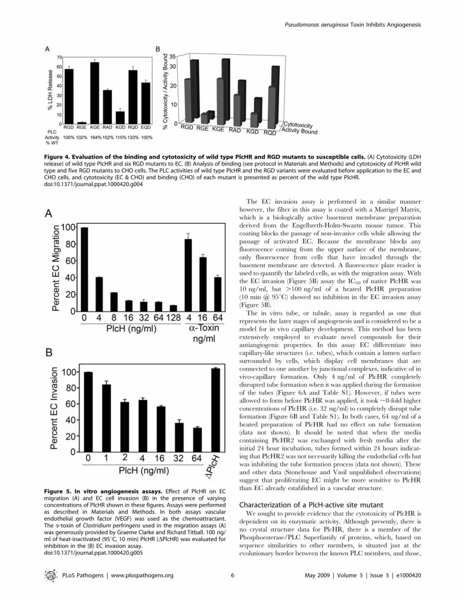
Additionally, several RGD variants of PlcHR were constructed.
Various plcH genes encoding PlcH with altered RGD motifs were
expressed, along with the wild type plcR gene, and the resulting
PlcHR-RGD mutant proteins were purified. Each contain variant
RGD motifs (e.g. RGE, RAD, RQD) that are found in other PlcH
homologs in the NCBI database (Figure 4). Several RGD variants
were then evaluated for their enzymatic activity (Figure 4A), as
well as, their binding (Figure 4B) and cytotoxicity to EC or CHO
cells (Figure 4A & 4B). In support of the hypothesis that the RGD
motif of PlcHR is a critical factor in its ability to kill EC or CHO
cells, some variants (i.e. RGE, KGE), still retained wild type PLC
activity, but were significantly decreased in their binding or
lethality to EC or CHO cells by comparison to the wild type
PlcHR (Figure 4A & 4B). Finally, we found that a resistant cell line
(i.e. L929 fibroblasts) bound significantly less toxin (16% of total
PlcHR added) compared to EC (42% of total PlcHR added) and
that binding of PlcHR to EC was saturable. That is, there was no
increased binding despite higher concentrations of PlcHR added
to these cells (Stonehouse, M. doctoral thesis University of
Colorado Denver).
PlcHR inhibits angiogenesis in vitroBased on the selective and potent toxicity of PlcHR toward EC,
it was of interest, particularly in the context of vasular disease
associated with P. aeruginosa sepsis (see Discussion below), to
examine whether this EC selective toxin inhibits the more complex
processes associated with angiogenesis (i.e. formation of new
vessels from the existing vasculature). There are assorted in vitro
angiogenesis assays (e.g. migration and invasion assays, tube
formation) to approximate individual mechanisms involved in the
formation of new blood vessels. See reference [39] for a detailed
description of the angiogenesis assays described below.
The EC migration assay is based on the movement of cells
through a fluorescence blocking, microporous inert filter coated
with human fibronectin. The pores of the membrane are not
occluded, which allows the EC to attach to the membrane and
freely migrate toward the angiogenic stimulus, vascular endothelial
growth factor (VEGF), in a chamber below. A fluorescent plate
reader is used to quantify EC, labeled with a fluorescent dye,
which migrated through the filter. In the EC migration assay
(Figure 5A) only 3 ng/ml of PlcHR was required to inhibit EC
migration by 50%, (IC50) while the IC50 for an unrelated PLC/
SMase, C. perfringens a-toxin (generously provided by Graeme
Clarke and Richard Titball) was 45 ng/ml (Figure 5A).
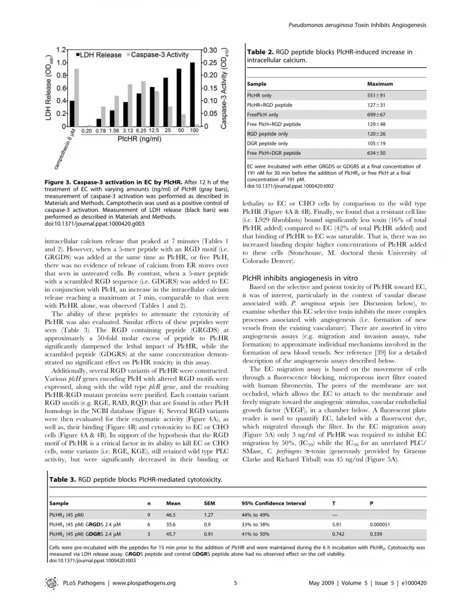
Figure 3. Caspase-3 activation in EC by PlcHR. After 12 h of thetreatment of EC with varying amounts (ng/ml) of PlcHR (gray bars),measurement of caspase-3 activation was performed as described inMaterials and Methods. Camptothecin was used as a positive control ofcaspase-3 activation. Measurement of LDH release (black bars) wasperformed as described in Materials and Methods.doi:10.1371/journal.ppat.1000420.g003
Table 2. RGD peptide blocks PlcHR-induced increase inintracellular calcium.
Sample Maximum
PlcHR only 551691
PlcHR+RGD peptide 127631
FreePlcH only 699667
Free PlcH+RGD peptide 129648
RGD peptide only 120626
DGR peptide only 105619
Free PlcH+DGR peptide 634650
EC were incubated with either GRGDS or GDGRS at a final concentration of191 nM for 30 min before the addition of PlcHR2 or free PlcH at a finalconcentration of 191 pM.doi:10.1371/journal.ppat.1000420.t002
Table 3. RGD peptide blocks PlcHR-mediated cytotoxicity.
Sample n Mean SEM 95% Confidence Interval T P
PlcHR2 (45 pM) 9 46.5 1.27 44% to 49% —
PlcHR2 (45 pM) GRGDS 2.4 mM 6 35.6 0.9 33% to 38% 5.91 0.000051
PlcHR2 (45 pM) GDGRS 2.4 mM 3 45.7 0.91 41% to 50% 0.742 0.339
Cells were pre-incubated with the peptides for 15 min prior to the addition of PlcHR and were maintained during the 6 h incubation with PlcHR2. Cytotoxicity wasmeasured via LDH release assay. GRGDS peptide and control GDGRS peptide alone had no observed effect on the cell viability.doi:10.1371/journal.ppat.1000420.t003
Pseudomonas aeruginosa Toxin Inhibits Angiogenesis
PLoS Pathogens | www.plospathogens.org 5 May 2009 | Volume 5 | Issue 5 | e1000420
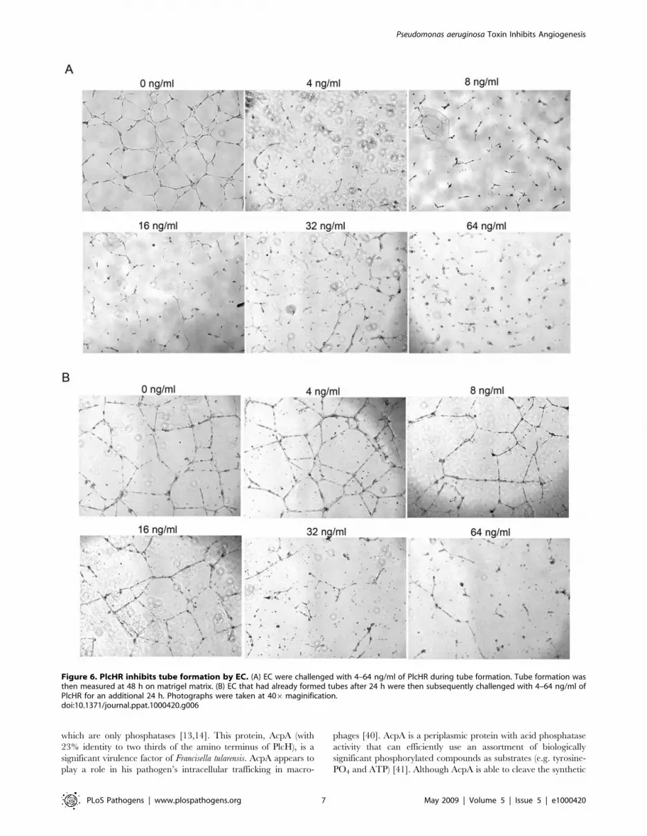
The EC invasion assay is performed in a similar manner
however, the filter in this assay is coated with a Matrigel Matrix,
which is a biologically active basement membrane preparation
derived from the Engelbreth-Holm-Swarm mouse tumor. This
coating blocks the passage of non-invasive cells while allowing the
passage of activated EC. Because the membrane blocks any
fluorescence coming from the upper surface of the membrane,
only fluorescence from cells that have invaded through the
basement membrane are detected. A fluorescence plate reader is
used to quantify the labeled cells, as with the migration assay. With
the EC invasion (Figure 5B) assay the IC50 of native PlcHR was
10 ng/ml, but .100 ng/ml of a heated PlcHR preparation
(10 min @ 95uC) showed no inhibition in the EC invasion assay
(Figure 5B).
The in vitro tube, or tubule, assay is regarded as one that
represents the later stages of angiogenesis and is considered to be a
model for in vivo capillary development. This method has been
extensively employed to evaluate novel compounds for their
antiangiogenic properties. In this assay EC differentiate into
capillary-like structures (i.e. tubes), which contain a lumen surface
surrounded by cells, which display cell membranes that are
connected to one another by junctional complexes, indicative of in
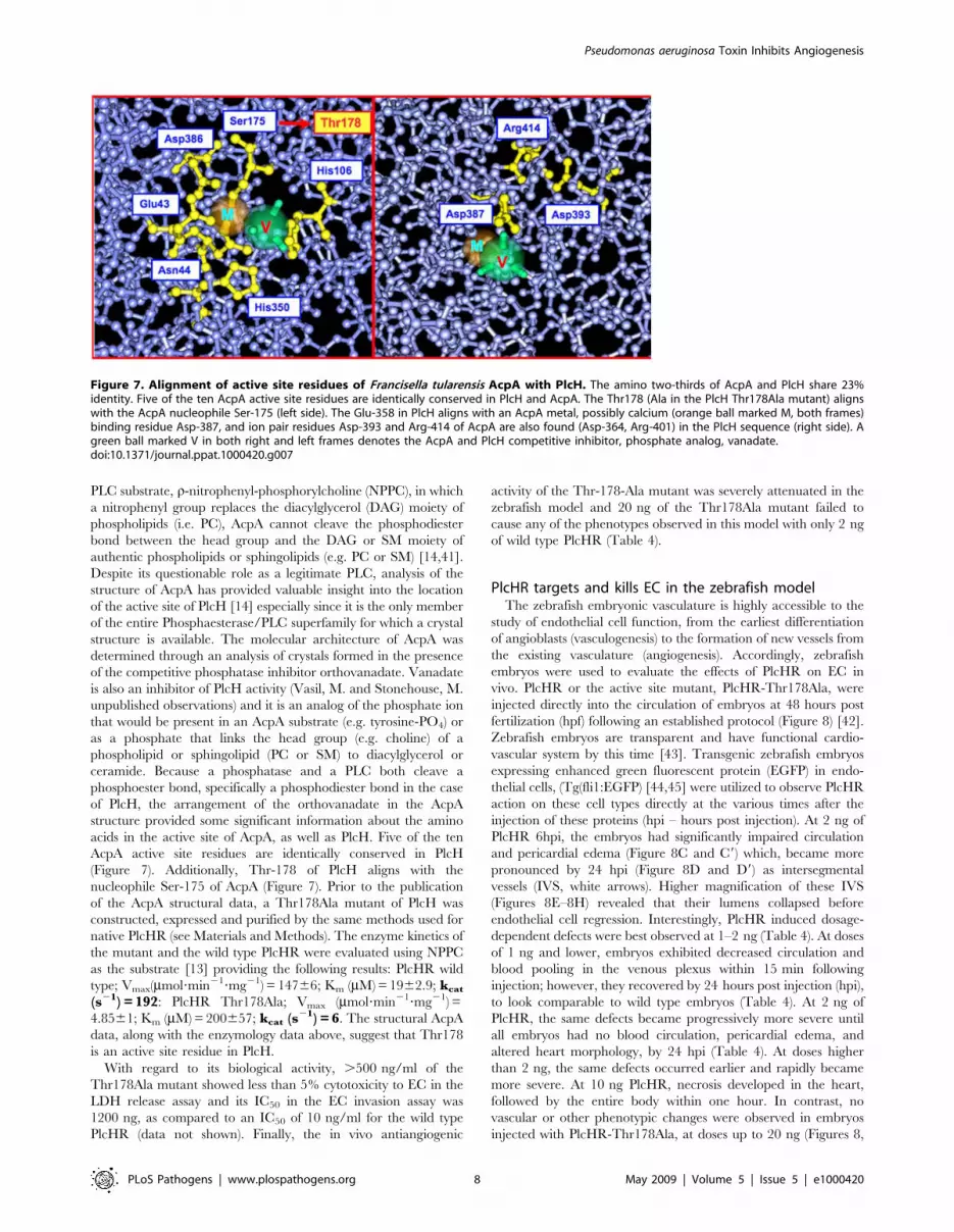
vivo-capillary formation. Only 4 ng/ml of PlcHR completely
disrupted tube formation when it was applied during the formation
of the tubes (Figure 6A and Table S1). However, if tubes were
allowed to form before PlcHR was applied, it took ,8-fold higher
concentrations of PlcHR (i.e. 32 ng/ml) to completely disrupt tube
formation (Figure 6B and Table S1). In both cases, 64 ng/ml of a
heated preparation of PlcHR had no effect on tube formation
(data not shown). It should be noted that when the media
containing PlcHR2 was exchanged with fresh media after the
initial 24 hour incubation, tubes formed within 24 hours indicat-
ing that PlcHR2 was not necessarily killing the endothelial cells but
was inhibiting the tube formation process (data not shown). These
and other data (Stonehouse and Vasil unpublished observations)
suggest that proliferating EC might be more sensitive to PlcHR
than EC already established in a vascular structure.
Characterization of a PlcH-active site mutantWe sought to provide evidence that the cytotoxicity of PlcHR is
dependent on its enzymatic activity. Although presently, there is
no crystal structure data for PlcHR, there is a member of the
Phosphoesterase/PLC Superfamily of proteins, which, based on
sequence similarities to other members, is situated just at the
evolutionary border between the known PLC members, and those,
Figure 4. Evaluation of the binding and cytotoxicity of wild type PlcHR and RGD mutants to susceptible cells. (A) Cytotoxicity (LDHrelease) of wild type PlcHR and six RGD mutants to EC. (B) Analysis of binding (see protocol in Materials and Methods) and cytotoxicity of PlcHR wildtype and five RGD mutants to CHO cells. The PLC activities of wild type PlcHR and the RGD variants were evaluated before application to the EC andCHO cells, and cytotoxicity (EC & CHO) and binding (CHO) of each mutant is presented as percent of the wild type PlcHR.doi:10.1371/journal.ppat.1000420.g004
Figure 5. In vitro angiogenesis assays. Effect of PlcHR on ECmigration (A) and EC cell invasion (B) in the presence of varyingconcentrations of PlcHR shown in these figures. Assays were performedas described in Materials and Methods. In both assays vascularendothelial growth factor (VEGF) was used as the chemoattractant.The a-toxin of Clostridium perfringens used in the migration assays (A)was generously provided by Graeme Clarke and Richard Titball. 100 ng/ml of heat-inactivated (95uC, 10 min) PlcHR (DPlcHR) was evaluated forinhibition in the (B) EC invasion assay.doi:10.1371/journal.ppat.1000420.g005
Pseudomonas aeruginosa Toxin Inhibits Angiogenesis
PLoS Pathogens | www.plospathogens.org 6 May 2009 | Volume 5 | Issue 5 | e1000420
which are only phosphatases [13,14]. This protein, AcpA (with
23% identity to two thirds of the amino terminus of PlcH), is a
significant virulence factor of Francisella tularensis. AcpA appears to
play a role in his pathogen’s intracellular trafficking in macro-
phages [40]. AcpA is a periplasmic protein with acid phosphatase
activity that can efficiently use an assortment of biologically
significant phosphorylated compounds as substrates (e.g. tyrosine-
PO4 and ATP) [41]. Although AcpA is able to cleave the synthetic
Figure 6. PlcHR inhibits tube formation by EC. (A) EC were challenged with 4–64 ng/ml of PlcHR during tube formation. Tube formation wasthen measured at 48 h on matrigel matrix. (B) EC that had already formed tubes after 24 h were then subsequently challenged with 4–64 ng/ml ofPlcHR for an additional 24 h. Photographs were taken at 406maginification.doi:10.1371/journal.ppat.1000420.g006
Pseudomonas aeruginosa Toxin Inhibits Angiogenesis
PLoS Pathogens | www.plospathogens.org 7 May 2009 | Volume 5 | Issue 5 | e1000420
PLC substrate, r-nitrophenyl-phosphorylcholine (NPPC), in which
a nitrophenyl group replaces the diacylglycerol (DAG) moiety of
phospholipids (i.e. PC), AcpA cannot cleave the phosphodiester
bond between the head group and the DAG or SM moiety of
authentic phospholipids or sphingolipids (e.g. PC or SM) [14,41].
Despite its questionable role as a legitimate PLC, analysis of the
structure of AcpA has provided valuable insight into the location
of the active site of PlcH [14] especially since it is the only member
of the entire Phosphaesterase/PLC superfamily for which a crystal
structure is available. The molecular architecture of AcpA was
determined through an analysis of crystals formed in the presence
of the competitive phosphatase inhibitor orthovanadate. Vanadate
is also an inhibitor of PlcH activity (Vasil, M. and Stonehouse, M.
unpublished observations) and it is an analog of the phosphate ion
that would be present in an AcpA substrate (e.g. tyrosine-PO4) or
as a phosphate that links the head group (e.g. choline) of a
phospholipid or sphingolipid (PC or SM) to diacylglycerol or
ceramide. Because a phosphatase and a PLC both cleave a
phosphoester bond, specifically a phosphodiester bond in the case
of PlcH, the arrangement of the orthovanadate in the AcpA
structure provided some significant information about the amino
acids in the active site of AcpA, as well as PlcH. Five of the ten
AcpA active site residues are identically conserved in PlcH
(Figure 7). Additionally, Thr-178 of PlcH aligns with the
nucleophile Ser-175 of AcpA (Figure 7). Prior to the publication
of the AcpA structural data, a Thr178Ala mutant of PlcH was
constructed, expressed and purified by the same methods used for
native PlcHR (see Materials and Methods). The enzyme kinetics of
the mutant and the wild type PlcHR were evaluated using NPPC
as the substrate [13] providing the following results: PlcHR wild
type; Vmax(mmol?min21?mg21) = 14766; Km (mM) = 1962.9; kcat
(s21) = 192: PlcHR Thr178Ala; Vmax (mmol?min21?mg21) =
4.8561; Km (mM) = 200657; kcat (s21) = 6. The structural AcpA
data, along with the enzymology data above, suggest that Thr178
is an active site residue in PlcH.
With regard to its biological activity, .500 ng/ml of the
Thr178Ala mutant showed less than 5% cytotoxicity to EC in the
LDH release assay and its IC50 in the EC invasion assay was
1200 ng, as compared to an IC50 of 10 ng/ml for the wild type
PlcHR (data not shown). Finally, the in vivo antiangiogenic
activity of the Thr-178-Ala mutant was severely attenuated in the
zebrafish model and 20 ng of the Thr178Ala mutant failed to
cause any of the phenotypes observed in this model with only 2 ng
of wild type PlcHR (Table 4).
PlcHR targets and kills EC in the zebrafish modelThe zebrafish embryonic vasculature is highly accessible to the
study of endothelial cell function, from the earliest differentiation
of angioblasts (vasculogenesis) to the formation of new vessels from
the existing vasculature (angiogenesis). Accordingly, zebrafish
embryos were used to evaluate the effects of PlcHR on EC in
vivo. PlcHR or the active site mutant, PlcHR-Thr178Ala, were
injected directly into the circulation of embryos at 48 hours post
fertilization (hpf) following an established protocol (Figure 8) [42].
Zebrafish embryos are transparent and have functional cardio-
vascular system by this time [43]. Transgenic zebrafish embryos
expressing enhanced green fluorescent protein (EGFP) in endo-
thelial cells, (Tg(fli1:EGFP) [44,45] were utilized to observe PlcHR
action on these cell types directly at the various times after the
injection of these proteins (hpi – hours post injection). At 2 ng of
PlcHR 6hpi, the embryos had significantly impaired circulation
and pericardial edema (Figure 8C and C9) which, became more
pronounced by 24 hpi (Figure 8D and D9) as intersegmental
vessels (IVS, white arrows). Higher magnification of these IVS
(Figures 8E–8H) revealed that their lumens collapsed before
endothelial cell regression. Interestingly, PlcHR induced dosage-
dependent defects were best observed at 1–2 ng (Table 4). At doses
of 1 ng and lower, embryos exhibited decreased circulation and
blood pooling in the venous plexus within 15 min following
injection; however, they recovered by 24 hours post injection (hpi),
to look comparable to wild type embryos (Table 4). At 2 ng of
PlcHR, the same defects became progressively more severe until
all embryos had no blood circulation, pericardial edema, and
altered heart morphology, by 24 hpi (Table 4). At doses higher
than 2 ng, the same defects occurred earlier and rapidly became
more severe. At 10 ng PlcHR, necrosis developed in the heart,
followed by the entire body within one hour. In contrast, no
vascular or other phenotypic changes were observed in embryos
injected with PlcHR-Thr178Ala, at doses up to 20 ng (Figures 8,
Figure 7. Alignment of active site residues of Francisella tularensis AcpA with PlcH. The amino two-thirds of AcpA and PlcH share 23%identity. Five of the ten AcpA active site residues are identically conserved in PlcH and AcpA. The Thr178 (Ala in the PlcH Thr178Ala mutant) alignswith the AcpA nucleophile Ser-175 (left side). The Glu-358 in PlcH aligns with an AcpA metal, possibly calcium (orange ball marked M, both frames)binding residue Asp-387, and ion pair residues Asp-393 and Arg-414 of AcpA are also found (Asp-364, Arg-401) in the PlcH sequence (right side). Agreen ball marked V in both right and left frames denotes the AcpA and PlcH competitive inhibitor, phosphate analog, vanadate.doi:10.1371/journal.ppat.1000420.g007
Pseudomonas aeruginosa Toxin Inhibits Angiogenesis
PLoS Pathogens | www.plospathogens.org 8 May 2009 | Volume 5 | Issue 5 | e1000420
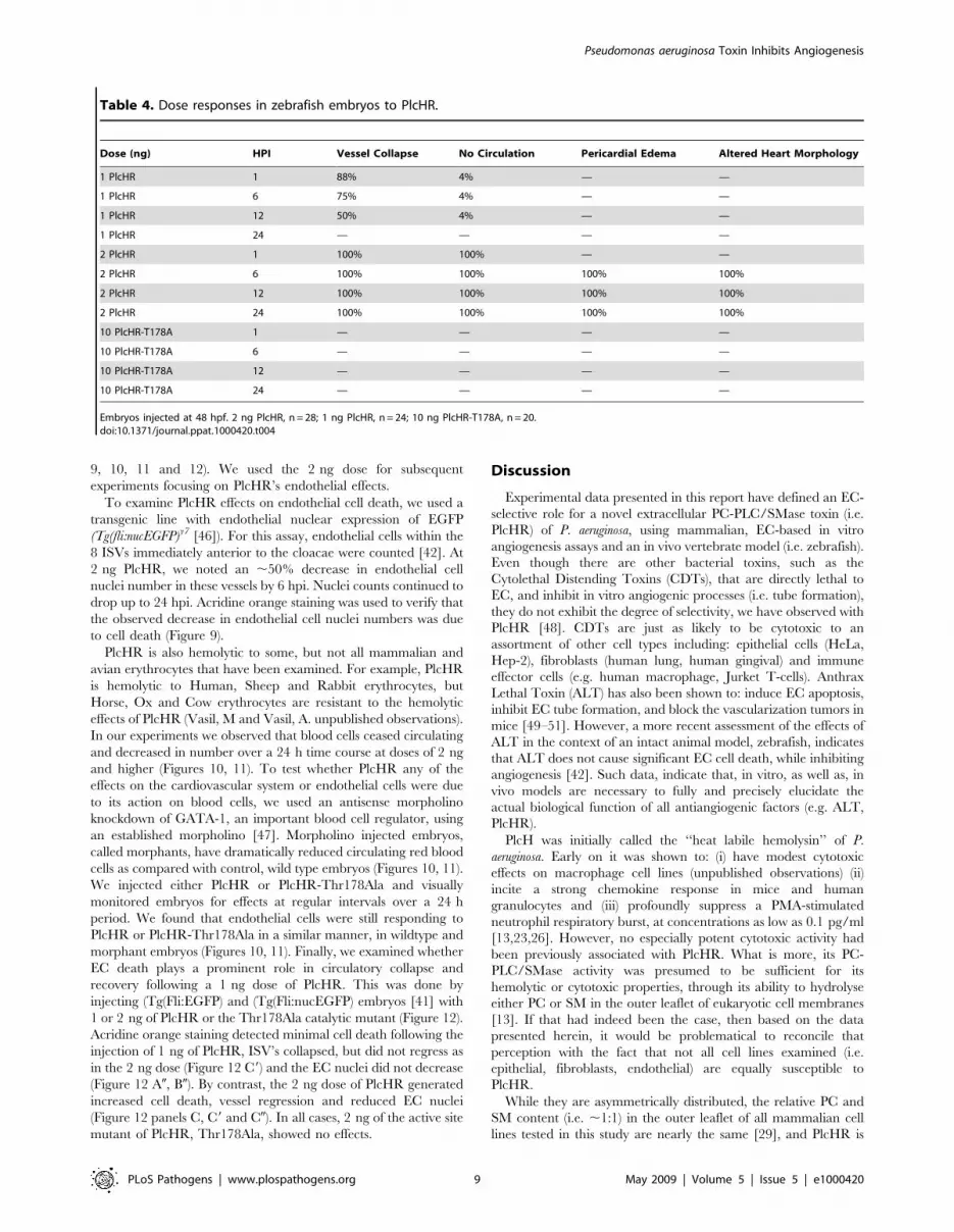
9, 10, 11 and 12). We used the 2 ng dose for subsequent
experiments focusing on PlcHR’s endothelial effects.
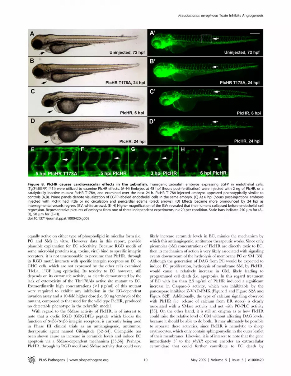
To examine PlcHR effects on endothelial cell death, we used a
transgenic line with endothelial nuclear expression of EGFP
(Tg(fli:nucEGFP)y7 [46]). For this assay, endothelial cells within the
8 ISVs immediately anterior to the cloacae were counted [42]. At
2 ng PlcHR, we noted an ,50% decrease in endothelial cell
nuclei number in these vessels by 6 hpi. Nuclei counts continued to
drop up to 24 hpi. Acridine orange staining was used to verify that
the observed decrease in endothelial cell nuclei numbers was due
to cell death (Figure 9).
PlcHR is also hemolytic to some, but not all mammalian and
avian erythrocytes that have been examined. For example, PlcHR
is hemolytic to Human, Sheep and Rabbit erythrocytes, but
Horse, Ox and Cow erythrocytes are resistant to the hemolytic
effects of PlcHR (Vasil, M and Vasil, A. unpublished observations).
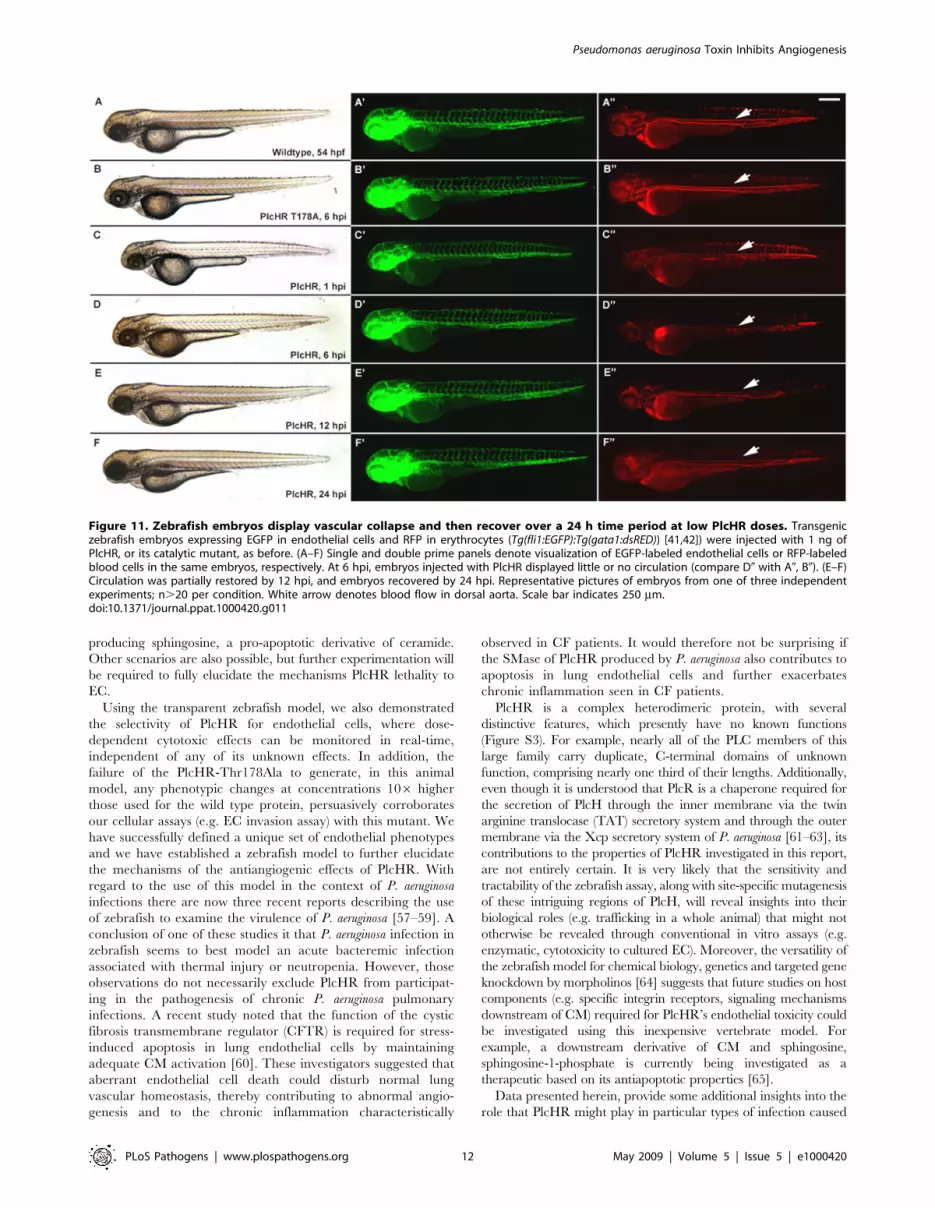
In our experiments we observed that blood cells ceased circulating
and decreased in number over a 24 h time course at doses of 2 ng
and higher (Figures 10, 11). To test whether PlcHR any of the
effects on the cardiovascular system or endothelial cells were due
to its action on blood cells, we used an antisense morpholino
knockdown of GATA-1, an important blood cell regulator, using
an established morpholino [47]. Morpholino injected embryos,
called morphants, have dramatically reduced circulating red blood
cells as compared with control, wild type embryos (Figures 10, 11).
We injected either PlcHR or PlcHR-Thr178Ala and visually
monitored embryos for effects at regular intervals over a 24 h
period. We found that endothelial cells were still responding to
PlcHR or PlcHR-Thr178Ala in a similar manner, in wildtype and
morphant embryos (Figures 10, 11). Finally, we examined whether
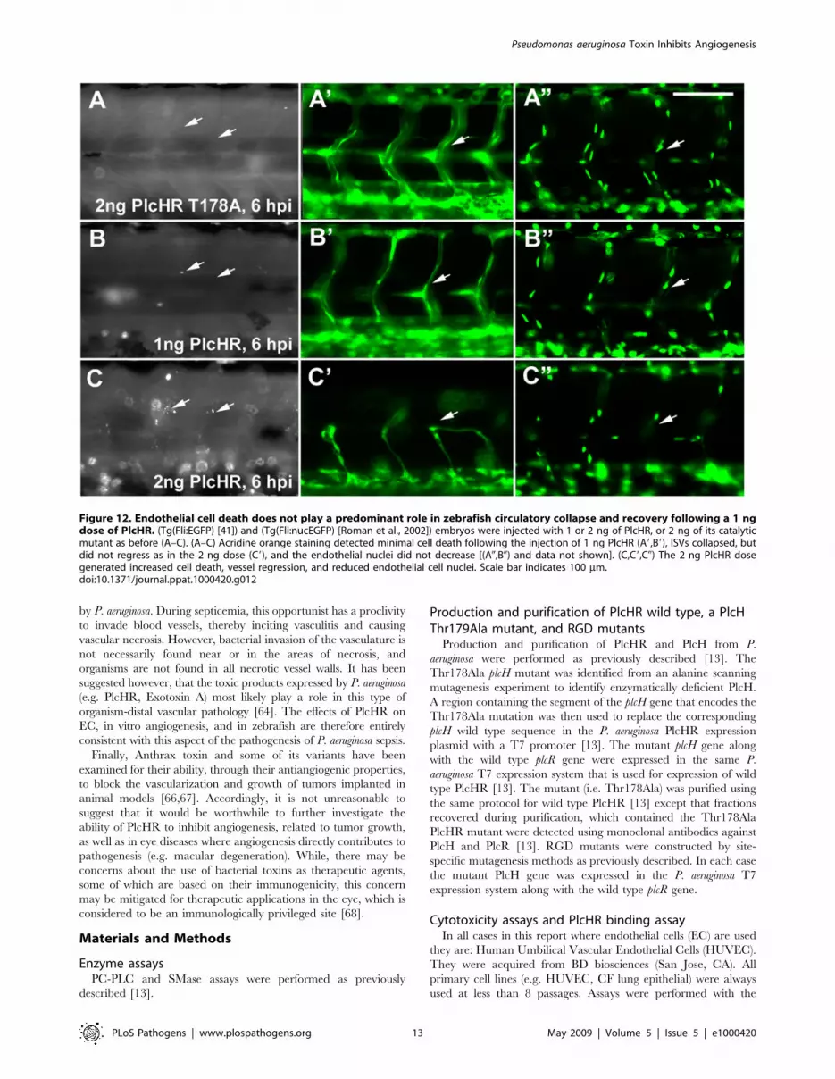
EC death plays a prominent role in circulatory collapse and
recovery following a 1 ng dose of PlcHR. This was done by
injecting (Tg(Fli:EGFP) and (Tg(Fli:nucEGFP) embryos [41] with
1 or 2 ng of PlcHR or the Thr178Ala catalytic mutant (Figure 12).
Acridine orange staining detected minimal cell death following the
injection of 1 ng of PlcHR, ISV’s collapsed, but did not regress as
in the 2 ng dose (Figure 12 C9) and the EC nuclei did not decrease
(Figure 12 A0, B0). By contrast, the 2 ng dose of PlcHR generated
increased cell death, vessel regression and reduced EC nuclei
(Figure 12 panels C, C9 and C0). In all cases, 2 ng of the active site
mutant of PlcHR, Thr178Ala, showed no effects.
Discussion
Experimental data presented in this report have defined an EC-
selective role for a novel extracellular PC-PLC/SMase toxin (i.e.
PlcHR) of P. aeruginosa, using mammalian, EC-based in vitro
angiogenesis assays and an in vivo vertebrate model (i.e. zebrafish).
Even though there are other bacterial toxins, such as the
Cytolethal Distending Toxins (CDTs), that are directly lethal to
EC, and inhibit in vitro angiogenic processes (i.e. tube formation),
they do not exhibit the degree of selectivity, we have observed with
PlcHR [48]. CDTs are just as likely to be cytotoxic to an
assortment of other cell types including: epithelial cells (HeLa,
Hep-2), fibroblasts (human lung, human gingival) and immune
effector cells (e.g. human macrophage, Jurket T-cells). Anthrax
Lethal Toxin (ALT) has also been shown to: induce EC apoptosis,
inhibit EC tube formation, and block the vascularization tumors in
mice [49–51]. However, a more recent assessment of the effects of
ALT in the context of an intact animal model, zebrafish, indicates
that ALT does not cause significant EC cell death, while inhibiting
angiogenesis [42]. Such data, indicate that, in vitro, as well as, in
vivo models are necessary to fully and precisely elucidate the
actual biological function of all antiangiogenic factors (e.g. ALT,
PlcHR).
PlcH was initially called the ‘‘heat labile hemolysin’’ of P.
aeruginosa. Early on it was shown to: (i) have modest cytotoxic
effects on macrophage cell lines (unpublished observations) (ii)
incite a strong chemokine response in mice and human
granulocytes and (iii) profoundly suppress a PMA-stimulated
neutrophil respiratory burst, at concentrations as low as 0.1 pg/ml
[13,23,26]. However, no especially potent cytotoxic activity had
been previously associated with PlcHR. What is more, its PC-
PLC/SMase activity was presumed to be sufficient for its
hemolytic or cytotoxic properties, through its ability to hydrolyse
either PC or SM in the outer leaflet of eukaryotic cell membranes
[13]. If that had indeed been the case, then based on the data
presented herein, it would be problematical to reconcile that
perception with the fact that not all cell lines examined (i.e.
epithelial, fibroblasts, endothelial) are equally susceptible to
PlcHR.
While they are asymmetrically distributed, the relative PC and
SM content (i.e. ,1:1) in the outer leaflet of all mammalian cell
lines tested in this study are nearly the same [29], and PlcHR is
Table 4. Dose responses in zebrafish embryos to PlcHR.
Dose (ng) HPI Vessel Collapse No Circulation Pericardial Edema Altered Heart Morphology
1 PlcHR 1 88% 4% — —
1 PlcHR 6 75% 4% — —
1 PlcHR 12 50% 4% — —
1 PlcHR 24 — — — —
2 PlcHR 1 100% 100% — —
2 PlcHR 6 100% 100% 100% 100%
2 PlcHR 12 100% 100% 100% 100%
2 PlcHR 24 100% 100% 100% 100%
10 PlcHR-T178A 1 — — — —
10 PlcHR-T178A 6 — — — —
10 PlcHR-T178A 12 — — — —
10 PlcHR-T178A 24 — — — —
Embryos injected at 48 hpf. 2 ng PlcHR, n = 28; 1 ng PlcHR, n = 24; 10 ng PlcHR-T178A, n = 20.doi:10.1371/journal.ppat.1000420.t004
Pseudomonas aeruginosa Toxin Inhibits Angiogenesis
PLoS Pathogens | www.plospathogens.org 9 May 2009 | Volume 5 | Issue 5 | e1000420
equally active on either type of phospholipid in micellar form (i.e.
PC and SM) in vitro. However data in this report, provide
plausible explanation for EC selectivity. Because RGD motifs of
some microbial proteins (e.g. toxins, viral) bind to specific integrin
receptors, it is not unreasonable to presume that PlcHR, through
its RGD motif, interacts with specific integrin receptors on EC or
CHO cells, which are not expressed by the other cells examined
(HeLa, 1uCF lung epithelia). Its toxicity to EC however, still
depends on its enzymatic activity, as clearly demonstrated by the
lack of cytotoxicity of the Thr178Ala active site mutant to EC.
Extraordinarily high concentrations (.1 mg/ml) of this mutant
were required to exhibit any inhibition in the EC-dependent
invasion assay and a 10-fold higher dose (i.e. 20 ng/embryo) of the
mutant, compared to that used for the wild type PlcHR, produced
no detectable phenotype in the zebrafish model.
With regard to the SMase activity of PlcHR, is of interest to
note that a cyclic RGD (GRGDFL) peptide which blocks the
function of avb3/avb5 integrin receptors, is currently being used
in Phase III clinical trials as an antiangiogenic, antitumor,
therapeutic agent named Cilengitide [52–54]. Cilengitide has
been shown cause an increase in ceramide levels and induce EC
apoptosis via a SMase-dependent mechanism [55,56]. Perhaps,
PlcHR, through its RGD motif and SMase activity that could very
likely increase ceramide levels in EC, mimics the mechanism by
which this antiangiogenic, antitumor therapeutic works. Since only
picomolar (pM) concentrations of PlcHR are directly toxic to EC,
then its mechanism of action is very likely associated with signaling
events downstream of the hydrolysis of membrane PC or SM [33].
Although the generation of DAG from PC would be expected to
induce EC proliferation, hydrolysis of membrane SM, by PlcHR,
would cause a relatively increase in CM, likely leading to
programmed cell death (i.e. apoptosis). In this regard treatment
of EC with less than 2.5 ng/ml of PlcHR induced a significant
increase in Caspase-3 activity, which was inhibitable by the
pancaspase inhibitor Z-VAD-FMK (Figure 3 and Figure S2A and
Figure S2B). Additionally, the type of calcium signaling observed
with PlcHR (i.e. release of calcium from ER stores) is clearly
associated with a SMase activity and not with PC-PLC activity
[33]. On the other hand, it is still an enigma as to how PlcHR
could raise the relative level of CM without affecting DAG levels,
because it should be able to do both,. It may ultimately be possible
to separate these activities, since PlcHR is hemolytic to sheep
erythrocytes, which only contain sphingomyelin in the outer leaflet
of their membranes. Likewise, it is of interest to note that the gene
immediately 59 to the plcHR operon encodes an extracellular
ceramidase that could further contribute to EC death by
Figure 8. PlcHR causes cardiovascular effects in the zebrafish. Transgenic zebrafish embryos expressing EGFP in endothelial cells,(Tg(Fli:EGFP) [41]) were utilized to examine PlcHR effects. (A–H) Embryos at 48 hpf (hours post-fertilization) were injected with 2 ng of PlcHR, or acatalytically inactive mutant PlcHR T178A, and examined over the next 24 h. PlcHR T178A-injected embryos appeared phenotypically similar tocontrols (A,B). Prime panels denote visualization of EGFP-labeled endothelial cells in the same embryo. (C) At 6 hpi (hours post-injection), embryosinjected with PlcHR had little or no circulation and pericardial edema (black arrows). (D) Effects became more pronounced by 24 hpi asintersegmental vessels regress (ISV, white arrows). (E–H) Higher magnification of the ISVs revealed that their lumens collapsed before endothelial cellregression. Representative pictures of embryos from one of three independent experiments; n.20 per condition. Scale bars indicate 250 mm for (A–D), 50 mm for (E–H).doi:10.1371/journal.ppat.1000420.g008
Pseudomonas aeruginosa Toxin Inhibits Angiogenesis
PLoS Pathogens | www.plospathogens.org 10 May 2009 | Volume 5 | Issue 5 | e1000420
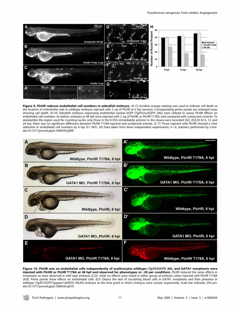
Figure 9. PlcHR reduces endothelial cell numbers in zebrafish embryos. (A–C) Acridine orange staining was used to indicate cell death atthe location of endothelial cells in wildtype embryos injected with 2 ng of PlcHR at 6 hpi (arrows). Corresponding prime panels are enlarged areasshowing cell death. (D–H) Zebrafish embryos expressing endothelial nuclear EGFP (Tg(Fli:nucEGFP) [46]) were utilized to assess PlcHR effects onendothelial cell numbers. As before, embryos at 48 hpf were injected with 2 ng of PlcHR, or PlcHR T178A, and compared with uninjected controls. Tostandardize the region used for counting nuclei, only those in the 8 ISVs immediately anterior to the cloaca were recorded [42]. (D,E,H) At 6, 12, and24 hpi, there was no significant difference between PlcHR T178A-injected and uninjected animals. (C–F) Those injected with PlcHR showed a clearreduction in endothelial cell numbers by 6 hpi (P,.001). (H) Data taken from three independent experiments; n = 6, statistics performed by t-test.doi:10.1371/journal.ppat.1000420.g009
Figure 10. PlcHR acts on endothelial cells independently of erythrocytes wildtype (Tg(Fli:EGFP)) [44], and GATA1 morphants wereinjected with PlcHR or PlcHR T178A at 48 hpf and observed for phenotypes (n.20 per condition). PlcHR induced the same effects inmorphants as were observed in wild type embryos (C,D), while no effects were noted in either group of embryos when injected with PlcHR T178A(A,B). Prime panels show effects on endothelial cells. (E,F) Depict the lack of circulating blood cells in GATA1 morphants and their presence inwildtype (Tg(fli1:EGFP):Tg(gata1:dsRED)) [44,45] embryos at the time point in which embryos were scored, respectively. Scale bar indicates 250 mm.doi:10.1371/journal.ppat.1000420.g010
Pseudomonas aeruginosa Toxin Inhibits Angiogenesis
PLoS Pathogens | www.plospathogens.org 11 May 2009 | Volume 5 | Issue 5 | e1000420
producing sphingosine, a pro-apoptotic derivative of ceramide.
Other scenarios are also possible, but further experimentation will
be required to fully elucidate the mechanisms PlcHR lethality to
EC.
Using the transparent zebrafish model, we also demonstrated
the selectivity of PlcHR for endothelial cells, where dose-
dependent cytotoxic effects can be monitored in real-time,
independent of any of its unknown effects. In addition, the
failure of the PlcHR-Thr178Ala to generate, in this animal
model, any phenotypic changes at concentrations 106 higher
those used for the wild type protein, persuasively corroborates
our cellular assays (e.g. EC invasion assay) with this mutant. We
have successfully defined a unique set of endothelial phenotypes
and we have established a zebrafish model to further elucidate
the mechanisms of the antiangiogenic effects of PlcHR. With
regard to the use of this model in the context of P. aeruginosa
infections there are now three recent reports describing the use
of zebrafish to examine the virulence of P. aeruginosa [57–59]. A
conclusion of one of these studies it that P. aeruginosa infection in
zebrafish seems to best model an acute bacteremic infection
associated with thermal injury or neutropenia. However, those
observations do not necessarily exclude PlcHR from participat-
ing in the pathogenesis of chronic P. aeruginosa pulmonary
infections. A recent study noted that the function of the cystic
fibrosis transmembrane regulator (CFTR) is required for stress-
induced apoptosis in lung endothelial cells by maintaining
adequate CM activation [60]. These investigators suggested that
aberrant endothelial cell death could disturb normal lung
vascular homeostasis, thereby contributing to abnormal angio-
genesis and to the chronic inflammation characteristically
observed in CF patients. It would therefore not be surprising if
the SMase of PlcHR produced by P. aeruginosa also contributes to
apoptosis in lung endothelial cells and further exacerbates
chronic inflammation seen in CF patients.
PlcHR is a complex heterodimeric protein, with several
distinctive features, which presently have no known functions
(Figure S3). For example, nearly all of the PLC members of this
large family carry duplicate, C-terminal domains of unknown
function, comprising nearly one third of their lengths. Additionally,
even though it is understood that PlcR is a chaperone required for
the secretion of PlcH through the inner membrane via the twin
arginine translocase (TAT) secretory system and through the outer
membrane via the Xcp secretory system of P. aeruginosa [61–63], its
contributions to the properties of PlcHR investigated in this report,
are not entirely certain. It is very likely that the sensitivity and
tractability of the zebrafish assay, along with site-specific mutagenesis
of these intriguing regions of PlcH, will reveal insights into their
biological roles (e.g. trafficking in a whole animal) that might not
otherwise be revealed through conventional in vitro assays (e.g.
enzymatic, cytotoxicity to cultured EC). Moreover, the versatility of
the zebrafish model for chemical biology, genetics and targeted gene
knockdown by morpholinos [64] suggests that future studies on host
components (e.g. specific integrin receptors, signaling mechanisms
downstream of CM) required for PlcHR’s endothelial toxicity could
be investigated using this inexpensive vertebrate model. For
example, a downstream derivative of CM and sphingosine,
sphingosine-1-phosphate is currently being investigated as a
therapeutic based on its antiapoptotic properties [65].
Data presented herein, provide some additional insights into the
role that PlcHR might play in particular types of infection caused
Figure 11. Zebrafish embryos display vascular collapse and then recover over a 24 h time period at low PlcHR doses. Transgeniczebrafish embryos expressing EGFP in endothelial cells and RFP in erythrocytes (Tg(fli1:EGFP):Tg(gata1:dsRED)) [41,42]) were injected with 1 ng ofPlcHR, or its catalytic mutant, as before. (A–F) Single and double prime panels denote visualization of EGFP-labeled endothelial cells or RFP-labeledblood cells in the same embryos, respectively. At 6 hpi, embryos injected with PlcHR displayed little or no circulation (compare D0 with A0, B0). (E–F)Circulation was partially restored by 12 hpi, and embryos recovered by 24 hpi. Representative pictures of embryos from one of three independentexperiments; n.20 per condition. White arrow denotes blood flow in dorsal aorta. Scale bar indicates 250 mm.doi:10.1371/journal.ppat.1000420.g011
Pseudomonas aeruginosa Toxin Inhibits Angiogenesis
PLoS Pathogens | www.plospathogens.org 12 May 2009 | Volume 5 | Issue 5 | e1000420
by P. aeruginosa. During septicemia, this opportunist has a proclivity
to invade blood vessels, thereby inciting vasculitis and causing
vascular necrosis. However, bacterial invasion of the vasculature is
not necessarily found near or in the areas of necrosis, and
organisms are not found in all necrotic vessel walls. It has been
suggested however, that the toxic products expressed by P. aeruginosa
(e.g. PlcHR, Exotoxin A) most likely play a role in this type of
organism-distal vascular pathology [64]. The effects of PlcHR on
EC, in vitro angiogenesis, and in zebrafish are therefore entirely
consistent with this aspect of the pathogenesis of P. aeruginosa sepsis.
Finally, Anthrax toxin and some of its variants have been
examined for their ability, through their antiangiogenic properties,
to block the vascularization and growth of tumors implanted in
animal models [66,67]. Accordingly, it is not unreasonable to
suggest that it would be worthwhile to further investigate the
ability of PlcHR to inhibit angiogenesis, related to tumor growth,
as well as in eye diseases where angiogenesis directly contributes to
pathogenesis (e.g. macular degeneration). While, there may be
concerns about the use of bacterial toxins as therapeutic agents,
some of which are based on their immunogenicity, this concern
may be mitigated for therapeutic applications in the eye, which is
considered to be an immunologically privileged site [68].
Materials and Methods
Enzyme assaysPC-PLC and SMase assays were performed as previously
described [13].
Production and purification of PlcHR wild type, a PlcHThr179Ala mutant, and RGD mutants
Production and purification of PlcHR and PlcH from P.
aeruginosa were performed as previously described [13]. The
Thr178Ala plcH mutant was identified from an alanine scanning
mutagenesis experiment to identify enzymatically deficient PlcH.
A region containing the segment of the plcH gene that encodes the
Thr178Ala mutation was then used to replace the corresponding
plcH wild type sequence in the P. aeruginosa PlcHR expression
plasmid with a T7 promoter [13]. The mutant plcH gene along
with the wild type plcR gene were expressed in the same P.
aeruginosa T7 expression system that is used for expression of wild
type PlcHR [13]. The mutant (i.e. Thr178Ala) was purified using
the same protocol for wild type PlcHR [13] except that fractions
recovered during purification, which contained the Thr178Ala
PlcHR mutant were detected using monoclonal antibodies against
PlcH and PlcR [13]. RGD mutants were constructed by site-
specific mutagenesis methods as previously described. In each case
the mutant PlcH gene was expressed in the P. aeruginosa T7
expression system along with the wild type plcR gene.
Cytotoxicity assays and PlcHR binding assayIn all cases in this report where endothelial cells (EC) are used
they are: Human Umbilical Vascular Endothelial Cells (HUVEC).
They were acquired from BD biosciences (San Jose, CA). All
primary cell lines (e.g. HUVEC, CF lung epithelial) were always
used at less than 8 passages. Assays were performed with the
Figure 12. Endothelial cell death does not play a predominant role in zebrafish circulatory collapse and recovery following a 1 ngdose of PlcHR. (Tg(Fli:EGFP) [41]) and (Tg(Fli:nucEGFP) [Roman et al., 2002]) embryos were injected with 1 or 2 ng of PlcHR, or 2 ng of its catalyticmutant as before (A–C). (A–C) Acridine orange staining detected minimal cell death following the injection of 1 ng PlcHR (A9,B9), ISVs collapsed, butdid not regress as in the 2 ng dose (C9), and the endothelial nuclei did not decrease [(A0,B0) and data not shown]. (C,C9,C0) The 2 ng PlcHR dosegenerated increased cell death, vessel regression, and reduced endothelial cell nuclei. Scale bar indicates 100 mm.doi:10.1371/journal.ppat.1000420.g012
Pseudomonas aeruginosa Toxin Inhibits Angiogenesis
PLoS Pathogens | www.plospathogens.org 13 May 2009 | Volume 5 | Issue 5 | e1000420
designated cell lines according to manufacturer’s recommendation
using the CytoTox 96 Assay Kit (Promega Corporation). Percent
activity was determined by performing a total lysis control in which
cells were lysed with 1% triton X-100. Percent lysis was determined
via the formula. Percent lysis = [(experimental2spontaneous con-
trol)/(Total lysis2spontaneous control)]6100 To measure binding
of PlcHR to CHO cells (Figure 5B) were grown in 24 well tissue
culture dishes until they reached 80–90% confluence. The media
was replaced with 300 ml of media containing 7.5 ng/ml wild type or
a mutant PlcHR. The cells were incubated for 2 h at 37uC with 5%
CO2. Control samples consisted of 7.5 ng/ml mutant or wild type
PlcHR2 incubated in 300 ml media alone. To determine the percent
of activity (enzyme) bound by the cells, the PLC activity in the
supernatants from the wild type or mutant PlcHR samples incubated
with cells was compared to the control samples. PLC activity was
detected using the synthetic substrate r-nitrophenyl-phosphorylcho-
line as previously described [13], which detects PlcHR activity, but it
does not react at all with CHO cells that are not exposed to PlcHR.
The difference in recovered activity between the controls and the
samples with cells was then used to calculate the percent of activity
bound to the cells. Percent activity bound = [(control activity -
activity with cells)/control activity]*100. Lysing the cells with 1%
Triton X-100 recovered all activity.
Caspase-3 activation assayCaspase-3 activity was assayed with the colorimetric CaspACE
Assay system (Promega, Madison, WI). The colorimetric substrate
(Ac-DEVD-pNA) is hydrolyzed by activated caspase-3 releasing
pNA producing a yellow color that is quantified at an absorbance of
405 nm. Camptothecin, a topoisomerase I inhibitor, was used as a
positive control for activation of caspase-3 and induction of
apoptosis. For inhibition of apoptosis ZVAD- FMK was added to
samples at a final concentration of 50 mM. HUVEC were cultivated
in 6 well tissue culture dishes to 80–90% confluency at which time
the media was exchanged with 2 ml of fresh media containing
PlcHR2 or other compounds to be examined. The cultures were
allowed to incubate at 37uC in 5% CO2 for 3 to 16 hours. The cells
were harvested by trypsin/EDTA treatment, washed with ice cold
PBS and suspended in lysis buffer at a concentration of 16108 cells/
ml. To prepare lysates cells were freeze-thawed four times and
sonicated twice for 15 seconds at level 10 in a Sonic Dismembrator
Model 100 (Fisher Scientific, Hampton, NH). The lysates were
incubated on ice for 15 minutes before the cell lysate supernatant
was harvested by centrifuge at 16,1006g for 20 minutes at 4uC.
Caspase-3 activity in the cell lysates was assayed for by the
manufactures’ recommended protocol.
Angiogenesis assaysEC migration assays, EC invasion assays and EC tube
formation assays were performed according to manufacturer’s
recommendations using the BD Biocoat Angiogenesis System (BD
Biosciences) with primary cultures of HUVEC.
AnimalsAll animal protocols were approved by the Institutional Animal
Care and Use Committee of Children’s Hospital Boston. Breeding
fish, wild type (AB strain), or transgenic lines, were maintained at
28.5uC on a 14 h light/10 h dark cycle. Embryos were collected
by natural spawning, and raised in 10% Hank’s saline at 32uC.
Microinjection of toxin proteins and inhibitorsMicroinjections into the zebrafish vasculature at 48 hpf were
carried out as described by Bolcome et al. (2008). PlcHR or
PlcHR-Thr178Ala were diluted immediately before, and kept at
4uC until injection. Injected amounts are indicated in the figure
legends for each experiment. Phenol red (0.05%) was added to
each condition for visibility during microinjection. Volumes of
40 nl or less were delivered into the common cardinal vein of
embryos anesthetized with tricaine (Sigma) at 48 hpf using a gas
driven microinjector (Medical Systems Corp.). After injection,
embryos were transferred into fresh medium for recovery,
maintained at 32uC, and scored for toxin action at time points
indicated in the text.
Acridine orange stainingDechorionated embryos were placed in 5 mL of a 50 mg/mL
solution of acridine orange (acridinium chloride hemi-[zinc
chloride], Sigma) in 10% Hank’s saline solution at room
temperature. After 30 minutes of staining while protected from
light, embryos were washed three times for 10 minutes with 10%
Hank’s saline. Following the third wash, embryos were mounted
on glass slides for fluorescence microscopy.
Supporting Information
Figure S1 A detailed analysis of PlcHR cytotoxicity of two
susceptible (HUVEC, CHO) and two resistant cell lines (L929 and
Hela). Time and dose killing of various cell types. The cells
indicated were treated with increasing concentrations of PlcHR for
increasing lengths of time. The arrows indicate increasing time of
treatment. HUVECs, CHO, and L929 were treated for 2, 4, 6, 8,
10, and 22 h, and the HeLa cells were treated for 3, 6, 9, 12, and
22 h.
Found at: doi:10.1371/journal.ppat.1000420.s001 (1.41 MB TIF)
Figure S2 Effect of PlcHR on caspase-3 expression. (A) The
pan-caspase inhibitor Z-VAD-FMK completely inhibits PlcHR
activation of caspase-3 and reduced the level of LDH release in
HUVECs. Caspase-3 activation and LDH release were measured
at 16 h post-treatment of HUVEC with 2.5 ng PlcHR. Z-VAD-
FMK is a potent, irreversible, and cell-permeable pan-caspase
inhibitor. (B) PlcHR activates caspase-3 between 3 and 6 h.
HUVEC were treated with 2.5 ng/ml PlcHR2 for 3, 6, and 9 h.
At each time point, caspase-3 activity was assayed as described in
Materials and Methods.
Found at: doi:10.1371/journal.ppat.1000420.s002 (0.21 MB TIF)
Figure S3 Key structural features of PlcHR and members of the
Phosphodiester/PLC Superfamily. PlcH is the only known
member of this family to be associated with a PlcR-like protein.
Found at: doi:10.1371/journal.ppat.1000420.s003 (9.25 MB TIF)
Table S1 Inhibition of EC tube formation by PlcHR. Pre-tube
formation: Endothelial cells were challenged with 4–64 ng/ml
during tube formation (24 h) on matrigel. Post-tube formation:
Endothelial cells that had already formed tubes after 48 h were
then challenged with 4–64 ng/ml of PlcHR for an additional 20 h.
Photographs were taken at 406 magnification. Tube length was
measured with Metamorph Ver 7.1.6.0 software (Molecular
Devices, Sunnyvale, CA).
Found at: doi:10.1371/journal.ppat.1000420.s004 (0.04 MB
DOC)
Author Contributions
Conceived and designed the experiments: MLV MJS HG JC. Performed
the experiments: MLV MJS AIV SJW REB JC. Analyzed the data: MLV
MJS AIV SJW HG REB JC. Contributed reagents/materials/analysis
tools: AIV SJW HG REB. Wrote the paper: MLV JC.
Pseudomonas aeruginosa Toxin Inhibits Angiogenesis
PLoS Pathogens | www.plospathogens.org 14 May 2009 | Volume 5 | Issue 5 | e1000420
References
1. Clarke CJ, Snook CF, Tani M, Matmati N, Marchesini N, et al. (2006) Theextended family of neutral sphingomyelinases. Biochemistry 45: 11247–11256.
2. Glunde K, Serkova NJ (2006) Therapeutic targets and biomarkers identified incancer choline phospholipid metabolism. Pharmacogenomics 7: 1109–1123.
3. Whatley RE, Zimmerman GA, McIntyre TM, Prescott SM (1990) Lipid
metabolism and signal transduction in endothelial cells. Prog Lipid Res 29:
45–63.
4. Teichgraber V, Ulrich M, Endlich N, Riethmuller J, Wilker B, et al. (2008)Ceramide accumulation mediates inflammation, cell death and infection
susceptibility in cystic fibrosis. Nat Med 14: 382–391.
5. Yu H, Zeidan YH, Wu BX, Jenkins RW, Flotte TR, et al. (2009) Defective Acid
Sphingomyelinase Pathway with Pseudomonas aeruginosa Infection in CysticFibrosis. Am J Respir Cell Mol Biol. e-pub ahead of print January 23, 2009.
6. Titball RW (1998) Bacterial phospholipases. Symp Ser Soc Appl Microbiol 27:127S–137S.
7. Titball RW, Naylor CE, Basak AK (1999) The Clostridium perfringens alpha-toxin.
Anaerobe 5: 51–64.
8. Smith GA, Marquis H, Jones S, Johnston NC, Portnoy DA, et al. (1995) The two
distinct phospholipases C of Listeria monocytogenes have overlapping roles in escapefrom a vacuole and cell-to-cell spread. Infect Immun 63: 4231–4237.
9. Martin SF, Follows BC, Hergenrother PJ, Trotter BK (2000) The cholinebinding site of phospholipase C (Bacillus cereus): Insights into substrate specificity.
Biochemistry 39: 3410–3415.
10. Alape-Giron A, Flores-Diaz M, Guillouard I, Naylor CE, Titball RW, et al.
(2000) Identification of residues critical for toxicity in Clostridium perfringens
phospholipase C, the key toxin in gas gangrene. Eur J Biochem 267: 5191–5197.
11. Titball RW, Fearn AM, Williamson ED (1993) Biochemical and immunological
properties of the C-terminal domain of the alpha-toxin of Clostridium perfringens.
FEMS Microbiol Lett 110: 45–50.
12. Titball RW, Leslie DL, Harvey S, Kelly D (1991) Hemolytic and sphingomy-elinase activities of Clostridium perfringens alpha-toxin are dependent on a domain
homologous to that of an enzyme from the human arachidonic acid pathway.
Infect Immun 59: 1872–1874.
13. Stonehouse MJ, Cota-Gomez A, Parker SK, Martin WE, Hankin JA, et al.(2002) A novel class of microbial phosphocholine-specific phospholipases C. Mol
Microbiol 46: 661–676.
14. Felts RL, Reilly TJ, Tanner JJ (2006) Structure of Francisella tularensis AcpA:
Prototype of a unique superfamily of acid phosphatases and phospholipases C. JBiol Chem 281: 30289–30298.
15. Korbsrisate S, Tomaras AP, Damnin S, Ckumdee J, Srinon V, et al. (2007)Characterization of two distinct phospholipase C enzymes from Burkholderia
pseudomallei. Microbiology 153: 1907–1915.
16. Raynaud C, Guilhot C, Rauzier J, Bordat Y, Pelicic V, et al. (2002)Phospholipases C are involved in the virulence of Mycobacterium tuberculosis.
Mol Microbiol 45: 203–217.
17. Viana-Niero C, de Haas PE, van Soolingen D, Leao SC (2004) Analysis of
genetic polymorphisms affecting the four phospholipase C (plc) genes inMycobacterium tuberculosis complex clinical isolates. Microbiology 150: 967–978.
18. Yang Z, Yang D, Kong Y, Zhang L, Marrs CF, et al. (2005) Clinical relevance ofMycobacterium tuberculosis plcD gene mutations. Am J Respir Crit Care Med 171:
1436–1442.
19. Ostroff RM, Wretlind B, Vasil ML (1989) Mutations in the hemolytic-
phospholipase C operon result in decreased virulence of Pseudomonas aeruginosa
PAO1 grown under phosphate-limiting conditions. Infect Immun 57:
1369–1373.
20. Rahme LG, Stevens EJ, Wolfort SF, Shao J, Tompkins RG, et al. (1995)
Common virulence factors for bacterial pathogenicity in plants and animals.Science 268: 1899–1902.
21. Hogan DA, Kolter R (2002) Pseudomonas-Candida interactions: An ecological
role for virulence factors. Science 296: 2229–2232.
22. Montes LR, Lopez DJ, Sot J, Bagatolli LA, Stonehouse MJ, et al. (2008)
Ceramide-enriched membrane domains in red blood cells and the mechanism ofsphingomyelinase-induced hot-cold hemolysis. Biochemistry 47: 11222–11230.
23. Montes LR, Ibarguren M, Goni FM, Stonehouse M, Vasil ML, et al. (2007)Leakage-free membrane fusion induced by the hydrolytic activity of PlcHR(2), a
novel phospholipase C/sphingomyelinase from Pseudomonas aeruginosa. BiochimBiophys Acta 1768: 2365–2372.
24. Soave R, Murray HW, Litrenta MM (1978) Bacterial invasion of pulmonaryvessels. Pseudomonas bacteremia mimicking pulmonary thromboembolism with
infarction. Am J Med 65: 864–867.
25. Ziegler EJ, Douglas H (1979) Pseudomonas aeruginosa vasculitis and bacteremia
following conjunctivitis: A simple model of fatal pseudomonas infection inneutropenia. J Infect Dis 139: 288–296.
26. Terada LS, Johansen KA, Nowbar S, Vasil AI, Vasil ML (1999) Pseudomonas
aeruginosa hemolytic phospholipase C suppresses neutrophil respiratory burst
activity. Infect Immun 67: 2371–2376.
27. Wieland CW, Siegmund B, Senaldi G, Vasil ML, Dinarello CA, et al. (2002)Pulmonary inflammation induced by Pseudomonas aeruginosa lipopolysaccharide,
phospholipase C, and exotoxin A: Role of interferon regulatory factor 1. InfectImmun 70: 1352–1358.
28. Zabner J, Karp P, Seiler M, Phillips SL, Mitchell CJ, et al. (2003) Development
of cystic fibrosis and noncystic fibrosis airway cell lines. Am J Physiol Lung CellMol Physiol 284: L844–L854.
29. Alberts B, Johnson A, Lewis J, Raff M, Roberts K, et al. (2002) The Molecular
Biology of the Cell. 4th ed. New York: Garland Science. pp 583–614.
30. Wadsworth SJ, Goldfine H (1999) Listeria monocytogenes phospholipase C-dependent calcium signaling modulates bacterial entry into J774 macrophage-
like cells. Infect Immun 67: 1770–1778.
31. Wadsworth SJ, Goldfine H (2002) Mobilization of protein kinase C inmacrophages induced by Listeria monocytogenes affects its internalization and
escape from the phagosome. Infect Immun 70: 4650–4660.
32. Pinton P, Ferrari D, Rapizzi E, Di Virgilio F, Pozzan T, et al. (2001) The Ca2+concentration of the endoplasmic reticulum is a key determinant of ceramide-
induced apoptosis: Significance for the molecular mechanism of Bcl-2 action.
EMBO J 20: 2690–2701.33. Hannun YA, Obeid LM (2008) Principles of bioactive lipid signalling: Lessons
from sphingolipids. Nat Rev Mol Cell Biol 9: 139–150.
34. Boatright KM, Salvesen GS (2003) Mechanisms of caspase activation. CurrOpin Cell Biol 15: 725–731.
35. Takada Y, Ye X, Simon S (2007) The integrins. Genome Biol 8: 215.
36. Assa-Munt N, Jia X, Laakkonen P, Ruoslahti E (2001) Solution structures and
integrin binding activities of an RGD peptide with two isomers. Biochemistry 40:2373–2378.
37. van der Most RG, Corver J, Strauss JH (1999) Mutagenesis of the RGD motif in
the yellow fever virus 17D envelope protein. Virology 265: 83–95.
38. Zhao Q, Pacheco JM, Mason PW (2003) Evaluation of genetically engineeredderivatives of a Chinese strain of foot-and-mouth disease virus reveals a novel
cell-binding site which functions in cell culture and in animals. J Virol 77:
3269–3280.39. Staton CA, Lewis C, Bricknell R, eds (2006) Angiogenesis Assays: A critical
appraisal of current techniques. 1st ed. Weimar (Texas): Culinary and
Hospitality Industry Publications Services. pp 390.40. Mohapatra NP, Balagopal A, Soni S, Schlesinger LS, Gunn JS (2007) AcpA is a
Francisella acid phosphatase that affects intramacrophage survival and
virulence. Infect Immun 75: 390–396.41. Reilly TJ, Baron GS, Nano FE, Kuhlenschmidt MS (1996) Characterization and
sequencing of a respiratory burst-inhibiting acid phosphatase from Francisella
tularensis. J Biol Chem 271: 10973–10983.
42. Bolcome RE III, Sullivan SE, Zeller R, Barker AP, Collier RJ, et al. (2008)Anthrax lethal toxin induces cell death-independent permeability in zebrafish
vasculature. Proc Natl Acad Sci U S A 105: 2439–2444.
43. Isogai S, Horiguchi M, Weinstein BM (2001) The vascular anatomy of thedeveloping zebrafish: An atlas of embryonic and early larval development. Dev
Biol 230: 278–301.
44. Lawson ND, Weinstein BM (2002) In vivo imaging of embryonic vasculardevelopment using transgenic zebrafish. Dev Biol 248: 307–318.
45. Traver D, Paw BH, Poss KD, Penberthy WT, Lin S, et al. (2003)
Transplantation and in vivo imaging of multilineage engraftment in zebrafishbloodless mutants. Nat Immunol 4: 1238–1246.
46. Roman BL, Pham VN, Lawson ND, Kulik M, Childs S, et al. (2002) Disruption
of acvrl1 increases endothelial cell number in zebrafish cranial vessels.Development 129: 3009–3019.
47. Galloway JL, Wingert RA, Thisse C, Thisse B, Zon LI (2005) Loss of gata1 but
not gata2 converts erythropoiesis to myelopoiesis in zebrafish embryos. Dev Cell8: 109–116.
48. Smith JL, Bayles DO (2006) The contribution of cytolethal distending toxin to
bacterial pathogenesis. Crit Rev Microbiol 32: 227–248.
49. Abi-Habib RJ, Singh R, Leppla SH, Greene JJ, Ding Y, et al. (2006) Systemicanthrax lethal toxin therapy produces regressions of subcutaneous human
melanoma tumors in athymic nude mice. Clin Cancer Res 12: 7437–7443.
50. Kirby JE (2004) Anthrax lethal toxin induces human endothelial cell apoptosis.Infect Immun 72: 430–439.
51. Rogers MS, Christensen KA, Birsner AE, Short SM, Wigelsworth DJ, et al.
(2007) Mutant anthrax toxin B moiety (protective antigen) inhibits angiogenesisand tumor growth. Cancer Res 67: 9980–9985.
52. MacDonald TJ, Stewart CF, Kocak M, Goldman S, Ellenbogen RG, et al.
(2008) Phase I clinical trial of cilengitide in children with refractory brain tumors:Pediatric Brain Tumor Consortium Study PBTC-012. J Clin Oncol 26:
919–924.
53. Reardon DA, Nabors LB, Stupp R, Mikkelsen T (2008) Cilengitide: An integrin-targeting arginine-glycine-aspartic acid peptide with promising activity for
glioblastoma multiforme. Expert Opin Investig Drugs 17: 1225–1235.
54. Smith JW (2003) Cilengitide Merck. Curr Opin Investig Drugs 4: 741–745.
55. Avraamides CJ, Garmy-Susini B, Varner JA (2008) Integrins in angiogenesis andlymphangiogenesis. Nat Rev Cancer 8: 604–617.
56. Erdreich-Epstein A, Tran LB, Cox OT, Huang EY, Laug WE, et al. (2005)
Endothelial apoptosis induced by inhibition of integrins alphavbeta3 andalphavbeta5 involves ceramide metabolic pathways. Blood 105: 4353–4361.
57. Brannon MK, Davis JM, Mathias JR, Hall CJ, Emerson JC, et al. (2009)
Pseudomonas aeruginosa Type III secretion system interacts with phagocytes tomodulate systemic infection of zebrafish embryos. Cell Microbiol 11: 755–768.
Pseudomonas aeruginosa Toxin Inhibits Angiogenesis
PLoS Pathogens | www.plospathogens.org 15 May 2009 | Volume 5 | Issue 5 | e1000420
58. Clatworthy AE, Lee JS, Leibman M, Kostun Z, Davidson AJ, et al. (2009)
Pseudomonas aeruginosa infection of zebrafish involves both host and pathogendeterminants. Infect Immun 77: 1293–1303.
59. Rawls JF, Mahowald MA, Goodman AL, Trent CM, Gordon JI (2007) In vivo
imaging and genetic analysis link bacterial motility and symbiosis in the zebrafishgut. Proc Natl Acad Sci U S A 104: 7622–7627.
60. Noe J, Petrusca D, Rush N, Deng P, Vandemark M, et al. (2009) CFTRRegulation of Intracellular pH and Ceramides is Required for Lung Endothelial
Cell Apoptosis. Am J Respir Cell Mol Biol [January 23, 2009, e-pub ahead of
print].61. Cota-Gomez A, Vasil AI, Kadurugamuwa J, Beveridge TJ, Schweizer HP, et al.
(1997) PlcR1 and PlcR2 are putative calcium-binding proteins required forsecretion of the hemolytic phospholipase C of Pseudomonas aeruginosa. Infect
Immun 65: 2904–2913.62. Ochsner UA, Snyder A, Vasil AI, Vasil ML (2002) Effects of the twin-arginine
translocase on secretion of virulence factors, stress response, and pathogenesis.
Proc Natl Acad Sci U S A 99: 8312–8317.
63. Snyder A, Vasil AI, Zajdowicz SL, Wilson ZR, Vasil ML (2006) Role of the
Pseudomonas aeruginosa PlcH Tat signal peptide in protein secretion, transcription,and cross-species Tat secretion system compatibility. J Bacteriol 188:
1762–1774.
64. Lieschke GJ, Currie PD (2007) Animal models of human disease: Zebrafish swiminto view. Nat Rev Genet 8: 353–367.
65. Takabe K, Paugh SW, Milstien S, Spiegel S (2008) ‘‘Inside-out’’ signaling ofsphingosine-1-phosphate: Therapeutic targets. Pharmacol Rev 60: 181–195.
66. Liu S, Wang H, Currie BM, Molinolo A, Leung HJ, et al. (2008) Matrix
metalloproteinase-activated anthrax lethal toxin demonstrates high potency intargeting tumor vasculature. J Biol Chem 283: 529–540.
67. Alfano RW, Leppla SH, Liu S, Bugge TH, Duesbery NS, et al. (2008) Potentinhibition of tumor angiogenesis by the matrix metalloproteinase-activated
anthrax lethal toxin: Implications for broad anti-tumor efficacy. Cell Cycle 7:745–749.
68. Ferguson TA, Green DR, Griffith TS (2002) Cell death and immune privilege.
Int Rev Immunol 21: 153–172.
Pseudomonas aeruginosa Toxin Inhibits Angiogenesis
PLoS Pathogens | www.plospathogens.org 16 May 2009 | Volume 5 | Issue 5 | e1000420
Copyright © 2022 FDOKUMEN